Guidance for the Content of Premarket Notifications (510(k ... of Kidney and Ureteral Calculi ......
30
Guidance for Industry and FDA Reviewers Guidance for the Content of Premarket Notifications (510(k)s) for Extracorporeal Shock Wave Lithotripters Indicated for the Fragmentation of Kidney and Ureteral Calculi Document issued on August 9, 2000 This document supersedes document, Draft Guidance for Information on Clinical Safety and Effectiveness Data for Extracorporeal Shock Wave Lithotripsy of Upper Urinary Tract (Renal Pelvis, Renal Calyx and Upper Ureteral) Calculi February 5, 1992 and Guidance for the Content of Premarket Notifications (510(k)s) for Extracorporeal Shock Wave Lithotripters Indicated for the Fragmentation of Kidney and Ureteral Calculi February 8, 1999. U.S. Department of Health and Human Services Food and Drug Administration Center for Devices and Radiological Health Urology and Lithotripsy Devices Branch Division of Reproductive, Abdominal, and Radiological Devices Office of Device Evaluation
Transcript of Guidance for the Content of Premarket Notifications (510(k ... of Kidney and Ureteral Calculi ......
Guidance for Industry and FDA Reviewers
Guidance for the Content ofPremarket Notifications (510(k)s)for Extracorporeal Shock WaveLithotripters Indicated for theFragmentation of Kidney and
Ureteral CalculiDocument issued on August 9, 2000
This document supersedes document, Draft Guidance for Information on Clinical Safetyand Effectiveness Data for Extracorporeal Shock Wave Lithotripsy of Upper UrinaryTract (Renal Pelvis, Renal Calyx and Upper Ureteral) Calculi February 5, 1992 andGuidance for the Content of Premarket Notifications (510(k)s) for Extracorporeal Shock WaveLithotripters Indicated for the Fragmentation of Kidney and Ureteral Calculi February 8, 1999.
U.S. Department of Health and Human ServicesFood and Drug Administration
Center for Devices and Radiological Health
Urology and Lithotripsy Devices Branch Division of Reproductive, Abdominal, and Radiological Devices
Office of Device Evaluation
Preface
Public Comment
Comments and suggestions may be submitted at any time for Agencyconsideration to Mr. John Baxley, Center for Devices and RadiologicalHealth, Urology and Lithotripsy Devices Branch, HFZ-470, 9200 CorporateBlvd., Rockville, Maryland, 20850. Comments may not be acted upon bythe Agency until the document is next revised or updated. For questionsregarding the use or interpretation of this guidance, contact Mr. JohnBaxley at (240) 276-4161 or by e-mail at [email protected].
Additional CopiesWorld Wide Web/CDRH home page:http://www.fda.gov/cdrh/ode/guidance/1226.pdf, or CDRH Facts onDemand at 1-800-899-0381 or 301-827-0111, specify number 1226 whenprompted for the document shelf number.
Table of Contents
Page1. Introduction ................................................................................... 1 2. Sponsor/Device Identification .......................................................2 3. Classification/Product Code ..........................................................3 4. Special Controls .............................................................................3 5. Device Description ........................................................................ 3 6. Claim of Substantial Equivalence ................................................. 5 7. Conformance to Standards ............................................................ 5 8. Performance Testing ..................................................................... 5 9. Labeling ......................................................................................... 9 10. Training Program .......................................................................... 14
11. Other Administrative Requirements ............................................. 14 12. Device Modifications .................................................................... 15
Appendix 1: SWL Specifications Sheet ...............................................16
Appendix 2: SWL Labeling Template ………….................................20
Page 1
Guidance1 for the Content of PremarketNotifications (510(k)s) for ExtracorporealShock Wave Lithotripters Indicated forthe Fragmentation of Kidney andUreteral Calculi1. Introduction
A. Background
The purpose of this guidance document is to identify the information that should beprovided to the Food and Drug Administration (FDA) in a premarket notification (510(k))to support a determination of substantial equivalence for an extracorporeal shock wavelithotripter (SWL).
B. Device Design/Principle of Operation
An SWL system focuses ultrasonic shock waves into the body to noninvasively fragmenturinary calculi (i.e., kidney and ureteral stones). SWL systems incorporate a shock wavegenerator, high voltage generator, control console, imaging/localization system, andpatient table. Prior to treatment, the stone is targeted using either an integral or stand-alone localization/imaging system. Shock waves are typically generated using one ofthree methods: (1) electrostatic spark discharge (spark gap), (2) electromagneticallyrepelled membranes, or (3) piezoelectric crystal arrays. These shock waves are focusedonto the stone with either a specially designed reflector/dish or acoustic lens. The shockwaves are created under water by the shock wave generator, and are transferred to thepatient’s body through either a water-filled rubber cushion or by direct contact of thepatient’s skin with the water. Once the stone is fragmented by the focused shock waves,the fragments pass out of the body with the patient’s urine.
SWL devices are available in stationary, mobile, and transportable configurations.Stationary systems are indicated for use in a single health care facility. Mobile devicesare permanently housed in a mobile trailer, which functions as a mobile lithotripsy suiteand can be driven from site to site. Transportable devices are also intended to be drivenfrom site to site, but unloaded and transported into the health care facility for patienttreatment.
1 This document is intended to provide guidance. It represents the Agency’s current thinking on the above.It does not create or confer any rights for or on any person and does not operate to bind FDA or the public.An alternative approach may be used if such approach satisfies the requirements of the applicable statute,regulations, or both.
Page 2
C. Regulatory History
SWL devices are post-amendment devices, and, therefore, were originally classified intoclass III by section 513(f) of the Federal Food, Drug, and Cosmetic Act (the act). On[date], FDA published a final rule in the Federal Register reclassifying SWL devices intoclass II (XX FR XXXXX).
D. Devices Not Included
This guidance document only addresses SWL devices that are indicated for thefragmentation of urinary calculi in the kidney or ureter. Devices with other indications,such as biliary/gallstone lithotripsy or orthopedic lithotripsy, are currently class III and arenot included within the scope of this guidance.
E. Additional Sources of Information
General guidance concerning the information required to be in a 510(k) may be obtainedfrom the Center for Devices and Radiological Health’s Division of Small ManufacturersAssistance (DSMA) at (800) 638-2041 or (301) 443-6597, or at its Internet address(http://www.fda.gov/cdrh/dsma/dsmamain.html#contents).
For further information, contact DSMA or:
Urology and Lithotripsy Devices Branch (ULDB)Division of Reproductive, Abdominal, Ear, Nose and Throat, and Radiological DevicesCenter for Devices and Radiological Health9200 Corporate Boulevard (HFZ-470)Rockville, Maryland 20850(301) 594-2194 (phone)(301) 594-2339 (fax)
2. Sponsor/Device Identification
FDA regulations (21 CFR 807.87) prescribe information that must appear in each 510(k)submission. This information includes:
A. Sponsor/Manufacturer Information
The name, contact person, address, telephone number, and (if available) facsimile numberof both the sponsor of the 510(k) application and (if different from the sponsor) thedevice manufacturer.
Page 3
B. Proposed Device
The trade or proprietary name of the device proposed for marketing, as well as thecommon device name, i.e., extracorporeal shock wave lithotripter.
C. Predicate Device
The legally marketed device(s) to which the proposed device is being compared. To be asspecific as possible, the 510(k) should include the following information to identify eachpredicate device and support the claim of substantial equivalence:
• Trade/proprietary name,• Model number,• Manufacturer,• 510(k)/PMA reference number (if known),• Intended use,• Technological characteristics/performance specifications, and• Labeling. 3. Classification/Product Code The Code of Federal Regulations (CFR) number, regulatory class, and product codeapplicable to the extracorporeal shock wave lithotripter (listed below) should be providedin the 510(k): • CFR Number: 21 CFR 876.5990• Regulatory Class: Class II (special controls)• Product Code: 78 LNS 4. Special Controls As stated in the final rule reclassifying extracorporeal shock wave lithotripters intoclass II (XX FR XXXXX), SWL devices are subject to the special control of thisguidance document†. † This guidance document describes a means by which SWL devices may comply with
the requirement of special controls for class II devices. Designation of this guidancedocument as a special control means that manufacturers attempting to establish thattheir device is substantially equivalent to a predicate SWL device should demonstratethat the proposed device complies with either the specific recommendations of thisguidance or some alternate control that provides equivalent assurances of safety andeffectiveness.
Page 4
5. Device Description A. Reason for 510(k) The sponsor should clearly state the reason for the submission of the 510(k), e.g., newSWL system, change in intended use, or design modifications to an existing SWL system.
Page 5
B. Intended Use The 510(k) should provide a clear statement of the proposed device’s intended use, suchas:
“The [device trade name] is intended to fragment urinary stones in the kidney(renal pelvis and renal calyces) and ureter (upper, middle, and lower ureter).”
The intended use should be identically worded in the following sections of the 510(k):• the physician’s labeling,• the “Indications for Use” form, and• (if provided) the “510(k) Summary.” C. Technical Characteristics The sponsor should provide a technical summary of the device (or device modification, ifapplicable) and its major components. This section of the 510(k) should include, but notnecessarily be limited to, the following information: • General overview of the entire SWL system.• Diagrams of the SWL system and its major components.• Description of all safety features.• Description of each of the SWL system’s major components/subassemblies.• Description of the localization procedure, its accuracy, and user calibration method.• Description of the system’s software/firmware (if applicable) in accordance with the
FDA guidance document entitled “Guidance for the Content of PremarketSubmissions for Software Contained in Medical Devices” (5/29/98) (available fromDSMA or its Internet address).
• Summary of the device’s acoustic output, as described in Section 8.A., “PerformanceTesting.”
• Description of the method(s) used to verify electrical safety and electromagneticcompatibility.
• Comparative descriptions of each of the device configurations for which marketingclearance is proposed (i.e., stationary, mobile, and transportable versions), as well asdescriptions of the following (as applicable): installation requirements, siterequirements, room/mobile suite requirements, and transportation vehiclespecifications.
For new models of SWL devices, this technical summary should also include theinformation requested on the “SWL Specifications Sheet” (Appendix 1). For devicemodifications, however, the sponsor should only supplement the technical summary withthe information from the “SWL Specifications Sheet” that is applicable to the particulartechnological change(s) being proposed in the 510(k).
Page 6
6. Claim of Substantial Equivalence In order to permit a determination of substantial equivalence, all intended uses andtechnological characteristics, including performance test results and labeling, should becompared to a legally marketed device. It is recommended that such comparisons includethe technical information requested in the “SWL Specifications Sheet” (Appendix 1), andbe presented in tabular format. 7. Conformance to Standards Manufacturers of SWL systems should conform to the following consensus standards: • IEC 60601-2-36, “Medical electrical equipment - Part 2: Particular requirements for
the safety of equipment for extracorporeally induced lithotripsy” (1997). • IEC 61846, “Ultrasonics - Pressure pulse lithotripters - Characteristics of fields”
(1998). Conformance to the above standards can be accomplished by submitting “declarations ofconformity” in the 510(k). For guidance on the preparation of declarations of conformityto recognized standards, manufacturers should refer to the following documents(available from DSMA or the listed CDRH Internet addresses):
“A New 510(k) Paradigm - Alternate Approaches to Demonstrating SubstantialEquivalence in Premarket Notifications” (3/20/98). (http://www.fda.gov/cdrh/reengine.html) “Guidance on the Recognition and Use of Consensus Standards” (2/19/98). (http://www.fda.gov/cdrh/modact/modguid.html)
8. Performance Testing Manufacturers of SWL devices should submit the results of the following performancetests to demonstrate substantial equivalence between the proposed and predicate devices: A. Shock Wave Characterization Measurements [NOTE: Measurements of shock wave characteristics, as described in Section 8.A. of theguidance, should be submitted in 510(k)s for (1) new SWL systems or (2) modifications tothe specifications of the shock wave generator, high voltage generator, or focusingmechanism of an existing device. If the 510(k) is being submitted for other changes to thedevice or its labeling, however, the sponsor should only reference the shock wave
Page 7
characterization measurements that were submitted in a previous marketing applicationfor the existing device model.] The purpose of this test is to quantify those shock wave characteristics that are believed tocontribute to stone fragmentation and adverse tissue effects, and, thus, are indicators ofclinical performance. These acoustic quantities (listed below) should bemeasured/calculated using the methodology described in the consensus standard IEC61846 “Ultrasonics - Pressure pulse lithotripters - Characteristics of fields” (1998).Furthermore, these acoustic quantities should be reported for minimum, typical, andmaximum shock wave generator output settings, and (to the extent possible) compared tothose reported for the predicate device(s). • Peak-positive acoustic pressure• Peak-negative acoustic pressure• Rise time• Compressional pulse duration• Maximum focal width• Orthogonal focal width• Focal extent• Focal volume• Distance between the focus and target location• Derived focal acoustic pulse energy• Derived acoustic pulse energy
NOTE: The above acoustic quantities are defined in Clause 3 of IEC 61846. To facilitate comparison of this information to that of the identified predicate SWLsystem(s), FDA recommends that these acoustic quantities be reported in standard metricunits of measure, and summarized in tabular format with accompanying graphs/plotsattached. B. Assessment of Localization Accuracy [NOTE: The assessment of localization accuracy, as described in Section 8.B. of theguidance, should be submitted in 510(k)s for (1) new SWL systems or (2) modifications toeither the spatial position of the shock wave focus or the specifications of thelocalization/stone targeting system of an existing device. If the 510(k) is being submittedfor other changes to the device or its labeling, however, the sponsor should onlyreference the localization accuracy assessment that was submitted in a previousmarketing application for the existing device model.] The purpose of this test is to verify that the localization/stone targeting system is capableof locating the shock wave focus with sufficient accuracy to target stones as small as4 mm in largest dimension within the focal volume. For this test, the manufacturershould quantitatively assess the maximum deviation of the “target location” (i.e., the
Page 8
location in space where the manufacturer intends the operator to locate the stone) fromthe “shock wave focus” (i.e., the point of peak-positive acoustic pressure) after aligningthe localization/stone targeting system with the shock wave focus in accordance with thepretreatment procedures described in the device’s instructions for use. This evaluationincludes measurement of the distance between the focus and target location, as defined inSection 8.A. of this guidance, along with an assessment of the errors/uncertaintiesinherent in the localization/stone targeting system and acoustic measurements. C. Road Testing [NOTE: Road testing, as described in Section 8.C. of the guidance, only applies to510(k)s for SWL systems that are seeking marketing clearance for mobile and/ortransportable use.] The purpose of this test is to verify that the mobile/transportable SWL system canwithstand the stresses and vibrations experienced during unloading, set-up, transportation,loading, and stowing without significant degradation in performance specifications. Thefollowing information should be considered when designing this test: • Using a final SWL system, baseline measures of the (1) mean peak-positive acoustic
pressure at the shock wave focus at the typical shock wave generator output setting(n=30 shocks), (2) localization/stone targeting system accuracy, and (3) overallsystem functionality should be obtained. (Since only relative measures of peak-positive acoustic pressure are needed for this test, robust hydrophones, such astourmaline crystal models, may be used.)
• Prepare the device for transport, consistent with the directions specified in thelabeling.
• Perform a road test of the device by transporting the device for at least 1 hour. Thecourse used for this test should represent a worst-case scenario, e.g., speed bumps,train tracks, sudden application of the breaks, highways, secondary and gravel roads,stop-and-go traffic, etc.
• Prepare the device for use, consistent with the directions specified in the labeling.• Repeat the baseline tests of (1) mean peak-positive acoustic pressure at the shock
wave focus at the typical shock wave generator output setting (n=30 shocks),(2) localization/stone targeting system accuracy, and (3) overall system functionality.(The post-road test measurement of peak-positive pressure should be performed usingthe same hydrophone design used during the baseline measurements.) These testresults should be compared to those obtained prior to the road test, and shoulddemonstrate that there is no difference in system performance specifications beforeand after transportation.
Page 9
D. Clinical Performance Testing [NOTE: Clinical performance testing, as described in Section 8.D. of the guidance,should be submitted in 510(k)s for (1) new SWL systems; (2) modifications to thespecifications of the shock wave generator, high voltage generator, or focusingmechanism of an existing device; or (3) the addition of clinical performance claims to thelabeling of an existing device. If the 510(k) is being submitted for other changes to thedevice or its labeling, however, the sponsor should only reference the clinicalperformance testing that was submitted in a previous marketing application for theexisting device model.] Clinical performance testing, as described in this section, should take the form of either aconfirmatory clinical study or a larger clinical investigation of safety and effectiveness,depending upon the technological characteristics and labeling claims of the particulardevice. If the proposed SWL system (1) employs a similar mechanism of action for thegeneration of shock waves as compared to predicate SWL systems, and (2) has shockwave characteristics (specified in Section 8.A. of this guidance) that are within the rangeof predicate SWL systems, a confirmatory clinical study should be performed todemonstrate substantial equivalence. The following information should be consideredwhen designing a confirmatory clinical study: • The objectives of this clinical study are to confirm the functionality of the device and
adequacy of the proposed labeling.• The study should enroll a total of 20 patients with urinary stone disease at
2 investigational sites, with follow-up early post-procedure (e.g., 48 hours to2 weeks). All subjects should be candidates for SWL, and should satisfy the device’sproposed indications and contraindications for use.
• Investigators should record appropriate data during the SWL treatment session and atthe early post-procedure follow-up exam to document the operational status of thedevice, such as:◊ the incidence, cause, and resolution of device malfunctions;◊ treatment parameters used;◊ stone fragmentation results;◊ the incidence of complications;◊ anesthesia/analgesia usage;◊ radiation exposure; and◊ evaluation of system ergonomics.
If the proposed SWL system (1) employs a new mechanism of action for the generation ofshock waves as compared to predicate SWL systems, or (2) has shock wavecharacteristics that are outside of the range of predicate SWL systems (as measured inSection 8.A. of this guidance), a larger clinical investigation should be performed todemonstrate that the new technological characteristics are as safe and as effective as those
Page 10
of the predicate device. When planning a large-scale clinical trial, consult the Urologyand Lithotripsy Devices Branch for guidance on the appropriate study design to addressthe particular technological characteristics offered by the proposed device. Lastly, if the sponsor is requesting the addition of device-specific claims regarding theclinical performance of the SWL system, clinical data sufficient to statistically supportsuch claims should be submitted. Consult the Urology and Lithotripsy Devices Branchfor guidance on the appropriate study design to address the particular clinicalperformance claims proposed for the SWL system. Any U.S. clinical investigation of a SWL system that is not legally marketed must beconducted in accordance with the investigational device exemptions (IDE) regulations fora significant risk device. Reports of foreign clinical experience are acceptable providedthat the investigation was conducted in accordance with the provisions of the IDEregulations regarding the protection of human research subjects (commonly referred to asthe “Declarations of Helsinki”), and the data are applicable to the U.S. population andmedical practice. 9. Labeling A. General Labeling Considerations Proposed labels, labeling, operator’s manuals, and any promotional information sufficientto describe the proposed extracorporeal shock wave lithotripter, its intended use, and itsdirections for use should be submitted in the 510(k), consistent with 21 CFR 807.87(e).The label of the device packaging must bear the prescription device statement inaccordance with 21 CFR 801.109(b)(1) under the authority of section 520(e) of the act:
“CAUTION: Federal law restricts this device to sale, distribution, and use onlyupon the lawful order of a physician trained and/or experienced in the use of thisdevice as outlined in an appropriate training program.”
Listed below are available sources that may provide useful information regarding theinformation to be included in the labeling of medical devices: (1) “Device LabelingGuidance,” ODE Blue Book Memorandum #G91-1; and (2) “Labeling: RegulatoryRequirements for Medical Devices,” HHS Publication FDA 89-4203. This information isavailable from DSMA. B. Labeling Requirements Specific to SWL Systems Labeling for the proposed SWL device should include the following Contraindications,Warnings, Precautions, Patient Selection and Treatment, and Adverse Eventsinformation, in addition to other appropriate labeling information:
Page 11
Contraindications:
Do not use the device in patients with:• Confirmed or suspected pregnancy.• Coagulation abnormalities (as indicated by abnormal prothrombin time, partial
thromboplastin time, or bleeding time) or those currently receiving anticoagulants(including aspirin).
• Arterial calcification or vascular aneurysm in the lithotripter’s shock wave path.• Urinary tract obstruction distal to the stone.• Anatomy which precludes focusing the device at the target stone, such as severe obesity
or excessive spinal curvature. Warnings:
Anticoagulants: Patients receiving anticoagulants (including aspirin) should temporarilydiscontinue such medication prior to extracorporeal shock wave lithotripsy to prevent severehemorrhage. Cardiac monitoring: Always perform cardiac monitoring during lithotripsy treatment, since theuse of extracorporeal shock wave lithotripsy has been reported to cause ventricular cardiacarrhythmias in some individuals. This warning is especially important for patients who may beat risk of cardiac arrhythmia due to a history of cardiac irregularities or heart failure. Cardiac arrhythmia during treatment: If a patient experiences cardiac arrhythmia duringtreatment at a fixed shock wave repetition rate, shock wave delivery should either beterminated or switched to an ECG-gated mode (i.e., delivery of the shock wave during therefractory period of the patient’s cardiac cycle). As a general practice, patients with a historyof cardiac arrhythmia should be treated in the ECG-gated mode. [Note: Only applicable if thesystem is capable of delivering shock waves at a fixed frequency.] Pacemaker or implantable defibrillator: To reduce the incidence of malfunction to apacemaker or implantable defibrillator, the pulse generator should be programmed to a singlechamber, non-rate responsive mode (pacemakers) or an inactive mode (implantabledefibrillators) prior to lithotripsy, and evaluated for proper function post-treatment. Do notfocus the lithotripter’s shock wave through or near the pulse generator. Bilateral stones: Do not perform bilateral treatment of kidney stones in a single treatmentsession, because either bilateral renal injury or total urinary tract obstruction by stonefragments may result. Patients with bilateral kidney stones should be treated using aseparate treatment session for each side. In the event of total urinary obstruction, correctiveprocedures may be needed to assure drainage of urine from the kidney. Air-filled interfaces in shock wave path: Do not apply shock waves to air-filled areas of thebody, i.e., intestines or lungs. Shock waves are rapidly dispersed by passage through an air-filled interface, which can cause bleeding and other harmful side effects. Infected stones: Prophylactic antibiotics should be administered prior to treatment wheneverthe possibility of stone infection exists. Extracorporeal shock wave lithotripsy treatment ofpathogen-harboring calculi could result in systemic infection. Cardiac disease, immunosuppression, and diabetes mellitus: Prophylactic antibiotics shouldbe administered prior to extracorporeal shock wave lithotripsy treatment to patients withcardiac disease (including valvular disease), immunosuppression, and diabetes mellitus, toprevent bacterial and/or subacute endocarditis.
Precautions:
Page 12
Renal injury: To reduce the risk of injury to the kidney and surrounding tissues, it isrecommended that: (1) the number of shock waves administered during each treatmentsession be minimized; (2) retreatment to the same kidney/anatomical site occur no soonerthan 1 month after the initial treatment; and (3) each kidney/anatomical site be limited to atotal of three treatment sessions. Radiographic follow-up: All patients should be followed radiographically after treatment untilstone-free or there are no remaining stone fragments which are likely to cause silentobstruction and loss of renal function. Impacted or embedded stones: The effectiveness of extracorporeal shock wave lithotripsymay be limited in patients with impacted or embedded stones. Alternative procedures arerecommended for these patients. Staghorn stones: The effectiveness of extracorporeal shock wave lithotripsy may be limited inpatients with either staghorn or large (> 20 mm in largest dimension) stones. Alternativeprocedures are recommended for these patients. Small ureteral stones: Small middle and lower ureteral stones, 4 to 6 mm in largestdimension, are likely to pass spontaneously. Therefore, the risks and benefits ofextracorporeal shock wave lithotripsy should be carefully assessed in this patient population.
Patient Selection and Treatment:
Children: The safety and effectiveness of this device in the treatment of urolithiasis in childrenhave not been demonstrated. Although children have been treated with shock wave therapyfor upper urinary tract stones, experience with lithotripsy in such cases is limited. Studiesindicate that there are growth plate disturbances in the epiphyses of developing long bones inrats subjected to shock waves. The significance of this finding to human experience isunknown. Women of childbearing potential: The treatment of lower ureteral stones should be avoided inwomen of childbearing potential. The application of shock wave lithotripsy to this patientpopulation could possibly result in irreversible damage to the female reproductive system andto the unborn fetus in the undiagnosed pregnancy.
Adverse Events:
Potential adverse events associated with the use of extracorporeal shock wave lithotripsyinclude those listed below, categorized by frequency and individually described:
Page 13
Potential Adverse Events with Extracorporeal Shock Wave Lithotripsy Commonly reported (> 20% of patients)
• Hematuria• Pain/renal colic• Skin redness at shock wave entry site
Occasionally reported (1-20% of patients)• Cardiac arrhythmia• Urinary tract infection• Urinary obstruction/steinstrasse• Skin bruising at shock wave entry site• Fever (> 38°C)• Nausea/vomiting
Infrequently reported (< 1% of patients)• Hematoma (perirenal/intrarenal)• Renal injury
Hematuria: Hematuria occurs following most treatments, is believed to be secondary totrauma to the renal parenchyma, and usually resolves spontaneously within 24 to 48 hours oftreatment. Pain/renal colic: Pain/renal colic commonly occurs during and immediately after treatment,and typically resolves spontaneously. Temporary pain/renal colic may also occur secondaryto the passage of stone fragments, and can be managed with medication. Skin redness at shock wave entry site: Skin redness at the shock wave entry site commonlyoccurs during and immediately after treatment, and typically resolves spontaneously. Cardiac arrhythmia: Cardiac arrhythmias, most commonly premature ventricular contractions,are generally reported during extracorporeal shock wave lithotripsy at fixed shock wavedelivery in 2 to 20% of patients. These cardiac disturbances rarely pose a serious risk to thehealthy patient, and typically resolve spontaneously upon synchronizing the shock waves withthe refractory period of the ventricular cycle (i.e., ECG gating) or terminating treatment. Urinary tract infection: Urinary tract infection (UTI) occurs in 1-7% of patients followingextracorporeal shock wave lithotripsy as a result of the release of bacteria from thefragmentation of infected calculi, and infrequently results in pyelonephritis or sepsis. The riskof infectious complications secondary to extracorporeal shock wave lithotripsy can beminimized through the use of prophylactic antibiotics in patients with UTI and infection stones. Urinary obstruction/steinstrasse: Urinary obstruction occurs in up to 6% of patients followinglithotripsy due to stone fragments becoming lodged in the ureter, and may be the result ofeither a single stone fragment or the accumulation of multiple small stone particles (i.e.,steinstrasse). Patients with urinary obstruction typically present with persistent pain, and maybe at risk of developing hydronephrosis with subsequent renal failure if the obstruction is notpromptly treated. Intervention is necessary if the obstructing fragments do not passspontaneously. Skin bruising at shock wave entry site: Skin bruising at the shock wave entry site occasionallyoccurs after treatment, and typically resolves spontaneously. Fever (> 38°C): Fever is occasionally reported after lithotripsy, and may be secondary toinfection. Nausea/vomiting: Transient nausea and vomiting are occasionally reported immediately afterlithotripsy, and may be associated with either pain or the administration of sedatives oranalgesia.
Page 14
Hematoma (perirenal/intrarenal): Clinically significant intrarenal or perirenal hematomasoccur in < 1% of lithotripsy treatments. These patients typically present with severe, chronicflank pain. Although clinically significant hematomas often resolve with conservativemanagement, severe hemorrhage and death have been reported. Management of severerenal hemorrhage includes the administration of blood transfusions, percutaneous drainage,or surgical intervention. Renal injury: Extracorporeal shock wave lithotripsy procedures have been known to causedamage to the treated kidney. The potential for injury, its long-term significance, and itsduration are unknown.
C. Other Labeling Considerations Specific to SWL Systems To assist manufacturers in the preparation of labeling for extracorporeal shock wavelithotripters, it is recommended that Appendix 2 of this guidance (“SWL LabelingTemplate”) be used as a template for the following sections of the operator’s manual:Device Description, Indications and Usage, Contraindications, Warnings and Precautions,Adverse Events, Patient Selection and Treatment, and Safe Radiation Practices. Additionally, FDA recommends that the labeling of SWL systems include the general andtechnical description information outlined in consensus standard IEC 60601-2-36,Clause 6. Although not specified in IEC 60601-2-36, FDA believes that the informationcalled for in Clause 6.8.3 should be provided for minimum, typical, and maximum shockwave generator output settings. D. Labeling Considerations for Mobile/Transportable Configurations If either mobile or transportable versions of the device are proposed, labeling specific tothese device configurations should be prepared and submitted. These additional labelingmaterials should include: • a description of how to prepare the device for use after transportation;• the system evaluation protocol that the user must follow prior to patient treatment to
verify that the device has not been damaged during transportation, includingverification of the alignment of the localization/stone targeting system with the shockwave focus;
• a description of how to prepare the device for transportation;• a description of the transportation vehicle; and• (for mobile versions) a description of the mobile lithotripsy suite.
Page 15
E. Patient Labeling Patient labeling is not required for SWL systems. However, if patient labeling is intendedto be distributed with the device, it should be submitted in the 510(k) for review. Formanufacturers wishing to develop patient labeling, the following items should beconsidered: • labeling should be written and formatted so as to be easily read and understood by
most patients (i.e., 7th grade reading level);• it should give readers realistic expectations of the benefits and risks of extracorporeal
shock wave lithotripsy treatment, and briefly describe each of the potentialcomplications; and
• it should briefly describe the alternative treatments. F. Promotional Literature Any promotional literature regarding the proposed device should be submitted in the510(k) for review. 10. Training Program Manufacturers of SWL systems should submit a description of a physician trainingprogram within the 510(k). This training program should provide potential users withdetailed instructions regarding (1) how to operate the proposed SWL system and (2) thegeneral practices for the safe and effective use of extracorporeal shock wave lithotripters. 11. Other Administrative Requirements Each 510(k) submission should contain the following administrative items: • a completed “Indications for Use” form;• a signed “Truthful and Accurate Statement”; and• either a “510(k) Summary” or “510(k) Statement.” Information regarding each of the above items is available from DSMA.
Page 16
12. Device Modifications Guidance concerning the premarket requirements for device modifications is available inthe document entitled “Deciding When to Submit a 510(k) for a Change to an ExistingDevice” (1/10/97). A copy is available from DSMA or CDRH’s Internet address(http://www.fda.gov/cdrh).
Page 17
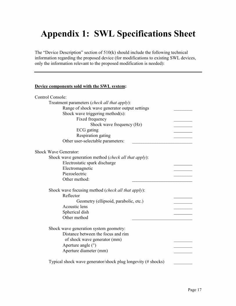
Appendix 1: SWL Specifications Sheet The “Device Description” section of 510(k) should include the following technicalinformation regarding the proposed device (for modifications to existing SWL devices,only the information relevant to the proposed modification is needed): Device components sold with the SWL system: Control Console: Treatment parameters (check all that apply): Range of shock wave generator output settings ________ Shock wave triggering method(s):
Fixed frequency ________ Shock wave frequency (Hz) ________ ECG gating ________ Respiration gating ________ Other user-selectable parameters: __________________________ Shock Wave Generator: Shock wave generation method (check all that apply): Electrostatic spark discharge ________ Electromagnetic ________ Piezoelectric ________ Other method: __________________________ Shock wave focusing method (check all that apply): Reflector ________
Geometry (ellipsoid, parabolic, etc.) ________ Acoustic lens ________ Spherical dish ________ Other method __________________________ Shock wave generation system geometry:
Distance between the focus and rim of shock wave generator (mm) ________ Aperture angle (°) ________
Aperture diameter (mm) ________ Typical shock wave generator/shock plug longevity (# shocks) ________
Page 18
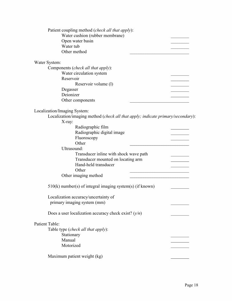
Patient coupling method (check all that apply): Water cushion (rubber membrane) ________ Open water basin ________ Water tub ________ Other method __________________________ Water System: Components (check all that apply):
Water circulation system ________ Reservoir ________ Reservoir volume (l) ________ Degasser ________ Deionizer ________ Other components __________________________ Localization/Imaging System: Localization/imaging method (check all that apply; indicate primary/secondary): X-ray: Radiographic film ________ Radiographic digital image ________ Fluoroscopy ________ Other __________________________ Ultrasound:
Transducer inline with shock wave path ________ Transducer mounted on locating arm ________ Hand-held transducer ________ Other __________________________ Other imaging method __________________________ 510(k) number(s) of integral imaging system(s) (if known) ________ Localization accuracy/uncertainty of
primary imaging system (mm) ________ Does a user localization accuracy check exist? (y/n) ________ Patient Table: Table type (check all that apply): Stationary ________ Manual ________
Motorized ________ Maximum patient weight (kg) ________
Page 19
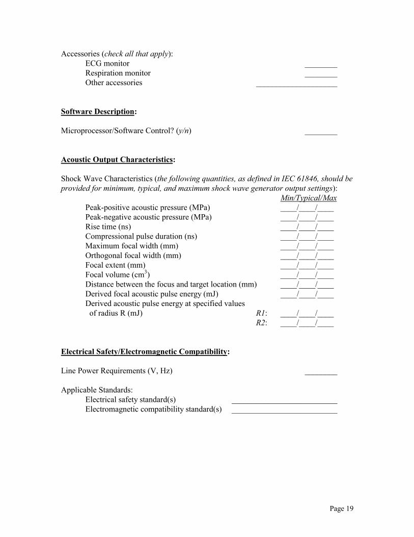
Accessories (check all that apply): ECG monitor ________ Respiration monitor ________ Other accessories ____________________ Software Description: Microprocessor/Software Control? (y/n) ________ Acoustic Output Characteristics: Shock Wave Characteristics (the following quantities, as defined in IEC 61846, should beprovided for minimum, typical, and maximum shock wave generator output settings): Min/Typical/Max
Peak-positive acoustic pressure (MPa) ____/____/____ Peak-negative acoustic pressure (MPa) ____/____/____ Rise time (ns) ____/____/____ Compressional pulse duration (ns) ____/____/____ Maximum focal width (mm) ____/____/____ Orthogonal focal width (mm) ____/____/____ Focal extent (mm) ____/____/____ Focal volume (cm3) ____/____/____ Distance between the focus and target location (mm) ____/____/____ Derived focal acoustic pulse energy (mJ) ____/____/____ Derived acoustic pulse energy at specified values of radius R (mJ) R1: ____/____/____
R2: ____/____/____ Electrical Safety/Electromagnetic Compatibility: Line Power Requirements (V, Hz) ________ Applicable Standards:
Electrical safety standard(s) __________________________ Electromagnetic compatibility standard(s) __________________________
Page 20
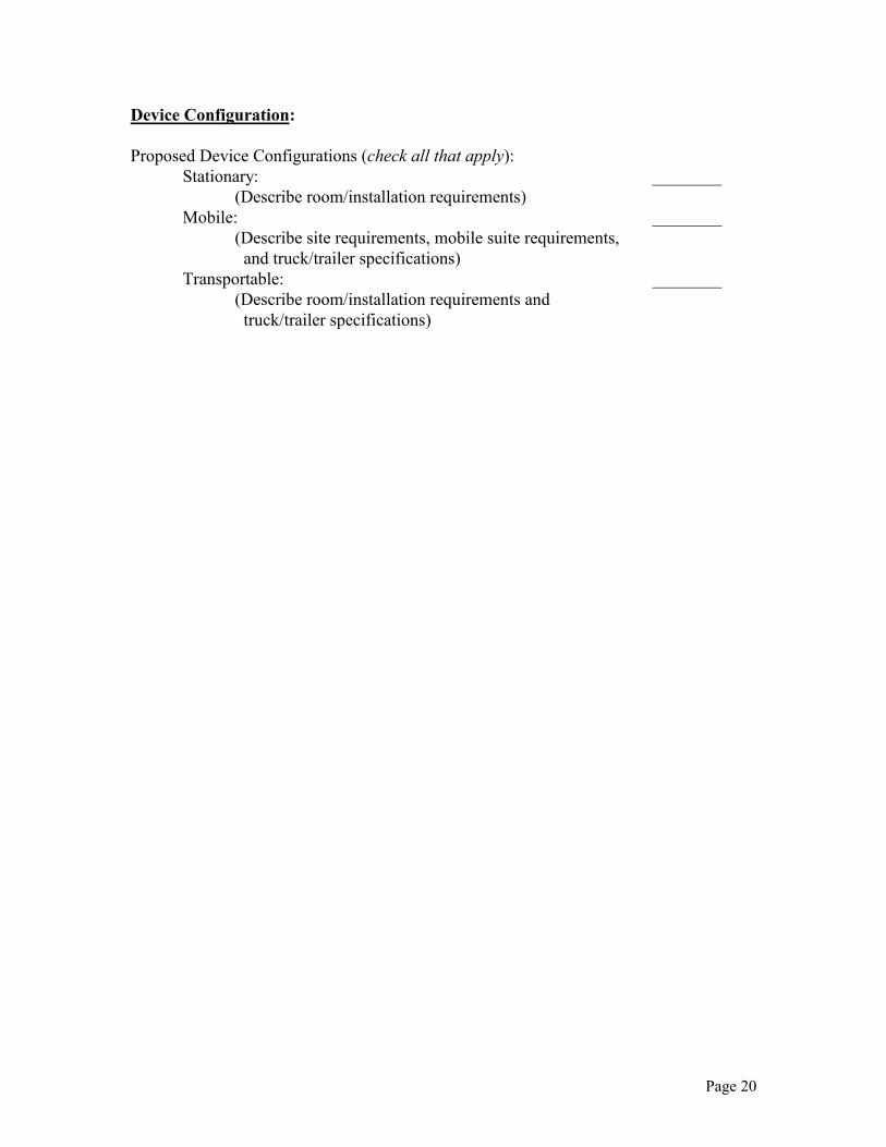
Device Configuration: Proposed Device Configurations (check all that apply): Stationary: ________ (Describe room/installation requirements) Mobile: ________ (Describe site requirements, mobile suite requirements,
and truck/trailer specifications) Transportable: ________ (Describe room/installation requirements and
truck/trailer specifications)
Page 21
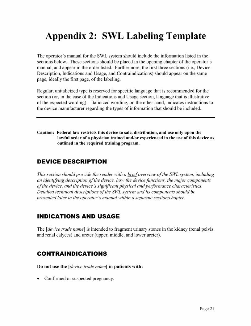
Appendix 2: SWL Labeling Template The operator’s manual for the SWL system should include the information listed in thesections below. These sections should be placed in the opening chapter of the operator’smanual, and appear in the order listed. Furthermore, the first three sections (i.e., DeviceDescription, Indications and Usage, and Contraindications) should appear on the samepage, ideally the first page, of the labeling. Regular, unitalicized type is reserved for specific language that is recommended for thesection (or, in the case of the Indications and Usage section, language that is illustrativeof the expected wording). Italicized wording, on the other hand, indicates instructions tothe device manufacturer regarding the types of information that should be included. Caution: Federal law restricts this device to sale, distribution, and use only upon the
lawful order of a physician trained and/or experienced in the use of this device asoutlined in the required training program.
DEVICE DESCRIPTION This section should provide the reader with a brief overview of the SWL system, includingan identifying description of the device, how the device functions, the major componentsof the device, and the device’s significant physical and performance characteristics.Detailed technical descriptions of the SWL system and its components should bepresented later in the operator’s manual within a separate section/chapter.
INDICATIONS AND USAGE The [device trade name] is intended to fragment urinary stones in the kidney (renal pelvisand renal calyces) and ureter (upper, middle, and lower ureter).
CONTRAINDICATIONS Do not use the [device trade name] in patients with:
• Confirmed or suspected pregnancy.
Page 22
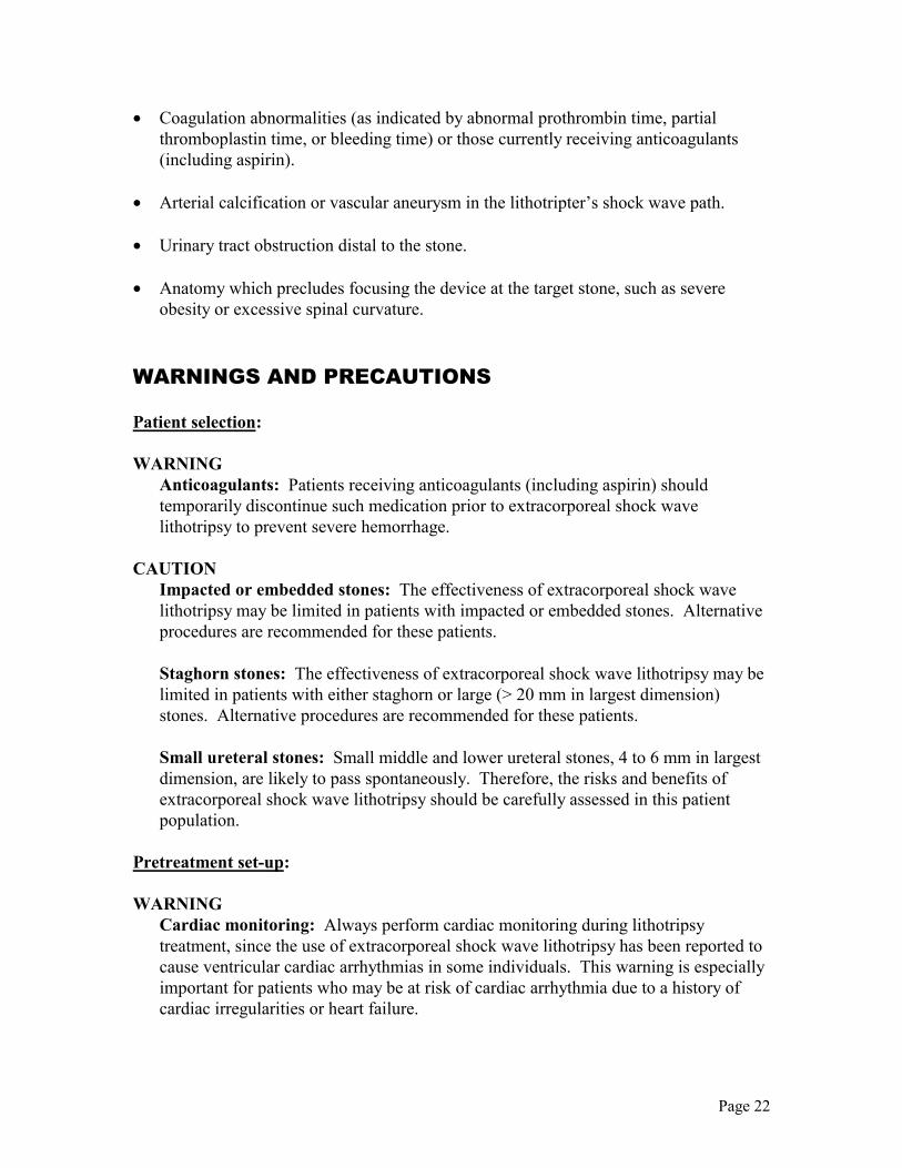
• Coagulation abnormalities (as indicated by abnormal prothrombin time, partialthromboplastin time, or bleeding time) or those currently receiving anticoagulants(including aspirin).
• Arterial calcification or vascular aneurysm in the lithotripter’s shock wave path. • Urinary tract obstruction distal to the stone. • Anatomy which precludes focusing the device at the target stone, such as severe
obesity or excessive spinal curvature.
WARNINGS AND PRECAUTIONS Patient selection: WARNING
Anticoagulants: Patients receiving anticoagulants (including aspirin) shouldtemporarily discontinue such medication prior to extracorporeal shock wavelithotripsy to prevent severe hemorrhage.
CAUTION
Impacted or embedded stones: The effectiveness of extracorporeal shock wavelithotripsy may be limited in patients with impacted or embedded stones. Alternativeprocedures are recommended for these patients. Staghorn stones: The effectiveness of extracorporeal shock wave lithotripsy may belimited in patients with either staghorn or large (> 20 mm in largest dimension)stones. Alternative procedures are recommended for these patients. Small ureteral stones: Small middle and lower ureteral stones, 4 to 6 mm in largestdimension, are likely to pass spontaneously. Therefore, the risks and benefits ofextracorporeal shock wave lithotripsy should be carefully assessed in this patientpopulation.
Pretreatment set-up: WARNING
Cardiac monitoring: Always perform cardiac monitoring during lithotripsytreatment, since the use of extracorporeal shock wave lithotripsy has been reported tocause ventricular cardiac arrhythmias in some individuals. This warning is especiallyimportant for patients who may be at risk of cardiac arrhythmia due to a history ofcardiac irregularities or heart failure.
Page 23
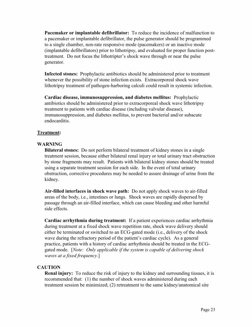
Pacemaker or implantable defibrillator: To reduce the incidence of malfunction toa pacemaker or implantable defibrillator, the pulse generator should be programmedto a single chamber, non-rate responsive mode (pacemakers) or an inactive mode(implantable defibrillators) prior to lithotripsy, and evaluated for proper function post-treatment. Do not focus the lithotripter’s shock wave through or near the pulsegenerator. Infected stones: Prophylactic antibiotics should be administered prior to treatmentwhenever the possibility of stone infection exists. Extracorporeal shock wavelithotripsy treatment of pathogen-harboring calculi could result in systemic infection. Cardiac disease, immunosuppression, and diabetes mellitus: Prophylacticantibiotics should be administered prior to extracorporeal shock wave lithotripsytreatment to patients with cardiac disease (including valvular disease),immunosuppression, and diabetes mellitus, to prevent bacterial and/or subacuteendocarditis.
Treatment: WARNING
Bilateral stones: Do not perform bilateral treatment of kidney stones in a singletreatment session, because either bilateral renal injury or total urinary tract obstructionby stone fragments may result. Patients with bilateral kidney stones should be treatedusing a separate treatment session for each side. In the event of total urinaryobstruction, corrective procedures may be needed to assure drainage of urine from thekidney. Air-filled interfaces in shock wave path: Do not apply shock waves to air-filledareas of the body, i.e., intestines or lungs. Shock waves are rapidly dispersed bypassage through an air-filled interface, which can cause bleeding and other harmfulside effects. Cardiac arrhythmia during treatment: If a patient experiences cardiac arrhythmiaduring treatment at a fixed shock wave repetition rate, shock wave delivery shouldeither be terminated or switched to an ECG-gated mode (i.e., delivery of the shockwave during the refractory period of the patient’s cardiac cycle). As a generalpractice, patients with a history of cardiac arrhythmia should be treated in the ECG-gated mode. [Note: Only applicable if the system is capable of delivering shockwaves at a fixed frequency.]
CAUTION
Renal injury: To reduce the risk of injury to the kidney and surrounding tissues, it isrecommended that: (1) the number of shock waves administered during eachtreatment session be minimized; (2) retreatment to the same kidney/anatomical site
Page 24
occur no sooner than 1 month after the initial treatment; and (3) eachkidney/anatomical site be limited to a total of three treatment sessions. Use of fluoroscopy: While fluoroscopy must be used during the procedure, cautionshould be used to minimize the exposure. [Note: Only applicable if fluoroscopiclocalization is required for use of the device.] Electromagnetic interference: If electromagnetic interference between theextracorporeal shock wave lithotripter and nearby electronic equipment is suspected(as evidenced by erratic behavior with either device), it is recommended that theirdistance be increased until proper operation resumes. If it is necessary to operate anelectronic device in close proximity to the lithotripsy system during treatment, thedevice and the lithotripter should be tested for proper simultaneous operation prior toclinical use.
Post-treatment: CAUTION
Radiographic follow-up: All patients should be followed radiographically aftertreatment until stone-free or there are no remaining stone fragments which are likelyto cause silent obstruction and loss of renal function.
Device maintenance: CAUTION
Electrical shock hazard: Never remove any of the cabinet covers to the system’selectronics. The high voltage power supply circuits utilized by extracorporeal shockwave lithotripters use voltages that are capable of causing serious injury or death fromelectric shock.
ADVERSE EVENTS Potential adverse events associated with the use of extracorporeal shock wave lithotripsyinclude those listed below, categorized by frequency and individually described:
Page 25
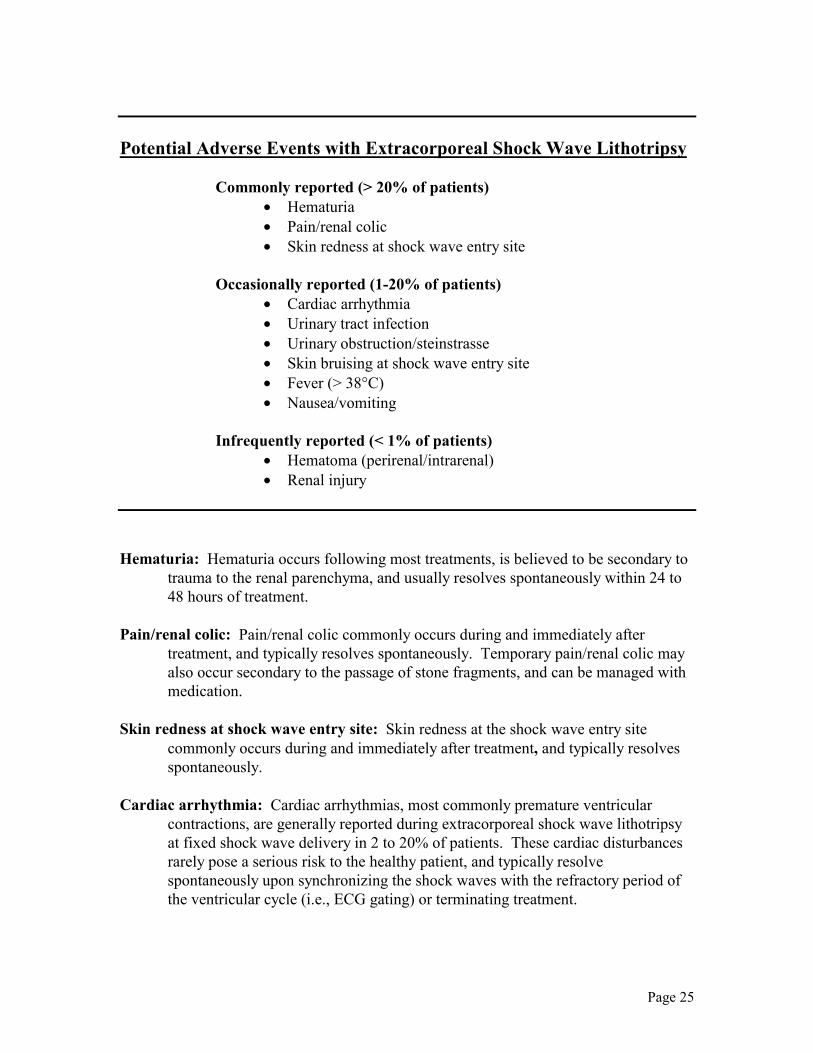
Potential Adverse Events with Extracorporeal Shock Wave Lithotripsy
Commonly reported (> 20% of patients)• Hematuria• Pain/renal colic• Skin redness at shock wave entry site
Occasionally reported (1-20% of patients)• Cardiac arrhythmia• Urinary tract infection• Urinary obstruction/steinstrasse• Skin bruising at shock wave entry site• Fever (> 38°C)• Nausea/vomiting
Infrequently reported (< 1% of patients)
• Hematoma (perirenal/intrarenal)• Renal injury
Hematuria: Hematuria occurs following most treatments, is believed to be secondary to
trauma to the renal parenchyma, and usually resolves spontaneously within 24 to48 hours of treatment.
Pain/renal colic: Pain/renal colic commonly occurs during and immediately after
treatment, and typically resolves spontaneously. Temporary pain/renal colic mayalso occur secondary to the passage of stone fragments, and can be managed withmedication.
Skin redness at shock wave entry site: Skin redness at the shock wave entry site
commonly occurs during and immediately after treatment, and typically resolvesspontaneously.
Cardiac arrhythmia: Cardiac arrhythmias, most commonly premature ventricular
contractions, are generally reported during extracorporeal shock wave lithotripsyat fixed shock wave delivery in 2 to 20% of patients. These cardiac disturbancesrarely pose a serious risk to the healthy patient, and typically resolvespontaneously upon synchronizing the shock waves with the refractory period ofthe ventricular cycle (i.e., ECG gating) or terminating treatment.
Page 26
Urinary tract infection: Urinary tract infection (UTI) occurs in 1-7% of patientsfollowing extracorporeal shock wave lithotripsy as a result of the release ofbacteria from the fragmentation of infected calculi, and infrequently results inpyelonephritis or sepsis. The risk of infectious complications secondary toextracorporeal shock wave lithotripsy can be minimized through the use ofprophylactic antibiotics in patients with UTI and infection stones.
Urinary obstruction/steinstrasse: Urinary obstruction occurs in up to 6% of patients
following lithotripsy due to stone fragments becoming lodged in the ureter, andmay be the result of either a single stone fragment or the accumulation of multiplesmall stone particles (i.e., steinstrasse). Patients with urinary obstruction typicallypresent with persistent pain, and may be at risk of developing hydronephrosis withsubsequent renal failure if the obstruction is not promptly treated. Intervention isnecessary if the obstructing fragments do not pass spontaneously.
Skin bruising at shock wave entry site: Skin bruising at the shock wave entry site
occasionally occurs after treatment, and typically resolves spontaneously. Fever (> 38°°°°C): Fever is occasionally reported after lithotripsy, and may be secondary to
infection. Nausea/vomiting: Transient nausea and vomiting are occasionally reported immediately
after lithotripsy, and may be associated with either pain or the administration ofsedatives or analgesia.
Hematoma (perirenal/intrarenal): Clinically significant intrarenal or perirenal
hematomas occur in < 1% of lithotripsy treatments. These patients typicallypresent with severe, chronic flank pain. Although clinically significanthematomas often resolve with conservative management, severe hemorrhage anddeath have been reported. Management of severe renal hemorrhage includes theadministration of blood transfusions, percutaneous drainage, or surgicalintervention.
Renal injury: Extracorporeal shock wave lithotripsy procedures have been known to
cause damage to the treated kidney. The potential for injury, its long-termsignificance, and its duration are unknown.
PATIENT SELECTION AND TREATMENT Specific patient populations: Children: The safety and effectiveness of this device in the treatment of urolithiasis inchildren have not been demonstrated. Although children have been treated with shockwave therapy for upper urinary tract stones, experience with lithotripsy in such cases is
Page 27
limited. Studies indicate that there are growth plate disturbances in the epiphyses ofdeveloping long bones in rats subjected to shock waves. The significance of this findingto human experience is unknown. Women of childbearing potential: The treatment of lower ureteral stones should beavoided in women of childbearing potential. The application of shock wave lithotripsy tothis patient population could possibly result in irreversible damage to the femalereproductive system and to the unborn fetus in the undiagnosed pregnancy.
SAFE RADIATION PRACTICES [Note: Only include this section if the SWL system is labeled for x-ray stonelocalization.] This section should present an overview of the practices for the safe use of ionizingradiation. As part of this section, the following information should be conveyed:• the importance of using the minimum technique factors and exposure durations
necessary for adequate stone imaging and localization;• the maximum technique factors and exposure durations that should be used, as well
as an indication of the maximum radiation dosage that would be expected to bedelivered to the patient during a single SWL treatment; and
• guidelines on how to minimize radiation exposure to the device operator and otherhealth care staff.