National Consensus on Management of Dyspepsia and...
9
CLINICAL PRACTICE 279 Acta Med Indones - Indones J Intern Med • Vol 49 • Number 3 • July 2017 National Consensus on Management of Dyspepsia and Helicobacter pylori Infection Ari F. Syam, Marcellus Simadibrata, Dadang Makmun, Murdani Abdullah, Achmad Fauzi, Kaka Renaldi, Hasan Maulahela, Amanda P. Utari e Indonesian Society of Gastroenterology, Jakarta, Indonesia. Indonesian Helicobacter pylori Study Group, Jakarta, Indonesia. Corresponding Author: Ari Fahrial Syam, MD., PhD. Division of Gastroenterology, Department of Internal Medicine, Faculty of Medicine Universitas Indonesia – Cipto Mangunkusumo Hospital. Jl. Diponegoro 71, Jakarta 10430, Indonesia. email: [email protected]. ABSTRAK Dispepsia merupakan salah satu dari berbagai keluhan umum yang dapat ditemui oleh dokter di berbagai bidang, tidak terbatas hanya pada ahli saluran cerna saja dalam praktik kesehariannya. Pengertian mengenai patofisiologi dispepsia terus berkembang sejak dimulainya investigasi secara ilmiah pada 1980-an sampai dengan saat ini yang memandang infeksi Helicobacter pylori sebagai salah satu faktor kunci dalam menangani dispepsia, baik terkait ulkus maupun non-ulkus. Penatalaksanaan dispepsia tidak bisa dilepaskan dari penatalaksaan infeksi H.pylori, serta penambahan pengetahuan baru terkait definisi, patofisiologi, diagnosis dan penatalaksanaan dispepsia dan infeksi H.pylori. Konsensus penatalaksanaan dispepsia dan infeksi H.pylori di Indonesia ini dibuat berdasarkan evidence based medicine, sehingga dapat digunakan sebagai rujukan para dokter dalam menangani kasus-kasus dispepsia dan infeksi H.pylori di tempat praktik sehari-hari. Dengan adanya konsensus terbaru ini diharapkan para dokter dapat lebih meningkatkan pelayanannya kepada pasien-pasien dispepsia dan infeksi H.pylori. Kata kunci: dispepsia, H. Pylori, saluran cerna. ABSTRACT Dyspepsia is one of numerous general complaints, which is commonly encountered by doctors of various disciplines. In daily practice, the complaint is not only limited for gastroenterologists. Knowledge on pathophysiology of dyspepsia have been developing continuously since a scientific investigation has been started in 1980’s, which considers Helicobacter pylori as one of key factor in managing dyspepsia, either it is associated with ulcer or non-ulcer. The management of dyspepsia cannot be separated from the management of H. pylori and there is an additional new knowledge associated with definition, pathophysiology, diagnosis and treatment of both dyspepsia and H. pylori infection. This consensus document on the management of dyspepsia and H. pylori infection in Indonesia has been developed using the evidence-based medicine principles; therefore, it can be used as a reference for doctors in dealing with dyspepsia and H. pylori infection cases in their daily practice. It is expected that with the new consensus, doctors can provide greater service to their patients who have dyspepsia and H. pylori infection. Keywords: dyspepsia, H. Pylori, gastrointestinal tract.
Transcript of National Consensus on Management of Dyspepsia and...
CLINICAL PRACTICE
279Acta Med Indones - Indones J Intern Med • Vol 49 • Number 3 • July 2017
National Consensus on Management of Dyspepsia and Helicobacter pylori Infection
Ari F. Syam, Marcellus Simadibrata, Dadang Makmun, Murdani Abdullah, Achmad Fauzi, Kaka Renaldi, Hasan Maulahela, Amanda P. UtariThe Indonesian Society of Gastroenterology, Jakarta, Indonesia.Indonesian Helicobacter pylori Study Group, Jakarta, Indonesia.
Corresponding Author:Ari Fahrial Syam, MD., PhD. Division of Gastroenterology, Department of Internal Medicine, Faculty of Medicine Universitas Indonesia – Cipto Mangunkusumo Hospital. Jl. Diponegoro 71, Jakarta 10430, Indonesia. email: [email protected].
ABSTRAKDispepsia merupakan salah satu dari berbagai keluhan umum yang dapat ditemui oleh dokter di berbagai
bidang, tidak terbatas hanya pada ahli saluran cerna saja dalam praktik kesehariannya. Pengertian mengenai patofisiologi dispepsia terus berkembang sejak dimulainya investigasi secara ilmiah pada 1980-an sampai dengan saat ini yang memandang infeksi Helicobacter pylori sebagai salah satu faktor kunci dalam menangani dispepsia, baik terkait ulkus maupun non-ulkus. Penatalaksanaan dispepsia tidak bisa dilepaskan dari penatalaksaan infeksi H.pylori, serta penambahan pengetahuan baru terkait definisi, patofisiologi, diagnosis dan penatalaksanaan dispepsia dan infeksi H.pylori.
Konsensus penatalaksanaan dispepsia dan infeksi H.pylori di Indonesia ini dibuat berdasarkan evidence based medicine, sehingga dapat digunakan sebagai rujukan para dokter dalam menangani kasus-kasus dispepsia dan infeksi H.pylori di tempat praktik sehari-hari. Dengan adanya konsensus terbaru ini diharapkan para dokter dapat lebih meningkatkan pelayanannya kepada pasien-pasien dispepsia dan infeksi H.pylori.
Kata kunci: dispepsia, H. Pylori, saluran cerna.
ABSTRACTDyspepsia is one of numerous general complaints, which is commonly encountered by doctors of
various disciplines. In daily practice, the complaint is not only limited for gastroenterologists. Knowledge on pathophysiology of dyspepsia have been developing continuously since a scientific investigation has been started in 1980’s, which considers Helicobacter pylori as one of key factor in managing dyspepsia, either it is associated with ulcer or non-ulcer. The management of dyspepsia cannot be separated from the management of H. pylori and there is an additional new knowledge associated with definition, pathophysiology, diagnosis and treatment of both dyspepsia and H. pylori infection.
This consensus document on the management of dyspepsia and H. pylori infection in Indonesia has been developed using the evidence-based medicine principles; therefore, it can be used as a reference for doctors in dealing with dyspepsia and H. pylori infection cases in their daily practice. It is expected that with the new consensus, doctors can provide greater service to their patients who have dyspepsia and H. pylori infection.
Keywords: dyspepsia, H. Pylori, gastrointestinal tract.
Ari F. Syam Acta Med Indones-Indones J Intern Med
280
INTRODUCTIONDyspepsia is a common symptom found
in daily practice and it has been known for a long period of time. There is a continuously developing definition starting from all symptoms originated from the upper gastrointestinal tract up to the exclusion of reflux symptoms; while the most recent definition refers to the Rome III criteria.1
H. pylori (Hp) infection nowadays has been considered as one of important factors in dyspepsia management, both the organic or functional dyspepsia; therefore, any discussion on dyspepsia should be correlated with the management of Hp infection. Various meta-analysis studies have demonstrated that there is a correlation between Hp infection and gastroduodenal diseases, which is characterized by symptoms/signs of dyspepsia.2,3 The prevalence of Hp infection in Asia is relatively high and thus it should be noticed in the diagnosis approach and management of dyspepsia. Hp eradication has been proven effective in eliminating symptoms of organic dyspepsia; however, for functional dyspepsia, further studies are still necessary.4
The consensus is developed to provide a guideline for general physicians, specialists, and consultants regarding the management of dyspepsia. The consensus has combined the management of dyspepsia and Hp infection; therefore, it will achieve better results.
EPIDEMIOLOGYDyspepsia is an uncomfortable feeling
(discomfort) originated from the upper part of abdomen. The discomfort can be one of or some of following symptoms, i.e. epigastric pain, epigastric burn, feeling of fullness after having meal, early satiated and bloating in the upper gastrointestinal tract area, nausea, vomiting and blurping.5 For functional dyspepsia, those abovementioned symptoms must occur for at least the last three months with symptom onset of six months before the diagnosis is defined.
The prevalence of dyspepsia in health care units reaches 30% of services offerec by general physicians and 50% of services by gastroenterologists. Most of Asian patients
with uninvestigated dyspepsia and without any alarm signs experience functional dyspepsia. The results of studies in Asian countries (China, Hong Kong, Indonesia, Korea, Malaysia, Singapore, Taiwan, Thailand and Vietnam) show that 43 to 79.5% patients with dyspepsia have dysfunctional dyspepsia.5
From the results of endoscopy, which had been performed in 550 patients with dyspepsia at some centers in Indonesia between January 2003 and April 2004, there were 44.7% cases with minimal disorder of gastritis and duodenitis; 6.5^ cases with gastric ulcer and 8.2% of normal cases.6 In Indonesia, the data on prevalence of Hp infection in patients with peptic ulcers (without a history of using non-steroid anti-inflammatory drugs (NSAIDS)) varies between 90 and 100% and for patients with functional dyspepsia, the prevalence is 20-40% with various diagnostic methods (serology examination, culture and histopathology).7
The prevalence of Hp infection in patients with dyspepsia who underwent endoscopic examination at various teaching hospital in Indonesia between 2003 and 2004 was 10.2%. A relatively high prevalence was found in Makassar, which was 55% (2011) and in Solo as many as 51.8% (2008). The high prevalence was also found in Yogyakarta of 30.6% and Surabaya of 23.5% in 2013; while the lowest prevalence has been found in Jakarta (8%).6,8-10
PATHOPHYSIOLOGY
The pathophysiiology of peptic ulcer caused by Hp and non-steroid anti-inflammatory drugs (NSAID) has been widely known.1 Functional dyspepsia is caused by some major factors such as gastroduodenal motility disorder, Hp infection, gastric acid, visceral hypersensitivity and psychological factors. Other factors that may have role are genetics, life style, environment, diet and history of previous gastrointestinal infection.11,12
The Role of Gastroduodenal Motility Disorders The gastroduodenal motility disorder consists
of reduced gastric capacity in accommodating meal (impaired gastric accommodation), antroduodenal incoordination and slow gastric

Vol 49 • Number 3 • July 2017 National consensus on management of dyspepsia and H. pylori infection
281
emptying. It is also one of main mechanism in the pathophysiology of functional dyspepsia, which is associated with bloating after meal and it may be found in the form of abdominal distension, bloating and fullness.5,12
The Role of Visceral HypersensitivityVisceral hypersensitivity has an important role
in the pathophysiology of functional dyspepsia, particularly in increasing the sensitivity of peripheral and central sensory nerves against chemical and intraluminal receptor stimuli at the proximal part of the stomach. It can cause or aggravate dyspepsia symptoms.5
The Role of Psychosocial FactorsPsychosocial disorder is one of inducers
that may have roles in functional dyspepsia. The severity of psychosocial disorder is consistent with the severity of dyspepsia. Various studies show that depression and anxiety have roles in the development of functional dyspepsia.5,12
The Role of Gastric AcidGastric acid may have a role in developing
symptoms of functional dyspepsia. It has also becomes an underlying reason for effective treatment using anti-secretory agents as shown by some studies in patients with functional dyspepsia. There is little data of studies on the gastric secretion and some reports in Asia are still controversial.5
The Role of Hp InfectionThe prevalence of Hp infection in patients
with functional dyspepsia varies from 39% to 87%. The correlation between Hp infection and motility disorder is inconsistent; however, eradication of Hp improves the symptoms of functional dyspepsia. Biological markers such as ghrelin and leptin as well as altered expression of muscle-specific microRNAs are correlated with pathophysiological process of functional dyspepsia, which still needs further studies.5,13
DIAGNOSIS
Diagnosis of DyspepsiaDyspepsia that has been investigated consists
of organic and functional dyspepsia. Organic dyspepsia consists of gastric ulcer, duodenal ulcer, erosive gastritis, gastritis, duodenitis and
a process of malignancy. Functional dyspepsia refers to the Rome III criteria, which has not been validated in Indonesia. The Asia-Pacific Consensus (2012) has decided to follow the concept in the Rome III diagnosis criteria with some additional symptoms of bloating in the upper abdominal part, which is commonly found as the symptom of functional dyspepsia.5
Dyspepsia according to the Rome III criteria is a disease with one or more symptoms associated with gastroduodenal abnormalities of: • Epigastric pain• Epigastric burn sensation• Fullness or discomfort after meal• Early satiety
The symptoms must occur at least in the last three months with an onset of symptoms in six months before the diagnosis is established.
The Rome III criteria categorize functional dyspepsia into 2 subgroups, i.e. the epigatric pain syndrome and postprandial distress syndrome. However, the most recent evidence demonstrates that there is an overlapping of diagnosis in two third of patients with dyspepsia.1
Uninvestigated dyspepsia
Workups (consistent with indications)- Blood test- Endoscopy- Urea breath test- Abdominal USG
Functional dyspepsia- Organic dyspepsia- Peptic ulcer- Erosive gastritis- Moderate to severegastritis
- Gastric cancer
Distress sydromeafter meal
Epigastric painsyndrome
Figure 1. Algorithm for diagnosis of uninvestigated dyspepsia
Evaluation on the alarm signs must always be part of evaluation of the patients who come with a complaint of dyspepsia. The alarm signs for dyspepsia include weight loss (unintended weight loss), progressive dysphagia, recurrent or persistent vomiting, gastrointestinal bleeding, anemia, fever, a mass in the upper abdominal area, a family history of gastric cancer, dyspepsia with new onset in patients aged more than 45 years. Patients with those complaints must be investigated first using endoscopy.5

Ari F. Syam Acta Med Indones-Indones J Intern Med
282
Diagnosis of Hp Infection14
Diagnostic test of Hp infection can be performed both directly using endoscopy (rapid urease test, histological test, culture and PCR) and indirectly without endoscopy (urea breath test, stool test, urine test and serology). Urea breat test has now become the gold standard for evaluating Hp. One of the available urea breath tests is the 13CO breath analyzer. There is a requirement for conducting Hp evaluation, i.e. the patient must be free from antibiotic and PPI (proton-pump inhibitor) treatment for 2 weeks and there are some other factors that need to be taken into consideration such as clinical situation, prevalence of the infection, prevalence of the infection in
a population, probability of pre-test infection, differences in test performance and factors that can affect the result of the test such as the use of anti-secretory and antibiotic treatment.
MANAGEMENTThe management of dyspepsia is initiated
by carrying out efforts on identification of pathophysiology and etiological factors as many as possible.11 Treatment of dyspepsia can already be started based on the dominant clinical symptoms (although those symptoms have not been investigated) and the treatment is subsequently continued in consistent with the results of investigation.
Tabel 1. A comparison of various diagnostic tests for Hp infection
Tests Sn Sp Notes
With endoscopy
Rapid urease test >98% 99% - Fast and inexpensive - Reduced sensitivity following treatment - Specimens are obtained from antrum
Histology >95% >95% - Increased detection using special staining
Warthin- Starry/ hematoxylin-eosin/ Giemsa) - Specimens are obtained from antrum and corpus
Culture
- Very specific, poor sensitivity when transport medium is not available
- Requires experience - Expensive, often unavailable - Specimens are obtained from antrum and corpus - Using media such as Sparrow
PCR
- Sensitive and specific - No standard - Specimens were obtained from antrum and corpus - Considered
Without endoscopy
ELISA serology 85-92% 79-83%
- Less accurate and does not demonstrate active infection - A reliable predictor of infection in developing countries with
high prevalencei - It is not recommended for a period after therapy - Inexpensive and available
13C urea breath test (UBT) such as: 13CO2 breath analyzer
95% 96%
- It is recommended for establishing the diagnosis of Hp infection before commencing therapy14
- The test of choice for confirming eradication - Patient is not allowed having PPI and antibiotic treatment for
2 weeks prior to the examination.15,16
- Varied availability
Fecal antigen 95% 94% Rarely used although it has high sensitivity and specificity, before and after therapy
Finger-stick serology Very poor and can not match the ELISA serology
Urinary antibody: Urine-baed Rapid Urine Test17-19 73.2-82% 78.6-90.7%
Right now, the urine test has not been available in Indonesia
Sn: sensitivity, Sp: specificity, ELISA: enzyme-linked immunosorbent assay,PCR: polymerase chain reaction, PPI: proton-pump inhibitor

Vol 49 • Number 3 • July 2017 National consensus on management of dyspepsia and H. pylori infection
283
Uninvestigated DyspepsiaThe optimum management strategy for
this phase is by providing empirical therapy for 1-4 weeks before receiving results of initial investigation, i.e. the examination evaluating the evidence of Hp presence.11,13 For certain region and ethnicity and patients with high risk, the Hp evaluation must be performed earlier. Medications that can be used are antacids, gastric acid antisecretory agents (PPI such as omeprazole, rabeprazole and lansoprazole and/or H2-Receptor Antagonist [H2RA]), prokinetics and cytoprotectors (such as rebamipide), in which the selection of the regimen is based on the dominant symptoms and previous medication. There is an ongoing development of a new drug which acts on the down-regulation proton pump that has been expected to have better mechanism of action than the PPI, i.e. the DLBS.24,11
Associated with the high prevalence of Hp infection, the test-and-treat strategy is applied for patients with dyspepsia symptoms without any alarm sign. The test-and-treat strategy is performed for:20
• Dyspepsia patients without any complication, who do not response to life style changes, 2-4 weeks of treatment using single PPI and without any alarm sign.
• Patients with a history of gastric or duodenal ulcer, but have never been investigated.
• Patients who will receive NSAID, particularly those with a history of gastroduodenal ulcer.
• Patients with unexplained iron deficiency anemia, idiopathic thrombocytopenic purpura and vitaminB12 deficiency.
Test and treat is NOT performed in:20
• Patients with gastroesophageal reflux disease (GERD).
• Children with functional dyspepsia.
Dyspepsia That Has Been InvestigatedDyspepsia patients with alarm signs receive
no empirical therapy; instead, they should be investigated first by endoscopy with or without histopathological assessment before they are treated as patients with functional dyspepsia.
After the investigation, it does not exclude the possibility that in some of dyspepsia cases, GERD is found as the abnormality.
Organic DyspepsiaWhen a lesion of mucosa (mucosal
damage) is found, which is consistent with the endoscopy findings, therapy is given based on the abnormalities that have been found. Abnormalities that have been included into the organic dyspepsia group are gastritis, hemorrhagic gastritis, duodenitis, gastric ulcer, duodenal ulcer or malignancy process. In peptic ulcer (gastric and/or duodenal ulcer), the medication that can be given are a combination of PPI such as rabeprazole 2 x 20 mg or
Table 2. Therapy regimen for Hp eradication14,23
Drug Dose DurationFirst line:
PPI* 2x1 7-14 days
Amoxicillin 1000 mg (2x1)
Clarithromycin 500 mg (2x1)
In an area where resistance to clarithromycin >20%
PPI* 2x1 7-14 days
Bismuth subsalicylate 2x2 tablet
Metronidazole 500 mg (3x1)
Tetracyline 250 mg (4x1)
When bismuth is not available:
PPI* 2x1 7-14 days
Amoxicillin 1000 mg (2x1)
Clarithromycin 500 mg (2x1)
Second line: this class of drugs is used when there is a failure with clarithromycin-based regimen
PPI* 2x1 7-14 days
Bismuth subsalicylate 2x2 tablets
Metronidazole 500 mg (3x1)
PPI* 2x1 7-14 days
Amoxicillin 1000 mg (2x1)
Levofloxacin 500 mg (2x1)
Third line: when the second line regimen fails. Whenever possible, the selection is based on resistance test and/or clinical changes
PPI* 7-14 days
Amoxicillin 2x1
Levofloxacin 1000 mg (2x1)
Rifabutin 500 mg (2x1)
*PPI agents that have been used are rabeprazole 20 mg, lansoprazole 30 mg, omeprazole 20 mg, pantoprazole 40 mg and esomeprazole 40 mg.Notes: Sequential therapy (can be given as the first lilne when there is no data about resistance to clarithromycin): PPI + amoxicillin for 5 days followed by PPI + clarithromycin and nitroimidazole (tinidazole) for 5 days.

Ari F. Syam Acta Med Indones-Indones J Intern Med
284
lanzoprazole 2 x 30 mg and mucoprotectors such as rebamipide 3 x 100 mg.
Functional DyspepsiaWhen there is no mucosal damage found
after the investigation, the treatment can be given in consistent with the presence of functional dyspepsia.
The use of prokinetics such as metoclopramide, domperidone, cisapride, itopride and others can improve the symptoms of patients with functional dyspepsia. It may be associated with slow gastric emptying as one of pathophysiology in functional dyspepsia. Caution must always be applied when using cisapride as it may have potency for cardiovascular complication.11
Data about the use of antidepressants or antianxiolytic in patient siwht functional dyspepsia is still very limited. A recent study in Japan has demonstrated a significant improved symptom in patients with functional dyspepsia
who have receivied 5-HT1 agonists compared to placebo. On the other hand, the use of venlafaxin, a serotonine and norepinephrin inhibitor, does not show better results compared to placebo.5
Psychological problems as well as sleep disturbance and defects on central serotonin sensitivity may become important factors in the response of antidepressant therapy in patients with functional dyspepsia.5
Management of dyspepsia with Hp infectionHp eradication can provide long-term
remission of dyspepsia symptoms.20 A cross-sectional study conducted in 21 patients at Cipto Mangunkusumo Hospital, Jakarta (2010) found that the eradication therapy had improved the symptoms in a majority of dyspepsia patients with a percentage of symptom improvement as much as 76% and 81% had negative Hp results when they were evaluated using UBT.21
Figure 2. Algorithm of managing dyspepsia at various levels of health care units5
Vol 49 • Number 3 • July 2017 National consensus on management of dyspepsia and H. pylori infection
285
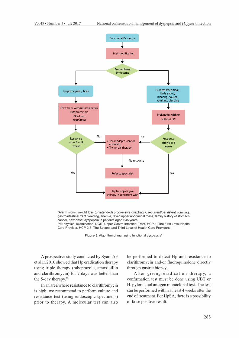
*Alarm signs: weight loss (unintended) progressive dysphagia, recurrent/persistent vomiting, gastrointestinal tract bleeding, anemia, fever, upper abdominal mass, family history of stomach cancer, new onset dyspepsia in patients aged >45 years.PE: physical examination; UGIT: Upper Gastro Intestinal Tract, HCP-1: The First Level Health Care Provider, HCP-2-3: The Second and Third Level of Health Care Providers.
Figure 3. Algorithm of managing functional dyspepsia5
A prospective study conducted by Syam AF et al in 2010 showed that Hp eradication therapy using triple therapy (rabeprazole, amoxicillin and clarithromycin) for 7 days was better than the 5-day therapy.22
In an area where resistance to clarithromycin is high, we recommend to perform culture and resistance test (using endoscopic specimens) prior to therapy. A molecular test can also
be performed to detect Hp and resistance to clarithromycin and/or fluoroquinolone directly through gastric biopsy.
After giving eradication therapy, a confirmation test must be done using UBT or H. pylori stool antigen monoclonal test. The test can be performed within at least 4 weeks after the end of treatment. For HpSA, there is a possibility of false positive result.

Ari F. Syam Acta Med Indones-Indones J Intern Med
286
Figure 4. Algorithm of managing eradication of Hp infection5
ACKNOWLEDGMENTSWe express our gratitude to Prof. Dr. dr.
Daldiyono, Prof. Dr. H.A. Aziz Rani, Dr. dr. Chudahman Manan and Mrs. Darwi in preparing the manuscript of this consensus.
REFERENCES1. Ford AC, Moayyedi P. Dyspepsia. Curr Opin
Gastroenterol. 2013;29:662-8.2. Saad AM, Choudhary A, Bechtold ML. Effect of
Helicobacter pylori treatment on gastroesophageal reflux disease (GERD): meta-analysis of randomized controlled trials. Scand J Gastroenterol. 2012;47:129-35.
3. Tang CL, Ye F, Liu W, Pan XL, Qian J, Zhang GX. Eradication of Helicobacter pylori infection reduces the incidence of peptic ulcer disease in patients using nonsteroidal anti-inflammatory drugs: a meta-analysis. Helicobacter. 2012;17:286-96.
4. Lee YY, Chua AS. Investigating functional dyspepsia in Asia. J Neurogastroenterol Motil. 2012;18:239-45.
5. Miwa H, Ghoshal UC, Gonlachanvit S, et al.
Asian consensus report on functional dyspepsia. J Neurogastroenterol Motil. 2012;18:150-68.
6. Syam AF, Abdullah M, Rani AA, et al. Evaluation of the use of rapid urease test: Pronto Dry to detect H pylori in patients with dyspepsia in several cities in Indonesia. World J Gastroenterol 2006;12:6216-8.
7. Rani AA, Fauzi A. Infeksi Helicobacter pylori dan penyakit gastro-duodenal. In: Sudoyo AW, Setyohadi B, Alwi I, Simadibrata M, Setiati S, eds. Buku ajar ilmu penyakit dalam. Jakarta: Pusat Penerbitan Ilmu Penyakit Dalam FKUI; 2006. p. 331-6.
8. Hidayati PS, Iswan Abbas Nusi IA, Maimunah U. Hubungan seropositivitas CagA H. pylori dengan derajat keparahan gastritis pada pasien dispepsia. Divisi Gastroenterohepatologi Departemen Ilmu Penyakit Dalam FK UNAIR – RSU Dr Soetomo Surabaya; 2013. (Unpublished manuscript).
9. Jumlah data Helicobacter pylori positif. RSUD Dr Moewardi Surakarta; 2008. (Unpublished raw data).
10. Parewangi AML. Jumlah data Helicobacter pylori positif di Makassar. Makassar: RSUP dr. Wahidin Sudirohusodo; 2011. (Unpublished raw data).
11. Futagami S, Shimpuku M, Yin Y, et al. Pathophysiology of functional dyspepsia. J Nippon Med Sch.

Vol 49 • Number 3 • July 2017 National consensus on management of dyspepsia and H. pylori infection
287
2011;78:280-5.12. Choung RS, Talley NJ. Novel mechanisms in functional
dyspepsia. World J Gastroenterol. 2006;12:673-7.13. Harmon RC, Peura DA. Evaluation and management
of dyspepsia. Therap Adv Gastroenterol. 2010;3:87-98.14. Hunt RH, Xiao SD, Megraud F, et al. Helicobacter
pylori in developing countries. World Gastroenterology Organisation Global Guideline. J Gastrointestin Liver Dis. 2011;20:299-304.
15. Altschuler S, Peura DA. Helicobacter pylori and peptic ulcer disease. In: McNally PR, ed. GI/Liver secrets plus. 4th ed. Philadelphia, Pa: Elsevier Mosby; 2010. p. 11.
16. Chey WD, Woods M, Scheiman JM, Nostrant TT, Del Valle J. Lansoprazole and ranitidine affect the accuracy of the 14C-urea breath test by a pH dependent mechanism. Am J Gastroenterol. 1997;92:446-50.
17. Nguyen LT, Uchida T, Tsukamoto Y, et al. Evaluation of rapid urine test for the detection of Helicobacter pylori infection in the Vietnamese population. Dig Dis Sci. 2010;55:89-93.
18. Leodolter A, Vaira D, Bazzoli F, et al. European multicentre validation trial of two new non-invasive tests for the detection of Helicobacter pylori antibodies: urine-based ELISA and rapid urine test. Aliment Pharmacol Ther. 2003;18:927-31.
19. Demiray Gurbuz E, Gonen C, Bekmen N, et al. The diagnostic accuracy of urine IgG antibody tests for the detection of Helicobacter pylori infection in Turkish dyspeptic patients. Turk J Gastroenterol. 2012;23:753-8.
20. Malfertheiner P, Megraud F, O’Morain CA, et al. Management of Helicobacter pylori infection--the Maastricht IV/Florence Consensus Report. Gut. 2012;61:646-64.
21. Utia K, Syam AF, Simadibrata M, Setiati S, Manan C. Clinical evaluation of dyspepsia in patients with functional dyspepsia, with the history of Helicobacter pylori eradication therapy in Cipto Mangunkusumo Hospital, Jakarta. Acta Med Indones. 2010;42:86-93.
22. Syam AF, Abdullah M, Rani AA, et al. A comparison of 5 or 7 days of rabeprazole triple therapy for eradication of Helicobacter pylori. Med J Indones. 2010:113-7.
23. Chey WD, Wong BC, Practice Parameters Committee of the American College of G. American College of Gastroenterology guideline on the management of Helicobacter pylori infection. Am J Gastroenterol. 2007;102:1808-25.