Cell Structure Part 2: Eukaryotic Cells (Animal Cells vs. Plant Cells)
Upload
tyler-phillipsCategory
view
220download
1
Mitosis and DNALearning Targets 1-12

The Nucleus• The central structure in most eukaryotic cells• Usually the largest structure in most
eukaryotic cells• The “boss” of the cell; running all cell
functions• Has 2 phospholipid bilayers (2 x 2 = 4 layers)• Layers pierced with holes called nuclear
pores
LT 1

The Nucleus

The Nucleus (cont.)Nuclear envelope – outer
membrane that contains genetic material; 2 layers thick; has pores
Nuclear Pore – hole in the nuclear membrane where large molecules pass through

The Nucleus (cont.)Nucleolus – sphere within
the nucleus; where ribosome parts are made
Chromatin – unorganized genetic material; the instruction manual to the cell

Cell DivisionQ: How does an organism grow in size?
-by its cell(s) getting bigger in size?OR
-by making more cells?Q: Why is it that cells are so small?
Q: Why are you made of trillions of microscopic cells ?
Q: Why aren’t you made of 1-2 giant, oozing cells?
LT 2

Why So Small?
A: In most cases the cells will divide and make more small cells instead of becoming large cells.

Why do Cells Divide?1. Large cells put more demands on the DNA– Think of OLSD. As we get more and more
students, could one principal, at one building effectively manage all of them? Not likely, so what do we do…we split. We have three principals at three HS’s. Same goes for the cell.
2. Diffusion is only good over short distances

3. The cell will have difficulty moving nutrients and wastes across the membrane.
Area of
One Face
Total Surfa
ce Area
Volume of
the Cell
Distance
from the
Center
Weight of Cell
SA to Volume
Ratio
SA to Weig
ht Ratio
s = 1 1 6 1 0.5 2.1 6 2.86
s = 2 4 24 8 1 12.9 3 1.86
s = 3 9 54 27 1.5 34.6 2 1.58
s = 4 16 96 64 2 87.1 1.5 1.10
Let’s look at our lab data1. Which best
represents the cell’s membrane?
Column 2 = Tot. Surface Area= Cell Membrane = Roads
2. Which represents the cell’s contents?
Column 3 & 5 = Vol & Wt= Cell Size = City Size

Why do Cells Divide?Think of the Columbus/I-71 example? As the
city grew faster than the road system, we had traffic jams (more cars trying to come in or go out than the roads could support). How is this like question 4 and 5?

Why do Cells Divide?• As the cell size increases, the SA-Vol ratio
decreases!! This is like Columbus growing but the roads not growing as quickly. If your SA –Vol ratio is not large, then you cannot move materials in or out very well. This like a traffic jam. This means the cell – would starve for lack of food, – or be poisoned by not being able to get rid of
wastes fast enough!!

Why do Cells Divide?
• So in order to not overload the DNA, to allow for the fastest diffusion, and to keep the Surface Area to Volume Ratio large, the cell will DIVIDE.
• The parent cell will divide into 2 daughter cells
• The DNA in the parent cell will be copied
• One copy of DNA will go to each new daughter cell
VIDEO

Lab Quiz TimeLearning Target 11. This structure is responsible for
making ribosomes2. This substance is the master
molecule of the cell and in an unorganized form
3. This contains pores by which materials can leave the nucleus
4. This structure is only found in eukaryotic cells
a. nuclear envelope
b. nucleus
c. chromatin
d. nucleolus

Lab Quiz TimeLearning Target 2:5. Roads : City :: ________ : Cell 6. Total Surface Area : Cell Membrane :: ______ & ______ : Cell Size 7. As the cell grows larger and gets more cell
content, will it need more or less cell membrane? _______________
Lab Quiz Time8. According to our data, as the cell size increased, did
the Total Surface Area to Volume ratio, get larger, smaller, or stay the same? ____________
9. Which cell (s=1, s=2, s=3, or s-4) had the best total surface area to volume ratio? __________
10. Based on your response to questions #9, what does this mean for the cell? Would it be better to have an organism made of many small cells or one large cell? ______________; because _______________________________________

Lab Quiz Time11. Besides the factor keeping cells small
discussed in the lab, what other 2 factors keep cells small?
a. ______________________________b. ______________________________

Answers1. D2. C3. A4. B5. Cell Membrane6. Volume and Weight (1/2 pt each)
7. More8. SMALLER !!! (that’s a problem)9. s-=1; the ratio was 6:1, this means there are plenty of roads for
the city or cell membrane for the cell size10. Many small cells BECAUSE the Tot. Surface Area to Volume
ratio is HIGHEST/BIGGEST. (1/2 pt per part)
11. a. DNA overload b. Diffusion is not good over long distances (1/2 pt each)

Old Dead DNA Dudes & Dudettes
Recall that DNA found in the nucleus is the molecule responsible for all cell functions.
But how do we know this?• This took some time for scientists to
determine.The question was is the genetic material PROTEIN or a NUCLEIC ACID (like DNA)??
LT 3

Old Dead DNA Dudes & DudettesFrederick Griffith (1928)-studied the pneumonia bacteria-found there were two kinds;
S-strain (smooth) and R-strain (rough)-Both strains injected into mice
1. S-strain = mice died of pneumonia2. R-strain = mice lived3. Heat-killed S-strain = mice lived4. Heat-killed S-strain + R-strain = mice DIED!!!
Somehow the deadly “factor” from the S-strain had “TRANSFORMED” the R-strain and made the combo deadly!! – called the TRANSFORMATION FACTOR
-also because the S-strain was heated, and heat denatures proteins, Griffith’s work suggested the “transforming factor” might be DNA and not proteins.

Old Dead DNA Dudes & Dudettes
Oswald Avery (1944)-Re-did Griffith’s work -found only the DNA of the S-Strain
transformed the R-strain-met with much skepticism because scientists
did not know much about the structure and function of DNA

Old Dead DNA Dudes & DudettesHershey & Chase (1952)-studied viruses that infect bacteria
because viruses are very simple; a little DNA or RNA and a protein coat-called BACTERIOPHAGES
Experiment:-took the protein of a T2 bacteriophage,
marked it, and injected it into E. coli-took the DNA of the T2 bacteriophage,
marked it with another tag, and injected it into E. coli
-let both E. coli cultures divide and found the bacteriophage DNA, not the protein, was found in the E. coli cells
Therefore, DNA is the genetic material!

Old Dead DNA Dudes & Dudettes
Erwin Chargaff (1947)-the structure of DNA is many units called
nucleotides-nucleotides have one of four nitrogen bases;
A = adenine, T = thymine, C = cytosine , G = guanine
-the amount of the A, T, C, and G’s are not equal to one another and are different by species
-BUT…the amount of A = T and C = GCalled the
Chargaff Rules
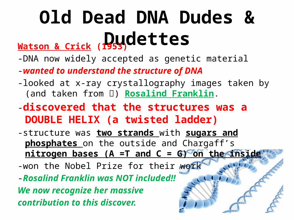
Old Dead DNA Dudes & DudettesWatson & Crick (1953)-DNA now widely accepted as genetic material-wanted to understand the structure of DNA-looked at x-ray crystallography images taken by (and taken from )
Rosalind Franklin.
-discovered that the structures was a DOUBLE HELIX (a twisted ladder)
-structure was two strands with sugars and phosphates on the outside and Chargaff’s nitrogen bases (A =T and C = G) on the inside
-won the Nobel Prize for their work -Rosalind Franklin was NOT included!! We now recognize her massive contribution to this discover.

Parts and Structure of DNA• Recall that inside the nucleus is the genetic
material, DNA. • DNA is the master molecule of the cell and is
responsible for running all cell functions. • DNA stands for Deoxyribonucleic Acid
So how is the structure of DNA related to its functions?
LT 4

Parts and Structure of DNAGene- a segment of the DNA molecule that
produces a particular protein and gives a particular trait. Ex: We have two genes for eye color: the Gey Gene and the Bey2 gene
Jobs:1. Will carry on them the information to
be passed from one generation to the next
2. Use this information to determine your traits
3. Be easily copied to be sure all the info gets to the next generation

Parts and Structure of DNAThe Facts:Long molecule Made up of parts called nucleotides• There are 3 parts to a nucleotide
1. Sugar (deoxyribose)2. Phosphate group3. Nitrogen base
LT 5

Parts and Structure of DNAThere are 4 kinds of nitrogen bases, divided
into 2 categories• Adenine (A) and Thymine (T)• Guanine (G)and Cytosine (C)

Parts and Structure of DNA
It is made up of two strands bound together in the middle•The backbone is made up of the phosphate group and deoxyribose sugar•The middle is made up of the nitrogen bases (A, T, C, and G)
LT 4 & 5

DNA ModelsYou will make…• 1 strand • Each strand will be 4 nucleotides long (any order you desire)• Side A ONLY - Tape your 4-nucleotide strand to the 4-nucleotide strands of your
table mates• Side B-DO NOT tape ANY nucleotides together
• Sides A and B – join at Table B. Side B people, make your nucleotide base pair according to Chargaff’s Rule to match Side A’s strand.
• Sugars will be WHITE• Phosphates BLACK• Bases
– Cytosine Red– Guanine Green– Adenine Blue– Thymine Yellow
LT 4
& 5

Prokaryotic v. Eukaryotic Cell Division
PROKARYOTES BOTH EUKARYOTES
•Single, circular molecule of DNA•No nuclei•DNA unconfined within cell membrane•Grow rapidly•DNA copies throughout the cell cycle•DNA replication stops briefly when cell divides•No mitotic spindles
•Have DNA•Replicate the DNA before cell division
•DNA is in the nucleus•Divide at varying rates•Rates are controlled by growth factors•Has periods of growth and rest•More complex than prokaryotic cells•Have programmed cell death

How Do Cells Divide?Recall that when cells get too big, the DNA is copied to
be placed into each new cell, then the cell divides.
DNA
In prokaryotes:•A simpler division process
•A form of ASEXUAL reproduction (cell produced is genetically identical to the parent cell)
•Called binary fission (copy DNA & split cell in 2)
In eukaryotes:•More complex division process
•A form of ASEXUAL reproduction
•3 Stages of Division• Interphase• Mitosis• Cytokinesis
•Replaces cells and grows the organism
LT 6

How Do Cells Divide?Copying and moving the DNA
into the daughter cells needs to be efficient.
So cells will condense the long DNA molecule into structures called
CHROMOSOMES – condensed, organized DNA on which genetic information is carried from the parent cell to the daughter cells.
LT 6

How Do Cells Divide? Chromosomes:
• Made up of DNA• Only visible at certain
points in the cell’s life• Made up of two identical
halves called sister chromatids
• Sister chromatids are joined at the centromere
LT 6

How Do Cells Divide?•Each species has its own number of chromosomes that are in each of their cells.
•This is called the DIPLOID NUMBER
Common Name Genus and Species
Diploid # of Chromosomes
Buffalo Bison bison 60
Cat Felis catus 38
Cattle Bos taurus, B. indicus
60
Dog Canis familiaris 78
Pig Sus scrofa 38
Human Homo sapiens 46
LT 6

How Do Cells Divide?There are 3 Stages to Cell Division
This is called the… Cell Cycle-a series of events that cells go through as
they grow and divide
1. Interphase Growth phase Longest portion of the cell cycle
2. Mitosis Division of the nucleus Shortest phase of
the cell cycle3. Cytokinesis Division of the cytoplasm
LT 7

How Do Cells Divide?
Interphase is the first stage of Cell Division.
BUT…the cell is not dividing here. It is GROWING!
3 Phases:
1. G1-cell grows (increase in size, new proteins and organelles)
2. S-chromosomes are copied to make 2 DNA molecules
3. G2-shortest phase; molecules and organelles for cell division are produced
LT 7

How Do Cells Divide?
Cells in Interphase
LT 7

How Do Cells Divide?• Cells spend most of the cell cycle in Interphase– Ex: Skin Cells divide about one time a day, but are
in Interphase for 22 of the 24hrs.– Ex: Nerve cells may stay in Interphase for decades
(think of paralysis!)• In Interphase…– The DNA is in the form of Chromatin– The nuclear envelope is intact– DNA is copying
LT 7

How Does DNA Copy?Q: So how do we actually copy DNA?
A: Each strand has all the info needed to copy it; IT’S A TEMPLATE!!
LT 8

How Does DNA Copy?LT 8
• The idea that each strand of DNA is a template is based on base pairing
A’s pair with T’sC’s pair with G’s
this strand is to this strandCOMPLIMENTARY
In Prokaryotes In EukaryotesReplication begins at Replication begins at1 pt and goes in both 100’s of pts and goesdirections both directions

How Does DNA Copy?• The location where the DNA molecule starts to
copy is called the replication fork– In prokaryotes, one replication fork– In eukaryotes, 100’s of replication forks
LT 8

How Does DNA Copy?• To begin replication the
bonds must be cut between the base pairs. These are weak hydrogen bonds– The “unzipping” and later
“re-zipping” of the base pairs is done by enzymes.
– Helicase- “unzips” the base pairs
– DNA Polymerase- “re-zips” the base pairs
LT 8

How Does DNA Copy?Steps to DNA Replication:1. At the replication fork, helicase “unzips” the DNA molecule.2. The DNA molecule begins to separate into two strands (the
parent strands or templates).3. Free nucleotides from within the nucleus match up with the
parent strand according to the base pairing rules.4. DNA polymerase bonds the free nucleotides to the parent
strands.5. More of the molecule is unzipped.6. More free nucleotides pair up and are bonded until both
strands have been copied completely.
LT 8

How Does DNA Copy?Let’s See This
Results:Two new IDENTICAL strands of DNAFor each new molecule formed ½ is the parent strand
and ½ is new
LT 8

Cell Division-MITOSISRecall that there are 3 parts to the cell cycle1. Interphase –when the cell is growing and DNA is copying2. Mitosis-when the NUCLEUS and its contents are split 3. Cytokinesis-when the CELL AND CYTOPLASM ARE split
Biologists divide Mitosis into 4 phases4. Prophase5. Metaphase6. Anaphase7. Telophase
LT 9

Cell Division-MITOSISPROPHASE
• The first phase of mitosis• Longest phase of mitosis• Chromatin form of DNA
becomes CHROMOSOMES• Centrioles separate and move
toward the ends/poles of the cell (plant cells do not have these)
• Nucleolus disappears • Nuclear envelope breaks
downChromosomes
Centrioles Nuclear Envelope

Cell Division-MITOSISMETAPHASE
“Meta” = Middle• The second phase of
mitosis• Often the shortest phase • Centrioles are at the poles• Sister chromatids line up in
the middle of the cell• Spindle fibers that come
from the centrioles attach to the centromeres of the sisters
• Nuclear envelope is gone
Sister Chromatids
CentriolesSpindle Fibers

Cell Division-MITOSISANAPHASE
“Ana” = Away• Third phase of mitosis• Spindle fibers attached at
the centromere,• Spindle fibers pull the
sister chromatids apart (now individual chromosomes)
• Chromosomes go to the poles of the cell
• Ends when chromosomes stop moving
LT 9
Spindle fibers
Sister Chromatids

Cell Division-MITOSISTELOPHASE
• Fourth and final phase of mitosis
• Chromosomes start to relax back into chromatin
• Nuclear envelope starts to reform around chromosomes
• Spindle fibers break apart• Nucleolus reforms
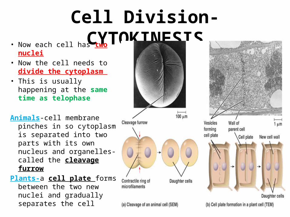
Cell Division-CYTOKINESIS• Now each cell has two nuclei• Now the cell needs to divide
the cytoplasm • This is usually happening at
the same time as telophase
Animals-cell membrane pinches in so cytoplasm is separated into two parts with its own nucleus and organelles-called the cleavage furrow
Plants-a cell plate forms between the two new nuclei and gradually separates the cell