Histology of Liver, Gallbladder & Pancreas of Liver, Gallbladder & Pancreas 2 S l i d e 4 Learning...
16
S l i d e 1 HISTOLOGY OF THE LIVER, GALLBLADDER & PANCREAS Liver, Gallbladder and Pancreas Robert W. Ogilvie, Ph.D. Professor Emeritus Medical University of South Carolina Visiting Professor University of South Carolina Roger H. Sawyer, Ph.D. Professor , Biological Sciences Executive Dean & Senior Associate Dean for Graduate Education, College of Arts & Sciences University of South Carolina This lecture will present the histology of the liver, gallbladder and pancreas. S l i d e 2 Relevant Resources Text PDF containing slides and narrative text of this lecture WebMic Program WebMic Study Guide Liver, Gallbladder & Pancreas: Unit 15, pages 189 - 200 These are the resources for text reading and practical lab exercises that will complement this lecture. S l i d e 3 Vocabulary Liver Hepatocyte von Kupffer cell Ito cell Sinusoid Portal area / periportal space Central vein Hepatic artery Hepatic veins Portal vein Bile duct Bile canaliculus Classic lobule Portal lobule Liver acinus Space of Disse Gallbladder Mucosa Muscularis externa Adventitia Mucosal folds Pancreas Islets of Langerhans Central acinar cell Intercalated ducts Excretory ducts This is the vocabulary of terms related to the content of this lecture.
Transcript of Histology of Liver, Gallbladder & Pancreas of Liver, Gallbladder & Pancreas 2 S l i d e 4 Learning...

Slide 1
HISTOLOGY OF THE LIVER, GALLBLADDER & PANCREAS
Liver, Gallbladder and Pancreas
Robert W. Ogilvie, Ph.D.Professor EmeritusMedical University of South CarolinaVisiting ProfessorUniversity of South Carolina
Roger H. Sawyer, Ph.D.Professor , Biological SciencesExecutive Dean & Senior Associate Dean for Graduate Education, College of Arts & SciencesUniversity of South Carolina
This lecture will present the histology of the liver, gallbladder and pancreas.
Slide 2
Relevant Resources
Text PDF containing slides and narrative text of this lecture
WebMic Program WebMic Study Guide Liver, Gallbladder & Pancreas: Unit 15, pages 189 - 200
These are the resources for text reading and practical lab exercises that will complement this lecture.
Slide 3
Vocabulary Liver
Hepatocyte von Kupffer cell Ito cell Sinusoid Portal area / periportal space Central vein Hepatic artery Hepatic veins Portal vein Bile duct Bile canaliculus Classic lobule Portal lobule Liver acinus Space of Disse
Gallbladder Mucosa Muscularis externa Adventitia Mucosal folds
Pancreas Islets of Langerhans Central acinar cell Intercalated ducts Excretory ducts
This is the vocabulary of terms related to the content of this lecture.

Histology of Liver, Gallbladder & Pancreas
2
Slide 4
Learning Outcomes
After completing a study of this lecture and working through the related laboratory exercise, you should be able to: describe, identify and give the function of the parts of a classic liver lobule. distinguish between and give the functional implications of a classic liver
lobule, a portal lobule and a liver acinus. explain the relationship between a hepatocyte, a liver sinusoid, a bile
canaliculus, the space of Disse, and the Ito cell. describe, identify and give the function of the von Kupffer cell. list the structures that occupy the periportal space (portal area). describe the histology of the mucosa of the gallbladder. define and identify the centroacinar cell of the pancreas describe, identify and explain the relationship between the exocrine and
endocrine parts of the pancreas.
These are the learning outcomes of this lecture. At the end of the lecture you will be presented with a few questions to aid in assessing your understanding and retention of the important concepts in this lecture.
Slide 5
Lecture Topics
Liver Anatomical Location & General Information Liver Functions The liver triad Portal system Liver lobule Classic lobule vs. portal lobule vs. liver acinus Liver parenchymal cells Bile canaliculi
Gall Bladder Pancreas Quiz
This is the outline of topics presented in this lecture. Each topic listed on this slide is hyperlinked to the first slide in that topic sequence with return hyperlinks back to this lecture outline slide.

Histology of Liver, Gallbladder & Pancreas
3
Slide 6
LiverAnatomical Location & General Information
Location Upper right quadrant abdomen, on top
of stomach, right kidney & intestines –small part in upper left quadrant
General Information Weighs 1500 grams
Largest internal organ
Performs on order of 500 functions Liver functions remain normal even if
70% is removed Only organ in the body that can
regenerate itself Small remnants of liver grow back to normal
size within a few weeks
The light pink areas in this liver suggest fatty change due to alcohol.
Return to outline
liver beer
The liver is located in the upper right quadrant of the abdomen as you can see in this photograph of a ‘transparent’ man found on the Internet as a part of an ABC news report. The liver is the largest mass of glandular tissue in the body. The liver performs over 500 functions. Even if 70% of the liver is destroyed it can maintain normal function. It is the only organ in the body that can regenerate itself. Observe the encircled light pink areas in the liver of this man. The light pink areas in this liver of the ‘transparent’ man suggest fatty change due to alcohol. Too much beer / liquor drinking causes liver cells to be engorged with fat that compromises liver function. This eventually leads to cirrhosis (scar tissue) that permanently damages the liver. Cirrhosis can eventually overcome the liver’s ability to regenerate. Liver cancer and infection of the liver are also very damaging to the liver. While the liver has the ability to regenerate itself, it does have limits. For example bacterial infection of the liver can completely overwhelm it. The blood sinuses of the liver contain cells that have great phagocytic properties and normally clear the blood coming from the intestines of any bacteria in one pass of the blood through the liver. However, interestingly enough, chronic alcohol drinking depresses the function of these cells that sets the stage for vulnerability to infection.
Slide 7
Liver Functions
Produces most of plasma proteins Albumins, lipoproteins, glycoproteins, prothrombin, fibrinogen, nonimmune alpha and
beta globulins
Stores and converts vitamins and iron Vitamins A, D and K
Clears blood of undesirable chemicals/drugs and microorganisms Oxidation and conjugation of chemicals/drugs to make them water soluble so
they can be secreted by the kidney E-coli bacteria, for example, that can gain entrance to blood by defective
intestine epithelial lining, are removed by special cells lining the blood vessels of the liver
Secretes bile into the intestine. Bile is essential for digestion of fats
Converts and stores extra sugar (glucose) in the form of starch (glycogen) that can be returned to glucose in times of starvation
Return to outline
With 500 different functions of the liver it is not possible to list them all. These presented are some of the most important functions.

Histology of Liver, Gallbladder & Pancreas
4
Slide 8
LiverBlood Supply/Drainage & Bile Secretion/Storage
Blood Supply Hepatic artery from the aorta
Oxygenated blood to supply parenchyma and stroma
Portal Vein from the intestines 75% of blood supply from portal vein
Brings absorbed nutrients
Blood drainage All by the hepatic veins
Bile secretion and storage Bile is secreted by liver cells
Drains to the intestines by common bile duct
Concentrated and stored in the gall bladder
This drawing illustrates the blood supply/drainage & the storage and drainage of bile from the liver. Observe that the liver has two vessels supplying it with blood and one draining it. One is the hepatic artery supplying the liver with oxygen and the other is the portal vein that brings blood from the intestines carrying molecules absorbed after digestion to the liver. Blood is collected / drained from the liver by the hepatic vein that returns blood to the heart. Bile is collected from the liver and stored in the gallbladder where it is concentrated and released on demand into the duodenum via the common bile duct. From this point on in the lecture please take note of the names of two vessels and a duct – 1) portal vein, 2) hepatic (portal) artery (arteriole), and 3) bile duct. As you learn the histology of the liver you will see that these three structures form a ‘triad’ that is integral in understanding liver histology.
Slide 9
The ‘Triad’Hepatic Vein
Hepatic arteryBile duct
Portal vein
A “triad” of structures
The liver parenchyma is organized at the histological level into lobules. Triads are located at the periphery of each lobule. Blood is drained from each lobule by a tributary of the hepatic vein – the central vein, located in the center of each lobule.
Return to outline
This drawing shows the relationship of the bile duct, portal vein and hepatic artery as they course through the liver. Liver parenchyma is organized into lobules, two of which are indicated in this drawing. The ‘triad’ is intimately related to the liver lobule. They are distributed around the peripheral aspect of each lobule providing blood from the hepatic artery and portal vein into the lobule and draining blood from it by a tributary of the hepatic vein – the central vein. Bile secreted by the liver cells is drained from the lobule by the bile ducts.

Histology of Liver, Gallbladder & Pancreas
5
Slide 10
Portal system
Brings nutrient rich blood from gut
Accounts for high incidence of liver metastasis from cancers of the intestine.
Return to outline
This is an illustration of the portal system that brings blood from the intestine and delivers it to the liver. The portal system begins in capillaries in the lamina propria of the intestine, continues in the different portal veins draining different parts of the intestine and ends in sinusoids in the liver. The blood is rich is nutrients from the intestine. Malignant cancer in the intestine can easily metastasize to the liver by the portal system.
Slide 11
The Liver Lobule
lobule lobule lobule
Liver Lobule2 mm long x 700 microns wide2 dimensional shape is a hexagonComposed of liver cells (hepatocytes) and sinusoidslobules are separated by connective tissue (stroma)
triad
Liver TriadsHepatic artery, portal vein & bile ductLocated around the peripheral aspect of each lobule
interlobular veinCentral veins
Central veinsLocated at center of each lobuleDrain lobule sinusoidsCentral veins are drained by interlobular veins
Return to outline
This drawing illustrates three liver lobules, their blood supply & drainage, and drainage of bile via the bile ducts. A liver lobule, the repeating unit of organization of the liver parenchyma, is 3 dimensional. Its dimensions average 2 mm long and 700 microns (0.7 mm) wide. In 2 dimensions as viewed in a histological section, the lobules are hexagonal in shape. Each lobule is composed of liver cells that are termed hepatocytes and sinusoids. The lobules are surrounded and separated from each other by connective tissue (stroma) that is continuous with the capsule of the liver. At the peripheral aspect of each lobule are triads commonly located at the points of the hexagon. The hepatic artery and portal veins of the triad supply the sinusoids of each lobule with blood and the bile duct drains bile secreted by the liver's cells from each lobule. Central veins are located in the center of each lobule. The sinusoids in each lobule drain into the central vein. Blood is collected from the central veins of several lobules by an interlobular vein. In the lobule, blood flows from the peripheral aspect and bile secreted by hepatocytes flows in the opposite direction, toward the peripheral aspect or the portal areas.
Histology of Liver, Gallbladder & Pancreas
6
Slide 12
Classic Lobule vs. Portal Lobule vs. Liver Acinus
Alternate terminology Hepatic artery
also interlobular artery/arteriole Portal vein
Also interlobular vein/venule Bile duct
Also interlobular duct Central Vein
also Intralobular vein / venule
12
Classic LobuleAxis center = central veinBlood collection
Portal LobuleAxis center = portal areaBile collection
Liver AcinusAxis center portal area to portal areaBlood supply
Return to outline
1 2 33 2 1
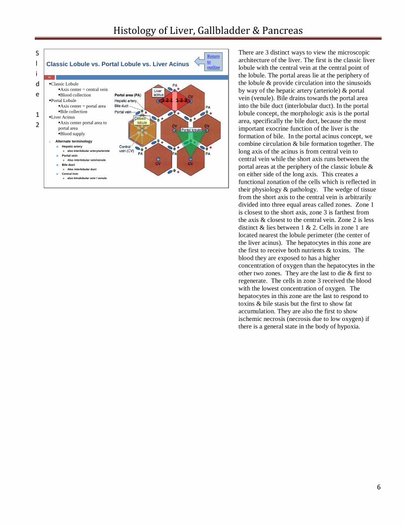
There are 3 distinct ways to view the microscopic architecture of the liver. The first is the classic liver lobule with the central vein at the central point of the lobule. The portal areas lie at the periphery of the lobule & provide circulation into the sinusoids by way of the hepatic artery (arteriole) & portal vein (venule). Bile drains towards the portal area into the bile duct (interlobular duct). In the portal lobule concept, the morphologic axis is the portal area, specifically the bile duct, because the most important exocrine function of the liver is the formation of bile. In the portal acinus concept, we combine circulation & bile formation together. The long axis of the acinus is from central vein to central vein while the short axis runs between the portal areas at the periphery of the classic lobule & on either side of the long axis. This creates a functional zonation of the cells which is reflected in their physiology & pathology. The wedge of tissue from the short axis to the central vein is arbitrarily divided into three equal areas called zones. Zone 1 is closest to the short axis, zone 3 is farthest from the axis & closest to the central vein. Zone 2 is less distinct & lies between 1 & 2. Cells in zone 1 are located nearest the lobule perimeter (the center of the liver acinus). The hepatocytes in this zone are the first to receive both nutrients & toxins. The blood they are exposed to has a higher concentration of oxygen than the hepatocytes in the other two zones. They are the last to die & first to regenerate. The cells in zone 3 received the blood with the lowest concentration of oxygen. The hepatocytes in this zone are the last to respond to toxins & bile stasis but the first to show fat accumulation. They are also the first to show ischemic necrosis (necrosis due to low oxygen) if there is a general state in the body of hypoxia.

Histology of Liver, Gallbladder & Pancreas
7
Slide 13
Classic Liver Lobule
•Liver Lobule•2 mm x 0.7 mm•Stacks of anastomosing plates of hepatocytes one cell thick•Hepatocytes separated by anastomosing sinusoids•Connective tissue surrounds portal vessels and between portal areas
PA
PA
PAPA
PA
PA
CV
CV = central veinPA = Portal Area
hexagoncross-section shape of liver lobule
This drawing of a wedge of a lobule illustrates the relationship between the hepatocytes, the sinusoids (that receive blood from the hepatic artery and portal vein) and the bile duct that drains bile from the lobule. A liver lobule consists of stacks of anastomosing plates of hepatocyte one cell thick separated by anastomosing sinusoids. Blood flows from the hepatic artery into the sinusoids and in collected by the central vein. Bile, secreted into small channels called bile canaliculi by liver cells (hepatocytes) flows in the opposite direction into slightly larger channels first, bile ductules and finally into bile ducts located in the portal areas. A liver lobule is on the order of 2mm high and 0.7 mm in diameter. It is actually a 3 dimensional hexagon. In this drawing of an ideal liver lobule sectioned across its long axis, we see that it is shaped like a hexagon. The central vein is at the center and the six portal areas form the six points of a hexagon. Connective tissue encloses the structures in the portal areas and extends also to wrap the liver lobule at its perimeter. In humans there is only a small amount of connective tissue at the perimeter so that it cannot be observed readily in an H & E section. A pig’s liver has much more connective tissue that is easily observed. A cow’s liver has an amount between the human and pig. Pig liver is tough when cooked for eating but a cow’s liver is tender.
Slide 14
Classic Liver LobuleCentral Vein & Portal Area
portal area
central vein
bile duct
hepatic artery
portal vein
central vein
sinusoid
The center of a classic liver lobule is the central vein. The peripheral extent of the lobule is determined by lines drawn between the portal areas that contain the hepatic arteries, bile ducts and portal veins. In human liver there is very little connective tissue surrounding the structures in the portal areas and even less extending from one portal area to another. One has to visualize the liver lobule by connecting the portal areas drawing imaginary lines as was done in this specimen. Observe that only 4 portal areas could be observed as is often the case. It is very difficult to find an ideal liver lobule with 6 portal areas surrounding a central vein in a typical histological section. An enlargement of the central vein area shows the central vein that is lined by endothelial cells as are the sinusoids that connect to the central vein. An enlargement of a portal area shows a portal vein containing blood cells, a bile duct lined by simple cuboidal epithelium and a hepatic artery. Observe that there is very little connective tissue in this portal area. To be precise the hepatic artery in the portal areas surrounding the central vein in a liver lobule should be called a hepatic arteriole because technically it is an arteriole. Text books and the literature often just refer to the vessel as hepatic artery.

Histology of Liver, Gallbladder & Pancreas
8
Slide 15
Liver Parenchymal Cells15
Return to outline
Viewed in the two dimensional plane the liver lobule appears as a plate. Within that plate the hepatocytes are arranged in cords which border the hepatic sinusoids. The hepatocytes are the most numerous of all of the cell types in the lobule. Hepatocytes have an intimate relationship with the sinusoids. The sinusoids are lined by endothelial cells. Blood flowing through the sinusoids comes from the nutrient rich low oxygen venous blood of the portal vein & oxygenated blood from the hepatic artery. Blood flows through the sinusoids into the central vein. Bile formed by the hepatocytes, drains in the opposite direction through special channels called canaliculi which drain into the bile duct by way of bile ductules (also calledHering’s canal). Kupffer cells are macrophages (Mononuclear phagocytic system) located strategically within the sinusoids loosely attached to the endothelial cells. Their function is to ingest foreign materials such as any wayward bacteria which may have escaped from the GI lumen. They also serve to remove abnormal and aged rbc’s after a splenectomy. The fat storing cells (cells of Ito) are responsible for the storage of vitamin A, a component in the synthesis of retinal pigments. They may also be responsible for most of the reticular fibers in the internal portion of the liver & would be involved in any fibrosis in response to a pathological condition. There is a gap between the hepatocytes and the lining of the sinusoids that is called the space of Disse that will be illustrated and explained in the next slide.
Slide 16
glycogen
rER
sER
bile canaliculus
adjacent hepatocyte
adjacent hepatocyte
adjacent hepatocyte
Hepatocyte & Liver Sinusoidsinusoid endothelium
Kupffer cells(phagocytes)
Microvilli projecting into the space of Disse
Ito cell
sinusoid lumen
sinusoid lumen
The details of the relationship between a hepatocyte and bile canaliculi and sinusoids is illustrated in this drawing. This view is as if a liver cord with its adjacent sinusoids, bile canaliculi and other hepatocytes in neighboring cords were cut in cross-section. Glycogen (stored in clumps), rough endoplasmic reticulum and smooth endoplasmic recticulum, aside from the nucleus, are three very important and essential organelles in the hepatocyte. Observe the three bile canaliculi that are always located between two adjacent hepatocytes. The canaliculi are formed by tight junctions between the hepatocytes at these locations. This makes a very tiny canal, 1 – 1.5 microns in diameter, that carries bile away from the hepatocyte. Bile is secreted into these canaliculi from the hepatocyte by exocytosis. The other surfaces are bordered by sinusoids that are lined with an endothelium. Within the sinusoid are special phagocytic cells call Kupffer cells. These cells attach themselves to the endothelial cells often with their processes spanning

Histology of Liver, Gallbladder & Pancreas
9
the sinusoid to catch and take in foriegn substances like bacteria. Observe the gaps in the endothelial lining. The gaps are large enough so that plasma bathes the surface of the hepatocyte in a space called the space of Disse, but small enough that formed elements of the blood cannot pass into this space. Microvilli project into the space of Disse. The microvilli provide an increase in surface area for interchange between the plasma and the hepatocyte. Thus, formed elements of the blood can be whizzing by at a pretty good clip while the hepatocyte is constantly exposed to plasma. In the space of Disse there is a special cell called the Ito cell (alias ‘fat storing cell’ or ‘stellate cell’). These cells store vitamin A. Vitamin A is released from these cells, transported to the retina where it contributes to the formation of rhodopsin, the visual pigment of the rods and cones. In certain pathological conditions these cells lose their pigment and, in the case of inflammation or cirrhosis, secrete collagen that forms scar tissue in the place of hepatocytes that were damaged by alcohol or infection.
Slide 17
Liver Parenchyma & Stroma
K
HepatocytesLarge-small nuclei- arrows
Binucleated cell -circledvon Kupffer cell – K
lymphocyte in sinusoid - L Silver Stain
StromaReticular Fibers
inSpace of Disse
HepatocyteNucleiEuchromatic
The parenchymal cells of the liver are the hepatocyte, the von Kupffer cell and the fat storing cell of Ito. Hepatocyte cytoplasm includes patches of rough ER to synthesize a variety of proteins the include albumin and fibrinogen among many others. Hepatocyte nuclei vary in size from small to large and some cells may be have two nuclei (binucleated). One binculeated hepatocyte is encircled in the left upper iimage. Hepatocyte nuclei are euchromatic that correlates with the function of protein synthesis. The enlargement in the lower left small image shows the detail of several euchromatic nuclei. Von Kupffer cells can be identified by their stellate shape and having more cytoplasm visible than the endothelial lining cells as indicated in the middle large image. Von Kupffer cells function to phagocytize bacteria and other foreign substances that may inadvertantly enter the liver sinusoids from the intestines. The middle lower image shows a von Kupffer cell clearly located within a sinusoid. The stroma is composed of reticular fibers that surround the blood vessels, nerves and bile ducts in the portal areas and also in the space of Disse as illustrated in the silver stained specimen on the right.

Histology of Liver, Gallbladder & Pancreas
10
Slide 18
Kupffer Cell Distributionafter intravascular injection of india ink
Low Powerobserve black spots
High PowerIndia Ink in von Kupffer cells
Small part of Kupffer cell
nucleus showing
This slide shows a low and high magnification of a liver specimen taken from a rat that was injected with india ink intravenously. India ink is a suspension of very small carbon particles. The carbon particles were phagocytized by von Kupffer cells. The nuclei of von Kupffer cells that are engorged with a foreign substance like carbon particles or bacteria etc. is usually obscured by the substance phagocytized. One von Kupffer cell in the right image indicated by the yellow arrow reveals a very small part of it nucleus (the light staining area). This demonstrates that Kupffer cells are quite numerous and it is easy to understand how effective they can be in taking bacteria and other undesirable components of the blood that may have entered into the portal venous system from the gastrointestinal tract.
Slide 19
Transmission Electron Micrograph
bile canaliculus lumen
Bile Canaliculi: 3 views
Histological Section: H &E
Alkaline Phosphatase
Return to outline
Bile is collected from several adjacent hepatocytes into a very tiny channel called a bile canaliculus. This slide shows three different views of a bile canaliculus by three different methods. The first view is a histological section stained with H&E where a cross-section of a single bile canaliculus is indicated by the arrow. The canaliculus is bordered by three hepatocytes, one of which has two nuclei, a not uncommon finding in normal hepatocytes. Recall that the diameter of a canaliculus is on the order of 2 micrometers. The next view is the bile canaliculi in a whole mount of liver cells where the liver cells are yellow and the bile canaliculi are black due to a histochemical reaction for alkaline phosphatase, a chemical that resides within that part of the plasma membrane of the hepatocytes forming the canaliculi. The third view is as seen with the aid of an electron microscope. Observe the microvilli projecting into the lumen of the canaliculus. The heptocytes forming a canaliculus are joined together by tight junctions as illustrated in the higher magnification in the inset.

Histology of Liver, Gallbladder & Pancreas
11
Slide 20
Bile Formation and Secretion20
This diagram shows the basic metabolic pathway for the formation of bile. Bilirubin, a breakdown product of hemoglobin, is brought into the cell & complexed with glucuronate via glucuronyltransferase to form the water soluble bilirubin glucuronide. This, along with bile salts such as cholic acid makes up bile. Bile is transported through the canaliculi, then by bile ducts in the triads, then large ducts and finally into the gallbladder for storage. Water makes up a good portion of bile. In the gall bladder a significant portion of the water is reabsorbed by the gall bladder epithelium so that the stored bile is a concentration of bile salts and bile pigment.
Slide 21
Normal Vs. Fatty Liver
Normal Liver Fatty Liver
Fatty liver is a condition in which the hepatocytes become engorged with lipid even to the extent that they can resemble adipocytes. Observe in the normal liver the relatively even reddish-bluish staining of the H&E stained specimen. Compare this to the fatty liver specimen in which many light stained discrete areas are present. An enlarged view of both specimens clearly shows that fat has replaced most of the normal liver cytoplasm so that the liver is beginning to look like adipose tissue. When spread throughout the liver, the function of the liver is severely compromised. Fatty liver is associated with diabetes, hypertension and obesity. Chronic alcohol drinking is definitely a cause. A fatty liver can lead to cirrhosis, a severe scarring of the liver where most of the hepatocytes are replaced by scar tissue. This has at least two results: 1) liver function is compromised and 2) due to the fact that the scarring obliterates many of the sinusoids, the resistance of blood flow through the liver dramatically increases causing a condition known as ascites, the build up of fluid in the abdominal cavity.

Histology of Liver, Gallbladder & Pancreas
12
Slide 22
The gall bladder is a pear-shaped, distensible sac with a volume of about 50 ml in humans. It is attached to the visceral surface of the liver.
Gallbladder
Return to outline
Slide 23
Gall Bladder and common bile duct
Stores bile Concentrates bile Releases bile to small intestine Bile emulsifies lipid in the
intestine
Cystic duct
GB
Hepatic duct
CommonBile duct
duodenum
This slide shows the location of the gallbladder and it intimate association with the liver. It is attached to the visceral surface of the liver. It is a blind pouch that stores, concentrates and releases bile into the duodenum. It receives dilute bile from the liver by way of the myriad of bile ducts that empty first into the hepatic duct and finally into a cystic duct that is connected to the gallbladder. When caused to do so by the action of smooth muscle in the wall of the gallbladder concentrated bile from the gallbladder is transported to the duodenum by the common bile duct. A histological cross-section shows the wall of the gallbladder surrounded by liver tissue. The next slide will illustrate the gallbladder wall structure in detail.
Slide 24
24
GALLBLADDERWall Structure
mucosa
muscularisexterna
serosa
The gallbladder is a hollow organ that has three different layers surrounding its lumen. The mucosa does not have a muscularis mucosa. The simple columnar epithelium of the mucosa has NO GOBLET CELLS. The folded mucosa resembles villi, but these are not villi. There are no glands at the base of the folds. The absence of a muscularis mucosa, goblet cells and intestinal glands helps distinguish the gall bladder from the jejunum and ileum of the small intestine. The muscularis externa contains bands of smooth muscle that function to squeeze bile out of the gallbladder into the duodenum via of the common bile duct. A serosa wraps the gallbladder.

Histology of Liver, Gallbladder & Pancreas
13
Slide 25
25
Gall Bladder Mucosa
water absorption
The mucosa of the gallbladder includes the epithelium consisting of tall simple columnar cells & lamina propria which is directly associated with the muscular externa layer. There is no muscularis mucosa included in the mucosa of the gallbladder. An enlargement of part of one of the folds shows the mucosa in greater detail. Observe that the columnar cells are tall and that there are no goblet cells. Sodium and chloride (and bicarbonate) are actively transported from the lumen across the epithelium into the lamina propria. The protein pumps that move these molecules are located in the baso-lateral cell membranes.
Slide 26
26
Inactive Pumping Na +
Gall Bladder EpitheliumInactive Vs. Active absorption of water
This slide shows two electron micrographs of the simple columnar epithelium of the gallbladder. The left image shows the epithelium in its inactive phase. The right image shows the epithelium in its active phase. Observe the large spaces between the baso-lateral aspects of the lining cells where the long arrows are located. In order to concentrate bile, the lining epithelium actively pumps Na+ to the base where it passively draws water with it, thus concentrating the bile contents. The basa-lateral plasma membranes of these cells contain the active transport molecules that actively transport sodium and chlorid ions into the interstitium. Water follows passively. This results in concentrated bile stored in the gallbladder.
Slide 27
The pancreas closely resembles the parotid gland with which it can be confused because all of its secretory units have serous cells making up the acini. The pancreas differs from the parotid gland in that it has no striated ducts and it contains an endocrine gland within itself, the islets of Langerhans.
Pancreas
Return to outline

Histology of Liver, Gallbladder & Pancreas
14
Slide 28
28
no striated ducts
PANCREAS
Grossly the pancreas has a head and a tail. The head is the larger portion and it lies in the C-shaped curve of the duodenum. The pancreas is a mixed gland exhibiting both endocrine & exocrine functions. It is a histology look alike for parotid gland but can be distinguished based on 2 characteristics: no striated ducts in the pancreas but there are in the parotid & the endocrine function is delineated by the islets of Langerhans which are found only in the pancreas, not in parotid. The beginning of its duct system in the pancreas, the intercalated duct, is actually enveloped by the acinar cells not attached to the end of the acinus as in the parotid. Therefore in cross-sections of the acini in the pancreas a duct cell is often present and this is called the 'centroacinar' cell, a unique histological feature of the pancreas.
Slide 29
29
PANCREAS: SURVEY VIEW
This low magnification survey view shows the islets of Langerhans, the endocrine portion (dark arrow heads), while the remaining parenchyma arranged as acini comprises the exocrine portion. The 2 small black arrows point to intralobular ducts which will drain into the much larger interlobular duct marked by the letter I. The asterisks note the arterial/venous drainage of the gland.
Slide 30
30
CENTROACINAR CELL
The very beginning of the duct system in the pancreas is defined by the centroacinar cell at the mouth of the acinus. They are the intra-acinar portion of the intercalated duct. Circled structures are centroacinar cells.

Histology of Liver, Gallbladder & Pancreas
15
Slide 31
31
PANCREAS: EXOCRINE & ENDOCRINE
(NOTE THE ISLET)
The islet of Langerhans is easily identified in this slide. The surrounding tissue is arranged into acini & comprises the exocrine portion. Islets are usually lighter staining in histological sections and they are often oval-shaped.
Slide 32
32
Cell Type Mallory-Azan Stain Product
α Red Glucagon
β Brown/Orange Insulin
δ Blue Somatostatin
PRINCIPAL CELL TYPES IN ISLETS
This table describes the basic functions of the cell types found in the islets. The alpha cell produces glucagon which increases the level of sugar in the blood. The ß cell produces insulin which tends to lower the level of sugar in the blood. The d cell produces somatostatin which tends to inhibit by paracrine secretion the release of other hormones. A mallory-azan stain can be used to distinguish the cell types. A more specific distinction between the cells must be done by immunohistochemical reactions. The islets of Langerhans will be presented again in the lecture on the endocrine organs.
Slide 33
Summary This lecture began with a presentation of liver histology emphasizing the lobular
organization of the liver. The blood supply and drainage of the liver were described and illustrated emphasizing the portal triad that consists of the hepatic artery, portal vein and bile duct.
The relationship between the neighboring hepatocytes was presented and bile duct formation was demonstrated.
The effect of fatty change on liver histology and function was presented and illustrated.
The three ways to view liver organization, the classic lobule, the portal lobule and the acinus were presented to emphasize different functions of the liver.
Gallbladder histology was presented with the emphasis on its role in taking up water to store bile in a concentrated form until needed for fat emulsification in the duodenum
Finally, the histology of the endocrine and exocrine parts of the pancreas was presented.
Histology of Liver, Gallbladder & Pancreas
16