Defects in the Fanconi Anemia Pathway and Chromatid...
12
Molecular and Cellular Pathobiology Defects in the Fanconi Anemia Pathway and Chromatid Cohesion in Head and Neck Cancer Chantal Stoepker 1 , Najim Ameziane 1 , Petra van der Lelij 1 , Irsan E. Kooi 1 , Anneke B. Oostra 1 , Martin A. Rooimans 1 , Saskia E. van Mil 1 , Arjen Brink 2 , Ralf Dietrich 3 , Jesper A. Balk 1 , Bauke Ylstra 4 , Hans Joenje 1 , Stephan M. Feller 5 , and Ruud H. Brakenhoff 2 Abstract Failure to repair DNA damage or defective sister chromatid cohesion, a process essential for correct chromosome segregation, can be causative of chromosomal instability (CIN), which is a hallmark of many types of cancers. We investigated how frequent this occurs in head and neck squamous cell carcinoma (HNSCC) and whether specific mechanisms or genes could be linked to these phenotypes. The genomic instability syndrome Fanconi anemia is caused by mutations in any of at least 16 genes regulating DNA interstrand crosslink (ICL) repair. Since patients with Fanconi anemia have a high risk to develop HNSCC, we investigated whether and to which extent Fanconi anemia path- way inactivation underlies CIN in HNSCC of non–Fanconi ane- mia individuals. We observed ICL-induced chromosomal break- age in 9 of 17 (53%) HNSCC cell lines derived from patients without Fanconi anemia. In addition, defective sister chromatid cohesion was observed in five HNSCC cell lines. Inactivation of FANCM was responsible for chromosomal breakage in one cell line, whereas in two other cell lines, somatic mutations in PDS5A or STAG2 resulted in inadequate sister chromatid cohesion. In addition, FANCF methylation was found in one cell line by screening an additional panel of 39 HNSCC cell lines. Our data demonstrate that CIN in terms of ICL-induced chromosomal breakage and defective chromatid cohesion is frequently observed in HNSCC. Inactivation of known Fanconi anemia and chromatid cohesion genes does explain CIN in the minor- ity of cases. These findings point to phenotypes that may be highly relevant in treatment response of HNSCC. Cancer Res; 75(17); 3543–53. Ó2015 AACR. Introduction Chromosomal instability (CIN) is a widespread phenomenon in many cancer types and can be caused by replication stress, inappropriate mitosis, aberrant telomere maintenance, or defec- tive DNA double-strand break repair (1–3). It can drive tumor- igenesis by facilitating the loss of tumor suppressors and gain of oncogenes. The rare disorder Fanconi anemia is one of several genetic syndromes associated with CIN. This disorder is charac- terized by a broad variety of congenital malformations, bone marrow failure, and high risk of early-onset cancer, in particular acute myeloid leukemia and squamous cell carcinoma of the head and neck region (HNSCC; refs. 4, 5). Currently, biallelic muta- tions in any of at least 16 Fanconi anemia genes can cause Fanconi anemia (6, 7). The observed CIN in Fanconi anemia cells is believed to emerge from impaired DNA interstrand crosslink (ICL) repair, leading to spontaneous and genotoxic-induced chromosomal breaks (8). As a consequence, Fanconi anemia–deficient cells are hypersensitive to DNA crosslinkers and endogenously produced aldehydes (9– 11). Moreover, ICL sensitivity has been reported for a different class of syndromes: the cohesinopathies (e.g., Robert syndrome and the Fanconi anemia–like Warsaw breakage syndrome; refs. 12–14). These syndromes are caused by mutations in genes involved in sister chromatid cohesion, which is a tightly regulated process and is effectuated by the cohesin complex (15). This complex consists of several subunits and holds newly replicated chromatids together from S-phase until they separate in mitosis. Thereby, sister chromatid cohesion is essential in regulating the proper distribution of chromosomes over the two dividing daugh- ter cells and preventing CIN (3, 15). The high frequency of HNSCC in individuals with Fanconi anemia shows the importance of the Fanconi anemia pathway in maintaining genomic stability and preventing cancer in squa- mous cells. Inactivation of the Fanconi anemia pathway is not exclusive to inherited cancer (16), but its significance in sporadic cancer, in particular HNSCC, remains unclear. Epigenetic silenc- ing of FANCF has been reported in 15% of sporadic HNSCC (17), but this result may be an overestimation as the method used for methylation detection is prone to generate false-positive results (18). Nevertheless, a comprehensive examination of Fanconi 1 Department of Clinical Genetics, VU University Medical Center, Amsterdam, the Netherlands. 2 Department of Otolaryngology-Head and Neck Surgery, VU University Medical Center, Amsterdam, the Netherlands. 3 German Fanconi Anemia Support Group and Research Fund, Unna-Siddinghausen, Germany. 4 Department of Pathology, VU University Medical Center, Amsterdam, the Netherlands. 5 Biological Systems Architecture Group, Weatherall Institute of Molecular Medi- cine, Department of Oncology, University of Oxford, United Kingdom. Note: Supplementary data for this article are available at Cancer Research Online (http://cancerres.aacrjournals.org/). Current address for S.M. Feller: Tumor Biology Group, Institute of Molecular Medicine, Centre for Applied Medical Research (ZAMED), Martin-Luther-Uni- versity Halle-Wittenberg, Halle 06120, Germany. Corresponding Author: Ruud H. Brakenhoff, Department of Otolaryngology- Head and Neck Surgery, VU University Medical Center, PO Box 7057, 1007 MB Amsterdam, the Netherlands. Phone: 31-20-4440953; Fax: 31-20-4443688; E-mail: [email protected] doi: 10.1158/0008-5472.CAN-15-0528 Ó2015 American Association for Cancer Research. Cancer Research www.aacrjournals.org 3543 on May 3, 2019. © 2015 American Association for Cancer Research. cancerres.aacrjournals.org Downloaded from Published OnlineFirst June 29, 2015; DOI: 10.1158/0008-5472.CAN-15-0528
Transcript of Defects in the Fanconi Anemia Pathway and Chromatid...

Molecular and Cellular Pathobiology
Defects in the Fanconi Anemia Pathway andChromatid Cohesion in Head and Neck CancerChantal Stoepker1, Najim Ameziane1, Petra van der Lelij1, Irsan E. Kooi1, Anneke B. Oostra1,Martin A. Rooimans1, Saskia E. van Mil1, Arjen Brink2, Ralf Dietrich3, Jesper A. Balk1,Bauke Ylstra4, Hans Joenje1, Stephan M. Feller5, and Ruud H. Brakenhoff2
Abstract
Failure to repair DNA damage or defective sister chromatidcohesion, a process essential for correct chromosome segregation,can be causative of chromosomal instability (CIN), which is ahallmark of many types of cancers. We investigated how frequentthis occurs in head and neck squamous cell carcinoma (HNSCC)and whether specific mechanisms or genes could be linked tothese phenotypes. The genomic instability syndrome Fanconianemia is caused by mutations in any of at least 16 genesregulating DNA interstrand crosslink (ICL) repair. Since patientswith Fanconi anemia have a high risk to develop HNSCC, weinvestigated whether and to which extent Fanconi anemia path-way inactivation underlies CIN in HNSCC of non–Fanconi ane-mia individuals. We observed ICL-induced chromosomal break-age in 9 of 17 (53%) HNSCC cell lines derived from patients
without Fanconi anemia. In addition, defective sister chromatidcohesion was observed in five HNSCC cell lines. Inactivation ofFANCM was responsible for chromosomal breakage in one cellline, whereas in two other cell lines, somatic mutations in PDS5Aor STAG2 resulted in inadequate sister chromatid cohesion. Inaddition, FANCF methylation was found in one cell line byscreening an additional panel of 39 HNSCC cell lines. Our datademonstrate that CIN in terms of ICL-induced chromosomalbreakage and defective chromatid cohesion is frequentlyobserved in HNSCC. Inactivation of known Fanconi anemiaand chromatid cohesion genes does explain CIN in the minor-ity of cases. These findings point to phenotypes that maybe highly relevant in treatment response of HNSCC. CancerRes; 75(17); 3543–53. �2015 AACR.
IntroductionChromosomal instability (CIN) is a widespread phenomenon
in many cancer types and can be caused by replication stress,inappropriate mitosis, aberrant telomere maintenance, or defec-tive DNA double-strand break repair (1–3). It can drive tumor-igenesis by facilitating the loss of tumor suppressors and gain ofoncogenes. The rare disorder Fanconi anemia is one of severalgenetic syndromes associated with CIN. This disorder is charac-terized by a broad variety of congenital malformations, bonemarrow failure, and high risk of early-onset cancer, in particular
acutemyeloid leukemia and squamous cell carcinomaof the headand neck region (HNSCC; refs. 4, 5). Currently, biallelic muta-tions in any of at least 16 Fanconi anemia genes can cause Fanconianemia (6, 7).
The observedCIN in Fanconi anemia cells is believed to emergefrom impaired DNA interstrand crosslink (ICL) repair, leading tospontaneous and genotoxic-induced chromosomal breaks (8). Asa consequence, Fanconi anemia–deficient cells are hypersensitiveto DNA crosslinkers and endogenously produced aldehydes (9–11). Moreover, ICL sensitivity has been reported for a differentclass of syndromes: the cohesinopathies (e.g., Robert syndromeand the Fanconi anemia–like Warsaw breakage syndrome;refs. 12–14). These syndromes are caused by mutations in genesinvolved in sister chromatid cohesion, which is a tightly regulatedprocess and is effectuated by the cohesin complex (15). Thiscomplex consists of several subunits and holds newly replicatedchromatids together from S-phase until they separate in mitosis.Thereby, sister chromatid cohesion is essential in regulating theproper distribution of chromosomesover the twodividingdaugh-ter cells and preventing CIN (3, 15).
The high frequency of HNSCC in individuals with Fanconianemia shows the importance of the Fanconi anemia pathway inmaintaining genomic stability and preventing cancer in squa-mous cells. Inactivation of the Fanconi anemia pathway is notexclusive to inherited cancer (16), but its significance in sporadiccancer, in particular HNSCC, remains unclear. Epigenetic silenc-ing of FANCF has been reported in 15% of sporadic HNSCC (17),but this result may be an overestimation as the method used formethylation detection is prone to generate false-positive results(18). Nevertheless, a comprehensive examination of Fanconi
1Department of Clinical Genetics, VU University Medical Center,Amsterdam, the Netherlands. 2Department of Otolaryngology-Headand Neck Surgery, VU University Medical Center, Amsterdam, theNetherlands. 3German Fanconi Anemia Support Group and ResearchFund, Unna-Siddinghausen, Germany. 4Department of Pathology,VUUniversity Medical Center, Amsterdam, the Netherlands. 5BiologicalSystems Architecture Group,Weatherall Institute of Molecular Medi-cine, Department of Oncology, University of Oxford, United Kingdom.
Note: Supplementary data for this article are available at Cancer ResearchOnline (http://cancerres.aacrjournals.org/).
Current address for S.M. Feller: Tumor Biology Group, Institute of MolecularMedicine, Centre for Applied Medical Research (ZAMED), Martin-Luther-Uni-versity Halle-Wittenberg, Halle 06120, Germany.
Corresponding Author: Ruud H. Brakenhoff, Department of Otolaryngology-Head and Neck Surgery, VU University Medical Center, PO Box 7057, 1007 MBAmsterdam, the Netherlands. Phone: 31-20-4440953; Fax: 31-20-4443688;E-mail: [email protected]
doi: 10.1158/0008-5472.CAN-15-0528
�2015 American Association for Cancer Research.
CancerResearch
www.aacrjournals.org 3543
on May 3, 2019. © 2015 American Association for Cancer Research. cancerres.aacrjournals.org Downloaded from
Published OnlineFirst June 29, 2015; DOI: 10.1158/0008-5472.CAN-15-0528

anemia pathway inactivation in sporadic HNSCC is desirable, asmore Fanconi anemia genes have been discovered in recent years.Defects in this pathway may be exploited to improve anticancertherapies and yield opportunities to personalize treatment, asFanconi anemia–deficient cells are hypersensitive to the com-monly used chemotherapeutic drug cisplatin. Therefore,we exam-ined CIN in terms of ICL-induced chromosomal breakage in headand neck tumors derived from individuals without Fanconianemia and whether Fanconi anemia pathway inactivation mayunderlie this phenotype. Because of the diagnostic overlap interms of ICL sensitivity between Fanconi anemia and cohesino-pathies, we investigated the role of inadequate sister chromatidcohesion in HNSCC in parallel and analyzed candidate genes toexplain the observed phenotypes.
Materials and MethodsCell culture
All HNSCC cell lines (Table 1) and control fibroblast cell lines(wild-type controls LN9SV and VU-41-F, ESCO2-deficientVU1199-F SV40, and PALB2-deficient EUFA1341FSV) were cul-tured in DMEM supplemented with 10% FBS and 1 mmol/Lsodium pyruvate (Gibco). Sporadic and Fanconi anemia HNSCCcell lines were established as described previously (19–21). Allthree FA-HNSCC cell lines were genetically corrected. Cell linesUM-SCC-11B, UM-SCC-14A, UM-SCC-14B, UM-SCC-14C, UM-SCC22A, and UM-SCC-35 were obtained from Dr. T. Carrey(University of Michigan, Ann Arbor, MI; refs. 22, 23) and FaDucells from the ATCC. HNSCC cell lines were authenticated bymicrosatellite profiling and TP53 mutation analysis. For infor-mation on cell line UPCI-SCC-154, see previously publisheddata (24).
Chromosomal breakage assayHNSCC cell lines were cultured for 48 hours in the absence
or presence of 50 nmol/L mitomycin C (Kyowa Hakko Co.) or
1 mmol/L cisplatin (Pharmachemie BV Haarlem). After treatmentwith 200 ng/mL demecolcin (Sigma) for 30 minutes, cells wereharvested, treated with 0.075 mol/L KCl for 20 minutes at roomtemperature, and fixed with 75% methanol, 25% acetic acid.Subsequently, cells were dropped onto glass slides and stainedwith 5% Giemsa (Merck). For each cell culture, 50 metaphaseswere analyzed for chromosomal breakage events and sister chro-matid cohesiondefects. All scoringwas performedon coded slidesto prevent counting bias.
Western blot and immunoprecipitation analysisHNSCC cell lines were treated with or without 200 nmol/L
mitomycin C overnight and harvested to examine FANCD2monoubiquitination. Protein expression of FANCM, PDS5A,STAG1, and STAG2 was only examined in untreated cells.Whole-cell extracts were prepared in lysis buffer [50 mmol/L Tris(pH 7.5), 150mmol/L NaCl, and 1% Triton X-100 supplementedwith protease (cOmplete EDTA free tablets, Roche) and phos-phatase (PhosSTOP, Roche) inhibitors. For immunoprecipitationreactions, cells were lysed and incubated with antiserum againstFANCM (gift from Dr. W. Wang) for 2 hours at 4�C, followed byincubation with Protein A/G PLUS-Agarose (sc-2003, SantaCruz Biotechnologies) for 30 minutes. Prior to immunoblotanalysis, lysateswere washed three timeswith lysis buffer. Proteinswere separated on a 3% to 8% Tris-Acetate NUPAGE gradient gel(Invitrogen) and transferred to Immobilon-P membrane over-night. After blocking with 5% dry milk in TBST [10 mmol/LTRIS-HCl (pH 7.5), 150 mmol/L NaCl, 0.05% Tween-20], themembrane was incubated with the indicated primary antibodiesovernight [1:500mouse anti-FANCD2(Fl17, sc-20022, SantaCruzBiotechnologies), 1:1,000 rabbit anti-PDS5A (ab17960, Abcam),1:5,000 goat anti-STAG1 (A300-156A, Bethyl), 1:1,000 goat anti-STAG2(A300-159A, Bethyl)], followed by washing with TBST andincubation with horseradish peroxidase–conjugated secondaryantibodies to visualize with ECL (GE Healthcare). Mouse mono-clonal anti-a-tubulin (1:5,000, B-5-1-2, SC23948, Santa Cruz
Table 1. Panel of 18 sporadic HNSCC and three FA-HNSCC cell lines
Cell line Gender Stage Primary tumor site
Sporadic HNSCC cell linesVU-SCC-40 Female T3N0 TongueVU-SCC-41 Male T3N2 Floor of mouthVU-SCC-78 Female T3N2b TongueVU-SCC-80 Male Recurrence Base of tongueVU-SCC-94 Female T3N1 TongueVU-SCC-96A Male T4N1 Retromolar trigoneVU-SCC-120 Female T3N1 TongueVU-SCC-147a Male T4N2 Floor of mouthVU-SCC-9917 Female T2N2b Oral cavityVU-SCC-OE Male Lymph node metastasis Floor of mouthUM-SCC-11B Male T2N2a Supraglottic larynxUM-SCC-14Ab Female Recurrence Floor of mouthUM-SCC-14Bb Female Recurrence Floor of mouthUM-SCC-14Cb Female Recurrence Floor of mouthUM-SCC-22A Female T2N1 HypopharynxUM-SCC-35 Male T4N1 Tonsillar fossaFaDu Male NA Hypopharynx
FA-HNSCC cell linesVU-SCC-1131 (FA-C) Female Recurrence Floor of mouthVU-SCC-1365 (FA-A) Male NA Mouth mucosaVU-SCC-1604 (FA-L) Female NA Tongue
Abbreviation: NA, not annotated.aHPV-positive.bLocal recurrences of a T1N0 carcinoma in the floor of mouth.
Stoepker et al.
Cancer Res; 75(17) September 1, 2015 Cancer Research3544
on May 3, 2019. © 2015 American Association for Cancer Research. cancerres.aacrjournals.org Downloaded from
Published OnlineFirst June 29, 2015; DOI: 10.1158/0008-5472.CAN-15-0528
Biotechnologies) was used as a control to ensure loading of equalamounts of protein in each Western blot lane.
FANCD2 and RAD51 immunofluorescenceCells were cultured on sterile chamber slides (Nunc) and
treated with 200 nmol/L mitomycin C for 16 hours. After pre-permeabilizationwith 0.25%Triton X-100 in PBS for 1minute onice, cells were fixed with 4% formaldehyde [16% formaldehydesolution (w/v), methanol-free (Thermo Scientific Pierce, dilutedin PBS)] for 15 minutes at room temperature prior to permeabi-lization with 0.5% Triton X-100 in PBS (20 minutes at roomtemperature). Unspecific binding sites were blocked by incubat-ing with 10% FBS (1 hour at room temperature) followed byincubation with rabbit anti-FANCD2 (1:200 NB100-182 NovusBiologicals) or rabbit anti-RAD51 (1:1,000, gift from Dr. R.Kanaar) for 2 hours at room temperature. Slides were washed(0.2% Triton X-100 in PBS) and incubated with goat anti-rabbitALEXA488 (1:1,000, A-11008, Invitrogen) for 1 hour, washedwith 0.2%Triton X-100 in PBS, and counterstainedwith TO-PRO-3 iodide (1:1,000, T3605, Invitrogen) for 30minutes. Slides werewashed with PBS, mounted by Vectashield (Vector Laboratories),and analyzed by a confocal microscope (Carl Zeiss).
Methylation-specific multiplex ligase–mediated probeamplification
FANCF promoter methylation was analyzed by using a MS-MLPA test (MRC-Holland), as described previously (25).
Whole-exome sequencingDNA samples were prepared for whole-exome sequencing with
NimbleGen SeqCap EZ V3 enrichment kit as described previously(26) and sequenced on Illumina's HiSeq 2000. Three samplelibraries were pooled per lane. Sequencing reads were mapped toUCSC genome version hg19 with BWA in paired-end mode. Forvariant calling, GATK was used to recalibrate base call scores, torealign reads around INDELs, and to call variants using thehaplotype caller. Variants with low coverage (depth < 5 reads),low GATK variant quality (GATK variant QUAL < 50), and/orstrand bias (FisherStrandBias > 60)were discarded and remainingvariants were annotated with ANNOVAR. Annotated variantswere further filtered by discarding synonymous variants, mapp-ability > 0.9,mutant allele frequency in exome sequencing project(ESP) < 2%, and the number of samples carrying the variant < 3.For selection of sequence variants in Fanconi anemia and cohe-sion (-associated) genes, a cutoff ofmutant allele frequency in ESP< 2% was used. For copy number analysis, log2 ratios betweentumor samples and in-house generated sex-matched nontumorbaselines were calculated for each target bait and were segmentedusing CGH call and CHG regions bioconductor packages into 5copy number levels. LOH was determined by calculating mutantallele frequencies of polymorphic sites (snp138Common).
IHCIHC was performed on paraffin-embedded sections of an
HNSCC tumor biopsy, of which cell line VU-SCC-78 was alsoderived. Sections were deparaffinized and subjected to Tris-EDTA(pH 9.0) antigen retrieval. A standard protocol was performedusing STAG2 antibody (1:25, clone J-12, sc-81852, Santa CruzBiotechnologies) and theBrightVisionþPoly-HRP-AntiMs/Rb/RtIgG kit (Immunologic BV). The staining was developed withdiaminobenzidine as chromogen and counterstained with
hematoxylin. The specificity of this antibody was previouslyverified by others (27).
Sanger sequencingThe presence of FANCM, PDS5A, or STAG2 mutations was
analyzed by Sanger sequencing. PCR products were purified usinga SAP/EXO treatment (Amersham Biosciences) according to themanufacturer's instructions. Sequencing reactions were preparedusing specific primers (available on request) and Big Dye Termi-nator Cycle Sequencing Kit (Applied Biosystems).
ResultsChromosomal breakage and defective sister chromatidcohesion in the majority of sporadic HNSCC cell lines
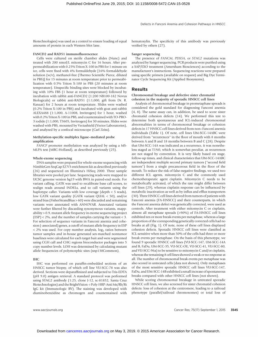
Analysis of chromosomal breakage in prometaphase spreads isconsidered the gold standard for diagnosing Fanconi anemia(4, 8). The same assay can, in addition, be used to score sisterchromatid cohesion defects (14). We performed this test todetermine both spontaneous and ICL-induced chromosomalabnormalities in terms of chromosomal breakage or cohesiondefects in 17HNSCC cell lines derived fromnon–Fanconi anemiaindividuals (Table 1). Of note, cell lines UM-SCC-14ABC werederived from "recurrences" in the floor of mouth with 8 monthsbetween A and B and 18 months between B and C (28). Despitethat UM-SCC-14A was indicated as a recurrence, it was nonethe-less staged as T1N0, which is somewhat peculiar, as recurrencesare not staged by convention. It is very likely based on stage,follow-up times, and clinical characteristics that UM-SCC-14ABCare independent multiple second primary tumors ("second fieldtumors") from a single precancerous field in the floor of themouth. To reduce the risk of false-negative findings, we used twodifferent ICL agents, mitomycin C and the commonly usedchemotherapeutic agent cisplatin. Mitomycin C needs to bemetabolically activated, of which the rate might differ betweencell lines (29), whereas cisplatin response can be influenced bymetabolic inactivation as well as by influx and efflux transporters(30). ThreeHNSCCcell lines derived from tumors of patientswithFanconi anemia (FA-HNSCC) and their counterparts, in whichthe Fanconi anemia defect was genetically corrected, were used ascontrols. After treatment with either mitomycin C or cisplatin,almost all metaphase spreads (>90%) of FA-HNSCC cell linesexhibited ten ormore break events permetaphase, whereas a largeproportion of the corresponding genetically corrected cells hadnobreaks at all (Fig. 1). Of note, none of these cell lines displayedcohesion defects. Sporadic HNSCC cell lines were classified asICL-sensitive when more than 50% of the cells had three or morebreak events per metaphase. On the basis of this phenotype, wefound 9 sporadic HNSCC cell lines (VU-SCC-147, UM-SCC-14Aand B, FaDu, UM-SCC-35, VU-SCC-OE, VU-SCC-41, VU-SCC-80,and VU-SCC-96a) to be sensitive tomitomycin C and/or cisplatin,whereas the remaining 8 cell lines showed aweak or no response atall. The number of chromosomal break events per metaphase wasalso scored in untreated cells (data not shown). Only metaphasesof the most sensitive sporadic HNSCC cell lines VU-SCC-147,FaDu, andUM-SCC-14Bexhibited a small increaseof spontaneousbreaks compared with other HNSCC cell lines (not shown).
While scoring chromosomal breakage in untreated sporadicHNSCC cell lines, we also screened for sister chromatid cohesiondefects: loss of cohesion at the centromere, leading to a railroadphenotype (parallel/railroad chromosomes) or total loss of
Defects in Fanconi Anemia and Cohesion Pathways in HNSCC
www.aacrjournals.org Cancer Res; 75(17) September 1, 2015 3545
on May 3, 2019. © 2015 American Association for Cancer Research. cancerres.aacrjournals.org Downloaded from
Published OnlineFirst June 29, 2015; DOI: 10.1158/0008-5472.CAN-15-0528
Stoepker et al.
Cancer Res; 75(17) September 1, 2015 Cancer Research3546
on May 3, 2019. © 2015 American Association for Cancer Research. cancerres.aacrjournals.org Downloaded from
Published OnlineFirst June 29, 2015; DOI: 10.1158/0008-5472.CAN-15-0528

cohesion between sister chromatids, leading to premature chro-matid separation (PCS). Quantification of railroad chromosomesandPCS revealed that 5of 17 sporadicHNSCCcell lines (VU-SCC-147, UM-SCC-14B, VU-SCC-OE, VU-SCC-78, and VU-SCC-120)displayed a substantial level (�70%) of cohesion defects, almostasmuch as in thepositive control cell lineVU1199-F SV40 (Fig. 1).This cell linewas derived froman individual suffering fromRobertsyndrome, a cohesinopathy caused by mutations in ESCO2 (31).Together, these data demonstrate the presence of ICL-inducedchromosomal breakage and/or defective chromatid cohesion intwo thirds of the sporadic HNSCC cell lines examined.
ICL-induced accumulation of HNSCC cells in the G2–M phaseof the cell cycle
In addition to increased chromosomal breakage, normal cellsfrom individuals with Fanconi anemia are characterized by anICL-induced accumulation of cells in the G2–M phase of the cellcycle. We performed cell-cycle analysis to determine whether FA-HNSCC and sporadic HNSCC cell lines treated with either mito-mycin C or cisplatin also arrested in the G2–M phase of the cellcycle (Fig. 1 and Supplementary Fig. S1). Indeed, despite thepresence of tumor-promoting genetic changes, FA-HNSCC cellsstill accumulated in the G2–M phase of the cell cycle aftertreatment with cross-linking agents, whereas this accumulationwas not observed in the corrected cell lines. Of the 9 sporadicHNSCCcell lines that showed chromosomal breakage in responseto mitomycin C and/or cisplatin, 5 cell lines arrested in the G2–Mphase of the cell cycle after treatment with mitomycin C and/orcisplatin (Fig. 1 and Supplementary Fig. S1). One plausibleexplanation for the absence of G2–Marrest in the other 4 sensitivecell lines is a defective G2–M cell cycle checkpoint, enabling thecells to continue growing in the presence of DNA damage.Conversely, of the 8 cell lines that were classified as ICL-resistantin terms of chromosomal breakage, 4 still showed G2–M arrest inresponse to mitomycin C and/or cisplatin (Fig. 1 and Supple-mentary Fig. S1). Untreated VU-SCC-78 cells already showed ahigh 4n peak, which will be discussed below.
Somewhat remarkably, ICL-induced G2–M arrest did not corre-late with ICL-induced chromosomal breakage. This might be dueto the variable genetic alterations that accumulated in these tumorcell lines. Because the chromosomal breakage assay is consideredthe gold standard in diagnosing Fanconi anemia, cell lines showingchromosomal breakage were classified as ICL-sensitive.
FANCM mutations in the sporadic head and neck tumor cellline FaDu
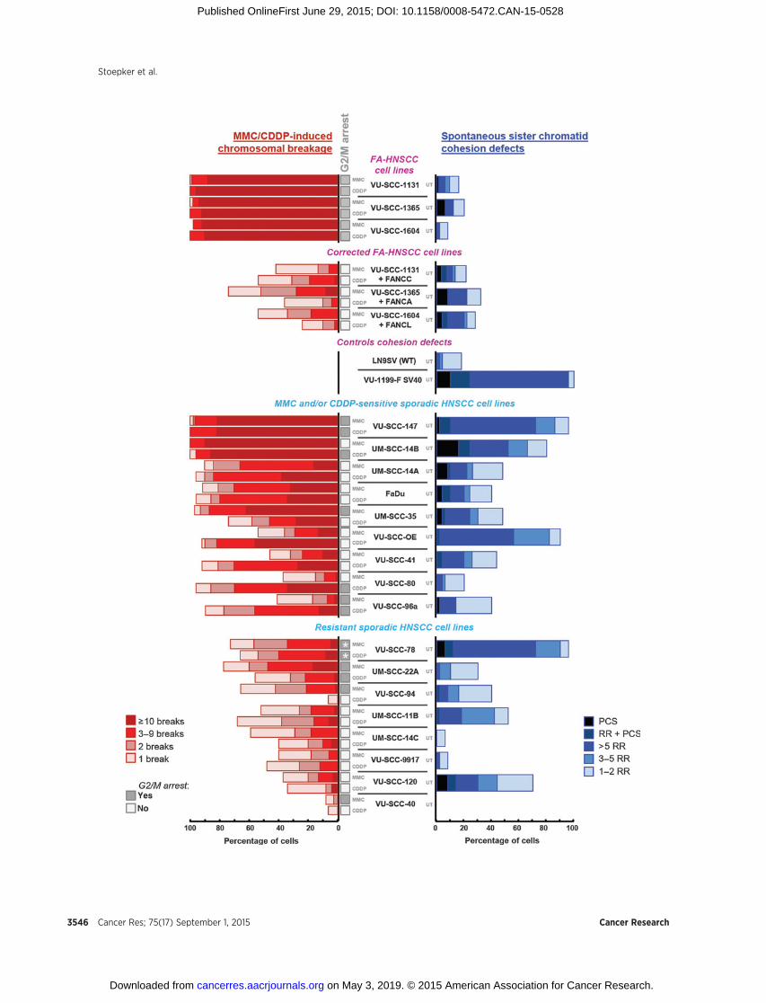
An ICL-induced increase in chromosomal breakage could beindicative for Fanconi anemia pathway inactivation. The Fanconianemia pathway can be divided into two components: theupstream part, which is required for FANCD2 monoubiquitina-
tion, and a downstream part that is not essential for this post-translational modification (6). To test for a functional defect inthe upstream part of the Fanconi anemia pathway, FANCD2monoubiquitination and focus formation were analyzed. Muta-tions in anyof the 8upstreamFanconi anemia core complex genes(FANCA, -B, -C, -E, -F, -G, -L, and -M) abolish or reduce (in case ofFANCM mutations) monoubiquitination and nuclear focus for-mation of FANCD2 (6). Five of the 9 ICL-sensitive sporadicHNSCC cell lines (UM-SCC-14B, FaDu, UM-SCC-35, VU-SCC-41, and VU-SCC96a) and 1 ICL-resistant cell line (UM-SCC-14C)appeared only moderately able to monoubiquitinate FANCD2(Fig. 2A). Notwithstanding, FANCD2 nucleus focus formationwas observed in all cell lines tested, although slight differences inICL-induced induction of FANCD2 foci cannot be excluded(Fig. 2B). A previous report (32) already showed that FaDu cellshave abnormal FANCD2 monoubiquitination, which is consis-tent with our data. However, the underlying cause remainedunknown. By using the Cancer Cell Line Encyclopedia, we foundthat FaDu harbors a homozygous nonsense mutation in FANCM(p.Ser1618�), which we confirmed by Sanger sequencing. As aresult of thismutation, FaDu cells lack FANCMprotein expression(Supplementary Fig. S2), which might explain the observedchromosomal breakage in this cell line.
Whole-exome sequencing revealed several putative pathogenicmutations in Fanconi anemia genes
Nine cell lines in our panel of 17 sporadic HNSCC cell lineswere sensitive to mitomycin C and/or cisplatin in terms ofchromosomal breakage. To assess whether besides FaDu, otherICL-sensitive HNSCC cell lines contained mutations in any of the16knownFanconi anemiaor 5 Fanconi anemia–associated genes,we sequenced the whole exomes of 6 ICL-sensitive cell lines(VU-SCC-80, VU-SCC-96a, VU-SCC-147, VU-SCC-OE, andUM-SCC-14B). This revealed possible pathogenic mutations inTP53, HRAS, NOTCH1, PIK3CA, SMAD4, and FAT1, which arefrequently altered in HNSCC (Supplementary Fig. S3a; refs.33–35). Moreover, copy number alterations that often occur inHNSCC were found (e.g., deletions encompassing TP53 andCDKN2A and amplifications of CCND1, MDM2, and EGFR;Supplementary Fig. S3b). These results show that the cell linesare representative HNSCC cell lines.
Besides these oncogenic driver mutations, one hemizygousnonsense mutation and 8 missense variants in various Fanconianemia genes were found (Table 2). The hemizygous nonsensemutation in FANCD1/BRCA2 (p.Lys3326�), identified in cell lineVU-SCC-147, is known as a polymorphic stop (36) and did notconfer mitomycin C or cisplatin sensitivity in a mouse embryonicstem cell–based assay (37). To further exclude this variant aspossible pathogenic, we investigated PARP inhibitor responseand nuclear RAD51 focus formation in VU-SCC-147 cells, as
Figure 1.ICL-induced chromosomal breakage, G2–M arrest, and spontaneous sister chromatid cohesion defects in HNSCC cell lines. Metaphase spreads of mitomycin-C(MMC)- and cisplatin (CDDP)-treated HNSCC cell lines were examined and scored for chromosomal breakage events. On the basis of this assay, two groups ofsporadic HNSCC cell lines could be distinguished, mitomycin C- and/or cisplatin-sensitive and resistant cell lines. FA-HNSCC cell lines and the correspondingcorrected cells were used as controls. Metaphase spreads of untreated (UT) cells were scored for sister chromatid cohesion defects, railroad chromosomes (RR) andPCS. LN9SV [wild-type (WT)] and VU1199-F SV40 fibroblasts were used as negative and positive controls, respectively. Percentage of cells with the indicatednumber of chromosomal break events, railroad chromosomes, and PCS are shown. Mitomycin C- and cisplatin-induced accumulation in the G2–M phase wasalso analyzed in Fanconi anemia as well as sporadic HNSCC cell lines (see also Supplementary Fig. S1). G2–M arrest was classified positive (filled boxes) as thepercentage of cells in G2–M � G1. Asterisks indicate that untreated VU-SCC-78 already had a high 4n peak.
Defects in Fanconi Anemia and Cohesion Pathways in HNSCC
www.aacrjournals.org Cancer Res; 75(17) September 1, 2015 3547
on May 3, 2019. © 2015 American Association for Cancer Research. cancerres.aacrjournals.org Downloaded from
Published OnlineFirst June 29, 2015; DOI: 10.1158/0008-5472.CAN-15-0528
BRCA2-deficient cells are PARP inhibitor–sensitive and lacknuclear RAD51 focus formation (38–40). VU-SCC-147 cells wereresistant to PARP inhibitor (Supplementary Fig. S4a) and wereable to form RAD51 foci (Supplementary Fig. S4b), suggestingthat the resulting truncating BRCA2mutation did most likely notcause the observed ICL sensitivity in this cell line.
To assess thepathogenic potential ofmissense variants, weusedthree tools: (i) the in silico algorithms SIFT, MutationTaster, andPolyPhen for the prediction of deleterious mutations, (ii) minorallele frequencies (MAF; Exome Variant Server, http://evs.gs.washington.edu/EVS/), and (iii) the occurrence of these variantsin patients with Fanconi anemia (Fanconi anemia gene variantdatabase, www.rockefeller.edu/fanconi/). Variants with low
MAFs (�0.02) and predicted to be pathogenic by at least 2 of3 in silico algorithms were classified as possibly damaging. Four of8 identified variants with MAFs below 0.02 were predicted to bepathogenic by at least two prediction algorithms (Table 2).Although these variants in FANCA, FANCJ, FANCP, and FANCQoccurred heterozygously in the respective cell lines and Fanconianemia is a recessive disease, haploinsufficiency or a dominant-negative effect cannot be excluded, particularly not in cancer cellswith their damaged genomes. Interestingly, the heterozygousvariant in FANCA (p.Cys625Ser) in cell line UM-SCC-14B hadbeen reported to occur in a patient with Fanconi anemia (Table 2;ref. 41). Therefore, this variant might be disease-causing, whichis strengthened by the observation that the cell line containing
Figure 2.FANCD2 monoubiquitination and focus formation. A, Western blotting for FANCD2 monoubiquitination. Sporadic HNSCC cell lines were treated with orwithout 200 nmol/L mitomycin C (MMC) for 16 hours and whole-cell lysates were immunoblotted. The upper band represents the monoubiquitinated form ofFANCD2. B, immunofluorescent analysis of FANCD2 focus formation (green). HNSCC cell lines were treated with 200 nmol/L mitomycin C for 16 hours. TO-PRO-3(red) was used for nuclear counterstaining. Representative confocal images are shown. CDDP, cisplatin.
Stoepker et al.
Cancer Res; 75(17) September 1, 2015 Cancer Research3548
on May 3, 2019. © 2015 American Association for Cancer Research. cancerres.aacrjournals.org Downloaded from
Published OnlineFirst June 29, 2015; DOI: 10.1158/0008-5472.CAN-15-0528

this variant had poor FANCD2 monoubiquitination (Fig. 2A).The other variants were not identified in patients with Fanconianemia.
Taken together, several sequence variants in Fanconi anemiagenes were found, but their pathogenic nature remains elusive. Inparticular, the variants in FANCA, FANCJ, FANCP, and FANCQpredicted to be pathogenic might be disease causing, but theirheterozygous nature indicates that they cannot explain the ICL-induced chromosomal breakage phenotype.
Absence of FANCD2 monoubiquitination in sporadic HNSCCcell line UPCI-SCC-154 due to FANCF methylation
On the basis of hydroxyurea (HU)-induced FANCD2 mono-ubiquitination analysis of a separate panel of 39HNSCC cell lines(Supplementary Table S1), one cell line (UPCI-SCC-154) wasselected for more detailed investigation. Upon mitomycin Ctreatment, FANCD2 monoubiquitination and nuclear FANCD2focus formation were absent in UPCI-SCC-154 cells (Fig. 3A andB). As with the 17 other sporadic HNSCC cell lines, chromosomalbreakage and cell-cycle analysis were performed to examine ICLsensitivity. Upon treatment with either mitomycin C or cisplatin,UPCI-SCC-154 cells accumulated in the G2–M phase of the cellcycle and exhibited in approximately 70% of scored metaphasesmore than three break events per metaphase (Fig. 3C). This wasalmost as sensitive as the FANCC-deficient control cell line VU-SCC-1131. Furthermore, UPCI-SCC-154 cells were sensitive tocisplatin in a growth inhibition assay (Supplementary Fig. S5).Wealso found in 60% of metaphases of untreated UPCI-SCC-154cells sister chromatid cohesion defects, mainly railroad chromo-somes (Fig. 3C).
Massive parallel sequencing of all known upstream Fanconianemia genes in UPCI-SCC-154 cells did not reveal mutationsthat could explain the absence of FANCD2 monoubiquitination.Because promotermethylation of some Fanconi anemia genes hasbeen observed in various tumors (16), we analyzed the methyl-ation of Fanconi anemia genes using methylation-specific mul-tiplex ligation-dependent probe amplification (MS-MLPA). Pro-
motermethylation of the FANCF genewas detected inUPCI-SCC-154 cells and transfection of this cell line with FANCF cDNArestored FANCD2 monoubiquitination (Fig. 3D) and reducedmitomycin C-induced chromosomal breakage (Fig. 3C). Theseresults demonstrate that FANCFmethylation is responsible for theobserved Fanconi anemia phenotype in cell line UPCI-SCC-154.
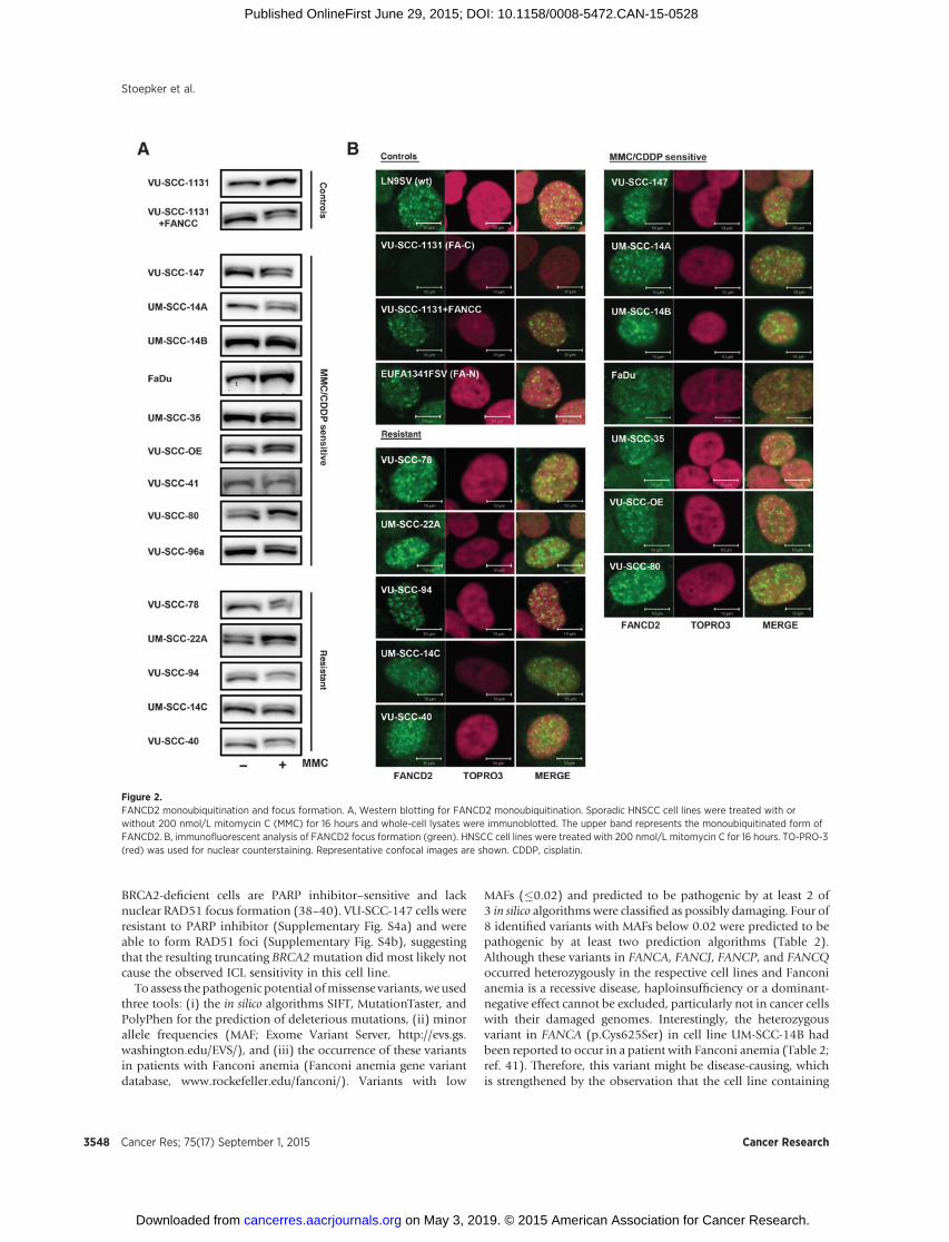
Sister chromatid cohesion defects due to mutations in PDS5Aand STAG2
As shown in Fig. 1, we also found sister chromatid cohesiondefects in several sporadic HNSCC cell lines. To assess the molec-ular basis of cohesion defects, we followed a candidate geneapproachbyfirst investigating the levels of several knowncohesionproteins (SMC1, SMC3, ESCO2, PDS5A, PDS5B, DDX11, STAG1,STAG2, andRAD21) byWesternblotting, followedbyDNASangersequencing. Remarkably, PDS5A protein expression was absent inone cell line (VU-SCC-41) with only moderate cohesion defects(Figs. 1 and 4A). In contrast, no cohesion defects (data not shown)and normal PDS5A protein expression were observed in primaryfibroblasts (VU-41-F) from the same patient (Fig. 4A), suggestingthat a somatic mutation in PDS5A had occurred in the tumor. Byusing cDNA primers spanning exons 3 to 7, we detected a shorterPCR product in VU-SCC-41 cells compared with the wild-typefibroblasts from the same patient (Supplementary Fig. S6a). Theshorter cDNA appeared to lack exon 6 sequence, resulting in aframeshift and premature stop (Supplementary Fig. S6b). Furtherexamination of the PDS5A gene revealed an inversion combinedwith a duplication of part of the inverted sequence at the intron–exon boundary of exon 6, which removes the splice donor site(Supplementary Fig. S6c). This mutation was also found in par-affin-embedded tumor material (data not shown), indicating thatthemutationwasnot inducedby cell culture. In the tumor cell line,wild-type PDS5A sequence was absent, suggesting that the otherPDS5A allele was deleted. Subsequent comparative genomehybridization (array CGH) indeed showed a heterozygous loss ofthe entire p-arm of chromosome 4, which is the region where thePDS5A gene is located (Supplementary Fig. S6d).
Table 2. Sequence variants with MAF � 0.02 in Fanconi anemia genes
Gene Sequence variant SIFT MT PP MAF EA/AA % Reads Remarks
Cell line VU-SCC-80FAAP16 c.370A>G; p.Asn124Asp T T T — 60/86 (70%) Predicted toleratedFANCD2 c.577A>G; p.Thr193Ala T T T 0.0007/0.0009 19/20 (95%) Predicted toleratedFANCQ/XPF c.241G>T; p.Val81Phe P P P —/0.0009 24/53 (45%) Predicted pathogenic
Cell line VU-SCC-147FANCD1/BRCA2 c.9976A>T; p.Lys3326� 0.0084/0.0027 39/41 (95%) Hemizygous nonsense mutation,
polymorphic stop (36)FANCM c.527C>T; p.Thr176Ile T T T 0.0051/0.0005 27/71 (38%) Predicted tolerated
Cell line VU-SCC-OEFANCI c.480G>C; p.Leu160Phe T P T — 66/66 (100%) Predicted tolerated
Cell line UM-SCC-14BFANCA c.1874G>C; p.Cys625Ser P P P 0.0031/0.0007 12/31 (39%) Reported in Fanconi
anemia patient (41)FANCP/SLX4 c.2854_2855delinsAT;
p.Ala952Met— 8/23 (35%) Predicted pathogenic
Cell line UM-SCC35FANCJ/BRIP1 c.517C>T; p.Arg173Cys P P P 0.0049/0.0011 39/125 (31%) Predicted pathogenic
NOTE: Transcript refseq ID: FAAP16 NM_199294.2; FANCA NM_000135.2; FANCD1/BRCA2 NM_000059; FANCD2 NM_033084.3; FANCI NM_001113378.1; FANCJ/BRIP1 NM_032043; FANCM NM_020937.2; FANCP/SLX4 NM_032444.2 and FANCQ/XPF NM_005236.2. MAF EA/AA represents minor allele frequency European/American and African/American populations, respectively.Abbreviations: MT, MutationTaster; PP, polyphen; P, pathogenic; T, tolerated.
Defects in Fanconi Anemia and Cohesion Pathways in HNSCC
www.aacrjournals.org Cancer Res; 75(17) September 1, 2015 3549
on May 3, 2019. © 2015 American Association for Cancer Research. cancerres.aacrjournals.org Downloaded from
Published OnlineFirst June 29, 2015; DOI: 10.1158/0008-5472.CAN-15-0528

Absence of STAG2 protein expression was observed in sporadicHNSCC cell line VU-SCC-78 that showed severe cohesion defects(Figs. 1 and 4B). In this cell line, a heterozygous 1 base pair (bp)insertion in STAG2 leading to a frameshift was identified bysequence analysis of gDNA (Supplementary Fig. S7a). BecauseSTAG2 maps to the X chromosome, a single mutational eventcould be enough for its complete inactivation. Sequencing ofSTAG2 cDNA indeed revealed that STAG2 mRNA expression wasderived entirely of the mutant allele, indicating that the wild-typeallele was not functional by X chromosome inactivation (Sup-plementary Fig. S7b). To exclude that the mutation was induced
during cell culture, STAG2 protein expression was examined andindeed missing in paraffin-embedded tumor material derivedfrom the same tumor of which cell line VU-SCC-78 was derived(Fig. 4C). In addition, it was previously shown that knockdown ofSTAG2 resulted in the generationof anoctaploid population (42).We detected a similar phenotype in VU-SCC-78 (SupplementaryFig. S1b).
Together, these results strongly suggest that the cohesiondefects observed in VU-SCC-41 were due to an acquiredmutationin the PDS5A gene, accompanied by a loss of the wild-type allele,whereas the cohesion defects in VU-SCC-78 were caused by a
Figure 3.ICL sensitivity, FANCD2 monoubiquitination, and focus formation of sporadic HNSCC cell line, UPCI-SCC-154. A and B, untreated, or mitomycin C (MMC)-treated(200 nmol/L for 16 hours) UPCI-SCC-154 cells were immunoblotted for FANCD2 (A) or analyzed for FANCD2 focus formation (green; B). VU-SCC-1131 andVU-SCC-1131 þ FANCC were used as controls. Nuclei were counterstained by TO-PRO-3 (red). C, ICL-induced chromosomal breakage events and spontaneoussister chromatid cohesion defects were observed in metaphase spreads of UPCI-SCC-154 and FANCF cDNA corrected UPCI-SCC-154 cells. CDDP, cisplatin.D, ectopic expression of FANCF in UPCI-SCC-154 cells restored FANCD2 monoubiquitination.
Stoepker et al.
Cancer Res; 75(17) September 1, 2015 Cancer Research3550
on May 3, 2019. © 2015 American Association for Cancer Research. cancerres.aacrjournals.org Downloaded from
Published OnlineFirst June 29, 2015; DOI: 10.1158/0008-5472.CAN-15-0528
somatic mutation in STAG2 in combination with X chromosomeinactivation of the other allele.
DiscussionIn the present study, we investigated the occurrence of Fanconi
anemia pathway inactivation in sporadic HNSCC by analyzing apanel of Fanconi anemia and sporadic HNSCC cell lines. Wefound in 9 of 17 (53%) of the sporadic HNSCC cell lines ICL-induced chromosomal breakage, which can be indicative of adefective Fanconi anemia pathway, and in 29% (5 of 17) severesister chromatid cohesion defects. However, this may be anoverestimation as some of these cell lines are related. The UM-SCC-14ABC cell lines are most likely derived from multipleprimary tumors from a large preneoplastic field based on theclinical history and tumor characteristics (28). Hence, these couldbe considered as separate tumors but nonetheless UM-SCC-14Aand B share a similar phenotype, whereas UM-SCC-14C clearlybehaves differently. Mutational inactivation of FANCM andmethylation of FANCF were observed in two sporadic HNSCCcell lines from screened panels of 17 and 39 HNSCC cell lines,respectively. Although a few possible disease-causing variants inFanconi anemia genes were found in the 6 ICL-sensitive HNSCCcell lines that were analyzed by whole-exome sequencing, their
heterozygous nature suggests that they cannot explain the ICL-induced chromosomal breakage phenotype. This indicates thatdespite a frequent Fanconi anemia–like phenotype (ICL-inducedchromosomal breakage), the Fanconi anemia pathway itselfseems rarely involved. Silencing of FANCF by promoter hyper-methylation has been demonstrated in a wide range of malig-nancies, including HNSCC (17). However, the importance ofFANCF promoter methylation in HNSCC is contradictory, as itwas demonstrated that the methylation-specific PCR methodthat is routinely used to investigate FANCF methylation is liableto produce false-positive results (18). By using the more specificmethod MS-MLPA, we showed that FANCF methylation doesoccur in sporadic HNSCC, albeit infrequently. Screening of largesample series by more robust methods, such as quantitativemethylation-specific PCR and sequencing, might substantiate ourfindings.
Interestingly, we found besides ICL-induced chromosomalbreakage also spontaneous sister chromatid cohesion defects ina subset of sporadic HNSCC. Because of the role of sister chro-matid cohesion in chromatid separation, DNA repair, and generegulation, this pathway is important for chromosomal stability(15). Mutations in cohesion genes could therefore lead to CIN bydifferentmechanisms:mis-segregation of chromosomes followedby aneuploidy due to partial or total loss of cohesion or as a
Figure 4.Mutational inactivation of PDS5A and STAG2 in two HNSCC cell lines. A, Western blot analysis showing a truncated PDS5A protein in VU-SCC-41 and normalexpression of PDS5A in fibroblasts (VU-41-F) of the same patient. B,Western blot analysis showing absence of STAG2 protein expression in VU-SCC-78. Tubulinwasused as a loading control. C, immunohistochemical analysis of STAG2 expression in paraffin-embedded tumor material of the same tumor of which cell lineVU-SCC-78 was derived. STAG2 protein expression was absent in tumor (T) material but present in normal tissue surrounding the tumor.
Defects in Fanconi Anemia and Cohesion Pathways in HNSCC
www.aacrjournals.org Cancer Res; 75(17) September 1, 2015 3551
on May 3, 2019. © 2015 American Association for Cancer Research. cancerres.aacrjournals.org Downloaded from
Published OnlineFirst June 29, 2015; DOI: 10.1158/0008-5472.CAN-15-0528

consequence of impaired DNA repair as well as altered geneexpression. Recently, in several studies, alterations in sister chro-matid cohesion genes were identified in a variety of cancers(27, 43–46). In particular, inactivating alterations in STAG2 havebeen frequently found. Likewise, we found mutational inactiva-tion of STAG2 in one HNSCC cell line, whereas in another cellline, PDS5A was mutated. The cohesion defects in VU-SCC-147and UPCI-SCC-154 could be explained by the presence of thehuman papillomavirus (HPV) and subsequent inactivation of theretinoblastoma tumor suppressor proteins (pRb) in these celllines. Loss of pRb has been demonstrated to alter H4K20 meth-ylation and lead to sister chromatid cohesiondefects (47–49). Thepresence of HPV in these cell lines might well explain the inad-equate sister chromatid cohesion.
Fanconi anemia and cohesinopathies have some overlappingfeatures. ICL-induced chromosomal breakage is not an exclusivehallmark of Fanconi anemia cells and has been observed in Tlymphocytes from individuals with Robert syndrome or Warsawbreakage syndrome as well (14). Moreover, mitomycin C-induced, but not spontaneous cohesion, defects have beenreported to occur in Fanconi anemia cells (14). Whether theseoverlapping phenotypes also occur in cancer cells remains to bedetermined in functional studies. This is of relevance, as thescreening for sequence variants in Fanconi anemia genes andcohesion genes might be exploited as biomarkers for cisplatinresponse. This is supported by previous work that demonstratedthat FANCM-deficient cell line FaDu was approximately 6-foldmore sensitive to cisplatin than themost resistantHNSCCcell linetested, although not as sensitive as FA-HNSCC cell lines (50). Inaddition, in the present study, we showed sensitivity to cisplatinfor the FANCF-deficient cell line UPCI-SCC-154 as well.
In summary, CIN in terms of ICL-induced chromosomal break-age or defective sister chromatid cohesion occurs frequently inHNSCC. In few cases, this is caused by defects in the Fanconianemia pathway or mutational inactivation of chromatid cohe-sion genes. Further unraveling of the relevantmechanisms and/orgenes causing these phenotypes may open new avenues for
treatment and provide the biomarkers to predict treatmentresponse.
Disclosure of Potential Conflicts of InterestNo potential conflicts of interest were disclosed.
Authors' ContributionsConception and design: C. Stoepker, P. van der Lelij, R. Dietrich, S.M. Feller,R.H. BrakenhoffDevelopment of methodology: B. YlstraAcquisition of data (provided animals, acquired and managed patients,provided facilities, etc.): C. Stoepker, N. Ameziane, S.E. van Mil, A. Brink,R. Dietrich, B. Ylstra, H. Joenje, R.H. BrakenhoffAnalysis and interpretation of data (e.g., statistical analysis, biostatistics,computational analysis): C. Stoepker, N. Ameziane, I.E. Kooi, R.H. BrakenhoffWriting, review, and/or revision of themanuscript: C. Stoepker, N. Ameziane,P. van der Lelij, A.B. Oostra, R. Dietrich, R.H. BrakenhoffAdministrative, technical, or material support (i.e., reporting or organizingdata, constructing databases): A.B. Oostra, M.A. Rooimans, A. Brink,R. Dietrich, R.H. BrakenhoffStudy supervision: R.H. BrakenhoffOther (practical support): J.A. BalkOther (primarily involved in the initial stages of this project as a supervisor):H. JoenjeOther (generation of experimental data): S.M. Feller
AcknowledgmentsThe authors thank Prof. Dr. Johan de Winter who initiated these studies, but
died during the completion of the experimental work and the drafting ofthe final version of the manuscript. They also thank E. Polder, R. van der Willik,J. Steltenpool, M. Blijlevens, and P.P. Eijk for technical assistance, E. Velleuer forcollecting tumormaterial, andH. teRiele for helpful discussions and comments.
Grant SupportFinancial support was provided from CCA/V-ICI Amsterdam, Dutch Cancer
Society and Fanconi Anemia Research Fund (FARF), Portland, Oregon.The costs of publication of this articlewere defrayed inpart by the payment of
page charges. This article must therefore be hereby marked advertisement inaccordance with 18 U.S.C. Section 1734 solely to indicate this fact.
Received March 5, 2015; revised May 13, 2015; accepted May 19, 2015;published OnlineFirst June 29, 2015.
References1. Lengauer C, Kinzler KW, Vogelstein B. Genetic instabilities in human
cancers. Nature 1998;396:643–9.2. Negrini S, Gorgoulis VG,Halazonetis TD.Genomic instability–an evolving
hallmark of cancer. Nat Rev Mol Cell Biol 2010;11:220–8.3. McGranahanN, Burrell RA, Endesfelder D, Novelli MR, Swanton C. Cancer
chromosomal instability: therapeutic and diagnostic challenges. EMBORep 2012;13:528–38.
4. Auerbach AD. Fanconi anemia and its diagnosis. Mutat Res 2009;668:4–10.
5. KutlerDI, AuerbachAD, Satagopan J,Giampietro PF, Batish SD,Huvos AG,et al. High incidence of head and neck squamous cell carcinoma in patientswith Fanconi anemia. Arch Otolaryngol Head Neck Surg 2003;129:106–12.
6. Kottemann MC, Smogorzewska A. Fanconi anaemia and the repair ofWatson and Crick DNA crosslinks. Nature 2013;493:356–63.
7. Bogliolo M, Schuster B, Stoepker C, Derkunt B, Su Y, Raams A, et al.Mutations in ERCC4, encoding the DNA-repair endonuclease XPF, causeFanconi anemia. Am J Hum Genet 2013;92:800–6.
8. Oostra AB, Nieuwint AWM, Joenje H, de Winter JP. Diagnosis of fanconianemia: chromosomal breakage analysis. Anemia 2012;2012:238731.
9. Sasaki MS, Tonomura A. A high susceptibility of Fanconi's anemia tochromosome breakage by DNA cross-linking agents. Cancer Res 1973;33:1829–36.
10. Auerbach AD, Wolman SR. Susceptibility of Fanconi's anaemia fibroblaststo chromosome damage by carcinogens. Nature 1976;261:494–6.
11. Langevin F,CrossanGP,Rosado IV,ArendsMJ, Patel KJ. Fancd2 counteractsthe toxic effects of naturally produced aldehydes in mice. Nature 2011;475:53–8.
12. Van den Berg DJ, Francke U. Sensitivity of Roberts syndrome cells togamma radiation, mitomycin C, and protein synthesis inhibitors. SomatCell Mol Genet 1993;19:377–92.
13. Van der Lelij P, Chrzanowska KH, Godthelp BC, RooimansMA, Oostra AB,Stumm M, et al. Warsaw breakage syndrome, a cohesinopathy associatedwith mutations in the XPD helicase family member DDX11/ChlR1. Am JHum Genet 2010;86:262–6.
14. Van der Lelij P, Oostra AB, Rooimans MA, Joenje H, de Winter JP.Diagnostic overlap between fanconi anemia and the cohesinopathies:Roberts syndrome and warsaw breakage syndrome. Anemia 2010;2010:565268.
15. Peters J-M, Nishiyama T. Sister chromatid cohesion. Cold Spring HarbPerspect Biol 2012;4: pii: a011130. doi.
16. Valeri A, Martínez S, Casado JA, Bueren JA. Fanconi anaemia: from amonogenic disease to sporadic cancer. Clin Transl Oncol 2011;13:215–21.
17. Marsit CJ, Liu M, Nelson HH, Posner M, Suzuki M, Kelsey KT. Inactivationof the Fanconi anemia/BRCA pathway in lung and oral cancers: implica-tions for treatment and survival. Oncogene 2004;23:1000–4.
Cancer Res; 75(17) September 1, 2015 Cancer Research3552
Stoepker et al.
on May 3, 2019. © 2015 American Association for Cancer Research. cancerres.aacrjournals.org Downloaded from
Published OnlineFirst June 29, 2015; DOI: 10.1158/0008-5472.CAN-15-0528
18. Ameziane N, Chen F, Leemans CR, Brakenhoff RH, Joenje H. No evidencefor FANCF gene silencing in head-and-neck squamous cell carcinomas.Cell Oncol 2009;31:53–6.
19. HermsenMA, Joenje H, Arwert F,WeltersMJ, Braakhuis BJ, BagnayM, et al.Centromeric breakage as amajor cause of cytogenetic abnormalities in oralsquamous cell carcinoma. Genes Chromosomes Cancer 1996;15:1–9.
20. Van Zeeburg HJT, Snijders PJF, Pals G, Hermsen MAJA, Rooimans MA,Bagby G, et al. Generation and molecular characterization of head andneck squamous cell lines of fanconi anemia patients. Cancer Res 2005;65:1271–6.
21. Martens-de Kemp SR, Dalm SU, Wijnolts FMJ, Brink A, Honeywell RJ,Peters GJ, et al. DNA-bound platinum is themajor determinant of cisplatinsensitivity in head and neck squamous carcinoma cells. PLoS One 2013;8:e61555.
22. Lin CJ, Grandis JR, Carey TE, Gollin SM, Whiteside TL, Koch WM, et al.Head and neck squamous cell carcinoma cell lines: establishedmodels andrationale for selection. Head Neck 2007;29:163–88.
23. Brenner JC, GrahamMP, Kumar B, Saunders LM, Kupfer R, Lyons RH, et al.Genotyping of 73 UM-SCC head and neck squamous cell carcinoma celllines. Head Neck 2010;32:417–26.
24. Wu Z, Doondeea JB, Gholami AM, Janning MC, Lemeer S, Kramer K, et al.Quantitative chemical proteomics reveals new potential drug targets inhead and neck cancer. Mol Cell Proteomics 2011;10:M111.011635.
25. Hess CJ, Ameziane N, Schuurhuis GJ, Errami A, Denkers F, Kaspers GJL,et al. Hypermethylation of the FANCC and FANCL promoter regions insporadic acute leukaemia. Cell Oncol 2008;30:299–306.
26. Knies K, Schuster B, Ameziane N, Rooimans M, Bettecken T, de Winter J,et al. Genotyping of fanconi anemia patients by whole exome sequencing:advantages and challenges. PLoS One 2012;7:e52648.
27. Solomon DA, Kim T, Diaz-Martinez LA, Fair J, Elkahloun AG, Harris BT,et al. Mutational inactivation of STAG2 causes aneuploidy in humancancer. Science 2011;333:1039–43.
28. Gr�enman R, Carey TE, McClatchey KD, Wagner JG, Pekkola-Heino K,SchwartzDR, et al. In vitro radiation resistance among cell lines establishedfrom patients with squamous cell carcinoma of the head and neck. Cancer1991;67:2741–7.
29. Seow HA, Penketh PG, Baumann RP, Sartorelli AC. Bioactivation andresistance to mitomycin C. Methods Enzymol 2004;382:221–33.
30. Kelland L. The resurgence of platinum-based cancer chemotherapy. NatRev Cancer 2007;7:573–84.
31. Vega H, Waisfisz Q, Gordillo M, Sakai N, Yanagihara I, Yamada M, et al.Roberts syndrome is caused bymutations in ESCO2, a human homolog ofyeast ECO1 that is essential for the establishment of sister chromatidcohesion. Nat Genet 2005;37:468–70.
32. Van Der Heijden MS, Brody JR, Kern SE. Functional screen of the fanconianemia pathway in cancer cells by Fancd2 immunoblot. Cancer Biol Ther2004;3:534–7.
33. Stransky N, Egloff AM, Tward AD, Kostic AD, Cibulskis K, Sivachenko A,et al. Themutational landscape of head andneck squamous cell carcinoma.Science 2011;333:1157–60.
34. Agrawal N, Frederick MJ, Pickering CR, Bettegowda C, Chang K, Li RJ,et al. Exome sequencing of head and neck squamous cell carcinomareveals inactivating mutations in NOTCH1. Science 2011;333:1154–7.
35. Leemans CR, Braakhuis BJM, Brakenhoff RH. The molecular biology ofhead and neck cancer. Nat Rev Cancer 2011;11:9–22.
36. Mazoyer S, Dunning AM, Serova O, Dearden J, Puget N, Healey CS, et al. Apolymorphic stop codon in BRCA2. Nat Genet 1996;14:253–4.
37. Kuznetsov SG, Liu P, Sharan SK. Mouse embryonic stem cell-based func-tional assay to evaluate mutations in BRCA2. Nat Med 2008;14:875–81.
38. Bryant HE, Schultz N, Thomas HD, Parker KM, Flower D, Lopez E, et al.Specific killing of BRCA2-deficient tumours with inhibitors of poly(ADP-ribose) polymerase. Nature 2005;434:913–7.
39. Farmer H,McCabe N, Lord CJ, Tutt ANJ, Johnson DA, Richardson TB, et al.Targeting the DNA repair defect in BRCA mutant cells as a therapeuticstrategy. Nature 2005;434:917–21.
40. YuanSS, Lee SY,ChenG, SongM, TomlinsonGE, LeeEY. BRCA2 is requiredfor ionizing radiation-induced assembly of Rad51 complex in vivo. CancerRes 1999;59:3547–51.
41. Castella M, Pujol R, Call�en E, Trujillo JP, Casado JA, Gille H, et al. Origin,functional role, and clinical impact of Fanconi anemia FANCAmutations.Blood 2011;117:3759–69.
42. Barber TD, McManus K, Yuen KWY, Reis M, Parmigiani G, Shen D, et al.Chromatid cohesion defects may underlie chromosome instability inhuman colorectal cancers. Proc Natl Acad Sci U S A 2008;105:3443–8.
43. Guo G, Sun X, Chen C, Wu S, Huang P, Li Z, et al. Whole-genome andwhole-exome sequencing of bladder cancer identifies frequent alterationsin genes involved in sister chromatid cohesion and segregation. Nat Genet2013;45:1459–63.
44. Solomon DA, Kim J-S, Bondaruk J, Shariat SF, Wang Z-F, Elkahloun AG,et al. Frequent truncatingmutations of STAG2 in bladder cancer. Nat Genet2013;45:1428–30.
45. Kon A, Shih L-Y, Minamino M, Sanada M, Shiraishi Y, Nagata Y, et al.Recurrent mutations in multiple components of the cohesin complex inmyeloid neoplasms. Nat Genet 2013;45:1232–7.
46. Thol F, Bollin R, Gehlhaar M, Walter C, Dugas M, Suchanek KJ, et al.Mutations in the cohesin complex in acute myeloid leukemia: clinical andprognostic implications. Blood 2014;123:914–20.
47. VanHarn T, Foijer F, van VugtM, Banerjee R, Yang F, Oostra A, et al. Loss ofRb proteins causes genomic instability in the absence of mitogenic sig-naling. Genes Dev 2010;24:1377–88.
48. Manning AL, Longworth MS, Dyson NJ. Loss of pRB causes centromeredysfunction and chromosomal instability. Genes Dev 2010;24:1364–76.
49. Manning AL, Yazinski SA, Nicolay B, Bryll A, Zou L, DysonNJ. Suppressionof genome instability in pRB-deficient cells by enhancement of chromo-some cohesion. Mol Cell 2014;53:993–1004.
50. Nagel R, Martens-de Kemp SR, Buijze M, Jacobs G, Braakhuis BJM,Brakenhoff RH. Treatment response of HPV-positive and HPV-negativehead and neck squamous cell carcinoma cell lines. Oral Oncol 2013;49:560–6.
www.aacrjournals.org Cancer Res; 75(17) September 1, 2015 3553
Defects in Fanconi Anemia and Cohesion Pathways in HNSCC
on May 3, 2019. © 2015 American Association for Cancer Research. cancerres.aacrjournals.org Downloaded from
Published OnlineFirst June 29, 2015; DOI: 10.1158/0008-5472.CAN-15-0528
2015;75:3543-3553. Published OnlineFirst June 29, 2015.Cancer Res Chantal Stoepker, Najim Ameziane, Petra van der Lelij, et al. Head and Neck CancerDefects in the Fanconi Anemia Pathway and Chromatid Cohesion in
Updated version
10.1158/0008-5472.CAN-15-0528doi:
Access the most recent version of this article at:
Material
Supplementary
http://cancerres.aacrjournals.org/content/suppl/2015/06/27/0008-5472.CAN-15-0528.DC1
Access the most recent supplemental material at:
Cited articles
http://cancerres.aacrjournals.org/content/75/17/3543.full#ref-list-1
This article cites 50 articles, 12 of which you can access for free at:
Citing articles
http://cancerres.aacrjournals.org/content/75/17/3543.full#related-urls
This article has been cited by 2 HighWire-hosted articles. Access the articles at:
E-mail alerts related to this article or journal.Sign up to receive free email-alerts
Subscriptions
Reprints and
To order reprints of this article or to subscribe to the journal, contact the AACR Publications Department at
Permissions
Rightslink site. Click on "Request Permissions" which will take you to the Copyright Clearance Center's (CCC)
.http://cancerres.aacrjournals.org/content/75/17/3543To request permission to re-use all or part of this article, use this link
on May 3, 2019. © 2015 American Association for Cancer Research. cancerres.aacrjournals.org Downloaded from
Published OnlineFirst June 29, 2015; DOI: 10.1158/0008-5472.CAN-15-0528