IMPAIRED FUNCTION OF FANCONI ANEMIA TYPE C DEFICIENT ...
146
IMPAIRED FUNCTION OF FANCONI ANEMIA TYPE C DEFICIENT MACROPHAGES Ying Liu Submitted to the faculty of the University Graduate School in partial fulfillment of the requirements for the degree Doctor of Philosophy in the Department of Microbiology and Immunology, Indiana University September 2011
Transcript of IMPAIRED FUNCTION OF FANCONI ANEMIA TYPE C DEFICIENT ...
IMPAIRED FUNCTION OF FANCONI ANEMIA TYPE C DEFICIENT
MACROPHAGES
Ying Liu
Submitted to the faculty of the University Graduate School in partial fulfillment of the requirements
for the degree Doctor of Philosophy
in the Department of Microbiology and Immunology, Indiana University
September 2011
ii
Accepted by the Faculty of Indiana University, in partial fulfillment of the requirements for the degree of Doctor of Philosophy.
Laura S. Haneline, M.D., Chair
Doctoral Committee
Alexander L. Dent, Ph.D.
May 19, 2011
Johnny J. He, Ph.D.
Edward F. Srour, Ph.D.
Mervin C. Yoder, M.D.

iii
ACKNOWLEDGEMENTS
The work presented in this thesis was carried out at Indiana University School of
Medicine. I would like to thank all the people who helped me to finish this research
project. It has also been pleasant to work with so many nice people in this research
community! Herein, I would like to especially express my appreciation for these people:
I would like to express my deepest gratitude to my advisor, Dr. Laura S. Haneline
for her mentoring me through this entire training process. Importantly, I thank her for
giving me the opportunity to think freely and devise my own experiments. She always
shows her warm support for me when I feel frustrated with failed experiments. She is
always patient with me even when some times when I made a similar mistake. She taught
me not only how to do research, how to present a scientific work, but more importantly,
how to understand research, how to be a good researcher in all means.
I would like to thank the members of my committee for their continual support of
my graduate work and for being there whenever I needed them.
To Dr. Mervin C. Yoder,
Thanks very much for so many insightful suggestions. I really appreciate your
time and your contribution. I know how busy you always are, but you still spend quite
some time with me in discussing the project once a while.
To Dr. Johnny J. He,
Thanks for your great scientific inputs into this project. I greatly appreciate the
tremendous support and encouragement, especially when I felt discouraged with failing
experiments.

iv
To Dr. Edward F. Srour,
Thanks for your numerous valuable suggestions for the research, which are so
important for me to finish my project smoothly.
To Dr. Alexander L. Dent,
Thanks for so many insightful suggestions. I am also grateful for you to help me
to grow as a prudent scientific researcher.
I am truly happy and grateful to have you all as my committee advisors! I will
never forget all the generous help and support you gave me!
To my lab mates,
I am grateful to all the people in Dr. Haneline’s lab, including Kimberly K.
Ballman, Ethel Catherine Derr-Yellin, Shehnaz Khan, and Emily K. Blue. I have received
so much help and good suggestions from all of them. I will miss the pleasant lab
environment in the future.
To my collaborators,
I wish to express my appreciation for many colleagues in the Department of
Microbiology and Immunology and in the Wells Center for Pediatric Research for their
knowledge and friendship, especially Melody Zeng (Dinauer Lab), Lin Wang (Carlesso
Lab), Charles Goodwin (Chan Lab), Sasidhar Vemula (Kapur Lab), Weiguo Yao (Kaplan
Lab), and Mike Ferkowicz (Yoder Lab).
Finally, I am continually thankful for the love and support of my family and
friends. Above all, my husband, Deqiang Li, who has emotionally and scientifically
helped me throughout the years while I was working towards my Ph.D degree!
v
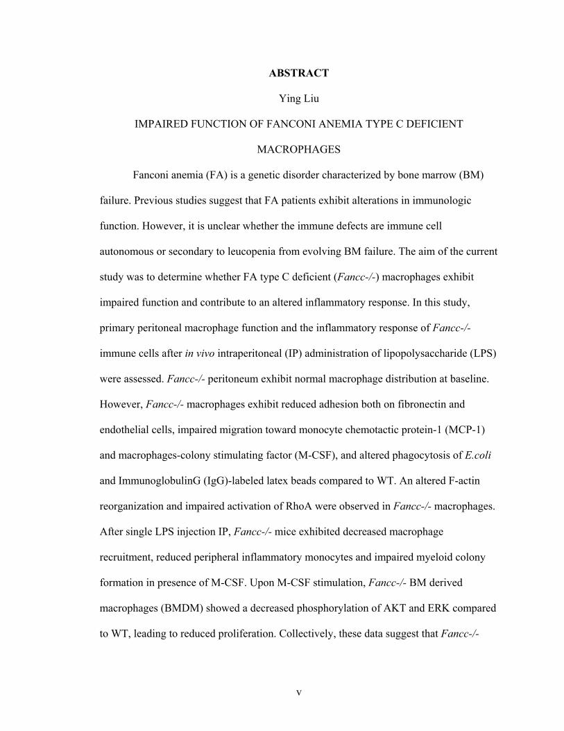
ABSTRACT
Ying Liu
IMPAIRED FUNCTION OF FANCONI ANEMIA TYPE C DEFICIENT
MACROPHAGES
Fanconi anemia (FA) is a genetic disorder characterized by bone marrow (BM)
failure. Previous studies suggest that FA patients exhibit alterations in immunologic
function. However, it is unclear whether the immune defects are immune cell
autonomous or secondary to leucopenia from evolving BM failure. The aim of the current
study was to determine whether FA type C deficient (Fancc-/-) macrophages exhibit
impaired function and contribute to an altered inflammatory response. In this study,
primary peritoneal macrophage function and the inflammatory response of Fancc-/-
immune cells after in vivo intraperitoneal (IP) administration of lipopolysaccharide (LPS)
were assessed. Fancc-/- peritoneum exhibit normal macrophage distribution at baseline.
However, Fancc-/- macrophages exhibit reduced adhesion both on fibronectin and
endothelial cells, impaired migration toward monocyte chemotactic protein-1 (MCP-1)
and macrophages-colony stimulating factor (M-CSF), and altered phagocytosis of E.coli
and ImmunoglobulinG (IgG)-labeled latex beads compared to WT. An altered F-actin
reorganization and impaired activation of RhoA were observed in Fancc-/- macrophages.
After single LPS injection IP, Fancc-/- mice exhibited decreased macrophage
recruitment, reduced peripheral inflammatory monocytes and impaired myeloid colony
formation in presence of M-CSF. Upon M-CSF stimulation, Fancc-/- BM derived
macrophages (BMDM) showed a decreased phosphorylation of AKT and ERK compared
to WT, leading to reduced proliferation. Collectively, these data suggest that Fancc-/-

vi
macrophages and subsequent defects in adhesion, migration, phagocytosis, and
recruitment in vivo. These data also support a Fancc-/- macrophage cells autonomous
defect predisposing to an altered inflammatory response.
Laura S. Haneline M.D., Chair
vii
TABLE OF CONTENTS
LIST OF TABLES .......................................................................................................... xii
LIST OF FIGURES ....................................................................................................... xiii
LIST OF ABBREVIATIONS ....................................................................................... xvi
INTRODUCTION..............................................................................................................1
I. Overview of Fanconi anemia ............................................................................................1
A. Clinical features of FA ..............................................................................................1
B. Molecular mechanisms of FA ...................................................................................2
C. Fanconi anemia type C (FANCC) .............................................................................4
D. Immune defects in FA ...............................................................................................5
II. Monocytes/Macrophages ................................................................................................6
A. Function and generation of macrophages .................................................................6
B. Innate immune response during inflammation ........................................................10
C. The generation of monocytes/macrophages during inflammation ..........................12
III. Cytoskeletal reorganization regulates macrophage function .......................................15
A. Cytoskeletal rearrangement in macrophages ..........................................................15
B. Regulators of cytoskeletal rearrangement in macrophages .....................................17
1. Overview of Rho GTPases.................................................................................17
2. Role of RhoA, Rac, and Cdc42 in macrophages ..............................................17
IV. RESEARCH RATIONALE.........................................................................................20
V. HYPOTHESIS AND AIMS..........................................................................................21
MATERIALS AND METHODS ....................................................................................22
1. Mice ..............................................................................................................................22
viii
2. Treatment of mice and isolation of resident peritoneal cells ........................................22
3. Differential counts of peritoneal cells ...........................................................................23
4. Flow cytometry .............................................................................................................25
5. Myeloid colony formation assay ...................................................................................26
6. Differentiation of bone marrow derived macrophages (BMDM) .................................28
7. Apoptosis assay .............................................................................................................28
8. Proliferation assay .........................................................................................................29
9. Immunoblot analysis .....................................................................................................29
10. Adhesion assays ...........................................................................................................30
11. Transwell motility assay ..............................................................................................31
12. Phagocytosis assay .......................................................................................................31
13. Superoxide detection ....................................................................................................32
14. F-actin reorganization immunocytochemistry .............................................................32
15. GST-pull down assay for activated RhoA, Rac1 and Cdc42 .......................................33
16. RNA isolation and reverse transcription- PCR (RT-PCR) ..........................................34
17. Cytokine multiplex analysis of mouse serum ..............................................................36
18. Statistical analyses .......................................................................................................36
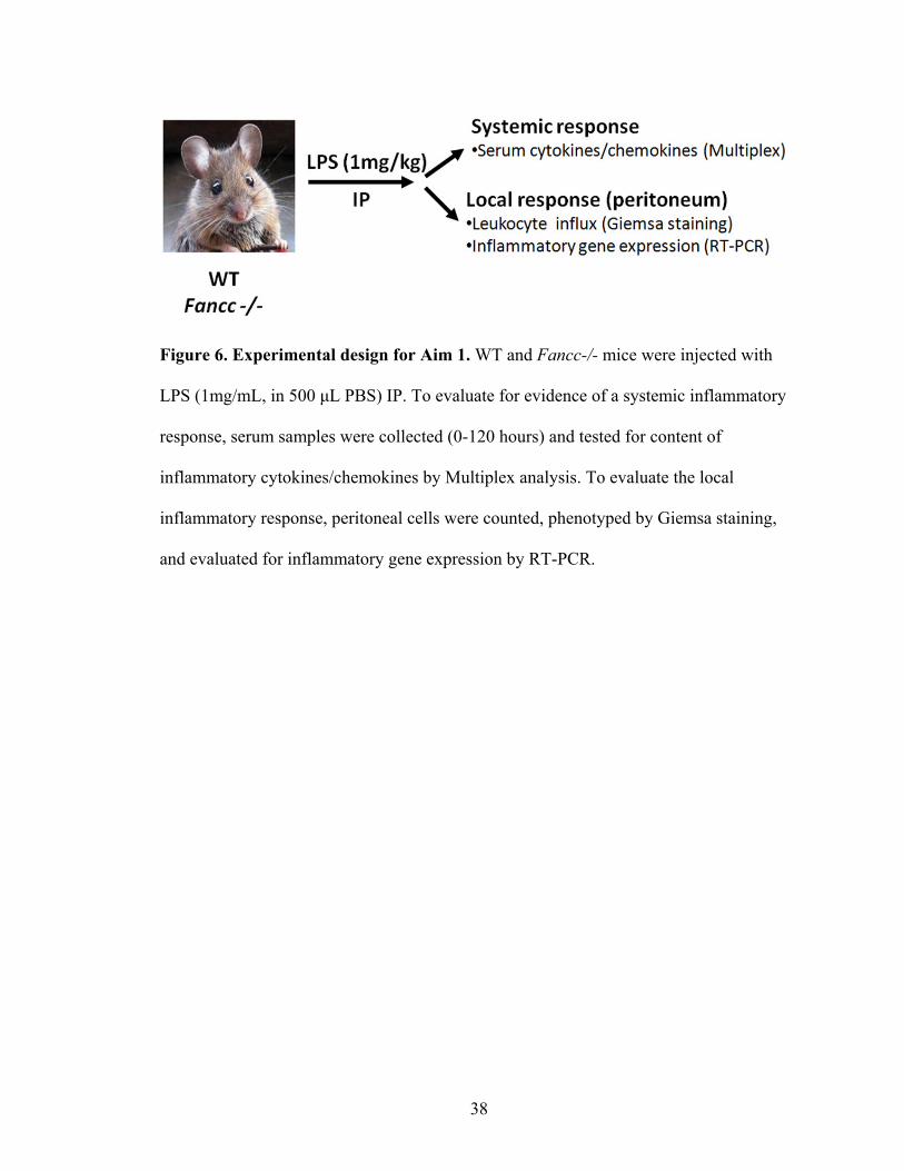
RESULTS .........................................................................................................................37
AIM 1: To examine whether Fancc-/- mice exhibit an altered
inflammatory response in vivo ...........................................................................................37
I. Fancc-/- mice exhibited an altered inflammatory response in vivo. ...............................39
A. Normal systemic levels of inflammatory cytokines/chemokines after
LPS injection ................................................................................................................39
ix
B. Local accumulation of monocytes/macrophages in LPS induced
peritonitis .....................................................................................................................41
C. Accumulation of monocytes/macrophages in sodium periodate
induced inflammatory response ...................................................................................45
1. Reduced levels of inflammatory cytokines/chemokines in the
early phase of inflammation ...................................................................................48
2. Decreased inflammatory monocytes in Fancc-/- mice ......................................51
a. Inflammatory moncoytes in the peripheral blood ........................................51
b. Inflammatory monocytes in the BM ............................................................53
3. Reduced monocyte progenitors in the BM ........................................................55
a. BM cellularity after LPS treatment ..............................................................55
b. Phenotypic characterization of monocyte progenitors in the BM
after LPS treatment ..........................................................................................57
c. Functional analysis of BM progenitors after LPS treatment ........................59
II. Fancc-/- BM cells exhibited an impaired response to M-CSF in vitro .........................63
A. Decreased number of BMDM in the presence of M-CSF ......................................63
B. Reduced M-CSF stimulated proliferation ...............................................................63
C. Impaired phosphorylation of ERK and AKT upon M-CSF stimulation .................66
SUMMARY OF AIM 1 .....................................................................................................68
AIM 2. To determine whether Fancc-/- macrophages exhibit cell
autonomous defects in adhesion, migration and phagocytosis. .........................................69
III. Impaired motility and function of Fancc-/- monocytes/macrophages .........................71
A. Characterization of Fancc-/- peritoneal cells at the steady state ............................71
x
B. Impaired adhesion of Fancc-/- peritoneal macrophages .........................................73
1. Impaired adhesion on FN ...................................................................................73
2. Impaired adhesion on endothelial cells ..............................................................75
C. Impaired migration of Fancc-/- peritoneal macrophages ........................................77
D. Impaired phagocytosis of Fancc-/- peritoneal macrophages ..................................79
1. Phagocytosis of E.coli bioparticles ....................................................................79
2. Phagocytosis of IgG latex beads ........................................................................81
3. Reduced phagocytosis mediated superoxide production ...................................83
E. Dysfunctional cytoskeleton reorganization in Fancc-/- macrophages ....................85
F. Impaired activation of RhoA in Fancc-/- macrophages ..........................................89
SUMMARY OF AIM 2 .....................................................................................................91
DISCUSSION ...................................................................................................................92
1. Altered inflammatory response in Fancc-/- mice ..........................................................92
2. Impaired adhesion, migration and phagocytosis of Fancc-/-
macrophages ......................................................................................................................95
3. Classical and alternative macrophage activation ...........................................................96
4. Association of altered cytoskeleton rearrangement and decreased
RhoA activation in Fancc-/- macrophages ........................................................................98
5. Attenuated M-CSF responsiveness of Fancc-/- progenitors and
macrophages ......................................................................................................................99
6. A limitation of our study: the molecular function of FANCC was not
evaluated ..........................................................................................................................101
xi
FUTURE STUDIES .......................................................................................................102
1. To evaluate whether Fancc-/- mice have altered inflammation and
immunity to bacterial pathogen challenged in vivo .........................................................102
2. To determine whether the altered function of macrophages is due to
the intrinsic role of FANCC .............................................................................................103
3. To assess whether the dysfunctional cell adhesion of Fancc-/-
macrophage is secondary to deficient RhoA activation ...................................................103
4. To measure whether M-CSF signaling is altered in myeloid progenitors ...................104
5. To examine whether Fancc-/- macrophages exhibit altered M1 or M2
activation. .........................................................................................................................105
REFERENCES ...............................................................................................................107
CURRICULUM VITAE

xii
LIST OF TABLES
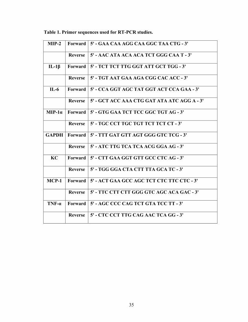
Table 1. Primer sequences used for RT-PCR studies ........................................................35
xiii
LIST OF FIGURES
Figure 1. Simplified schematic of monocytes/macrophages generation
in the BM .............................................................................................................................9
Figure 2. Leukocyte recruitment after LPS treatment ......................................................11
Figure 3. Life-cycle of macrophages ................................................................................14
Figure 4. Differential counts of peritoneal cells ...............................................................24
Figure 5. Representative photographs of individual hematopoietic
progenitor colony ...............................................................................................................27
Figure 6. Experimental design for Aim 1 .........................................................................38
Figure 7. Fancc-/- and WT mice had similar serum levels of inflammatory
mediators after LPS treatment ...........................................................................................40
Figure 8. Fancc-/- mice had altered number of leukocytes in LPS inflamed
peritoneal cavity .................................................................................................................43
Figure 9. Fancc-/- mice exhibited impaired monocyte/macrophage accumulation
in sodium periodate induced peritonitis .............................................................................47
Figure 10. Fancc-/- peritoneal cells exhibited a significant decrease in
gene expression of multiple inflammatory cytokines/chemokines
after LPS treatment ............................................................................................................50
Figure 11. Fancc-/- mice exhibited decreased inflammatory monocytes
in the peripheral blood .......................................................................................................52
Figure 12. Fancc-/- mice exhibited no significant alterations in the number
of BM inflammatory monocytes ........................................................................................54
Figure 13. Fancc-/- mice had normal BM cellularity after LPS injection .........................56
xiv
Figure 14. Phenotypically-defined progenitors from Fancc-/- mice
exhibited normal LPS-induced expansion .........................................................................58
Figure 15. Fancc-/- progenitors exhibited impaired colony forming ability
after in vivo LPS exposure .................................................................................................61
Figure 16. Fancc-/- progenitors exhibited altered distribution after in vivo
LPS treatment.....................................................................................................................62
Figure 17. Fancc-/- BM cells exhibited reduced proliferation in response
to M-CSF ...........................................................................................................................65
Figure 18. Fancc-/- BMDM exhibited impaired phosphorylation of ERK
and AKT after M-CSF stimulation ....................................................................................67
Figure 19. Experimental strategy for Aim 2 ......................................................................70
Figure 20. Fancc-/- peritoneum exhibited a normal leukocyte distribution ......................72
Figure 21. Fancc-/- macrophages exhibited reduced adhesion on FN ..............................74
Figure 22. Fancc-/- macrophages exhibited reduced adhesion on
endothelial cells .................................................................................................................76
Figure 23. Fancc-/- macrophages exhibited reduced migration ........................................78
Figure 24. Fancc-/- macrophages exhibited decreased phagocytosis of
E.coli bioparticels ..............................................................................................................80
Figure 25. Fancc-/- macrophages exhibited decreased phagocytosis of
IgG beads ...........................................................................................................................82
Figure 26. Fancc-/- macrophages exhibited reduced superoxide production ....................84
Figure 27. Fancc-/- peritoneal macrophages exhibited normal F-actin content ................87

xv
Figure 28. Fancc-/- peritoneal macrophages exhibited impaired elongation ...................88
Figure 29. Impaired activation of RhoA was observed in Fancc-/-
macrophages ......................................................................................................................90
xvi
LIST OF ABBREVIATIONS
BMDM Bone marrow derived macrophage
CFSE Carboxy-fluorescein diacetate, succinimidyl ester
CFU-G Colony forming unit–granulocyte
CFU-M Colony forming unit–macrophage
CFU-GM Colony forming unit–granulocyte macrophage
CMP Common myeloid progenitor
DAPI 4',6-Diamidino-2-phenylindole
DEB Diepoxybutane
FA Fanconi anemia
FANCC Fanconi anemia type C
Fancc-/- FA type C deficient
GEF Guanosine exchange factor
GM-CSF Granulocyte macrophage-colony stimulating factor
GST Glutathione S Transferase
HSC Hematopoietic stem cell
ID FANCI/FANCD2
IMDM Iscove modified Dulbecco medium
IP Intraperitoneally
LPS Lipopolysaccharide
LSK Lin-Sca-1+c-Kit+
MCP-1 Monocyte chemotactic protein-1
M-CSF Macrophage-colony stimulating factor
xvii
MMC Mitomycin C
MMP Multipotent progenitor
MNC Mononuclear cells
NO nitric oxide
PAMPs Pathogen associated molecular patterns
PBD P21 binding domain
PBS Phosphate buffered saline
PCR Polymerase chain reaction
PRRs Pathogen recognition receptors
RBD Rho binding domain
ROS Reactive oxygen species
RPMI Roswell Park Memorial Institute medium
RT-PCR Reverse transcription polymerase chain reaction
SEM Standard error of the mean
SOZ Serum opsonized zymosan
TLR2 Toll-like receptor 2
TLR4 Toll-like receptor 8
TLR8 Toll-like receptor 4
1
INTRODUCTION
I. Overview of Fanconi anemia
A. Clinical features of FA
Fanconi anemia (FA) was first reported by Dr. Guido Fanconi (Fanconi et al.,
1927), who described 3 siblings with short stature, skin pigmentation, and pancytopenia.
Subsequently, FA is defined as a rare genetic disorder characterized by progressive bone
marrow failure, developmental anomalies, and increased predisposition to malignancy
(Alter, 2002). Although multiple systems are affected in FA, the life-threatening features
of this disease are hematologic. Clinically, 98% of FA patients will develop
myelodysplastic syndromes, acute myeloid leukemia or bone marrow (BM) failure by the
age of 40 (Young and Alter, 1994). Though blood counts are usually normal at birth, the
size of red blood cells is often large (macrocytosis) within the first decade of life. By 20
years of age, over 50% of patients will develop pancytopenia (Young and Alter, 1994).
The symptoms appear progressively and often lead to BM failure. The incidence of FA is
estimated to be 1 per 350,000 live births (Verlander et al., 1995). Though FA is found in
all ethnic groups, it is highly prevalent in two populations: Ashkenazi Jews and the
Afrikaans of South Africa (Moustacchi et al., 1990) (Moustacchi et al.,2002).
A key cellular feature of FA is chromosomal instability. Chromosomal breakage
induced by DNA cross-linking agents, including mitomycin C (MMC) and
diepoxybutane (DEB), has been exploited to study mechanisms underlying the disease
manifestations and to confirm the clinical suspicion for FA. Indeed, DEB testing of
peripheral blood cells is the gold-standard clinical test used to make a definitive diagnosis
2
of FA. FA cells treated with DEB exhibit marked chromosome breakage on cytogenetic
analyses.
The long-term treatment for FA is hematopoietic stem cell transplantation. Over
the last two decades, the modifications to standard allogeneic stem cell transplantation
have reduced regimen-related toxicity and increased survival . However, finding a
suitable donor (HLA compatibility) only occurs in 30% of patients (Gluckman et al.,
1995), which obviously remains a major hurdle for treatment. Alternatively, umbilical
cord blood offers a potential source of hematopoietic stem cells for FA patients without
an HLA match (Broxmeyer et al., 1989). Though hematopoietic stem cell transplantation
improves hematologic outcomes, patients are still at increased risk of developing
malignancies including head and neck, gynecologic, and gastrointestinal cancers.
Therefore, further pathogenetic investigations of FA are needed to improve clinical
outcomes for patients.
B. Molecular mechanisms of FA
For the past two decades, extensive investigation has focused on elucidating the
function of FA proteins. Current data demonstrate that FA is caused by autosomal
recessive mutations in any of 15 FA genes (FANCA, C, D1, D2, E, F, G, I, J, L, M,N and
O) (de Winter and Joenje, 2009; Green and Kupfer, 2009; Mace et al., 2007;
Smogorzewska et al., 2007; Meindl et al., 2010; Vaz et al., 2010; Stoepker et al., 2011)
or a rare X-linked recessive mutation in FANCB. FA proteins are involved in a common
pathway which plays multiple distinct roles (Li and Stollar, 2007). Eight FA proteins (A,
B, C, E, F, G, L, and M) form a nuclear multi-protein core complex after activation by
3
DNA damage. The FA core complex functions as an E3 ubiquitin ligase and mediates the
monoubiquitination of FANCD2 and FANCI (Levitus et al., 2006; Dorsman et al., 2007),
the so-called ID complex (FANCI/FANCD2) (Sims et al., 2007). The activated ID
complex subsequently co-localizes with downstream FA proteins (FANCD1/BRCA2,
FANCN, and FANCJ) and other DNA-repair proteins such as RAD51. Defects in one of
the FA core complex proteins results in lack of FA core complex formation and ID
complex monoubiquitination.
Understanding FA pathway function in DNA damage response has received
significant attention in recent years. However, numerous studies demonstrate that several
FA proteins have alternate functions that are independent of DNA damage response. For
instance, Fanconi anemia type C (FANCC) has an important role in cellular response to
oxidative stress, which is mediated in part through altered redox regulation and ASK1
activation (Saadatzadeh et al., 2004). Other novel investigations have shown that FANCC
facilitates phosphorylation of STAT proteins, which has been suggested to indicate that
FA proteins participate in cell growth arrest, cell cycle progression, and survival (Pang et
al., 2000; Fagerlie et al., 2001; Fagerlie et al., 2004). In addition, FANCC forms a
cytoplasmic subcomplex with FANCA, which stabilizes nucleophosmin, a protein that
regulates centrosome duplication and genomic stability (Du et al., 2010; Grisendi et al.,
2005; Grisendi and Pandolfi, 2005). In addition, FANCC is involved in the regulation of
telomere length (Rhee et al., 2010). Collectively, these findings indicate that FANCC
possesses functions independent of DNA damage response.
4
C. Fanconi anemia type C (FANCC)
FANCC is a commonly mutated FA gene, which is conserved among vertebrates
(de Winter and Joenje, 2009; Green and Kupfer, 2009; Mace et al., 2007; Smogorzewska
et al., 2007; Meindl et al., 2010). FANCC encodes a 63 kDa protein with no recognizable
motifs or domains (Patel and Joenje, 2007; Wang, 2007). It is localized in both
cytoplasmic and nuclear cellular compartments (Yamashita et al., 1994; Youssoufian,
1994; Hoatlin et al., 1998). The biologic consequences of lacking FANCC have been
investigated in a murine model containing homozygous disruption of the Fancc gene
(Chen et al., 1996). This model recapitulates many aspects of the human disease
including hypersensitivity to DNA cross-linking agents (Carreau et al., 1998; Marathi et
al., 1996; Haneline et al., 1998; Haneline et al., 2003; Bijangi-Vishehsaraei et al., 2005;
Rathbun et al., 1997), defects in hematopoiesis (Haneline et al., 1999; Haneline et al.,
2003), and a predisposition to malignancies (Freie et al., 2003; Rogers et al., 2004; van
der Heijden et al., 2004; Wreesmann et al., 2007). This model is frequently used as a
preclinical platform to examine FA hematologic disease pathogenesis in vivo.
Specifically, several studies demonstrate that Fancc-/- BM cells display the characteristic
hypersensitivity to MMC and DEB (Chen et al., 1996; Carreau et al., 1998; Marathi et
al., 1996; Haneline et al., 1998; Haneline et al., 2003; Bijangi-Vishehsaraei et al., 2005).
Fancc-/- hematopoietic stem cells (HSCs) exhibit decreased self-renewal capacity,
impaired short- and long-term multilineage repopulating ability, and reduced survival (Li
et al., 2007; Haneline et al., 1999; Haneline et al., 1998; Haneline et al., 2003). In
addition, Fancc-/- progenitors are exquisitely sensitive to low doses of TNF-α, IFN-γ,
reactive oxygen species (ROS), and other inflammatory mediators in vitro and in vivo
5
(Haneline et al., 1998; Li et al., 2004; Haneline et al., 2003; Haneline et al., 1999).
Furthermore, long-term exposure of Fancc-/- HSCs to TNF-α causes the outgrowth of
cytogenetically abnormal clones and predisposition to leukemic transformation after
transplantation (Li et al., 2007). In summary, studies using the Fancc-/- murine model
have yielded novel insights into hematopoietic disease pathogenesis in FA. Importantly,
emerging data from FA patients and Fancc-/- mice suggest that FA patients may have
defects in immunity as well.
D. Immune defects in FA
A growing body of literature suggests that mutations in FA genes may also impair
the immune system, which requires intact function of differentiated hematopoietic cells
(Hadjur and Jirik, 2003; Sejas et al., 2007; Zhang et al., 2007). A number of clinical
studies indicate that FA patients have altered levels of circulating cytokines (Stark et al.,
1993; Bagnara et al., 1993; Rosselli et al., 1994). The most recent study from Dufour and
colleagues demonstrated that BM mononuclear cells from FA patients overproduce TNF-
α and IFN-γ after in vitro activation (Dufour et al., 2003). In addition, it has been
suggested that FA patients may have an increased susceptibility to a variety of pathogens
(Fagerlie and Bagby, 2006; MacMillan et al., 2000; Dufour et al., 2003; Schultz and
Shahidi, 1993; Rosselli et al., 1994; Sejas et al., 2007; Froom et al., 1987; Hersey et al.,
1982; Lebbe et al., 1993; Castello et al., 1998; Perussia et al., 1987; Pedersen et al.,
1977), though it is unclear whether this observation is due to a subtle immunodeficiency
or secondary to leukopenia from evolving BM failure.
6
Studies in Fancc-/- mice provide compelling support for a primary defect in the
innate immune response in FA. Fancc-/- mice challenged in vivo with lipopolysaccharide
(LPS) exhibit increased systemic inflammation and death compared to controls (Sejas et
al., 2007), though it remains unknown what cells are responsible. Limited data suggest
that Fancc-/- macrophage function may be altered. Fancc-/- macrophages stimulated in
vitro with IFN-γ and LPS have increased inducible nitric oxide synthase expression and
nitrite release (Hadjur and Jirik, 2003). In addition, Vanderwerf et al. demonstrated that
splenic macrophages from Fancc-/- mice overexpress TNF-α in response to Toll-like
receptor 8 (TLR8) agonists, but not TLR4 agonists (Vanderwerf et al., 2009). While
these studies show dysfunction of Fancc-/- macrophages in vitro, other important
macrophage functions required for an intact immune response were not examined.
Furthermore, no studies have been done to examine whether the in vivo physiologic
function of Fancc-/- macrophages is altered. Therefore, the overall hypothesis being
tested in this proposal is that Fancc-/- macrophages exhibit defective function
predisposing to an altered inflammatory response.
II. Monocytes/Macrophages
A. Function and generation of macrophages
The immunologic system is divided into two major branches: the innate immune
system and the adaptive immune system. Innate immunity is not specific to the type of
organism it fights and is ready to be activated upon initial infection. Macrophages are a
major component of innate immunity. They are equipped with a broad range of pathogen
recognition receptors (PRR) and are capable of increasing phagocytic efficiency and

7
inflammatory cytokine production after pathogen exposure (Greaves and Gordon, 2002;
Gordon and Read, 2002; Platt et al., 2002). Macrophages identify a “danger” by detecting
an organism’s pathogen-associated molecular patterns and endogenous signals released
from damaged tissues via PRR. Additionally, macrophages also play essential roles in
tissue homeostasis at steady state via clearance of apoptotic cells and cellular debris as
well as through production of growth factors (Swirski et al., 2009; Gordon and Taylor,
2005). Last but not least, activated macrophages initiate an acute inflammatory response
to recruit neutrophils and natural killer cells and to facilitate the maturation,
differentiation and migration of dendritic cells, which proceed to activate the adaptive
immune response (Ni and O'Neill, 2001; Matzinger, 2002; Whelan et al., 2003).
Therefore, macrophages are the unique cell type that serves as a direct link between the
innate and adaptive immune systems.
As professional mononuclear phagocytes, macrophages are primarily described as
a population of long-lived myeloid cells that are derived from the BM. Like other
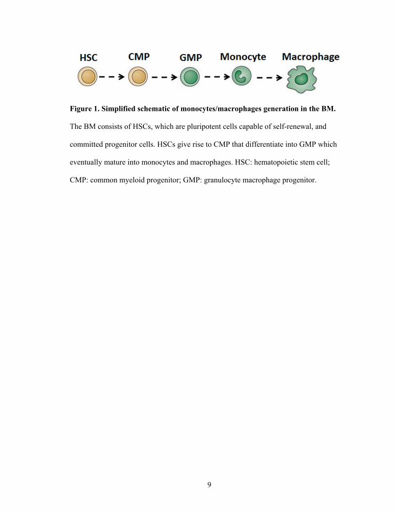
hematopoietic cells, macrophages originate from HSCs, which sequentially differentiate
into multipotent progenitors incapable of self-renewal. Successive lineage commitment
steps during monocyte/macrophage development include common myeloid progenitor
(CMP), granulocyte macrophage progenitor (GMP), and BM monocytes, which are
illustrated in Figure 1(Varol et al., 2007; Iwasaki and Akashi, 2007; Geissmann et al.,
2003). CMPs are multipotent cells that generate megakaryocytes, erythrocytes,
granulocytes and monoycte/macrophages (Chang, 2009). GMPs are more committed and
give rise to granulocytes and macrophages only (Varol et al., 2007; Iwasaki and Akashi,
2007). The process of lineage designation and restriction are regulated by selective and
8
gradual modulation of lineage-specific transcription factors and cytokine signaling
pathways.
Colony stimulating factor-1 [(CSF-1) also known as macrophage colony
stimulating factor (M-CSF)] regulates differentiation, proliferation, and survival of the
monocyte/macrophage lineage (Bonifer and Hume, 2008). The critical role for M-CSF in
macrophage generation is documented in CSF-1 and CSF-1 receptor deficient mice,
which have a reduction in several macrophage populations (Umeda et al., 1996) (Yu et
al., 2008). As an essential growth factor for macrophage differentiation, M-CSF
stimulates BM cells to differentiate into macrophages in vitro, bone marrow derived
macrophages (BMDM), which serve as a powerful model system to study macrophage
differentiation and function (Zhu et al., 2008; Ikeda et al., 2008; Uemura et al., 1993).
9
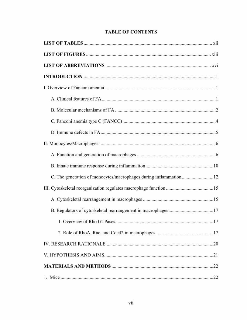
Figure 1. Simplified schematic of monocytes/macrophages generation in the BM.
The BM consists of HSCs, which are pluripotent cells capable of self-renewal, and
committed progenitor cells. HSCs give rise to CMP that differentiate into GMP which
eventually mature into monocytes and macrophages. HSC: hematopoietic stem cell;
CMP: common myeloid progenitor; GMP: granulocyte macrophage progenitor.
10
B. Innate immune response during inflammation
Inflammation is a tightly controlled process that is initiated following tissue injury
or infection. The synchronized action of professional phagocytes, e.g., neutrophils,
macrophages, and monocytes, is crucial for effective elimination of intruders and cell
debris. To initiate an inflammatory response, resident macrophages recognize pathogens
and produce inflammatory cytokines/chemokines. Subsequently, neutrophils are recruited
during inflammation and release a large array of granules and then become apoptotic,
which in turn promotes monocyte influx. After removal of cellular debris, the
inflammatory response is terminated by macrophages via release of anti-inflammatory
mediators following ingestion of apoptotic neutrophils (Gordon and Taylor, 2005). Multi-
functional macrophages also manipulate the adaptive immune response and repair
damaged tissues during inflammation (Gordon, 2007; Gordon and Martinez, 2010;
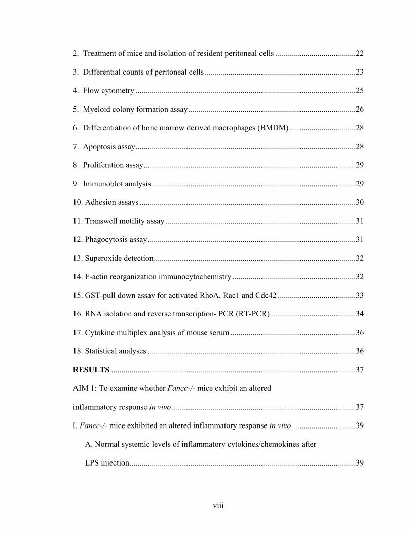
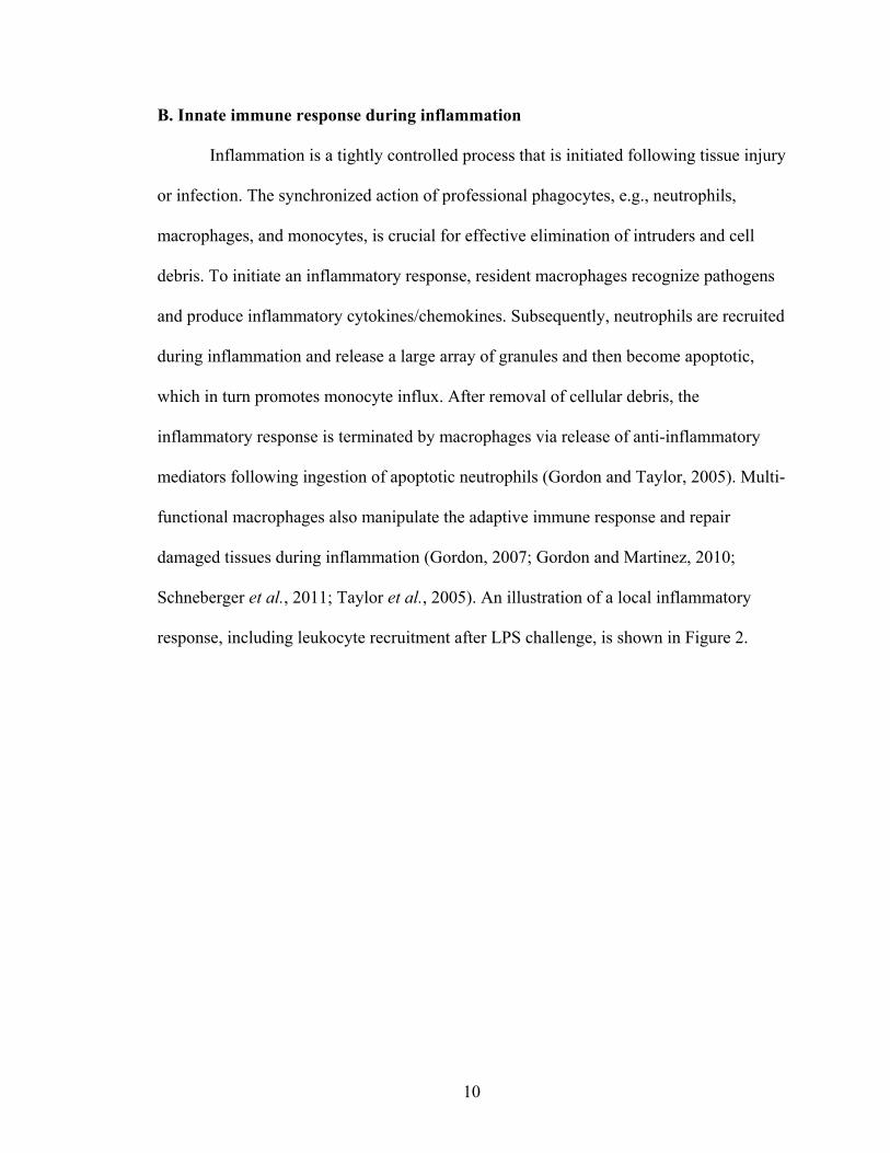
Schneberger et al., 2011; Taylor et al., 2005). An illustration of a local inflammatory
response, including leukocyte recruitment after LPS challenge, is shown in Figure 2.
11
Figure 2. Leukocyte recruitment after LPS treatment. LPS activated resident
macrophages release inflammatory cytokines/chemokines which promote the recruitment
of neutrophils to the inflamed site. Neutrophils release granules, which recruits
monocytes from the peripheral blood and promotes the activation of monocytes to
macrophages. Activated macrophages control the life-span of neutrophils by producing
GM-CSF and are also responsible for removing apoptotic cells by phagocytosis.
Macrophages produce anti-inflammatory cytokines and growth factors during
phagocytosis to promote tissue-repair and leave the inflamed site from draining lymph
nodes.
12
C. The generation of monocytes/macrophages during inflammation
During inflammation, the origination of monocytes/macrophages from the BM is
influenced by the immunologic storm and stress (Chang, 2009). For instance, in vivo
studies show that lipopolysaccharide (LPS) is sufficient to activate tissue resident
macrophages to produce a large amount of inflammatory cytokines/chemokines.
Mobilization of white cells from the BM to the peripheral blood is increased immediately
as a result. Inflammatory monocytes are an important inflammatory cell type that is
mobilized with subsequent migration towards local inflammatory loci. Based on Ly6C
expression, murine monocytes can be differentiated into two discrete subpopulations:
Ly6Chi and Ly6Clo (Yona et al., 2010; Ziegler-Heitbrock, 2007; Auffray et al., 2007;
Nahrendorf et al., 2007). The ratio of Ly6Clo to Ly6Chi is fairly stable at steady state.
However, following inflammation, Ly6Chi monocytes (also known as inflammatory
monocytes) egress from the BM into the peripheral blood in a CCR2-dependent fashion
(Tsou et al., 2007; Serbina et al., 2003; Serbina and Pamer, 2006; Geissmann et al.,
2003), which is likely determined by the inflammatory milieu and PRRs (Auffray et al.,
2009).
In vivo studies also demonstrate that local inflammatory response is sufficient to
induce an expansion of HSCs and progenitors in the BM as defined by the LSK
phenotype (cells negative for lineage markers and positive for Sca1 and c-kit) (Rodriguez
et al., 2009). HSC and progenitor expansion is primarily due to enhanced
cytokine/chemokine production during inflammation. In addition, Nagai et al., showed
that CMP and GMP differentiate into F4/80hi macrophages in vitro in the presence of
LPS or pam3csk4 (a TLR2 agonist) (Abramson et al., 1977), suggesting that TLR ligands
13
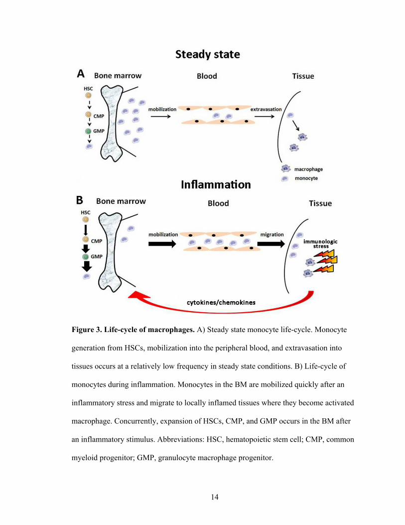
directly regulate hematopoietic cell proliferation and fate determination. In summary,
TLR activation of hematopoietic progenitors constitutes an important innate immune
response to expand the monocyte/macrophage lineage during an infection. The
differentiation and proliferation of monocytes/macrophages are regulated by a network of
cytokines, chemokines, and immunological stresses (Figure 3A-B).
14
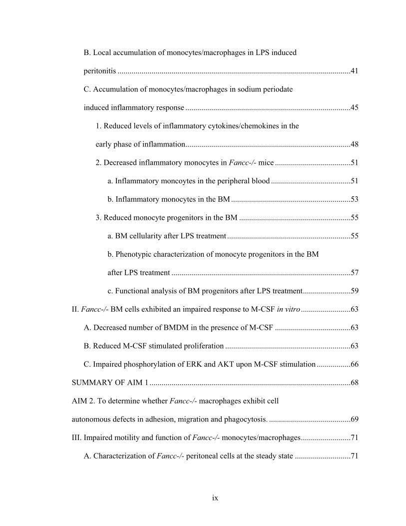
Figure 3. Life-cycle of macrophages. A) Steady state monocyte life-cycle. Monocyte
generation from HSCs, mobilization into the peripheral blood, and extravasation into
tissues occurs at a relatively low frequency in steady state conditions. B) Life-cycle of
monocytes during inflammation. Monocytes in the BM are mobilized quickly after an
inflammatory stress and migrate to locally inflamed tissues where they become activated
macrophage. Concurrently, expansion of HSCs, CMP, and GMP occurs in the BM after
an inflammatory stimulus. Abbreviations: HSC, hematopoietic stem cell; CMP, common
myeloid progenitor; GMP, granulocyte macrophage progenitor.
15
III. Cytoskeletal reorganization regulates macrophage function
A. Cytoskeletal rearrangement in macrophages
Actin cytoskeletal rearrangement is fundamental for many aspects of immune
response to infection. For instance, adhesion, spreading of immune cells on the vascular
endothelium, migration to the inflammatory site, and formation of phagosomes are
dependent on proper organization of the actin cytoskeleton (Burkhardt et al., 2008;
Fooksman et al., 2010; Batista et al., 2010; Hidalgo and Frenette, 2007; Bokoch, 2005).
The clinical significance of active reorganization of the actin cytoskeleton in immune
cells is accentuated by several human diseases where this process is disrupted. For
instance, individuals with mutations in the Wiskott–Aldrich syndrome gene (WASP)
suffer recurrent infections due to altered cytoskeletal rearrangements resulting in
dysfunctional migration, proliferation, and survival of immune cells (Notarangelo et al.,
2008; Thrasher and Burns, 2010). Likewise, a dominant-negative mutation in the RAC2
gene results in recurrent infections due to a cytoskeletal alteration of phagocytes
(Williams et al., 2000).
Several cell structures composed of F-actin including lamellipodia and filopodia
are present in macrophages. Lamellipodia are thin, broad projections at the edge of a
mobile cell and are dynamic structures that constantly change shape. They contain an
extensively branched array of actin filaments, oriented with their plus ends toward the
plasma membrane. Forward extension of a lamellipodium occurs by growth of actin
filaments adjacent to the plasma membrane. Filopodia (microspikes) are long, thin and
transient processes that extend out from the cell surface. They are enriched with bundles
16
of parallel actin filaments that are closely spaced and provide stiffness (DeFife et al.,
1999) .
In fact, a successive migration of macrophages requires the synergistic
coordination of all cytoskeletal structures described above. Specifically, the forward
movement of a cell can be divided into several sequential steps. First is the protrusion of
filopodia and lamellipodia, which are full of actin filaments located at the leading edge of
a cell. Secondly, the protrusions adhere on the substratum via focal complexes. Lastly,
the cell contracts cytoplasmic actomyosin and releases contact sites at the tail of the cell
(Allen et al., 1998; Sheetz et al., 1999). Dynamic and integrated coordination of the actin
cytoskeleton in macrophages has an essential role in cell movement.
In addition to adhesion and migration having an integral role in regulating the in
vivo trafficking of monocytes/macrophages, a major function of macrophages once in
tissue beds is phagocytosis of cell debris, pathogens, and apoptotic cells (Savill et al.,
1989; Dockrell et al., 2001). Phagocytosis is also tightly regulated by the actin
cytoskeleton (Yamada et al., 1989; Haberzettl et al., 2007). The phagocytic process is
comprised of several sequential and complex events initiated by specific receptors on the
surface of phagocytic cells. The signal generated by the receptors clustering in turn leads
to the local polymerization of actin filaments and to particle internalization (Niedergang
and Chavrier, 2005). Formation of phagosomes requires the assistance of local
reorganization of the actin cytoskeleton. Although the mechanisms associated with
ingestion are diverse according to different particles and receptors, studies have
highlighted the significance of small GTP-binding proteins of the Rho family in
phagocytosis (Niedergang and Chavrier, 2005).
17
B. Regulators of cytoskeletal rearrangement in macrophages
1. Overview of Rho GTPases
Rho GTPases belong to the Ras-like families that link signal transduction
pathways through multiple cell-surface receptors to a variety of intracellular signaling
proteins. Rho GTPases function as molecular switches between an inactive form (GDP-
bound) and an active form (GTP-bound) (Bar-Sagi and Hall, 2000). They are regulated
by guanosine exchange factors (GEFs) and GTPase-activating proteins which promote
GTP loading and accelerate GTP hydrolysis, respectively (Cerione and Zheng, 1996).
Along with a variety of effectors, the GTP-bound Rho GTPases regulate actin
cytoskeletal reorganization, cell shape, elongation, polarization, and motility (del Pozo et
al., 1999; Yang et al., 2001a). To date, the best studied Rho GTPases are RhoA, Rac, and
Cdc42. All of these small GTPases are of importance for cell shape, adhesion, motility
and cell-cycle progression. Specifically, RhoA is essential for cell shape and adhesion by
assembling contractile actin microfilaments into stress fibers (Hall, 1998). Formation of
lamellipodial and filopodial protrusions are mainly dependent on Rac and Cdc42
GTPases (Hall, 1998). In summary, the balance of Rho GTPase activities determines
cellular morphology and migratory behavior (Evers et al., 2000).
2. Role of RhoA, Rac, and Cdc42 in macrophages
Three Rho isoforms have been identified in mammals, RhoA, RhoB and RhoC
(Wheeler and Ridley, 2004). Macrophages only express RhoA and RhoB (Wheeler and
Ridley, 2007), which have distinct cellular localization. RhoA is mainly in the cytosol,
complexed to Rho GDP-dissociation inhibitors which maintain Rho GTPases as soluble

18
inactive cytosolic proteins. RhoB is mainly restricted in membranous compartments
(Adamson et al., 1992). Macrophages expressing a RhoA dominant-negative mutant
display impaired CR3-mediated phagocytosis and migration. Mice with conditionally
deleted RhoA in macrophages have not been described to date. RhoB is unique among
this subclass of small GTPases because expression is induced in response to genotoxic
agents as well as mitogens (Fritz et al., 1999). Indeed, RhoB is required for DNA-
damage-induced apoptosis of mouse embryonic fibroblasts (Liu et al., 2001).
The Rac subfamily of GTPase includes Rac1, Rac2, and Rac3 that share high
homology. Macrophages lacking Rac1 are elongated but have normal migration (Wells et
al., 2004). Macrophages expressing a Rac1 dominant-negative mutant exhibit impaired
migration and Fc-γ-dependent phagocytosis (Jones et al., 1998; Caron and Hall, 1998).
Similarly, Rac1/Rac2-null macrophages appear to be elongated and star-like cells with
multiple long protrusions, but with normal migration (Wheeler et al., 2006).
Macrophages lacking only Rac2 have a relatively normal shape, but are deprived of
podosomes, implicating a migration defect upon contact with the extracellular matrix
(Linder, 2007). Rac2 null mice display hematopoietic defects in formyl-methionyl-
leucyl-phenylalanine-induced migration and superoxide production, chemokine-induced
chemotaxis, and cell survival (Yang et al., 2001a; Roberts et al., 1999; Yang et al., 2000;
Yang et al., 2001b; Kim and Dinauer, 2001). However, in murine macrophages, studies
have shown that Rac1 is the predominant isoform (Yamauchi et al., 2004; Pradip et al.,
2003).
In dendritic cells and macrophages, Cdc42 is required for podosome assembly
through WASP complex-induced actin polymerization. Indeed, the interaction with

19
WASP separates Cdc42 from Rac (Weed et al., 1998). Macrophages with Cdc42
dominant-negative mutants exhibit disrupted polarity and reduced chemotaxis (Allen et
al., 1998). In summary, Rho, Rac, and Cdc42 small GTPases are essential for cell shape,
adhesion, and motility of macrophages.
20
IV. RESEARCH RATIONALE
Although FA hematologic dysfunction is primarily considered to be a disease
affecting the function of HSCs and progenitors, studies in both FA patients and FA gene
murine models suggest that immune defects exist (Hadjur and Jirik, 2003; Sejas et al.,
2007; Zhang et al., 2007; Stark et al., 1993; Bagnara et al., 1993; Rosselli et al., 1994;
Dufour et al., 2003; Fagerlie and Bagby, 2006; MacMillan et al., 2000; Schultz and
Shahidi, 1993; Froom et al., 1987; Hersey et al., 1982; Lebbe et al., 1993; Castello et al.,
1998; Perussia et al., 1987; Pedersen et al., 1977). However, it is not clear from previous
studies whether innate immune defects observed in FA are immune cell autonomous or
secondary to leukopenia from evolving BM failure. Limited studies in Fancc-/- mice
provide support for a primary defect in macrophages. Fancc-/- mice challenged in vivo
with LPS have increased serum inflammatory mediators (Sejas et al., 2007; Vanderwerf
et al., 2009), though it remains unknown what cells are responsible. Fancc-/-
macrophages stimulated in vitro with IFN-γ and LPS have increased inducible nitric
oxide synthase expression and nitrite release (Hadjur and Jirik, 2003). In addition, splenic
macrophages from Fancc-/- mice overexpress TNF-α in response to toll-like receptor 8
(TLR8) agonists, but not TLR4 agonists (Vanderwerf et al., 2009). While these studies
show dysfunction of Fancc-/- macrophages in vitro, other important macrophage
functions required for an intact immune response have not been examined. Furthermore,
no studies have examined whether the in vivo physiologic function of Fancc-/-
macrophages is altered. Therefore, the overall goal of these studies was to assess whether
Fancc-/- macrophages exhibit altered function, thereby leading to an impaired
inflammatory response in vivo.

21
V. HYPOTHESIS AND AIMS
Hypothesis: Fancc-/- macrophages exhibit defective function predisposing to an
altered inflammatory response.
Aim 1: To examine whether Fancc-/- mice exhibit an altered inflammatory response in
vivo.
Aim 2: To determine whether Fancc-/- macrophages exhibit cell autonomous defects on
adhesion, migration, and phagocytosis.
22
MATERIALS AND METHODS
1. Mice
Fancc-/- mice were previously described (Chen et al., 1996). Fancc+/- mice were
intercrossed with C57BI/6J mice for more than 10 generations to develop an inbred
strain. Since Fancc-/- mice are infertile, Fancc+/- mice were bred to generate Fancc-/-
and WT mice. The genotype of mice was determined by PCR as described (Haneline et
al., 1998). The following primers were used: 5’CCTGCCATCTTCAGAATTGT3’ (exon
8 primer), 5’GAGCAACACAAATGGTAAGG3’ (intron 8 primer), and
5’TTGAATGGAAGGATTGGAGC3’ (neomycin resistance gene primer).
These mice were maintained under specific pathogen-free conditions in the Indiana
University Laboratory Animal Research Center, Indianapolis, IN. All studies were
approved by the Indiana University School of Medicine Animal Care and Use
Committee.
2. Treatment of mice and isolation of resident peritoneal cells
Age-matched (6 to 12 weeks) WT and Fancc-/- mice were used for all
experiments. Mice were injected intraperitoneally (IP) with either a single dose of LPS (1
mg/kg suspended in 500 μL PBS) or a single dose of sodium periodate (5 mM, 1 mL) to
induce inflammation (Sejas et al., 2007; Suh et al., 2006). An equal volume of the vehicle
control (phosphate buffered saline, PBS) was used for both models. Peritoneal lavage was
conducted with 10 mL of ice cold PBS to collect cellular exudates for in vitro analyses at
times indicated. Peritoneal cells were washed with PBS twice and resuspended in
Roswell Park Memorial Institute medium (RPMI) 1640 (Invitrogen, Carlsbad, CA)
23
containing 10% fetal calf serum (FCS, Lonza Walkersville, Walkersville, MD).
Recovered peritoneal cells were diluted and counted on an electronic cell counter
(Beckman Coulter). Each sample was counted in duplicate. Cells were then prepared for
adhesion, migration, and flow cytometry assays. An aliquot of cells was plated with
macrophage media (RPMI containing 10% FCS). After a 2-hour incubation at 37°C, 5%
CO2, non-adherent cells were removed by washing with PBS, and the remaining adherent
cells were cultured for 24 hours before conducting functional assays. For phagocytosis
assays, fresh peritoneal cells were plated in either chamber slides (Thermo Scientific,
Rochester, NY) for phagocytosing E.coli bioparticles or coverslips (Schott, Louisville,
KY) for uptake of IgG latex beads.
3. Differential counts of peritoneal cells
An aliquot of recovered peritoneal cells was used to make cytospin preparations
and for flow cytometry analyses. The cytospin slides were stained with Giemsa for
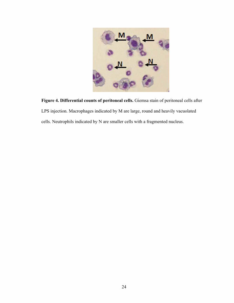
differential counts. A representative photomicrograph is shown in Figure 4. Two hundred
cells per slide were counted.
24
Figure 4. Differential counts of peritoneal cells. Giemsa stain of peritoneal cells after
LPS injection. Macrophages indicated by M are large, round and heavily vacuolated
cells. Neutrophils indicated by N are smaller cells with a fragmented nucleus.
25
4. Flow cytometry
Resident peritoneal cells were incubated with antibodies that recognize
macrophage antigens including CD11b or Mac1, CD115, and F4/80. Specific antibodies
used for these studies were FITC-conjugated mouse CD11b (BD, San Diego, CA), PE-
conjugated mouse CD115 (eBioscience, San Diego, CA), and APC-conjugated mouse
F4/80 (Invitrogen). To quantitate inflammatory monocytes, peripheral blood was
collected from WT and Fancc-/- mice. A small aliquot was used for total white blood cell
count determination as described (Haneline et al., 1999). The majority of sample was
treated with ACK lysing buffer (Lonza Walkersville, Walkersville, MD) to remove red
blood cells. The remaining cells were incubated with antibodies that distinguish
inflammatory monocytes (Hokeness et al., 2005). Specific antibodies used for these
studies were FITC-conjugated mouse Ly-6C (BD), PE-conjugated CD11b (BD), and
APC-conjugated F4/80 (Invitrogen). Isotype control antibodies were used to correct
background fluorescence and set analysis gates. Cells were acquired using a
FACSCalibur (BD) and analyzed with CellQuest software (BD).
26
5. Myeloid colony formation assay
WT and Fancc-/- BM low-density mononuclear cells (MNCs) were prepared and
plated in hematopoietic progenitor assays as described with minor modifications in the
cytokines used (Haneline et al., 1998; Haneline et al., 2003). Briefly, MNCs from the
BM of WT and Fancc-/- mice were resuspended in Iscove modified Dulbecco medium
(IMDM; GIBCO) supplemented with 20% FCS. Cells were plated (5 × 10⁴ cells/mL) in
methylcellulose (STEMCELL Technologies Inc, Vancouver, British Columbia, Canada)
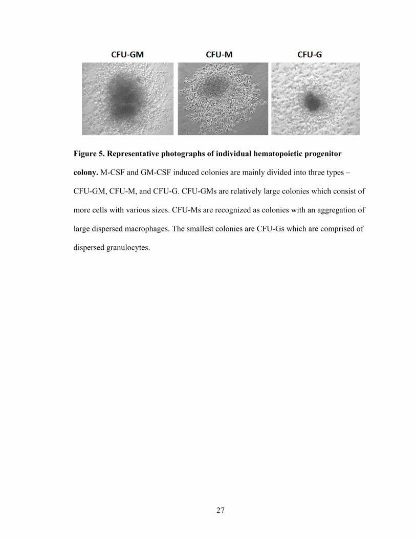
in the presence of M-CSF (50 ng/mL) and/or GM-CSF (10 ng/mL). Total colonies,
including colony forming unit–granulocyte (CFU-G), colony forming unit–macrophage
(CFU-M), and colony forming unit–granulocyte macrophage (CFU-GM), were scored 7
days after plating under a light microscope. Colony morphology was used to determine
individual hematopoietic colony type as illustrated in Figure 5.
27
Figure 5. Representative photographs of individual hematopoietic progenitor
colony. M-CSF and GM-CSF induced colonies are mainly divided into three types –
CFU-GM, CFU-M, and CFU-G. CFU-GMs are relatively large colonies which consist of
more cells with various sizes. CFU-Ms are recognized as colonies with an aggregation of
large dispersed macrophages. The smallest colonies are CFU-Gs which are comprised of
dispersed granulocytes.
28
6. Differentiation of bone marrow derived macrophages (BMDM)
BMDM were obtained using the conditions adapted from Racoosin and Swanson
(Racoosin and Swanson, 1989). Total bone marrow cells flushed from mouse femurs
were washed twice in IMDM. Total bone marrow cells from each mouse were suspended
in 20 mL IMDM with 20% FCS, 50 ng/mL M-CSF, 1% penicillin, and 100 μg/mL
streptomycin and plated on a single 100 mm petri dish (non-tissue culture plastic) (Day
0). Following 12 hours at 37°C, non-adherent cells were collected and transferred to a
new 100 mm tissue culture dish, whereas adherent cells were discarded. After 3 days of
differentiation, non-adherent cells were collected, centrifuged, re-suspended in fresh
differentiation media, and added back to the original tissue culture dish. On day 6 of
differentiation, adherent cells were either collected using cold PBS for experiments or
new media added. Day 6-8 differentiated BMDMs were used for all experiments.
7. Apoptosis assay
Apoptosis of BMDM was examined by terminal deoxynucleotidyl transferase
(tDt)-mediated dUTP nick end-labeling (TUNEL) assay (Roche Diagnostics,
Indianapolis, IN) (Saadatzadeh et al., 2009; Li et al., 2004). Cells were treated with
mitomycin C (50 nM and 500 nM) as positive controls. In detail, BMDM were fixed with
4% formaldehyde for 30 minutes at room temperature and permeabilized with 0.1%
sodium citrate (Fisher Scientific, Fair Lawn, NJ), 0.1% Triton-X100 (Boehringer
Mannheim, Mannheim, Germany) for 2 minutes at 4°C. Cells were incubated with Tdt
and dUTP-FITC in a humidified environment at 37°C, 5% CO2 for 1 hour. After
incubation, cytospins were evaluated by fluorescence microscopy. Photographs were
29
obtained of each condition and scored for apoptotic cells. A total of at least 100 cells
from at least three independent experiments were analyzed for apoptotic cells.
Apoptosis assay of peritoneal cells were tested by expression of annexin V (BD)
and 7AAD (BD) in cells. Cells stain positive for both antibodies by flow cytometry are
defined as early apoptotic cells.
8. Proliferation assay
Starved BMDM were stimulated with M-CSF (50 ng/mL) for 24 hours and pulsed
with 0.037 MBq (1.0 µCi) of [3H]-thymidine (Amersham Biosciences, Piscataway, NJ)
for 6 hours at 37°C prior to harvesting. The cells were washed 4 to 5 times with PBS.
Thymidine incorporation was measured using a Beckman Coulter LS 6500 Multipurpose
Scintillation counter (Beckman Coulter, Fullerton, CA) (Munugalavadla et al., 2005).
9. Immunoblot analysis
BMDMs and peritoneal cells were lysed and quantitated using a bicinchoninic
acid (BCA) protein assay (Pierce Chemical, San Diego, CA). Lysis buffer includes: 0.5
M Hepes, 5 M NaCl, 50% Glycerol, 25% Triton X, 1 M MgCl2, 250 mM EDTA, and
Protease inhibitors and PMSF. Equal amounts of proteins were separated on a 4-12%
sodium dodecyl sulfate (SDS)–polyacrylamide gradient gel (Invitrogen) by
electrophoresis and transferred to nitrocellulose membranes. Membranes were blocked
with Tris-buffered saline Tween containing 5% nonfat milk for 1 hour. For
immunodetection of ERK and AKT, anti-ERK (phospho and total), and anti-AKT
(phospho and total) were used at a 1:1000 dilution for 12 hours. All primary antibodies
30
were from Cell Signaling (Danvers, MA). The secondary antibody, anti-rabbit-
horesradish peroxidase (HRP) (Amersham Biosciences), was used at a 1:5000 dilution for
1 hour before visualizing by ECL chemiluminescence.
10. Adhesion assays
Adhesion assays were performed on fibronectin (FN, recombinant human
fragment CH296, Takara Bio Inc, Madison, WI) or on endothelial cells as described
(Munugalavadla et al., 2005; Hollingsworth et al., 2007). For assay on FN, flat-bottom
96-well polystyrene plates were coated with FN at a concentration of 2 μg/mL at 37°C
for 1 hour and then washed twice. Peritoneal cells (1 × 10⁵) were added to each well and
allowed to adhere at 37°C for 1 hour. Unbound cells were removed by aspiration, and
wells were washed twice with cold PBS. Adherent cells were fixed with 4%
paraformaldehyde (Sigma, St. Louis, MO) and stained with 0.1% crystal violet (Sigma).
Adherent cells were photographed in 5-6 random fields/well with a Leica microscope
using a 10X objective lens. For assay on activated endothelial cells, human endothelial
colony-forming cells (Ingram et al., 2008) were cultured in a 96-well plate to 100%
confluence followed by treatment with LPS (1 μg/mL) or vehicle control for 24 hours.
Carboxy-fluorescein diacetate, succinimidyl ester (CFSE, Sigma) was used to label
peritoneal cells at a concentration of 1 μM for 8 minutes. CSFE-stained peritoneal cells (5
× 104 cells) were added to endothelial monolayers and co-cultured for 1 and 2 hours.
Non-adherent cells were washed off, and adherent cells were photographed in 5-6
random fields/well with a 10X objective lens.
31
11. Transwell motility assay
Migration assays were performed in transwell cell culture chambers with
polycarbonate filters (24-well, 8-μm pore, corning Costar, Cambridge, MA). The filters
were coated with FN (20 µg/mL) for 2 hours and washed with PBS. Peritoneal cells (5 ×
105 cells) in 100 µL RPMI were added to the upper chamber. RPMI (500 µL) with or
without chemotaxis reagents (100 ng/mL M-CSF, 20 ng/mL MCP1) was placed in the
lower chamber. After 24 hours of culture, cells remaining on the membrane in the upper
chamber were scraped off, and cells that had migrated to the membrane in the lower
chamber were stained with crystal violet and counted under a Leica microscope. Ten
random fields/well were counted.
12. Phagocytosis assay
Phagocytosis was conducted with either Escherichia coli (E.coli, K-12 strain)
bioparticles BODIPY FL conjugate (Sigma) or human serum coated latex beads (3.30
μm, Bangs Lab, Fishers, IN). E.coli bioparticles were suspended at a concentration of 20
mg/mL in PBS containing 2 mM sodium azide and incubated with macrophages for 1
hour. Latex beads were incubated with human serum at a concentration of 4 mg/mL in
PBS at 37°C for 1 hour (Suh et al., 2006). Serum coated latex beads were then re-
suspended in RPMI 1640 medium and incubated with macrophages. Phagocytosis was
stopped by placing cells on ice and non-phagocytosed bioparticles and beads were
washed off with cold PBS. Cells were fixed with 4% paraformaldehyde and stained with
rhodamine phalloidin (Invitrogen) and diamidino-2-phenylindole (DAPI) (Sigma).
Phagocytosis of E.coli was imaged using a fluorescent microscope with a 20X objective
32
lens. Ingestion of latex beads was analyzed by confocal microscopy (Olympus FV1000-
MPE confocal/multiphoton Microscope) after phalloidin staining. Images were quantified
by counting at least 100 cells in each condition for each experiment.
13. Superoxide detection
The production of superoxide was monitored by a lucigenin chemiluminescence
assay (Suh et al., 2006; Vazquez-Torres et al., 2000). WT and Fancc-/- peritoneal
macrophages were plated in a 96-well flat bottom tissue culture-treated plate at a density
of 2 × 10⁵ cells per well (Corning Inc.) for 24 hours. After the addition of Zymosan
particles (Invitrogen) and serum opsonized Zymosan (SOZ), cells were then incubated at
37°C for 60 minutes in an Lma microplate luminometer. The relative amount of
superoxide produced over 60 minutes was determined by integrating the
chemiluminescence unit signals using a SoftMax PRO software (Molecular Devices).
Human serum (Sigma) was used to coat Zymosan particles (Sigma) at 37°C for one hour
to achieve SOZ.
14. F-actin reorganization immunocytochemistry
Peritoneal macrophage were adhered to glass coverslips previously coated with
FN (2 µg/mL) for indicated times. Adherent cells were fixed with 4% paraformaldehyde
for 10 minutes, washed with PBS, and stained with rhodamine phalloidin (100 ng/mL,
Sigma) at 37°C for 10 minutes. The slides were washed with PBS, air dried, and mounted
in mounting solution (DAKO, Cambridgeshire, United Kingdom). Confocal microscopic
imaging with Olympus FV1000-MPE confocal/multiphoton equipped with an Argon
33
laser and three diode lasers were used to determine phalloidin staining. Image J was used
to process the micrographs. Images acquired from confocal systems were opened in
Image J and single plane images were saved as individual TIFF files only after cropping
without any alterations. A total of at least 100 cells from at least three independent
experiments were analyzed unless otherwise stated in the figure legend.
15. GST pull-down assay for activated RhoA, Rac1 and Cdc42
Activation of RhoA, Rac1 and Cdc42 was determined using kits from Millipore
following the instructions provided by the manufacturer (Roberts et al., 1999). Briefly,
WT and Fancc-/- peritoneal macrophages were harvested from 10 mice per genotype per
experiment. Macrophages were quiesced by serum deprivation for 12 hours. Cells were
activated by the addition of serum and then harvested at multiple time points by lysing
with ice-cold buffer (50 mM Tris-HCl, pH 7.2, 1% TritonX-100, 0.1% SDS, 0.5%
sodium deoxycholate, 500 mM NaCl, 10 mM MgCl2, 5 μg/mL each of leupeptin and
aprotinin, and 1 mM PMSF). Rac1 and Cdc42 activation were analyzed by affinity
precipitation of a GST fusion-protein corresponding to p21-binding domain (PBD 67-
150) of human PAK-1 bound to glutathione agarose (GST-PBD). RhoA-GTP was
immunoprecipitated with a GST-tagged fusion protein, corresponding to residues 7-89 of
mouse Rhotekin Rho Binding Domain (GST-RBD). Cell lysates were
immunoprecipitated at 4°C for 1 hour. Immunoprecipitated complexes were subjected to
4-12% SDS-PAGE electrophoresis. Activated Rho GTPases were detected by
immunoblotting using antibodies against RhoA (Santa Cruz, CA), Rac1 (BD
34
Pharmingen, San Diego, CA), and Cdc42 (Santa Cruz). Blots were developed by
enhanced chemiluminescence and bands were quantified with ImageJ software.
16. RNA isolation and reverse transcription-PCR (RT-PCR)
Total RNA from cells was isolated using RNeasy Micro Kit (QIAGEN, Valencia,
CA). Isolated RNA was quickly evaluated by agarose gel, and RNA concentration was
measured using a spectrophotometer. cDNA was generated by reverse transcription with
oligo dT as a primer using the SuperScript First-Strand synthesis system (Invitrogen,
Carlsbad, CA). The cDNA were then used as templates for PCR to detect mRNA
expression. The RT-PCR primer sets are listed in Table 1. PCR amplification was carried
out in a total volume of 15 μL, containing 2 μL of cDNA, 10 μL of 2X FastStart
Universal SYBR Green Master Mix (Roche), and 0.2 μM of each primer. The PCR
cycling conditions were 95°C for 5 minutes followed by two-step cycling 40 cycles of
95°C for 10 seconds, and 60°C for 30 seconds. All PCR assays were performed in
duplicate or triplicate. Fold change of gene expression was analyzed using 2-∆∆CT method
(Livak and Schmittgen, 2001). ∆∆CT = (CT.Target - CT.Actin) Time ᵡ - (CT.Target -
CT.Actin) Time 0. Time ᵡ is any time point and Time 0 represents the expression of the
target gene normalized to β-actin.
35
Table 1. Primer sequences used for RT-PCR studies.
MIP-2 Forward 5' - GAA CAA AGG CAA GGC TAA CTG - 3'
Reverse 5' - AAC ATA ACA ACA TCT GGG CAA T - 3'
IL-1β Forward 5' - TCT TCT TTG GGT ATT GCT TGG - 3'
Reverse 5' - TGT AAT GAA AGA CGG CAC ACC - 3'
IL-6 Forward 5' - CCA GGT AGC TAT GGT ACT CCA GAA - 3'
Reverse 5' - GCT ACC AAA CTG GAT ATA ATC AGG A - 3'
MIP-1α Forward 5' - GTG GAA TCT TCC GGC TGT AG - 3'
Reverse 5' - TGC CCT TGC TGT TCT TCT CT - 3'
GAPDH Forward 5' - TTT GAT GTT AGT GGG GTC TCG - 3'
Reverse 5' - ATC TTG TCA TCA ACG GGA AG - 3'
KC Forward 5' - CTT GAA GGT GTT GCC CTC AG - 3'
Reverse 5' - TGG GGA CTA CTT TTA GCA TC - 3'
MCP-1 Forward 5' - ACT GAA GCC AGC TCT CTC TTC CTC - 3'
Reverse 5' - TTC CTT CTT GGG GTC AGC ACA GAC - 3'
TNF-α Forward 5' - AGC CCC CAG TCT GTA TCC TT - 3'
Reverse 5' - CTC CCT TTG CAG AAC TCA GG - 3'

36
17. Cytokine multiplex analysis of mouse serum
Blood was collected at indicated times after LPS treatment from WT and Fancc-/-
mice. Serum was separated by centrifugation (3,000 RPM, 10 minutes). Levels of TNF-α,
IL-6, IL-1β, MCP-1, IFN-γ, and KC were measured using Multiplex Bead Immunoassays
as per the manufactuer’s protocol (Millipore, Billerica, MA). A luminex 200 instrument
(Luminex Corp, Austin, TX) was used for data acquisition and analysis. Cytokine
concentrations were calculated using a Bio-Plex Manager 2.3 software (Bio-Rad,
Hercules, CA) with a 5-parameter curve-fitting algorithm applied for standard curve
calculations.
18. Statistical analyses
Parametric data are presented as mean plus or minus standard error of the mean
(SEM), unless otherwise stated. For all data, an unpaired Student t test was conducted to
evaluate for differences between treatment groups. A P value less than 0.05 was
considered significant.
37
RESULTS
AIM 1: To examine whether Fancc-/- mice exhibit an altered inflammatory
response in vivo. To create a model of peritoneal inflammation, a low dose of LPS (1
mg/kg suspended in 500 μL PBS) was injected into WT and Fancc-/- mice
intraperitoneally (IP) similar to previous studies (Sejas et al., 2007; Hollingsworth et al.,
2007; Hadjur and Jirik, 2003). Figure 6 illustrates the strategy of experiments for this
aim. In brief, after one LPS injection, serum and peritoneal cells were collected at
indicated time points to assess the systemic and local inflammatory response in vivo.
38
Figure 6. Experimental design for Aim 1. WT and Fancc-/- mice were injected with
LPS (1mg/mL, in 500 μL PBS) IP. To evaluate for evidence of a systemic inflammatory
response, serum samples were collected (0-120 hours) and tested for content of
inflammatory cytokines/chemokines by Multiplex analysis. To evaluate the local
inflammatory response, peritoneal cells were counted, phenotyped by Giemsa staining,
and evaluated for inflammatory gene expression by RT-PCR.
39
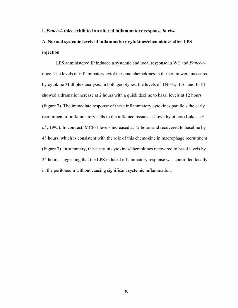
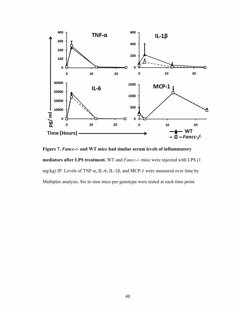
I. Fancc-/- mice exhibited an altered inflammatory response in vivo.
A. Normal systemic levels of inflammatory cytokines/chemokines after LPS
injection
LPS administered IP induced a systemic and local response in WT and Fancc-/-
mice. The levels of inflammatory cytokines and chemokines in the serum were measured
by cytokine Multiplex analysis. In both genotypes, the levels of TNF-α, IL-6, and Il-1β
showed a dramatic increase at 2 hours with a quick decline to basal levels at 12 hours
(Figure 7). The immediate response of these inflammatory cytokines parallels the early
recruitment of inflammatory cells to the inflamed tissue as shown by others (Lukacs et
al., 1995). In contrast, MCP-1 levels increased at 12 hours and recovered to baseline by
48 hours, which is consistent with the role of this chemokine in macrophage recruitment
(Figure 7). In summary, these serum cytokines/chemokines recovered to basal levels by
24 hours, suggesting that the LPS induced inflammatory response was controlled locally
in the peritoneum without causing significant systemic inflammation.
40
Figure 7. Fancc-/- and WT mice had similar serum levels of inflammatory
mediators after LPS treatment. WT and Fancc-/- mice were injected with LPS (1
mg/kg) IP. Levels of TNF-α, IL-6, IL-1β, and MCP-1 were measured over time by
Multiplex analysis. Six to nine mice per genotype were tested at each time point.
41
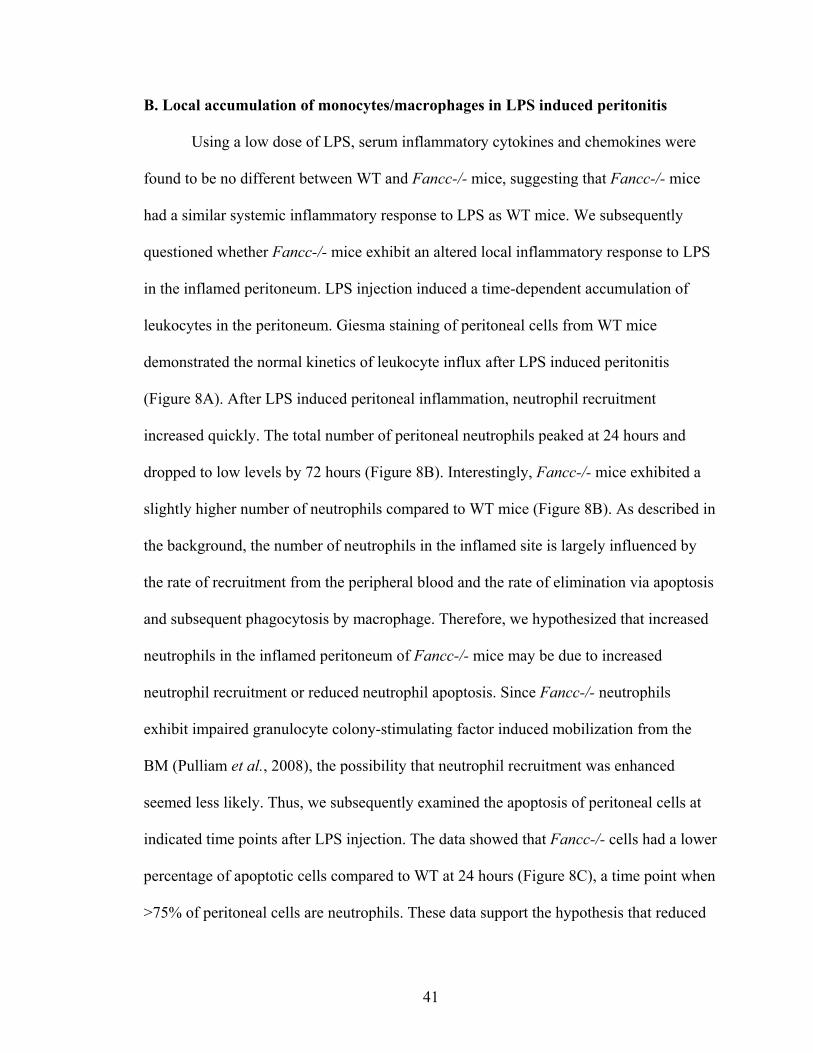
B. Local accumulation of monocytes/macrophages in LPS induced peritonitis
Using a low dose of LPS, serum inflammatory cytokines and chemokines were
found to be no different between WT and Fancc-/- mice, suggesting that Fancc-/- mice
had a similar systemic inflammatory response to LPS as WT mice. We subsequently
questioned whether Fancc-/- mice exhibit an altered local inflammatory response to LPS
in the inflamed peritoneum. LPS injection induced a time-dependent accumulation of
leukocytes in the peritoneum. Giesma staining of peritoneal cells from WT mice
demonstrated the normal kinetics of leukocyte influx after LPS induced peritonitis
(Figure 8A). After LPS induced peritoneal inflammation, neutrophil recruitment
increased quickly. The total number of peritoneal neutrophils peaked at 24 hours and
dropped to low levels by 72 hours (Figure 8B). Interestingly, Fancc-/- mice exhibited a
slightly higher number of neutrophils compared to WT mice (Figure 8B). As described in
the background, the number of neutrophils in the inflamed site is largely influenced by
the rate of recruitment from the peripheral blood and the rate of elimination via apoptosis
and subsequent phagocytosis by macrophage. Therefore, we hypothesized that increased
neutrophils in the inflamed peritoneum of Fancc-/- mice may be due to increased
neutrophil recruitment or reduced neutrophil apoptosis. Since Fancc-/- neutrophils
exhibit impaired granulocyte colony-stimulating factor induced mobilization from the
BM (Pulliam et al., 2008), the possibility that neutrophil recruitment was enhanced
seemed less likely. Thus, we subsequently examined the apoptosis of peritoneal cells at
indicated time points after LPS injection. The data showed that Fancc-/- cells had a lower
percentage of apoptotic cells compared to WT at 24 hours (Figure 8C), a time point when
>75% of peritoneal cells are neutrophils. These data support the hypothesis that reduced
42
neutrophil apoptosis may contribute to the increased number of neutrophils observed in
the peritoneum of Fancc-/- mice after LPS treatment. These data also suggest that
Fancc-/- neutrophils may exhibit autonomous defects that alter the inflammatory
response in vivo.
As expected, LPS induced peritonitis caused an influx of monocytes/macrophages
into the peritoneum at later time points. In WT mice, LPS caused a significant
extravasation of monocytes/macrophages into the peritoneal cavity, peaking at 96 hours
and declining sharply to near basal levels by 120 hours, similar to previous studies
(Figure 8D) (Ajuebor et al., 1999; Takahashi et al., 2009; Parsons et al., 2007; Mortier et
al., 2005). Fancc-/- mice displayed similar kinetics for monocyte/macrophage
recruitment after LPS injection (Figure 8D). However, the number of
monoyctes/macrophages was significantly lower in Fancc-/- mice compared to WT in the
later phase of inflammation (i.e. 72 to 120 hours). These data are intriguing given that
Fancc-/- mice had more neutrophils in the peritoneum after LPS treatment, which should
correlate with increased macrophage recruitment. We speculate that intrinsic defects in
monocytes/macrophages may account for this observation.
43
Figure 8. Fancc-/- mice had altered number of leukocytes in LPS inflamed
peritoneal cavity. (A) Leukocyte distribution in LPS inflamed peritoneum. At baseline
and after LPS injection, peritoneal cells were harvested (0-72 hours) and stained with
Giemsa for morphologic evaluation of peritoneal leukocyte distribution. Representative
photomicrographs of peritoneal cells from WT mice are shown (M: macrophage, N:
neutrophil). (B) Total number of peritoneal neutrophils. Neutrophil number was obtained
by multiplying total peritoneal cells by the neutrophil percentage determined by Giemsa
staining. Six to twelve mice per genotype were used in each of the three independent
experiments, *P<0.05. (C) Total number of monocytes/macrophages.
44
Monocyte/macrophage number was calculated by multiplying total peritoneal cells by the
monocyte/macrophage percentage determined by Giemsa staining. Six to twelve mice per
genotype were used in each of the three independent experiments, *P<0.05. (D)
Apoptosis of peritoneal cells after LPS treatment. Apoptosis was evaluated by flow
cytometry. Cells staining positively for both annexinV and 7AAD were scored as
apoptotic cells. Three mice per genotype were used in each of the three independent
experiments, *P<0.05.
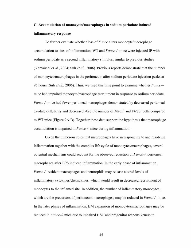
45
C. Accumulation of monocytes/macrophages in sodium periodate induced
inflammatory response
To further evaluate whether loss of Fancc alters monocyte/macrophage
accumulation to sites of inflammation, WT and Fancc-/- mice were injected IP with
sodium periodate as a second inflammatory stimulus, similar to previous studies
(Yamauchi et al., 2004; Suh et al., 2006). Previous reports demonstrate that the number
of monocytes/macrophages in the peritoneum after sodium periodate injection peaks at
96 hours (Suh et al., 2006). Thus, we used this time point to examine whether Fancc-/-
mice had impaired monocyte/macrophage recruitment in response to sodium periodate.
Fancc-/- mice had fewer peritoneal macrophages demonstrated by decreased peritoneal
exudate cellularity and decreased absolute number of Mac1+ and F4/80+ cells compared
to WT mice (Figure 9A-B). Together these data support the hypothesis that macrophage
accumulation is impaired in Fancc-/- mice during inflammation.
Given the numerous roles that macrophages have in responding to and resolving
inflammation together with the complex life cycle of monocytes/macrophages, several
potential mechanisms could account for the observed reduction of Fancc-/- peritoneal
macrophages after LPS induced inflammation. In the early phase of inflammation,
Fancc-/- resident macrophages and neutrophils may release altered levels of
inflammatory cytokines/chemokines, which would result in decreased recruitment of
monocytes to the inflamed site. In addition, the number of inflammatory monocytes,
which are the precursors of peritoneum macrophages, may be reduced in Fancc-/- mice.
In the later phases of inflammation, BM expansion of monocytes/macrophages may be
reduced in Fancc-/- mice due to impaired HSC and progenitor responsiveness to

46
inflammatory stimuli. The next series of experiments were designed to test each of these
possibilities.
47
Figure 9. Fancc-/- mice exhibit impaired monocyte/macrophage accumulation in
sodium periodate induced peritonitis. Peritoneal cells were harvested 96 hours after IP
injection of sodium periodate. (A) Total peritoneal cell number is shown. Eight mice per
genotype were used in each of the three independent experiments, *P<0.05. (B) The
absolute number of peritoneal cells expressing macrophage antigens, Mac1 or F4/80, is
shown. The absolute number of Mac1+ or F4/80+ is calculated by multiplying the total
cell number by percentage of Mac1+ or F4/80+ cells determined by flow cytometry. Six
mice per genotype were used in each of the three independent experiments, *P<0.05.
48
1. Reduced levels of inflammatory cytokines/chemokines in the early phase of
inflammation
Because accumulation of leukocytes in the inflamed site is largely regulated by
cytokine/chemokine production, gene expression of multiple inflammatory mediators was
assessed in local peritoneal cells. Initially, gene expression was examined in peritoneal
cells at baseline and 2 hours after LPS treatment because resident macrophage account
for the majority of cells in the peritoneum at these time points (see Figure 8). No
differences were detected between WT and Fancc-/- peritoneal cells in the baseline
mRNA expression of cytokines/chemokines tested. In both WT and Fancc-/-peritoneal
cells, most transcripts of inflammatory cytokines/chemokines had a significant increase
after LPS treatment compared to PBS controls (Figure 10A). Among them, KC had a
dramatic increase, consistent with the role that KC serves as a neutrophil chemoattractant.
Surprisingly, compared to WT, Fancc-/- peritoneal cells displayed a significant decrease
in mRNA expression of TNF-α, KC, MIP-1α, and MIP-2, with a similar trend for IL-6,
IL-1β, and MCP-1. To determine whether the reduced expression of inflammatory
cytokines/chemokines was either impaired or delayed, we assessed expression in
peritoneal cells at later time points (0-120 hours) (Figure 10B).These studies showed that
Fancc-/- peritoneal cells exhibited reduced inflammatory gene expression at multiple
time points, implying an altered inflammatory response in vivo. Additionally, we noticed
that KC, IL-6, TNF-α, and MIP-1α exhibit different kinetics of expression patterns after
LPS injection in both WT and Fancc-/- mice. KC expression peaked at 2 hours when
resident macrophages were the majority cell type, indicating resident macrophages are
important for neutrophil recruitment during inflammation. TNF-α and MIP-1α peaked at

49
24 hours when half of the peritoneal cells were neutrophils, suggesting that neutrophils
contribute to monocyte recruitment in LPS induced peritonitis.
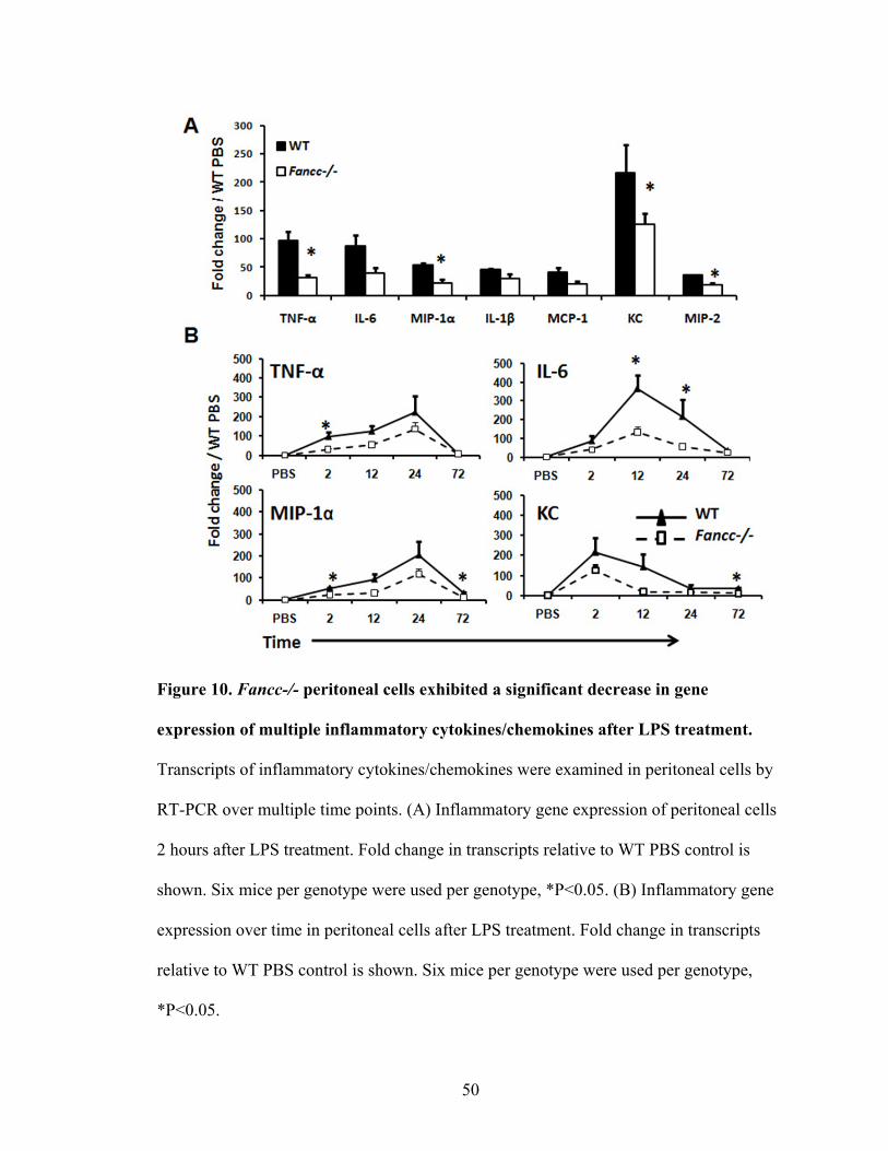
50
Figure 10. Fancc-/- peritoneal cells exhibited a significant decrease in gene
expression of multiple inflammatory cytokines/chemokines after LPS treatment.
Transcripts of inflammatory cytokines/chemokines were examined in peritoneal cells by
RT-PCR over multiple time points. (A) Inflammatory gene expression of peritoneal cells
2 hours after LPS treatment. Fold change in transcripts relative to WT PBS control is
shown. Six mice per genotype were used per genotype, *P<0.05. (B) Inflammatory gene
expression over time in peritoneal cells after LPS treatment. Fold change in transcripts
relative to WT PBS control is shown. Six mice per genotype were used per genotype,
*P<0.05.
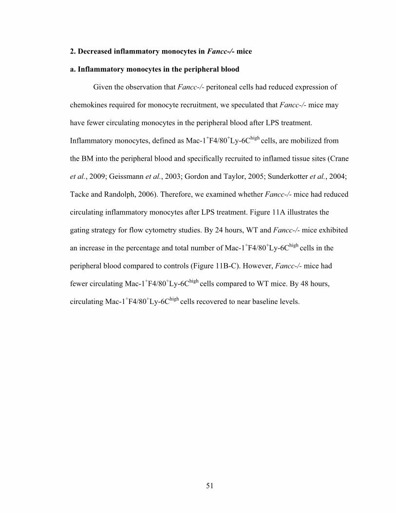
51
2. Decreased inflammatory monocytes in Fancc-/- mice
a. Inflammatory monocytes in the peripheral blood
Given the observation that Fancc-/- peritoneal cells had reduced expression of
chemokines required for monocyte recruitment, we speculated that Fancc-/- mice may
have fewer circulating monocytes in the peripheral blood after LPS treatment.
Inflammatory monocytes, defined as Mac-1+F4/80+Ly-6Chigh cells, are mobilized from
the BM into the peripheral blood and specifically recruited to inflamed tissue sites (Crane
et al., 2009; Geissmann et al., 2003; Gordon and Taylor, 2005; Sunderkotter et al., 2004;
Tacke and Randolph, 2006). Therefore, we examined whether Fancc-/- mice had reduced
circulating inflammatory monocytes after LPS treatment. Figure 11A illustrates the
gating strategy for flow cytometry studies. By 24 hours, WT and Fancc-/- mice exhibited
an increase in the percentage and total number of Mac-1+F4/80+Ly-6Chigh cells in the
peripheral blood compared to controls (Figure 11B-C). However, Fancc-/- mice had
fewer circulating Mac-1+F4/80+Ly-6Chigh cells compared to WT mice. By 48 hours,
circulating Mac-1+F4/80+Ly-6Chigh cells recovered to near baseline levels.
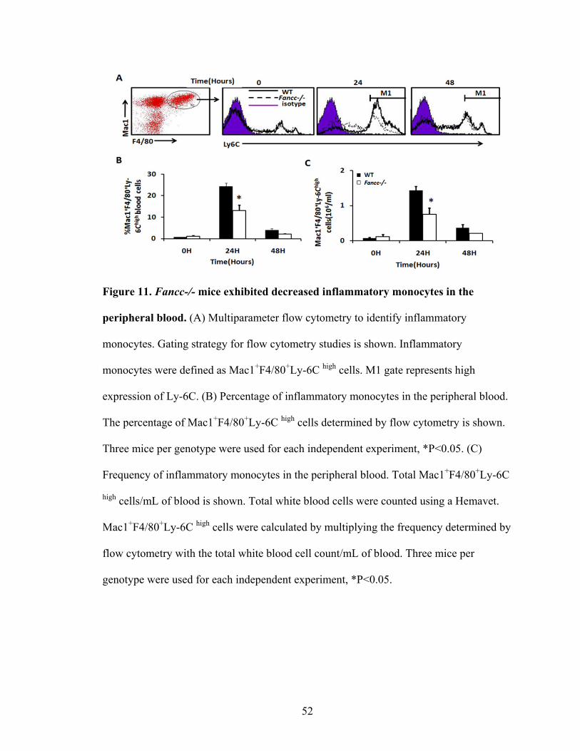
52
Figure 11. Fancc-/- mice exhibited decreased inflammatory monocytes in the
peripheral blood. (A) Multiparameter flow cytometry to identify inflammatory
monocytes. Gating strategy for flow cytometry studies is shown. Inflammatory
monocytes were defined as Mac1+F4/80+Ly-6C high cells. M1 gate represents high
expression of Ly-6C. (B) Percentage of inflammatory monocytes in the peripheral blood.
The percentage of Mac1+F4/80+Ly-6C high cells determined by flow cytometry is shown.
Three mice per genotype were used for each independent experiment, *P<0.05. (C)
Frequency of inflammatory monocytes in the peripheral blood. Total Mac1+F4/80+Ly-6C
high cells/mL of blood is shown. Total white blood cells were counted using a Hemavet.
Mac1+F4/80+Ly-6C high cells were calculated by multiplying the frequency determined by
flow cytometry with the total white blood cell count/mL of blood. Three mice per
genotype were used for each independent experiment, *P<0.05.
53
b. Inflammatory monocytes in the BM
Because inflammatory monocytes are mobilized from the BM during
inflammation, we subsequently evaluated the BM for Mac-1+F4/80+Ly-6Chigh cells after
LPS injection. These studies demonstrated that no dramatic changes were observed
within the BM of mice from either genotype (Figure 12). By 48 hours, Fancc-/- BM cells
had a slightly higher percentage of inflammatory monocytes compared to WT, though the
absolute number of inflammatory monocytes in a femur was not different between
genotypes. Collectively, these data suggest that defective monocyte mobilization from the
BM into the peripheral blood or impaired monocyte activation to an inflammatory
phenotype may occur in Fancc-/- mice during inflammatory stress. However, further
studies need to be done to understand the underlying mechanisms.
54
Figure 12. Fancc-/- mice exhibited no significant alterations in the number of BM
inflammatory monocytes. BM cells were collected at indicated time points after LPS
injection. Inflammatory monocytes were defined as Mac1+F4/80+Ly-6C high cells. (A)
Percentage of inflammatory monocytes in the BM. The percentage of Mac1+F4/80+Ly-6C
high cells determined by flow cytometry is shown. Three mice per genotype were used for
each independent experiment, *P<0.05. (B) Absolute number of inflammatory monocytes
per femur. Total Mac1+F4/80+Ly-6C high cells/femur is shown. Three mice per genotype
were used for each independent experiment, *P<0.05.
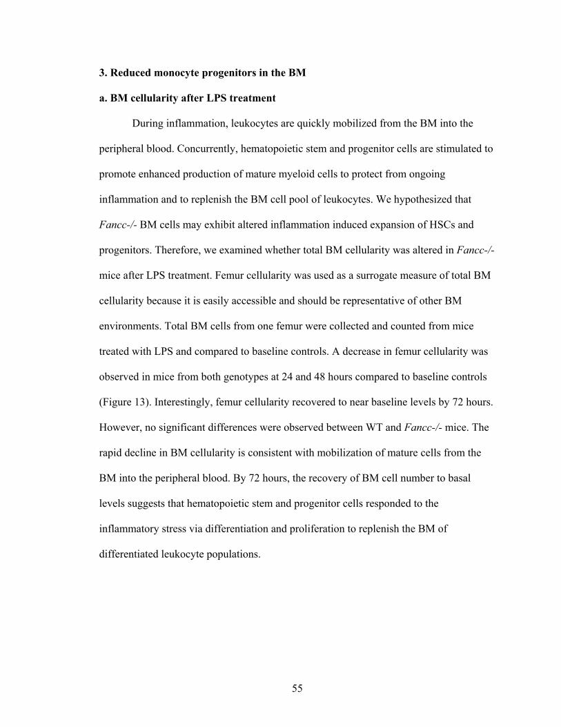
55
3. Reduced monocyte progenitors in the BM
a. BM cellularity after LPS treatment
During inflammation, leukocytes are quickly mobilized from the BM into the
peripheral blood. Concurrently, hematopoietic stem and progenitor cells are stimulated to
promote enhanced production of mature myeloid cells to protect from ongoing
inflammation and to replenish the BM cell pool of leukocytes. We hypothesized that
Fancc-/- BM cells may exhibit altered inflammation induced expansion of HSCs and
progenitors. Therefore, we examined whether total BM cellularity was altered in Fancc-/-
mice after LPS treatment. Femur cellularity was used as a surrogate measure of total BM
cellularity because it is easily accessible and should be representative of other BM
environments. Total BM cells from one femur were collected and counted from mice
treated with LPS and compared to baseline controls. A decrease in femur cellularity was
observed in mice from both genotypes at 24 and 48 hours compared to baseline controls
(Figure 13). Interestingly, femur cellularity recovered to near baseline levels by 72 hours.
However, no significant differences were observed between WT and Fancc-/- mice. The
rapid decline in BM cellularity is consistent with mobilization of mature cells from the
BM into the peripheral blood. By 72 hours, the recovery of BM cell number to basal
levels suggests that hematopoietic stem and progenitor cells responded to the
inflammatory stress via differentiation and proliferation to replenish the BM of
differentiated leukocyte populations.
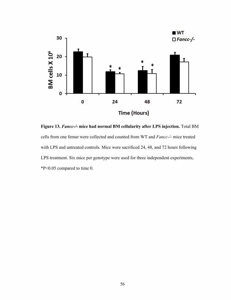
56
Figure 13. Fancc-/- mice had normal BM cellularity after LPS injection. Total BM
cells from one femur were collected and counted from WT and Fancc-/- mice treated
with LPS and untreated controls. Mice were sacrificed 24, 48, and 72 hours following
LPS treatment. Six mice per genotype were used for three independent experiments,
*P<0.05 compared to time 0.
57
b. Phenotypic characterization of monocyte progenitors in the BM after LPS
treatment
Previous data from our laboratory demonstrate that Fancc-/- HSC and myeloid
progenitors exhibit defective function (Haneline et al., 1999; Haneline et al., 1998;
Saadatzadeh et al., 2004; Saadatzadeh et al., 2009; Haneline et al., 2006; Haneline et al.,
2003). Therefore, we hypothesized that Fancc-/- myeloid progenitors may inadequately
expand in response to inflammatory stimuli. As described in the background (Figure 1),
the BM HSCs give rise to CMP that differentiate into GMP which eventually mature into
monocytes and macrophages. Thus, we next questioned whether Fancc-/- BM cells
exhibit altered myeloid population during LPS induced inflammatory response.
Phenotypically-defined populations including LSK (Lin-Sca-1+c-Kit+) which is enriched
with stem cells, CMP (LKS- Fc-γRloCD34+IL-7R-) and GMP (LKS- Fc-γR+CD34+IL-7R-)
were examined by multiparameter flow cytometry. Both WT and Fancc-/- BM had an
increase in the absolute number of LSK cells by 24 hours, and recovered to steady state
levels by 72 hours (Figure 14). The number of CMP and GMP population, on the other
hand, showed a dramatic increase at 72 hours, suggesting that the expansion of CMP and
GMP takes about three days after LPS treatment. However, no significant differences
were observed between WT and Fancc-/- mice.
58
Figure 14. Phenotypically-defined progenitors from Fancc-/- mice exhibited normal
LPS-induced expansion. Total BM cells from one femur were counted from control and
LPS treated WT and Fancc-/- mice. BM cells were labeled with antibodies for the
measurement of LSK, CMP, and GMP by multi parameter flow cytometry. Absolute
numbers were calculated by multiplying percentage of each cell population determined
by flow cytometry by femur cellularity. LSK was defined as lin-sca1+c-kit+. CMP was
defined as LKS- Fc-γRloCD34+IL-7R-. GMP was defined as LKS- Fc-γR+CD34+IL-7R-.
Six mice per genotype were used in three independent experiments.
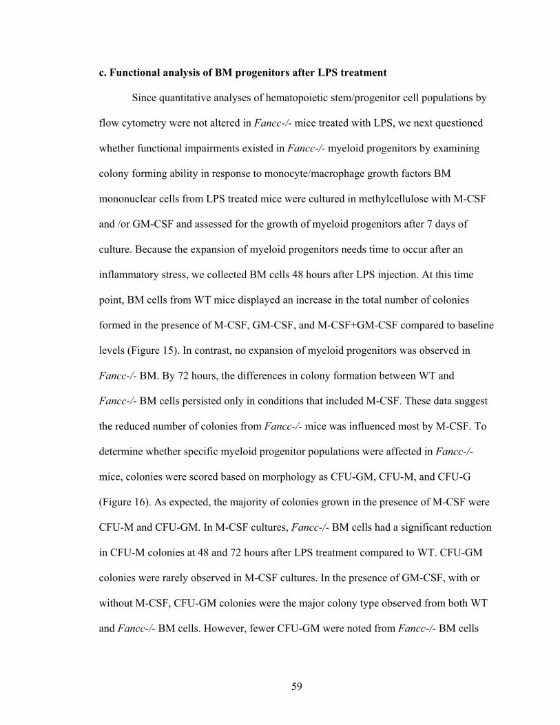
59
c. Functional analysis of BM progenitors after LPS treatment
Since quantitative analyses of hematopoietic stem/progenitor cell populations by
flow cytometry were not altered in Fancc-/- mice treated with LPS, we next questioned
whether functional impairments existed in Fancc-/- myeloid progenitors by examining
colony forming ability in response to monocyte/macrophage growth factors BM
mononuclear cells from LPS treated mice were cultured in methylcellulose with M-CSF
and /or GM-CSF and assessed for the growth of myeloid progenitors after 7 days of
culture. Because the expansion of myeloid progenitors needs time to occur after an
inflammatory stress, we collected BM cells 48 hours after LPS injection. At this time
point, BM cells from WT mice displayed an increase in the total number of colonies
formed in the presence of M-CSF, GM-CSF, and M-CSF+GM-CSF compared to baseline
levels (Figure 15). In contrast, no expansion of myeloid progenitors was observed in
Fancc-/- BM. By 72 hours, the differences in colony formation between WT and
Fancc-/- BM cells persisted only in conditions that included M-CSF. These data suggest
the reduced number of colonies from Fancc-/- mice was influenced most by M-CSF. To
determine whether specific myeloid progenitor populations were affected in Fancc-/-
mice, colonies were scored based on morphology as CFU-GM, CFU-M, and CFU-G
(Figure 16). As expected, the majority of colonies grown in the presence of M-CSF were
CFU-M and CFU-GM. In M-CSF cultures, Fancc-/- BM cells had a significant reduction
in CFU-M colonies at 48 and 72 hours after LPS treatment compared to WT. CFU-GM
colonies were rarely observed in M-CSF cultures. In the presence of GM-CSF, with or
without M-CSF, CFU-GM colonies were the major colony type observed from both WT
and Fancc-/- BM cells. However, fewer CFU-GM were noted from Fancc-/- BM cells
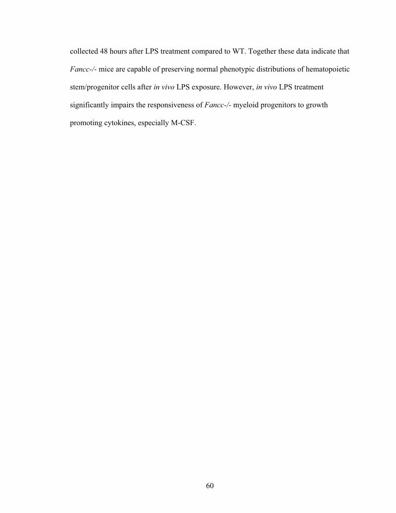
60
collected 48 hours after LPS treatment compared to WT. Together these data indicate that
Fancc-/- mice are capable of preserving normal phenotypic distributions of hematopoietic
stem/progenitor cells after in vivo LPS exposure. However, in vivo LPS treatment
significantly impairs the responsiveness of Fancc-/- myeloid progenitors to growth
promoting cytokines, especially M-CSF.
61
Figure 15. Fancc-/- progenitors exhibited impaired colony forming ability after in
vivo LPS exposure. BM cells were collected from untreated and LPS treated mice and
were cultured in methylcellulose (50,000/dish) in the presence of M-CSF (50 ng/mL),
GM-CSF (10 ng/mL), or M-CSF and GM-CSF for 7 days. Colonies were scored under an
Olympus microscope with a 4X objective lens. Total colonies per femur were calculated
by multiplying femur cellularity with the number of colonies scored per 50,000 BM cells
plated. Three mice were used for each of the three independent experiments, *P<0.05
compared to time 0, **P<0.05 compared to WT.
62
Figure 16. Fancc-/- progenitors exhibited altered distribution after in vivo LPS
treatment. BM cells were collected from untreated and LPS treated mice and were
cultured in methylcellulose in the presence of M-CSF (50 ng/mL), GM-CSF (10 ng/mL),
or M-CSF and GM-CSF for 7 days. Colonies were differentially scored by morphology
as CFU-M, CFU-G, and CFU-GM. Colony type frequencies are shown. Three mice were
used for each of the three independent experiments, *P<0.05.
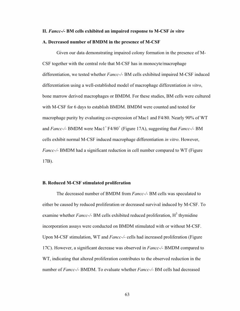
63
II. Fancc-/- BM cells exhibited an impaired response to M-CSF in vitro
A. Decreased number of BMDM in the presence of M-CSF
Given our data demonstrating impaired colony formation in the presence of M-
CSF together with the central role that M-CSF has in monocyte/macrophage
differentiation, we tested whether Fancc-/- BM cells exhibited impaired M-CSF induced
differentiation using a well-established model of macrophage differentiation in vitro,
bone marrow derived macrophages or BMDM. For these studies, BM cells were cultured
with M-CSF for 6 days to establish BMDM. BMDM were counted and tested for
macrophage purity by evaluating co-expression of Mac1 and F4/80. Nearly 90% of WT
and Fancc-/- BMDM were Mac1+ F4/80+ (Figure 17A), suggesting that Fancc-/- BM
cells exhibit normal M-CSF induced macrophage differentiation in vitro. However,
Fancc-/- BMDM had a significant reduction in cell number compared to WT (Figure
17B).
B. Reduced M-CSF stimulated proliferation
The decreased number of BMDM from Fancc-/- BM cells was speculated to
either be caused by reduced proliferation or decreased survival induced by M-CSF. To
examine whether Fancc-/- BM cells exhibited reduced proliferation, H3 thymidine
incorporation assays were conducted on BMDM stimulated with or without M-CSF.
Upon M-CSF stimulation, WT and Fancc-/- cells had increased proliferation (Figure
17C). However, a significant decrease was observed in Fancc-/- BMDM compared to
WT, indicating that altered proliferation contributes to the observed reduction in the
number of Fancc-/- BMDM. To evaluate whether Fancc-/- BM cells had decreased

64
survival in response to M-CSF, TUNEL assays were conducted to measure apoptosis.
WT and Fancc-/- BMDM exhibited very low rates of apoptosis (<2%). Together, these
data suggest that the major factor contributing to decreased BMDM cell number is
impaired M-CSF induced proliferation and not a survival defect.
65
Figure 17. Fancc-/- BM cells exhibited reduced proliferation in response to M-CSF.
BM mononuclear cells were cultured in the presence of M-CSF (50 ng/mL) for 6 days to
establish BMDM. (A) Phenotypic analysis. A representative dot plot illustrating Mac1
and F4/80 expression is shown. (B) BMDM cellularity. The number of viable cells is
shown. Six independent experiments were performed, *P<0.05. (C) Proliferation assays.
Thymidine incorporation assays were used to evaluate proliferation of BMDM at baseline
and after M-CSF stimulation (50 ng/mL for 24 hours). Six independent experiments were
performed, *P<0.05, compared to WT, **P<0.05, compared to no M-CSF.
66
C. Impaired phosphorylation of ERK and AKT upon M-CSF stimulation
ERK and AKT activation are the major downstream targets of M-CSF signaling
that regulate the proliferation of BMDM (Pixley and Stanley, 2004). Therefore, we next
examined whether Fancc-/- BMDM exhibited altered phosphorylation of ERK and AKT.
BMDM established from WT mice exhibited robust phosphorylation of ERK and AKT
after M-CSF stimulation (Figure 18A-B). In contrast, Fancc-/- BMDM had reduced
phosphorylation of ERK and AKT (Figure 18A-C). To ensure that differences detected in
ERK and AKT phosphorylation were not due to impaired expression of the M-CSF
receptor or C-Fms (CD115), flow cytometric analysis of CD115 on BMDM was
conducted. Fancc-/- BMDM had similar expression levels of the M-CSF receptor
compared to WT (Figure 18D). Collectively, these data indicate that Fancc-/- BMDM
have altered M-CSF induced ERK and AKT phosphorylation which likely contributes to
the proliferation defect observed.
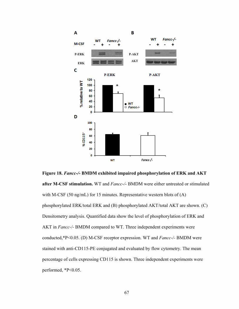
67
Figure 18. Fancc-/- BMDM exhibited impaired phosphorylation of ERK and AKT
after M-CSF stimulation. WT and Fancc-/- BMDM were either untreated or stimulated
with M-CSF (50 ng/mL) for 15 minutes. Representative western blots of (A)
phosphorylated ERK/total ERK and (B) phosphorylated AKT/total AKT are shown. (C)
Densitometry analysis. Quantified data show the level of phosphorylation of ERK and
AKT in Fancc-/- BMDM compared to WT. Three independent experiments were
conducted,*P<0.05. (D) M-CSF receptor expression. WT and Fancc-/- BMDM were
stained with anti-CD115-PE conjugated and evaluated by flow cytometry. The mean
percentage of cells expressing CD115 is shown. Three independent experiments were
performed, *P<0.05.
68
SUMMARY OF AIM 1:
In the peritoneum after LPS induced peritonitis, Fancc-/- mice had increased
neutrophils in early inflammatory stages and fewer monocytes/macrophages in later
phases of inflammation. The inflammatory gene expression of Fancc-/- peritoneal cells
was reduced, which was associated with fewer circulating inflammatory monocytes in the
peripheral blood of Fancc-/- mice. In addition, Fancc-/- mice were capable of preserving
normal phenotypic distributions of hematopoietic stem/progenitor cells after in vivo LPS
exposure, however Fancc-/- myeloid progenitor responsiveness to M-CSF was
significantly impaired. Studies using an in vitro BMDM model showed that Fancc-/-
BMDM had reduced M-CSF induced proliferation and decreased phosphorylation of
ERK and AKT. Overall, these observations indicate that Fancc-/- mice have an altered
LPS induced inflammatory response and impaired M-CSF signaling. However, the
question of whether Fancc-/- macrophages exhibit autonomous defects in function that
contributes to the in vivo phenotype remains.
69
AIM 2: To determine whether Fancc-/- macrophages exhibit cell autonomous
defects in adhesion, migration and phagocytosis.
Macrophage motility is essential for normal development and immune function.
We questioned whether Fancc-/- peritoneal macrophages have impaired cell motility
including adhesion and migration as well as phagocytosis since this is a critical cellular
function for macrophages. The experimental design is illustrated in Figure 19. In brief,
peritoneal macrophages were collected and assessed for adhesion on FN and endothelial
cells, migration toward MCP-1 and M-CSF, and phagocytosis of E.coli particles and IgG
latex beads. These analyses were instrumental to determine whether Fancc-/-
macrophages exhibit intrinsic alterations in function. These studies also help to clarify
whether reduced monocyte/macrophage in the local inflamed site is due to impaired
motility of Fancc-/- cells.
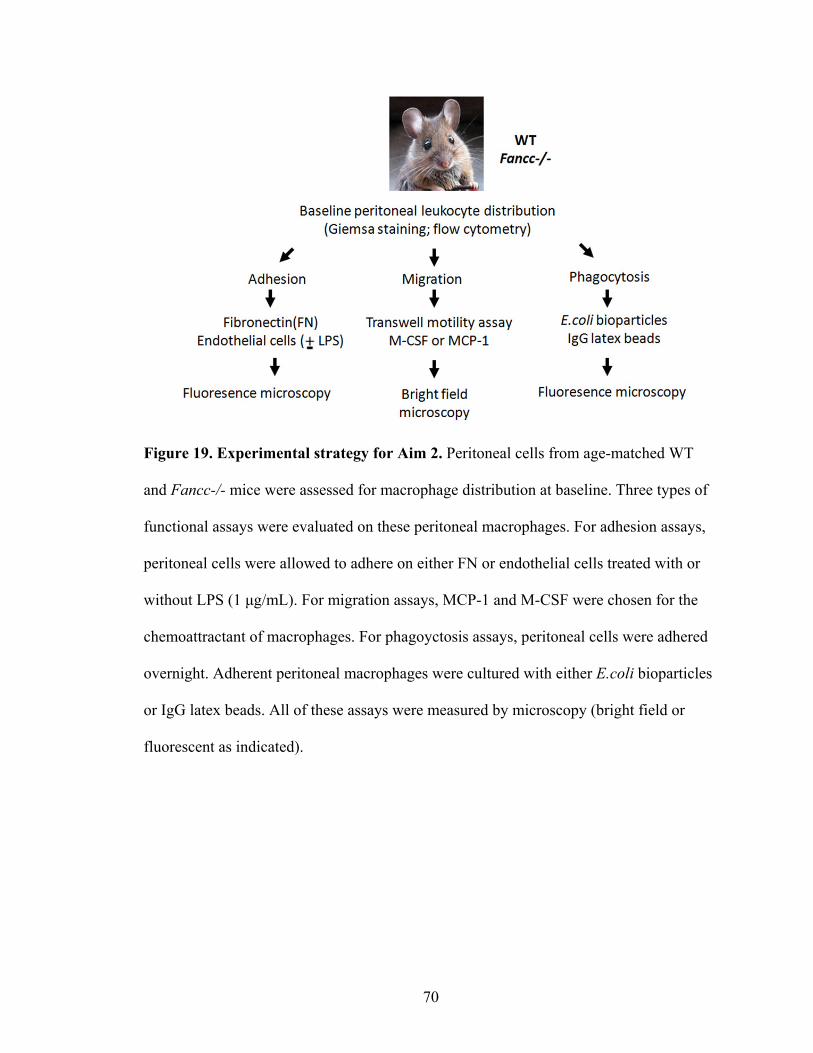
70
Figure 19. Experimental strategy for Aim 2. Peritoneal cells from age-matched WT
and Fancc-/- mice were assessed for macrophage distribution at baseline. Three types of
functional assays were evaluated on these peritoneal macrophages. For adhesion assays,
peritoneal cells were allowed to adhere on either FN or endothelial cells treated with or
without LPS (1 μg/mL). For migration assays, MCP-1 and M-CSF were chosen for the
chemoattractant of macrophages. For phagoyctosis assays, peritoneal cells were adhered
overnight. Adherent peritoneal macrophages were cultured with either E.coli bioparticles
or IgG latex beads. All of these assays were measured by microscopy (bright field or
fluorescent as indicated).
71
III. Impaired motility and function of Fancc-/- monocytes/macrophages
A. Characterization of Fancc-/- peritoneal cells at the steady state
Before examining whether Fancc-/- peritoneal macrophages exhibit altered
function, the steady-state distribution of macrophages in the peritoneal cavity was
examined. At baseline, Giemsa staining of peritoneal cells indicated that the majority of
cells in the peritoneum were macrophages, accounting for nearly 80% of cells, and no
differences were detected between WT and Fancc-/- mice (Figure 20A-B). Flow
cytometric analyses of peritoneal cells from WT and Fancc-/- mice showed a similar
phenotypic expression of macrophage antigens (Figure 20C) In addition, the absolute
number of these phenotypically-defined cells were also similar between both genotypes
(Figure 21D). These data demonstrate the distribution of monocyte/macrophage at steady
state is normal in Fancc-/- mice compared to WT.
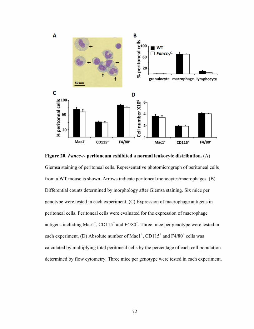
72
Figure 20. Fancc-/- peritoneum exhibited a normal leukocyte distribution. (A)
Giemsa staining of peritoneal cells. Representative photomicrograph of peritoneal cells
from a WT mouse is shown. Arrows indicate peritoneal monocytes/macrophages. (B)
Differential counts determined by morphology after Giemsa staining. Six mice per
genotype were tested in each experiment. (C) Expression of macrophage antigens in
peritoneal cells. Peritoneal cells were evaluated for the expression of macrophage
antigens including Mac1+, CD115+ and F4/80+. Three mice per genotype were tested in
each experiment. (D) Absolute number of Mac1+, CD115+ and F4/80+ cells was
calculated by multiplying total peritoneal cells by the percentage of each cell population
determined by flow cytometry. Three mice per genotype were tested in each experiment.
73
B. Impaired adhesion of Fancc-/- peritoneal macrophages
1. Impaired adhesion on FN
As professional phagocytes, monocytes/macrophages are recruited to the inflamed
site to effectively eliminate intruders and cell debris. Our data demonstrate that Fancc-/-
mice exhibit significantly reduced monocytes/macrophages in LPS induced peritonitis.
However, it is not clear whether this observation is due to cell autonomous defects in
Fancc-/- macrophages. To examine whether Fancc-/- macrophages exhibit cell
autonomous defects that contribute to the impaired accumulation in vivo, we next
examined whether Fancc-/- macrophages have altered adhesion and migration. To
evaluate whether adhesion of Fancc-/- macrophages is compromised, resident
macrophages were adhered to either FN, which is an important component of the
extracellular matrix, or to endothelial cells, which represents a physiologically-relevant
cell type. For adhesion to FN, fewer Fancc-/- macrophages adhered to FN after 1 hour
compared to WT (Figure 21A-B).

74
Figure 21. Fancc-/- macrophages exhibited reduced adhesion on FN. Fresh peritoneal
cells were collected from WT and Fancc-/- mice in the steady state. Cells were allowed
to adhere on FN (CH296) coated and uncoated wells for one hour. Nonadherent cells
were removed by washing with PBS. Adherent cells were estimated after Giemsa
staining. Photomicrographs were taken on a Leica microscope (20X). (A) Representative
photomicrographs are shown. (B) Quantitative data of levels of adhesion for Fancc-/-
cells compared to WT are shown. Three independent experiments were
performed,*P<0.05.
75
2. Impaired adhesion on endothelial cells
Given that circulating leukocytes are recruited to the inflamed endothelium,
where they adhere and traverse between endothelial cells to enter the inflammatory site
(Kubes, 2002), we next questioned whether cell-cell adhesion was also altered in
Fancc-/- macrophages by conducting macrophage-endothelial cell co-culture assays. In
addition to conducting assays under baseline conditions, endothelial cells were stimulated
with LPS (1 μg/mL) prior to co-culture with macrophages to model the upregulation of
adhesion molecules that occurs on endothelial cells during inflammation (Lush et al.,
2000). In conditions with untreated endothelial cells, WT macrophages had increased
adherence after 2 hours of co-culture compared to the 1 hour time point (Figure 22A-B).
In contrast, Fancc-/- macrophages had no observable increase under the same conditions
and were significantly decreased compared to WT cells. The impairment in Fancc-/-
macrophage adhesion was even more apparent when evaluated on LPS-activated
endothelial cells. WT macrophages exhibited enhanced adhesion to LPS-activated
endothelial cells at 1 and 2 hour time points compared to untreated endothelial cell
controls. However, Fancc-/- macrophages had a dramatic reduction in the number of
adherent cells at both time points compared to WT cells (Figure 22A-B). Taken together
these data indicate that Fancc-/- macrophages exhibit impaired adhesion to FN and
endothelial cells.
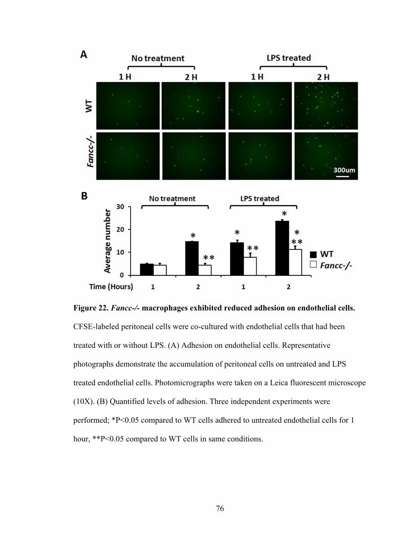
76
Figure 22. Fancc-/- macrophages exhibited reduced adhesion on endothelial cells.
CFSE-labeled peritoneal cells were co-cultured with endothelial cells that had been
treated with or without LPS. (A) Adhesion on endothelial cells. Representative
photographs demonstrate the accumulation of peritoneal cells on untreated and LPS
treated endothelial cells. Photomicrographs were taken on a Leica fluorescent microscope
(10X). (B) Quantified levels of adhesion. Three independent experiments were
performed; *P<0.05 compared to WT cells adhered to untreated endothelial cells for 1
hour, **P<0.05 compared to WT cells in same conditions.
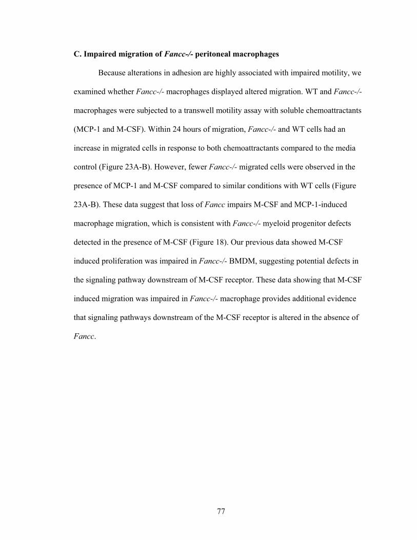
77
C. Impaired migration of Fancc-/- peritoneal macrophages
Because alterations in adhesion are highly associated with impaired motility, we
examined whether Fancc-/- macrophages displayed altered migration. WT and Fancc-/-
macrophages were subjected to a transwell motility assay with soluble chemoattractants
(MCP-1 and M-CSF). Within 24 hours of migration, Fancc-/- and WT cells had an
increase in migrated cells in response to both chemoattractants compared to the media
control (Figure 23A-B). However, fewer Fancc-/- migrated cells were observed in the
presence of MCP-1 and M-CSF compared to similar conditions with WT cells (Figure
23A-B). These data suggest that loss of Fancc impairs M-CSF and MCP-1-induced
macrophage migration, which is consistent with Fancc-/- myeloid progenitor defects
detected in the presence of M-CSF (Figure 18). Our previous data showed M-CSF
induced proliferation was impaired in Fancc-/- BMDM, suggesting potential defects in
the signaling pathway downstream of M-CSF receptor. These data showing that M-CSF
induced migration was impaired in Fancc-/- macrophage provides additional evidence
that signaling pathways downstream of the M-CSF receptor is altered in the absence of
Fancc.
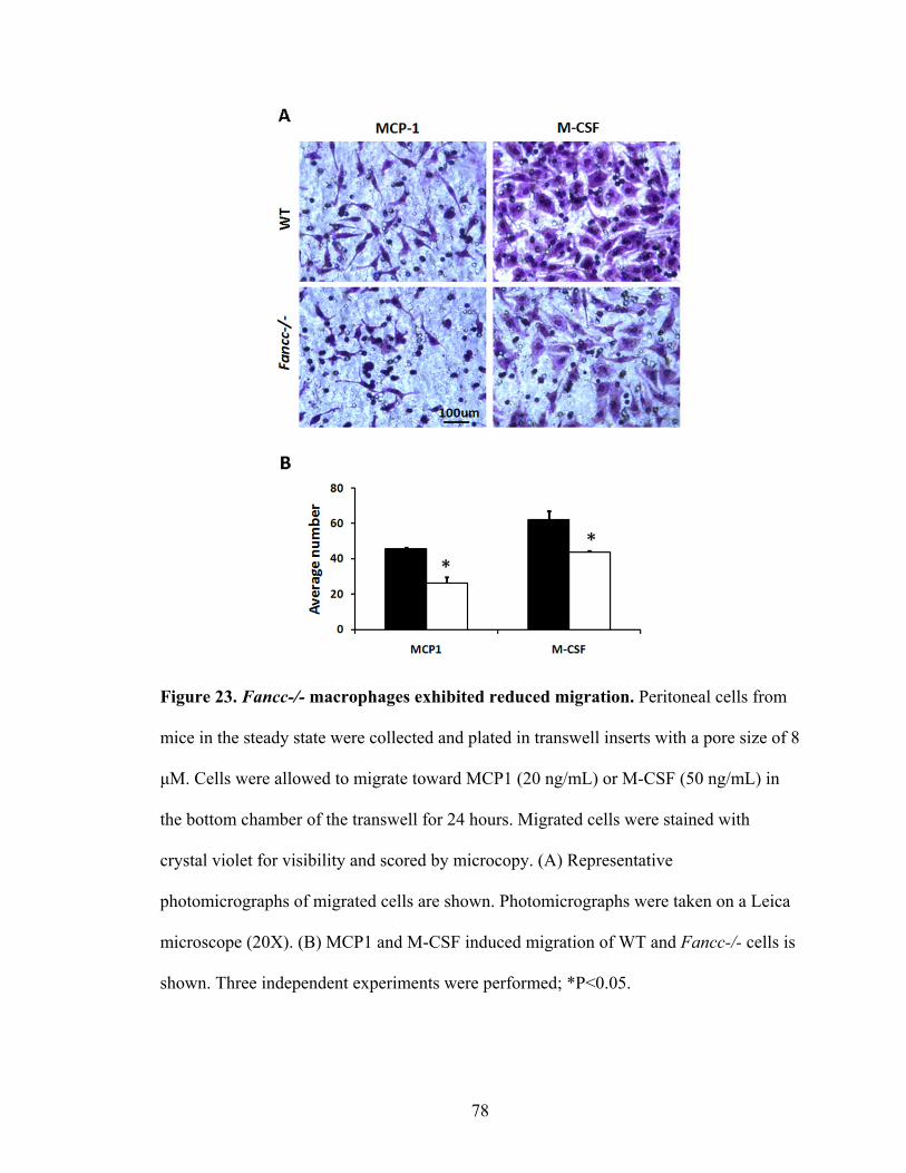
78
Figure 23. Fancc-/- macrophages exhibited reduced migration. Peritoneal cells from
mice in the steady state were collected and plated in transwell inserts with a pore size of 8
μM. Cells were allowed to migrate toward MCP1 (20 ng/mL) or M-CSF (50 ng/mL) in
the bottom chamber of the transwell for 24 hours. Migrated cells were stained with
crystal violet for visibility and scored by microcopy. (A) Representative
photomicrographs of migrated cells are shown. Photomicrographs were taken on a Leica
microscope (20X). (B) MCP1 and M-CSF induced migration of WT and Fancc-/- cells is
shown. Three independent experiments were performed; *P<0.05.
79
D. Impaired phagocytosis of Fancc-/- peritoneal macrophages
1. Phagocytosis of E.coli bioparticles
As a professional phagocyte, phagocytosis is a critical biologic function that
macrophages perform for pathogen clearance and for inflammation resolution. Therefore,
we evaluated whether Fancc-/- macrophages also exhibit impaired phagocytosis assessed
by the uptake of fluorescent E.coli bioparticles and IgG-coated latex beads. Compared to
WT macrophages, Fancc-/- macrophages had reduced cells that ingested fluorescent
E.coli particles after 1 hour of co-culture (Figure 24A-B), implying dysfunctional
phagocytosis mediated by TLR4.
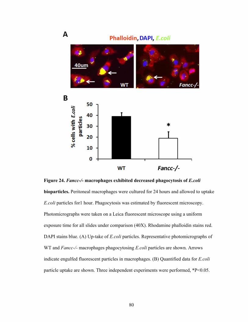
80
Figure 24. Fancc-/- macrophages exhibited decreased phagocytosis of E.coli
bioparticles. Peritoneal macrophages were cultured for 24 hours and allowed to uptake
E.coli particles for1 hour. Phagocytosis was estimated by fluorescent microscopy.
Photomicrographs were taken on a Leica fluorescent microscope using a uniform
exposure time for all slides under comparison (40X). Rhodamine phalloidin stains red.
DAPI stains blue. (A) Up-take of E.coli particles. Representative photomicrographs of
WT and Fancc-/- macrophages phagocytosing E.coli particles are shown. Arrows
indicate engulfed fluorescent particles in macrophages. (B) Quantified data for E.coli
particle uptake are shown. Three independent experiments were performed, *P<0.05.
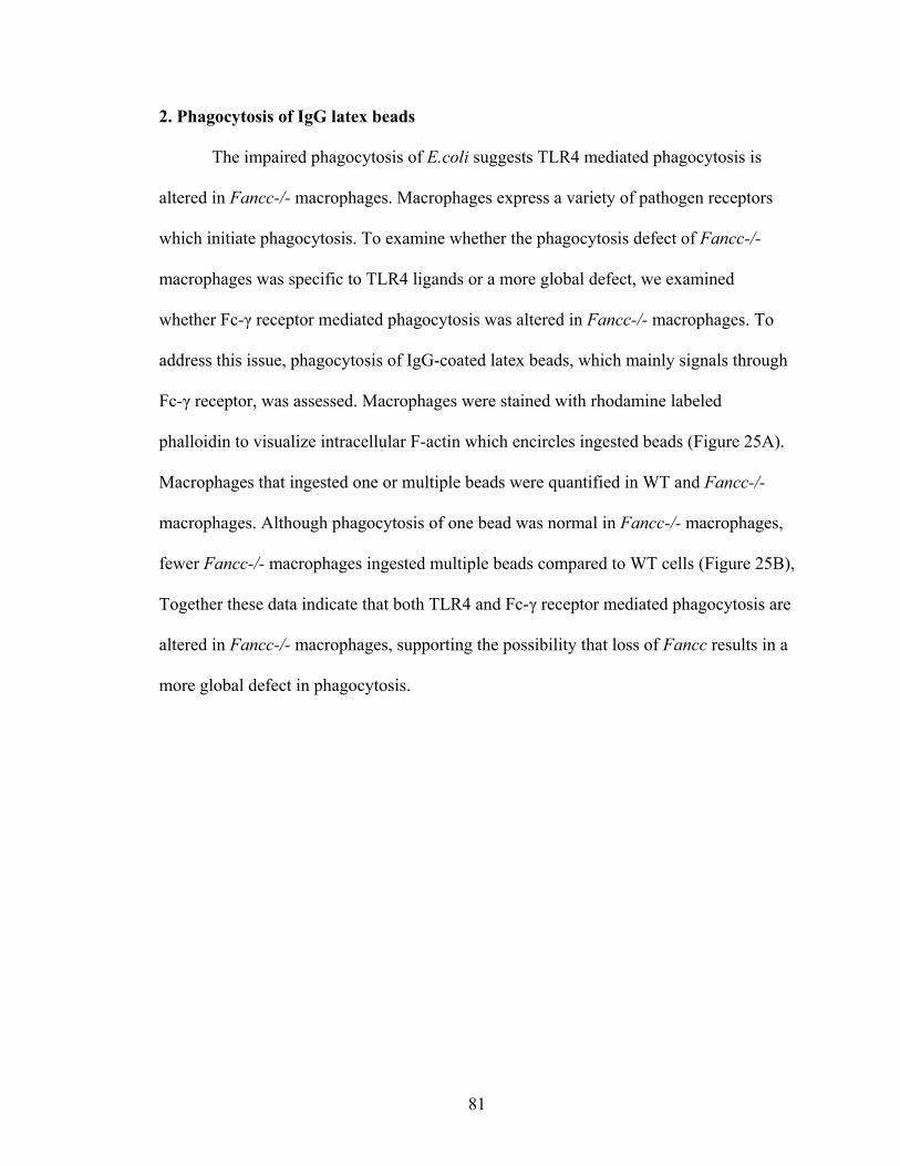
81
2. Phagocytosis of IgG latex beads
The impaired phagocytosis of E.coli suggests TLR4 mediated phagocytosis is
altered in Fancc-/- macrophages. Macrophages express a variety of pathogen receptors
which initiate phagocytosis. To examine whether the phagocytosis defect of Fancc-/-
macrophages was specific to TLR4 ligands or a more global defect, we examined
whether Fc-γ receptor mediated phagocytosis was altered in Fancc-/- macrophages. To
address this issue, phagocytosis of IgG-coated latex beads, which mainly signals through
Fc-γ receptor, was assessed. Macrophages were stained with rhodamine labeled
phalloidin to visualize intracellular F-actin which encircles ingested beads (Figure 25A).
Macrophages that ingested one or multiple beads were quantified in WT and Fancc-/-
macrophages. Although phagocytosis of one bead was normal in Fancc-/- macrophages,
fewer Fancc-/- macrophages ingested multiple beads compared to WT cells (Figure 25B),
Together these data indicate that both TLR4 and Fc-γ receptor mediated phagocytosis are
altered in Fancc-/- macrophages, supporting the possibility that loss of Fancc results in a
more global defect in phagocytosis.
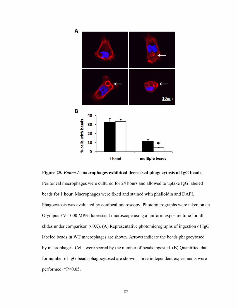
82
Figure 25. Fancc-/- macrophages exhibited decreased phagocytosis of IgG beads.
Peritoneal macrophages were cultured for 24 hours and allowed to uptake IgG labeled
beads for 1 hour. Macrophages were fixed and stained with phalloidin and DAPI.
Phagocytosis was evaluated by confocal microscopy. Photomicrographs were taken on an
Olympus FV-1000 MPE fluorescent microscope using a uniform exposure time for all
slides under comparison (60X). (A) Representative photomicrographs of ingestion of IgG
labeled beads in WT macrophages are shown. Arrows indicate the beads phagocytosed
by macrophages. Cells were scored by the number of beads ingested. (B) Quantified data
for number of IgG beads phagocytosed are shown. Three independent experiments were
performed, *P<0.05.
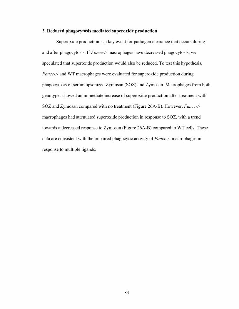
83
3. Reduced phagocytosis mediated superoxide production
Superoxide production is a key event for pathogen clearance that occurs during
and after phagocytosis. If Fancc-/- macrophages have decreased phagocytosis, we
speculated that superoxide production would also be reduced. To test this hypothesis,
Fancc-/- and WT macrophages were evaluated for superoxide production during
phagocytosis of serum opsonized Zymosan (SOZ) and Zymosan. Macrophages from both
genotypes showed an immediate increase of superoxide production after treatment with
SOZ and Zymosan compared with no treatment (Figure 26A-B). However, Fancc-/-
macrophages had attenuated superoxide production in response to SOZ, with a trend
towards a decreased response to Zymosan (Figure 26A-B) compared to WT cells. These
data are consistent with the impaired phagocytic activity of Fancc-/- macrophages in
response to multiple ligands.
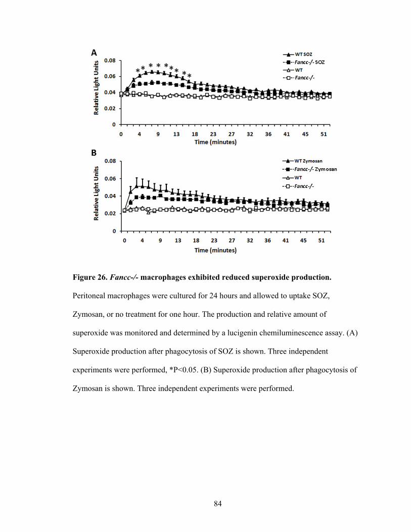
84
Figure 26. Fancc-/- macrophages exhibited reduced superoxide production.
Peritoneal macrophages were cultured for 24 hours and allowed to uptake SOZ,
Zymosan, or no treatment for one hour. The production and relative amount of
superoxide was monitored and determined by a lucigenin chemiluminescence assay. (A)
Superoxide production after phagocytosis of SOZ is shown. Three independent
experiments were performed, *P<0.05. (B) Superoxide production after phagocytosis of
Zymosan is shown. Three independent experiments were performed.
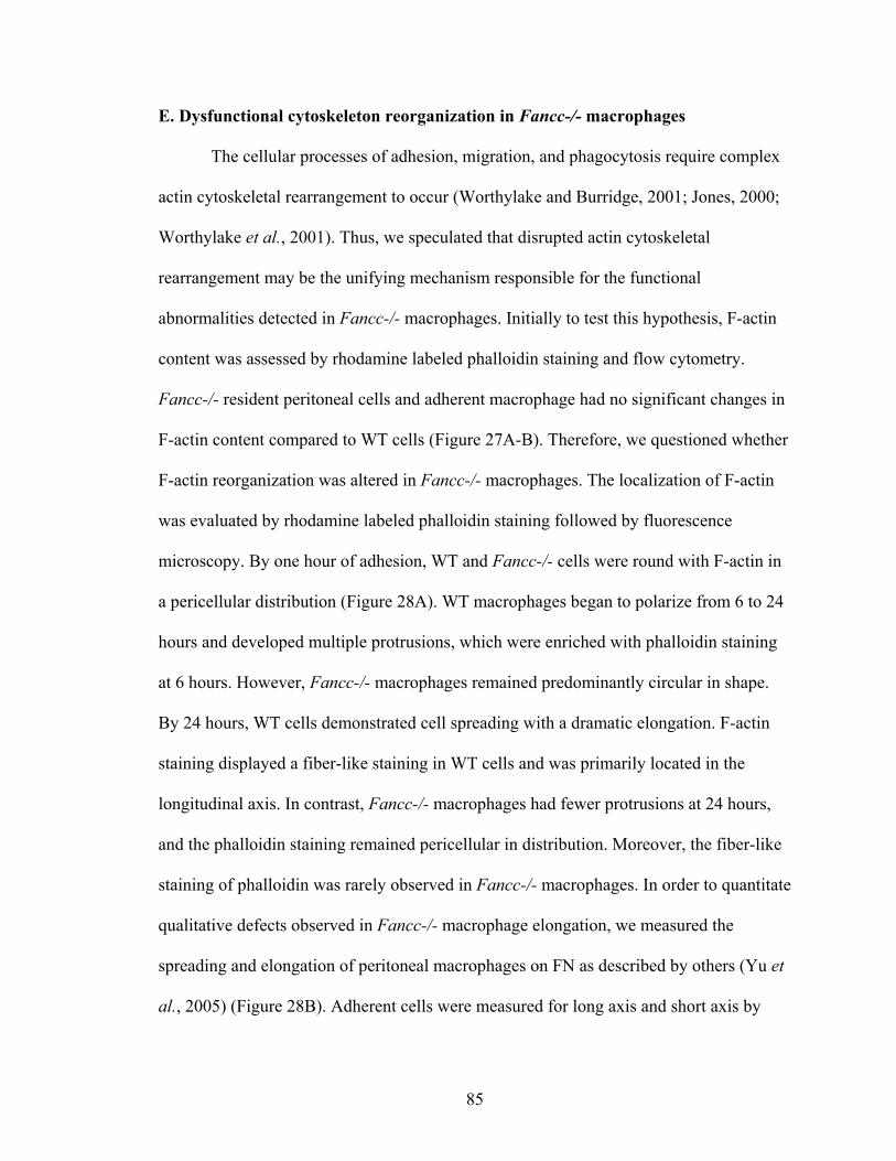
85
E. Dysfunctional cytoskeleton reorganization in Fancc-/- macrophages
The cellular processes of adhesion, migration, and phagocytosis require complex
actin cytoskeletal rearrangement to occur (Worthylake and Burridge, 2001; Jones, 2000;
Worthylake et al., 2001). Thus, we speculated that disrupted actin cytoskeletal
rearrangement may be the unifying mechanism responsible for the functional
abnormalities detected in Fancc-/- macrophages. Initially to test this hypothesis, F-actin
content was assessed by rhodamine labeled phalloidin staining and flow cytometry.
Fancc-/- resident peritoneal cells and adherent macrophage had no significant changes in
F-actin content compared to WT cells (Figure 27A-B). Therefore, we questioned whether
F-actin reorganization was altered in Fancc-/- macrophages. The localization of F-actin
was evaluated by rhodamine labeled phalloidin staining followed by fluorescence
microscopy. By one hour of adhesion, WT and Fancc-/- cells were round with F-actin in
a pericellular distribution (Figure 28A). WT macrophages began to polarize from 6 to 24
hours and developed multiple protrusions, which were enriched with phalloidin staining
at 6 hours. However, Fancc-/- macrophages remained predominantly circular in shape.
By 24 hours, WT cells demonstrated cell spreading with a dramatic elongation. F-actin
staining displayed a fiber-like staining in WT cells and was primarily located in the
longitudinal axis. In contrast, Fancc-/- macrophages had fewer protrusions at 24 hours,
and the phalloidin staining remained pericellular in distribution. Moreover, the fiber-like
staining of phalloidin was rarely observed in Fancc-/- macrophages. In order to quantitate
qualitative defects observed in Fancc-/- macrophage elongation, we measured the
spreading and elongation of peritoneal macrophages on FN as described by others (Yu et
al., 2005) (Figure 28B). Adherent cells were measured for long axis and short axis by
86
Adobe photoshop. The cells with the ratio of Long/Short axes greater than 1 were defined
as elongating or polarizing cells. Round cells were defined as having a ratio of 1. Figure
28C illustrates the percentage of elongated cells within 24 hours. WT cells had a dramatic
increase in the percentage of elongated cells within 6 hours compared to 1 hour. Over
90% of WT cells finished cell spreading by 24 hours. In contrast, Fancc-/- cells displayed
a dramatic reduction in percentage of elongated cells by 6 hours and only half of them
completed elongation by 24 hours. These data indicate that Fancc-/- macrophages have
insufficient F-actin cytoskeletal rearrangements induced by FN, which may be the
underlying mechanism responsible for dysfunctional adhesion, migration, and
phagocytosis.
87
Figure 27. Fancc-/- peritoneal macrophages exhibited normal F-actin content. Fresh
peritoneal cells from WT and Fancc-/- mice were stained with rhodamine phalloidin and
examined by flow cytometry to quantitate F-actin content. (A) Representative histogram
of rhodamin-phalloidin staining. (B) Mean fluorescence intensity (MFI) of rhodamine-
phalloidin staining. MFI of phalloidin staining is shown. Three independent experiments
were performed.
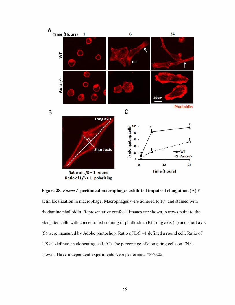
88
Figure 28. Fancc-/- peritoneal macrophages exhibited impaired elongation. (A) F-
actin localization in macrophage. Macrophages were adhered to FN and stained with
rhodamine phalloidin. Representative confocal images are shown. Arrows point to the
elongated cells with concentrated staining of phalloidin. (B) Long axis (L) and short axis
(S) were measured by Adobe photoshop. Ratio of L/S =1 defined a round cell. Ratio of
L/S >1 defined an elongating cell. (C) The percentage of elongating cells on FN is
shown. Three independent experiments were performed, *P<0.05.
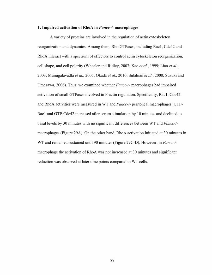
89
F. Impaired activation of RhoA in Fancc-/- macrophages
A variety of proteins are involved in the regulation of actin cytoskeleton
reorganization and dynamics. Among them, Rho GTPases, including Rac1, Cdc42 and
RhoA interact with a spectrum of effectors to control actin cytoskeleton reorganization,
cell shape, and cell polarity (Wheeler and Ridley, 2007; Kao et al., 1999; Liao et al.,
2003; Munugalavadla et al., 2005; Okada et al., 2010; Sulahian et al., 2008; Suzuki and
Umezawa, 2006). Thus, we examined whether Fancc-/- macrophages had impaired
activation of small GTPases involved in F-actin regulation. Specifically, Rac1, Cdc42
and RhoA activities were measured in WT and Fancc-/- peritoneal macrophages. GTP-
Rac1 and GTP-Cdc42 increased after serum stimulation by 10 minutes and declined to
basal levels by 30 minutes with no significant differences between WT and Fancc-/-
macrophages (Figure 29A). On the other hand, RhoA activation initiated at 30 minutes in
WT and remained sustained until 90 minutes (Figure 29C-D). However, in Fancc-/-
macrophage the activation of RhoA was not increased at 30 minutes and significant
reduction was observed at later time points compared to WT cells.
90
Figure 29. Impaired activation of RhoA was observed in Fancc-/- macrophages. For
a single small GTPase activation assay, peritoneal cells from 10 mice per genotype were
pooled. (A) Representative Rac1 and (B) Cdc42 activation assays are shown. GTP-Rac1
and GTP-Cdc42 were immunoprecipitated with GST-PBD and detected by Rac1 and
Cdc42 specific antibodies. (C) Representative RhoA activation assay is shown. GTP-
RhoA was immunoprecipitated with GST-RBD and detected by RhoA antibody. (D)
Quantification of densitometry data is shown. GTP-RhoA was normalized to total RhoA,
and the relative change was normalized to time 0. Three independent experiments were
performed,*P<0.005.
91
SUMMARY OF AIM2
Baseline leukocyte distribution in the peritoneum of Fancc-/- mice was normal,
however numerous functional deficits were elucidated in resident Fancc-/- macrophages,
including impaired adhesion, migration, phagocytosis, and superoxide production. The
unifying mechanism responsible for these phenotypes is speculated to be altered F-actin
reorganization, which is supported by our data showing abnormal elongation of Fancc-/-
peritoneal macrophages after adhesion to FN and impaired RhoA activation .
Collectively, these data support the contention that loss of Fancc not only affects
hematopoietic stem/progenitor cell functions, as previously thought, but also severely
impairs immune cell function resulting in an altered inflammatory response in vivo.
92
DISCUSSION
Astute clinical observations have long suggested that FA patients may have
immune defects (reviewed in Fagerlie SR, 2006). However, FA genetic heterogeneity
together with leukopenia from evolving BM failure has made formal proof of this
concept challenging. Limited previous studies in murine models of FA support altered
immunologic function (Sejas et al., 2007; Zhang et al., 2007; Hadjur and Jirik, 2003;
Vanderwerf et al., 2009). However, none of these reports included a systematic
functional evaluation of macrophages, which are an integral cellular component of the
innate immune system as well as an antigen presenting cell that participates in the
coordination of acquired immunity.
1. Altered inflammatory response in Fancc-/- mice
In the current studies, we show that LPS induced recruitment of Fancc-/-
monocytes/macrophages to an inflamed peritoneum in vivo is significantly reduced.
Previous studies demonstrated alterations in the response of Fancc-/- cells to toll-like
receptor ligands such as LPS and R848 (Vanderwerf et al., 2009; Sejas et al., 2007;
Hadjur and Jirik, 2003). Decreased Fancc-/- monocyte/macrophage recruitment was not
specific to toll receptor signaling since a similar response was detected after injection of
sodium periodate, which induces a non-specific, chemical inflammation. The decrease in
circulating Fancc-/- inflammatory monocytes after LPS injection suggests that impaired
monocytes mobilization from the BM may be a contributing factor for fewer
macrophages observed in the local inflammatory site. However, there are other possible
explanations for the reduced local monocytes/macrophages which have not been tested.
93
First of all, the protein level of cytokines/chemokines in the inflamed peritoneum cavity
has not been examined. We have tested the gene expression of cytokines/chemokines in
the peritoneal cells. Our data showed reduced mRNA production in Fancc-/- cells.
However, the total protein levels in the peritoneal fluid could be different from mRNA
expression in peritoneal cells, because other cell types in the peritoneum may contribute
to the protein levels of cytokines/chemokines (i.e. endothelial, epithelial and T cells). To
determine the levels of cytokines/chemokines, Multiplex technique could be used with
LPS injected peritoneal fluid samples. Secondly, the survival of peritoneal
monocytes/macrophages were not evaluated in our studies. We showed Fancc-/- mice
exhibited a decreased number of monocytes/macrophages in the later phase of
inflammation, which could be due to either decreased recruitment from peripheral blood
or increased apoptosis of monocytes/macrophages. Although we showed that Fancc-/-
peritoneal neutrophils had decreased apoptosis in the early phase, the survival of
macrophages was not assessed. A similar method of testing apoptosis of neutrophils
could be used for determining the monocytes/macrophages survival. Moreover, the
identification of Fancc-/- macrophage deficits in vitro, including impaired adhesion,
migration, and phagocytosis, support macrophage autonomous defects in Fancc-/- mice
that likely participate in the alterations observed in vivo. Collectively, these data provide
strong evidence for a primary defect in the function of Fancc-/- macrophage.
Previously, Sejas and colleagues examined Fancc-/- mice for evidence of
enhanced systemic inflammation after LPS treatment (Sejas et al., 2007). In these studies,
investigators reported high serum levels of inflammatory cytokines, especially TNF-α, in
Fancc-/- mice compared to WT after LPS injection. Therefore, our data demonstrating no
94
differences in serum inflammatory cytokines and reduced inflammatory
cytokine/chemokine expression in peritoneal cells from Fancc-/- mice after LPS was
initially surprising. However, the overall goal of our studies was to evaluate for
alterations in the local inflammatory response, not systemic inflammation. Therefore,
potential explanations for the apparent discrepant results between our studies and
previous data include type of inflammatory response initiated, type of LPS used, and
environment that Fancc-/- mice are housed. IP injections are highly reproducible for LPS
induced inflammatory response studies in mice (Holmgren et al., 2008; Marquez-Velasco
et al., 2007). However, handling mice for a proper IP injection can be challenging. To
keep the results consistent, we treated mice with isoflurane which anaesthesizes the mice
allowing for the proper treatment of LPS injection and avoiding subcutaneous or
abdominal organs injection which will cause complete different inflammatory responses
compared to peritoneal cavity injection. Additionally, we also adjusted the LPS dose
according to mouse body weight in order to have consistent results. Although we use the
LPS from the pharmaceutical company, the lot number and the potential quality may be
different from previous studies. Overall, these are the contributors for the difference we
observed in Fancc-/- mice from previous studies. However, our findings are consistent
with a more recent study that reported normal LPS induced TNF-α production from
Fancc-/- splenic macrophages (Vanderwerf et al., 2009). Interestingly, these studies did
show that Fancc-/- splenic macrophages treated with a TLR8 agonist overproduced TNF-
α, suggesting that TLR8 signaling may be altered. Taken together, these studies
emphasize that Fancc-/- immune cells do exhibit altered functions including higher levels
of TNF-α. Yet, the mechanisms involved needs to be further investigated.
95
2. Impaired adhesion, migration and phagocytosis of Fancc-/- macrophages
The implications of our findings are significant given the central role that
macrophages have in both innate and acquired immunity. Furthermore, macrophages
orchestrate the initiation and resolution phases of an acute inflammatory response
(Cailhier et al., 2005). The severity of local inflammation is dependent on the type of
pathogen exposure, efficiency of pathogen clearance, and competent resolution of
inflammation (Cox et al., 1995; Tabas, 2010). Crucial functions of macrophages in these
processes are to kill pathogens, phagocytose apoptotic neutrophils, and repair tissue
injury as a result of the inflammation. Therefore, the modest defects in Fancc-/-
macrophage function may not result in overt immunodeficiency, but rather subtle
inefficiencies in pathogen clearance and inflammation resolution, resulting in a
predisposition towards chronic inflammation or a lower threshold for progression to a
severe systemic inflammatory response. Studies demonstrating increased serum
inflammatory cytokines in FA patients are consistent with a chronic inflammatory state in
FA (Stark et al., 1993; Bagnara et al., 1993; Rosselli et al., 1994). In addition, studies
showing inflammatory cytokine overproduction as well as hypersensitivity to LPS
induced shock/death are consistent with a lower threshold for progression to a severe
systemic inflammatory response in FA (Fagerlie and Bagby, 2006; Sejas et al., 2007;
Zhang et al., 2007; Dufour et al., 2003). In our studies, LPS is the only stimulus that we
tested for an altered inflammatory response in Fancc-/- mice. Since LPS is only part of
gram-negative bacteria, the inflammation induced by real pathogen and endotoxin may be
very different. Therefore, it is necessary to examine whether Fancc-/- mice exhibit
altered inflammation in response to a bacterial (eg. E.coli ) infection.
96
3. Classical and alternative macrophage activation
In the acute-phase of inflammation, pro-inflammatory mediators are released
primarily by PMNs and inflammatory macrophages. Over time, the inflammatory
response is dampened by resolving macrophages. Consequently, macrophage activation
can be pro-inflammatory or anti-inflammatory, which is dependent on intrinsic
macrophage characteristics and extrinsic environmental cues. Dysregulated activation of
macrophages contributes to local tissue destruction, systemic inflammation, and chronic
inflammation. Thus, macrophages have an essential role in initiating, instructing, and
terminating the adaptive immune response.
Classically activated or M1 macrophages develop in response to concomitant
stimulation with IFN-γ and microbial products such as LPS (Adams, 1989; Yamazaki et
al., 2008; Jeannin et al., 2011). Functionally, M1 macrophages secrete significant
amounts of pro-inflammatory cytokines, such as TNF-α, IL-1β, and IL-12 (Benoit et al.,
2008). In addition, M1 macrophages are characterized by enhanced endocytic functions
and ability to kill intracellular pathogens, which is mediated by a variety of mechanisms
including phagocytosis, phagosome acidification, and nitric oxide (NO) synthesis
(Groesdonk et al., 2006; Raisanen et al., 2005). NO synthesis in macrophages is
regulated by the balance between inducible NO synthetase (iNOS) and arginase, which
sequesters NO by competing the same substrate L-Arginine (Colton et al., 2006). Similar
to M1 cells, M2 or alternatively activated macrophages change cellular morphology and
secretory pattern after being stimulated by multiple anti-inflammatory regulators such as
IL-4, IL-13, TGF-β, and IL-10 (Pixley and Stanley, 2004; Martinez et al., 2008).
Importantly, M-CSF stimulation of macrophages is critical for intact activation towards
97
an M2 phenotype (Kinashi et al., 1991; Yamazaki et al., 2008). M2 macrophages release
chemokines including CCL17, and CCL18 (Imai et al., 1999). In addition, M2
macrophages counteract ongoing inflammation by releasing factors such as Ym1, Ym2,
IL-10, and TGF-β, which also indirectly promotes tissue repair (Raes et al., 2002a; Raes
et al., 2002b; Stein et al., 1992).
Interestingly, previous studies in Fancc-/- peritoneal macrophages demonstrated
increased iNOS expression and NO production in response to LPS and IFN-γ stimulation.
Though not discussed in the manuscript, these findings suggest that Fancc-/-
macrophages exhibit increased M1 activation. Our data showing reduced M-CSF induced
proliferation, ERK and AKT phosphorylation in Fancc-/- macrophages suggest reduced
M2 activation. Therefore, we hypothesize that Fancc-/- macrophages have increased
classical activation and reduced alternative activation leading to an altered inflammatory
response. Future studies to test this hypothesis will be important.
98
4. Association of altered cytoskeleton rearrangement and decreased RhoA activation
in Fancc-/- macrophages
Dramatic morphologic differences were identified between Fancc-/- macrophage
and controls, which are attributed to disrupted actin rearrangement in Fancc-/-
macrophage. Complex cytoskeletal rearrangements are essential for numerous cellular
processes to be intact including adhesion, migration, and phagocytosis. Given that the
functional defects in Fancc-/- macrophage were induced by numerous extracellular
stimuli (i.e. FN, M-CSF, MCP-1, SOZ, E.coli), we speculate that dysregulation of
cytoskeletal organization is the underlying mechanism responsible for the functional
defects in Fancc-/- macrophage in vitro as well as altered trafficking in vivo. The
recruitment of monocytes/macrophages to local inflammatory sites is dependent on
dynamic cell shape changes, which promote cell movement from the blood through the
endothelium into local tissue environments. Several important steps are involved in this
process with each individual step requiring dynamic changes in the actin cytoskeleton
(Vicente-Manzanares and Sanchez-Madrid, 2004; Worthylake et al., 2001; Worthylake
and Burridge, 2001). Reduced adhesion of Fancc-/- monocytes/macrophages to
endothelial cells as well as decreased motility of macrophages through the endothelium
will significantly impact mobilization and recruitment.
A variety of proteins are involved in the complex regulation of the cytoskeleton.
Among them, Rho GTPases have a central role in controlling actin cytoskeleton
reorganization, cell shape, contractility, and polarity (Wheeler and Ridley, 2007; Kao et
al., 1999; Liao et al., 2003; Munugalavadla et al., 2005; Okada et al., 2010; Sulahian et
al., 2008; Suzuki and Umezawa, 2006). Interestingly, Fancc-/- macrophage had reduced
99
RhoA-GTP, while Rac1-GTP and Cdc42-GTP levels were normal. Consistent with these
data, macrophages expressing a dominant negative RhoA mutant exhibit reduced
migration and phagocytosis (Caron and Hall, 1998; Jones et al., 1998). While no previous
studies have demonstrated alterations in the activation of Rho GTPases in Fancc-/-
macrophages, a previous study reported decreased cdc42 activity in FA-A Epstein-Barr
virus (EBV)–transformed lymphoblastoid cell lines. Reduced Cdc42 activity was
associated with impaired adhesion of Fanca-/- hematopoietic stem/progenitor cells
(Zhang et al., 2008). In addition, previous data showed granulocyte-colony stimulating
factor induced mobilization of neutrophils and HSC/progenitors was decreased in FA
patients and murine models (Croop et al., 2001; Kang et al., 2001; Pulliam et al., 2008;
Muller and Williams, 2009). Our data together with these previous observations support
the hypothesis that deregulated cytoskeletal organization may have a more global
mechanistic role in the FA hematopoietic phenotype.
5. Attenuated M-CSF responsiveness of Fancc-/- myeloid progenitors and
macrophages
Interestingly, our studies demonstrate that Fancc-/- mice challenged with LPS
exhibit reduced expansion of M-CSF responsive progenitors in the BM. However,
quantification of hematopoietic stem and progenitor cells by multi-parameter flow
cytometry showed that absolute numbers of LSK, CMP, and GMP in the BM of WT and
Fancc-/- mice treated with LPS are not different. We speculated that these findings were
due to altered M-CSF intracellular signaling in myeloid progenitors. While our studies do
not directly address whether M-CSF signaling is abnormal in Fancc-/- myeloid
100
progenitors, data from BMDM are supportive of this hypothesis. Fewer numbers of
Fancc-/- BMDM were obtained after 6 days in culture with M-CSF compared to WT.
Although M-CSF regulates differentiation, survival, and proliferation of BMDM, only
proliferation was impaired in Fancc-/- cells. Reduced M-CSF induced ERK and AKT
phosphorylation were observed in Fancc-/- BMDM, which is consistent with the central
role that these two proteins have in regulating proliferation of BMDM. Taken together
these data suggest that monocyte/macrophage generation in vivo is altered under
conditions of stress. To determine whether M-CSF signaling is also impaired in Fancc-/-
myeloid progenitors, the next set of experiments would require isolation of LSK, CMP,
and GMP by flow cytometry followed by stimulation with M-CSF and analyzing for
activation of downstream signaling molecules such as ERK and AKT.
Additional clinical evidence suggests that FA patients may exhibit impaired
macrophage function. Skeletal studies of FA patients demonstrate a high prevalence of
osteopenia (Giri et al., 2007; Brim et al., 2007). Osteoclasts are reduced, and the balance
of osteoclast to osteoblast function is altered (Brim et al., 2007). However, the potential
mechanisms have not been elucidated. As specific BM resident macrophages, osteoclasts
have an important role in bone resorption. M-CSF is required for intact differentiation,
proliferation and survival of osteoclasts via activation of ERK and AKT (Bradley et al.,
2008; Umeda et al., 1996). Importantly, adhesion of osteoclasts to the bone surface and
migration along the bone are integral for intact bone remodeling to occur. Reduced
adhesion, migration, and phagocytosis of Fancc-/- peritoneal macrophages suggests that
osteoclasts may also have similar defects that disrupt the balance of bone remodeling in
101
vivo. Taken together, our data may provide a potential mechanism for skeletal defects
observed in FA, which warrants further investigation.
6. A limitation of our study: the molecular function of FANCC was not evaluated
A number of groups have shown that FANCC have non-FA binding partners
indicating that FANCC is involved in signaling pathways independent of DNA repair.
For instance, Fancc mutant hematopoietic and embryonic fibroblast cells exhibit
suppressed STAT1 activation in response to IFN-γ stimulation (Fagerlie et al., 2001;
Pang et al., 2000). These studies also showed that FANCC and STAT1 physically
interact in cells stimulated with cytokines. In our studies, we observed suppressed ERK
and AKT activation in response to M-CSF. To determine the actual link of FANCC and
M-CSF signaling, it is necessary to investigate where these alterations are initially
observed downstream of M-CSF stimulation. We speculate FANCC serves as a
chaperone protein to regulate multiple survival, growth, and proliferation pathways, as
has been previously suggested (Bagby, 2003).
102
FUTURE STUDIES
Given the data discussed above several future aims become evident. These aims
are outlined below with a brief rationale and approach comment.
1. To evaluate whether Fancc-/- mice have altered inflammation and immunity to
bacterial pathogen challenge in vivo
LPS is an integral component of the outer cell wall membrane of gram-negative
bacteria. The toxicity of LPS is associated with Lipid A, and LPS immunogenicity is
associated with the polysaccharide component (Freudenberg et al., 2008). Humans
injected with purified LPS develop a cytokine cascade in the serum reminiscent of
bacterial sepsis (Wilson et al., 1997). The early cytokine response (TNF-α, IL-6, and IL-
8) to LPS in humans coincides with the onset of fever and the activation of blood
neutrophils, monocytes and lymphocytes. Therefore, IP injection of LPS in mice is a
popular model to investigate the local inflammatory response followed by the recruitment
of white blood cells. However, an LPS induced inflammatory response is different from
gram-negative bacteria induced inflammation. For example, the components for antigen
presentation are not evaluable in the host by pure LPS injection. In addition, bacterial
pathogen infection significantly challenges the integrity of the immune system, which
needs to work as a synergistic network to clear pathogens and resolve ongoing
inflammation. Therefore, challenging Fancc-/- mice with a local gram negative bacterial
infection would provide a clinically-relevant test to determine whether immunity and
inflammation resolution were compromised in FA.
103
2. To determine whether the altered function of macrophages is due to the intrinsic
role of FANCC
We identified defects in Fancc-/- macrophages in vivo and in vitro. However,
these findings do not necessarily mean that the dysfunctional phenotypes observed are
caused by deficient Fancc expression. Rather, altered Fancc-/- macrophage function
could be secondary to other mutations given the well-established genomic instability of
Fancc-/- cells. To address this issue, it is essential to restore Fancc expression in Fancc-/-
macrophages and assess whether functional deficits of Fancc-/- macrophages are rescued.
The initial approach for these experiments would be to transduce Fancc-/- peritoneal
macrophage with a bicistronic lentiviral vector encoding recombinant Fancc and
enhanced green fluorescent protein. Transduced Fancc-/- cells would be green when
evaluated by fluorescent microscopy and would be examined for correction of adhesion
and migration defects, including an evaluation of cytoskeletal rearrangement by confocal
microscopy after adhesion to FN.
3. To assess whether the dysfunctional cell adhesion of Fancc-/- macrophage is
secondary to deficient RhoA activation
Our data demonstrated that Fancc-/- macrophages had impaired adhesion on FN,
altered F-actin distribution, and impaired RhoA activation, suggesting that defective
RhoA activation is responsible for altered cytoskeletal rearrangement and subsequent
dysfunctional adhesion. However, these studies did not assess whether the altered
adhesion of Fancc-/- macrophages is directly mediated through RhoA. To address this
important point, it will be necessary to restore RhoA function in Fancc-/- macrophage.
104
To accomplish this goal, we propose to transducer Fancc-/- macrophages with a
bicistronic lentiviral vector encoding a constitutively active RhoA cDNA and enhanced
green fluorescent protein (Ghiaur et al., 2006; Melendez et al., 2011) followed by an
assessment of adhesion and cytoskeletal reorganization. If macrophage elongation and
adhesion to FN of Fancc-/- macrophage are restored to WT levels, we will conclude that
altered adhesion and cytoskeletal defects are caused by reduced activation of RhoA.
4. To measure whether M-CSF signaling is altered in myeloid progenitors
We found that M-CSF signaling was altered in Fancc-/- BMDM characterized by
reduced phosphorylation of ERK and AKT. These data suggest that activation of ERK
and AKT may be partially responsible for the decreased number of Fancc-/- BMDM in
response to M-CSF. In addition, our data show that Fancc-/- myeloid progenitors exhibit
reduced M-CSF responsiveness after immunologic stress in vivo. However, a direct
evaluation of M-CSF signaling was not conducted in Fancc-/- myeloid progenitors.
Therefore, it is necessary to assess M-CSF signaling in monocyte/macrophage
progenitors. These experiments would require sorting CMPs and GMPs by flow
cytometry from the BM of WT and Fancc-/- mice injected with either LPS or PBS as
controls. Sorted cells would be stimulated with M-CSF. Then, the phosphorylation status
of ERK and AKT would be determined by flow cytometry as previously described
(Arroyo et al., 2010; Stritesky et al., 2011). Additional studies to evaluate whether M-
CSF induced survival and proliferation on CMP and GMP would also be conducted.
Again, sorted CMP and GMP from the BM of WT and Fancc-/- mice injected with either
LPS or PBS would be cultured with M-CSF for six days. During this time, proliferation
105
will be examined by thymidine incorporation assays, and survival will be measured using
a TUNEL assay.
5. To examine whether Fancc-/- macrophages exhibit altered M1 or M2 activation
As described in the discussion, M-CSF is important for M2 activation (Kinashi et
al., 1991; Yamazaki et al., 2008; Pixley and Stanley, 2004). Our data showing that
Fancc-/- BMDM and myeloid progenitors exhibit altered responsiveness to M-CSF
suggest that M1/M2 activation may be altered in Fancc-/- mice. Therefore, to determine
whether Fancc-/- macrophages exhibit altered M1 or M2 activation, we would evaluate
for mRNA markers of M1/M2 activation, ROS/NO production, and arginase activity in
WT and Fancc-/- macrophages. Additional details are outlined below.
To assess cytokine profile. Circulating monocytes will be collected by magnetic
activated cell sorting selection of CD14+ cells from the peripheral blood of WT and
Fancc-/- mice. These cells will be stimulated with either LPS and IFN-γ or IL-4 and
TGF-β to induce an M1 and M2 phenotype respectively, as previously described (Benoit
et al., 2008; Yamazaki et al., 2008). Cytokine profile will be performed by RT-PCR. M1
cytokines include TNF-α, IL-1β, IL-12, and IL-23. M2 cytokines include IL-10, IL-4, and
IL-13.
To evaluate expression of cluster genes for M1 and M2 activation. Emerging data
demonstrate that M1 and M2 macrophage exhibit distinct gene expression profiles
(Pixley and Stanley, 2004; Martinez et al., 2008). For example, M1 macrophages highly
express Cox2 and iNOS as well as inflammatory cytokines while M2 macrophages highly
express FIZZ1, Ym, MMR, and arginase as well as anti-inflammatory cytokines (Raes et
106
al., 2002a; Raes et al., 2002b; Stein et al., 1992), Using the same cells and RNA from a),
the gene expression of M1 and M2 markers would be performed by RT-PCR.
ROS and NO production.ROS and NO production are sensitive measures of M1
activation (Groesdonk et al., 2006; Raisanen et al., 2005). Production of ROS and NO
will be assessed using CM-H2DCFDA and DAF-FM fluorescent probes respectively.
The percentage of cells positive for ROS/NO probes and mean fluorescence intensity
would be evaluated by flow cytometry. In addition, ROS and NO production within
macrophages would be determined by confocal microscopy.
Arginase activity. Increased arginase activity is one of the most important
characteristics of alternative or M2 activation (Colton et al., 2006; Chang et al., 2001;
Abdallahi et al., 2001). Arginase activity assay is a functional analysis of arginase, which
catalyzes the conversion of arginine to orithine and urea.
107
REFERENCES
Abdallahi, O. M., Bensalem, H., Augier, R., Diagana, M., De Reggi, M. &Gharib, B. (2001). Arginase expression in peritoneal macrophages and increase in circulating polyamine levels in mice infected with Schistosoma mansoni. Cell Mol Life Sci 58(9): 1350-1357.
Abramson, S., Miller, R. G. &Phillips, R. A. (1977). The identification in adult bone marrow of pluripotent and restricted stem cells of the myeloid and lymphoid systems. J Exp Med 145(6): 1567-1579.
Adams, D. O. (1989). Molecular interactions in macrophage activation. Immunol Today 10(2): 33-35.
Adamson, P., Paterson, H. F. &Hall, A. (1992). Intracellular localization of the P21rho proteins. J Cell Biol 119(3): 617-627.
Ajuebor, M. N., Das, A. M., Virag, L., Flower, R. J., Szabo, C. &Perretti, M. (1999). Role of resident peritoneal macrophages and mast cells in chemokine production and neutrophil migration in acute inflammation: evidence for an inhibitory loop involving endogenous IL-10. J Immunol 162(3): 1685-1691.
Allen, W. E., Zicha, D., Ridley, A. J. &Jones, G. E. (1998). A role for Cdc42 in macrophage chemotaxis. J Cell Biol 141(5): 1147-1157.
Alter, B. P. (2002). Bone marrow failure syndromes in children. Pediatr Clin North Am 49(5): 973-988.
Arroyo, J. A., Anthony, R. V., Parker, T. A. &Galan, H. L. (2010). eNOS, NO, and the activation of ERK and AKT signaling at mid-gestation and near-term in an ovine model of intrauterine growth restriction. Syst Biol Reprod Med 56(1): 62-73.
Auffray, C., Fogg, D., Garfa, M., Elain, G., Join-Lambert, O., Kayal, S., Sarnacki, S., Cumano, A., Lauvau, G. &Geissmann, F. (2007). Monitoring of blood vessels and tissues by a population of monocytes with patrolling behavior. Science 317(5838): 666-670.
Auffray, C., Sieweke, M. H. &Geissmann, F. (2009). Blood monocytes: development, heterogeneity, and relationship with dendritic cells. Annu Rev Immunol 27: 669-692.
Bagby, G. C., Jr. (2003). Genetic basis of Fanconi anemia. Curr Opin Hematol 10(1): 68-76.
108
Bagnara, G. P., Bonsi, L., Strippoli, P., Ramenghi, U., Timeus, F., Bonifazi, F., Bonafe, M., Tonelli, R., Bubola, G., Brizzi, M. F. &et al. (1993). Production of interleukin 6, leukemia inhibitory factor and granulocyte-macrophage colony stimulating factor by peripheral blood mononuclear cells in Fanconi's anemia. Stem Cells 11 Suppl 2: 137-143.
Bar-Sagi, D. &Hall, A. (2000). Ras and Rho GTPases: a family reunion. Cell 103(2): 227-238.
Batista, F. D., Treanor, B. &Harwood, N. E. (2010). Visualizing a role for the actin cytoskeleton in the regulation of B-cell activation. Immunol Rev 237(1): 191-204.
Benoit, M., Desnues, B. &Mege, J. L. (2008). Macrophage polarization in bacterial infections. J Immunol 181(6): 3733-3739.
Bijangi-Vishehsaraei, K., Saadatzadeh, M. R., Werne, A., McKenzie, K. A., Kapur, R., Ichijo, H. &Haneline, L. S. (2005). Enhanced TNF-alpha-induced apoptosis in Fanconi anemia type C-deficient cells is dependent on apoptosis signal-regulating kinase 1. Blood 106(13): 4124-4130.
Bokoch, G. M. (2005). Regulation of innate immunity by Rho GTPases. Trends Cell Biol 15(3): 163-171.
Bonifer, C. &Hume, D. A. (2008). The transcriptional regulation of the Colony-Stimulating Factor 1 Receptor (csf1r) gene during hematopoiesis. Front Biosci 13: 549-560.
Bradley, E. W., Ruan, M. M., Vrable, A. &Oursler, M. J. (2008). Pathway crosstalk between Ras/Raf and PI3K in promotion of M-CSF-induced MEK/ERK-mediated osteoclast survival. J Cell Biochem 104(4): 1439-1451.
Brim, N. M., Cu-Uvin, S., Hu, S. L. &O'Bell, J. W. (2007). Bone disease and pathologic fractures in a patient with tenofovir-induced Fanconi syndrome. AIDS Read 17(6): 322-328, C323.
Broxmeyer, H. E., Douglas, G. W., Hangoc, G., Cooper, S., Bard, J., English, D., Arny, M., Thomas, L. &Boyse, E. A. (1989). Human umbilical cord blood as a potential source of transplantable hematopoietic stem/progenitor cells. Proc Natl Acad Sci U S A 86(10): 3828-3832.
Burkhardt, J. K., Carrizosa, E. &Shaffer, M. H. (2008). The actin cytoskeleton in T cell activation. Annu Rev Immunol 26: 233-259.
109
Cailhier, J. F., Partolina, M., Vuthoori, S., Wu, S., Ko, K., Watson, S., Savill, J., Hughes, J. &Lang, R. A. (2005). Conditional macrophage ablation demonstrates that resident macrophages initiate acute peritoneal inflammation. J Immunol 174(4): 2336-2342.
Caron, E. &Hall, A. (1998). Identification of two distinct mechanisms of phagocytosis controlled by different Rho GTPases. Science 282(5394): 1717-1721.
Carreau, M., Gan, O. I., Liu, L., Doedens, M., McKerlie, C., Dick, J. E. &Buchwald, M. (1998). Bone marrow failure in the Fanconi anemia group C mouse model after DNA damage. Blood 91(8): 2737-2744.
Castello, G., Gallo, C., Napolitano, M. &Ascierto, P. A. (1998). Immunological phenotype analysis of patients with Fanconi's anaemia and their family members. Acta Haematol 100(1): 39-43.
Cerione, R. A. &Zheng, Y. (1996). The Dbl family of oncogenes. Curr Opin Cell Biol 8(2): 216-222.
Chang, C. I., Liao, J. C. &Kuo, L. (2001). Macrophage arginase promotes tumor cell growth and suppresses nitric oxide-mediated tumor cytotoxicity. Cancer Res 61(3): 1100-1106.
Chang, Z. L. (2009). Recent development of the mononuclear phagocyte system: in memory of Metchnikoff and Ehrlich on the 100th Anniversary of the 1908 Nobel Prize in Physiology or Medicine. Biol Cell 101(12): 709-721.
Chen, M., Tomkins, D. J., Auerbach, W., McKerlie, C., Youssoufian, H., Liu, L., Gan, O., Carreau, M., Auerbach, A., Groves, T., Guidos, C. J., Freedman, M. H., Cross, J., Percy, D. H., Dick, J. E., Joyner, A. L. &Buchwald, M. (1996). Inactivation of Fac in mice produces inducible chromosomal instability and reduced fertility reminiscent of Fanconi anaemia. Nat Genet 12(4): 448-451.
Colton, C. A., Mott, R. T., Sharpe, H., Xu, Q., Van Nostrand, W. E. &Vitek, M. P. (2006). Expression profiles for macrophage alternative activation genes in AD and in mouse models of AD. J Neuroinflammation 3: 27.
Cox, G., Crossley, J. &Xing, Z. (1995). Macrophage engulfment of apoptotic neutrophils contributes to the resolution of acute pulmonary inflammation in vivo. Am J Respir Cell Mol Biol 12(2): 232-237.
110
Crane, M. J., Hokeness-Antonelli, K. L. &Salazar-Mather, T. P. (2009). Regulation of inflammatory monocyte/macrophage recruitment from the bone marrow during murine cytomegalovirus infection: role for type I interferons in localized induction of CCR2 ligands. J Immunol 183(4): 2810-2817.
Croop, J. M., Cooper, R., Fernandez, C., Graves, V., Kreissman, S., Hanenberg, H., Smith, F. O. &Williams, D. A. (2001). Mobilization and collection of peripheral blood CD34+ cells from patients with Fanconi anemia. Blood 98(10): 2917-2921.
de Winter, J. P. &Joenje, H. (2009). The genetic and molecular basis of Fanconi anemia. Mutat Res 668(1-2): 11-19.
DeFife, K. M., Jenney, C. R., Colton, E. &Anderson, J. M. (1999). Cytoskeletal and adhesive structural polarizations accompany IL-13-induced human macrophage fusion. J Histochem Cytochem 47(1): 65-74.
del Pozo, M. A., Vicente-Manzanares, M., Tejedor, R., Serrador, J. M. &Sanchez-Madrid, F. (1999). Rho GTPases control migration and polarization of adhesion molecules and cytoskeletal ERM components in T lymphocytes. Eur J Immunol 29(11): 3609-3620.
Dockrell, D. H., Lee, M., Lynch, D. H. &Read, R. C. (2001). Immune-mediated phagocytosis and killing of Streptococcus pneumoniae are associated with direct and bystander macrophage apoptosis. J Infect Dis 184(6): 713-722.
Dorsman, J. C., Levitus, M., Rockx, D., Rooimans, M. A., Oostra, A. B., Haitjema, A., Bakker, S. T., Steltenpool, J., Schuler, D., Mohan, S., Schindler, D., Arwert, F., Pals, G., Mathew, C. G., Waisfisz, Q., de Winter, J. P. &Joenje, H. (2007). Identification of the Fanconi anemia complementation group I gene, FANCI. Cell Oncol 29(3): 211-218.
Du, W., Li, J., Sipple, J., Chen, J. &Pang, Q. (2010). Cytoplasmic FANCA-FANCC complex interacts and stabilizes the cytoplasm-dislocalized leukemic nucleophosmin protein (NPMc). J Biol Chem 285(48): 37436-37444.
Dufour, C., Corcione, A., Svahn, J., Haupt, R., Poggi, V., Beka'ssy, A. N., Scime, R., Pistorio, A. &Pistoia, V. (2003). TNF-alpha and IFN-gamma are overexpressed in the bone marrow of Fanconi anemia patients and TNF-alpha suppresses erythropoiesis in vitro. Blood 102(6): 2053-2059.
Evers, E. E., Zondag, G. C., Malliri, A., Price, L. S., ten Klooster, J. P., van der Kammen, R. A. &Collard, J. G. (2000). Rho family proteins in cell adhesion and cell migration. Eur J Cancer 36(10): 1269-1274.
111
Fagerlie, S. R. &Bagby, G. C. (2006). Immune defects in Fanconi anemia. Crit Rev Immunol 26(1): 81-96.
Fagerlie, S. R., Diaz, J., Christianson, T. A., McCartan, K., Keeble, W., Faulkner, G. R. &Bagby, G. C. (2001). Functional correction of FA-C cells with FANCC suppresses the expression of interferon gamma-inducible genes. Blood 97(10): 3017-3024.
Fagerlie, S. R., Koretsky, T., Torok-Storb, B. &Bagby, G. C. (2004). Impaired type I IFN-induced Jak/STAT signaling in FA-C cells and abnormal CD4+ Th cell subsets in Fancc-/- mice. J Immunol 173(6): 3863-3870.
Fooksman, D. R., Vardhana, S., Vasiliver-Shamis, G., Liese, J., Blair, D. A., Waite, J., Sacristan, C., Victora, G. D., Zanin-Zhorov, A. &Dustin, M. L. (2010). Functional anatomy of T cell activation and synapse formation. Annu Rev Immunol 28: 79-105.
Freie, B., Li, X., Ciccone, S. L., Nawa, K., Cooper, S., Vogelweid, C., Schantz, L., Haneline, L. S., Orazi, A., Broxmeyer, H. E., Lee, S. H. &Clapp, D. W. (2003). Fanconi anemia type C and p53 cooperate in apoptosis and tumorigenesis. Blood 102(12): 4146-4152.
Freudenberg, M. A., Tchaptchet, S., Keck, S., Fejer, G., Huber, M., Schutze, N., Beutler, B. &Galanos, C. (2008). Lipopolysaccharide sensing an important factor in the innate immune response to Gram-negative bacterial infections: benefits and hazards of LPS hypersensitivity. Immunobiology 213(3-4): 193-203.
Fritz, G., Gnad, R. &Kaina, B. (1999). Cell and tissue-type specific expression of Ras-related GTPase RhoB. Anticancer Res 19(3A): 1681-1688.
Froom, P., Aghai, E., Dobinsky, J. B., Quitt, M. &Lahat, N. (1987). Reduced natural killer activity in patients with Fanconi's anemia and in family members. Leuk Res 11(2): 197-199.
Geissmann, F., Jung, S. &Littman, D. R. (2003). Blood monocytes consist of two principal subsets with distinct migratory properties. Immunity 19(1): 71-82.
Ghiaur, G., Lee, A., Bailey, J., Cancelas, J. A., Zheng, Y. &Williams, D. A. (2006). Inhibition of RhoA GTPase activity enhances hematopoietic stem and progenitor cell proliferation and engraftment. Blood 108(6): 2087-2094.
Giri, N., Batista, D. L., Alter, B. P. &Stratakis, C. A. (2007). Endocrine abnormalities in patients with Fanconi anemia. J Clin Endocrinol Metab 92(7): 2624-2631.

112
Gluckman, E., Auerbach, A. D., Horowitz, M. M., Sobocinski, K. A., Ash, R. C., Bortin, M. M., Butturini, A., Camitta, B. M., Champlin, R. E., Friedrich, W., Good, R. A., Gordon-Smith, E. C., Harris, R. E., Klein, J. P., Ortega, J. J., Pasquini, R., Ramsay, N. K., Speck, B., Vowels, M. R., Zhang, M. J. &Gale, R. P. (1995). Bone marrow transplantation for Fanconi anemia. Blood 86(7): 2856-2862.
Gordon, S. (2007). The macrophage: past, present and future. Eur J Immunol 37 Suppl 1: S9-17.
Gordon, S. &Martinez, F. O. (2010). Alternative activation of macrophages: mechanism and functions. Immunity 32(5): 593-604.
Gordon, S. &Taylor, P. R. (2005). Monocyte and macrophage heterogeneity. Nat Rev Immunol 5(12): 953-964.
Gordon, S. B. &Read, R. C. (2002). Macrophage defences against respiratory tract infections. Br Med Bull 61: 45-61.
Greaves, D. R. &Gordon, S. (2002). Macrophage-specific gene expression: current paradigms and future challenges. Int J Hematol 76(1): 6-15.
Green, A. M. &Kupfer, G. M. (2009). Fanconi anemia. Hematol Oncol Clin North Am 23(2): 193-214.
Grisendi, S., Bernardi, R., Rossi, M., Cheng, K., Khandker, L., Manova, K. &Pandolfi, P. P. (2005). Role of nucleophosmin in embryonic development and tumorigenesis. Nature 437(7055): 147-153.
Grisendi, S. &Pandolfi, P. P. (2005). NPM mutations in acute myelogenous leukemia. N Engl J Med 352(3): 291-292.
Groesdonk, H. V., Schlottmann, S., Richter, F., Georgieff, M. &Senftleben, U. (2006). Escherichia coli prevents phagocytosis-induced death of macrophages via classical NF-kappaB signaling, a link to T-cell activation. Infect Immun 74(10): 5989-6000.
Haberzettl, P., Duffin, R., Kramer, U., Hohr, D., Schins, R. P., Borm, P. J. &Albrecht, C. (2007). Actin plays a crucial role in the phagocytosis and biological response to respirable quartz particles in macrophages. Arch Toxicol 81(7): 459-470.
113
Hadjur, S. &Jirik, F. R. (2003). Increased sensitivity of Fancc-deficient hematopoietic cells to nitric oxide and evidence that this species mediates growth inhibition by cytokines. Blood 101(10): 3877-3884.
Hall, A. (1998). Rho GTPases and the actin cytoskeleton. Science 279(5350): 509-514.
Haneline, L. S., Broxmeyer, H. E., Cooper, S., Hangoc, G., Carreau, M., Buchwald, M. &Clapp, D. W. (1998). Multiple inhibitory cytokines induce deregulated progenitor growth and apoptosis in hematopoietic cells from Fac-/- mice. Blood 91(11): 4092-4098.
Haneline, L. S., Gobbett, T. A., Ramani, R., Carreau, M., Buchwald, M., Yoder, M. C. &Clapp, D. W. (1999). Loss of FancC function results in decreased hematopoietic stem cell repopulating ability. Blood 94(1): 1-8.
Haneline, L. S., Li, X., Ciccone, S. L., Hong, P., Yang, Y., Broxmeyer, H. E., Lee, S. H., Orazi, A., Srour, E. F. &Clapp, D. W. (2003). Retroviral-mediated expression of recombinant Fancc enhances the repopulating ability of Fancc-/- hematopoietic stem cells and decreases the risk of clonal evolution. Blood 101(4): 1299-1307.
Haneline, L. S., White, H., Yang, F. C., Chen, S., Orschell, C., Kapur, R. &Ingram, D. A. (2006). Genetic reduction of class IA PI-3 kinase activity alters fetal hematopoiesis and competitive repopulating ability of hematopoietic stem cells in vivo. Blood 107(4): 1375-1382.
Hersey, P., Edwards, A., Lewis, R., Kemp, A. &McInnes, J. (1982). Deficient natural killer cell activity in a patient with Fanconi's anaemia and squamous cell carcinoma. Association with defect in interferon release. Clin Exp Immunol 48(1): 205-212.
Hidalgo, A. &Frenette, P. S. (2007). Leukocyte podosomes sense their way through the endothelium. Immunity 26(6): 753-755.
Hoatlin, M. E., Christianson, T. A., Keeble, W. W., Hammond, A. T., Zhi, Y., Heinrich, M. C., Tower, P. A. &Bagby, G. C., Jr. (1998). The Fanconi anemia group C gene product is located in both the nucleus and cytoplasm of human cells. Blood 91(4): 1418-1425.
Hokeness, K. L., Kuziel, W. A., Biron, C. A. &Salazar-Mather, T. P. (2005). Monocyte chemoattractant protein-1 and CCR2 interactions are required for IFN-alpha/beta-induced inflammatory responses and antiviral defense in liver. J Immunol 174(3): 1549-1556.
114
Hollingsworth, J. W., Li, Z., Brass, D. M., Garantziotis, S., Timberlake, S. H., Kim, A., Hossain, I., Savani, R. C. &Schwartz, D. A. (2007). CD44 regulates macrophage recruitment to the lung in lipopolysaccharide-induced airway disease. Am J Respir Cell Mol Biol 37(2): 248-253.
Holmgren, C., Esplin, M. S., Hamblin, S., Molenda, M., Simonsen, S. &Silver, R. (2008). Evaluation of the use of anti-TNF-alpha in an LPS-induced murine model. J Reprod Immunol 78(2): 134-139.
Ikeda, O., Sekine, Y., Muromoto, R., Ohbayashi, N., Yoshimura, A. &Matsuda, T. (2008). Enhanced c-Fms/M-CSF receptor signaling and wound-healing process in bone marrow-derived macrophages of signal-transducing adaptor protein-2 (STAP-2) deficient mice. Biol Pharm Bull 31(9): 1790-1793.
Imai, T., Nagira, M., Takagi, S., Kakizaki, M., Nishimura, M., Wang, J., Gray, P. W., Matsushima, K. &Yoshie, O. (1999). Selective recruitment of CCR4-bearing Th2 cells toward antigen-presenting cells by the CC chemokines thymus and activation-regulated chemokine and macrophage-derived chemokine. Int Immunol 11(1): 81-88.
Ingram, D. A., Lien, I. Z., Mead, L. E., Estes, M., Prater, D. N., Derr-Yellin, E., DiMeglio, L. A. &Haneline, L. S. (2008). In vitro hyperglycemia or a diabetic intrauterine environment reduces neonatal endothelial colony-forming cell numbers and function. Diabetes 57(3): 724-731.
Iwasaki, H. &Akashi, K. (2007). Myeloid lineage commitment from the hematopoietic stem cell. Immunity 26(6): 726-740.
Jeannin, P., Duluc, D. &Delneste, Y. (2011). IL-6 and leukemia-inhibitory factor are involved in the generation of tumor-associated macrophage: regulation by IFN-gamma. Immunotherapy 3(4 Suppl): 23-26.
Jones, G. E. (2000). Cellular signaling in macrophage migration and chemotaxis. J Leukoc Biol 68(5): 593-602.
Jones, G. E., Allen, W. E. &Ridley, A. J. (1998). The Rho GTPases in macrophage motility and chemotaxis. Cell Adhes Commun 6(2-3): 237-245.
Kang, E. M., Hanazano, Y., Frare, P., Vanin, E. F., De Witte, M., Metzger, M., Liu, J. M. &Tisdale, J. F. (2001). Persistent low-level engraftment of rhesus peripheral blood progenitor cells transduced with the fanconi anemia C gene after conditioning with low-dose irradiation. Mol Ther 3(6): 911-919.
115
Kao, W. J., Hubbell, J. A. &Anderson, J. M. (1999). Protein-mediated macrophage adhesion and activation on biomaterials: a model for modulating cell behavior. J Mater Sci Mater Med 10(10/11): 601-605.
Kim, C. &Dinauer, M. C. (2001). Rac2 is an essential regulator of neutrophil nicotinamide adenine dinucleotide phosphate oxidase activation in response to specific signaling pathways. J Immunol 166(2): 1223-1232.
Kinashi, T., Lee, K. H., Ogawa, M., Tohyama, K., Tashiro, K., Fukunaga, R., Nagata, S. &Honjo, T. (1991). Premature expression of the macrophage colony-stimulating factor receptor on a multipotential stem cell line does not alter differentiation lineages controlled by stromal cells used for coculture. J Exp Med 173(5): 1267-1279.
Kubes, P. (2002). The complexities of leukocyte recruitment. Semin Immunol 14(2): 65-72.
Lebbe, C., Pinquier, L., Rybojad, M., Chomienne, C., Ochonisky, S., Miclea, J. M., Gluckman, E. &Morel, P. (1993). Fanconi's anaemia associated with multicentric Bowen's disease and decreased NK cytotoxicity. Br J Dermatol 129(5): 615-618.
Levitus, M., Joenje, H. &de Winter, J. P. (2006). The Fanconi anemia pathway of genomic maintenance. Cell Oncol 28(1-2): 3-29.
Li, J., Sejas, D. P., Zhang, X., Qiu, Y., Nattamai, K. J., Rani, R., Rathbun, K. R., Geiger, H., Williams, D. A., Bagby, G. C. &Pang, Q. (2007). TNF-alpha induces leukemic clonal evolution ex vivo in Fanconi anemia group C murine stem cells. J Clin Invest 117(11): 3283-3295.
Li, M. L. &Stollar, V. (2007). Distinct sites on the Sindbis virus RNA-dependent RNA polymerase for binding to the promoters for the synthesis of genomic and subgenomic RNA. J Virol 81(8): 4371-4373.
Li, X., Yang, Y., Yuan, J., Hong, P., Freie, B., Orazi, A., Haneline, L. S. &Clapp, D. W. (2004). Continuous in vivo infusion of interferon-gamma (IFN-gamma) preferentially reduces myeloid progenitor numbers and enhances engraftment of syngeneic wild-type cells in Fancc-/- mice. Blood 104(4): 1204-1209.
Liao, H., Bucala, R. &Mitchell, R. A. (2003). Adhesion-dependent signaling by macrophage migration inhibitory factor (MIF). J Biol Chem 278(1): 76-81.
116
Liu, A. X., Rane, N., Liu, J. P. &Prendergast, G. C. (2001). RhoB is dispensable for mouse development, but it modifies susceptibility to tumor formation as well as cell adhesion and growth factor signaling in transformed cells. Mol Cell Biol 21(20): 6906-6912.
Livak, K. J. &Schmittgen, T. D. (2001). Analysis of relative gene expression data using real-time quantitative PCR and the 2(-Delta Delta C(T)) Method. Methods 25(4): 402-408.
Lukacs, N. W., Strieter, R. M., Chensue, S. W., Widmer, M. &Kunkel, S. L. (1995). TNF-alpha mediates recruitment of neutrophils and eosinophils during airway inflammation. J Immunol 154(10): 5411-5417.
Lush, C. W., Cepinskas, G. &Kvietys, P. R. (2000). LPS tolerance in human endothelial cells: reduced PMN adhesion, E-selectin expression, and NF-kappaB mobilization. Am J Physiol Heart Circ Physiol 278(3): H853-861.
Mace, G., Briot, D., Guervilly, J. H. &Rosselli, F. (2007). [Fanconi anemia: cellular and molecular features]. Pathol Biol (Paris) 55(1): 19-28.
MacMillan, M. L., Auerbach, A. D., Davies, S. M., Defor, T. E., Gillio, A., Giller, R., Harris, R., Cairo, M., Dusenbery, K., Hirsch, B., Ramsay, N. K., Weisdorf, D. J. &Wagner, J. E. (2000). Haematopoietic cell transplantation in patients with Fanconi anaemia using alternate donors: results of a total body irradiation dose escalation trial. Br J Haematol 109(1): 121-129.
Marathi, U. K., Howell, S. R., Ashmun, R. A. &Brent, T. P. (1996). The Fanconi anemia complementation group C protein corrects DNA interstrand cross-link-specific apoptosis in HSC536N cells. Blood 88(6): 2298-2305.
Marquez-Velasco, R., Masso, F., Hernandez-Pando, R., Montano, L. F., Springall, R., Amezcua-Guerra, L. M. &Bojalil, R. (2007). LPS pretreatment by the oral route protects against sepsis induced by cecal ligation and puncture. Regulation of proinflammatory response and IgM anti-LPS antibody production as associated mechanisms. Inflamm Res 56(9): 385-390.
Martinez, F. O., Sica, A., Mantovani, A. &Locati, M. (2008). Macrophage activation and polarization. Front Biosci 13: 453-461.
Matzinger, P. (2002). An innate sense of danger. Ann N Y Acad Sci 961: 341-342.
117
Meindl, A., Hellebrand, H., Wiek, C., Erven, V., Wappenschmidt, B., Niederacher, D., Freund, M., Lichtner, P., Hartmann, L., Schaal, H., Ramser, J., Honisch, E., Kubisch, C., Wichmann, H. E., Kast, K., Deissler, H., Engel, C., Muller-Myhsok, B., Neveling, K., Kiechle, M., Mathew, C. G., Schindler, D., Schmutzler, R. K. &Hanenberg, H. (2010). Germline mutations in breast and ovarian cancer pedigrees establish RAD51C as a human cancer susceptibility gene. Nat Genet 42(5): 410-414.
Melendez, J., Stengel, K., Zhou, X., Chauhan, B. K., Debidda, M., Andreassen, P., Lang, R. A. &Zheng, Y. (2011). RhoA GTPase is dispensable for actomyosin regulation but is essential for mitosis in primary mouse embryonic fibroblasts. J Biol Chem 286(17): 15132-15137.
Mortier, S., Faict, D., Gericke, M., Lameire, N. &De Vriese, A. (2005). Effects of new peritoneal dialysis solutions on leukocyte recruitment in the rat peritoneal membrane. Nephron Exp Nephrol 101(4): e139-145.
Moustacchi, E., Guillouf, C., Fraser, D., Rosselli, F., Diatloff-Zito, C. &Papadopoulo, D. (1990). Fanconi's anemia: genetic and molecular aspects of the defect. Nouv Rev Fr Hematol 32(6): 387-389.
Muller, L. U. &Williams, D. A. (2009). Finding the needle in the hay stack: hematopoietic stem cells in Fanconi anemia. Mutat Res 668(1-2): 141-149.
Munugalavadla, V., Borneo, J., Ingram, D. A. &Kapur, R. (2005). p85alpha subunit of class IA PI-3 kinase is crucial for macrophage growth and migration. Blood 106(1): 103-109.
Nahrendorf, M., Swirski, F. K., Aikawa, E., Stangenberg, L., Wurdinger, T., Figueiredo, J. L., Libby, P., Weissleder, R. &Pittet, M. J. (2007). The healing myocardium sequentially mobilizes two monocyte subsets with divergent and complementary functions. J Exp Med 204(12): 3037-3047.
Ni, K. &O'Neill, H. C. (2001). Development of dendritic cells from GM-CSF-/- mice in vitro : GM-CSF enhances production and survival of cells. Dev Immunol 8(2): 133-146.
Niedergang, F. &Chavrier, P. (2005). Regulation of phagocytosis by Rho GTPases. Curr Top Microbiol Immunol 291: 43-60.
Notarangelo, L. D., Miao, C. H. &Ochs, H. D. (2008). Wiskott-Aldrich syndrome. Curr Opin Hematol 15(1): 30-36.
118
Okada, A., Yasui, T., Fujii, Y., Niimi, K., Hamamoto, S., Hirose, M., Kojima, Y., Itoh, Y., Tozawa, K., Hayashi, Y. &Kohri, K. (2010). Renal macrophage migration and crystal phagocytosis via inflammatory-related gene expression during kidney stone formation and elimination in mice: Detection by association analysis of stone-related gene expression and microstructural observation. J Bone Miner Res 25(12): 2425-2435.
Pang, Q., Fagerlie, S., Christianson, T. A., Keeble, W., Faulkner, G., Diaz, J., Rathbun, R. K. &Bagby, G. C. (2000). The Fanconi anemia protein FANCC binds to and facilitates the activation of STAT1 by gamma interferon and hematopoietic growth factors. Mol Cell Biol 20(13): 4724-4735.
Parsons, S. A., Mewburn, J. D., Truesdell, P. &Greer, P. A. (2007). The Fps/Fes kinase regulates leucocyte recruitment and extravasation during inflammation. Immunology 122(4): 542-550.
Patel, K. J. &Joenje, H. (2007). Fanconi anemia and DNA replication repair. DNA Repair (Amst) 6(7): 885-890.
Pedersen, F. K., Hertz, H., Lundsteen, C., Platz, P. &Thomsen, M. (1977). Indication of primary immune deficiency in Fanconi's anemia. Acta Paediatr Scand 66(6): 745-751.
Perussia, B., Kobayashi, M., Rossi, M. E., Anegon, I. &Trinchieri, G. (1987). Immune interferon enhances functional properties of human granulocytes: role of Fc receptors and effect of lymphotoxin, tumor necrosis factor, and granulocyte-macrophage colony-stimulating factor. J Immunol 138(3): 765-774.
Pixley, F. J. &Stanley, E. R. (2004). CSF-1 regulation of the wandering macrophage: complexity in action. Trends Cell Biol 14(11): 628-638.
Platt, N., Haworth, R., Darley, L. &Gordon, S. (2002). The many roles of the class A macrophage scavenger receptor. Int Rev Cytol 212: 1-40.
Pradip, D., Peng, X. &Durden, D. L. (2003). Rac2 specificity in macrophage integrin signaling: potential role for Syk kinase. J Biol Chem 278(43): 41661-41669.
Pulliam, A. C., Hobson, M. J., Ciccone, S. L., Li, Y., Chen, S., Srour, E. F., Yang, F. C., Broxmeyer, H. E. &Clapp, D. W. (2008). AMD3100 synergizes with G-CSF to mobilize repopulating stem cells in Fanconi anemia knockout mice. Exp Hematol 36(9): 1084-1090.
119
Raes, G., De Baetselier, P., Noel, W., Beschin, A., Brombacher, F. &Hassanzadeh Gh, G. (2002a). Differential expression of FIZZ1 and Ym1 in alternatively versus classically activated macrophages. J Leukoc Biol 71(4): 597-602.
Raes, G., Noel, W., Beschin, A., Brys, L., de Baetselier, P. &Hassanzadeh, G. H. (2002b). FIZZ1 and Ym as tools to discriminate between differentially activated macrophages. Dev Immunol 9(3): 151-159.
Raisanen, S. R., Alatalo, S. L., Ylipahkala, H., Halleen, J. M., Cassady, A. I., Hume, D. A. &Vaananen, H. K. (2005). Macrophages overexpressing tartrate-resistant acid phosphatase show altered profile of free radical production and enhanced capacity of bacterial killing. Biochem Biophys Res Commun 331(1): 120-126.
Rathbun, R. K., Faulkner, G. R., Ostroski, M. H., Christianson, T. A., Hughes, G., Jones, G., Cahn, R., Maziarz, R., Royle, G., Keeble, W., Heinrich, M. C., Grompe, M., Tower, P. A. &Bagby, G. C. (1997). Inactivation of the Fanconi anemia group C gene augments interferon-gamma-induced apoptotic responses in hematopoietic cells. Blood 90(3): 974-985.
Rhee, D. B., Wang, Y., Mizesko, M., Zhou, F., Haneline, L. &Liu, Y. (2010). FANCC suppresses short telomere-initiated telomere sister chromatid exchange. Hum Mol Genet 19(5): 879-887.
Roberts, A. W., Kim, C., Zhen, L., Lowe, J. B., Kapur, R., Petryniak, B., Spaetti, A., Pollock, J. D., Borneo, J. B., Bradford, G. B., Atkinson, S. J., Dinauer, M. C. &Williams, D. A. (1999). Deficiency of the hematopoietic cell-specific Rho family GTPase Rac2 is characterized by abnormalities in neutrophil function and host defense. Immunity 10(2): 183-196.
Rodriguez, S., Chora, A., Goumnerov, B., Mumaw, C., Goebel, W. S., Fernandez, L., Baydoun, H., HogenEsch, H., Dombkowski, D. M., Karlewicz, C. A., Rice, S., Rahme, L. G. &Carlesso, N. (2009). Dysfunctional expansion of hematopoietic stem cells and block of myeloid differentiation in lethal sepsis. Blood 114(19): 4064-4076.
Rogers, C. D., van der Heijden, M. S., Brune, K., Yeo, C. J., Hruban, R. H., Kern, S. E. &Goggins, M. (2004). The genetics of FANCC and FANCG in familial pancreatic cancer. Cancer Biol Ther 3(2): 167-169.
Rosselli, F., Sanceau, J., Gluckman, E., Wietzerbin, J. &Moustacchi, E. (1994). Abnormal lymphokine production: a novel feature of the genetic disease Fanconi anemia. II. In vitro and in vivo spontaneous overproduction of tumor necrosis factor alpha. Blood 83(5): 1216-1225.
120
Saadatzadeh, M. R., Bijangi-Vishehsaraei, K., Hong, P., Bergmann, H. &Haneline, L. S. (2004). Oxidant hypersensitivity of Fanconi anemia type C-deficient cells is dependent on a redox-regulated apoptotic pathway. J Biol Chem 279(16): 16805-16812.
Saadatzadeh, M. R., Bijangi-Vishehsaraei, K., Kapur, R. &Haneline, L. S. (2009). Distinct roles of stress-activated protein kinases in Fanconi anemia-type C-deficient hematopoiesis. Blood 113(12): 2655-2660.
Savill, J. S., Wyllie, A. H., Henson, J. E., Walport, M. J., Henson, P. M. &Haslett, C. (1989). Macrophage phagocytosis of aging neutrophils in inflammation. Programmed cell death in the neutrophil leads to its recognition by macrophages. J Clin Invest 83(3): 865-875.
Schneberger, D., Aharonson-Raz, K. &Singh, B. (2011). Monocyte and macrophage heterogeneity and Toll-like receptors in the lung. Cell Tissue Res 343(1): 97-106.
Schultz, J. C. &Shahidi, N. T. (1993). Tumor necrosis factor-alpha overproduction in Fanconi's anemia. Am J Hematol 42(2): 196-201.
Sejas, D. P., Rani, R., Qiu, Y., Zhang, X., Fagerlie, S. R., Nakano, H., Williams, D. A. &Pang, Q. (2007). Inflammatory reactive oxygen species-mediated hemopoietic suppression in Fancc-deficient mice. J Immunol 178(8): 5277-5287.
Serbina, N. V. &Pamer, E. G. (2006). Monocyte emigration from bone marrow during bacterial infection requires signals mediated by chemokine receptor CCR2. Nat Immunol 7(3): 311-317.
Serbina, N. V., Salazar-Mather, T. P., Biron, C. A., Kuziel, W. A. &Pamer, E. G. (2003). TNF/iNOS-producing dendritic cells mediate innate immune defense against bacterial infection. Immunity 19(1): 59-70.
Sheetz, M. P., Felsenfeld, D., Galbraith, C. G. &Choquet, D. (1999). Cell migration as a five-step cycle. Biochem Soc Symp 65: 233-243.
Sims, A. E., Spiteri, E., Sims, R. J., 3rd, Arita, A. G., Lach, F. P., Landers, T., Wurm, M., Freund, M., Neveling, K., Hanenberg, H., Auerbach, A. D. &Huang, T. T. (2007). FANCI is a second monoubiquitinated member of the Fanconi anemia pathway. Nat Struct Mol Biol 14(6): 564-567.
121
Smogorzewska, A., Matsuoka, S., Vinciguerra, P., McDonald, E. R., 3rd, Hurov, K. E., Luo, J., Ballif, B. A., Gygi, S. P., Hofmann, K., D'Andrea, A. D. &Elledge, S. J. (2007). Identification of the FANCI protein, a monoubiquitinated FANCD2 paralog required for DNA repair. Cell 129(2): 289-301.
Stark, R., Andre, C., Thierry, D., Cherel, M., Galibert, F. &Gluckman, E. (1993). The expression of cytokine and cytokine receptor genes in long-term bone marrow culture in congenital and acquired bone marrow hypoplasias. Br J Haematol 83(4): 560-566.
Stein, M., Keshav, S., Harris, N. &Gordon, S. (1992). Interleukin 4 potently enhances murine macrophage mannose receptor activity: a marker of alternative immunologic macrophage activation. J Exp Med 176(1): 287-292.
Stoepker, C., Hain, K., Schuster, B., Hilhorst-Hofstee, Y., Rooimans, M. A., Steltenpool, J., Oostra, A. B., Eirich, K., Korthof, E. T., Nieuwint, A. W., Jaspers, N. G., Bettecken, T., Joenje, H., Schindler, D., Rouse, J. &de Winter, J. P. (2011). SLX4, a coordinator of structure-specific endonucleases, is mutated in a new Fanconi anemia subtype. Nat Genet 43(2): 138-141.
Stritesky, G. L., Muthukrishnan, R., Sehra, S., Goswami, R., Pham, D., Travers, J., Nguyen, E. T., Levy, D. E. &Kaplan, M. H. (2011). The transcription factor STAT3 is required for T helper 2 cell development. Immunity 34(1): 39-49.
Suh, C. I., Stull, N. D., Li, X. J., Tian, W., Price, M. O., Grinstein, S., Yaffe, M. B., Atkinson, S. &Dinauer, M. C. (2006). The phosphoinositide-binding protein p40phox activates the NADPH oxidase during FcgammaIIA receptor-induced phagocytosis. J Exp Med 203(8): 1915-1925.
Sulahian, T. H., Imrich, A., Deloid, G., Winkler, A. R. &Kobzik, L. (2008). Signaling pathways required for macrophage scavenger receptor-mediated phagocytosis: analysis by scanning cytometry. Respir Res 9: 59.
Sunderkotter, C., Nikolic, T., Dillon, M. J., Van Rooijen, N., Stehling, M., Drevets, D. A. &Leenen, P. J. (2004). Subpopulations of mouse blood monocytes differ in maturation stage and inflammatory response. J Immunol 172(7): 4410-4417.
Suzuki, E. &Umezawa, K. (2006). Inhibition of macrophage activation and phagocytosis by a novel NF-kappaB inhibitor, dehydroxymethylepoxyquinomicin. Biomed Pharmacother 60(9): 578-586.
122
Swirski, F. K., Nahrendorf, M., Etzrodt, M., Wildgruber, M., Cortez-Retamozo, V., Panizzi, P., Figueiredo, J. L., Kohler, R. H., Chudnovskiy, A., Waterman, P., Aikawa, E., Mempel, T. R., Libby, P., Weissleder, R. &Pittet, M. J. (2009). Identification of splenic reservoir monocytes and their deployment to inflammatory sites. Science 325(5940): 612-616.
Tabas, I. (2010). Macrophage death and defective inflammation resolution in atherosclerosis. Nat Rev Immunol 10(1): 36-46.
Tacke, F. &Randolph, G. J. (2006). Migratory fate and differentiation of blood monocyte subsets. Immunobiology 211(6-8): 609-618.
Takahashi, M., Galligan, C., Tessarollo, L. &Yoshimura, T. (2009). Monocyte chemoattractant protein-1 (MCP-1), not MCP-3, is the primary chemokine required for monocyte recruitment in mouse peritonitis induced with thioglycollate or zymosan A. J Immunol 183(5): 3463-3471.
Taylor, P. R., Martinez-Pomares, L., Stacey, M., Lin, H. H., Brown, G. D. &Gordon, S. (2005). Macrophage receptors and immune recognition. Annu Rev Immunol 23: 901-944.
Thrasher, A. J. &Burns, S. O. (2010). WASP: a key immunological multitasker. Nat Rev Immunol 10(3): 182-192.
Tsou, C. L., Peters, W., Si, Y., Slaymaker, S., Aslanian, A. M., Weisberg, S. P., Mack, M. &Charo, I. F. (2007). Critical roles for CCR2 and MCP-3 in monocyte mobilization from bone marrow and recruitment to inflammatory sites. J Clin Invest 117(4): 902-909.
Uemura, N., Ozawa, K., Takahashi, K., Tojo, A., Tani, K., Harigaya, K., Suzu, S., Motoyoshi, K., Matsuda, H., Yagita, H. &et al. (1993). Binding of membrane-anchored macrophage colony-stimulating factor (M-CSF) to its receptor mediates specific adhesion between stromal cells and M-CSF receptor-bearing hematopoietic cells. Blood 82(9): 2634-2640.
Umeda, S., Takahashi, K., Naito, M., Shultz, L. D. &Takagi, K. (1996). Neonatal changes of osteoclasts in osteopetrosis (op/op) mice defective in production of functional macrophage colony-stimulating factor (M-CSF) protein and effects of M-CSF on osteoclast development and differentiation. J Submicrosc Cytol Pathol 28(1): 13-26.
van der Heijden, M. S., Brody, J. R., Gallmeier, E., Cunningham, S. C., Dezentje, D. A., Shen, D., Hruban, R. H. &Kern, S. E. (2004). Functional defects in the fanconi anemia pathway in pancreatic cancer cells. Am J Pathol 165(2): 651-657.
123
Vanderwerf, S. M., Svahn, J., Olson, S., Rathbun, R. K., Harrington, C., Yates, J., Keeble, W., Anderson, D. C., Anur, P., Pereira, N. F., Pilonetto, D. V., Pasquini, R. &Bagby, G. C. (2009). TLR8-dependent TNF-(alpha) overexpression in Fanconi anemia group C cells. Blood 114(26): 5290-5298.
Varol, C., Landsman, L., Fogg, D. K., Greenshtein, L., Gildor, B., Margalit, R., Kalchenko, V., Geissmann, F. &Jung, S. (2007). Monocytes give rise to mucosal, but not splenic, conventional dendritic cells. J Exp Med 204(1): 171-180.
Vaz, F., Hanenberg, H., Schuster, B., Barker, K., Wiek, C., Erven, V., Neveling, K., Endt, D., Kesterton, I., Autore, F., Fraternali, F., Freund, M., Hartmann, L., Grimwade, D., Roberts, R. G., Schaal, H., Mohammed, S., Rahman, N., Schindler, D. &Mathew, C. G. (2010). Mutation of the RAD51C gene in a Fanconi anemia-like disorder. Nat Genet 42(5): 406-409.
Vazquez-Torres, A., Xu, Y., Jones-Carson, J., Holden, D. W., Lucia, S. M., Dinauer, M. C., Mastroeni, P. &Fang, F. C. (2000). Salmonella pathogenicity island 2-dependent evasion of the phagocyte NADPH oxidase. Science 287(5458): 1655-1658.
Verlander, P. C., Kaporis, A., Liu, Q., Zhang, Q., Seligsohn, U. &Auerbach, A. D. (1995). Carrier frequency of the IVS4 + 4 A-->T mutation of the Fanconi anemia gene FAC in the Ashkenazi Jewish population. Blood 86(11): 4034-4038.
Vicente-Manzanares, M. &Sanchez-Madrid, F. (2004). Role of the cytoskeleton during leukocyte responses. Nat Rev Immunol 4(2): 110-122.
Wang, W. (2007). Emergence of a DNA-damage response network consisting of Fanconi anaemia and BRCA proteins. Nat Rev Genet 8(10): 735-748.
Wells, C. M., Walmsley, M., Ooi, S., Tybulewicz, V. &Ridley, A. J. (2004). Rac1-deficient macrophages exhibit defects in cell spreading and membrane ruffling but not migration. J Cell Sci 117(Pt 7): 1259-1268.
Wheeler, A. P. &Ridley, A. J. (2007). RhoB affects macrophage adhesion, integrin expression and migration. Exp Cell Res 313(16): 3505-3516.
Wheeler, A. P., Wells, C. M., Smith, S. D., Vega, F. M., Henderson, R. B., Tybulewicz, V. L. &Ridley, A. J. (2006). Rac1 and Rac2 regulate macrophage morphology but are not essential for migration. J Cell Sci 119(Pt 13): 2749-2757.
124
Whelan, K. T., Lin, C. L., Cella, M., McMichael, A. J., Austyn, J. M. &Rowland-Jones, S. L. (2003). The HIV protease inhibitor indinavir reduces immature dendritic cell transendothelial migration. Eur J Immunol 33(9): 2520-2530.
Williams, D. A., Tao, W., Yang, F., Kim, C., Gu, Y., Mansfield, P., Levine, J. E., Petryniak, B., Derrow, C. W., Harris, C., Jia, B., Zheng, Y., Ambruso, D. R., Lowe, J. B., Atkinson, S. J., Dinauer, M. C. &Boxer, L. (2000). Dominant negative mutation of the hematopoietic-specific Rho GTPase, Rac2, is associated with a human phagocyte immunodeficiency. Blood 96(5): 1646-1654.
Wilson, C. S., Seatter, S. C., Rodriguez, J. L., Bellingham, J., Clair, L. &West, M. A. (1997). In vivo endotoxin tolerance: impaired LPS-stimulated TNF release of monocytes from patients with sepsis, but not SIRS. J Surg Res 69(1): 101-106.
Worthylake, R. A. &Burridge, K. (2001). Leukocyte transendothelial migration: orchestrating the underlying molecular machinery. Curr Opin Cell Biol 13(5): 569-577.
Worthylake, R. A., Lemoine, S., Watson, J. M. &Burridge, K. (2001). RhoA is required for monocyte tail retraction during transendothelial migration. J Cell Biol 154(1): 147-160.
Wreesmann, V. B., Estilo, C., Eisele, D. W., Singh, B. &Wang, S. J. (2007). Downregulation of Fanconi anemia genes in sporadic head and neck squamous cell carcinoma. ORL J Otorhinolaryngol Relat Spec 69(4): 218-225.
Yamada, A., Dileepan, K. N., Stechschulte, D. J. &Suzuki, T. (1989). Regulation of Fc gamma 2a receptor-mediated phagocytosis by a murine macrophage-like cell line, P388D1: involvement of casein kinase II activity associated with Fc gamma 2a receptor. J Mol Cell Immunol 4(4): 191-199; discussion 199-201.
Yamashita, T., Barber, D. L., Zhu, Y., Wu, N. &D'Andrea, A. D. (1994). The Fanconi anemia polypeptide FACC is localized to the cytoplasm. Proc Natl Acad Sci U S A 91(14): 6712-6716.
Yamauchi, A., Kim, C., Li, S., Marchal, C. C., Towe, J., Atkinson, S. J. &Dinauer, M. C. (2004). Rac2-deficient murine macrophages have selective defects in superoxide production and phagocytosis of opsonized particles. J Immunol 173(10): 5971-5979.
Yamazaki, T., Nagata, K. &Kobayashi, Y. (2008). Cytokine production by M-CSF- and GM-CSF-induced mouse bone marrow-derived macrophages upon coculturing with late apoptotic cells. Cell Immunol 251(2): 124-130.

125
Yang, F. C., Atkinson, S. J., Gu, Y., Borneo, J. B., Roberts, A. W., Zheng, Y., Pennington, J. &Williams, D. A. (2001a). Rac and Cdc42 GTPases control hematopoietic stem cell shape, adhesion, migration, and mobilization. Proc Natl Acad Sci U S A 98(10): 5614-5618.
Yang, F. C., Kapur, R., King, A. J., Tao, W., Kim, C., Borneo, J., Breese, R., Marshall, M., Dinauer, M. C. &Williams, D. A. (2000). Rac2 stimulates Akt activation affecting BAD/Bcl-XL expression while mediating survival and actin function in primary mast cells. Immunity 12(5): 557-568.
Yang, F. C., Merlino, G. &Chin, L. (2001b). Genetic dissection of melanoma pathways in the mouse. Semin Cancer Biol 11(3): 261-268.
Yona, S., Hayhoe, R. &Avraham-Davidi, I. (2010). Monocyte and neutrophil isolation and migration assays. Curr Protoc Immunol Chapter 14: Unit 14 15.
Youssoufian, H. (1994). Localization of Fanconi anemia C protein to the cytoplasm of mammalian cells. Proc Natl Acad Sci U S A 91(17): 7975-7979.
Yu, R. Y., Wang, X., Pixley, F. J., Yu, J. J., Dent, A. L., Broxmeyer, H. E., Stanley, E. R. &Ye, B. H. (2005). BCL-6 negatively regulates macrophage proliferation by suppressing autocrine IL-6 production. Blood 105(4): 1777-1784.
Yu, W., Chen, J., Xiong, Y., Pixley, F. J., Dai, X. M., Yeung, Y. G. &Stanley, E. R. (2008). CSF-1 receptor structure/function in MacCsf1r-/- macrophages: regulation of proliferation, differentiation, and morphology. J Leukoc Biol 84(3): 852-863.
Zhang, X., Sejas, D. P., Qiu, Y., Williams, D. A. &Pang, Q. (2007). Inflammatory ROS promote and cooperate with the Fanconi anemia mutation for hematopoietic senescence. J Cell Sci 120(Pt 9): 1572-1583.
Zhang, X., Shang, X., Guo, F., Murphy, K., Kirby, M., Kelly, P., Reeves, L., Smith, F. O., Williams, D. A., Zheng, Y. &Pang, Q. (2008). Defective homing is associated with altered Cdc42 activity in cells from patients with Fanconi anemia group A. Blood 112(5): 1683-1686.
Zhu, N., Cui, J., Qiao, C., Li, Y., Ma, Y., Zhang, J. &Shen, B. (2008). cAMP modulates macrophage development by suppressing M-CSF-induced MAPKs activation. Cell Mol Immunol 5(2): 153-157.

126
Ziegler-Heitbrock, L. (2007). The CD14+ CD16+ blood monocytes: their role in infection and inflammation. J Leukoc Biol 81(3): 584-592.
CURRICULUM VITAE
YING LIU
Education
2011 Ph.D., Department of Microbiology & Immunology,
Indiana University, Indianapolis
2001 Bachelor Degree of Medicine, China Medical University
Honors and Fellowships
2009-2010 3rd in Sigma Xi Research Competition
2008-2009 IUSM Graduate Fellowship in “Translational Research”
2007-2008 IUSM Travel Grant
2006-2007 Indiana University Graduate Fellowship
Professional Experience
1. Indiana University-Purdue University Indianapolis (IUPUI): research assistant in
Dr. Gary Bellus’s lab (Dept. of Medical Genetics)
2. Institute de recherché cliniques de Montreal (University of Montreal, Canada):
Trainee in Dr. Marie Trudel’s lab (Dept. of Molecular Genetics and
Development)
3. The Montreal Children’s Hospital Research Institute (McGill University Health
Center, Canada): Research assistant in Dr. Rima Rozen’s lab (Dept. of Human
Genetcs).
4. Shanghai Tissue Engineering Research & development Center: Research assistant,
Faculty of the information section.
Conferences Attended
1. Ying Liu, Kimberly Ballman, Ethel Derr-Yellin, Shehnaz Khan and Laura
Haneline. Impaired Function of Fancc-/- Macrophage. The Pediatric Academic
Societies and Asian Society For Pediatric Research. Annual Joint Meeting,
Denver, CO, April, 2011 [poster]
2. Ying Liu, Kimberly Ballman, Ethel Derr-Yellin, Shehnaz Khan and Laura
Haneline. Impaired Function of Fancc-/- Immune Cells. The American Society of
Hematology, 50th Annual Meeting, Orlando, FL, December, 2010 [poster]
3. Twentieth Annual Fanconi Anemia Scientific Symposium, Eugene, OR, 2008
4. Nineteenth Annual Fanconi Anemia Scientific Symposium, Chicago, IL, 2007
Publications
1. Liu Y, Ballman K, Li D, Khan S, Derr-Yellin E, Shou W and Haneline LS.
Impaired Function of Fanconi Anemia Type C Deficient Macrophages. J Leukoc
Biol. (under review)
2. Li D, Liu Y, Maruyama M, Zhu W, Chen H, Zhang W, Reuter S, Lin SF,
Haneline LS, Field LJ, Chen PS and Shou W. Restrictive loss of Plakoglobin in
cardiomyocytes leads to Arrhythmognic Right Ventricular Cardiomyopathy. Hum
Mol Genet. (accepted)
3. Li D, Hallett MA, Zhu W, Rubart M, Liu Y, Yang Z, Chen H, Haneline LS, Chan
RJ, Schwartz RJ, Field LJ, Atkinson SJ and Shou W. Dishevelled-associated
activator of morphogenesis 1 (Daam1) is required for heart morphogenesis.
Development. 2011, 138(2):303-315.
4. Li D, Pickell L, Liu Y, Wu Q, Cohn JS and Rozen R. Maternal
methylenetetrahydrofolate reductase deficiency and low dietary folate lead to
adverse reproductive outcomes and congenital heart defects in mice. Am J Clin
Nutr. 2005, 82(1):188-95.
5. Li D, Pickell L, Liu Y and Rozen R. Impact of methylenetetrahydrofolate
reductase deficiency and low dietary folate on the development of neural tube
defects in splotch mice.Birth Defects Res A Clin Mol Teratol. 2006, 76(1):55-9.