The Fanconi anemia DNA maintenance pathway: focus on the ... dissertation.pdfThe Fanconi anemia DNA...
133
The Fanconi anemia DNA maintenance pathway: focus on the FANCE and FANCF proteins France Léveillé
Transcript of The Fanconi anemia DNA maintenance pathway: focus on the ... dissertation.pdfThe Fanconi anemia DNA...
The Fanconi anemia DNA maintenance pathway: focus on the FANCE and FANCF proteins
France Léveillé

The research performed in this thesis was performed at the Department of Clinical Genetics and Human Genetics of the VU University Medical Center, Amsterdam, The Netherlands. This study was financially supported by the Fanconi Anemia Research Fund (Eugene, OR).
VRIJE UNIVERSITEIT
The Fanconi anemia DNA maintenance pathway: focus on the FANCE and FANCF proteins
ACADEMISCH PROEFSCHRIFT
ter verkrijging van de graad Doctor aan de Vrije Universiteit Amsterdam, op gezag van de rector magnificus
prof.dr. L.M. Bouter, in het openbaar te verdedigen
ten overstaan van de promotiecommissie van de faculteit der Geneeskunde
op vrijdag 27 oktober 2006 om 15.45 uur in het auditorium van de universiteit,
De Boelelaan 1105
door
France Léveillé
geboren te Montreal, Canada

promotor: prof.dr. H. Joenje copromotor: dr. J.P. de Winter

à Pol
à mes parents


Table of contents
Chapter 1 Introduction 9
Fanconi anemia 1. Prevalence 11 2. Clinical aspects 11 3. Cellular aspects 12 4. FA genes and proteins 17 5. FA and carcinogenesis 28 6. Treatment 30 Outline of the thesis 32
Chapter 2 Isolation of a cDNA representing the Fanconi anemia
complementation group E gene 35 Chapter 3 A novel FANCE missense mutation in two patients 43 Chapter 4 The nuclear accumulation of the Fanconi anemia protein
FANCE depends on FANCC 53 Chapter 5 Fanconi anemia gene product FANCF is a flexible
adaptor protein 73 Chapter 6 General Discussion 95
References 109
Summary / Samenvatting 125
List of abbreviations 131
Acknowledgements 133


CHAPTER 1
INTRODUCTION


Introduction 11
FANCONI ANEMIA (FA) is a rare inherited disorder that was first reported in 1927 by the Swiss pediatrician Guido Fanconi. He described a familial form of aplastic anemia (bone marrow failure) in three brothers with short stature, hypogonadism and abnormal skin pigmentation (Fanconi, 1927).
The inheritance pattern of FA has traditionally been classified as autosomal recessive. However, the discovery of an FA gene on the X chromosome (FANCB) implicated an X-linked mode of inheritance in this disorder as well (Meetei et al., 2004).
1. PREVALENCE FA is a rare disorder with a worldwide prevalence of around 3 per million and an estimated heterozygote (carrier) frequency of one in 300 in Europe and the United States (Swift, 1971; Schroeder et al., 1976). However, in some isolated populations, such as the Ashkenazi Jews, South African Afrikaners and Spanish gypsies, the carrier frequency for FA is higher, due to common founder mutations (Whitney et al., 1993; Tipping et al., 2001; Callén et al., 2005).
Since the first report in 1927 by Dr. Fanconi, over 1300 FA patients have been diagnosed worldwide and collected in the International Fanconi Anemia Registry (IFAR)*1 (Alter, 2003).
2. CLINICAL ASPECTS The main clinical manifestation of FA is related to the hematological abnormalities that become apparent early in life. FA is therefore classified as a bone marrow failure syndrome in children. In fact, at the median age of 7 years, children with FA frequently develop aplastic anemia (also called pancytopenia) resulting in a malfunction or suppression of multipotent myeloid stem cells. This hematopoietic deficit is associated with inadequate production or release of the differentiated cell lines, such as blood cells, platelets (thrombocytopenia) and leukocytes (neutropenia) (Tischkowitz & Hodgson, 2003) or loss of the stem cells.
In addition to ineffective hematopoiesis, FA patients have a high risk of developing acute myeloid leukemia (AML) and myelodysplasia syndrome (MDS). The risk of developing AML was evaluated to be 15 000-fold higher in FA patients compared to age-matched normal individuals (Auerbach & Allen, 1991). Bone marrow samples of FA patients with AML/MDS frequently show cytogenetic aberrations, such as trisomy 1q, monosomy 5 and monosomy 7. Furthermore, gain of chromosome 3q with monosomy 7 is strongly associated with an increased risk of developing AML/MDS and represents an important marker of poor prognosis (Tonnies et al., 2003).
FA patients may also develop non-hematological features, which are highly heterogeneous and vary among patients, even within the same family. Nevertheless, a typical FA patient exhibits physical abnormalities, such as short stature (growth retardation), abnormal thumbs and radius, microcephaly, and skin pigmentation problems (café-au-lait spots). The variety and frequency of congenital malformations seen in FA patients are summarized in Table 1. Importantly, up to thirty per cent of the patients do not show any obvious congenital abnormality, which makes it difficult to diagnose FA solely based on the clinical phenotype.
12 Chapter 1
TABLE 1. Frequency of abnormalities in Fanconi anemia* Abnormality Frequency (%) Skeletal (radial ray, hip, scoliosis, rib) 71 Skin pigmentation (café-au-lait spots, hyper-hypopigmentation) 64 Short stature 63 Eyes (microphtalmia) 38 Renal and urinary tract 34 Male genital 20 Mental retardation 16 Gastrointestinal (e.g. anorectal, duodenal atresia) 14 Cardiac 13 Hearing 11 Central nervous system (e.g. hydrocephalus, septum pellucidum) 8 No congenital abnormalities 30 * From Tischkowitz & Dokal, 2004
While AML is the most common malignancy in FA, patients that reach early adulthood are ~50 times more likely to develop solid tumors compared to the general population. These patients have a very high risk for developing squamous cell carcinomas particularly in the head and neck and anogenital region (Alter, 2003; Rosenberg et al., 2003). It has been shown that the human papillomavirus might play a role in the pathogenesis of these tumors (Kutler et al., 2003), although this is still a matter of debate (van Zeeburg et al., 2004). Moreover, high risk of hepatic tumors is also frequently observed in FA patients treated with androgens for bone marrow failure (Young & Alter, 1994).
The clinical phenotypes of FA patients depend on many factors, such as ethnic and individual genetic background, environmental factors or a particular type of mutation (Gillio et al., 1997; Futaki et al., 2000). The variety of malformations observed in FA patients suggests that the associated FA genes are involved in the normal development of various organ systems.
3. CELLULAR ASPECTS
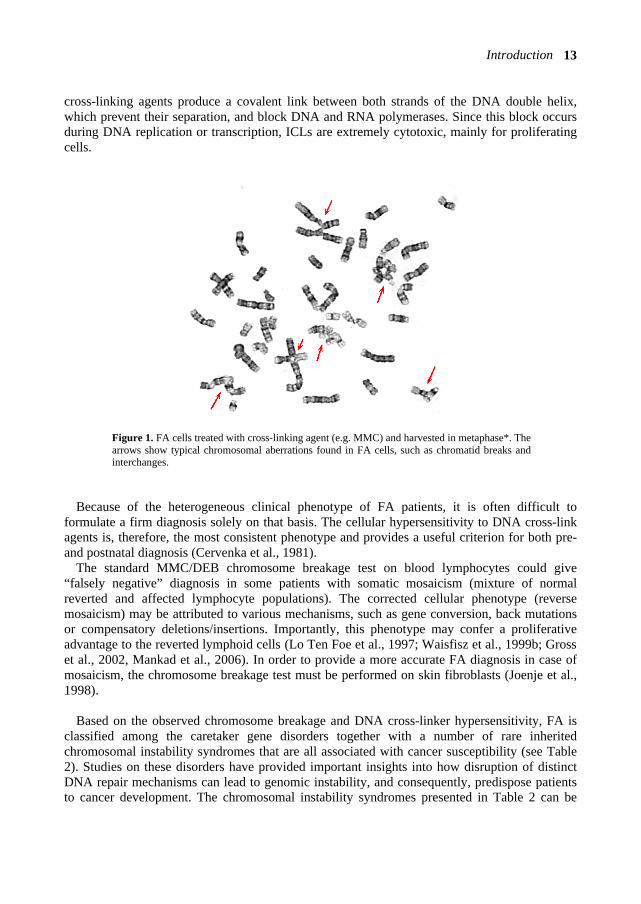
3.1. Genome instability and hypersensitivity to DNA cross-linking agents Cells from FA patients show spontaneous chromosomal instability. Analyzed by classical cytogenetic methods, metaphases from FA lymphocytes exhibit spontaneous chromosomal aberrations, including chromatid breaks and quadriradial chromosomes (Schroeder, et al., 1964). In addition, both primary and immortalized FA lymphocytes are extremely sensitive to antimitotic agents that induce interstrand cross-links (ICLs) in DNA (also called bifunctional alkylating agents), such as mitomycin C (MMC), diepoxybutane (DEB), cisplatin, photoactivated psoralens and derivatives of nitrogen mustard (Sasaki & Tonomura, 1973; Ishida & Buchwald, 1982). In response to these agents, the FA metaphases show an increased number of chromosome damages compared to metaphases of normal lymphocytes (Figure 1). The
Introduction 13
cross-linking agents produce a covalent link between both strands of the DNA double helix, which prevent their separation, and block DNA and RNA polymerases. Since this block occurs during DNA replication or transcription, ICLs are extremely cytotoxic, mainly for proliferating cells.
Figure 1. FA cells treated with cross-linking agent (e.g. MMC) and harvested in metaphase*. The arrows show typical chromosomal aberrations found in FA cells, such as chromatid breaks and interchanges.
Because of the heterogeneous clinical phenotype of FA patients, it is often difficult to formulate a firm diagnosis solely on that basis. The cellular hypersensitivity to DNA cross-link agents is, therefore, the most consistent phenotype and provides a useful criterion for both pre- and postnatal diagnosis (Cervenka et al., 1981).
The standard MMC/DEB chromosome breakage test on blood lymphocytes could give “falsely negative” diagnosis in some patients with somatic mosaicism (mixture of normal reverted and affected lymphocyte populations). The corrected cellular phenotype (reverse mosaicism) may be attributed to various mechanisms, such as gene conversion, back mutations or compensatory deletions/insertions. Importantly, this phenotype may confer a proliferative advantage to the reverted lymphoid cells (Lo Ten Foe et al., 1997; Waisfisz et al., 1999b; Gross et al., 2002, Mankad et al., 2006). In order to provide a more accurate FA diagnosis in case of mosaicism, the chromosome breakage test must be performed on skin fibroblasts (Joenje et al., 1998).
Based on the observed chromosome breakage and DNA cross-linker hypersensitivity, FA is classified among the caretaker gene disorders together with a number of rare inherited chromosomal instability syndromes that are all associated with cancer susceptibility (see Table 2). Studies on these disorders have provided important insights into how disruption of distinct DNA repair mechanisms can lead to genomic instability, and consequently, predispose patients to cancer development. The chromosomal instability syndromes presented in Table 2 can be
14 Chapter 1
inherited in either a dominant or a recessive mode. In the dominant mode of inheritance, the affected individual inherits only one mutant allele of a given gene (e.g. BRCA1/2 genes for the hereditary breast cancer and DNA mismatch repair genes for the hereditary non-polyposis colorectal cancer). In that case, the patient is heterozygous and the carcinogenesis process might be initiated by somatic inactivation of the other allele or by the haplotype insufficiency due to the presence of one mutant allele (Buchholz et al., 2002). In the recessive mode, each parent passes on one copy of an abnormal gene. This implies that both alleles of the affected individual have inherited mutations in a particular DNA repair gene (biallelic mutations).
Cancer development is a consequence of the alterations of three types of genes: oncogenes, tumor suppressor genes and DNA repair genes (Vogelstein & Kinzler, 2004). Oncogenes contribute to cancer by a single mutational activation, whereas the tumor suppressor genes and DNA repair genes contribute to the cancer process when they are inactivated. Genes involved in DNA repair prevent carcinogenesis by protecting the genome against alterations that activate oncogenes or inactivate tumor suppressor genes. Therefore, germline mutations in both alleles of DNA repair genes lead to genomic instability and are associated with a number of cancer-prone syndromes. For instance, Xeroderma pigmentosum (XP) patients have a defect in excision and repair of UV photoproducts in DNA and have a dramatically increased risk to develop skin cancer. The molecular DNA repair process affected in these genetic instability (or chromosomal fragility) syndromes are shown in Table 2.
The exact DNA repair pathway that is defective in FA patients is still unknown. In fact, the molecular genetics of FA proteins seems to define a previously unknown multi-protein complex that is involved in the response to DNA cross-linking agents. Although the exact role of the FA proteins in ICL repair is still not completely resolved, recent findings have provided new insights into the role of FA in DNA repair during replication. For example, the finding that BRCA2 protein corresponds to FANCD1, and that FANCJ and FANCM contain enzymatic domains that are involved in DNA processing, directly implicates FA in DNA repair through homologous recombination (HR) and translesion synthesis (TLS) (see, Tables 2 and 4) (Howlett et al., 2002; Levitus et al., 2005; Levran et al., 2005; Meetei et al., 2005). The role of these mechanisms in the FA pathway will be discussed in Chapter 6.
Most of the proteins involved in cellular DNA damage response pathways converge in a
common “tumor suppression network of interactions” that contribute to genome stability of the human cells (Surrallés et al., 2004). FA proteins interact (functionally and/or physically) with several other DNA repair proteins or pathways that are involved in genetic instability syndromes or DNA damage responses (Rosselli et al., 2003; Macé et al., 2004). These proteins include ATM, ATR, BLM, BRCA1, BRCA2, Rad50/MRE11/NBS1 complex, RAD51, RPA and XPF (Garcia-Higuera, et al., 2001; Digweed et al., 2002; Godthelp et al., 2002, 2005; Nakanishi et al., 2002; Pichierri et al., 2002, 2004; Tanuguchi et al., 2002a, 2002b; Meetei et al., 2003a; Sridharan et al., 2003; Wong et al., 2003; Andreassen et al., 2004; Wang et al., 2004; Howlett et al., 2005). The molecular cross-talk between the FA proteins and the other DNA repair proteins suggests that the FA proteins play a central role in the interactions within this network. Indeed, the phenotypic overlap between some FA, NBS, and BLM patients may indicate a tight molecular cross-talk between the proteins involved in these syndromes.
Introduction 15
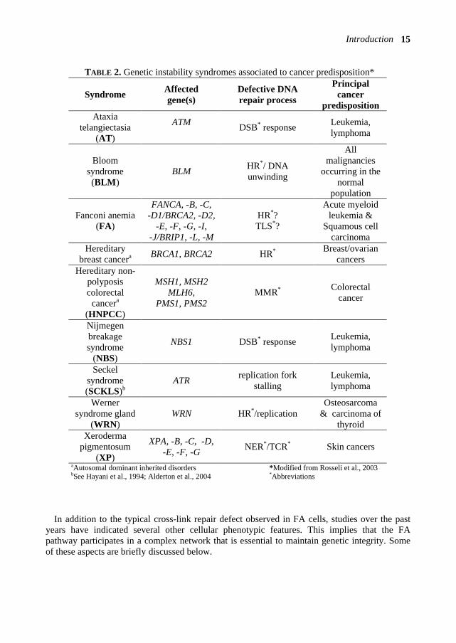
TABLE 2. Genetic instability syndromes associated to cancer predisposition*
aAutosomal dominant inherited disorders *Modified from Rosseli et al., 2003 bSee Hayani et al., 1994; Alderton et al., 2004 *Abbreviations
In addition to the typical cross-link repair defect observed in FA cells, studies over the past
years have indicated several other cellular phenotypic features. This implies that the FA pathway participates in a complex network that is essential to maintain genetic integrity. Some of these aspects are briefly discussed below.
Syndrome Affected gene(s)
Defective DNA repair process
Principal cancer
predisposition Ataxia
telangiectasia (AT)
ATM DSB* response Leukemia,
lymphoma
Bloom syndrome (BLM)
BLM HR*/ DNA unwinding
All malignancies
occurring in the normal
population
Fanconi anemia (FA)
FANCA, -B, -C, -D1/BRCA2, -D2,
-E, -F, -G, -I, -J/BRIP1, -L, -M
HR*? TLS*?
Acute myeloid leukemia &
Squamous cell carcinoma
Hereditary breast cancera BRCA1, BRCA2 HR* Breast/ovarian
cancers Hereditary non-
polyposis colorectal
cancera (HNPCC)
MSH1, MSH2 MLH6,
PMS1, PMS2 MMR* Colorectal
cancer
Nijmegen breakage syndrome
(NBS)
NBS1 DSB* response Leukemia, lymphoma
Seckel syndrome (SCKLS)b
ATR replication fork stalling
Leukemia, lymphoma
Werner syndrome gland
(WRN) WRN HR*/replication
Osteosarcoma & carcinoma of
thyroid Xeroderma
pigmentosum (XP)
XPA, -B, -C, -D, -E, -F, -G NER*/TCR* Skin cancers
16 Chapter 1
3.2. Cell cycle abnormalities Another important cellular characteristic in FA cells is the impairment of the cell cycle. This impairment occurs either spontaneously or after introduction of ICL. FA cells spontaneously arrest in late S or delay at G2/M transition of the cell cycle compared to wild-type cells (Dutrillaux et al., 1982), a phenomenon that is further exaggerated when cells are treated with DNA cross-linking agents (Heinrich et al., 1998). In normal cells, ICL treatment during the G2 phase of the cell cycle does not induce a G1 or G2/M arrest, but requires DNA replication to provoke growth arrest (Akkari et al., 2000). Similar to wild-type cells, the ICL-induced chromosome breakage seen in FA cells occurs only after DNA replication and only if cells fail to arrest (Centurion et al., 2000; Sala-Trepat et al., 2000; Akkari et al., 2001). Importantly, FA cells are not completely inefficient in repairing cross-linked DNA but are ~3 times slower than wild-type cells, indicating that FA has a primary defect in ICL repair during late S phase (Akkari et al., 2001). In other words, the ICLs seem to be tolerated in the double-strand DNA until they are hit by a replication fork.
3.3. Oxygen sensitivity Some studies showed that primary FA skin fibroblast cultures grow better in low oxygen (Schrindler & Hoehn, 1988) and that FA lymphocytes exhibit elevated chromosomal aberrations under higher oxygen concentrations (Joenje et al., 1981). These findings suggest that FA proteins are involved in the defense against oxygen toxicity and in the repair of oxidative damages (for review, see Pagano & Youssoufian, 2003). In line with this, some FA proteins have been related to the regulation of oxygen metabolism. The FANCC protein, for example, interacts both physically and functionally with a number of cytoplasmic proteins known to catalyse the production of reactive oxygen species, such as the nicotin-amide adenine dinucleotide phosphate (NADPH) cytochrome P450 reductase (Kruyt et al., 1998) and the glutathione S-transferase P1 (GSTP1) (Cumming et al., 2001). Similarly, the FANCG protein interacts directly with protein P450 cytochrome 2E1 (CYP2E1) and down-regulates the level of CYP2E1 (Futaki et al., 2002). These interactions suggest that some FA proteins play a role in detoxifying metabolism.
A recent study proposed that oxidative stress facilitates the FANCA-FANCG interaction and also induces multimerization of the FA proteins (FANCA, FANCC and FANCG) through intermolecular disulfide linkage(s) (Park et al., 2004). However, further studies are required to establish whether the FANCA-FANCG interaction via disulfide linkage(s) is essential for the normal function of FA proteins in response to DNA damage.
Since the DNA cross-link agents (e.g. MMC and DEB) produce reactive oxygen species, it has been proposed that the sensitivity of FA cells to those agents may be due to the incapacity to neutralize reactive oxygen species (Korkina et al., 2000). However, FA fibroblasts transformed with SV40 large T-antigen lack hypersensitivity to oxygen but maintain hypersensitivity to MMC (Saito et al., 1993), which suggests that the oxygen sensitive phenotype is a secondary defect in FA cells.

Introduction 17
3.4. Apoptosis and telomere maintenance The regulation of apoptotic mechanisms (programmed cell death) seems to be disrupted in FA cells. For example, the level of apoptotic cells that occur spontaneously or induced by MMC is significantly increased in FA cells (Kruyt et al., 1996; Ridet et al., 1997). The high level of apoptosis in bone marrow stem cells and during embryogenesis may account for the depletion of hematopoietic reserves and developmental abnormalities commonly seen in FA patients (for review, see Rosselli, 1998).
One of the FA proteins, FANCC, seems to be involved in the regulation of apoptosis in hematopoietic cells, since it protects the progenitor cells from the fas-mediated apoptotic pathway (Wang et al., 1998). FANCC also prevents apoptosis by augmenting the activity of the GSTP1 protein (Cumming et al., 2001). In addition, the GSTM1 genotype significantly influences the progression of bone marrow failure in FA complementation group C patients (median age 3 years vs 7 years) by increasing susceptibility to cytokine-induced apoptosis (Davies et al., 2005).
Telomeres protect the ends of the chromosomes by preventing fusion (Blasco et al., 1999). Therefore, telomeres have an important role in genomic stability, and more precisely, in adequate functioning of the hematopoietic system (Herrera et al., 1999). A study by Callén and colleagues (2002) on FA cells showed a higher rate of breakage at telomeric sequences and in chromosome end fusion (>10-fold), which indicates a defect in telomere maintenance (see also Li et al., 2003). This effect also occurs in similar hematopoietic syndromes, such as in acquired aplastic anemia, in MDS and in AT (a syndrome caused by mutations in the ATM gene, see Table 2) (Metcalfe et al., 1996; Boultwood et al., 1997; Brummendorf et al., 2001). A defect in telomere maintenance may therefore contribute to the genomic instability of FA cells.
4. FA GENES AND PROTEINS 4.1. Genetic heterogeneity The genetic heterogeneity in FA patients was traditionally assessed by complementation analysis through somatic cell fusion experiments. These experiments use the characteristic sensitivity of FA cells to DNA cross-linking agents such as MMC/DEB (Duckworth-Rysiecki & Taylor, 1985; Buchwald, 1995; Joenje et al., 1995, 1997). Briefly, if the fusion of two immortalized FA lymphoblastoid cell lines results in a MMC-resistant hybrid cell, the two patients have different defective FA genes, and therefore belong to different complementation groups. Otherwise, if the fusion results in a MMC-sensitive hybrid cell, the two patients have the same defective FA gene, and are therefore considered to belong to the same complementation group (Figure 2). This method, however, may result in mis-assignments due to phenotypic reversion or chromosome loss. Therefore, very stringent criteria have to be applied. Joenje and colleagues proposed that before declaring a new FA complementation group, at least two patient cell lines have to be excluded from all known subtypes and show to belong to the same group by cell fusion experiments (Joenje et al., 2000). Gene complementation or identification of pathogenic mutations in the associated FA gene provides definitive proof that a patient cell line belongs to the assigned complementation group.

18 Chapter 1
4.2. FA complementation groups To date, twelve FA complementation groups have been established: FA-A, -B, -C, -D1, -D2, -E, -F, -G, -I, -J, -L and -M, where each subtype represents a distinct FA gene. Complementation group A is the most common FA subtype, accounting for ~65% of the patients. The FA-C and FA-G groups represent less than 10% of the cases and the other subtypes are very rare (Table 3) (Levitus et al., 2004).
TABLE 3. FA complementation groups distribution (in %)*
A 64 F 2
B 2 G 8
C 9 I 2
D1 3 J 3
D2 3 L <1
E 3 M <1 *Results are based on the first 245 (updated to 251) FA families classified by the European Fanconi Anemia Research Program (1994-2003).
Figure 2. FA complementation analysis through somatic cell fusion experiments. The hybrid cell is subjected to a MMC test and the resulting phenotype determines whether the different FA cell lines belong to the same or different complementation group(s).

Introduction 19
There is some correlation between specific complementation groups, type of mutations and clinical features in FA. The FA-C patients with the IVS4+4A→T and L554P mutations are associated with early onset of hematological abnormalities and multiple birth defects, while individuals with the 322delG and Q13X mutations have milder clinical outcome with a reduced incidence of somatic abnormalities (Gillio et al., 1997; Faivre et al., 2000). Nevertheless, additional factors appear to influence the clinical phenotype, since the FANCC IVS4+4A→T mutation results in milder phenotype in Japanese patients (Futaki et al., 2000). In a genotype-phenotype study of 245 FA patients, Faivre and colleagues reported that the rare FA subtypes D, E and F have a higher incidence of somatic abnormalities (Faivre et al., 2000). In general, the patients from complementation group D1 have a more severe clinical phenotype compared to the other subtypes. The FA-D1 patients, who carry biallelic mutations in the BRCA2 gene, are associated with earlier onset of acute myeloid leukemias and solid tumors, such as Wilm’s tumors and medulloblastomas (Offit et al., 2003; Hirsch et al., 2004; Wagner et al., 2004). Patients from complementation group E exhibit malformations of the central nervous system (Faivre et al., 2000). These malformations appear to be represented mostly in FA-E patients (60% of cases), compared to the other FA patients (<4%). The patients with FANCG mutations and FANCA null mutations seem to represent a high-risk group with a poor hematologic outcome. In support of this, FA-A cells with FANCA null mutations have a MMC sensitivity severely impaired while FANCA mutations producing abnormal FANCA proteins complement the MMC sensitivity to different extends (Adachi et al., 2002). Therefore, the complete loss of FANCA function versus partial FANCA function might reflect the phenotypic variation seen in FA-A patients.
4.3. Gene identification A number of methods were used to identify FA genes, such as expression cloning, positional cloning, direct sequencing of candidate genes and protein complex purification. Currently, eleven of the twelve FA genes have been identified, as the FANCI gene remains to be cloned (Levitus et al., 2004). The principal features of the FA genes and related proteins are summarized in Table 4.
Many of the FA genes were identified using functional complementation of the cross-linker sensitive phenotype of FA cells with wild-type cDNAs. This technique uses a cDNA expression library cloned in an episomal mammalian expression vector (pREP4) that is transfected into a lymphoblastoid cell line derived from an FA patient (Strathdee et al., 1992). The transfected cell line is first selected for vector uptake using hygromycin and then subjected to MMC to select the cells that have corrected the FA phenotype. Thus, the cell line that becomes resistant to DNA cross-linking agents is complemented (corrected) by the cDNA that encodes the protein affected by the defective FA gene. The vector DNA is isolated from the complemented pool of cells and the cDNA insert analyzed. To further confirm that the candidate cDNA is indeed representing an FA gene, mutation screening in the corresponding FA patient cell lines should reveal pathogenic sequence alterations. This cloning strategy has, however, some limitations, since the overexpressed cDNA might be toxic for the cells or the cDNA library might not contain a specific FA cDNA. In addition, false positive cDNA might by found when a cell line becomes MMC resistant due to phenotypic reversion.
20 Chapter 1
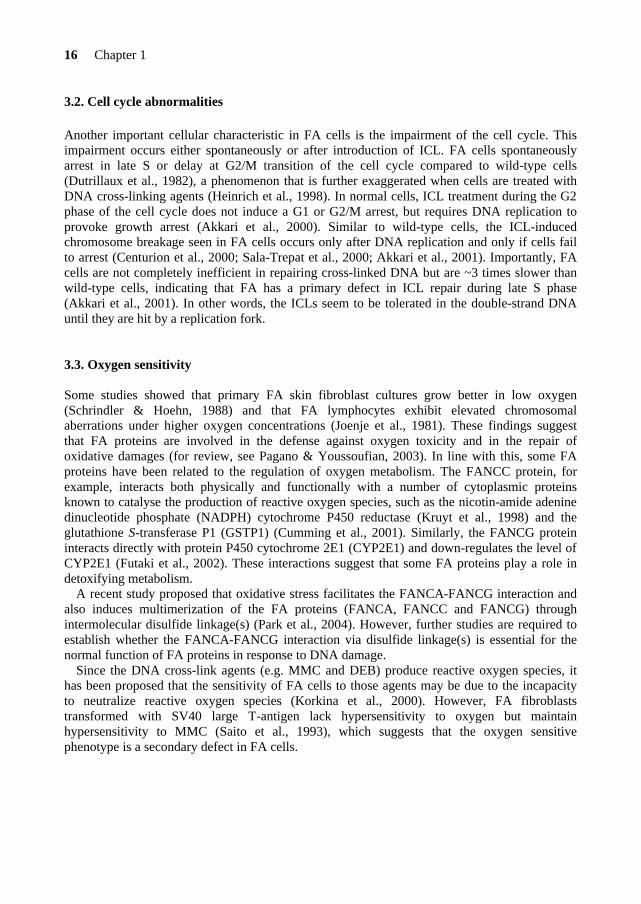
TABLE 4. FA complementation groups and FA genes Subtype FA gene Chromosomal Molecular Cellular Motifs/Domains location weight (kDa) localization or Function A FANCA 16q24.3 163 N/C bipartite NLS, leucine zipper B FANCB Xp22.3 95 N/C NLS C FANCC 9q22.3 63 N/C - D1 FANCD1/ 13q12.3 38 N 8 BRC repeats, BRCA2 RAD51 recruitment D2 FANCD2 3p26 155-162 N K561 monoubiquitinated, S222 phosphorylated E FANCE a 6p21.3 60 N 2 NLS F FANCF 11p15 42 N adaptor protein G FANCG 9p13 68 N/C leucine zipper, 7 TPR motifs I FANCI b ? ? ? ? J FANCJ/ 17q22 150 N 5’→ 3’DNA helicase, BRIP1 BRCA1binding L FANCL 2p16 43 N/C WD40 repeats,
RING finger E3 ubiquitin ligase
M FANCM 14q21.2 250 N DNA translocase, helicase domain adescribed in this thesis, Chapters 2 and 3 N = nuclear; C= cytoplasmic bnot identified NLS = nuclear localization signal
Another important method to identify new FA genes is through protein complex purification. These experiments in HeLa nuclear extract allowed the immunoisolation of a protein complex (BRAFT) that is associated with BLM, the helicase involved in Bloom syndrome (see Table 2). This protein complex contains BLM, replication protein A (RPA), FANCA and Topoisomerase IIIα (Topo IIIα) (Meetei et al., 2003a). The purification of BRAFT with FANCA antibody led to the isolation of five FA core complex proteins (FANCA, C, E, F and G), as well as several unidentified FANCA-associated polypeptides (FAAPs) or BLM-associated polypeptides (BLAPs). These polypeptides are termed FAAP or BLAP followed by a number that corresponds to their molecular weight. Some of these polypeptides, FAAP43, FAAP95 and FAAP250, were later classified as FA proteins by western blot and mutation analyses in FA patients, and represent FANCL, FANCB and FANCM, respectively (Meetei et al., 2003b, 2004, 2005).
Presently, two FA related polypeptides of this immunopurified BRAFT complex (i.e. FAAP75/BLAP75 and FAAP100) remain to be characterized. FAAP75 seems not to be involved in the FA pathway but functions together with Bloom’s helicase to preserve genomic stability (Yin et al., 2005). Furthermore, the FAAP100 protein cannot represent the FANCI gene product since a normal protein level was found in FA-I cells (Medhurst et al., unpublished results).
Introduction 21
4.4. Description of the FA genes and proteins In this section, the main characteristics of the currently known FA genes and proteins will be presented in order of identification. A comprehensive model for the FA protein-protein interactions inside a common pathway will be proposed in Chapter 6 (General Discussion). The hypothetical function of the FA pathway in the DNA cross-link repair process will also be discussed in Chapter 6.
FANCC FANCC is the first identified FA gene and it was isolated by cDNA expression cloning in 1992 (Strathdee et al., 1992). The FANCC cDNA encodes a protein of 558 amino acids with an estimated molecular weight of 63 kDa. The gene maps to chromosome 9q22.3 and consists of 14 exons. Diverse forms of FANCC mRNAs were detected, which indicates that the regulation of this gene might be complex. The role of these FANCC transcripts is still unknown (Gibson et al., 1993; Savoia et al, 1995).
The first studies on the intracellular localization of FANCC revealed that FANCC is localized to the cytoplasm (Yamashita et al., 1994; Youssoufian, 1996). Later, subcellular fractionation experiments and confocal microscopy analyses showed that FANCC is present in both compartments of the cells, with a weak trace of FANCC (about 10%) in the nucleus (Hoatlin et al., 1998). The expression of FANCC is regulated during cell cycle progression with the lowest levels at the G1/S boundary and maximal levels at the G2/M transition (Kupfer et al., 1997a; Heinrich et al., 2000). This cell cycle regulation appears to be subject of posttranslational mechanisms that use proteasome-dependent proteolysis (Heinrich et al., 2000).
The FANCC protein interacts with and regulates the function of various non-FA proteins, in the cytoplasm as well as in the nucleus of the cell. The cytosolic partners of FANCC include the mitotic cyclin-dependent kinase cdc2 (regulator of the transition from G2 to M phase), the stress response proteins and molecular chaperones GRP94 and Hsp70, proteins involved in oxygen radical metabolism (i.e. the NADPH cytochrome P450 reductase and GSTP1), and the signal transducer and activator of transcription, STAT1 (Kupfer et al., 1997a, Kruyt et al., 1998; Hoshino et al., 1998; Pang et al., 2000, 2002; Cumming et al., 2001). In the nucleus, FANCC interacts with a member of the BTB/POZ family of transcriptional repressor proteins, FAZF (Hoatlin et al., 1999).
The interaction of FANCC with non-FA proteins suggests that FANCC, in addition to its role in cross-link repair (as discussed earlier), is involved in cell cycle control, protein transport, anti-apoptotic pathway, detoxification mechanism and signal transduction (Bogliolo et al., 2002). Consistent with this multifunctional role, structurally separate functional domains implicated in the nuclear damage process and cytoplasmic actions of the cell were found in the FANCC protein (Pang et al., 2001). FANCA The FANCA gene, which is defective in most FA patients (Table 3), was mapped to chromosome band 16q24.3 (Pronk et al., 1995) and identified simultaneously by expression
22 Chapter 1
cloning and positional cloning strategies in 1996 (FA/Breast Cancer Consortium, 1996; Lo Ten Foe et al., 1996). FANCA contains 43 exons resulting in a mRNA transcript of 5.5-kb that encodes a protein of 1455 amino acids with a predicted molecular weight of ~163 kD. The primary amino acid sequence of FANCA reveals two overlapping bipartite nuclear localization signals (NLS) at its N-terminus (a.a 18-34 and 19-35) and a leucine-zipper motif between residues 1069 and 1090.
FANCA is present in both the cytoplasm and nucleus of the cell, but is predominantly localized in the nuclear compartment (Kupfer et al., 1997b). The nuclear accumulation of FANCA requires the bipartite NLS motifs, as well as the C-terminal region (Näf et al., 1998; Lightfoot et al., 1999; Kupfer et al., 1999). Moreover, the nuclear accumulation and function of FANCA seem to depend on its phosphorylation by an unknown cytoplasmic serine kinase (Yamashita et al., 1998; Yagasaki et al., 2001; Adachi et al., 2002). It is hypothesized that this phosphorylation event is stimulated by the FA core complex proteins (i.e. FANCB, FANCE, FANCF and FANCG), since it is lacking in FA cells of complementation groups B, E, F and G, but present in FA-D1 and -D2 cell lines (Yamashita et al., 1998; Kupfer et al., 1999). The nuclear accumulation of FANCA also depends on FANCB, -L and -M (de Winter et al., 2000b; Meetei et al., 2003b, 2005). Furthermore, FANCA contains nuclear export signals that seem to play a role in the nuclear accumulation of FANCA (Ferrer et al., 2005).
FANCG/XRCC9 In 1998, de Winter and colleagues identified the FA complementation group G gene. The FANCG gene appeared to be identical to the human XRCC9 gene, isolated on the basis of its capacity to complement the Chinese hamster ovary (CHO) mutant UV40 (Liu et al., 1997). This gene is located at chromosome band 9p13 and contains 14 exons that encode a protein of 622 amino acids (molecular weight ~70 kDa).
The FANCG protein localizes to the cytoplasm and nucleus of the cell and interacts directly with FANCA (Waisfisz et al., 1999a). Studies on the structure/function of FANCA and FANCG revealed that amino acids 1-428 of FANCG bind to the N-terminal NLS of the FANCA protein (Näf et al., 1998; Kuang et al., 2000). Additional functional analyses of patient-derived FANCG mutations also showed that FANCG has a C-terminal functional domain, which appears to be required for normal assembly of the FANCA/FANCC/FANCG protein complex (Kuang et al., 2000; Nakanishi et al., 2001).
Comparative amino acid sequence analysis of the zebrafish Fancg ortholog and human FANCG reveals seven tetratricopeptide repeat motifs (TPRs) in FANCG (Blom et al., 2002, 2004). TPRs are degenerate 34 amino acid repeat motifs that form a scaffold structure for protein-protein interactions, which suggests that FANCG is essential in the assembly and/or stability of the FA core complex. Four of the TPRs in FANCG are required for binding FANCA and are of functional importance.
FANCG is subjected to posttranslational modification, such as phosphorylation (Futaki et al., 2001). The phosphorylation sites in FANCG were mapped to serines 7, 383 and 387 (Qiao et al., 2004; Mi et al., 2004). During the cell cycle (at mitosis), the FA proteins detach from chromatin and FANCG becomes phosphorylated at serines 383 and 387 (Qiao et al., 2001, 2004). The G2/M kinase cdc2, which binds to FANCC, appears to phosphorylate FANCG-

Introduction 23
Ser387 (Kupfer et al., 1997a; Mi et al., 2004). However, the function of these phosphorylation steps and the overall consequences on the FA pathway remain to be clarified.
FANCF FANCF, the gene mutated in complementation group F, was identified in 2000 by expression cloning. This gene consists of one single exon and its cDNA of ~1.3 kb is translated into a protein of 374 amino acids (molecular weight of 42 kDa) (de Winter et al., 2000a). FANCF maps to chromosome 11p15 (de Winter et al., 2000a) in a hot-spot area for hypermethylation, indicating that the mRNA levels of FANCF partly depend on methylation (Taniguchi et al., 2003).
To date, 5 FA-F patients have been identified worldwide and the mutational analysis*2 of those patients did not reveal any missense mutation that could point out functional domains in FANCF (Table 5). The FANCF protein is predominantly localized in the nucleus of the cell, where it is able to form a complex with FANCA, FANCC and FANCG (de Winter et al., 2000b). The C-terminal region of FANCF is directly involved in the interaction with FANCG (Medhurst et al., 2001; Gordon & Buchwald, 2003). Furthermore, FANCF is present in FA cells derived from complementation groups A, B, C, D1, D2, E and G, and appears to stabilize the FANCA, FANCC, FANCG proteins in the complex (Siddique et al., 2001). In Chapter 5, I will present a site-directed mutagenesis study on the FANCF protein. This study reveals important findings on functional domains and residues in FANCF and provides new insights into the role of FANCF in the FA pathway. FANCE FANCE is the fifth and until now, last FA gene that has been cloned by the expression cloning method (Chapter 2). The FANCE protein directly binds both FANCC and FANCD2 (Medhurst et al., 2001; Pace et al., 2002). Moreover, FANCE is required for the nuclear accumulation of FANCC and links the FA core complex to the downstream FANCD2 protein (see below)
TABLE 5. Individuals with FANCF mutations* Patient Mutation Consequence for protein BD497 484-485dela deletion EUFA121 349-395del deletion
16C→T Gln6STOP EUFA698 230-252dela deletion EUFA927 327C→Ga Tyr109STOP EUFA1228 887-894dela deletion aHomozygous mutation

24 Chapter 1
(Taniguchi & D’Andrea, 2002; Pace et al., 2002). Further studies on the FANCE protein are presented in Chapter 4. FANCD2 A gene defective in FA-D cells was localized to chromosome 3 at the locus 3p22-26, using microcell-mediated chromosome transfer and positional cloning approaches (Whitney et al., 1995). The identification of the FA gene at this locus showed that the FA-D group is heterogenous and represents 2 FA genes: FANCD1 and FANCD2 (Timmers et al., 2001), the latter being the gene at 3p22-26.
FANCD2 contains 44 exons and encodes a 1451 amino acids nuclear protein. Normal human cells express the FANCD2 protein in two isoforms: FANCD2-S (short) of 155 kDa and FANCD2-L (long) of 162 kDa. The long form is generated by the attachment of a single ubiquitin molecule to lysine residue 561 of the FANCD2 protein (Timmers et al., 2001). This monoubiquitination reaction is an essential step in the FA pathway and requires most of the FA core complex components (FANCA, -B, -C, -E, -F, -G, -L and -M). In this complex, FANCL seems to be the catalytic subunit of the monoubiquitination reaction (Meetei et al., 2004b). USP1 is the deubiquitinating enzyme that removes the ubiquitin moiety (Nijman et al., 2005). Monoubiquitinated FANCD2 assembles in nuclear foci that exist normally in the S phase of the cell cycle and after cellular exposure to DNA damaging agents that block DNA replication, such as MMC, ionizing radiation (IR) and deoxynucleotide depletion by hydroxyurea (HU) (Garcia-Higuera et al., 2001; Taniguchi et al., 2002b). This FANCD2 modification allows its re-localization from a soluble nuclear compartment to the chromatin fraction and nuclear matrix (Meetei et al., 2004b), where it co-localizes in nuclear foci with numerous proteins involved in DNA repair and cell cycle checkpoint regulation, such as ATM, ATR, BLM, breast cancer proteins (BRCA1 and BRCA2), PCNA, RAD51 and the MRE11/RAD50/NBS1 (MRN) complex (Garcia-Higuera et al., 2001; Nakanishi et al., 2002; Pichierri et al., 2002, 2004; Taniguchi et al., 2002a, 2002b; Meetei et al., 2003a; Andreassen et al., 2004; Hussain et al., 2004; Wang et al., 2004).
FANCD2 appears to be involved in another pathway as well. In normal cells, when exposed to ionizing radiation, the ataxia telangiectasia kinase (ATM) phosphorylates FANCD2 on serine 222 with the cooperation of the MRN complex (Nakanishi et al., 2002), which results in the activation of an S phase checkpoint response (Taniguchi et al., 2002b). In addition, ATR, an ATM- and RAD3-related protein kinase responsible for Seckel syndrome (see Table 2), also mediates the FANCD2 phosphorylation (Pichierri & Rosselli, 2004) and seems also involved in FANCD2 monoubiquitination (Andreassen et al., 2004). FANCD1/BRCA2 The gene defective in FA complementation group D1 patients was found on the basis of a candidate gene approach. The BRCA1 and BRCA2 (breast cancer susceptibility gene 1 and 2) genes were good FA candidate genes since BRCA1 and BRCA2 deficient cell lines are hypersensitive to the interstrand cross-linking agent MMC (Patel et al., 1998; Moynahan et al., 2001). Mutation screening of both genes was performed in cell lines derived from FA-B and
Introduction 25
FA-D1 patients and biallelic BRCA2 mutations were detected in both cell lines (Howlett et al., 2002). Functional complementation of the FA-D1 fibroblasts with wild-type BRCA2 cDNA restored the MMC hypersensitive phenotype, indicating that BRCA2 corresponds to FANCD1.
Importantly, the FA-D1 cells express the monoubiquitinated form of FANCD2, suggesting that FANCD1/BRCA2 functions downstream of the FA core complex proteins (Garcia-Higuera et al., 2001). Recently, the FANCD2-L protein has been demonstrated to promote chromatin loading of FANCD1/BRCA2 (Wang et al., 2004). FANCD1/BRCA2 is a large nuclear protein of 3,418 amino acids that contains eight BRC repeat motifs, of which six bind the RAD51 protein, and three oligonucleotide-binding domains that interact with single-stranded DNA (ssDNA) (Wong et al., 1997). The C-terminal portion of the FANCD1/BRCA2 protein is also involved in the binding of RAD51 and includes a nuclear localization signal. The RAD51 protein functions as a helical polymer, composed of hundreds of monomers, that wraps around ssDNA and forms a nucleoprotein filament involved in homologous recombination (West, 2003). In addition, FANCD1/BRCA2 drives RAD51 to sites of DNA damage (Davies et al., 2001) and is required for stabilization of stalled DNA replication forks (Lomonosov et al., 2003).
Similar to FANCD2, FANCD1/BRCA2 is an ATM substrate and phosphorylation appears to activate the IR-inducible S phase checkpoint response (Wang et al., 2004). FANCL FANCL is the first FA gene that has been identified by biochemical approaches and the FANCL protein is the first FA protein with an enzymatic activity. Several FANCA associated proteins (FAAP43, FAAP95, FAAP100 and FAAP250) were discovered by immunoprecipitation with an FANCA specific antibody (Meetei et al., 2003a). FA complementation group L was identified by western blot analysis revealing the absence of FAAP43 in a single FA patient. This protein corresponds to PHF9 (PHD finger protein 9), a protein with three WD40 repeats and a RING-type zinc-finger motif that has E3-ubiquitin ligase activity (Meetei et al., 2003b).
The FANCL protein is present in both nuclear and cytoplasmic compartments of the cell (Meetei et al., 2003b) and appears to play an important role in a catalytic reaction within the FA pathway. A recent study by Meetei and colleagues (2004) showed that FANCL is the ubiquitin ligase responsible for the FANCD2 monoubiquitination (FANCD2-L), but not BRCA1, as previously thought. Furthermore, they found that FANCL is necessary, but not sufficient for the monoubiquitination of FANCD2. Consequently, all the components of the FA core complex seem to act as a “regulated ubiquitin ligase” in which FANCL represents the catalytic subunit. In line with this, the WD40 repeat regions in FANCL appear to be required for its interaction with the FA core complex and the RING motif, for the recruitment of a putative E2 ubiquitin-conjugating enzyme for monoubiquitination of FANCD2 (Gurtan et al., 2006). FANCB Similar to FANCL, the FANCB protein is one of the components of the purified FANCA complex (FAAP95). This polypeptide is mutated in FA patients belonging to complementation group B. The FANCB protein contains a bipartite nuclear localization signal at its C-terminus
26 Chapter 1
(Meetei et al., 2004). Consistent with this finding, FANCB is predominantly detected in the nuclear extract of HeLa cells.
The FANCB gene is located on the X chromosome at Xp22.31. In male individuals only one X chromosome is present and in females, the X chromosome is subject to X-inactivation. Thus, the FANCB gene is the only FA gene present in a single active copy in the human genome. Therefore, compared to the other autosomal FA genes, FANCB can be inactivated by a single mutation event, which could lead to genomic instability and cellular transformation. Since FANCB has a higher chance to be inactivated, it might be implicated in sporadic cancer in the general population (see below) and FA families belonging to complementation group B would necessitate clinical management (Rahman & Ashworth, 2004). FANCM Two separate research groups have found that the human ortholog of the archaebacterial protein HEF (Helicase-associated Endonuclease for Fork structured DNA) is a component of the FA core complex. The FANCA-associated polypeptide of 250 kDa (FAAP250) turned out to be closely related to Hef (Meetei et al., 2005). Immunoblotting of FAAP250 on several FA patient-derived cell lines, revealed that the protein was present in cells of patients from the eleven known FA subtypes, but absent in cells from one FA patient who was excluded from many complementation groups (Meetei et al., 2005). Subsequent genotyping of this FA individual’s cDNA confirmed that FAAP250 is the gene defective in this patient, which established a new FA complementation group, FA-M (Meetei et al., 2005). A second research group found that the vertebrate Hef ortholog is an important component of the FA pathway by using knockout approaches in chicken DT40 cells (Mosedale et al., 2005).
Like the other FA core complex proteins, FANCM is essential for the monoubiquitination of FANCD2. Furthermore, FANCM is a phosphoprotein that becomes hyperphosphorylated in response to DNA damaging agents. This posttranslational modification seems to occur independently of the other components of the FA core complex and may serve as a signal that regulates the ubiquitin ligase activity of the FA core complex (Meetei et al., 2005).
The discovery of FANCM (and FANCJ, see below) provided a breakthrough in the understanding of the FA pathway and strongly linked FA to the DNA repair process. FANCM possesses two potential DNA-metabolizing domains: a DEAH-box helicase domain and an endonuclease domain homologous to ERCC4/XPF (an endonuclease essential for nucleotide excision repair, see Table 2). The helicase domain of FANCM has an ATPase activity, which is stimulated by single- and double-stranded DNA, whereas its endonuclease domain seems to be inactive. FANCM is able to dissociate triplex DNA, possibly reflecting its ability to translocate on duplex DNA (Meetei et al., 2005).
FANCJ/BACH1/BRIP1 Using homozygosity mapping and candidate gene screening approaches, recent studies revealed that BACH1 (BRCA1 Associated C-terminal Helicase) or BRIP1 (BRCA1-Interacting Protein C-terminal helicase) is the gene defective in the FA complementation group J (Levitus et al., 2005; Levran et al., 2005; Litman et al., 2005). Moreover, the link between BRIP1 and the FA

Introduction 27
pathway has been established by knockout experiments in chicken DT40 cells (Bridge et al., 2005).
FANCJ/BRIP1 is a member of the RecQ DEAH-box helicase family, which efficiently unwinds non-Watson-Crick DNA structures, such as the Holliday junctions that result from DNA strand exchange during homologous recombination or repair of stalled replication forks (Risinger & Groden, 2004). Several RecQ helicases have been coupled to genomic instability disorders that predispose patients to tumor development, e.g. BLM, RECQL4 and WRN, which are responsible for Bloom syndrome, Rothmund-Thomson syndrome and Werner syndrome, respectively (Risinger & Groden, 2004).
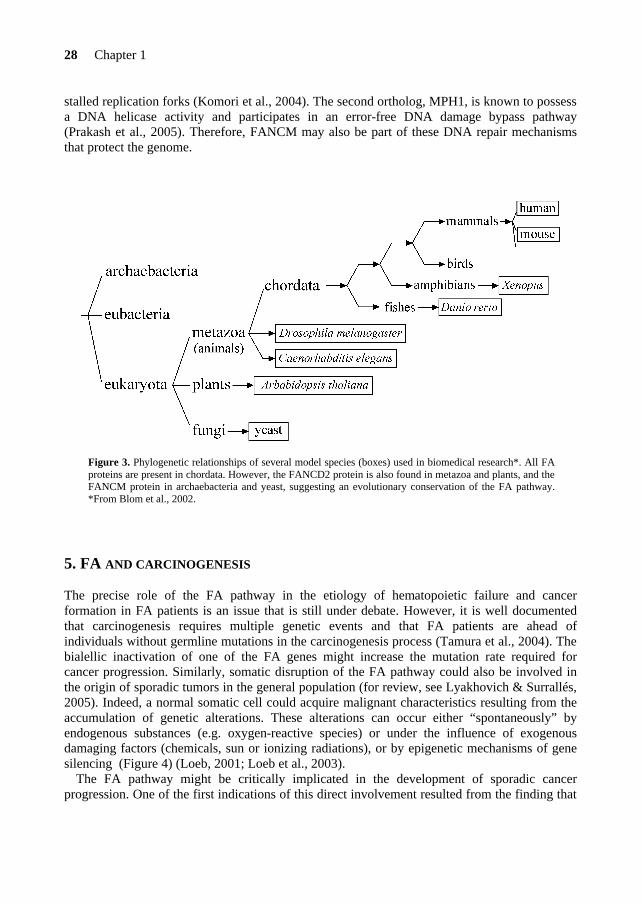
The FANCJ helicase is phosphorylated at serine 990 and binds the BRCT (BRCA1 C- terminal) domains of the BRCA1 protein during S phase of the cell cycle (Yu et al., 2003). This association seems to contribute to DSB repair via HR (Cantor et al., 2001). However, Bridge and colleagues (2005) showed that FANCJ functions independently of BRCA1 in the FA pathway. The type of structures that FANCJ preferentially binds to and unwinds is the forked duplexes (Gupta et al., 2005). In addition, FANCJ possesses an ATP-dependent DNA helicase that translocates in a 5’→ 3’ direction and therefore, may directly repair damaged DNA (Cantor et al., 2004). Since normal monoubiquitination of FANCD2 was reported in FA-J cells, FANCJ is positioned downstream of this event in the FA pathway. Nevertheless, how FANCJ is connected to the FA pathway remains to be determined. 4.5. Evolution of the FA genes An important limitation in understanding the molecular basis of FA is the lack of conservation of the FA proteins in lower eukaryotes (non-vertebrates) that could provide genetic models. The FANCA, FANCB, FANCC, FANCE, FANCF and FANCG genes are only conserved in vertebrates (chordata) implying that they have originated later during evolution (Figure 3). In addition, these FA genes encode novel proteins that lack sequence homologies or domains with known function. Therefore, orthologs of the human FA proteins with a high evolutionary distance have been looked for in order to find conserved residues/domains that highlight their biological importance (Blom et al., 2002, 2004). Orthologs are genes in different species that have originated from a common ancestral gene early in evolution and are thereby likely to perform a similar function. In fact, most of the FA proteins have been found in lower vertebrates, such as in zebrafish (Danio rerio) (Titus et al., 2006) and amphibia (Xenopus laevis) (Chapter 5; Sobeck et al., 2006). Moreover, FANCG orthologs have been used for the identification of multiple TPR (tetratricopeptide repeat) motifs essential for FANCG (Blom et al., 2002, 2004).
The identification of more FA genes has, however, modified the notion that the FA pathway evolved late in evolution. Orthologs of some FA genes have been found in lower eukaryotes and even in archaebacteria, suggesting a possible conservation of the FA pathway throughout evolution (see Figure 3). These include the FANCD2 gene, which has orthologs in the fruit fly Drosophila melanogaster, in the nematode Caenorhabditis elegans and even in the plant Arabidopsis thaliana, but is absent in yeast (Timmers et al., 2001; Castillo et al., 2003; Dequen et al., 2005). Another example is FANCM (Meetei et al., 2005), which has orthologs in archaeabacteria and in yeast, Hef and MPH1, respectively. The first FANCM ortholog, Hef, is a DNA repair protein that contains both helicase and endonuclease activities and is able to restore
28 Chapter 1
stalled replication forks (Komori et al., 2004). The second ortholog, MPH1, is known to possess a DNA helicase activity and participates in an error-free DNA damage bypass pathway (Prakash et al., 2005). Therefore, FANCM may also be part of these DNA repair mechanisms that protect the genome.
Figure 3. Phylogenetic relationships of several model species (boxes) used in biomedical research*. All FA proteins are present in chordata. However, the FANCD2 protein is also found in metazoa and plants, and the FANCM protein in archaebacteria and yeast, suggesting an evolutionary conservation of the FA pathway. *From Blom et al., 2002.
5. FA AND CARCINOGENESIS
The precise role of the FA pathway in the etiology of hematopoietic failure and cancer formation in FA patients is an issue that is still under debate. However, it is well documented that carcinogenesis requires multiple genetic events and that FA patients are ahead of individuals without germline mutations in the carcinogenesis process (Tamura et al., 2004). The bialellic inactivation of one of the FA genes might increase the mutation rate required for cancer progression. Similarly, somatic disruption of the FA pathway could also be involved in the origin of sporadic tumors in the general population (for review, see Lyakhovich & Surrallés, 2005). Indeed, a normal somatic cell could acquire malignant characteristics resulting from the accumulation of genetic alterations. These alterations can occur either “spontaneously” by endogenous substances (e.g. oxygen-reactive species) or under the influence of exogenous damaging factors (chemicals, sun or ionizing radiations), or by epigenetic mechanisms of gene silencing (Figure 4) (Loeb, 2001; Loeb et al., 2003).
The FA pathway might be critically implicated in the development of sporadic cancer progression. One of the first indications of this direct involvement resulted from the finding that

Introduction 29
FANCF is inactivated by promoter hypermethylation in a subset of ovarian epithelial cancers (Taniguchi et al., 2003; Wang et al., 2006). FANCF gene silencing and disruption of the FA pathway may contribute to chromosome instability and selective cisplatin sensitivity observed in these tumors. Epigenetic silencing of FANCF by aberrant methylation of multiple CpG islands in the FANCF promoter might be an important mechanism in sporadic malignancies, as it was found in several other types of cancer, such as non-small-cell lung cancer (NSCLC), head and neck squamous carcinomas (HNSCC), cervical carcinomas and also in a case of sporadic AML (Tischkowitz et al., 2003; Marsit et al., 2004; Narayan et al., 2004).
Figure 4. Causes, cellular responses and consequences of DNA damage. Slightly modified from van Gent et al., 2001.
Another significant finding that implicates the FA pathway in cancer development, is the increased risk of Fancd2-deficient mice to develop epithelial cancers, mainly breast, ovarian, and liver cancers (Houghtaling et al., 2003).
The high incidence of AML in FA patients (15 000-fold) suggests that FA genes may play a specific role in non-FA individuals with AML. Acquired rather than inherited disruption of the FA pathway, by deletion, reduction of expression or point mutation in a FA gene may represent an early event in the development of sporadic AML (Xie et al., 2000; Condie et al., 2002). Indeed, dysfunction of the FANCA protein has been found in sporadic AML cases (Lensch et al., 2003; Tischkowitz et al., 2004).
Genetic alterations of the FANCD1/BRCA2, FANCC and FANCG genes seem also to predispose cells toward malignant transformations in the general population. Inactivation of one of these genes occurs in 5-10% of sporadic pancreatic carcinomas (Goggins et al., 1996; van der

30 Chapter 1
Heijden et al., 2003, 2004; Couch et al., 2005). Similarly, inherited mutations in FANCC and FANCG appear to be implicated in a subset of patients with a family history of pancreatic cancer (van der Heijen et al., 2003). With the exception of families with germline mutations in the FANCD1/BRCA2 gene, which predisposes to a number of epithelial cancers, such as breast, ovary, prostate or pancreas (Turner et al., 2004; Wagner et al., 2004), there is little evidence for an increased risk of breast cancer in relatives of FA patients (Seal et al., 2003).
In most cells, only a single functional copy of FANCB is present because of its location on chromosome X and the random inactivation of one X chromosome in females (Meetei et al., 2004). FANCB, which is an essential component of the FA pathway, might potentially be inactivated by point mutation, deletion or methylation. Therefore, somatic mutational inactivation of FANCB could generate an FA-like cellular phenotype that might contribute to the oncogenesis process.
Importantly, patients harboring tumors with a defect in the FA pathway are expected to be sensitive to DNA cross-linking-based therapy (such as cisplatin) and are therefore curable (Lyakhovich & Surrallés, 2005). Recent encouraging data from van der Heijden and colleagues (2005) support the use of cross-linker drugs in clinical trial for those patients. One might imagine the potential therapeutic use of drugs that enhance cross-linker sensitivity of tumors (e.g. by FA pathway inhibitors) to the cytolytic effect of cisplatin in specific targeted cells.
6. TREATMENT
The treatments offered to FA patients are mainly directed to their hematological complications. In the short term, the use of hematopoietic growth factors, such as G-CSF and GM-CSF (granulocyte-macrophage colony-stimulating factor) can improve blood counts. However, their effects on the rate of hemoglobulin and platelet stimulation are variable and often disappointing (Guinan et al., 1994; Rackoff et al., 1996). Androgen therapy is another therapeutic alternative, which can be applied in conjunction with hematopoietic growth factors. However, most of treated patients become refractory over the long term and, because of the treatment, may have a higher risk to develop liver adenomas and tumors (Young & Alter, 1994).
The treatment of choice for FA is bone marrow transplantation (BMT) with a histocompatible sibling donor, which replaces the defective stem cells. The stem cells of the donor can be collected from bone marrow, peripheral blood or umbilical cord blood. The standard protocol for transplantation has to be adjusted for FA patients, due to their hypersensitivity to cyclophosphamide, an alkylating agent used to deplete the body from host BM and blood cells prior to transplant. In histocompatible HLA-matched siblings, this therapy is very effective with a success rate of ~70% (Guardiola et al., 1998). However, for patients without HLA-compatible sibling donor, the prognosis is relatively poor with 33% survival at 3 years (Guardiola et al., 2000). Strikingly, the overall survival prognosis for FA individuals is also poor, where the median survival age is reduced to ~20 years (range 0-50 years) (Joenje & Patel, 2001).
Even though the BMT successfully cures the bone marrow failure, FA patients still have a high risk to develop solid tumors. In addition, treatment of leukemia and/or solid tumors in FA patients is complicated because of their hypersensitivity to the cross-linking chemotherapeutic agents (e.g. MMC, cisplatinum and cyclophosphamide).
Introduction 31
Gene therapy represents an attractive substitute for patients lacking HLA-matched donors. This approach attempts to transfer, ex vivo, a normal cDNA copy of the defective FA gene into the hematopoietic progenitors or stem cells collected from the patient in a viral vector. Since blood from mosaic FA patients displays a selective growth advantage for wild-type cells (reverted from the pathogenic allele), the FA cells corrected by gene therapy may also have proliferative advantage in vivo (Waisfisz et al., 1999b; Cohen-Haguenauer et al., 2006). A recent study has given promising results in transduced cells with a lentiviral vector and to date this technology is adapted for FA patients (Galimi et al., 2002). However, like BMT, gene therapy would not correct physical abnormalities nor reduce the risk to develop solid tumors. *1IFAR: http://clinfo.rockefeller.edu/fanconi/ptrecrt.html *2Mutation status: www.rockefeller.educ/Fanconi/mutate/
32 Chapter 1
7. OUTLINE OF THE THESIS The discovery of new FA genes provides a great opportunity to understand the molecular basis of FA and could also be helpful for clinical diagnosis, gene therapy and preventive measures for patients and families with FA. In the present thesis, we have isolated the Fanconi anemia group E gene, FANCE, by expression cloning and functional complementation of MMC hypersensitivity in FA-E cells (Chapter 2).
Mutation screening in an FA gene can define important domains in the protein and on the biochemical features of the FA pathway. Moreover, a particular FA genotype might guide the clinical management of patients. In Chapter 3, we describe the identification of a novel homozygous FANCE missense mutation (R371W) in two patients belonging to complementation group E.
The FANCF gene was previously described (de Winter et al., 2000a). Both FANCE and
FANCF proteins are ‘orphans’, with unknown function. To gain more insight in the molecular functions of these two FA proteins, subcellular localization, domain structures and protein interactions were examined. In Chapter 4, we investigated the nuclear localization of the FANCE protein and analyzed the domains involved in the interaction with its two direct binding partners: FANCC and FANCD2. To determine functionally important domains in the FANCF protein, we performed an extensive mutagenesis study (Chapter 5). Since functional domains in proteins are often highly conserved between species, we used a Xenopus leavis FANCF homolog to predict important residues and domains in FANCF.
Finally, in the General Discussion (Chapter 6) a model for the assembly of the FA core
complex is presented. In addition, the possible function of the FA pathway in DNA repair is discussed.


CHAPTER 2
FANCONI ANEMIA (FA) is an autosomal recessive chromosomal instability syndrome with at least seven different complementation groups. Four FA genes (FANCA, FANCC, FANCF, and FANCG) have been identified, and two other FA genes (FANCD and FANCE) have been mapped. Here we report the identification, by complementation cloning, of the gene mutated in FA complementation group E (FANCE). FANCE has 10 exons and encodes a novel 536-amino acid protein with two potential nuclear localization signals.
Johan P. de Winter, France Léveillé, Carola G.M. van Berkel, Martin A. Rooimans, Laura van der Weel, Jurgen Steltenpool, Ilja Demuth, Neil V. Morgan, Noa Alon, Lucine Bosnoyan-Collins, Jeff
Lightfoot, Peter A. Leegwater, Quinten Waisfisz, Kenshi Komatsu, Fré Arwert, Jan C. Pronk, Christopher G. Mathew, Martin Digweed, Manuel Buchwald, and Hans Joenje
American Journal of Human Genetics 67, 1306-1308 (2000)
Isolation of a cDNA representing the Fanconi
anemia complementation group E gene


Identification of FANCE
37
INTRODUCTION
Fanconi anemia (FA) is characterized by bone marrow failure, developmental abnormalities, cancer predisposition and cellular hypersensitivity to DNA cross-linking agents such as mitomycin C (Auerbach et al. 1998 [MIM 227650]). Complementation analysis has indicated that mutations in at least 7 different genes can cause FA (Joenje et al. 1997, 2000). Four FA genes have been identified: FANCA (Fanconi Anemia/Breast Consortium 1996; Lo ten Foe et al. 1996 [MIM 227650]), FANCC (Strathdee et al. 1992 [MIM 227645]), FANCF (de Winter et al. 2000 [MIM 603467]) and FANCG (de Winter et al. 1998 [MIM 602956]). Intriguingly, none of these genes has revealed any decisive clue towards a molecular function of the FA pathway, since they encode novel proteins that lack significant functional domains. The recently described homology between FANCF and the RNA binding protein ROM (de Winter et al. 2000) appeared to be nonsignificant, because mutations in the FANCF region homologous to ROM did not affect the function of FANCF (J.P. de Winter, unpublished data). Two other FA genes, FANCD and FANCE, have been mapped to chromosome 3p25.3 (Whitney et al. 1995; Hejna et al. 2000 [MIM 227646]) and 6p21-22 (Waisfisz et al. 1999 [MIM 600901]), respectively.
Here we report the cloning of a cDNA representing FANCE, by complementation of the FA-
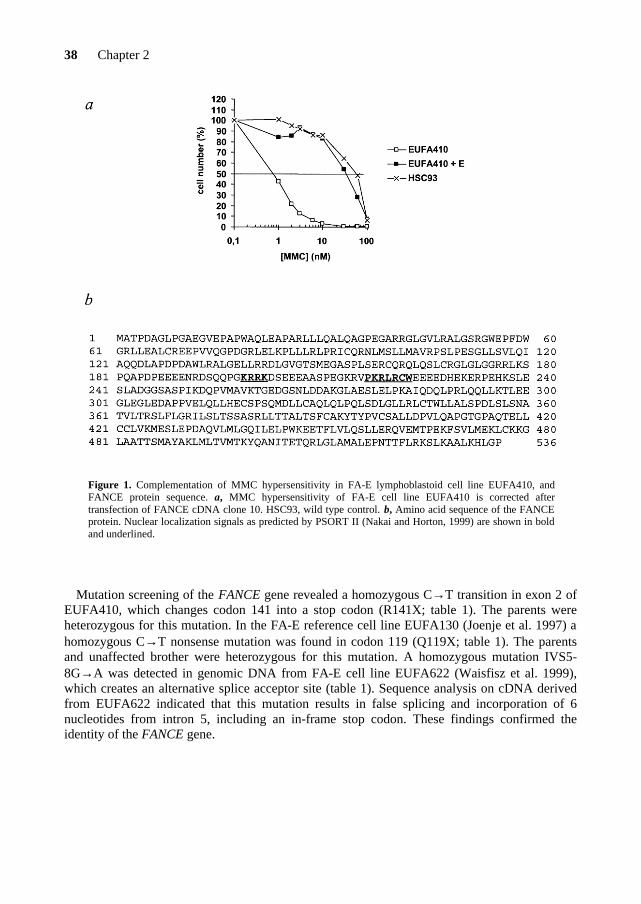
E lymphoblastoid cell line EUFA410 (Waisfisz et al. 1999) with an episomal expression library (Strathdee et al. 1992). Following selection for library uptake in hygromycin-containing medium (100 µg/ml) and subsequent selection for resistance to mitomycin C (15 nM), 4 of the 12 cDNA clones that we recovered from the pool of complemented cells had identical inserts of ~2.5 kb. Secondary transfection of one of these cDNA clones (clone10 [AF265210]) into EUFA410 cells again complemented their MMC-hypersensitive phenotype (Fig. 1a). The cDNA insert has a 1,611-nt open reading frame, encoding a 536-amino acid protein (Fig. 1b). The predicted FANCE protein contains two potential nuclear localization signals, but, like the other FA proteins, lacks any significant homology to other proteins.
The Stanford high-resolution TNG3 radiation-hybrid panel was used to position FANCE
between microsatellite markers D6S439 and D6S1645 in agreement with the genetic map location of FANCE (Waisfisz et al. 1999). The FANCE cDNA appeared identical to a human genomic DNA sequence (clone 109F14 [Genbank accession number AL022721]; Tripodis et al. 2000) on chromosome 6p21.2-21.3. A comparison between this genomic DNA sequence and the FANCE cDNA revealed that the FANCE gene has 10 exons spanning ~15 kb of genomic sequence. FANCE appears to be located between the genes encoding the 60S ribosomal protein RPL10A (Csa-19) and a ZNF127 like protein, a region where cDNA selection, exon trapping, and exon prediction programs failed to detect a gene (Tripodis et al 2000).
38 Chapter 2
Figure 1. Complementation of MMC hypersensitivity in FA-E lymphoblastoid cell line EUFA410, and FANCE protein sequence. a, MMC hypersensitivity of FA-E cell line EUFA410 is corrected after transfection of FANCE cDNA clone 10. HSC93, wild type control. b, Amino acid sequence of the FANCE protein. Nuclear localization signals as predicted by PSORT II (Nakai and Horton, 1999) are shown in bold and underlined.
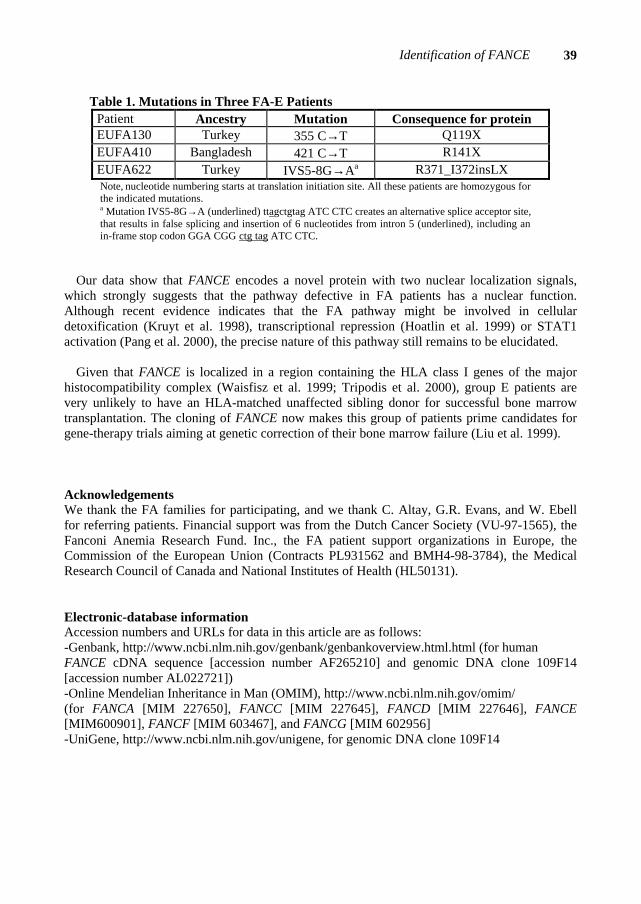
Mutation screening of the FANCE gene revealed a homozygous C→T transition in exon 2 of EUFA410, which changes codon 141 into a stop codon (R141X; table 1). The parents were heterozygous for this mutation. In the FA-E reference cell line EUFA130 (Joenje et al. 1997) a homozygous C→T nonsense mutation was found in codon 119 (Q119X; table 1). The parents and unaffected brother were heterozygous for this mutation. A homozygous mutation IVS5-8G→A was detected in genomic DNA from FA-E cell line EUFA622 (Waisfisz et al. 1999), which creates an alternative splice acceptor site (table 1). Sequence analysis on cDNA derived from EUFA622 indicated that this mutation results in false splicing and incorporation of 6 nucleotides from intron 5, including an in-frame stop codon. These findings confirmed the identity of the FANCE gene.
Identification of FANCE
39
Table 1. Mutations in Three FA-E Patients Patient Ancestry Mutation Consequence for protein EUFA130 Turkey 355 C→T Q119X EUFA410 Bangladesh 421 C→T R141X EUFA622 Turkey IVS5-8G→Aa R371_I372insLX Note, nucleotide numbering starts at translation initiation site. All these patients are homozygous for the indicated mutations. a Mutation IVS5-8G→A (underlined) ttagctgtag ATC CTC creates an alternative splice acceptor site, that results in false splicing and insertion of 6 nucleotides from intron 5 (underlined), including an in-frame stop codon GGA CGG ctg tag ATC CTC.
Our data show that FANCE encodes a novel protein with two nuclear localization signals, which strongly suggests that the pathway defective in FA patients has a nuclear function. Although recent evidence indicates that the FA pathway might be involved in cellular detoxification (Kruyt et al. 1998), transcriptional repression (Hoatlin et al. 1999) or STAT1 activation (Pang et al. 2000), the precise nature of this pathway still remains to be elucidated.
Given that FANCE is localized in a region containing the HLA class I genes of the major
histocompatibility complex (Waisfisz et al. 1999; Tripodis et al. 2000), group E patients are very unlikely to have an HLA-matched unaffected sibling donor for successful bone marrow transplantation. The cloning of FANCE now makes this group of patients prime candidates for gene-therapy trials aiming at genetic correction of their bone marrow failure (Liu et al. 1999).
Acknowledgements We thank the FA families for participating, and we thank C. Altay, G.R. Evans, and W. Ebell for referring patients. Financial support was from the Dutch Cancer Society (VU-97-1565), the Fanconi Anemia Research Fund. Inc., the FA patient support organizations in Europe, the Commission of the European Union (Contracts PL931562 and BMH4-98-3784), the Medical Research Council of Canada and National Institutes of Health (HL50131). Electronic-database information Accession numbers and URLs for data in this article are as follows: -Genbank, http://www.ncbi.nlm.nih.gov/genbank/genbankoverview.html.html (for human FANCE cDNA sequence [accession number AF265210] and genomic DNA clone 109F14 [accession number AL022721]) -Online Mendelian Inheritance in Man (OMIM), http://www.ncbi.nlm.nih.gov/omim/ (for FANCA [MIM 227650], FANCC [MIM 227645], FANCD [MIM 227646], FANCE [MIM600901], FANCF [MIM 603467], and FANCG [MIM 602956] -UniGene, http://www.ncbi.nlm.nih.gov/unigene, for genomic DNA clone 109F14

40 Chapter 2
REFERENCES Auerbach AD, Buchwald M, Joenje, H. (1998) Fanconi anemia. In: Vogelstein B., Kinzler KW
(eds) The Genetic Basis of Human Cancer. McGraw-Hill, New York, pp 317-332 de Winter JP, Waisfisz Q, Rooimans MA, van Berkel CG, Bosnoyan-Collins L, Alon N,
Carreau M, Bender O, Demuth I, Schindler D, Pronk JC, Arwert F, Hoehn H, Digweed M, Buchwald M, Joenje H (1998) The Fanconi anemia group G gene FANCG is identical with XRCC9. Nature Genet 20:281-283
de Winter JP, Rooimans MA, van der Weel L, van Berkel CG, Alon N, Bosnoyan-Collins L, de Groot J, Zhi Y, Waisfisz Q, Pronk JC, Arwert F, Mathew CG, Scheper RJ, Hoatlin ME, Buchwald M, Joenje H (2000) The Fanconi anaemia gene FANCF encodes a novel protein with homology to ROM. Nature Genet 24:15-16
Hejna JA, Timmers CD, Reifsteck C, Bruun DA, Lucas LW, Jakobs PM, Toth-Fejel S, Unsworth N, Clemens SL, Garcia DK, Naylor SL, Thayer MJ, Olson SB, Grompe M, Moses RE (2000) Localization of the Fanconi anemia complementation group D gene to a 200-kb region on chromosome 3p25.3. Am J Hum Genet 66:1540-1551
Hoatlin ME, Zhi Y, Ball H, Silvey K, Melnick A, Stone S, Arai S, Hawe N, Owen G, Zelent A, Licht JD (1999) A novel BTB/POZ transcriptional repressor protein interacts with the Fanconi anemia group C protein and PLZF. Blood 94:3737-3747
Joenje H, Levitus M, Waisfisz Q, D'Andrea A, Garcia-Higuera I, Pearson T, van Berkel CGM, Rooimans MA, Morgan N, Mathew CG, Arwert F (2000) Complementation analysis in Fanconi anemia: assignment of the reference FA-H patient to group A. Am J Hum Genet 67:759-762
Joenje H, Oostra AB, Wijker M, di Summa FM, van Berkel CG, Rooimans MA, Ebell, van Weel M, Pronk JC, Buchwald M, Arwert F (1997) Evidence for at least eight Fanconi anemia genes. Am J Hum Genet 61:940-944
Kruyt FAE, Hoshino T, Liu JM, Joseph P, Jaiswal AK, Youssoufian H (1998) Abnormal microsomal detoxification implicated in Fanconi anemia group C by interaction of the FAC protein with NADPH cytochrome P450 reductase. Blood 92:3050-3056
Liu JM, Kim S, Read EJ, Futaki M, Dokal I, Carter CS, Leitman SF, Pensiero M, Young NS, Walsh CE (1999) Engraftment of hematopoietic progenitor cells transduced with the Fanconi anemia group C gene (FANCC). Hum Gene Ther 10:2337-2346
Lo ten Foe JR, Rooimans MA, Bosnoyan-Collins L, Alon N, Wijker M, Parker L, Lightfoot J, Carreau M, Callen DF, Savoia A, Cheng NC, Van Berkel CGM, Strunk MHP, Gille JJP, Pals G, Kruyt FAE, Pronk JC, Arwert F, Buchwald M, Joenje H (1996) Expression cloning of a cDNA for the major Fanconi anemia gene, FAA. Nature Genet 14:320-323
Nakai K, Horton P (1999) PSORT: a program for detecting the sorting signals of proteins and predicting their subcellular localization. Trends Biochem Sci 24:34-35
Pang Q, Fagerlie S, Christianson TA, Keeble W, Faulkner G, Diaz J, Rathbun RK, Bagby GC (2000) The Fanconi anemia protein FANCC binds to and facilitates the activation of STAT1 by gamma interferon and hematopoietic growth factors. Mol Cell Biol 20:4724-4735
Strathdee CA, Gavish H, Shannon WR, Buchwald M (1992) Cloning of cDNAs for Fanconi’s anemia by functional complementation. Nature 356:763-767
The Fanconi anemia/Breast cancer consortium (1996) Positional cloning of Fanconi anemia group A gene. Nature Genet 14:324-328

Identification of FANCE
41
Tripodis N, Palmer S, Phillips S, Milne S, Beck S, Ragoussis J (2000) Construction of a high-resolution 2.5-Mb transcript map of the human 6p21.2-6p21.3 region immediately centromeric of the major histocompatibility complex. Genome Res 10:454-472
Waisfisz Q, Saar K, Morgan NV, Altay C, Leegwater PA, de Winter JP, Komatsu K, Evans DGR, Wegner R-D, Reis A, Joenje H, Arwert F, Mathew CG, Pronk JC, Digweed M (1999) The Fanconi anemia group E gene, FANCE, maps to chromosome 6p. Am J Hum Genet 64:1400-1405
Whitney M, Thayer M, Reifsteck C, Olson S, Smith L, Jacobs PM, Leach R, Naylord S, Joenje H, Grompe M (1995) Microcell mediated chromosome transfer maps the Fanconi anemia group D gene to chromosome 3p. Nature Genet 11:341-343


CHAPTER 3
FANCONI ANEMIA (FA) is a recessive chromosomal instability syndrome characterized by bone marrow failure, diverse congenital malformations and cancer predisposition. To date, twelve FA complementation groups (A-C, D1, D2, E-G, I, J, L and M) have been assigned and eleven associated genes have been identified. FANCE is the gene mutated in FA complementation group E. Here we report a novel FANCE mutation (1111C→T) leading to an amino acid substitution (R371W) and disruption of the FA pathway. The defective FANCE protein is undetectable by immunoblot analysis. This FANCE missense mutation was found homozygous in a German and Dutch FA-E patient. The possibility of an ancestral founder mutation in FANCE remains to be elucidated.
France Léveillé, Najim Ameziane, Martin A. Rooimans, Patrick Bier, Bertus Kuyt, Gerald Pals, Hans Joenje, and Johan P. de Winter
A novel FANCE missense mutation in two Fanconi anemia patients

FANCE mutation in two patients 45
INTRODUCTION Fanconi anemia (FA) is rare recessive chromosomal instability syndrome characterized by bone marrow failure, congenital abnormalities and cancer predisposition (for reviews, see Joenje & Patel, 2001; D’Andrea & Grompe, 2003). The cells from FA patients are hypersensitive to DNA cross-linking agents, such as mitomycin C (MMC) and diepoxybutane, which suggest a role of these proteins in the repair of DNA interstrand cross-links. To date, 12 FA complementation groups have been described (Levitus et al., 2004) and 11 associated genes have been identified: FANCC (Strathdee et al., 1992), FANCA (FAB, 1996; Lo Ten Foe et al., 1996), FANCG (de Winter et al., 1998), FANCF (de Winter et al., 2000a), FANCE (de Winter et al., 2000b), FANCD2 (Timmers et al., 2001), FANCD1/BRCA2 (Howlett et al., 2002), FANCL (Meetei et al., 2003), FANCB (Meetei et al., 2004), FANCJ (Levitus et al., 2005; Levran et al., 2005; Litman et al., 2005) and FANCM (Meetei et al., 2005).
Most FA patients belong to complementation group A (65%), G (13%) and C (10%), in a
population of 241 subtyped FA families (classified by the European Fanconi Anemia Research Programme), while the other subtypes are very rare (Levitus et al., 2004). Nevertheless, the worldwide prevalence of one FA complementation group compared to another often varies with ethnic background (Joenje & Patel, 2001). For instance, different founder mutations in FANCA were reported in the Afrikaner population of South Africa (Tipping et al., 2001) and in Gypsy families from Spain (Callén et al., 2005), while the majority of the FA Ashkenazi-Jewish families share a common mutation in FANCC (Whitney et al., 1993). In addition, the clinical manifestations and outcomes of FA patients are also variable between and inside complementation groups, and could be associated to a particular type of mutation in an FA gene (Faivre et al., 2000; Kutler et al., 2003). Therefore, the identification of a mutation in an FA gene could significantly improve the clinical management of those patients.
The FA complementation group E is a rare subtype. To date, only seven FA-E patients have
been identified worldwide (Levitus et al., 2004; Ameziane et al., 2006). FANCE is the gene defective in the FA complementation group E and is localized to chromosome band 6p21.2-21.3 (Waisfisz et al., 1999; de Winter et al., 2000b). The FANCE gene has 10 exons spanning ~ 15 kb of genomic DNA and a 1.611-nucleotide open reading frame encoding a protein of 536-amino acids (de Winter et al., 2000b). Here we report a novel FANCE mutation in two FA-E patients (1111C→T) that leads to an amino acid change in codon 371 (R371W). Intriguingly, these 2 patients originate from Germany (EUFA279) and The Netherlands (EUFA1278) and therefore, may represent a founder mutation in FANCE. The results of the present report are included in Ameziane et al., 2006. MATERIALS AND METHODS Patients, cell lines and complementation analysis The FA diagnosis of the patients was based on clinical symptoms and chromosomal breakages tests. Cell fusion studies (as described in Joenje et al., 1995), functional complementation, protein analysis and sequencing allowed us to assign the two FA patients EUFA279 and EUFA1278 to complementation group E. Lymphoblastoid cell lines were established from
46 Chapter 3
blood by Epstein-Barr virus (EBV) transformation. The MMC-induced growth inhibition test was performed as previously described (Ishida et al., 1982; Joenje et al., 1986).
Sequencing and mutation analysis of FANCE Genomic DNA was isolated from lymphoblastoid cells by standard procedures and total RNA by using the RNase Out ribonuclease (Invitrogen). To synthesize cDNA we used Superscript II RNase H reverse transcriptase (Invitrogen) and Oligo dT priming (Roche) according to standard protocols. Sequence change in patient EUFA279 was identified at the genomic level and confirmed on cDNA by direct sequencing using the Thermo Sequenase Cy5.5 dye terminator cycle sequencing kit (Amersham) and the Visible Genetics OpenGene automatic sequencer (Visible Genetics Inc, Toronto, Ontario, Canada) to analyze the products. Mutation screening in the patient EUFA1278 was done by the denaturing high-performance liquid chromatography (DHPLC) approach (Ameziane et al., 2006).
Protein analysis Cell lysates (~400 000 cells) from lymphoblastoid cell lines were obtained as previously described (Léveillé et al., 2004) and proteins were separated on SDS-polyacrylamide gel. Proteins were transferred to a PVDF membrane and immunoblotted with FANCE antibody (kindly provided by Dr. K.J. Patel).
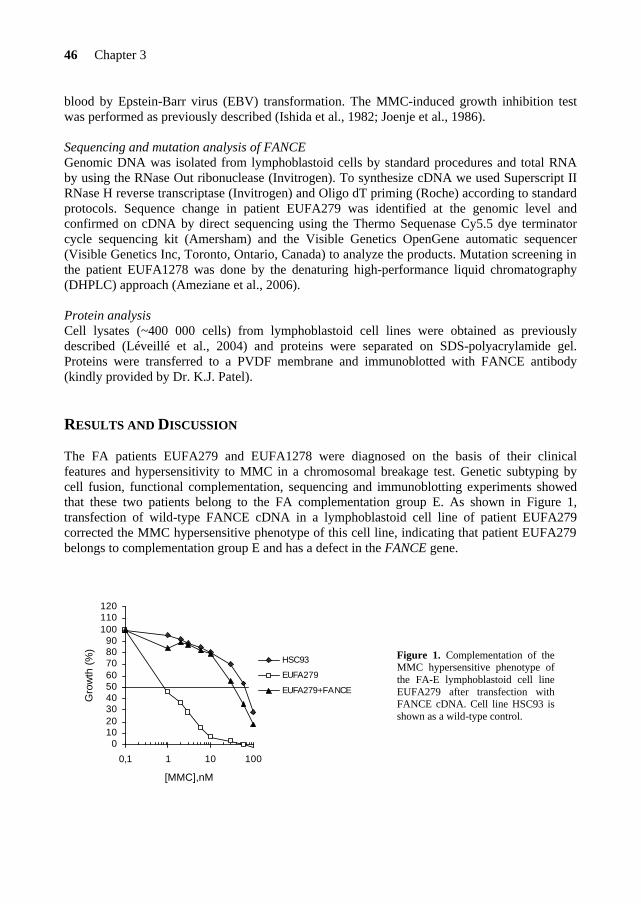
RESULTS AND DISCUSSION The FA patients EUFA279 and EUFA1278 were diagnosed on the basis of their clinical features and hypersensitivity to MMC in a chromosomal breakage test. Genetic subtyping by cell fusion, functional complementation, sequencing and immunoblotting experiments showed that these two patients belong to the FA complementation group E. As shown in Figure 1, transfection of wild-type FANCE cDNA in a lymphoblastoid cell line of patient EUFA279 corrected the MMC hypersensitive phenotype of this cell line, indicating that patient EUFA279 belongs to complementation group E and has a defect in the FANCE gene.
0102030405060708090
100110120
0,1 1 10 100
[MMC],nM
Gro
wth
(%
)
HSC93
EUFA279
EUFA279+FANCE
Figure 1. Complementation of the MMC hypersensitive phenotype of the FA-E lymphoblastoid cell line EUFA279 after transfection with FANCE cDNA. Cell line HSC93 is shown as a wild-type control.
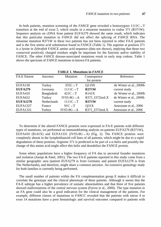
FANCE mutation in two patients 47
In both patients, mutation screening of the FANCE gene revealed a homozygous 1111C→T transition at the end of exon 5, which results in a missense mutation in codon 371 (R371W). Sequence analysis on cDNA from patient EUFA279 showed the same result, which indicates that this particular mutation in FANCE did not affect the splicing of FANCE RNA. The missense mutation R371W in these two patients has not been reported in other FA-E patients and is the first amino acid substitution found in FANCE (Table 1). The arginine at position 371 is a lysine in Zebrafish FANCE amino acid sequence (data not shown), implying that those two conserved positively charged residues might be important for the function and/or stability of FANCE. The other FANCE disease-associated mutations result in early stop codons. Table 1 shows the spectrum of FANCE mutations in known FA patients.
TABLE 1. Mutations in FANCE FA-E Patient Ancestry Mutation Consequence Reference for protein EUFA130 Turkey 355C→T Q119X de Winter et al., 2000b EUFA279 Germany 1111C→T R371W current study EUFA410 Bangladesh 421C→T R141X de Winter et al., 2000b EUFA622 Turkey IVS5-8G→A R371_I372insLX de Winter et al., 2000b EUFA1278 Netherlands 1111C→T R371W current study EUFA1327 France 91C→T Q31X Ameziane et al., 2006 EUFA1331 Turkey IVS5-8G→A R371_I372insLX Ameziane et al., 2006
To determine if the altered FANCE proteins were expressed in FA-E patients with different types of mutations, we performed an immunoblotting analysis on patients EUFA279 (R371W), EUFA410 (R141X) and EUFA1331 (IVS5-8G→A) (Fig. 2). The FANCE proteins were completely absent in the lymphoblastoid cell lines of all patients, which might be due to a rapid degradation of these proteins. Arginine 371 is predicted to be part of a α-helix and possibly the change of this amino acid might affect this helix and destabilize the FANCE protein.
Some ethnic populations have a higher frequency of FA due to ancestral founder mutations
and isolation (Joenje & Patel, 2001). The two FA-E patients reported in this study come from a similar geographic area (patient EUFA279 is from Germany and patient EUFA1278 is from The Netherlands), and therefore, might share a common ancestor. An extensive genealogy study for both families is currently being performed.
The small number of patients within the FA complementation group E makes it difficult to
correlate the genotype and the clinical phenotype of these patients. Although it seems that the FA-E subtype has a higher prevalence of somatic abnormalities and that three of five patients showed malformations of the central nervous system (Faivre et al., 2000). The type mutation in an FA gene could also be a good indication for the clinical management of the patients. For example, different classes of mutations in FANCC revealed that the patients with intron 4 or exon 14 mutations have a poor hematologic and survival outcomes compared to patients with
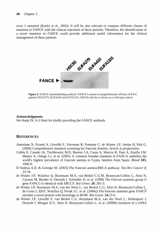
48 Chapter 3
exon 1 mutation (Kutler et al., 2003). It will be also relevant to compare different classes of mutation in FANCE with the clinical outcomes of these patients. Therefore, the identification of a novel mutation in FANCE could provide additional useful information for the clinical management of these patients.
Figure 2. FANCE immunoblotting analysis. FANCE is absent in lymphoblastoid cell lines of FA-E patients EUFA279, EUFA410 and EUFA1331. HSC93 cell line is shown as a wild-type control.
Acknowledgments We thank Dr. K.J Patel for kindly providing the FANCE antibody. REFERENCES Ameziane N, Errami A, Léveillé F, Vervenne R, Fontaine C, de Winter J.P, Joenje H, Pals G.
(2006) Comprehensive mutation screening for Fanconi Anemia. Article in preparation Callén E, Casado JA, Tischkowitz M.D, Bueren J.A, Creus A, Marcos R, Dasi A, Estella J.M,
Munoz A, Ortega J.J. et al. (2005) A common founder mutation in FANCA underlies the world's highest prevalence of Fanconi anemia in Gypsy families from Spain. Blood 105, 1946-9.
D’Andrea A.D. & Grompe M. (2003) The Fanconi anemia/BRCA pathway. Nat Rev Cancer 3, 23-34.
de Winter J.P, Waisfisz Q, Rooimans M.A, van Berkel C.G.M, Bosnoyan-Collins L, Alon N, Carreau M, Bender O, Demuth I, Schindler D. et al. (1998) The Fanconi anaemia group G gene FANCG is identical with XRCC9. Nat Genet. 20, 281-3.
de Winter J.P, Rooimans M.A, van der Weel L, van Berkel C.G, Alon N, Bosnoyan-Collins L, de Groot J, ZhiY, Waisfisz Q, Pronk J.C. et al. (2000a) The Fanconi anaemia gene FANCF encodes a novel protein with homology to ROM. Nat Genet. 24,15-6.
de Winter J.P, Léveillé F, van Berkel C.G, Rooimans M.A, van der Weel L, Steltenpool J, Demuth I, Morgan N.V, Alon N, Bosnoyan-Collins L. et al. (2000b) Isolation of a cDNA
HSC93
EUFA13
31
EUFA41
0
EUFA27
9
FANCE
HSC93
EUFA13
31
EUFA41
0
EUFA27
9
FANCE

FANCE mutation in two patients 49
representing the Fanconi anemia complementation group E gene. Am J Hum Genet. 67, 1306-8.
FAB; The Fanconi Anaemia/Breast Cancer Consortium (1996) Positional cloning of the Fanconi anaemia group A gene. Nat Genet. 14, 324-8.
Faivre L, Guardiola P, Lewis C, Dokal I, Ebell W, Zatterale A, Altay C, Poole J, Stones D, Kwee M.L. et al. (2000) Association of complementation group and mutation type with clinical outcome in fanconi anemia. European Fanconi Anemia Research Group. Blood 96, 4064-70.
Howlett N.G, Taniguchi T, Olson S, Cox B, Waisfisz Q, de Die-Smulders C, Persky N, Grompe M, Joenje H, Pals G. et al. (2002) Biallelic inactivation of BRCA2 in Fanconi anemia. Science 297, 606-9.
Ishida R. & Buchwald M. (1982) Susceptibility of Fanconi's anemia lymphoblasts to DNA- cross-linking and alkylating agents. Cancer Res. 42, 4000-6.
Joenje H, Lo Ten Foe J.R, Oostra A.B, van Berkel C.G.M, Rooimans M.A, Schroeder-Kurth T, Wegner R-D, Gille J.J.P, Buchwald M, Arwert F. (1995) Classification of Fanconi anemia patients by complementation analysis: evidence for a fifth genetic subtype. Blood 86, 2156-60.
Joenje H. & Patel K.J. (2001) The emerging genetic and molecular basis of Fanconi anaemia. Nat Rev Genet. 2, 446-57.
Kutler D.I, Singh B, Satagopan J, Batish S.D, Berwick M, Giampietro P.F, Hanenberg H, Auerbach A.D. (2003) A 20-year perspective on the International Fanconi Anemia Registry (IFAR). Blood 101, 1249-56.
Léveillé F, Blom E, Medhurst A.L, Bier P, Laghmani E.H, Johnson M, Rooimans M.A, Sobeck A, Waisfisz Q, Arwert F. et al. (2004) The Fanconi anemia gene product FANCF is a flexible adaptor protein. J Biol Chem. 38, 39421-30.
Levitus M, Rooimans M.A, Steltenpool J, Cool N.F.C, Oostra A.B, Mathew C.G, Hoatlin M.E, Waisfisz Q, Arwert F, de Winter J.P, Joenje,H. (2004) Heterogeneity in Fanconi anemia: evidence for two new genetic subtypes. Blood 103, 2498-503.
Levitus M, Waisfisz Q, Godthelp B.C, de Vries Y, Hussain S, Wiegant W.W, Elghabzouri-Maghrani E, Steltenpool J, Rooimans M.A, Pals G. et al. (2005) The DNA helicase BRIP1 is defective in Fanconi anemia complementation group J. Nat Genet. 37, 934-5 .
Levran O, Attwooll C, Henry R.T, Milton K.L, Neveling K, Rio P, Batish S.D, Kalb R, Velleuer E, Barral S. et al. (2005) The BRCA1-interacting helicase BRIP1 is deficient in Fanconi anemia. Nat Genet. 37,1296.
Litman R, Peng M, Jin Z, Zhang F, Zhang J, Powell S, Andreassen P.R, Cantor,S.B. (2005) BACH1 is critical for homologous recombination and appears to be the Fanconi anemia gene product FANCJ. Cancer Cell 8, 255-65.
Lo Ten Foe J.R, Rooimans M.A, Bosnoyan-Collins L, Alon N, Wijker M, Parker L, Lightfoot J, Carreau M, Callen D.F, Savoia A. et al. (1996) Expression cloning of cDNA for the major Fanconi anaemia gene, FAA. Nat Genet. 14, 320-3.
Meetei A.R, de Winter J.P, Medhurst A.L, Wallisch M, Waisfisz Q, van de Vrugt H.J, Oostra A.B, Yan Z, Ling C, Bishop C.E. et al. (2003) A novel ubiquitin ligase is deficient in Fanconi anemia. Nat Genet. 35, 165-70.
Meetei A.R, Levitus M, Xue Y, Medhurst A.L, Zwaan M, Ling C, Rooimans M.A, Bier P, Hoatlin M, Pals G. et al. (2004) X-linked inheritance of Fanconi anemia complementation group B. Nat Genet. 36, 1219-24.

50 Chapter 3
Meetei A.R, Medhurst A.L, Ling C, Xue Y, Singh T.R, Bier P, Steltenpool J, Stone S, Dokal I, Mathew C.G. et al. (2005) A Human Orthologue of Archaeal DNA Repair Protein Hef is Defective in Fanconi Anemia Complementation Group M. Nat Genet. 37, 958-63.
Strathdee C.A, Gavish H, Shannon W.R. and Buchwald M. (1992) Cloning of cDNAs for Fanconi's anaemia by functional complementation. Nature 358, 434.
Timmers C, Taniguchi T, Hejna J, Reifsteck C, Lucas L, Bruun D, Thayer M, Cox B, Olson S, D’Andrea A.D. et al. (2001) Positional cloning of a novel Fanconi anemia gene, FANCD2. Mol Cell 7, 241-8.
Tipping A.J, Pearson T, Morgan N.V, Gibson R.A, Kuyt L.P, Havenga C, Gluckman E, Joenje H, de Ravel T, Jansen S, Mathew C.G. (2001) Molecular and genealogical evidence for a founder effect in Fanconi anemia families of the Afrikaner population of South Africa. Proc Natl Acad Sci. 98, 5734-9.
Waisfisz Q, Saar K, Morgan N.V, Altay C, Leegwater P.A, de Winter J.P, Komatsu K, Evans G.R, Wegner R-D, Reis A. et al. (1999) The Fanconi anemia group gene, FANCE, maps to chromosome 6p. Am J Hum Genet. 64, 1400-5.
Whitney M.A, Saito H, Jakobs P.M, Gibson R.A, Moses R.E, Grompe M. (1993) A common mutation in the FACC gene causes Fanconi anaemia in Ashkenazi Jews. Nat Genet. 4, 202-5.


CHAPTER 4
FANCONI ANEMIA (FA) protein FANCE is an essential component of the nuclear FA core complex, which is required for monoubiquitination of the downstream target FANCD2, an important step in the FA pathway of DNA cross-link repair. FANCE is predominantly localized in the nucleus and acts as a molecular bridge between the FA core complex and FANCD2, through direct binding of both FANCC and FANCD2. At present, it is poorly understood how the nuclear accumulation of FANCE is regulated and therefore we investigated the nuclear localization of this FA protein. We found that FANCE has a strong tendency to localize in the nucleus, since the addition of a nuclear export signal does not interfere with the nuclear localization of FANCE. We also demonstrate that the nuclear accumulation of FANCE does not rely solely on its nuclear localization signal motifs, but also on FANCC. The other FA proteins are not involved in the nuclear accumulation of FANCE, indicating a tight relationship between FANCC and FANCE, as suggested from their direct interaction. Finally, we show that the region of FANCE interacting with FANCC appears to be different from the region involved in binding FANCD2. This strengthens the idea that FANCE recruits FANCD2 to the core complex, without interfering with the binding of FANCC.
France Léveillé, Miriam Ferrer, Annette L. Medhurst, El Houari Laghmani, Martin A. Rooimans, Patrick Bier, Jurgen Steltenpool, Tom A. Titus, John H. Postlethwait, Maureen E. Hoatlin, Hans Joenje, and Johan P. de Winter
DNA repair 5, 556-565 (2006)
The nuclear accumulation of the Fanconi anemia protein FANCE
depends on FANCC

The nuclear accumulation of FANCE depends on FANCC 55
INTRODUCTION
Fanconi anemia (FA) is a recessive inherited chromosomal instability disorder characterized by congenital anomalies, progressive bone marrow failure, and predisposition to acute myeloid leukemia and squamous cell carcinomas (for review, see Refs. [1-3]). FA cells display excessive chromosomal aberrations and hypersensitivity to DNA cross-linking agents such as mitomycin C (MMC) and diepoxybutane (DEB), indicative of a molecular defect in DNA-damage response.
To date, 12 FA complementation groups have been identified and all the associated genes have been cloned (FANCA, -B, -C, -D1/BRCA2, -D2, -E, -F, -G, -J, -L and -M) [4-15], except for FANCI [16]. The FA proteins collaborate in a common pathway in which the FANCA, -B, -C, -E, -F, -G, -L and -M proteins assemble in a nuclear core complex that functions as an ubiquitin ligase for FANCD2 [12,14,17]. The monoubiquitinated form of FANCD2 colocalizes with proteins involved in DNA repair (such as BRCA1, RAD51, and NBS1) during S-phase or in response to DNA damaging agents [18,19]. In addition, the FANCD2 protein binds double strand DNA ends and Holliday junctions [21], and assembles with FANCD1/BRCA2 and FANCE in a stable chromatin complex [22]. The USP1 protein appears to be the deubiquitinating enzyme of FANCD2 and may therefore play a critical role in the regulation of the FA pathway [20]. The FA proteins have been implicated in the repair of interstrand cross-link (ICL) lesions through diverse repair pathways, such as homologous recombination (HR), translesion synthesis and the BLM helicase pathway [23-27]. Importantly, recent studies showed that the FANCJ and FANCM proteins have a helicase and DNA translocase activity, respectively [14,15]. These findings indicate that FA proteins may play a direct role in DNA-damage response.
The specific functions of the FA proteins in the FA core complex and the structural features
of the various protein-protein interactions within this complex are not fully understood. FANCC, one of the FA core complex components, has been shown to localize in both cytoplasmic and nuclear compartments of the cell [28-31]. The nuclear localization of FANCC is crucial for the function of the FA core complex and depends on the presence of FANCE [32,33]. FANCE directly interacts with both FANCC and FANCD2 and, consequently, links the downstream FANCD2 protein to the putative ligase activity of the nuclear FA core complex [33-35]. The FANCE protein contains 2 putative nuclear localization signals (NLS) [9] and localizes to nuclear foci [33], but it is unclear how the nuclear localization of FANCE is regulated. A better insight in the nuclear accumulation of the FANCE protein will improve our understanding of the FA core complex assembly.
Here we present a study on the nuclear localization of FANCE and the interaction between
FANCE, FANCC and FANCD2. We found that the FANCE and FANCC proteins are functionally linked in that they require each other to be stable in the nucleus of the cell. The nuclear accumulation of FANCE depends not only on its nuclear localization signals, but also on FANCC, and not on the other FA proteins. By mammalian two-hybrid assay, we determined that different regions of FANCE seem to mediate the direct interactions with FANCC and FANCD2.
56 Chapter 4
MATERIALS AND METHODS
1. Cell culture, transfection and MMC assay Epstein-Barr virus (EBV)-immortalized lymphoblasts were maintained in RPMI 1640 media supplemented with 1 mM glutamine (Invitrogen, Carlsbad, CA, USA) and 10% fetal calf serum (FCS) (Invitrogen). Lymphoblastoid cell lines were transfected by electroporation using an ECM830 electrosquareporator (BTX, San Diego, CA, USA) and cultured in selection media containing hygromycin B (100 µg/ml; Roche, Basel, Switzerland). The MMC-induced growth inhibition assays were performed as previously described [36,37]. The HeLa cells were grown in Dulbecco modified Eagle medium (DMEM) supplemented with 10% FCS and the fibroblasts (BeBu and FA-C fibroblasts EUFA1233) were maintained in F10 medium. 2. Plasmid constructs A wild-type or a mutated nuclear export signal (NES) was cloned to the C-terminus of pSK-FANCE [see Ref. 38] and then subcloned in the pEGFP-N1 expression vector (Clontech, Palo Alto, CA, USA). For the generation of the FANCE construct in which the nuclear localization signal 1 was mutated (mNLS1), we used the QuickChange site-directed mutagenesis kit (Stratagene, La Jolla, CA, USA) according to the manufacturer’s instructions. Oligonucleotides with mutations in NLS2 were used to create FANCE-mNLS2 and FANCE mNLS1-2 by PCR using pN1-FANCE-GFP and pN1-FANCE-mNLS1-GFP as a template. All FANCE mutant constructs were subcloned into the episomal expression vector pMEP4 (Clontech). The FANCE fragments used in the mammalian two-hybrid assay were generated by using internal restriction sites in FANCE or by PCR with primers that contained specific restriction sites and subcloned into pM-GAL4 DNA-binding domain and pVP16-GAL4 activation domain vectors (Clontech). The sequences of the FANCE NLS mutants and the correct reading frame of all the FANCE fragments were verified using the GeneAmp PCR system 9700 (Applied Biosystems) and analyzed on an ABI 3730 DNA analyzer (Applied Biosystems). 3. DNA transfections and fluorescence microscopy analysis For transient transfections, 2.5 x 10⁵ HeLa cells were seeded onto sterile glass coverlips in six-well plates, and transfected with 1 µg of pEGFP-N1 alone (Invitrogen) or pEGFP-N1 encoding FANCE-GFP, FANCE-NES/mNES-GFP or FANCE NLS mutants using Lipofectamine Plus (Invitrogen), according to manufacturer’s guidelines. When indicated, FANCE constructs were cotransfected with pCMV-Flag-CRM1 (a generous gift from Dr. Jose A. Rodriguez, VUMC, Amsterdam). The primary wild type (BeBu) and FA-C (EUFA1233) fibroblast cell lines were transiently transfected with 1 µg of pN1-FANCE-GFP and pN1-FANCEmNLS1-2-GFP by electroporation (Amaxa). At 24h (HeLa cells) or 48h (fibroblasts) after transfection, cells with the EGFP-tagged proteins were fixed with 3.7% formaldehyde in PBS for 30 min, washed with PBS, and stained with the chromosomal dye Hoechst 33342 (Sigma, St. Louis, MO) for 30 min in order to counterstain the nuclei, and mounted onto microscope slides using Vectashield (Vector Laboratories, Inc., Burlingame, CA). Immunocytochemical detection of Flag-CRM1 was performed with the anti-Flag M2 monoclonal antibody (Stratagene, La Jolla, CA) and an Alexa Fluor 594-conjugated goat anti-mouse secondary antibody (Molecular Probes, Eugene, OR), using a previously described immunostaining method [39]. Slides were examined under UV light on an inverted Leica DMIRB/E fluorescence microscope (Leica Heidelberg, Heidelberg, Germany). Images (magnification X 400) were collected using Leica Q500MC
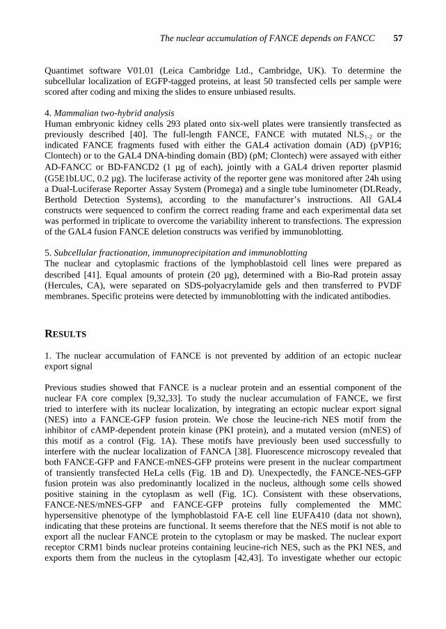
The nuclear accumulation of FANCE depends on FANCC 57
Quantimet software V01.01 (Leica Cambridge Ltd., Cambridge, UK). To determine the subcellular localization of EGFP-tagged proteins, at least 50 transfected cells per sample were scored after coding and mixing the slides to ensure unbiased results. 4. Mammalian two-hybrid analysis Human embryonic kidney cells 293 plated onto six-well plates were transiently transfected as previously described [40]. The full-length FANCE, FANCE with mutated NLS1-2 or the indicated FANCE fragments fused with either the GAL4 activation domain (AD) (pVP16; Clontech) or to the GAL4 DNA-binding domain (BD) (pM; Clontech) were assayed with either AD-FANCC or BD-FANCD2 (1 µg of each), jointly with a GAL4 driven reporter plasmid (G5E1bLUC, 0.2 µg). The luciferase activity of the reporter gene was monitored after 24h using a Dual-Luciferase Reporter Assay System (Promega) and a single tube luminometer (DLReady, Berthold Detection Systems), according to the manufacturer’s instructions. All GAL4 constructs were sequenced to confirm the correct reading frame and each experimental data set was performed in triplicate to overcome the variability inherent to transfections. The expression of the GAL4 fusion FANCE deletion constructs was verified by immunoblotting. 5. Subcellular fractionation, immunoprecipitation and immunoblotting The nuclear and cytoplasmic fractions of the lymphoblastoid cell lines were prepared as described [41]. Equal amounts of protein (20 µg), determined with a Bio-Rad protein assay (Hercules, CA), were separated on SDS-polyacrylamide gels and then transferred to PVDF membranes. Specific proteins were detected by immunoblotting with the indicated antibodies. RESULTS
1. The nuclear accumulation of FANCE is not prevented by addition of an ectopic nuclear export signal Previous studies showed that FANCE is a nuclear protein and an essential component of the nuclear FA core complex [9,32,33]. To study the nuclear accumulation of FANCE, we first tried to interfere with its nuclear localization, by integrating an ectopic nuclear export signal (NES) into a FANCE-GFP fusion protein. We chose the leucine-rich NES motif from the inhibitor of cAMP-dependent protein kinase (PKI protein), and a mutated version (mNES) of this motif as a control (Fig. 1A). These motifs have previously been used successfully to interfere with the nuclear localization of FANCA [38]. Fluorescence microscopy revealed that both FANCE-GFP and FANCE-mNES-GFP proteins were present in the nuclear compartment of transiently transfected HeLa cells (Fig. 1B and D). Unexpectedly, the FANCE-NES-GFP fusion protein was also predominantly localized in the nucleus, although some cells showed positive staining in the cytoplasm as well (Fig. 1C). Consistent with these observations, FANCE-NES/mNES-GFP and FANCE-GFP proteins fully complemented the MMC hypersensitive phenotype of the lymphoblastoid FA-E cell line EUFA410 (data not shown), indicating that these proteins are functional. It seems therefore that the NES motif is not able to export all the nuclear FANCE protein to the cytoplasm or may be masked. The nuclear export receptor CRM1 binds nuclear proteins containing leucine-rich NES, such as the PKI NES, and exports them from the nucleus in the cytoplasm [42,43]. To investigate whether our ectopic
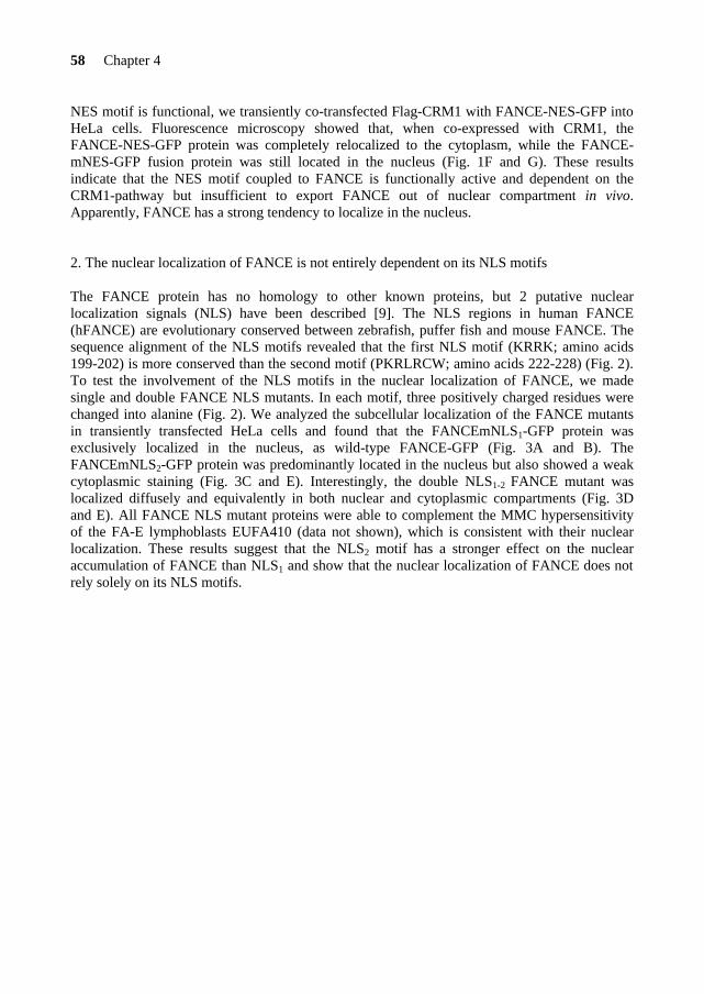
58 Chapter 4
NES motif is functional, we transiently co-transfected Flag-CRM1 with FANCE-NES-GFP into HeLa cells. Fluorescence microscopy showed that, when co-expressed with CRM1, the FANCE-NES-GFP protein was completely relocalized to the cytoplasm, while the FANCE-mNES-GFP fusion protein was still located in the nucleus (Fig. 1F and G). These results indicate that the NES motif coupled to FANCE is functionally active and dependent on the CRM1-pathway but insufficient to export FANCE out of nuclear compartment in vivo. Apparently, FANCE has a strong tendency to localize in the nucleus.
2. The nuclear localization of FANCE is not entirely dependent on its NLS motifs The FANCE protein has no homology to other known proteins, but 2 putative nuclear localization signals (NLS) have been described [9]. The NLS regions in human FANCE (hFANCE) are evolutionary conserved between zebrafish, puffer fish and mouse FANCE. The sequence alignment of the NLS motifs revealed that the first NLS motif (KRRK; amino acids 199-202) is more conserved than the second motif (PKRLRCW; amino acids 222-228) (Fig. 2). To test the involvement of the NLS motifs in the nuclear localization of FANCE, we made single and double FANCE NLS mutants. In each motif, three positively charged residues were changed into alanine (Fig. 2). We analyzed the subcellular localization of the FANCE mutants in transiently transfected HeLa cells and found that the FANCEmNLS1-GFP protein was exclusively localized in the nucleus, as wild-type FANCE-GFP (Fig. 3A and B). The FANCEmNLS2-GFP protein was predominantly located in the nucleus but also showed a weak cytoplasmic staining (Fig. 3C and E). Interestingly, the double NLS1-2 FANCE mutant was localized diffusely and equivalently in both nuclear and cytoplasmic compartments (Fig. 3D and E). All FANCE NLS mutant proteins were able to complement the MMC hypersensitivity of the FA-E lymphoblasts EUFA410 (data not shown), which is consistent with their nuclear localization. These results suggest that the NLS2 motif has a stronger effect on the nuclear accumulation of FANCE than NLS1 and show that the nuclear localization of FANCE does not rely solely on its NLS motifs.
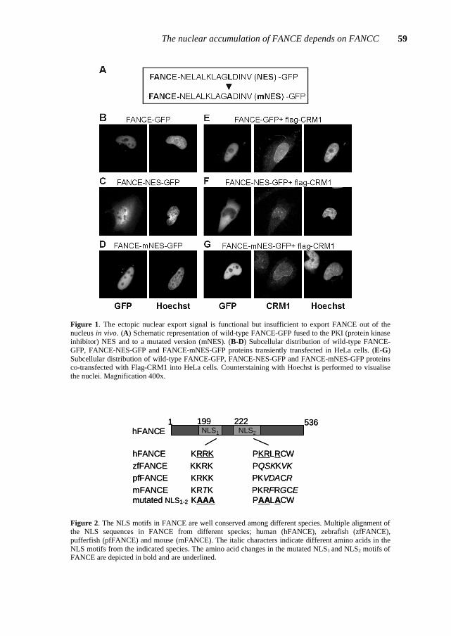
The nuclear accumulation of FANCE depends on FANCC 59
Figure 1. The ectopic nuclear export signal is functional but insufficient to export FANCE out of the nucleus in vivo. (A) Schematic representation of wild-type FANCE-GFP fused to the PKI (protein kinase inhibitor) NES and to a mutated version (mNES). (B-D) Subcellular distribution of wild-type FANCE-GFP, FANCE-NES-GFP and FANCE-mNES-GFP proteins transiently transfected in HeLa cells. (E-G) Subcellular distribution of wild-type FANCE-GFP, FANCE-NES-GFP and FANCE-mNES-GFP proteins co-transfected with Flag-CRM1 into HeLa cells. Counterstaining with Hoechst is performed to visualise the nuclei. Magnification 400x.
hFANCE KRRK PKRLRCWzfFANCE KKRK PQSKKVKpfFANCE KRKK PKVDACRmFANCE KRTK PKRFRGCEmutated NLS1-2 KAAA PAALACW
1 536NLS1 NLS2
199 222hFANCE
hFANCE KRRK PKRLRCWzfFANCE KKRK PQSKKVKpfFANCE KRKK PKVDACRmFANCE KRTK PKRFRGCEmutated NLS1-2 KAAA PAALACW
1 536NLS1 NLS2
199 222hFANCE
Figure 2. The NLS motifs in FANCE are well conserved among different species. Multiple alignment of the NLS sequences in FANCE from different species; human (hFANCE), zebrafish (zfFANCE), pufferfish (pfFANCE) and mouse (mFANCE). The italic characters indicate different amino acids in the NLS motifs from the indicated species. The amino acid changes in the mutated NLS1 and NLS2 motifs of FANCE are depicted in bold and are underlined.
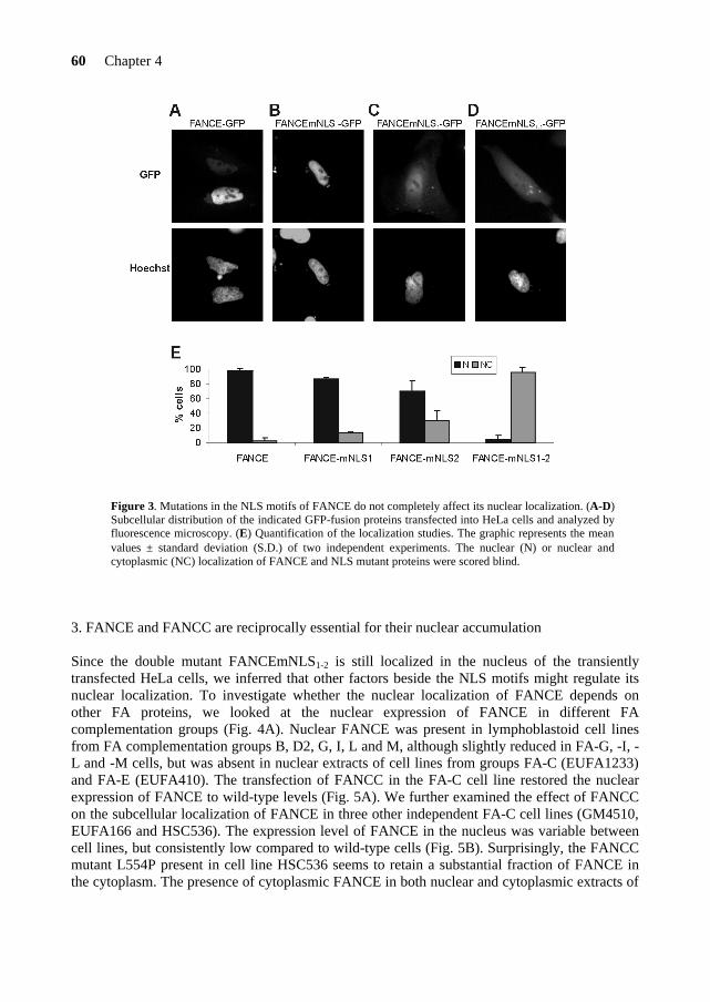
60 Chapter 4
Figure 3. Mutations in the NLS motifs of FANCE do not completely affect its nuclear localization. (A-D) Subcellular distribution of the indicated GFP-fusion proteins transfected into HeLa cells and analyzed by fluorescence microscopy. (E) Quantification of the localization studies. The graphic represents the mean values ± standard deviation (S.D.) of two independent experiments. The nuclear (N) or nuclear and cytoplasmic (NC) localization of FANCE and NLS mutant proteins were scored blind.
3. FANCE and FANCC are reciprocally essential for their nuclear accumulation Since the double mutant FANCEmNLS1-2 is still localized in the nucleus of the transiently transfected HeLa cells, we inferred that other factors beside the NLS motifs might regulate its nuclear localization. To investigate whether the nuclear localization of FANCE depends on other FA proteins, we looked at the nuclear expression of FANCE in different FA complementation groups (Fig. 4A). Nuclear FANCE was present in lymphoblastoid cell lines from FA complementation groups B, D2, G, I, L and M, although slightly reduced in FA-G, -I, -L and -M cells, but was absent in nuclear extracts of cell lines from groups FA-C (EUFA1233) and FA-E (EUFA410). The transfection of FANCC in the FA-C cell line restored the nuclear expression of FANCE to wild-type levels (Fig. 5A). We further examined the effect of FANCC on the subcellular localization of FANCE in three other independent FA-C cell lines (GM4510, EUFA166 and HSC536). The expression level of FANCE in the nucleus was variable between cell lines, but consistently low compared to wild-type cells (Fig. 5B). Surprisingly, the FANCC mutant L554P present in cell line HSC536 seems to retain a substantial fraction of FANCE in the cytoplasm. The presence of cytoplasmic FANCE in both nuclear and cytoplasmic extracts of
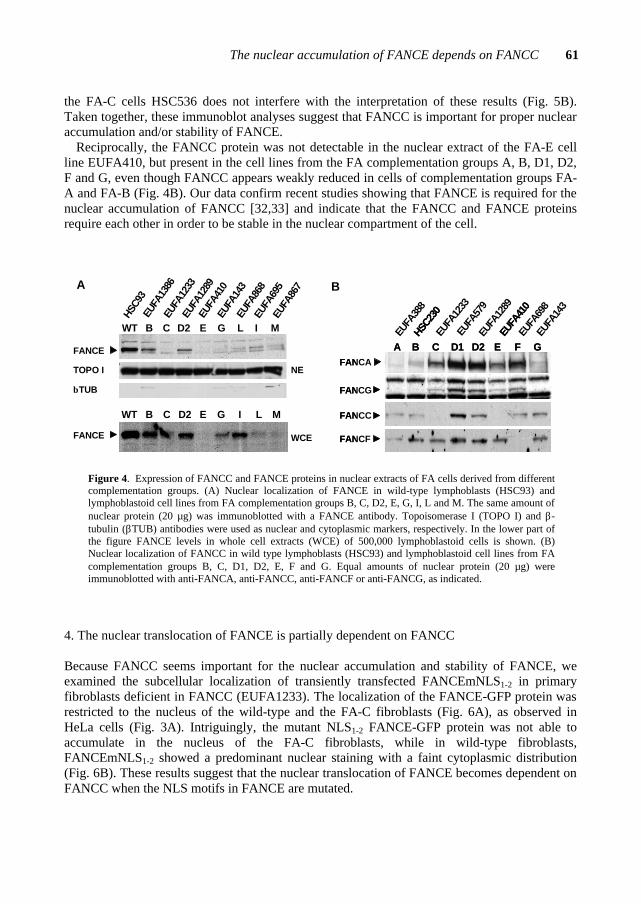
The nuclear accumulation of FANCE depends on FANCC 61
the FA-C cells HSC536 does not interfere with the interpretation of these results (Fig. 5B). Taken together, these immunoblot analyses suggest that FANCC is important for proper nuclear accumulation and/or stability of FANCE.
Reciprocally, the FANCC protein was not detectable in the nuclear extract of the FA-E cell line EUFA410, but present in the cell lines from the FA complementation groups A, B, D1, D2, F and G, even though FANCC appears weakly reduced in cells of complementation groups FA-A and FA-B (Fig. 4B). Our data confirm recent studies showing that FANCE is required for the nuclear accumulation of FANCC [32,33] and indicate that the FANCC and FANCE proteins require each other in order to be stable in the nuclear compartment of the cell.
B
EUFA
388
EUFA
1233
EUFA
579
EUFA
1289
EUFA
698
EUFA
143
CA B D1 D2 E F G
HSC2
30
EUFA
410
FANCG
FANCA
FANCC
FANCF
CA B D1 D2 E F G
HSC2
30
EUFA
410
FANCG
FANCA
FANCC
FANCF
A
βTUB
WT B C D2 E G L I M
HSC9
3EU
FA13
86EU
FA12
33EU
FA12
89EU
FA41
0EU
FA14
3EU
FA86
8EU
FA69
5EU
FA86
7
FANCE
TOPO I
FANCE
WT B C D2 E G LI M
NE
WCE
Figure 4. Expression of FANCC and FANCE proteins in nuclear extracts of FA cells derived from different complementation groups. (A) Nuclear localization of FANCE in wild-type lymphoblasts (HSC93) and lymphoblastoid cell lines from FA complementation groups B, C, D2, E, G, I, L and M. The same amount of nuclear protein (20 µg) was immunoblotted with a FANCE antibody. Topoisomerase I (TOPO I) and β-tubulin (βTUB) antibodies were used as nuclear and cytoplasmic markers, respectively. In the lower part of the figure FANCE levels in whole cell extracts (WCE) of 500,000 lymphoblastoid cells is shown. (B) Nuclear localization of FANCC in wild type lymphoblasts (HSC93) and lymphoblastoid cell lines from FA complementation groups B, C, D1, D2, E, F and G. Equal amounts of nuclear protein (20 µg) were immunoblotted with anti-FANCA, anti-FANCC, anti-FANCF or anti-FANCG, as indicated.
4. The nuclear translocation of FANCE is partially dependent on FANCC Because FANCC seems important for the nuclear accumulation and stability of FANCE, we examined the subcellular localization of transiently transfected FANCEmNLS1-2 in primary fibroblasts deficient in FANCC (EUFA1233). The localization of the FANCE-GFP protein was restricted to the nucleus of the wild-type and the FA-C fibroblasts (Fig. 6A), as observed in HeLa cells (Fig. 3A). Intriguingly, the mutant NLS1-2 FANCE-GFP protein was not able to accumulate in the nucleus of the FA-C fibroblasts, while in wild-type fibroblasts, FANCEmNLS1-2 showed a predominant nuclear staining with a faint cytoplasmic distribution (Fig. 6B). These results suggest that the nuclear translocation of FANCE becomes dependent on FANCC when the NLS motifs in FANCE are mutated.
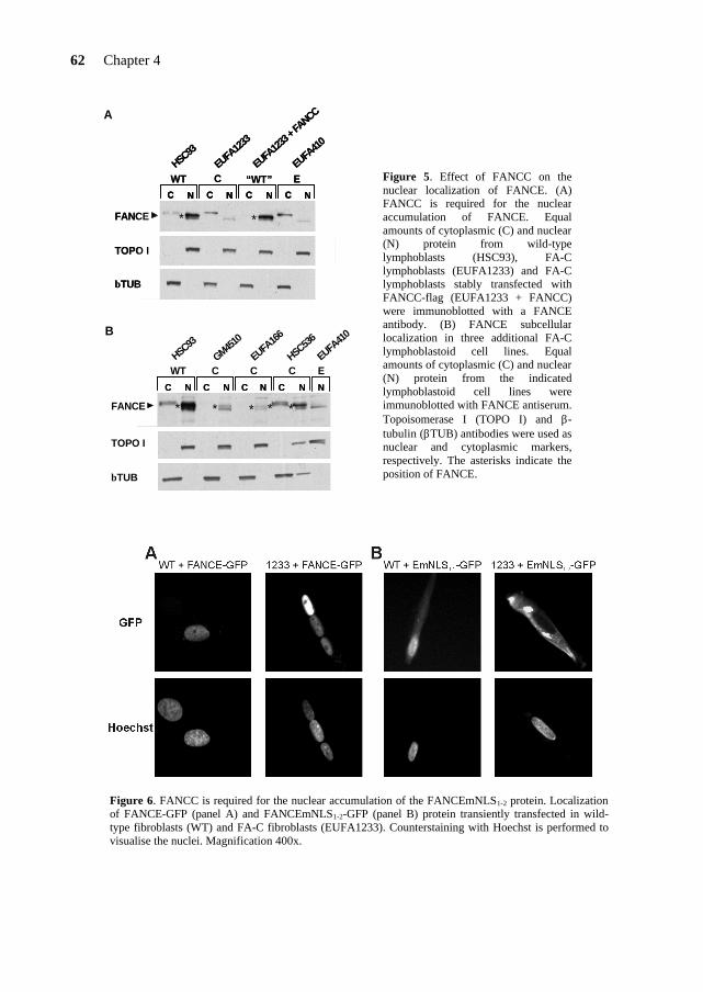
62 Chapter 4
C N C N C N C N
WT C “WT” EHSC93
EUFA12
33
EUFA41
0
EUFA12
33 +
FANCC
TOPO I
βTUB
FANCE * *
C NC N C NC N C NC N C NC N
WT C “WT” EHSC93
EUFA12
33
EUFA41
0
EUFA12
33 +
FANCC
TOPO I
βTUB
FANCE * *
A
WT C C CHSC93
GM4510
HSC536
EUFA16
6
EEUFA
410
TOPO I
βTUB
FANCE
C NC N C NC N C NC N C NC N NN
* * * * *
B
Figure 6. FANCC is required for the nuclear accumulation of the FANCEmNLS1-2 protein. Localization of FANCE-GFP (panel A) and FANCEmNLS1-2-GFP (panel B) protein transiently transfected in wild-type fibroblasts (WT) and FA-C fibroblasts (EUFA1233). Counterstaining with Hoechst is performed to visualise the nuclei. Magnification 400x.
Figure 5. Effect of FANCC on the nuclear localization of FANCE. (A) FANCC is required for the nuclear accumulation of FANCE. Equal amounts of cytoplasmic (C) and nuclear (N) protein from wild-type lymphoblasts (HSC93), FA-C lymphoblasts (EUFA1233) and FA-C lymphoblasts stably transfected with FANCC-flag (EUFA1233 + FANCC) were immunoblotted with a FANCE antibody. (B) FANCE subcellular localization in three additional FA-C lymphoblastoid cell lines. Equal amounts of cytoplasmic (C) and nuclear (N) protein from the indicated lymphoblastoid cell lines were immunoblotted with FANCE antiserum. Topoisomerase I (TOPO I) and β-tubulin (βTUB) antibodies were used as nuclear and cytoplasmic markers, respectively. The asterisks indicate the position of FANCE.

The nuclear accumulation of FANCE depends on FANCC 63
0
10
20
30
empty
FANCD2
FANCE
E+D2
EmNLS
1-2
EmNLS
1-2+
D2
Fold
indu
ctio
n re
lativ
e to
FA
NC
D2
0
2040
60
80100120
140
empty
FANCC
FANCE
E+C
EmNLS
1-2
EmNLS
1-2+
C
Fold
indu
ctio
n re
lativ
e to
FA
NC
C
A
B
0
10
20
30
empty
FANCD2
FANCE
E+D2
EmNLS
1-2
EmNLS
1-2+
D2
Fold
indu
ctio
n re
lativ
e to
FA
NC
D2
0
2040
60
80100120
140
empty
FANCC
FANCE
E+C
EmNLS
1-2
EmNLS
1-2+
C
Fold
indu
ctio
n re
lativ
e to
FA
NC
C
A
B
Figure 7. FANCEmNLS1-2 interacts with FANCC and FANCD2 in mammalian two-hybrid assays. Protein pairs were co-transfected in 293 cells in the presence of a luciferase reporter construct to test for direct interactions. (A) Interaction between FANCEmNLS1-2 and FANCC. (B) Interaction between FANCEmNLS1-2 and FANCD2. Fold induction is expressed relative to the luciferase activity obtained with the pVP16-AD-FANCC or pM-BD-FANCD2 vector alone. Results shown are mean ± S.D. and derived from an experiment in triplicate.
5. FANCE has different interaction domains for FANCC and FANCD2 The direct binding partners of FANCE are the FANCC and FANCD2 proteins [33-35]. To investigate the nature of these associations, we mapped contact sites implicated in the binding of FANCE by using the mammalian two-hybrid (M2H) assay. We first looked whether the NLS1-2 motifs in FANCE are involved in a direct interaction with either FANCC or FANCD2. Like wild-type FANCE, the FANCEmNLS1-2 protein showed a strong induction of the reporter gene with both FANCC and FANCD2 proteins (Fig. 7A and B), an indication that the NLS motifs are not directly involved in FANCC or FANCD2 interaction. Then, five FANCE deletion constructs were tested for interaction with wild-type FANCC and FANCD2 (Fig. 8). We found that only two FANCE fragments were able to interact with FANCC: an internal fragment of FANCE including the amino acids 149-371, and a fragment that lacks the first 148 amino acids of FANCE (149-536), although the reporter gene induction was weak for the latest (Fig. 8A and B). The FANCD2 protein interacted only with the FANCE fragment lacking the N-terminus (149-536) (Fig. 8C and D). Surprisingly, this interaction produced a very strong induction of the reporter gene, which was stronger than the association between FANCD2 and
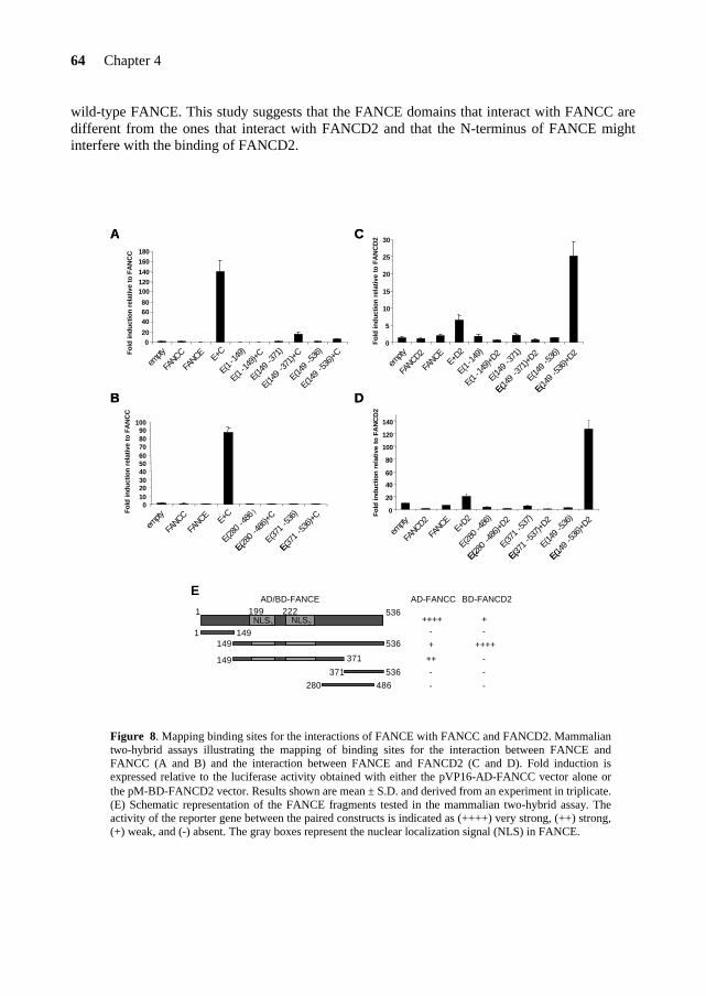
64 Chapter 4
wild-type FANCE. This study suggests that the FANCE domains that interact with FANCC are different from the ones that interact with FANCD2 and that the N-terminus of FANCE might interfere with the binding of FANCD2.
0
5
10
15
20
25
30
empty
FANCD2
FANCE
E+D2
E(1-1
49)
E(1-1
49)+D
2
E(149
-371)
E(149
-371)+
D2
E(149
-536)
E(149 -53
6)+D2
Fold
indu
ctio
n re
lativ
e to
FA
NC
D2
020
406080
100120140
160180
empty
FANCC
FANCE
E+C
E(1-1
49)
E(1-14
9)+C
E(149 -3
71)
E(149
-371)+
C
E(149 -5
36)
E(149
-536)+
CFold
indu
ctio
n re
lativ
e to
FA
NC
C
0102030405060708090
100
empty
FANCC
FANCE E+
C
E(280 -4
86)
E(280 -48
6)+C
E(371 -53
6)
E(371 -53
6)+C
Fold
indu
ctio
n re
lativ
e to
FA
NC
C
0
20
40
60
80
100
120
140
empty
FANCD2
FANCE
E+D2
E(280 -4
86)
E(280
-486
)+D2
E(371 -5
37)
E(371
-537)+
D2
E(149 -5
36)
E(149 -5
36)+D
2
Fold
indu
ctio
n re
lativ
e to
FA
NC
D2
A
B
C
D
0
5
10
15
20
25
30
empty
FANCD2
FANCE
E+D2
E(1-1
49)
E(1-1
49)+D
2
E(149
-371)
E(149
-371)+
D2
E(149
-536)
E(149 -53
6)+D2
Fold
indu
ctio
n re
lativ
e to
FA
NC
D2
020
406080
100120140
160180
empty
FANCC
FANCE
E+C
E(1-1
49)
E(1-14
9)+C
E(149 -3
71)
E(149
-371)+
C
E(149 -5
36)
E(149
-536)+
CFold
indu
ctio
n re
lativ
e to
FA
NC
C
0102030405060708090
100
empty
FANCC
FANCE E+
C
E(280 -4
86)
E(280 -48
6)+C
E(371 -53
6)
E(371 -53
6)+C
Fold
indu
ctio
n re
lativ
e to
FA
NC
C
0
20
40
60
80
100
120
140
empty
FANCD2
FANCE
E+D2
E(280 -4
86)
E(280
-486
)+D2
E(371 -5
37)
E(371
-537)+
D2
E(149 -5
36)
E(149 -5
36)+D
2
Fold
indu
ctio
n re
lativ
e to
FA
NC
D2
A
B
C
D
AD-FANCC
-++++
BD-FANCD2
-+
-
-
++
-
-
-
+ ++++
1
1491
536NLS1 NLS2
199 222
371280 486
149 371
149 536
AD/BD-FANCE
371
E
536
Figure 8. Mapping binding sites for the interactions of FANCE with FANCC and FANCD2. Mammalian two-hybrid assays illustrating the mapping of binding sites for the interaction between FANCE and FANCC (A and B) and the interaction between FANCE and FANCD2 (C and D). Fold induction is expressed relative to the luciferase activity obtained with either the pVP16-AD-FANCC vector alone or the pM-BD-FANCD2 vector. Results shown are mean ± S.D. and derived from an experiment in triplicate. (E) Schematic representation of the FANCE fragments tested in the mammalian two-hybrid assay. The activity of the reporter gene between the paired constructs is indicated as (++++) very strong, (++) strong, (+) weak, and (-) absent. The gray boxes represent the nuclear localization signal (NLS) in FANCE.
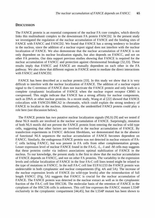
The nuclear accumulation of FANCE depends on FANCC 65
DISCUSSION
The FANCE protein is an essential component of the nuclear FA core complex, which directly links this multisubunit complex to the downstream FA protein FANCD2. In the present study we investigated the regulation of the nuclear accumulation of FANCE and the binding sites of FANCE with FANCC and FANCD2. We found that FANCE has a strong tendency to localize in the nucleus, since the addition of a nuclear export signal does not interfere with the nuclear localization of FANCE. We also demonstrate that the nuclear accumulation of FANCE is not only dependent on its nuclear localization signals, but also depends on FANCC, and not on other FA proteins. Our data support previous studies showing that FANCE is required for the nuclear accumulation of FANCC and protection against chromosomal breakage [32,33]. These results imply that FANCC and FANCE are mutually dependent on each other in the FA pathway. We also show that different regions in FANCE seem to mediate the direct interactions with FANCC and FANCD2.
FANCE has been described as a nuclear protein [33]. In this study we show that it is very
difficult to interfere with the nuclear localization of FANCE. The addition of a nuclear export signal to the C-terminus of FANCE does not inactivate the FANCE protein and only leads to a complete cytoplasmic localization of FANCE when the nuclear export receptor CRM1 is coexpressed. This might indicate that FANCE has a strong affinity for nuclear components, such as DNA or other nuclear proteins. In a recent study, Wang et al [22] showed that FANCE colocalizes with FANCD1/BRCA2 in chromatin, which could explain the strong tendency of FANCE to localize in the nucleus. Alternatively, the unidentified FANCI protein could play a role here (see discussion below).
The FANCE protein has two putative nuclear localization signals (NLS) [9] and we tested if
these NLS motifs are involved in the nuclear accumulation of FANCE. Surprisingly, mutation of both NLS motifs did not prevent the FANCE protein from entering the nucleus of wild type cells, suggesting that other factors are involved in the nuclear accumulation of FANCE. By transfection experiments in FANCC deficient fibroblasts, we demonstrated that in the absence of functional NLS sequences the nuclear accumulation of FANCE becomes dependent on FANCC. Similarly, the endogenous FANCE protein was not detected in nuclear extracts of FA-C cells lacking FANCC, but was present in FA cells from other complementation groups. Lesser expression level of nuclear FANCE found in the FA-G, -L, -I and -M cells may suggests that these proteins confer via indirect associations optimal stabilization of FANCE in the nucleus. To our knowledge, the present study is the first to show that the nuclear accumulation of FANCE depends on FANCC, and not on other FA proteins. The variability in the expression levels and cellular localization of FANCE in the four FA-C cell lines tested might be related to the type of mutations in FANCC. In the null FA-C cell line EUFA1233 the FANCE protein was undetectable in both cytoplasmic and nuclear compartments (Fig. 4A and 5A). The increase of the nuclear expression levels of FANCE (to wild-type levels) after the reintroduction of full length FANCC (Fig. 5A) suggests that FANCC is crucial for the nuclear accumulation of FANCE. The FANCE protein was detected in the nuclear extract as well as in the cytoplasmic fraction of the FA-C cell line HSC536. The molecular basis of the retention of FANCE in the cytoplasm of the HSC536 cells is unknown. This cell line expresses the FANCC mutant L554P exclusively in the cytoplasmic compartment [44,45], but the L554P mutant has been shown to

66 Chapter 4
be defective in binding FANCE using two hybrid assays [33,35]. One possible explanation is that FANCE weakly binds the L554P mutant in the presence of other FA proteins, which partially stabilizes and retains FANCE in the cytoplasmic compartment. This is consistent with the presence of a normal FANCA/FANCG complex in both cellular compartments of HSC536 cells [31] and the observation that FANCE has weak and/or transient interactions with both FANCA and FANCG [34]. It seems unlikely that the truncated FANCC mutants of the FA-C cell lines EUFA166 (322delG) and GM4510 (IVS4+4A→T) [46] were able to bind to FANCE, since FANCC deletion constructs failed to interact with FANCE [35]. In the nuclear extracts of those two FA-C cell lines, the level of FANCE was markedly reduced and FANCE was not retained in the cytoplasmic fraction (Fig. 5B). Taken together, these results indicate that FANCC is important for the nuclear accumulation and/or stability of FANCE. Reciprocally, previous studies demonstrated that FANCC needs the FANCE protein to accumulate in the nucleus of the cell [32,33], which we confirmed for the endogenous FANCC protein. Other FA core complex members, such as FANCA and FANCB might also produce a stabilizing effect on FANCC in the nucleus, since nuclear FANCC levels appeared to be slightly reduced in FA-A and -B cells. Similarly, lower FANCE expression levels were found in the nucleus of FA-G, -I, -L and -M cells, suggesting that several proteins are needed for stabilization of FANCE in the nucleus. Surprisingly, in FA-I cells cellular FANCE levels were normal, while nuclear FANCE levels were reduced. Interestingly, we also found reduced nuclear FANCC and FANCD2 levels in FA-I cells (de Winter, unpublished results). This could indicate that the unidentified FANCI protein is involved in the nuclear retention of FANCC, FANCE and FANCD2.
The expression of FANCC is regulated during the cell cycle and seems to involve
proteosomal degradation [47]. We hypothesize that the FANCC/FANCE interaction occurs in the nucleus upon a cell cycle regulated nuclear translocation of FANCC. In the absence of FANCC, the nuclear FANCE protein is unstable and similarly, FANCC needs to be stabilized in the nucleus by binding to FANCE. Inactivation of the NLS motifs in FANCE makes the nuclear translocation of FANCE dependent on the interaction with FANCC (piggy bag riding) and expression of a FANCC mutant defective in nuclear accumulation (like in HSC536) is able to retain a proportion of FANCE in the cytoplasm.
Does FANCE bind FANCD2 alone or in complex with FANCC? Although our study does
not distinguish between these possibilities, the mammalian two hybrid experiments suggest that different FANCE fragments interact with FANCC and FANCD2 (Fig. 8). The FANCC protein appears to interact with an internal region of FANCE (149-371), which includes the NLS motifs (see also Ref. [35]), but the interaction with full length FANCE is much stronger. FANCD2 interacts with the FANCE fragment lacking the first 148 amino acids (149-536) and all the other FANCE fragments failed to interact with FANCD2. This interaction is much stronger than with the full length FANCE, indicating that the N-terminus of FANCE might interfere with the binding of FANCD2. Possibly, the conformation of FANCE changes upon binding of FANCC and other FA proteins, thereby exposing the FANCD2 binding site. Further experiments need to be performed to prove this hypothesis, as we cannot exclude that removing the N-terminus of FANCE results in a more favorable positioning of the VP16 activation domain (AD) with regard to the GAL4 DNA-binding domain (BD) fused to FANCD2. In a recent study using the yeast two hybrid assay three FANCE mutants (Q312X, E263K and L348M) have been

The nuclear accumulation of FANCE depends on FANCC 67
described that had lost their ability to bind FANCD2, but were still able to bind FANCC [48]. This strengthens the idea that the FANCC and FANCD2 binding sites of FANCE are different.
Based on this and previous studies, we propose a model for the sequential assembly of the
nuclear FA core complex. The cytoplasmic FANCA/FANCG [41,49] and FANCB/FANCL [13] subcomplexes are probably translocated to the nucleus independently. In the nucleus these complexes interact, which needs the recently identified FANCM protein. The FANCE protein directly interacts with FANCC in the nucleus and then associates with the FANCA/FANCB/FANCG/FANCL subcomplex, an interaction that is stabilized by FANCF [31,40]. The association of the nuclear FA core complex with other factors (e.g. FANCI [16]) or the association of the FANCC/FANCE subcomplex with FANCF [40] could induce a structural change in FANCE resulting in the binding and monoubiquitination of FANCD2. It is also possible that posttranslational modifications of FANCD2 during S-phase (e.g. phosphorylation) permit the FANCE/FANCD2 interaction to occur.
We conclude that the FANCC and FANCE proteins are functionally linked since they require
each other to be stable in the nucleus of the cell. FANCC is important for the nuclear accumulation and/or stability of FANCE and similarly, FANCE is essential for the function of FANCC in the nucleus. Relationships between the FA proteins inside the FA core complex emerge as a complicated network of protein-protein interactions where FANCC and FANCE are tightly interrelated. A similar relationship has been found for the FANCA/FANCG and FANCB/FANCL protein pairs.
Acknowledgments We would like to thank Dr. K.J. Patel for kindly providing the FANCE antibody. This work was supported by the Fanconi Anemia Research Fund (Eugene, OR), the Dutch Cancer Society and the Netherlands Organization for Health Research and Development.
REFERENCES [1] H. Joenje, K.J. Patel, The emerging genetic and molecular basis of Fanconi anaemia,
Nat. Rev. Genet. 2 (2001) 446-457. [2] A.D. D’Andrea, M. Grompe, The Fanconi anemia/BRCA pathway, Nat.Rev.Cancer 3
(2003) 23-34. [3] M.D. Tischkowitz, S.V. Hodgson, Fanconi anaemia, J. Med. Genet. 40 (2003) 1-10. [4] C.A. Strathdee, H. Gavish, W.R. Shannon, M. Buchwald, Cloning of cDNAs for
Fanconi's anaemia by functional complementation, Nature 358 (1992) 434. [5] FAB; The Fanconi Anaemia/Breast Cancer Consortium, Positional cloning of the Fanconi
anaemia group A gene, Nat. Genet. 14 (1996) 324-328. [6] J.R. Lo Ten Foe, M.A. Rooimans, L. Bosnoyan-Collins, N. Alon, M. Wijker, L.
Parker, J. Lightfoot, M. Carreau, D.F. Callen, A. Savoia, N.C. Cheng, C.G.M. van Berkel, M.H. Strunk, J.J.P. Gille, G. Pals, F.A.E Kruyt, J.C. Pronk, F. Arwert, M. Buchwald, H. Joenje,
68 Chapter 4
Expression cloning of cDNA for the major Fanconi anaemia gene, FAA. Nat. Genet. 14 (1996) 320-323.
[7] J.P. de Winter, Q. Waisfisz, M.A. Rooimans, C.G.M. van Berkel, L. Bosnoyan-Collins, N. Alon, M. Carreau, O. Bender, I. Demuth, D. Schindler, J.C. Pronk, F. Arwert, H. Hoehn, M. Digweed, M. Buchwald, H. Joenje, The Fanconi anaemia group G gene FANCG is identical with XRCC9, Nat. Genet. 20 (1998) 281-283.
[8] J.P. de Winter, M.A. Rooimans, L. van der Weel, C.G van Berkel, N. Alon, L. Bosnoyan-Collins, J. de Groot, Y. Zhi, Q. Waisfisz, J.C. Pronk, F. Arwert, C.G. Mathew, R.K. Scheper, M.E. Hoatlin, M. Buchwald, H. Joenje, The Fanconi anaemia gene FANCF encodes a novel protein with homology to ROM, Nat. Genet. 24 (2000) 15-16.
[9] J.P. de Winter, F. Léveillé, C.G. van Berkel, M.A Rooimans, L van der Weel, J. Steltenpool, I. Demuth, N.V. Morgan, N. Alon, L. Bosnoyan-Collins, J. Lightfoot, P.A. Leegwater, Q. Waisfisz, K. Komatsu, F. Arwert, J.C. Pronk, C.G. Mathew, M. Digweed, M. Buchwald, H. Joenje, Isolation of a cDNA representing the Fanconi anemia complementation group E gene, Am. J. Hum. Genet. 67 (2000) 1306-1308.
[10] C. Timmers, T. Taniguchi, J. Hejna, C. Reifsteck, L. Lucas, D. Bruun, M. Thayer, B. Cox, S. Olson, A.D. D’Andrea, R. Moses, M. Grompe, Positional cloning of a novel Fanconi anemia gene, FANCD2, Mol. Cell 7 (2001) 241-248.
[11] N.G. Howlett, T. Taniguchi, S. Olson, B. Cox, Q. Waisfisz, C. de Die-Smulders, N. Persky, M. Grompe, H. Joenje, G. Pals, H. Ikeda, E.A Fox, A.D. D’Andrea, Biallelic inactivation of BRCA2 in Fanconi anemia, Science 297 (2002) 606-609.
[12] A.R. Meetei, J.P. de Winter, A.L Medhurst, M. Wallisch, Q. Waisfisz, H.J. van de Vrugt, A.B. Oostra, Z. Yan, C. Ling, C.E. Bishop, M.E. Hoatlin, H. Joenje, W. Wang, A novel ubiquitin ligase is deficient in Fanconi anemia, Nat. Genet. 35 (2003) 165-170.
[13] A.R. Meetei, M. Levitus, Y. Xue, A.L. Medhurst, M. Zwaan, C. Ling, M.A. Rooimans, P. Bier, M. Hoatlin, G. Pals, J.P. de Winter, W. Wang, H. Joenje, X-linked inheritance of Fanconi anemia complementation group B, Nat. Genet. 36 (2004) 1219-1224.
[14] A.R. Meetei, A.L. Medhurst, C. Ling, Y. Xue, T.R. Singh, P. Bier, J. Steltenpool, S. Stone, I. Dokal, C.G. Mathew, M. Hoatlin, H. Joenje, J.P de Winter, W. Wang, A Human Orthologue of Archaeal DNA Repair Protein Hef is Defective in Fanconi Anemia Complementation Group M, Nat. Genet. 37 (2005) 958-963.
[15] M. Levitus, Q. Waisfisz, B.C. Godthelp, Y. de Vries, S. Hussain, W.W. Wiegant, E. Elghabzouri-Maghrani, J. Steltenpool, M.A. Rooimans, G. Pals, F. Arwert, C.G. Mathew, M.Z. Zdzienicka, K. Hiom, J.P. de Winter, H. Joenje, The DNA helicase BRIP1 is defective in Fanconi anemia complementation group J, Nat. Genet. 37 (2005) 934-935.
[16] M. Levitus, M.A. Rooimans, J. Steltenpool, N.F.C. Cool, A.B. Oostra, C.G. Mathew, M.E. Hoatlin, Q.Waisfisz, F. Arwert, J.P. de Winter, H. Joenje, Heterogeneity in Fanconi anemia: evidence for two new genetic subtypes, Blood 103 (2004) 2498-2503.
[17] I. Garcia-Higuera, T. Taniguchi, S. Ganesan, M.S. Meyn, C. Timmers, J. Hejna, M. Grompe, A.D. D’Andrea, Interaction of the Fanconi anemia proteins and BRCA1 in a common pathway, Mol. Cell 7 (2001) 249-269.
[18] T. Taniguchi, I. Garcia-Higuera, P.R. Andreassen, R.C. Gregory, M. Grompe, A.D. D’Andrea, S-phase-specific interaction of the Fanconi anemia protein, FANCD2, with BRCA2 and RAD51, Blood 100 (2002) 2414-2420.
The nuclear accumulation of FANCE depends on FANCC 69
[19] K. Nakanishi, T. Taniguchi, V. Ranganathan, H.H. New, L.A. Moreau, M. Stotsky, C.G. Mathew, M.B. Kastan, D.T. Weaver, A.D. D’Andrea, Interaction of FANCD2 and NBS1 in the DNA damage response, Nat. Cell Biol. 4 (2002) 913-920.
[20] S.M. Nijman, T.T. Huang, A.M. Dirac, T.R. Brummelkamp, R.M. Kerkhoven, A.D. D'Andrea, R. Bernards, The deubiquitinating enzyme USP1 regulates the Fanconi anemia pathway, Mol. Cell 7 (2005) 331-339.
[21] W.H. Park, S. Margossian, A.A. Horwitz, A.M. Simons, A.D. D'Andrea, J.D. Parvin, Direct DNA binding activity of the Fanconi anemia D2 protein, J. Biol. Chem. 25 (2005) 23593-23598.
[22] X.Z. Wang, P.R. Andreassen, A.D. D’Andrea, Functional Interaction of monoubiquitination FANCD2 and BRCA2/FANCD1 in chromatin, Mol. Cell. Biol. 24 (2004) 5850-5862.
[23] S. Hirano, K. Yamamoto, M. Ishiai, M. Yamazoe, M. Seki, N. Matsushita, M. Ohzeki, Y. M. Yamashita, H. Arakawa, J-M. Buerstedde, T. Enomoto, S. Takeda, L.H. Thompson, M. Takata, Functional relationships of FANCC to homologous recombination, translation synthesis, and BLM, EMBO J. 24 (2005) 418-427.
[24] W. Niedwiedz, G. Mosedale, M. Johnson, C. Yi Ong, P. Pace, K.J. Patel, The Fanconi anaemia gene FANCC promotes homologous recombination and error-prone DNA repair, Mol. Cell 15 (2004) 607-620.
[25] K. Nakanishi, Y-G. Yang, A.J. Pierce, T. Taniguchi, M. Digweed, A.D. D’Andrea, Z-Q. Wang, M. Jasin, Human Fanconi anemia monoubiquitination pathway promotes homologous DNA repair, Proc. Natl. Acad. Sci. 102 (2005) 1110-1115.
[26] A. Ohashi, M.Z. Zdzienicka, J. Chen, F.J. Couch, Fanconi anemia complementation group D2 (FANCD2) functions independently of BRCA2- and RAD51-associated homologous recombination in response to DNA damage, J. Biol. Chem. 15 (2005) 14877-14883.
[27] K. Yamamoto, S. Hirano, M. Ishiai, K. Morishima, H. Kitao, K. Namikoshi, M. Kimura, N. Matsushita, H. Arakawa, J-M. Buerstedde, K. Komatsu, L.H. Thompson, M. Takata, Fanconi anemia protein FANCD2 promotes immunoglobulin gene conversion and DNA repair through a mechanism related to homologous recombination, Mol. Cell. Biol. 25 (2005) 34-43.
[28] T. Yamashita, D.L. Baber, Y. Zhu, H. Wu, A.D. D’Andrea, The Fanconi anemia polypeptide FACC is localized to the cytoplasm of human cells, Proc. Natl. Acad. Sci. 91 (1994) 6712-6716.
[29] H. Youssoufian, Localization of Fanconi anemia C protein to the cytoplasm of mammalian cells, Proc. Natl. Acad. Sci. 91 (1994) 7975-7979.
[30] M.E. Hoatlin, T.A. Christianson, W.W. Keeble, A.T. Hammond, Y. Zhi, M.C. Heinrich, P.A. Tower, Jr,G.C. Bagby, The Fanconi anemia group C gene product is located in both the nucleus and cytoplasm of human cells, Blood 91 (1998) 1418-1425.
[31] J.P. de Winter, L. van der Weel, J. de Groot, S. Stone, Q. Waisfisz, F. Arwert, R.J. Scheper, F.A.E. Kruyt, M.E. Hoatlin, H. Joenje, The Fanconi anemia protein FANCF forms a nuclear complex with FANCA, FANCC and FANCG, Hum. Mol. Genet. 18 (2000) 2665-2674.
[32] T. Taniguchi, A.D. D’Andrea, The Fanconi anemia protein, FANCE, promotes the nuclear accumulation of FANCC, Blood 100 (2002) 2457-2462.
[33] P. Pace, M. Johnson, W.M. Tan, G. Mosedale, C. Sng, M. Hoatlin, J. de Winter, H. Joenje, F. Gergely, K.J. Patel, FANCE: the link between Fanconi anaemia complex assembly and activity, EMBO J. 21 (2002) 3414-3423.

70 Chapter 4
[34] A.L. Medhurst, P.A.J. Huber, Q. Waisfisz, J.P. de Winter, C.G. Mathew, Direct interactions of the five known Fanconi anaemia proteins suggest a common functional pathway, Hum. Mol. Genet. 10 (2001) 423-429.
[35] S.M. Gordon, M. Buchwald, Fanconi anemia protein complex: mapping protein interactions in the yeast 2- and 3-hybrid systems, Blood 102 (2003) 136-141.
[36] R. Ishida, M. Buchwald, Susceptibility of Fanconi's anemia lymphoblasts to DNA-cross-linking and alkylating agents, Cancer Res. 42 (1982) 4000-4006.
[37] H. Joenje, J.R. Lo Ten Foe, A.B. Oostra, C.G.M. van Berkel, M.A. Rooimans, T. Schroeder-Kurth, R-D. Wegner, J.J.P. Gille, M. Buchwald, F. Arwert,. Classification of Fanconi anemia patients by complementation analysis: evidence for a fifth genetic subtype, Blood 86 (1995) 2156-2160.
[38] F.A.E. Kruyt, H. Youssoufian, The Fanconi anemia proteins FAA and FAC function in different cellular compartments to protect against cross-linking agent cytotoxicity, Blood 92 (1998) 2229-2236.
[39] M. Ferrer, J.A. Rodriguez, E.A. Spierings, J.P. de Winter, G. Giaccone, F.A. Kruyt, Identification of multiple nuclear export sequences in Fanconi anemia group A protein that contribute to CRM1-dependent nuclear export, Hum. Mol. Genet. 14 (2005) 1271-1281.
[40] F. Léveillé, E. Blom, A.L. Medhurst, P. Bier, E.H. Laghmani, M. Johnson, M.A. Rooimans, A. Sobeck, Q. Waisfisz, F. Arwert, K.J. Patel, M.E. Hoatlin, H. Joenje, J.P. de Winter, The Fanconi anemia gene product FANCF is a flexible adaptor protein, J. Biol. Chem. 38 (2004) 39421-39430.
[41] Q. Waisfisz, J.P. de Winter, F.A.E. Kruyt, J. de Groot, L. van der Weel, L. M. Dijkmans, Y. Zhi, F. Arwert, R.J. Scheper, H. Youssoufian, A physical complex of the Fanconi anemia proteins FANCG/XRCC9 and FANCA, Proc. Natl. Acad. Sci. 96 (1999) 10320-10325.
[42] J. Moroianu, Nuclear import and export pathway, J. Cell. Biochem. Suppl. 32-33 (1999) 76-83.
[43] B.R. Henderson, A. Eleftheriou, A comparison of the activity, sequence specificity, and CRM1-dependence of different nuclear export signals, Exp. Cell Res. 256 (2000) 213-224.
[44] M.E. Hoatlin, Y. Zhi, H. Ball, K. Silvey, A. Melnick, S. Stone, S. Arai, N. Hawe, G. Owen, A. Zelent, J.D. Licht, A novel BTB/POZ transcriptional repressor protein interacts with the Fanconi anemia group C protein and PLZF, Blood 94 (1999) 3737-3747.
[45] I. Garcia-Higuera, A.D. D’Andrea, Nuclear localization of the Fanconi anemia protein FANCC is required for functional activity, Blood 93 (1999) 4025-4026.
[46] T. Yamashita, N. Wu, G.M. Kupfer, C. Corless, H. Joenje, M. Grompe, A.D. D’Andrea, Clinical variability of Fanconi anemia (type C) results from expression of an amino terminal truncated Fanconi anemia complementation group C polypeptide with partial activity, Blood 87 (1996) 4424-4432.
[47] M.C. Heinrich, K.V. Silvey, S. Stone, A.J. Zigler, D.J. Griffith, M. Montalto, L. Chai, Y. Zhi, M.E. Hoatlin, Posttranscriptional cell cycle-dependent regulation of human FANCC expression, Blood 95 (2000) 3970-3977.
[48] S.M. Gordon, N. Alon, M. Buchwald, FANCC, FANCE and FANCD2 form a ternary complex essential to the integrity of the Fanconi anemia DNA damage response pathway, J. Biol. Chem. 280 (2005) 36118-36125.
[49] I. Garcia-Higuera, Y. Kuang, D. Näf, J. Wasik, A.D. D’Andrea, Fanconi anemia proteins FANCA, FANCC, and FANCG/XRCC9 interact in a functional nuclear complex, Mol. Cell. Biol. 19 (1999) 4866-4873.


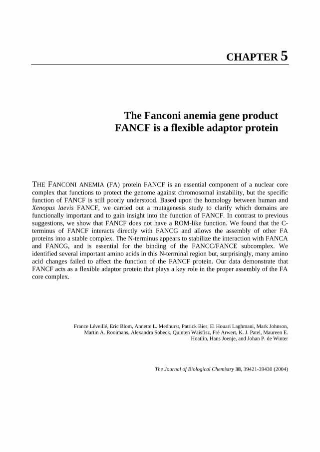
CHAPTER 5
THE FANCONI ANEMIA (FA) protein FANCF is an essential component of a nuclear core complex that functions to protect the genome against chromosomal instability, but the specific function of FANCF is still poorly understood. Based upon the homology between human and Xenopus laevis FANCF, we carried out a mutagenesis study to clarify which domains are functionally important and to gain insight into the function of FANCF. In contrast to previous suggestions, we show that FANCF does not have a ROM-like function. We found that the C-terminus of FANCF interacts directly with FANCG and allows the assembly of other FA proteins into a stable complex. The N-terminus appears to stabilize the interaction with FANCA and FANCG, and is essential for the binding of the FANCC/FANCE subcomplex. We identified several important amino acids in this N-terminal region but, surprisingly, many amino acid changes failed to affect the function of the FANCF protein. Our data demonstrate that FANCF acts as a flexible adaptor protein that plays a key role in the proper assembly of the FA core complex.
France Léveillé, Eric Blom, Annette L. Medhurst, Patrick Bier, El Houari Laghmani, Mark Johnson, Martin A. Rooimans, Alexandra Sobeck, Quinten Waisfisz, Fré Arwert, K. J. Patel, Maureen E.
Hoatlin, Hans Joenje, and Johan P. de Winter
The Journal of Biological Chemistry 38, 39421-39430 (2004)
The Fanconi anemia gene product FANCF is a flexible adaptor protein


FANCF is a flexible adaptor protein 75
INTRODUCTION
Fanconi anemia (FA) is an autosomal recessive chromosomal instability syndrome. The clinical phenotype of FA patients is characterized by congenital abnormalities, progressive bone marrow failure, and a predisposition to cancer; particularly acute myeloid leukemia and squamous cell carcinoma (1,2). The spontaneous cytogenetic aberrations specific for FA cells are exacerbated upon treatment with DNA cross-linking agents, such as mitomycin C (MMC)
and diepoxybutane, which suggests a DNA maintenance defect particularly in the handling of cross-link damage.
To date, somatic cell fusion studies have demonstrated 11 FA complementation groups (A-C,
D1, D2, E-G, I, J, L) (3) and eight of the FA associated genes have been identified: FANCA, FANCC, FANCD1/BRCA2, FANCD2, FANCE, FANCF, FANCG and FANCL (4-12). These FA genes encode orphan proteins and no functional domains have been found in their primary amino acid sequences, except for a RING finger in FANCL (10) and multiple TPR motifs in FANCG (13). In FANCF a region with homology to RNA-binding protein ROM has been suggested to provide a clue for its function (5).
FANCA, FANCC, FANCE, FANCF, FANCG and FANCL assemble in a nuclear core
complex (10, 14-17), in which FANCG binds directly to FANCA and FANCF and FANCC binds to FANCE (15, 16). The FA core complex is essential for the monoubiquitination of FANCD2 (18) and this modified form of FANCD2 colocalizes with BRCA1, RAD51 (19) and PCNA (20) in foci that also contain other DNA repair proteins. Nevertheless, it is still unknown how monoubiquitinated FANCD2 is involved in DNA repair and MMC resistance. Recently, biallelic mutations in BRCA2 have been found in cell lines derived from FA-D1 patients, adding BRCA2 to the list of FA proteins (8). The BRCA proteins are known to be involved in a multitude of biological functions including DNA repair, recombination, cell cycle control and transcription (21). BRCA2 appears to be directly linked to the repair of double-stranded breaks (DSBs) by homologous recombination (22). FANCD2 has also been identified as a target of the ataxia telangiectasia (AT) signaling pathway (23) and seems to be functionally connected to the RAD50/MRE11/NBS1 (RMN) protein complex (24, 25), which plays an important role in the repair of DSBs (26). Taken together, these data suggest that the FA proteins serve to maintain genomic stability and integrity, in concert with other protein complexes.
FANCF plays an important role in the FA pathway and its functional disruption seems to be
involved in specific types of cancer, as suggested from the hypermethylation of the FANCF promoter in a subset of ovarian, oral, lung and cervical cancers (27, 28, 29). Nevertheless, the function of FANCF is poorly understood. Patient-derived mutant forms of FA proteins and structure/function analysis of FANCA, FANCC and FANCG have been informative in finding important functional residues (30-34), but for FANCF this information is lacking, since none of the FA-F patients have missense mutations that could provide insight into its function (5). For this reason, we started a site-directed mutagenesis study to obtain more information about functional domains in the FANCF protein. Because such domains are expected to be relatively conserved during evolution, we searched for FANCF homologs in lower vertebrates that might highlight important residues in FANCF. We found a Xenopus laevis homolog of FANCF (xFANCF), which has a relatively low overall homology with human FANCF. However, two

76 Chapter 5
relatively conserved regions were located at the N- and C- terminus. Based upon this conservation and amino acid properties, we generated FANCF a large panel of FANCF mutants and identified several functionally important amino acids and domains in FANCF. This study reveals that FANCF is an adaptor protein that plays a key role in the proper assembly of the FA core complex. To be able to perform this function the N-terminus of FANCF interacts with the FANCC/FANCE subcomplex, whereas the C-terminus binds to the FANCA/FANCG subcomplex.
EXPERIMENTAL PROCEDURES Cell Culture and Transfection Epstein-Barr virus (EBV)-transformed lymphoblasts were cultured in RPMI 1640 media supplemented with 1 mM glutamine (Gibco, Invitrogen, Carlsbad, CA, USA) and 10% fetal calf serum (FCS; Gibco). Selection medium to obtain stable cell lines also contained hygromycin B (100 µg/ml; Roche, Basel, Switzerland). For stable expression lymphoblastoid cell lines were transfected by electroporation using an ECM830 electro square porator (BTX, San Diego, CA, USA). The MMC-induced growth inhibition assays were performed as previously described (35-36). Generation of FANCF Mutant Constructs The FANCF mutant constructs were generated by polymerase chain reaction (PCR) with oligonucleotides encoding the amino acid substitutions or deleted regions of the FANCF sequence. We used as a template the FANCF cDNA clone 10 obtained by expression cloning (5). The FANCF mutant R47A+F48A was made using the QuikChange site-directed mutagenesis kit (Stratagene, La Jolla, CA, USA) according to the manufacturer’s instructions. All FANCF mutants have been analyzed by sequencing to confirm that the desired changes had occurred and to exclude the presence of other PCR derived alterations. The mutants were subcloned into the expression vector pCEP4 (Invitrogen) and transfected in FA-F lymphoblasts, EUFA698. FANCF mutants 1-15del and delC31, and wild type FANCF-Flag were also subcloned into the expression vector pIRESneo (Clontech, Palo Alto, CA, USA) to generate stable cell lines. Expression of the mutant proteins in the stable cell lines was confirmed by immunoprecipitation and immunoblotting with FANCF-specific antibodies. Sequence Analysis FANCF mutants and the Xenopus laevis FANCF IMAGE clone 3200942 (obtained from HGMP Resource Centre, Cambridge, UK), were subcloned in pBluescript SK- and were sequenced with CY5.5 labeled T7 and T3 primers using a Thermo Sequenase primer cycle sequencing kit (Amersham). Products were analysed on a Visible Genetics automatic DNA sequencer (Visible Genetics Inc, Toronto, Ontario, Canada). Yeast Two-hybrid Analysis The MATCHMAKER Two-Hybrid System 3 (Clontech) was used according to the manufacturer’s instructions as previously described (15). In brief, bait and prey constructs were sequentially transformed into AH109 yeast cells and selected on -Trp-Leu-His-Ade medium. The resulting colonies were tested for β- galactosidase expression with 5-bromo-4-chloro-3-
FANCF is a flexible adaptor protein 77
indolyl-β-D-galactopyranoside (X-gal). To confirm interactions a yeast-mating assay was used where constructs were transformed separately into two different yeast strains and mating cultures plated onto selection medium as above. All constructs were tested for self-activation against a series of control plasmids and expression of the mutant proteins was verified by immunoblotting. Mammalian Two- and Three-hybrid Analysis Human embryonic kidney cells 293, either untransfected or stably transfected with wild type or mutant (L554P) FANCC were plated onto six well plates. After 48h, the cells were transiently transfected with FANCE cDNA fused to the GAL-4 activation domain (pVP16; Clontech) and the indicated FANCF constructs fused in frame to the GAL4 DNA-binding domain (pM; Clontech) (1 µg of each), together with a GAL4 driven reporter plasmid (G5E1bLUC, 0.2 µg). The luciferase activity was monitored after 24 h using a Dual-Luciferase Reporter Assay System (Promega) and a single tube luminometer (DLReady, Berthold Detection Systems), according to the manufacturer’s instructions. All GAL4 constructs were sequenced to confirm the correct reading frame and each experimental data set was performed in triplicate to overcome the variability inherent to transfections. Transient expression of the FANCF constructs and stable expression of FANCC were confirmed by immunoblotting with FANCF and FANCC specific antisera. Subcellular Fractionation, Immunoprecipitation and Immunoblotting Nuclear and cytoplasmic fractions of the lymphoblastoid cell lines were obtained as described previously (17). Cell extracts (~107 cells) were prepared in lysis buffer (50 mM Tris pH 7.4, 150 mM NaCl and 1% Nonidet NP40 supplemented with protease inhibitors) and subjected to immunoprecipitation using the indicated antibodies, as previously described (14). Anti-FLAG M2 affinity gel (Sigma-Aldrich, Saint-Louis, MO, USA) was used to immunoprecipitate FLAG-tagged proteins. Immunoprecipitates or lysates were then separated on 8 % SDS-polyacrylamide gels, transferred to PVDF membranes and specific proteins were detected by immunoblotting with the indicated antibodies.
RESULTS
Homology between the Human and Xenopus FANCF Proteins Since functionally important amino acids are well conserved between species, we searched several databases for FANCF homologs to obtain clues about functional domains. In the NCBI database an IMAGE clone (3200942) was found, which represents the full-length Xenopus laevis homolog of FANCF (xFANCF). The cDNA was sequenced and the predicted protein was aligned with the human FANCF sequence (Fig. 1). The overall homology between both sequences appeared to be limited (27% identity, 49% similarity) and dispersed over the whole molecule. However, relatively high sequence conservation was found in both the N- and C-terminal regions of the protein. To test if this limited homology was sufficient to function in a human background, FLAG-tagged xFANCF was transfected into a cell line from an FA-F patient (EUFA698). Unlike the human protein, xFANCF was unable to complement the MMC
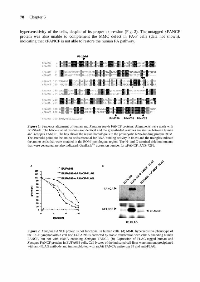
78 Chapter 5
hypersensitivity of the cells, despite of its proper expression (Fig. 2). The untagged xFANCF protein was also unable to complement the MMC defect in FA-F cells (data not shown), indicating that xFANCF is not able to restore the human FA pathway.
F1-15del
hFANCF 1 MESLLQHLDRFSELLAVSSTTYVSTWDPATVRRALQWARYLRHIHRRFGRHGPIRTALERxFANCF 1 MHTMLENLDRFIELLTLSQSVHVKDWDILCVRRGFEWGSYFQQVYHRFCANDCLRNALED
hFANCF 61 RLHNQWRQEGGFGRGPVPGLANFQALGHCDVLLSLRLLENRALGDAARYHLVQQLFPGPGxFANCF 61 HLRSKNQKLSTCIKN--YQYISFNDLRHSKDIFCTSLLQNKALSRNIFQYIVTKLK----
hFANCF 121 VRDADE ETLQESLARLARRRSAVHMLRFNGYRENPNLQEDSLMKTQAELLLERLQ-EVGKxFANCF 115 -NQSSGETNTCNMSNIMCQKAASALLLFPSSIDSKNPLENPCAVTEAEILKNHLEYRIHS
* *hFANCF 180 AEA ERPARFLSSLWERLPQNNFLKVIAVALLQPPLSRRPQEELEPGIHKSPGEG SQVLVHxFANCF 174 LKE DHQFAVVSDVLSGIPQPSIYNLLAILLLSSETCD-SQDQLS---------LSHLLLK
hFANCF 240 WLLGNSEVFAAFCRALPAGLLTLVTSRHPALSPVYLGLLTDWGQRLHYDLQKGIWVGTESxFANCF 224 WLLENDSVWTDFCSKIHSHLLSHLSSKYSIVRKAYFNFLIKLGESMELDVQG-KWVSN-S
hFANCF 300 Q DVPWEELHNRFQSLCQAPPPLKDKVLTALETCKAQDGDFEVPGLSIWTDLLLALRSGAFxFANCF 282 A ELSFDIFLNHFESLMKTSEDLQTATEATLEDLKRQDGDYNVPGISVWTDVLLEVKRI
hFANCF 360 RKRQVLGLSAGLSSV FdelC40 FdelC31 FdelC23
F1-15del
hFANCF 1 MESLLQHLDRFSELLAVSSTTYVSTWDPATVRRALQWARYLRHIHRRFGRHGPIRTALERxFANCF 1 MHTMLENLDRFIELLTLSQSVHVKDWDILCVRRGFEWGSYFQQVYHRFCANDCLRNALED
hFANCF 61 RLHNQWRQEGGFGRGPVPGLANFQALGHCDVLLSLRLLENRALGDAARYHLVQQLFPGPGxFANCF 61 HLRSKNQKLSTCIKN--YQYISFNDLRHSKDIFCTSLLQNKALSRNIFQYIVTKLK----
hFANCF 121 VRDADE ETLQESLARLARRRSAVHMLRFNGYRENPNLQEDSLMKTQAELLLERLQ-EVGKxFANCF 115 -NQSSGETNTCNMSNIMCQKAASALLLFPSSIDSKNPLE
CDVLLSLRLLENRALGDAARYHLVQQLFPGPGxFANCF 61 HLRSKNQKLSTCIKN--YQYISFNDLRHSKDIFCTSLLQNKALSRNIFQYIVTKLK----
hFANCF 121 VRDADE ETLQESLARLARRRSAVHMLRFNGYRENPNLQEDSLMKTQAELLLERLQ-EVGKxFANCF 115 -NQSSGETNTCNMSNIMCQKAASALLLFPSSIDSKNPLENPCAVTEAEILKNHLEYRIHS
* *hFANCF 180 AEA ERPARFLSSLWERLPQNNFLKVIAVALLQPPLSRRPQEELEPGIHKSPGEG SQVLVHxFANCF 174 LKE DHQFAVVSDVLSGIPQPSIYNLLAILLLSSETCD-SQDQLS---------LSHLLLK
hFANCF 240 WLLGNSEVFAAFCRALPAGLLTLVTSRHPALSPVYLGLLTDWGQRLHYD
NPCAVTEAEILKNHLEYRIHS* *
hFANCF 180 AEA ERPARFLSSLWERLPQNNFLKVIAVALLQPPLSRRPQEELEPGIHKSPGEG SQVLVHxFANCF 174 LKE DHQFAVVSDVLSGIPQPSIYNLLAILLLSSETCD-SQDQLS---------LSHLLLK
hFANCF 240 WLLGNSEVFAAFCRALPAGLLTLVTSRHPALSPVYLGLLTDWGQRLHYDLQKGIWVGTESxFANCF 224 WLLENDSVWTDFCSKIHSHLLSHLSSKYSIVRKAYFNFLIKLGESMELDVQG-KWVSN-S
hFANCF 300 Q DVPWEELHNRFQSLCQAPPPLKDKVLTALETCKAQDGDFEVPGLSIWTDLLLALRSGAFxFANCF 282 A ELSFDIFLNHFESLMKTSEDLQTATEATLEDLKRQDGDYNVPGISV
LQKGIWVGTESxFANCF 224 WLLENDSVWTDFCSKIHSHLLSHLSSKYSIVRKAYFNFLIKLGESMELDVQG-KWVSN-S
hFANCF 300 Q DVPWEELHNRFQSLCQAPPPLKDKVLTALETCKAQDGDFEVPGLSIWTDLLLALRSGAFxFANCF 282 A ELSFDIFLNHFESLMKTSEDLQTATEATLEDLKRQDGDYNVPGISVWTDVLLEVKRI
hFANCF 360 RKRQVLGLSAGLSSV FdelC40 FdelC31 FdelC23 Figure 1. Sequence alignment of human and Xenopus laevis FANCF proteins. Alignments were made with BoxShade. The black-shaded residues are identical and the gray-shaded residues are similar between human and Xenopus FANCF. The box shows the region homologous to the prokaryotic RNA-binding protein ROM. The asterisks point out the amino acids essential for RNA-binding activity in ROM and the triangles indicate the amino acids that were mutated in the ROM homologous region. The N- and C-terminal deletion mutants that were generated are also indicated. GenBankTM accession number for xFANCF: AY547288.
A BEUFA698EUFA698
EUFA698+hFANCF-FLAGEUFA698+hFANCF-FLAG
EUFA698+xFANCF-FLAGEUFA698+xFANCF-FLAG
010
2030
40
5060
7080
90100
110120
0 1 10 100
[MMC] (nM)
gro
wth
(%)
010
2030
40
5060
7080
90100
110120
0 1 10 100
[MMC] (nM)
gro
wth
(%)
hFANCF
EUFA
698
EUFA
698+
hFA
NCF-F
LAG
EUFA
698
+xFANCF
-FLA
G
IP: FLAG
FANCA
xFANCF
Figure 2. Xenopus FANCF protein is not functional in human cells. (A) MMC hypersensitive phenotype of the FA-F lymphoblastoid cell line EUFA698 is corrected by stable transfection with cDNA encoding human FANCF, but not with cDNA encoding Xenopus FANCF. (B) Expression of FLAG-tagged human and Xenopus FANCF proteins in EUFA698 cells. Cell lysates of the indicated cell lines were immunoprecipitated with anti-FLAG antibody and immunoblotted with rabbit FANCA antiserum 89 and anti-FLAG.
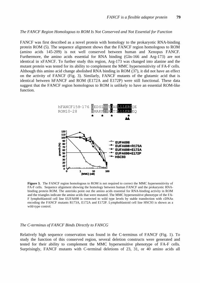
FANCF is a flexible adaptor protein 79
The FANCF Region Homologous to ROM Is Not Conserved and Not Essential for Function FANCF was first described as a novel protein with homology to the prokaryotic RNA-binding protein ROM (5). The sequence alignment shows that the FANCF region homologous to ROM (amino acids 145-209) is not well conserved between human and Xenopus FANCF. Furthermore, the amino acids essential for RNA binding (Gln-166 and Arg-173) are not identical in xFANCF. To further study this region, Arg-173 was changed into alanine and the mutant protein was tested for its ability to complement the MMC hypersensitivity of FA-F cells. Although this amino acid change abolished RNA binding in ROM (37), it did not have an effect on the activity of FANCF (Fig. 3). Similarly, FANCF mutants of the glutamic acid that is identical between hFANCF and ROM (E172A and E172P) were still functional. These data suggest that the FANCF region homologous to ROM is unlikely to have an essential ROM-like function.
****
0102030405060708090
100110120
0 1 10 100[MMC] (nM)
EUFA698EUFA698+R173AEUFA698+E172AEUFA698+E172PHSC93
gro
wth
(%)
hFANCF158-176 QEDSLMKTQAELLLERLQEROM10-28 NMAKFIRAQTLLLLERLEQ
****
0102030405060708090
100110120
0 1 10 100[MMC] (nM)
EUFA698EUFA698+R173AEUFA698+E172AEUFA698+E172PHSC93
gro
wth
(%)
****
0102030405060708090
100110120
0 1 10 100[MMC] (nM)
EUFA698EUFA698+R173AEUFA698+E172AEUFA698+E172PHSC93
gro
wth
(%)
hFANCF158-176 QEDSLMKTQAELLLERLQEROM10-28 NMAKFIRAQTLLLLERLEQ
Figure 3. The FANCF region homologous to ROM is not required to correct the MMC hypersensitivity of FA-F cells. Sequence alignment showing the homology between human FANCF and the prokaryotic RNA-binding protein ROM. The asterisks point out the amino acids essential for RNA-binding activity in ROM and the triangles indicate the amino acids that were mutated. The MMC hypersensitive phenotype of the FA-F lymphoblastoid cell line EUFA698 is corrected to wild type levels by stable transfection with cDNAs encoding the FANCF mutants R173A, E172A and E172P. Lymphoblastoid cell line HSC93 is shown as a wild-type control.
The C-terminus of FANCF Binds Directly to FANCG Relatively high sequence conservation was found in the C-terminus of FANCF (Fig. 1). To study the function of this conserved region, several deletion constructs were generated and tested for their ability to complement the MMC hypersensitive phenotype of FA-F cells. Surprisingly, FANCF mutants with C-terminal deletions of 23, 31, or 40 amino acids all
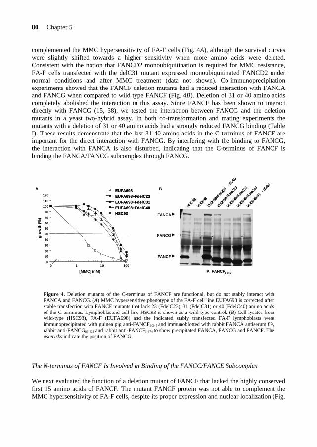
80 Chapter 5
complemented the MMC hypersensitivity of FA-F cells (Fig. 4A), although the survival curves were slightly shifted towards a higher sensitivity when more amino acids were deleted. Consistent with the notion that FANCD2 monoubiquitination is required for MMC resistance, FA-F cells transfected with the delC31 mutant expressed monoubiquitinated FANCD2 under normal conditions and after MMC treatment (data not shown). Co-immunoprecipitation experiments showed that the FANCF deletion mutants had a reduced interaction with FANCA and FANCG when compared to wild type FANCF (Fig. 4B). Deletion of 31 or 40 amino acids completely abolished the interaction in this assay. Since FANCF has been shown to interact directly with FANCG (15, 38), we tested the interaction between FANCG and the deletion mutants in a yeast two-hybrid assay. In both co-transformation and mating experiments the mutants with a deletion of 31 or 40 amino acids had a strongly reduced FANCG binding (Table I). These results demonstrate that the last 31-40 amino acids in the C-terminus of FANCF are important for the direct interaction with FANCG. By interfering with the binding to FANCG, the interaction with FANCA is also disturbed, indicating that the C-terminus of FANCF is binding the FANCA/FANCG subcomplex through FANCG.
A EUFA698EUFA698+FdelC23EUFA698+FdelC31EUFA698+FdelC40HSC93
EUFA698EUFA698+FdelC23EUFA698+FdelC31EUFA698+FdelC40HSC93
[MMC] (nM)
0
10
20
30
40
50
60
70
80
90
100
110
120
0 1 10 100
gro
wth
(%)
B
HSC93
VU06
98
VU06
98+F
ANCF-FL
AG
VU06
98+F
delC23
VU06
98+F
delC31
VU06
98+F
delC40
VU06
98+F
1-15
del
IP: FANCF1-245
FANCA
FANCG
FANCF
* * * *
Figure 4. Deletion mutants of the C-terminus of FANCF are functional, but do not stably interact with FANCA and FANCG. (A) MMC hypersensitive phenotype of the FA-F cell line EUFA698 is corrected after stable transfection with FANCF mutants that lack 23 (FdelC23), 31 (FdelC31) or 40 (FdelC40) amino acids of the C-terminus. Lymphoblastoid cell line HSC93 is shown as a wild-type control. (B) Cell lysates from wild-type (HSC93), FA-F (EUFA698) and the indicated stably transfected FA-F lymphoblasts were immunoprecipitated with guinea pig anti-FANCF1-245 and immunoblotted with rabbit FANCA antiserum 89, rabbit anti-FANCG83-622 and rabbit anti-FANCF1-374 to show precipitated FANCA, FANCG and FANCF. The asterisks indicate the position of FANCG.
The N-terminus of FANCF Is Involved in Binding of the FANCC/FANCE Subcomplex We next evaluated the function of a deletion mutant of FANCF that lacked the highly conserved first 15 amino acids of FANCF. The mutant FANCF protein was not able to complement the MMC hypersensitivity of FA-F cells, despite its proper expression and nuclear localization (Fig.
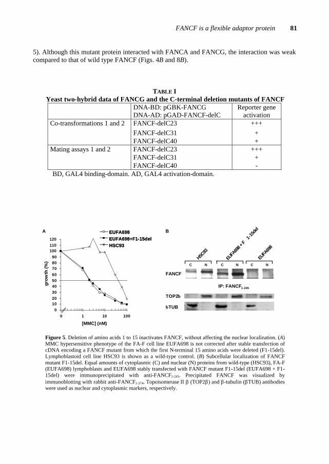
FANCF is a flexible adaptor protein 81
5). Although this mutant protein interacted with FANCA and FANCG, the interaction was weak compared to that of wild type FANCF (Figs. 4B and 8B).
TABLE I Yeast two-hybrid data of FANCG and the C-terminal deletion mutants of FANCF
DNA-BD: pGBK-FANCG DNA-AD: pGAD-FANCF-delC
Reporter gene activation
FANCF-delC23 +++ FANCF-delC31 +
Co-transformations 1 and 2
FANCF-delC40 + FANCF-delC23 +++ FANCF-delC31 +
Mating assays 1 and 2
FANCF-delC40 - BD, GAL4 binding-domain. AD, GAL4 activation-domain.
A
0102030405060708090
100110120
0 1 10 100
[MMC] (nM)
gro
wth
(%)
EUFA698EUFA698+F1-15delHSC93
EUFA698EUFA698+F1-15delHSC93
C C CN N N
HSC93
EUFA
698 +
F1-
15de
l
EUFA
698
FANCF
TOP2β
βTUB
IP: FANCF1-245
B
Figure 5. Deletion of amino acids 1 to 15 inactivates FANCF, without affecting the nuclear localization. (A) MMC hypersensitive phenotype of the FA-F cell line EUFA698 is not corrected after stable transfection of cDNA encoding a FANCF mutant from which the first N-terminal 15 amino acids were deleted (F1-15del). Lymphoblastoid cell line HSC93 is shown as a wild-type control. (B) Subcellular localization of FANCF mutant F1-15del. Equal amounts of cytoplasmic (C) and nuclear (N) proteins from wild-type (HSC93), FA-F (EUFA698) lymphoblasts and EUFA698 stably transfected with FANCF mutant F1-15del (EUFA698 + F1-15del) were immunoprecipitated with anti-FANCF1-245. Precipitated FANCF was visualized by immunoblotting with rabbit anti-FANCF1-374. Topoisomerase II β (TOP2β) and β-tubulin (βTUB) antibodies were used as nuclear and cytoplasmic markers, respectively.

82 Chapter 5
Unlike wild-type FANCF, which can be immunoprecipitated in a complex with FANCE, we did not detect any FANCE in a complex with the N-terminal deletion mutant of FANCF with antisera against FANCE (Fig. 6A). Furthermore, the mutant did not restore the interaction among FANCE, FANCA and FANCG that is observed in wild-type cells. Similarly, the C-terminal deletion mutant lacking the last 31 amino acids was also unable to bind FANCE and could not form a stable FA complex (Fig. 6A). In a reciprocal experiment, FANCC and FANCE did not co-immunoprecipitate with the N-terminal deletion mutant (Fig. 6B). Taken together, these results indicate that the N-terminus of FANCF is essential for its function and is involved in binding of FANCC and FANCE.
An important question for functional studies is how these proteins associate in the core
complex. Previously, no direct interaction between FANCF and FANCC or FANCE has been found in yeast two-hybrid assays (15, 38), suggesting that the association with FANCC and FANCE is indirect. We investigated these interactions in a mammalian two-hybrid assay and also in this assay FANCF did not interact with FANCC or FANCE (Fig. 6C), whereas an interaction between FANCC and FANCE was observed. We then sought to determine if the N-terminus of FANCF might be involved in the binding of this FANCC/FANCE subcomplex, using a mammalian three-hybrid (M3H) system, with 293 cells stably overexpressing FANCC. In this assay, we observed a strong induction of the reporter gene when FANCF was cotransfected with FANCE (Fig. 6D), indicating that FANCC acts as a molecular bridge between FANCF and FANCE. As a control experiment, we used a 293 cell line stably overexpressing FANCC mutant L554P, which is defective in binding FANCE (16, 38) and found no interaction between FANCF and FANCE (data not shown). To further extend these observations, we tested the FANCF N- and C-terminal deletion mutants in the M3H assay. In agreement with our co-immunoprecipitation experiments, the FANCF mutant 1-15del completely failed to interact with FANCE, whereas the FANCF mutant delC31 had a reduced interaction (Fig. 6D). Collectively, these observations indicate that the first 15 amino acids of FANCF are essential for the direct binding of the FANCC/FANCE subcomplex. Furthermore, they demonstrate that this interaction also partially depends on the C-terminal part of the FANCF protein.
The N-terminus of FANCF Contains Several Residues Essential for Its Function Several missense mutants were generated to delineate amino acids in the N-terminal region of FANCF essential for its interaction with other FA proteins and for its ability to complement the MMC defect in FA-F cells (see Table II). Secondary structure prediction programs indicate that the first 15 amino acids of human FANCF form an amphipathic α-helix (Fig. 7), with a strong negative charge at one side of the helix (Glu-2, Asp-9 and Glu-13). In the Xenopus homolog a similar structure was predicted, with Glu-6, Asp-9 and Glu-13 at the negative side of the helix.

FANCF is a flexible adaptor protein 83
EUFA
698+
L14A
FANCA
FANCG
FANCF
FANCE
HSC93
EUFA
698
EUFA
698+
FdelC
31
EUFA
698+
F1-1
5del
IP: FANCE
A C
0
10
20
30
40
50
60
70
80
E C F F+C F+E C+E
fold
ind
uct
ion
EUFA
698+
F1-1
5del
-FLAG
EUFA
698+
FANCF
-FLA
G
FANCF
FANCE
FANCC
FANCA
EUFA
698
IP: FLAG
EUFA
698+
F1-1
5del
-FLAG
EUFA
698+
FANCF
-FLA
G
FANCF
FANCE
FANCC
FANCA
EUFA
698
IP: FLAG
B D
0
5
10
15
20
25
30
35
40
F E+F delC31 E+delC31 1-15del E+1-15del
fold
in
du
ctio
n r
elat
ive
toF
AN
CE
**
Figure 6. Interaction of FANCF mutants with the FANCC/FANCE subcomplex. (A) Cell lysates from wild-type (HSC93), FA-F (EUFA698) lymphoblasts and FA-F cells stably transfected with the indicated FANCF mutants were immunoprecipitated with anti-FANCE and immunoblotted with anti-FANCA serum 89, anti-FANCG83-622, anti-FANCF1-374 and anti-FANCE to visualize FANCA, FANCG, FANCF and FANCE respectively. The asterisks indicate the position of FANCG. (B) EUFA698 cells were stably transfected with cDNAs encoding the FANCF mutant F1-15del-FLAG and wild-type FANCF-FLAG in expression vector pIRESneo. Immunoprecipitation was performed with anti-FLAG resin and immunoprecipitated FA proteins were visualized by immunoblotting with anti-FANCA serum 89, anti-FANCC106-558, anti-FANCE and anti-FANCF1-374. (C) Mammalian two-hybrid assay indicating a lack of direct interaction between FANCF and FANCC or FANCE. Fold induction is expressed relative to the luciferase activity obtained with empty vectors (pM and pVP16). (D) Mammalian three-hybrid assay illustrating the interaction of FANCF and FANCF mutants with the FANCC/FANCE subcomplex. Fold induction is expressed relative to the luciferase activity obtained with the pVP16-AD-FANCE vector alone. Results shown are mean ± standard deviation (SD) and derived from an experiment in triplicate.
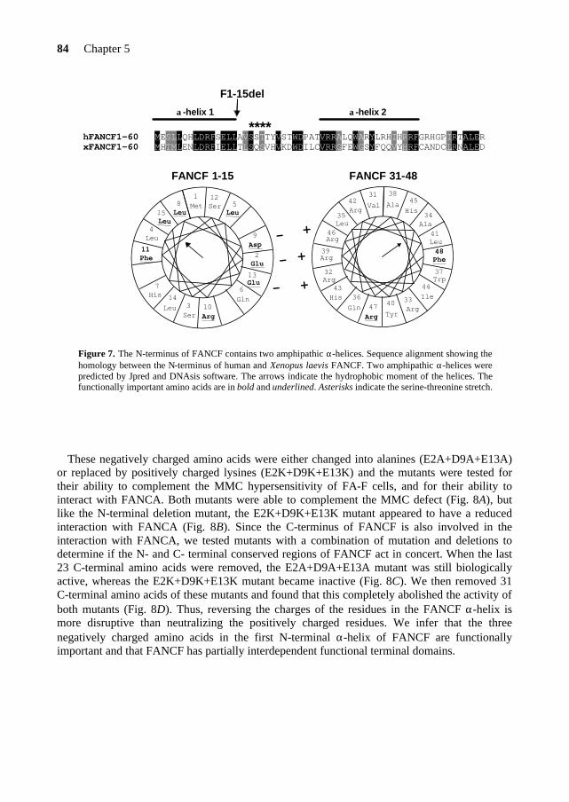
84 Chapter 5
hFANCF1-60 MESLLQHLDRFSELLAVSSTTYVSTWDPATVRRALQWARYLRHIHRRFGRHGPIRTALERxFANCF1-60 MHTMLENLDRFIELLTLSQSVHVKDWDILCVRRGFEWGSYFQQVYHRFCANDCLRNALED
α-helix 1 α-helix 2
F1-15del
****
FANCF 31-48FANCF 1-15
1Met
2Glu
3Ser
4Leu
5Leu
6Gln
7His
8Leu
9Asp
10Arg
11Phe
12Ser
13Glu
14Leu
15Leu
31Val
32Arg
33Arg
34Ala
35Leu
36Gln
37Trp
38Ala
39Arg
40Tyr
41Leu
42Arg
43His
44Ile
45His
46Arg
47Arg
48Phe+
+
-
-
-
+
Figure 7. The N-terminus of FANCF contains two amphipathic α-helices. Sequence alignment showing the homology between the N-terminus of human and Xenopus laevis FANCF. Two amphipathic α-helices were predicted by Jpred and DNAsis software. The arrows indicate the hydrophobic moment of the helices. The functionally important amino acids are in bold and underlined. Asterisks indicate the serine-threonine stretch.
These negatively charged amino acids were either changed into alanines (E2A+D9A+E13A)
or replaced by positively charged lysines (E2K+D9K+E13K) and the mutants were tested for their ability to complement the MMC hypersensitivity of FA-F cells, and for their ability to interact with FANCA. Both mutants were able to complement the MMC defect (Fig. 8A), but like the N-terminal deletion mutant, the E2K+D9K+E13K mutant appeared to have a reduced interaction with FANCA (Fig. 8B). Since the C-terminus of FANCF is also involved in the interaction with FANCA, we tested mutants with a combination of mutation and deletions to determine if the N- and C- terminal conserved regions of FANCF act in concert. When the last 23 C-terminal amino acids were removed, the E2A+D9A+E13A mutant was still biologically active, whereas the E2K+D9K+E13K mutant became inactive (Fig. 8C). We then removed 31 C-terminal amino acids of these mutants and found that this completely abolished the activity of both mutants (Fig. 8D). Thus, reversing the charges of the residues in the FANCF α-helix is more disruptive than neutralizing the positively charged residues. We infer that the three negatively charged amino acids in the first N-terminal α-helix of FANCF are functionally important and that FANCF has partially interdependent functional terminal domains.
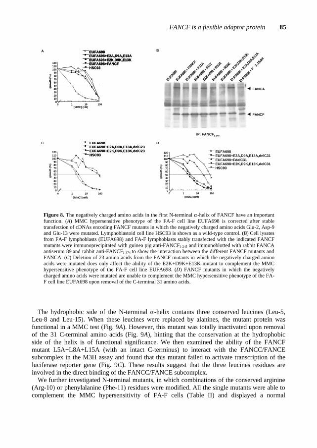
FANCF is a flexible adaptor protein 85
EUFA
698 +
F1-
15de
l
EUFA
698
EUFA
698 +
FANCF
EUFA
698 +
F11A
EUFA
698 +
F11Y
EUFA
698 +
R10
A
EUFA
698 +
R10E
EUFA
698 +
E2K,D9K
,E13K
EUFA
698 +
E2A,D9A
,E13A
IP: FANCF1-245
FANCA
FANCF
B
0102030405060708090
100110120
0 1 10 100[MMC] (nM)
gro
wth
(%)
A EUFA698EUFA698+E2A,D9A,E13AEUFA698+E2K,D9K,E13KEUFA698+FANCFHSC93
EUFA698EUFA698+E2A,D9A,E13AEUFA698+E2K,D9K,E13KEUFA698+FANCF
EUFA698EUFA698+E2A,D9A,E13AEUFA698+E2K,D9K,E13KEUFA698+FANCFHSC93
0102030405060708090
100110120
0 1 10 100[MMC] (nM)
EUFA698EUFA698+E2A,D9A,E13A,delC31EUFA698+FdelC31EUFA698+E2K,D9K,E13K,delC31HSC93
gro
wth
(%)
D
0102030405060708090
100110120
0 1 10 100[MMC] (nM)
EUFA698
HSC93
EUFA698+E2A,D9A,E13A,delC23EUFA698+E2K,D9K,E13K,delC23
EUFA698
HSC93
EUFA698+E2A,D9A,E13A,delC23EUFA698+E2K,D9K,E13K,delC23
gro
wth
(%)
C
Figure 8. The negatively charged amino acids in the first N-terminal α-helix of FANCF have an important function. (A) MMC hypersensitive phenotype of the FA-F cell line EUFA698 is corrected after stable transfection of cDNAs encoding FANCF mutants in which the negatively charged amino acids Glu-2, Asp-9 and Glu-13 were mutated. Lymphoblastoid cell line HSC93 is shown as a wild-type control. (B) Cell lysates from FA-F lymphoblasts (EUFA698) and FA-F lymphoblasts stably transfected with the indicated FANCF mutants were immunoprecipitated with guinea pig anti-FANCF1-245 and immunoblotted with rabbit FANCA antiserum 89 and rabbit anti-FANCF1-374 to show the interaction between the different FANCF mutants and FANCA. (C) Deletion of 23 amino acids from the FANCF mutants in which the negatively charged amino acids were mutated does only affect the ability of the E2K+D9K+E13K mutant to complement the MMC hypersensitive phenotype of the FA-F cell line EUFA698. (D) FANCF mutants in which the negatively charged amino acids were mutated are unable to complement the MMC hypersensitive phenotype of the FA-F cell line EUFA698 upon removal of the C-terminal 31 amino acids.
The hydrophobic side of the N-terminal α-helix contains three conserved leucines (Leu-5,
Leu-8 and Leu-15). When these leucines were replaced by alanines, the mutant protein was functional in a MMC test (Fig. 9A). However, this mutant was totally inactivated upon removal of the 31 C-terminal amino acids (Fig. 9A), hinting that the conservation at the hydrophobic side of the helix is of functional significance. We then examined the ability of the FANCF mutant L5A+L8A+L15A (with an intact C-terminus) to interact with the FANCC/FANCE subcomplex in the M3H assay and found that this mutant failed to activate transcription of the luciferase reporter gene (Fig. 9C). These results suggest that the three leucines residues are involved in the direct binding of the FANCC/FANCE subcomplex.
We further investigated N-terminal mutants, in which combinations of the conserved arginine (Arg-10) or phenylalanine (Phe-11) residues were modified. All the single mutants were able to complement the MMC hypersensitivity of FA-F cells (Table II) and displayed a normal
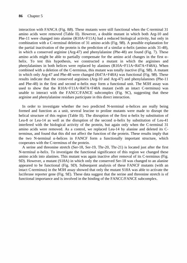
86 Chapter 5
interaction with FANCA (Fig. 8B). These mutants were still functional when the C-terminal 31 amino acids were removed (Table II). However, a double mutant in which both Arg-10 and Phe-11 were changed into alanine (R10A+F11A) had a reduced biological activity, but only in combination with a C-terminal deletion of 31 amino acids (Fig. 9B). A possible explanation for the partial inactivation of the protein is the prediction of a similar α-helix (amino acids 31-48), in which a conserved arginine (Arg-47) and phenylalanine (Phe-48) are found (Fig. 7). These amino acids might be able to partially compensate for the amino acid changes in the first α-helix. To test this hypothesis, we constructed a mutant in which the arginines and phenylalanines in both helices were replaced by alanines (R10A+F11A+R47A+F48A). When combined with a deletion of the C-terminus, this mutant was totally inactive (Fig. 9B). A mutant in which only Arg-47 and Phe-48 were changed (R47A+F48A) was functional (Fig. 9B). These results indicate that the conserved arginines (Arg-10 and Arg-47) and phenylalanines (Phe-11 and Phe-48) in the first and second α-helix may form a functional unit. The M3H assay was used to show that the R10A+F11A+R47A+F48A mutant (with an intact C-terminus) was unable to interact with the FANCC/FANCE subcomplex (Fig. 9C), suggesting that these arginine and phenylalanine residues participate in this direct interaction.
In order to investigate whether the two predicted N-terminal α-helices are really being
formed and function as a unit, several leucine to proline mutants were made to disrupt the helical structure of this region (Table II). The disruption of the first α-helix by substitution of Leu-8 or Leu-14 as well as the disruption of the second α-helix by substitution of Leu-41 interfered with the biological activity of the protein, but again only when the C-terminal 31 amino acids were removed. As a control, we replaced Leu-14 by alanine and deleted its C-terminus, and found that this did not affect the function of the protein. These results imply that the two N-terminal α-helices in FANCF form a functionally important structure, which cooperates with the C-terminus of the protein.
A serine and threonine stretch (Ser-18, Ser-19, Thr-20, Thr-21) is located just after the first N-terminal α-helix. To investigate the functional significance of this region we changed these amino acids into alanines. This mutant was again inactive after removal of its C-terminus (Fig. 9D). However, a mutant (S18A) in which only the conserved Ser-18 was changed to an alanine appeared to be functional (Fig. 9D). Subsequent analysis of these FANCF mutants (with an intact C-terminus) in the M3H assay showed that only the mutant S18A was able to activate the luciferase reporter gene (Fig. 9E). These data suggest that the serine and threonine stretch is of functional importance and is involved in the binding of the FANCC/FANCE subcomplex.

FANCF is a flexible adaptor protein 87
0102030405060708090
100110120
0 1 10 100
[MMC] (nM)
gro
wth
(%)
EUFA698EUFA698+S18A,delC31EUFA698+S18A,S19,T20A,T21A,delC31HSC93
EUFA698EUFA698+S18A,delC31EUFA698+S18A,S19,T20A,T21A,delC31HSC93
D
0102030405060708090
100110120
0 1 10 100
[MMC] (nM)
gro
wth
(%)
BEUFA698EUFA698+RF1delC31EUFA698+RF2delC31EUFA698+2XRFdelC31HSC93
EUFA698EUFA698+RF1delC31EUFA698+RF2delC31EUFA698+2XRFdelC31HSC93
0102030405060708090
100110120
0 1 10 100
[MMC] (nM)
gro
wth
%
EUFA698EUFA698+L5A,L8A,L15AEUFA698+L5A,L8A,L15A,delC31HCS93
EUFA698EUFA698+L5A,L8A,L15AEUFA698+L5A,L8A,L15A,delC31HCS93
A
0
5
10
15
20
F E+F 3xL E+3xL 2xRF E+2xRF
fold
ind
uct
ion
rel
ativ
e to
FA
NC
E
C
E
0
5
10
15
20
25
30
35
40
F E+F S18A E+S18A SSTT/A E+SSTT/A
fold
ind
uct
ion
rel
ativ
e to
FA
NC
E
Figure 9. Other functional important amino acids in the N-terminus of FANCF. (A) FANCF mutant in which Leu-5, Leu-8 and Leu-15 are changed into alanines is not able to complement the MMC hypersensitive phenotype of EUFA698 cells after removal of the C-terminal 31 amino acids. (B) Effect of arginine and phenylalanine-substitutions in FANCF on the ability to complement the MMC hypersensitive phenotype of EUFA698 cells. FANCF mutants R10A+F11A and R10A+F11A+R47A+R48A are inactivated upon removal of the C-terminus. (C) Mammalian three-hybrid assay illustrating the disturbed interaction between the FANCF mutants 3xL (L5A+L8A+L15A) and 2xRF (R10A+F11A+R47A+R48A) and the FANCC/FANCE subcomplex. Results shown are mean ± S.D. and derived from an experiment in triplicate. Fold induction is expressed relative to the luciferase activity obtained with the pVP16-AD-FANCE vector alone. (D) FANCF mutant in which Ser-18, Ser19, Thr-20 and Thr-21 are changed into alanine is not able to complement the MMC hypersensitive phenotype of EUFA698 cells after removal of the C-terminal 31 amino acids. (E) Mammalian three-hybrid assay showing interaction of the FANCC/FANCE subcomplex with wild type FANCF and FANCF mutant S18A, which is lost in the FANCF mutant S18A-S19A-T20A-T21A (SSTT/A).
Amino Acids 76 to 84 Are Not Essential for the Function of FANCF FA-F patient EUFA698 has a 23 bp deletion in the FANCF gene resulting in a frame shift at codon 77 (5). Since the deleted region is poorly conserved between human and Xenopus FANCF, we tried to overcome the defect by selection for a phenotypic revertant in vitro. After long-term culture of a lymphoblastoid cell line from patient EUFA698 in the presence of 15 nM MMC a MMC resistant cell line was obtained. Sequence analysis indicated that a deletion of nucleotide 225 (225delT) had restored the FANCF reading frame in one allele of the reverted
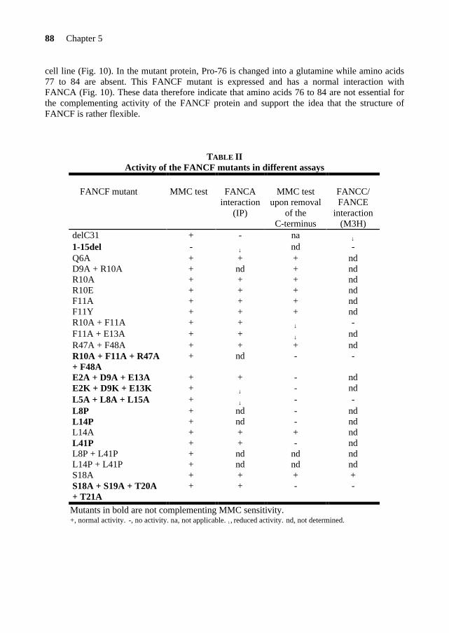
88 Chapter 5
cell line (Fig. 10). In the mutant protein, Pro-76 is changed into a glutamine while amino acids 77 to 84 are absent. This FANCF mutant is expressed and has a normal interaction with FANCA (Fig. 10). These data therefore indicate that amino acids 76 to 84 are not essential for the complementing activity of the FANCF protein and support the idea that the structure of FANCF is rather flexible.
TABLE II
Activity of the FANCF mutants in different assays
FANCF mutant
MMC test
FANCA
interaction (IP)
MMC test
upon removal of the
C-terminus
FANCC/ FANCE
interaction (M3H)
delC31 + - na ↓
1-15del - ↓ nd - Q6A + + + nd D9A + R10A + nd + nd R10A + + + nd R10E + + + nd F11A + + + nd F11Y + + + nd R10A + F11A + + ↓ - F11A + E13A + + ↓ nd R47A + F48A + + + nd R10A + F11A + R47A + F48A
+ nd - -
E2A + D9A + E13A + + - nd E2K + D9K + E13K + ↓ - nd L5A + L8A + L15A + ↓ - - L8P + nd - nd L14P + nd - nd L14A + + + nd L41P + + - nd L8P + L41P + nd nd nd L14P + L41P + nd nd nd S18A + + + + S18A + S19A + T20A + T21A
+ + - -
Mutants in bold are not complementing MMC sensitivity. +, normal activity. -, no activity. na, not applicable. ↓, reduced activity. nd, not determined.
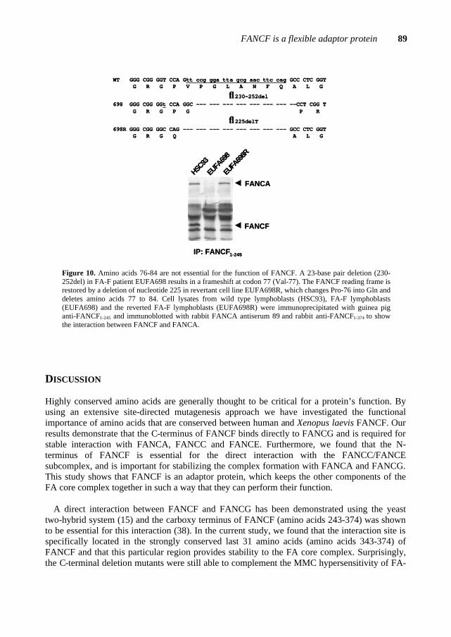
FANCF is a flexible adaptor protein 89
HSC93
EUFA
698
EUFA
698R
FANCA
FANCF
IP: FANCF1-245
WT GGG CGG GGT CCA Gtt ccg gga tta gcg aac ttc cag GCC CTC GGTG R G P V P G L A N F Q A L G
↓230-252del698 GGG CGG GGt CCA GGC --- --- --- --- --- --- --- --CCT CGG T
G R G P G P R
↓225delT698R GGG CGG GGC CAG --- --- --- --- --- --- --- --- GCC CTC GGT
G R G Q A L G
Figure 10. Amino acids 76-84 are not essential for the function of FANCF. A 23-base pair deletion (230-252del) in FA-F patient EUFA698 results in a frameshift at codon 77 (Val-77). The FANCF reading frame is restored by a deletion of nucleotide 225 in revertant cell line EUFA698R, which changes Pro-76 into Gln and deletes amino acids 77 to 84. Cell lysates from wild type lymphoblasts (HSC93), FA-F lymphoblasts (EUFA698) and the reverted FA-F lymphoblasts (EUFA698R) were immunoprecipitated with guinea pig anti-FANCF1-245 and immunoblotted with rabbit FANCA antiserum 89 and rabbit anti-FANCF1-374 to show the interaction between FANCF and FANCA.
DISCUSSION Highly conserved amino acids are generally thought to be critical for a protein’s function. By using an extensive site-directed mutagenesis approach we have investigated the functional importance of amino acids that are conserved between human and Xenopus laevis FANCF. Our results demonstrate that the C-terminus of FANCF binds directly to FANCG and is required for stable interaction with FANCA, FANCC and FANCE. Furthermore, we found that the N-terminus of FANCF is essential for the direct interaction with the FANCC/FANCE subcomplex, and is important for stabilizing the complex formation with FANCA and FANCG. This study shows that FANCF is an adaptor protein, which keeps the other components of the FA core complex together in such a way that they can perform their function.
A direct interaction between FANCF and FANCG has been demonstrated using the yeast
two-hybrid system (15) and the carboxy terminus of FANCF (amino acids 243-374) was shown to be essential for this interaction (38). In the current study, we found that the interaction site is specifically located in the strongly conserved last 31 amino acids (amino acids 343-374) of FANCF and that this particular region provides stability to the FA core complex. Surprisingly, the C-terminal deletion mutants were still able to complement the MMC hypersensitivity of FA-
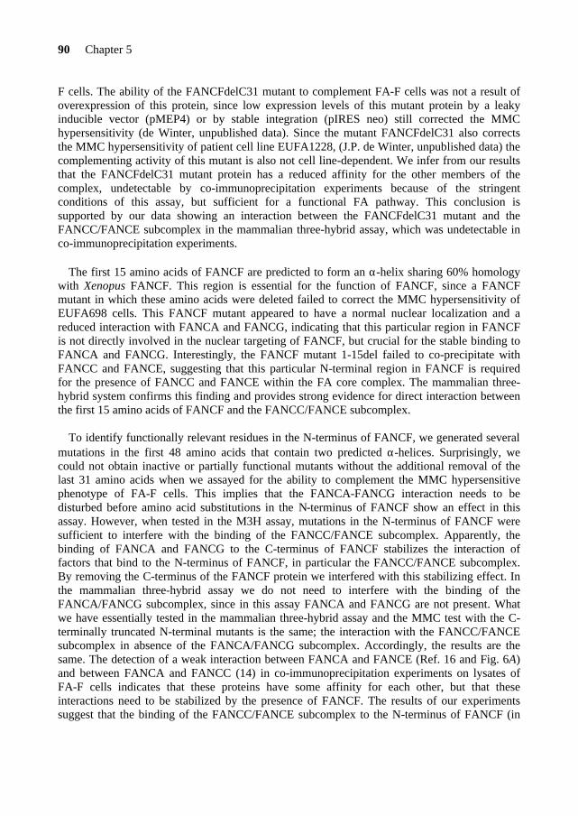
90 Chapter 5
F cells. The ability of the FANCFdelC31 mutant to complement FA-F cells was not a result of overexpression of this protein, since low expression levels of this mutant protein by a leaky inducible vector (pMEP4) or by stable integration (pIRES neo) still corrected the MMC hypersensitivity (de Winter, unpublished data). Since the mutant FANCFdelC31 also corrects the MMC hypersensitivity of patient cell line EUFA1228, (J.P. de Winter, unpublished data) the complementing activity of this mutant is also not cell line-dependent. We infer from our results that the FANCFdelC31 mutant protein has a reduced affinity for the other members of the complex, undetectable by co-immunoprecipitation experiments because of the stringent conditions of this assay, but sufficient for a functional FA pathway. This conclusion is supported by our data showing an interaction between the FANCFdelC31 mutant and the FANCC/FANCE subcomplex in the mammalian three-hybrid assay, which was undetectable in co-immunoprecipitation experiments.
The first 15 amino acids of FANCF are predicted to form an α-helix sharing 60% homology
with Xenopus FANCF. This region is essential for the function of FANCF, since a FANCF mutant in which these amino acids were deleted failed to correct the MMC hypersensitivity of EUFA698 cells. This FANCF mutant appeared to have a normal nuclear localization and a reduced interaction with FANCA and FANCG, indicating that this particular region in FANCF is not directly involved in the nuclear targeting of FANCF, but crucial for the stable binding to FANCA and FANCG. Interestingly, the FANCF mutant 1-15del failed to co-precipitate with FANCC and FANCE, suggesting that this particular N-terminal region in FANCF is required for the presence of FANCC and FANCE within the FA core complex. The mammalian three-hybrid system confirms this finding and provides strong evidence for direct interaction between the first 15 amino acids of FANCF and the FANCC/FANCE subcomplex.
To identify functionally relevant residues in the N-terminus of FANCF, we generated several
mutations in the first 48 amino acids that contain two predicted α-helices. Surprisingly, we could not obtain inactive or partially functional mutants without the additional removal of the last 31 amino acids when we assayed for the ability to complement the MMC hypersensitive phenotype of FA-F cells. This implies that the FANCA-FANCG interaction needs to be disturbed before amino acid substitutions in the N-terminus of FANCF show an effect in this assay. However, when tested in the M3H assay, mutations in the N-terminus of FANCF were sufficient to interfere with the binding of the FANCC/FANCE subcomplex. Apparently, the binding of FANCA and FANCG to the C-terminus of FANCF stabilizes the interaction of factors that bind to the N-terminus of FANCF, in particular the FANCC/FANCE subcomplex. By removing the C-terminus of the FANCF protein we interfered with this stabilizing effect. In the mammalian three-hybrid assay we do not need to interfere with the binding of the FANCA/FANCG subcomplex, since in this assay FANCA and FANCG are not present. What we have essentially tested in the mammalian three-hybrid assay and the MMC test with the C-terminally truncated N-terminal mutants is the same; the interaction with the FANCC/FANCE subcomplex in absence of the FANCA/FANCG subcomplex. Accordingly, the results are the same. The detection of a weak interaction between FANCA and FANCE (Ref. 16 and Fig. 6A) and between FANCA and FANCC (14) in co-immunoprecipitation experiments on lysates of FA-F cells indicates that these proteins have some affinity for each other, but that these interactions need to be stabilized by the presence of FANCF. The results of our experiments suggest that the binding of the FANCC/FANCE subcomplex to the N-terminus of FANCF (in
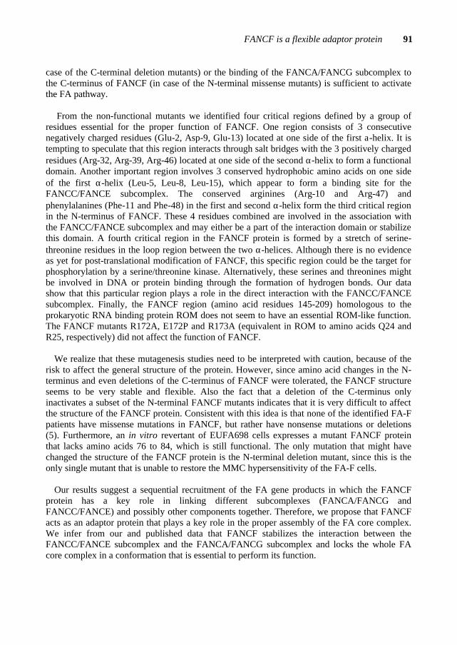
FANCF is a flexible adaptor protein 91
case of the C-terminal deletion mutants) or the binding of the FANCA/FANCG subcomplex to the C-terminus of FANCF (in case of the N-terminal missense mutants) is sufficient to activate the FA pathway.
From the non-functional mutants we identified four critical regions defined by a group of
residues essential for the proper function of FANCF. One region consists of 3 consecutive negatively charged residues (Glu-2, Asp-9, Glu-13) located at one side of the first a-helix. It is tempting to speculate that this region interacts through salt bridges with the 3 positively charged residues (Arg-32, Arg-39, Arg-46) located at one side of the second α-helix to form a functional domain. Another important region involves 3 conserved hydrophobic amino acids on one side of the first α-helix (Leu-5, Leu-8, Leu-15), which appear to form a binding site for the FANCC/FANCE subcomplex. The conserved arginines (Arg-10 and Arg-47) and phenylalanines (Phe-11 and Phe-48) in the first and second α-helix form the third critical region in the N-terminus of FANCF. These 4 residues combined are involved in the association with the FANCC/FANCE subcomplex and may either be a part of the interaction domain or stabilize this domain. A fourth critical region in the FANCF protein is formed by a stretch of serine-threonine residues in the loop region between the two α-helices. Although there is no evidence as yet for post-translational modification of FANCF, this specific region could be the target for phosphorylation by a serine/threonine kinase. Alternatively, these serines and threonines might be involved in DNA or protein binding through the formation of hydrogen bonds. Our data show that this particular region plays a role in the direct interaction with the FANCC/FANCE subcomplex. Finally, the FANCF region (amino acid residues 145-209) homologous to the prokaryotic RNA binding protein ROM does not seem to have an essential ROM-like function. The FANCF mutants R172A, E172P and R173A (equivalent in ROM to amino acids Q24 and R25, respectively) did not affect the function of FANCF.
We realize that these mutagenesis studies need to be interpreted with caution, because of the
risk to affect the general structure of the protein. However, since amino acid changes in the N-terminus and even deletions of the C-terminus of FANCF were tolerated, the FANCF structure seems to be very stable and flexible. Also the fact that a deletion of the C-terminus only inactivates a subset of the N-terminal FANCF mutants indicates that it is very difficult to affect the structure of the FANCF protein. Consistent with this idea is that none of the identified FA-F patients have missense mutations in FANCF, but rather have nonsense mutations or deletions (5). Furthermore, an in vitro revertant of EUFA698 cells expresses a mutant FANCF protein that lacks amino acids 76 to 84, which is still functional. The only mutation that might have changed the structure of the FANCF protein is the N-terminal deletion mutant, since this is the only single mutant that is unable to restore the MMC hypersensitivity of the FA-F cells.
Our results suggest a sequential recruitment of the FA gene products in which the FANCF
protein has a key role in linking different subcomplexes (FANCA/FANCG and FANCC/FANCE) and possibly other components together. Therefore, we propose that FANCF acts as an adaptor protein that plays a key role in the proper assembly of the FA core complex. We infer from our and published data that FANCF stabilizes the interaction between the FANCC/FANCE subcomplex and the FANCA/FANCG subcomplex and locks the whole FA core complex in a conformation that is essential to perform its function.
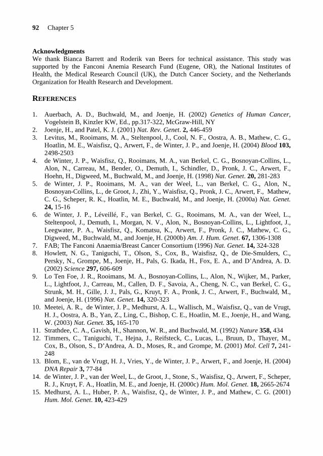
92 Chapter 5
Acknowledgments We thank Bianca Barrett and Roderik van Beers for technical assistance. This study was supported by the Fanconi Anemia Research Fund (Eugene, OR), the National Institutes of Health, the Medical Research Council (UK), the Dutch Cancer Society, and the Netherlands Organization for Health Research and Development. REFERENCES 1. Auerbach, A. D., Buchwald, M., and Joenje, H. (2002) Genetics of Human Cancer,
Vogelstein B, Kinzler KW, Ed., pp.317-322, McGraw-Hill, NY 2. Joenje, H., and Patel, K. J. (2001) Nat. Rev. Genet. 2, 446-459 3. Levitus, M., Rooimans, M. A., Steltenpool, J., Cool, N. F., Oostra, A. B., Mathew, C. G.,
Hoatlin, M. E., Waisfisz, Q., Arwert, F., de Winter, J. P., and Joenje, H. (2004) Blood 103, 2498-2503
4. de Winter, J. P., Waisfisz, Q., Rooimans, M. A., van Berkel, C. G., Bosnoyan-Collins, L., Alon, N., Carreau, M., Bender, O., Demuth, I., Schindler, D., Pronk, J. C., Arwert, F., Hoehn, H., Digweed, M., Buchwald, M., and Joenje, H. (1998) Nat. Genet. 20, 281-283
5. de Winter, J. P., Rooimans, M. A., van der Weel, L., van Berkel, C. G., Alon, N., Bosnoyan-Collins, L., de Groot, J., Zhi, Y., Waisfisz, Q., Pronk, J. C., Arwert, F., Mathew, C. G., Scheper, R. K., Hoatlin, M. E., Buchwald, M., and Joenje, H. (2000a) Nat. Genet. 24, 15-16
6. de Winter, J. P., Léveillé, F., van Berkel, C. G., Rooimans, M. A., van der Weel, L., Steltenpool, J., Demuth, I., Morgan, N. V., Alon, N., Bosnoyan-Collins, L., Lightfoot, J., Leegwater, P. A., Waisfisz, Q., Komatsu, K., Arwert, F., Pronk, J. C., Mathew, C. G., Digweed, M., Buchwald, M., and Joenje, H. (2000b) Am. J. Hum. Genet. 67, 1306-1308
7. FAB; The Fanconi Anaemia/Breast Cancer Consortium (1996) Nat. Genet. 14, 324-328 8. Howlett, N. G., Taniguchi, T., Olson, S., Cox, B., Waisfisz, Q., de Die-Smulders, C.,
Persky, N., Grompe, M., Joenje, H., Pals, G. Ikada, H., Fox, E. A., and D’Andrea, A. D. (2002) Science 297, 606-609
9. Lo Ten Foe, J. R., Rooimans, M. A., Bosnoyan-Collins, L., Alon, N., Wijker, M., Parker, L., Lightfoot, J., Carreau, M., Callen, D. F., Savoia, A., Cheng, N. C., van Berkel, C. G., Strunk, M. H., Gille, J. J., Pals, G., Kruyt, F. A., Pronk, J. C., Arwert, F., Buchwald, M., and Joenje, H. (1996) Nat. Genet. 14, 320-323
10. Meetei, A. R., de Winter, J. P., Medhurst, A. L., Wallisch, M., Waisfisz, Q., van de Vrugt, H. J., Oostra, A. B., Yan, Z., Ling, C., Bishop, C. E., Hoatlin, M. E., Joenje, H., and Wang, W. (2003) Nat. Genet. 35, 165-170
11. Strathdee, C. A., Gavish, H., Shannon, W. R., and Buchwald, M. (1992) Nature 358, 434 12. Timmers, C., Taniguchi, T., Hejna, J., Reifsteck, C., Lucas, L., Bruun, D., Thayer, M.,
Cox, B., Olson, S., D’Andrea, A. D., Moses, R., and Grompe, M. (2001) Mol. Cell 7, 241-248
13. Blom, E., van de Vrugt, H. J., Vries, Y., de Winter, J. P., Arwert, F., and Joenje, H. (2004) DNA Repair 3, 77-84
14. de Winter, J. P., van der Weel, L., de Groot, J., Stone, S., Waisfisz, Q., Arwert, F., Scheper, R. J., Kruyt, F. A., Hoatlin, M. E., and Joenje, H. (2000c) Hum. Mol. Genet. 18, 2665-2674
15. Medhurst, A. L., Huber, P. A., Waisfisz, Q., de Winter, J. P., and Mathew, C. G. (2001) Hum. Mol. Genet. 10, 423-429

FANCF is a flexible adaptor protein 93
16. Pace, P., Johnson, M., Tan, W. M., Mosedale, G., Sng, C., Hoatlin, M., de Winter, J., Joenje, H., Gergely, F., and Patel, K. J. (2002) EMBO J. 21, 3414-3423
17. Waisfisz, Q., de Winter, J. P., Kruyt, F. A., de Groot, J., van der Weel, L., Dijkmans, L.M., Zhi, Y., Arwert, F., Scheper, R. J., and Youssoufian, H. (1999) Proc. Natl. Acad. Sci. 96, 10320-10325
18. Garcia-Higuera, I., Taniguchi, T., Ganesan, S., Meyn, M. S., Timmers, C., Hejna, J., Grompe, M., and D’Andrea, A. D. (2001) Mol. Cell. 7, 249-262
19. Taniguchi, T., Garcia-Higuera, I., Andreassen, P. R., Gregory, R. C., Grompe, M., and D’Andrea, A. D. (2002a) Blood 100, 2414-2420
20. Hussain, S., Wilson, J. B., Medhurst, A. L., Hejna, J., Witt, E., Ananth, S., Davies, A., Masson, J. Y., Moses, R., West, S. C., de Winter, J. P., Ashworth, A., Jones, N. J., and Mathew, C. G. (2004) Hum. Mol. Genet. 13, 1241-1248
21. Venkitaraman, A. R. (2002) Cell 108, 171-182 22. Yang, H., Jeffrey, P. D., Miller, J., Kinnucan, E., Sun, Y., Thoma, N. H., Zheng, N., Chen,
P. L., Lee, W. H., and Pavletich, N. P. (2002) Science 297, 1837-48 23. Taniguchi, T., Garcia-Higuera, I., Andreassen, P. R., Gregory, R. C., Kim, S. T., lane, W.
S., Kastan, M. B., and D’Andrea, A. D. (2002b) Cell 109, 459-472 24. Nakanishi, K., Taniguchi, T., Ranganathan, V., New, H. H., Moreau, L. A., Stotsky, M.,
Mathew, C. G., Kastan, M. B., Weaver, D. T., and D’Andrea, A. D. (2002) Nat. Cell Biol. 4, 913-920
25. Pichierri, P., Averbeck, D., and Rosselli, F. (2002) Hum. Mol. Genet. 11, 2531-2546 26. D’Amours, D., and Jackson, S. P. (2002) Nat. Rev. Mol. Cell Biol. 3, 317-327 27. Taniguchi, T., Tischkowitz, M., Ameziane, N., Hodgson, S. V., Mathew, C. G., Joenje, H.,
Mok, S. C., and D'Andrea, A. D. (2002c) Nat. Med. 9, 568-574 28. Marsit, C. J., Liu, M., Nelson, H. H., Posner, M., Suzuki, M., and Kelsey, K. T. (2004)
Oncogene 23, 1000-1004 29. Narayan, G., Arias-Pulido, H., Nandula, S. V., Basso, K., Sugirtharaj, D. D. Vargas, H.,
Mansukhani, M., Villella, J., Meyer, L., Schneider, A., Gissmann, L., Durst, M., Pothuri, B., and Murty, V. V. (2004) Cancer Res. 64, 2994-2997
30. Kupfer, G. M., Naf, D., Garcia-Higuera, I., Wasik, J., Cheng, A., Yamashita, T., Tipping, A., Morgan, N., Mathew, C. G., and D’Andrea, A. D. (1999) Exp. Hematol. 27, 587-593
31. Kuang, Y., Garcia-Higuera, I., Moran, A., Mondoux, M., Digweed, M., and D’Andrea, A. D. (2000) Blood 96, 1625-1632
32. Nakanishi, K., Moran, A., Hays, T., Kuang, Y., Fox, E., Garneau, D., Monte de Oca, R., Grompe, M., and D’Andrea, A. D. (2001) Exp. Hematol. 29, 842-849
33. Pang, Q., Christianson, T. A., Keeble, W., Diaz, J., Faulkner, G. R., Reifsteck, C., Olson, S., and Bagby, G. C. (2001) Blood 98, 1392-1401
34. Adachi, D., Oda, T., Yagasaki, H., Nakasato, K., Taniguchi, T., D’andrea, A. D., Asano, S., and Yamashita, T. (2002) Hum. Mol. Genet. 11, 3125-3134
35. Ishida, R., and Buchwald, M. (1982) Cancer Res. 42, 4000-4006 36. Joenje, H., Lo Ten Foe, J. R., Oostra, A. B., van Berkel, C. G., Rooimans, M. A.,
Schroeder-Kurth, T., Wegner, R-D., Gille, J. J., Buchwald, M., and Arwert, F. (1995) Blood 86, 2156-2160
37. Predki, P. F., Nayak, L. M., Gottlieb, M. B., and Regan, L. (1995) Cell 80, 41-50 38. Gordon, S. M., and Buchwald, M. (2003) Blood 102,136-141


CHAPTER 6
GENERAL DISCUSSION

General Discussion
97
It is generally accepted that the FA proteins interact with each other to form a multiprotein nuclear complex that functions in a common cellular pathway. This pathway is activated during the S phase of the cell cycle and is required for the response to stalled replication forks (Kennedy & D’Andrea, 2005). In this Chapter, I will first present the molecular architecture of the FA pathway through a model that describes the sequential assembly and subcellular localization of the FA proteins. The proposed model is based on co-immunoprecipitation and two-/three-hybrid experiments as well as on results presented in this thesis. Secondly, I will describe a model for the function of the FA pathway in the repair of interstrand DNA cross-links (ICLs).
MOLECULAR ARCHITECTURE OF THE FA PATHWAY
To facilitate the description of the molecular architecture of the FA pathway, FA proteins are categorized according to their role in the monoubiquitination of FANCD2. Most of the FA proteins are required for this monoubiquitination step and are referred to as the upstream part of the pathway. However, FANCD1/BRCA2 and FANCJ/BRIP1 are not required for this event and are therefore positioned downstream of FANCD2 (Figure 1).
1. FA protein-protein interactions upstream of FANCD2 and the formation of the FA core complex Eight of the FA proteins (FANCA, B, C, E, F, G, L and M) and one FANCA-associated protein, FAAP100, assemble in an ordered sequence of events to form a nuclear ubiquitin ligase, “the FA core complex” (see Figure 1). This multisubunit complex is essential for the monoubiquitination of FANCD2. The FANCI protein, which has not been identified yet, seems to have a different role, since in the absence of FANCI, the FA core complex is properly formed but FANCD2 is not monoubiquitinated (Levitus et al., 2004). Therefore, FANCI is probably not a member of this FA core complex but may assist the complex in the monoubiquitination of FANCD2. In this respect, the FANCI protein is also considered to function upstream of FANCD2. At the initial stages of the sequential assembly of the FA core complex, specific interactions between FA proteins take place in the cytoplasm to form two FA subcomplexes (FANCA/FANCG and FANCB/FANCL/FAAP100). These subcomplexes might enter the nucleus through the nuclear pores via active transport, since their cumulative molecular size exceeds the limit of 60 kDa for passive transport through the nuclear pore. The transport of these subcomplexes probably takes place by recognition of nuclear localization signal (NLS) sequences within the FANCA and FANCB proteins. The nucleocytoplasmic shuttling of several FA proteins appears to be regulated in a cell cycle-dependent manner (Thomashevski et al., 2004; Mi & Kupfer, 2005).

98 Chapter 6
A physical association between FANCA and FANCG is believed to represent an early step in the assembly of the FA core complex, since their interaction is observed in all non A/G FA complementation groups (Waisfisz et al., 1999a; de Winter et al., 2000b). The direct interaction between these two proteins appears to be mediated by the N-terminus of FANCA (amino acids 1 to 40 including the NLS motif) and the tetratricopeptide repeat (TPR) motifs 5 and 6 at the C-terminus of FANCG (Kruyt et al., 1999; Garcia-Higuera et al., 1999, 2000; Kuang et al., 2000; Gordon & Buchwald, 2003; Blom et al., 2004, Hussain et al., 2006). This association might regulate the nuclear accumulation of FANCA, as the NLS is probably shielded after binding FANCG. In addition to the N-terminal region, the nuclear accumulation of FANCA also requires its C-terminal portion (Lighfoot et al., 1999). This region does not contain a NLS sequence and is not able to translocate a GFP fusion protein to the nucleus. Therefore, the C-terminus might be needed to replace FANCG from the N-terminal NLS sequence, perhaps by phosphorylation of FANCA at a site adjacent to its NLS (Yamashita et al., 1998; Yagasaki et al., 2001; Harreman et al., 2004).
The FANCG protein contains several TPR motifs, which function as scaffolds for protein-protein interactions (Blom et al., 2004). This suggests that FANCG plays an important role in the assembly and stability of the multiprotein FA core complex. Indeed, FANCG is able to mediate the interaction between FANCA and the C-terminus of FANCF (Medhurst et al., 2001; Gordon & Buchwald, 2003; Chapter 5). However, studies on the FANCG-FANCF interaction were unable to delineate the FANCG binding region, perhaps because the fragments of FANCG used in these studies disrupted the superhelical structure of the TPR domains required for binding FANCF (Medhurst et al., 2001; Gordon & Buchwald, 2003; Blom et al., 2004; Hussain et al., 2006). Furthermore, the C-terminal region of FANCG is important for recruitment of FANCC in the nuclear FA core complex (Kuang et al., 2000; Nakanishi et al., 2001), probably through its direct interaction with FANCF (see below). FANCG also appears to stabilize the interaction between FANCA and FANCL (Medhurst et al., 2006) and additional roles have been proposed for FANCG as it seems to have other direct binding partners such as FANCD1/BRCA2 and the RAD51 paralog XRCC3 (Hussain et al., 2003, 2006).
The FANCB, FANCL and FAAP100 proteins form a ternary complex (Medhurst et al.,
unpublished data) and the FANCB/FANCL complex directly binds to FANCA (Medhurst et al., 2006). The stability and the nuclear localization of this group of proteins seem to depend on each other, as well as on the nuclear FANCM protein. Recent studies showed that the nuclear accumulation of FANCL depends on FANCA FANCB and FANCM (Meetei et al., 2003b, Medhurst et al., 2006). Similarly, the nuclear accumulation of FANCA depends on FANCB, FANCM and, to a lesser extent, on FANCL (de Winter et al., 2000b; Meetei et al., 2005; Medhurst et al., 2006). The FANCA/FANCG dimer and the FANCB/FANCL/FAAP100 ternary complex probably enter the nucleus independently, since the interaction between FANCA and FANCL is only found in the nuclear compartment and FANCA mutants that are affected in their nuclear accumulation are unable to interact with FANCL (Medhurst et al., 2006). In the nucleus, it is likely that these two subcomplexes physically assemble around FANCM, as no interaction between FANCA, FANCG and FANCL is found in the absence of FANCM (Medhurst et al., 2006). The association of these subcomplexes with FANCM appears to occur in a succession of events, in which the FANCA/FANCG dimer first binds FANCM followed by the FANCB/FANCL/FAAP100 subcomplex. This is supported by the observation that in absence of FANCM, the expression levels of FANCA and FANCG are much more

General Discussion
99
affected than the levels of FANCL (Meetei et al., 2005). Furthermore, since the nuclear accumulation of FANCA is also regulated by FANCB and FANCL, it is likely that the association of the FANCB/FANCL subcomplex with FANCA masks the nuclear exports signals present in FANCA and maintains FANCA in the nucleus (Ferrer et al., 2005). The FANCL protein, which contains WD40 domains, also might play an important role in the assembly and stability of these subcomplexes, since such domains are known to mediate protein-protein interactions (Meetei et al., 2003b; Gurtan et al., 2006).
The other FA protein that partially resides in the cytoplasmic compartment of the cell is
FANCC. The expression of FANCC is regulated during the cell cycle and seems to be subjected to proteasome-dependent proteolysis (Heinrich et al., 2000). Therefore, the nuclear transport of FANCC is also cell cycle-dependent and might be initiated at the same time as the FANCA/FANCG and FANCB/FANCL/FAAP100 subcomplexes. However, little is known about how FANCC is imported into the nucleus. One possibility is that FANCC enters the nucleus through passive transport as its molecular size is about 60 kDa, or via an unidentified nuclear localization signal (NLS) within its sequence. Alternatively, the nuclear import of FANCC occurs via its binding to a NLS-cargo protein. This protein could be FANCE, which contains two NLS motifs and binds directly to FANCC (Chapters 2, 4 & 5; Medhurst et al., 2001; Pace et al., 2002; Taniguchi & D’Andrea, 2002; Godon & Buchwald, 2003, 2005). Our localization study reveals that the nuclear accumulation of FANCE is not exclusively dependent on its NLS motifs, but also relies on FANCC (Chapter 4). We found that the FANCC and FANCE proteins have a mutual interdependence in the FA pathway. In absence of FANCC, the expression level of nuclear FANCE is disturbed, and similarly, in absence of FANCE, FANCC is not able to accumulate in the nucleus (see also Pace et al., 2002; Taniguchi & D’Andrea, 2002). Furthermore, FANCC deletion and missense mutants are defective in FANCE binding and are incapable to accumulate in the nucleus (Pace et al., 2002; Gordon & Buchwald, 2003). Therefore, FANCC and FANCE affect each other in their nuclear accumulation and/or stability.
We have demonstrated that the FANCC/FANCE subcomplex is able to bind directly to the
FANCF protein (Chapter 5). FANCF is a small nuclear protein that was first thought to be a RNA binding protein based upon homology to the prokaryotic ROM protein (de Winter et al., 2000a). However, site-directed mutagenesis of the FANCF region homologous to ROM reveals that FANCF has a different function (Chapter 5). Results from two-hybrid studies show that neither FANCC nor FANCE binds to FANCF (Chapter 5). However, three-hybrid studies indicate that FANCC allows direct interaction between FANCF-FANCE (Chapter 5) and, similarly, FANCE mediates the interaction between FANCF-FANCC (Gordon et al., 2005). Therefore, a ternary complex exists between FANCC, FANCE and FANCF. We found that the first 15 amino acids of FANCF are necessary to bind and recruit the FANCC and FANCE proteins, which suggests that the N-terminus of FANCF binds both proteins simultaneously. It has been shown that the interaction between FANCA and FANCL depends on FANCB, FANCG and FANCM, but is independent of FANCC, FANCE and FANCF (Medhurst et al., 2006). Therefore, the formation of the FANCC/FANCE/FANCF subcomplex seems to represent a downstream event in the assembly of the FA core complex. The FANCA, FANCC, FANCE and FANCG proteins seem to have some affinity for each other but they require FANCF to stabilize their interaction. In the assembly of the FA core complex, FANCF plays a crucial role since it allows the formation of a larger and stable complex by bridging the FANCG

100 Chapter 6
protein, which is connected to the FANCM/FANCA/FANCB/FANCL/ FAAP100 subcomplex, and the FANCC/FANCE subcomplex.
The FANCC/FANCE subcomplex links to another FA protein, FANCD2, the key substrate for the ubiquitin ligase of the FA core complex (Chapter 4; Pace et al., 2002; Gordon & Buchwald, 2003; Gordon et al., 2005). In fact, FANCE binds both FANCC and FANCD2 but the FANCC and FANCD2 proteins do not directly interact with each other. Therefore, the ternary complex formation between these proteins seems to be mediated by FANCE (Gordon et al., 2005). Along this line, our study suggests that different regions of FANCE interact with FANCC and FANCD2 (Chapter 4). The N-terminus of FANCD2 binds FANCE toward a C-terminal region, while full-length FANCC binds to a minimal internal portion of FANCE (amino acids 150-311) encompassing its NLS motifs (Chapter 4; Gordon & Buchwald, 2003; Gordon et al., 2005). Consequently, the FANCC/FANCE subcomplex physically connects the FANCD2 protein to the FA core complex (see Figure 1). Interestingly, the N-terminal region of FANCD2 is also involved in binding FANCD1/BRCA2 (Gordon & Buchwald, 2003; Hussain et al., 2004). This overlapping region of protein-protein interactions in FANCD2 may be of functional importance (see below). In summary, the FANCC/FANCE subcomplex interacts with FANCF and FANCD2. However, it is still unclear whether this subcomplex mediates at the same time the interaction with FANCF and FANCD2, or whether it forms a trimeric complex with each protein independently. This issue can be clarified by performing a four-hybrid study of these proteins.
Monoubiquitination of FANCD2 Following the proper assembly and stability of the FA core complex, FANCL, through its RING domain, might allow the recruitment of an E2 ubiquitin-conjugating enzyme (Gurtan et al., 2006). This enzyme could be FANCI, which is involved in the monoubiquitination of FANCD2 (Levitus et al., 2004), but could also be an as yet unidentified E2 enzyme. There is no direct association between FANCL and its substrate FANCD2, but this interaction seems to be mediated by FANCE in collaboration with the rest of the FA core complex. The results reported in Chapter 4 suggest that FANCI is implicated in the nuclear retention of FANCC, FANCE and FANCD2. Therefore, it is possible that the interaction of the FANCC/FANCE/FANCD2 ternary complex with FANCI subsequently leads to the FANCL mediated transfer of the ubiquitin moiety to FANCD2.
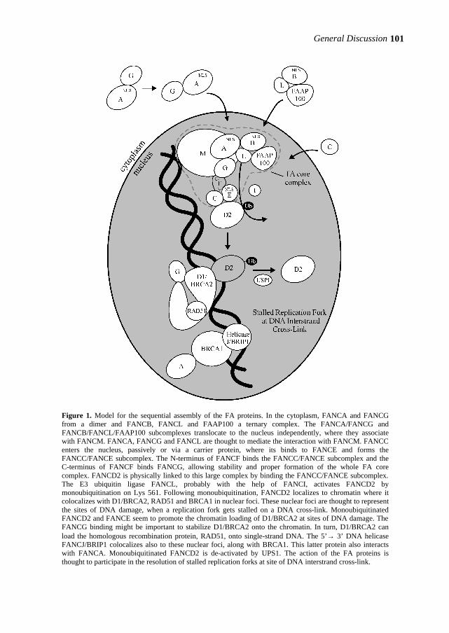
General Discussion
101
Figure 1. Model for the sequential assembly of the FA proteins. In the cytoplasm, FANCA and FANCG from a dimer and FANCB, FANCL and FAAP100 a ternary complex. The FANCA/FANCG and FANCB/FANCL/FAAP100 subcomplexes translocate to the nucleus independently, where they associate with FANCM. FANCA, FANCG and FANCL are thought to mediate the interaction with FANCM. FANCC enters the nucleus, passively or via a carrier protein, where its binds to FANCE and forms the FANCC/FANCE subcomplex. The N-terminus of FANCF binds the FANCC/FANCE subcomplex and the C-terminus of FANCF binds FANCG, allowing stability and proper formation of the whole FA core complex. FANCD2 is physically linked to this large complex by binding the FANCC/FANCE subcomplex. The E3 ubiquitin ligase FANCL, probably with the help of FANCI, activates FANCD2 by monoubiquitination on Lys 561. Following monoubiquitination, FANCD2 localizes to chromatin where it colocalizes with D1/BRCA2, RAD51 and BRCA1 in nuclear foci. These nuclear foci are thought to represent the sites of DNA damage, when a replication fork gets stalled on a DNA cross-link. Monoubiquitinated FANCD2 and FANCE seem to promote the chromatin loading of D1/BRCA2 at sites of DNA damage. The FANCG binding might be important to stabilize D1/BRCA2 onto the chromatin. In turn, D1/BRCA2 can load the homologous recombination protein, RAD51, onto single-strand DNA. The 5’→ 3’ DNA helicase FANCJ/BRIP1 colocalizes also to these nuclear foci, along with BRCA1. This latter protein also interacts with FANCA. Monoubiquitinated FANCD2 is de-activated by UPS1. The action of the FA proteins is thought to participate in the resolution of stalled replication forks at site of DNA interstrand cross-link.
102 Chapter 6
2. FA protein-protein interactions downstream of FANCD2 Following monoubiquitination, FANCD2 is targeted to chromatin and colocalizes in nuclear foci with numerous proteins involved in DNA processing, such as FANCD1/BRCA2, FANCJ/BRIP1, BRCA1, RAD51 and PCNA (see also Figure 2) (Garcia-Higuera et al., 2001; Cantor et al., 2001; Taniguchi et al., 2002b; Wang et al., 2004; Howlett et al., 2005). These nuclear foci seem to represent the sites of DNA damage where the replication fork stalls at a DNA cross-link. The FA core complex proteins also have the ability to associate with chromatin and play a crucial role in chromatin targeting of monoubiquitinated FANCD2 (Matsushita et al., 2005; Mi & Kupfer et al., 2005). Furthermore, efficient DNA damage-induced monoubiquitination and nuclear foci formation of FANCD2 requires the ATR (ATM-and Rad3-related) protein kinase signal transducer as well as the single-strand DNA binding protein RPA (Andreassen et al., 2004).
The FANCD1/BRCA2 protein is directly implicated in the repair of DNA double-strand
breaks (DSB) by homologous recombination (HR). This type of repair uses homology between two DNA strands to restore the original sequence of the broken DNA using one of the strands as a repair template (see Figure 2). In this HR repair process, D1/BRCA2 appears to regulate the RAD51 filament loading and extension onto the single-strand DNA (ssDNA) at sites of damage (for reviews, West, 2003; Shivji & Venkitaraman, 2004). The interaction model between D1/BRCA2 and RAD51 suggests that the internal BRC repeat region in D1/BRCA2 provides a structure for the assembly of multiple RAD51 monomers and that the binding of its C-terminal oligosaccharide or oligonucleotide binding (OB) folds to ssDNA provides a way to dislodge RPA from ssDNA, enabling the formation of the RAD51 filament on that ssDNA (Powell & Kachnic, 2003; Shin et al., 2003). Subsequently, RAD51 can catalyze the pairing and the exchange of ssDNA with an intact DNA template (either the sister chromatid or the homologous chromosome). Interestingly, although controversial (see also Digweed et al., 2002), normal RAD51 foci formation is observed in patient-derived cell lines from all FA complementation groups, except for the FA-D1 group (Godthelp et al., 2002, 2006; Litman et al., 2005). Since cells from the FA-J and FA-D1 patient groups show the monoubiquitinated form of FANCD2, this defective RAD51 process provides a test to distinguish between these two groups.
The D1/BRCA2 protein interacts directly with both FANCD2 and FANCG, and co-
immunoprecipitates with FANCE (Hussain et al., 2003, 2004; Wang et al., 2004). Two-hybrid analysis revealed that the FANCG protein binds the N-terminal as well as the C-terminal parts of D1/BRCA2, around the BRC repeats and colocalizes in nuclear foci with both D1/BRCA2 and RAD51 (Hussain et al., 2003). As mentioned earlier, FANCE and the C-terminus of D1/BRCA2 share the same N-terminal binding site of FANCD2 (Gordon & Buchwald, 2003; Hussain et al., 2004). The functional role of these D1/BRCA2 protein-protein interactions remains unclear. However, there is evidence that monoubiquitinated FANCD2 present in chromatin is involved in the chromatin loading of D1/BRCA2 (Wang et al., 2004). In addition, the interaction between FANCE and D1/BRCA2 appears to be independent of FANCD2 (Wang et al., 2004). Therefore, it is tempting to speculate that FANCE recruits monoubiquitinated FANCD2 to D1/BRCA2 and facilitates their targeting onto damaged chromatin. This process

General Discussion
103
might be followed by stabilization of the D1/BRCA2-RAD51 complex on ssDNA by FANCG, throughout its binding on both ends of D1/BRCA2 (Hussain et al., 2003, 2006).
In the same DNA damage-induced nuclear foci, the DNA 5’→ 3’ helicase FANCJ/BRIP1
binds to the C-terminal BRCT repeats of BRCA1 (Cantor et al., 2001). BRCA1 is also involved in homologous recombination repair and appears to regulate the 3’→ 5’exonuclease activity of the RAD50/MRE11/NBS1 (RMN) complex (Moynahan et al., 1999; Wu et al., 2000). FANCJ is not the only FA protein linked to BRCA1. Indeed, BRCA1 interacts physically with FANCA and associates with D1/BRCA2 (Chen et al., 1998; Folias et al., 2002). BRCA1 is important for the translocation/accumulation of monoubiquitinated FANCD2 at sites of DNA damage (Garcia-Higuera et al., 2001; Vandenberg et al., 2003). A recent model for the function of FANCJ and BRCA1 in the homologous recombination process proposed that FANCJ functions downstream of the RAD51 loading by D1/BRCA2 and that its activity is controlled by BRCA1 (Cantor & Andreassen, 2006). Thus, under the control of BRCA1, FANCJ prevents the release of the RAD51 filament from ssDNA until homologous recombination has been completed. It will be crucial to delineate the protein-protein interactions of FANCJ within the FA pathway.
The BRCA1 and FANCA proteins have also a common direct binding partner, the BRG1 (brm-related gene 1) protein, which is a subunit of the SWI/SNF chromatin-remodeling complex (Bochar et al., 2000; Otsuki et al., 2001). FANCA might therefore be involved in the initial phase of the ICL repair by sensing the lesion or the aberrant chromatin structure around the ICL lesion. FANCA might also be used as a docking point at DNA damage site for the assembly of BRCA1 and its associated proteins, such as FANCJ.
Finally, in the late S phase of the cell cycle, the monoubiquitinated FANCD2 is deactivated by the deubiquitinating enzyme USP1, thereby inactivating the FA pathway (Nijman et al., 2005). At mitosis, the FA core complex is released from chromatin (Mi & Kupfer, 2005).
FUNCTION OF THE FA PATHWAY IN DNA INTERSTRAND CROSS-LINK REPAIR
Accumulating evidence suggests that the FA pathway is activated by DNA interstrand cross-link (ICL) damage. In addition, the FA pathway seems directly involved in the handling of ICL together with proteins from several DNA repair pathways. These processes occur during the S phase of the cell cycle, when DNA is duplicated by DNA polymerases at multiple replication forks (for reviews, see Kennedy & D’Andrea, 2005; Niedernhofer et al., 2005). The key features on how the FA pathway cooperates with the nucleotide excision (NER), translesion synthesis (TLS) and homologous recombination (HR) repair pathways to restore the ICL damage are outlined in Figure 2.
The ICL lesion covalently links the two complementary strands of DNA and prevents their
separation, thereby obstructing the replication (or transcription) process (Figure 2, step 1). This replication block probably triggers the signaling to specific proteins, such as ATR (step 2). The activation of ATR represents an early response caused by the replicative stress, which results in
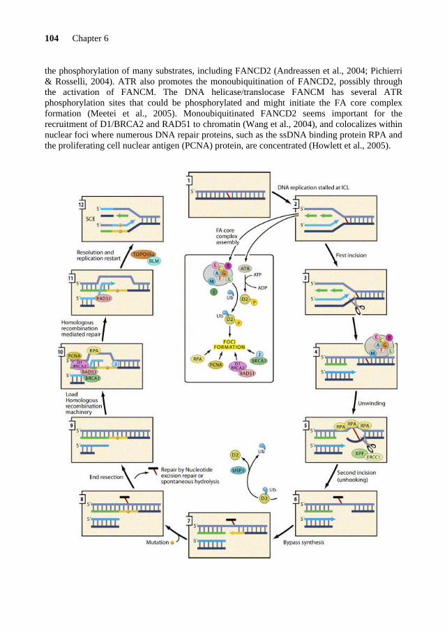
104 Chapter 6
the phosphorylation of many substrates, including FANCD2 (Andreassen et al., 2004; Pichierri & Rosselli, 2004). ATR also promotes the monoubiquitination of FANCD2, possibly through the activation of FANCM. The DNA helicase/translocase FANCM has several ATR phosphorylation sites that could be phosphorylated and might initiate the FA core complex formation (Meetei et al., 2005). Monoubiquitinated FANCD2 seems important for the recruitment of D1/BRCA2 and RAD51 to chromatin (Wang et al., 2004), and colocalizes within nuclear foci where numerous DNA repair proteins, such as the ssDNA binding protein RPA and the proliferating cell nuclear antigen (PCNA) protein, are concentrated (Howlett et al., 2005).
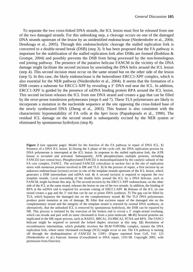
General Discussion
105
To separate the two cross-linked DNA strands, the ICL lesion must first be released from one of the two damaged strands. For this unhooking step, a cleavage occurs on one of the damaged DNA strands upstream of the lesion by an unidentified endonuclease (Niedernhofer et al., 2004; Dendouga et al., 2005). Through this endonucleolytic cleavage the stalled replication fork is converted to a double-strand break (DSB) (step 3). It has been proposed that the FA pathway is important for the stabilization of the stalled replication fork after DSBs are formed (Rothfuss & Grompe, 2004) and possibly prevents the DSB from being processed by the non-homologous end joining pathway. The presence of the putative helicase FANCM in the vicinity of the DNA damage might facilitate the second incision by unwinding the DNA helix around the ICL lesion (step 4). This second incision must occur on the same strand but on the other side of the lesion (step 5). In this case, the likely endonuclease is the heterodimer ERCC1-XPF complex, which is also essential for the NER pathway (Niedernhofer et al., 2004). It seems that the formation of a DSB creates a substrate for ERCC1-XPF by revealing a 3’ DNA end near the ICL. In addition, ERCC1-XPF is guided by the presence of ssDNA binding protein RPA around the ICL lesion. This second incision releases the ICL from one DNA strand and creates a gap that can be filled by the error-prone translesion polymerases (steps 6 and 7). These TLS polymerases are likely to incorporate a mutation in the nucleotide sequence at the site opposing the cross-linked base of the newly synthesized DNA (Zheng et al., 2003). This feature is also consistent with the characteristic hypomutability of FA cells at the hprt locus (Papadopoulo et al., 1990). The residual ICL damage on the second strand is subsequently excised by the NER system or eliminated by spontaneous hydrolysis (step 8).
Figure 2 (see opposite page). Model for the function of the FA pathway in repair of DNA ICL. 1) Presence of a DNA ICL lesion. 2) During the S phase of the cycle cell, the DNA replication process by DNA polymerase is interrupted at the ICL lesion. In response to the stalled replication fork, the ATR kinase is recruited and activated. Subsequently, ATR phosphorylates multiple proteins, including FANCD2 (see central box). Phosphorylated FANCD2 is monoubiquitinated by the catalytic subunit of the FA core complex, FANCL. The activated FANCD2 colocalizes in nuclear foci at the site of replication stress with numerous proteins involved in HR and TLS. 3) In the process of repair, a first incision by an unknown endonuclease (scissor) occurs in one of the template strands upstream of the ICL lesion, which generates a DSB intermediate and ssDNA end. 4) A second incision is required to separate the two template strands. Local unwinding of the double helix around the ICL by a DNA helicase, such as FANCM, might facilitate this step. 5) The second incision by the ERCC1-XPF endonuclease, on the other side of the ICL at the same strand, releases the lesion on one of the two strands. In addition, the binding of RPA at the ssDNA end is required for accurate cutting of ERCC1-XPF. 6) Release of the ICL on one strand creates a gap and the 3’ end that can be use to prime DNA synthesis. 7) The gap can be filled by TLS, which bypasses the damaged site on the complementary strand. 8) The TLS DNA polymerases produce point mutation at site of damage. 9) After that excision repair of the damaged site on the complementary strand and the integrity of the template strand is restored by normal DNA synthesis, or alternatively, that the unhooked ICL is removed by spontaneous hydrolysis, the DSB can be repaired by HR. This process is initiated by the resection of the broken end to reveal a 3’ single-strand overhang, which can invade and pair with its sister chromatid to form a joint molecule. 10-11) Several proteins are implicated in the HR repair process, such as RAD51, BRCA1, D1/BRCA2, PCNA and RPA. The FANCJ helicase might be required to unwind the forked duplex structure at this step. 12) Resolution of recombination intermediate, which probably involves the BLM-TOPOIIIα complex, restarts the DNA replication fork, where sister chromatid exchange (SCE) might occur or not. The FA pathway is turning off through the deubiquitination of FANCD2 by USP1. (Figure reprinted from Cell, Vol. 123 (Niedernhofer et al.) Fanconi Anemia (Cross)linked to DNA repair, 1191-98. Copyright 2005, with permission from Elsevier).
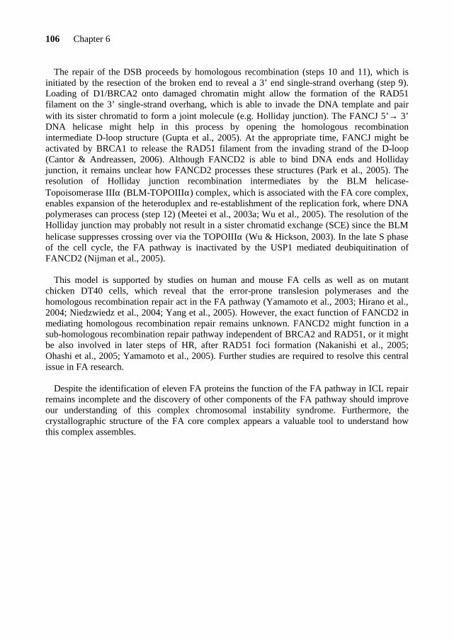
106 Chapter 6
The repair of the DSB proceeds by homologous recombination (steps 10 and 11), which is initiated by the resection of the broken end to reveal a 3’ end single-strand overhang (step 9). Loading of D1/BRCA2 onto damaged chromatin might allow the formation of the RAD51 filament on the 3’ single-strand overhang, which is able to invade the DNA template and pair with its sister chromatid to form a joint molecule (e.g. Holliday junction). The FANCJ 5’→ 3’ DNA helicase might help in this process by opening the homologous recombination intermediate D-loop structure (Gupta et al., 2005). At the appropriate time, FANCJ might be activated by BRCA1 to release the RAD51 filament from the invading strand of the D-loop (Cantor & Andreassen, 2006). Although FANCD2 is able to bind DNA ends and Holliday junction, it remains unclear how FANCD2 processes these structures (Park et al., 2005). The resolution of Holliday junction recombination intermediates by the BLM helicase-Topoisomerase IIIα (BLM-TOPOIIIα) complex, which is associated with the FA core complex, enables expansion of the heteroduplex and re-establishment of the replication fork, where DNA polymerases can process (step 12) (Meetei et al., 2003a; Wu et al., 2005). The resolution of the Holliday junction may probably not result in a sister chromatid exchange (SCE) since the BLM helicase suppresses crossing over via the TOPOIIIα (Wu & Hickson, 2003). In the late S phase of the cell cycle, the FA pathway is inactivated by the USP1 mediated deubiquitination of FANCD2 (Nijman et al., 2005).
This model is supported by studies on human and mouse FA cells as well as on mutant
chicken DT40 cells, which reveal that the error-prone translesion polymerases and the homologous recombination repair act in the FA pathway (Yamamoto et al., 2003; Hirano et al., 2004; Niedzwiedz et al., 2004; Yang et al., 2005). However, the exact function of FANCD2 in mediating homologous recombination repair remains unknown. FANCD2 might function in a sub-homologous recombination repair pathway independent of BRCA2 and RAD51, or it might be also involved in later steps of HR, after RAD51 foci formation (Nakanishi et al., 2005; Ohashi et al., 2005; Yamamoto et al., 2005). Further studies are required to resolve this central issue in FA research.
Despite the identification of eleven FA proteins the function of the FA pathway in ICL repair
remains incomplete and the discovery of other components of the FA pathway should improve our understanding of this complex chromosomal instability syndrome. Furthermore, the crystallographic structure of the FA core complex appears a valuable tool to understand how this complex assembles.


REFERENCES


References
111
Adachi D, Oda T, Yagasaki H, Nakasato K, Taniguchi T, D'Andrea AD, Asano S, Yamashita T. (2002) Heterogeneous activation of the Fanconi anemia pathway by patient-derived FANCA mutants. Hum Mol Genet. 11, 3125-34. Andreassen PR, D'Andrea AD, Taniguchi T. (2004) ATR couples FANCD2 monoubiquitination to the DNA-damage response. Genes Dev. 18, 1958-63. Akkari YM, Bateman RL, Reifsteck CA, Olson SB, Grompe M. (2000) DNA replication is required to elicit cellular responses to psoralen-induced DNA interstrand cross-links. Mol Cell Biol. 21, 8283-89. Akkari YM, Bateman RL, Reifsteck CA, D'Andrea AD, Olson SB, Grompe M. (2001) The 4N cell cycle delay in Fanconi anemia reflects growth arrest in late S phase. Mol Genet Metab. 74, 403-412. Alderton GK, Joenje H, Varon R, Borglum AD, Jeggo PA, O'Driscoll M. (2004) Seckel syndrome exhibits cellular features demonstrating defects in the ATR-signalling pathway. Hum Mol Genet. 13, 3127-38. Alter BP. (2003) Cancer in Fanconi anemia, 1927-2001. Cancer 97, 425-40. Review.
Alter BP, Greene MH, Velazquez I, Rosenberg PS. (2003) Cancer in Fanconi anemia. Blood 101, 2072. Auerbach AD & Allen RG. (1991) Leukemia and preleukemia in Fanconi anemia patients. A review of the literature and report of the International Fanconi Anemia Registry. Cancer Genet Cytogenet. 51, 1-12. Review. Blasco MA, Gasser SM, Lingner J. (1999) Telomeres and telomerase. Genes Dev. 13, 2353-9. Blom E, van de Vrugt HJ, de Winter JP, Arwert F, Joenje H. (2002) Evolutionary clues to the molecular function of Fanconi anemia genes. Acta Haematol. 108, 231-6. Blom E, van de Vrugt HJ, Vries Y, de Winter JP, Arwert F, Joenje H. (2004) Multiple TPR motifs characterize the Fanconi anemia FANCG protein. DNA Repair 5, 77-84. Bochar DA, Wang L, Beniya H, Kinev A, Xue Y, Lane WS, Wang W, Kashanchi F, Shiekhattar R. (2000) BRCA1 is associated with a human SWI/SNF-related complex: linking chromatin remodeling to breast cancer. Cell 102, 257-65. Bogliolo M, Cabre O, Callen E, Castillo V, Creus A, Marcos R, Surralles J. (2002) The Fanconi anaemia genome stability and tumour suppressor network. Mutagenesis 17, 529-38. Review. Boultwood J, Fidler C, Kusec R, Rack K, Elliott PJ, Atoyebi O, Chapman R, Oscier DG, Wainscoat JS. (1997) Telomere length in myelodysplastic syndromes. Am J Hematol. 56, 266-71. Bridge WL, Vandenberg CJ, Franklin RJ, Hiom K. (2005) The BRIP1 helicase functions independently of BRCA1 in the Fanconi anemia pathway for DNA crosslink repair. Nat Genet. 37, 953-7. Brummendorf TH, Rufer N, Holyoake TL, Maciejewski J, Barnett MJ, Eaves CJ, Eaves AC, Young N, Lansdorp PM. (2001) Telomere length dynamics in normal individuals and in patients with hematopoietic stem cell-associated disorders. Ann N Y Acad Sci. 938, 293-303; discussion 303-4. Buchholz TA, Wu X, Hussain A, Tucker SL, Mills GB, Haffty B, Bergh S, Story M, Geara FB, Brock WA. (2002) Evidence of haplotype insufficiency in human cells containing a germline mutation in BRCA1 or BRCA2. Int J Cancer 97, 557-61. Buchwald M. (1995) Complementation groups: one or more per gene? Nat Genet. 11, 228-30. Callen E, Samper E, Ramirez MJ, Creus A, Marcos R, Ortega JJ, Olive T, Badell I, Blasco MA, Surralles J. (2002) Breaks at telomeres and TRF2-independent end fusions in Fanconi anemia. Hum Mol Genet. 11, 439-44. Callen E, Casado JA, Tischkowitz MD, Bueren JA, Creus A, Marcos R, Dasi A, Estella JM, Munoz A, Ortega JJ, de Winter J, Joenje H, Schindler D, Hanenberg H, Hodgson SV, Mathew CG, Surralles J. (2005) A common founder
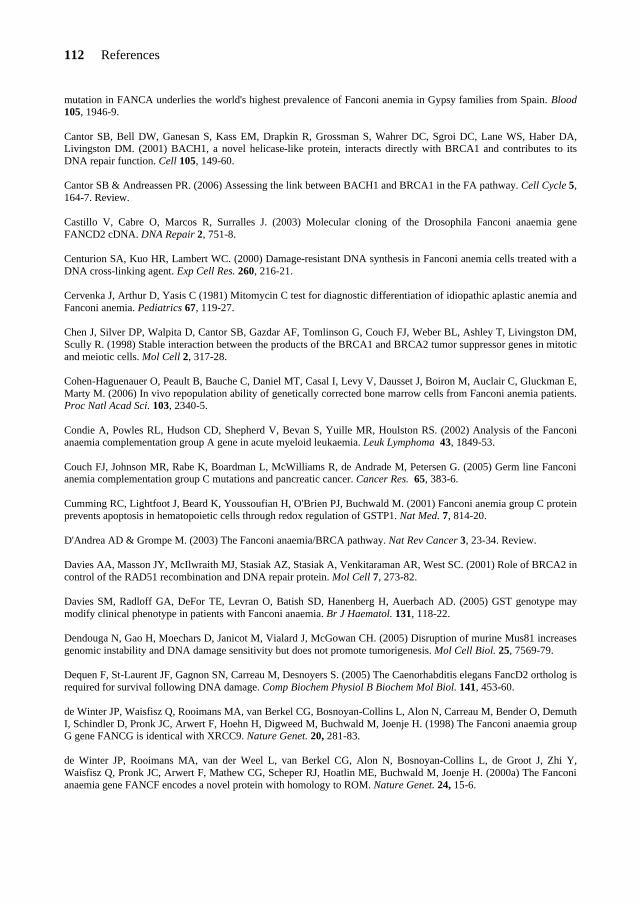
112 References
mutation in FANCA underlies the world's highest prevalence of Fanconi anemia in Gypsy families from Spain. Blood 105, 1946-9. Cantor SB, Bell DW, Ganesan S, Kass EM, Drapkin R, Grossman S, Wahrer DC, Sgroi DC, Lane WS, Haber DA, Livingston DM. (2001) BACH1, a novel helicase-like protein, interacts directly with BRCA1 and contributes to its DNA repair function. Cell 105, 149-60. Cantor SB & Andreassen PR. (2006) Assessing the link between BACH1 and BRCA1 in the FA pathway. Cell Cycle 5, 164-7. Review. Castillo V, Cabre O, Marcos R, Surralles J. (2003) Molecular cloning of the Drosophila Fanconi anaemia gene FANCD2 cDNA. DNA Repair 2, 751-8. Centurion SA, Kuo HR, Lambert WC. (2000) Damage-resistant DNA synthesis in Fanconi anemia cells treated with a DNA cross-linking agent. Exp Cell Res. 260, 216-21. Cervenka J, Arthur D, Yasis C (1981) Mitomycin C test for diagnostic differentiation of idiopathic aplastic anemia and Fanconi anemia. Pediatrics 67, 119-27. Chen J, Silver DP, Walpita D, Cantor SB, Gazdar AF, Tomlinson G, Couch FJ, Weber BL, Ashley T, Livingston DM, Scully R. (1998) Stable interaction between the products of the BRCA1 and BRCA2 tumor suppressor genes in mitotic and meiotic cells. Mol Cell 2, 317-28. Cohen-Haguenauer O, Peault B, Bauche C, Daniel MT, Casal I, Levy V, Dausset J, Boiron M, Auclair C, Gluckman E, Marty M. (2006) In vivo repopulation ability of genetically corrected bone marrow cells from Fanconi anemia patients. Proc Natl Acad Sci. 103, 2340-5. Condie A, Powles RL, Hudson CD, Shepherd V, Bevan S, Yuille MR, Houlston RS. (2002) Analysis of the Fanconi anaemia complementation group A gene in acute myeloid leukaemia. Leuk Lymphoma 43, 1849-53. Couch FJ, Johnson MR, Rabe K, Boardman L, McWilliams R, de Andrade M, Petersen G. (2005) Germ line Fanconi anemia complementation group C mutations and pancreatic cancer. Cancer Res. 65, 383-6. Cumming RC, Lightfoot J, Beard K, Youssoufian H, O'Brien PJ, Buchwald M. (2001) Fanconi anemia group C protein prevents apoptosis in hematopoietic cells through redox regulation of GSTP1. Nat Med. 7, 814-20. D'Andrea AD & Grompe M. (2003) The Fanconi anaemia/BRCA pathway. Nat Rev Cancer 3, 23-34. Review. Davies AA, Masson JY, McIlwraith MJ, Stasiak AZ, Stasiak A, Venkitaraman AR, West SC. (2001) Role of BRCA2 in control of the RAD51 recombination and DNA repair protein. Mol Cell 7, 273-82. Davies SM, Radloff GA, DeFor TE, Levran O, Batish SD, Hanenberg H, Auerbach AD. (2005) GST genotype may modify clinical phenotype in patients with Fanconi anaemia. Br J Haematol. 131, 118-22. Dendouga N, Gao H, Moechars D, Janicot M, Vialard J, McGowan CH. (2005) Disruption of murine Mus81 increases genomic instability and DNA damage sensitivity but does not promote tumorigenesis. Mol Cell Biol. 25, 7569-79. Dequen F, St-Laurent JF, Gagnon SN, Carreau M, Desnoyers S. (2005) The Caenorhabditis elegans FancD2 ortholog is required for survival following DNA damage. Comp Biochem Physiol B Biochem Mol Biol. 141, 453-60. de Winter JP, Waisfisz Q, Rooimans MA, van Berkel CG, Bosnoyan-Collins L, Alon N, Carreau M, Bender O, Demuth I, Schindler D, Pronk JC, Arwert F, Hoehn H, Digweed M, Buchwald M, Joenje H. (1998) The Fanconi anaemia group G gene FANCG is identical with XRCC9. Nature Genet. 20, 281-83. de Winter JP, Rooimans MA, van der Weel L, van Berkel CG, Alon N, Bosnoyan-Collins L, de Groot J, Zhi Y, Waisfisz Q, Pronk JC, Arwert F, Mathew CG, Scheper RJ, Hoatlin ME, Buchwald M, Joenje H. (2000a) The Fanconi anaemia gene FANCF encodes a novel protein with homology to ROM. Nature Genet. 24, 15-6.

References
113
de Winter JP, van der Weel L, de Groot J, Stone S, Waisfisz Q, Arwert F, Scheper RJ, Kruyt FAE, Hoatlin ME, Joenje H. (2000b) The Fanconi anemia protein FANCF forms a nuclear complex with FANCA, FANCC and FANCG. Hum Mol Genet. 18, 2665-74. Digweed M, Rothe S, Demuth I, Scholz R, Schindler D, Stumm M, Grompe M, Jordan A, Sperling K. (2002) Attenuation of the formation of DNA-repair foci containing RAD51 in Fanconi anaemia. Carcinogenesis 23, 1121-6. Dokal I. (2000) The genetics of Fanconi's anaemia. Baillieres Best Pract Res Clin Haematol. 13, 407-25. Review. Duckworth-Rysiecki G & Taylor AM (1985) Effects of ionizing radiation on cells from Fanconi's anemia patients. Cancer Res. 45, 416-20. Dutrillaux B, Aurias A, Dutrillaux AM, Buriot D, Prieur M. (1982) The cell cycle of lymphocytes in Fanconi anemia. Hum Genet. 62, 327-32. Fagerlie S, Lensch MW, Pang Q, Bagby GC Jr. (2001) The Fanconi anemia group C gene product: signaling functions in hematopoietic cells. Exp Hematol. 29, 1371-81. Review. Faivre L, Guardiola P, Lewis C, Dokal I, Ebell W, Zatterale A, Altay C, Poole J, Stones D, Kwee ML, van Weel-Sipman M, Havenga C, Morgan N, de Winter J, Digweed M, Savoia A, Pronk J, de Ravel T, Jansen S, Joenje H, Gluckman E, Mathew CG. (2000) Association of complementation group and mutation type with clinical outcome in fanconi anemia. European Fanconi Anemia Research Group. Blood 96, 4064-70. Fanconi Anaemia/Breast Cancer Consortium. (1996) Positional cloning of the Fanconi anaemia group A gene. Nat Genet. 14, 324-8. Fanconi G. (1927) Familiaere infantile perniziösartige Anaemie (pernizioeses Blutbild und Konstitution). Jahrbuch Kinderheil 117, 257-80. Ferrer M, Rodriguez JA, Spierings EA, de Winter JP, Giaccone G, Kruyt FA. (2005) Identification of multiple nuclear export sequences in Fanconi anemia group A protein that contribute to CRM1-dependent nuclear export. Hum Mol Genet. 14, 1271-81. Folias A, Matkovic M, Bruun D, Reid S, Hejna J, Grompe M, D'Andrea A, Moses R. (2002) BRCA1 interacts directly with the Fanconi anemia protein FANCA. Hum Mol Genet. 11, 2591-7. Futaki M, Yamashita T, Yagasaki H, Toda T, Yabe M, Kato S, Asano S, Nakahata T. (2000) The IVS4 + 4 A to T mutation of the fanconi anemia gene FANCC is not associated with a severe phenotype in Japanese patients. Blood 95, 1493-8. Futaki M, Igarashi T, Watanabe S, Kajigaya S, Tatsuguchi A, Wang J, Liu JM. (2002) The FANCG Fanconi anemia protein interacts with CYP2E1: possible role in protection against oxidative DNA damage. Carcinogenesis 23, 67-72. Galimi F, Noll M, Kanazawa Y, Lax T, Chen C, Grompe M, Verma IM. (2002) Gene therapy of Fanconi anemia: preclinical efficacy using lentiviral vectors. Blood 100, 2732-6. Garcia-Higuera I, Kuang Y, Naf D, Wasik J, D'Andrea AD. (1999) Fanconi anemia proteins FANCA, FANCC, and FANCG/XRCC9 interact in a functional nuclear complex. Mol Cell Biol. 19, 4866-73. Garcia-Higuera I, Kuang Y, Denham J, D'Andrea AD. (2000) The fanconi anemia proteins FANCA and FANCG stabilize each other and promote the nuclear accumulation of the Fanconi anemia complex. Blood 96, 3224-30. Garcia-Higuera I, Taniguchi T, Ganesan S, Meyn MS, Timmers C, Hejna J, Grompe M, D’Andrea AD. (2001) Interaction of the Fanconi anemia proteins and BRCA1 in a common pathway. Mol Cell 7, 249-62. Gibson RA, Hajianpour A, Murer-Orlando M, Buchwald M, Mathew CG. (1993) A nonsense mutation and exon skipping in the Fanconi anaemia group C gene. Hum Mol Genet. 2, 797-9.
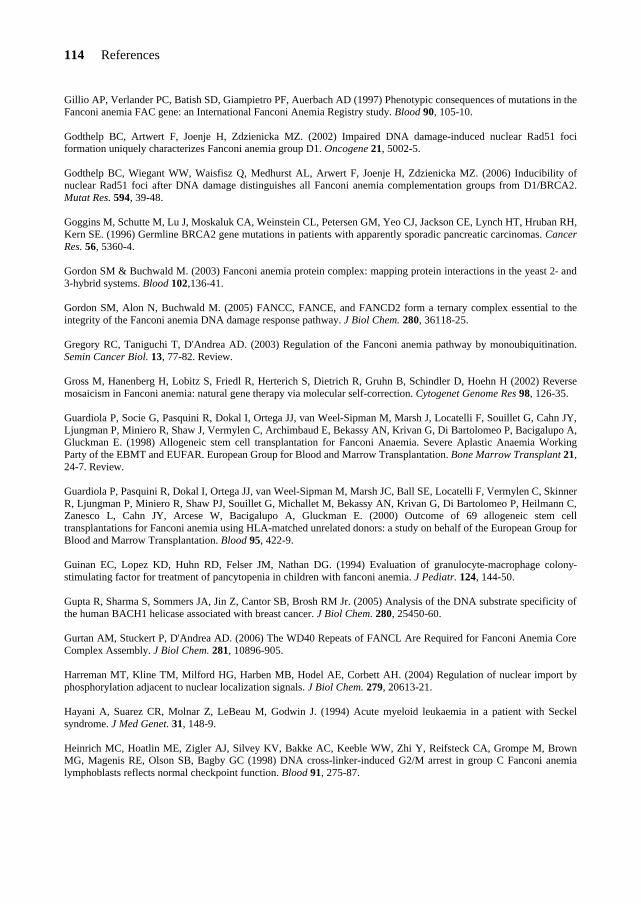
114 References
Gillio AP, Verlander PC, Batish SD, Giampietro PF, Auerbach AD (1997) Phenotypic consequences of mutations in the Fanconi anemia FAC gene: an International Fanconi Anemia Registry study. Blood 90, 105-10. Godthelp BC, Artwert F, Joenje H, Zdzienicka MZ. (2002) Impaired DNA damage-induced nuclear Rad51 foci formation uniquely characterizes Fanconi anemia group D1. Oncogene 21, 5002-5. Godthelp BC, Wiegant WW, Waisfisz Q, Medhurst AL, Arwert F, Joenje H, Zdzienicka MZ. (2006) Inducibility of nuclear Rad51 foci after DNA damage distinguishes all Fanconi anemia complementation groups from D1/BRCA2. Mutat Res. 594, 39-48. Goggins M, Schutte M, Lu J, Moskaluk CA, Weinstein CL, Petersen GM, Yeo CJ, Jackson CE, Lynch HT, Hruban RH, Kern SE. (1996) Germline BRCA2 gene mutations in patients with apparently sporadic pancreatic carcinomas. Cancer Res. 56, 5360-4. Gordon SM & Buchwald M. (2003) Fanconi anemia protein complex: mapping protein interactions in the yeast 2- and 3-hybrid systems. Blood 102,136-41. Gordon SM, Alon N, Buchwald M. (2005) FANCC, FANCE, and FANCD2 form a ternary complex essential to the integrity of the Fanconi anemia DNA damage response pathway. J Biol Chem. 280, 36118-25. Gregory RC, Taniguchi T, D'Andrea AD. (2003) Regulation of the Fanconi anemia pathway by monoubiquitination. Semin Cancer Biol. 13, 77-82. Review. Gross M, Hanenberg H, Lobitz S, Friedl R, Herterich S, Dietrich R, Gruhn B, Schindler D, Hoehn H (2002) Reverse mosaicism in Fanconi anemia: natural gene therapy via molecular self-correction. Cytogenet Genome Res 98, 126-35. Guardiola P, Socie G, Pasquini R, Dokal I, Ortega JJ, van Weel-Sipman M, Marsh J, Locatelli F, Souillet G, Cahn JY, Ljungman P, Miniero R, Shaw J, Vermylen C, Archimbaud E, Bekassy AN, Krivan G, Di Bartolomeo P, Bacigalupo A, Gluckman E. (1998) Allogeneic stem cell transplantation for Fanconi Anaemia. Severe Aplastic Anaemia Working Party of the EBMT and EUFAR. European Group for Blood and Marrow Transplantation. Bone Marrow Transplant 21, 24-7. Review. Guardiola P, Pasquini R, Dokal I, Ortega JJ, van Weel-Sipman M, Marsh JC, Ball SE, Locatelli F, Vermylen C, Skinner R, Ljungman P, Miniero R, Shaw PJ, Souillet G, Michallet M, Bekassy AN, Krivan G, Di Bartolomeo P, Heilmann C, Zanesco L, Cahn JY, Arcese W, Bacigalupo A, Gluckman E. (2000) Outcome of 69 allogeneic stem cell transplantations for Fanconi anemia using HLA-matched unrelated donors: a study on behalf of the European Group for Blood and Marrow Transplantation. Blood 95, 422-9. Guinan EC, Lopez KD, Huhn RD, Felser JM, Nathan DG. (1994) Evaluation of granulocyte-macrophage colony-stimulating factor for treatment of pancytopenia in children with fanconi anemia. J Pediatr. 124, 144-50. Gupta R, Sharma S, Sommers JA, Jin Z, Cantor SB, Brosh RM Jr. (2005) Analysis of the DNA substrate specificity of the human BACH1 helicase associated with breast cancer. J Biol Chem. 280, 25450-60. Gurtan AM, Stuckert P, D'Andrea AD. (2006) The WD40 Repeats of FANCL Are Required for Fanconi Anemia Core Complex Assembly. J Biol Chem. 281, 10896-905. Harreman MT, Kline TM, Milford HG, Harben MB, Hodel AE, Corbett AH. (2004) Regulation of nuclear import by phosphorylation adjacent to nuclear localization signals. J Biol Chem. 279, 20613-21. Hayani A, Suarez CR, Molnar Z, LeBeau M, Godwin J. (1994) Acute myeloid leukaemia in a patient with Seckel syndrome. J Med Genet. 31, 148-9. Heinrich MC, Hoatlin ME, Zigler AJ, Silvey KV, Bakke AC, Keeble WW, Zhi Y, Reifsteck CA, Grompe M, Brown MG, Magenis RE, Olson SB, Bagby GC (1998) DNA cross-linker-induced G2/M arrest in group C Fanconi anemia lymphoblasts reflects normal checkpoint function. Blood 91, 275-87.

References
115
Heinrich MC, Silvey KV, Stone S, Zigler AJ, Griffith DJ, Montalto M, Chai L, Zhi Y, Hoatlin ME. (2000) Posttranscriptional cell cycle-dependent regulation of human FANCC expression. Blood 95, 3970-7. Herrera E, Samper E, Martin-Caballero J, Flores JM, Lee HW, Blasco MA. (1999) Disease states associated with telomerase deficiency appear earlier in mice with short telomeres. EMBO J. 18, 2950-60. Hirano S, Yamamoto K, Ishiai M, Yamazoe M, Seki M, Matsushita N, Ohzeki M, Yamashita YM, Arakawa H, Buerstedde JM, Enomoto T, Takeda S, Thompson LH, Takata M. (2005) Functional relationships of FANCC to homologous recombination, translesion synthesis, and BLM. EMBO J. 24, 418-27. Hirsch B, Shimamura A, Moreau L, Baldinger S, Hag-alshiekh M, Bostrom B, Sencer S, D'Andrea AD. (2004) Association of biallelic BRCA2/FANCD1 mutations with spontaneous chromosomal instability and solid tumors of childhood. Blood 103, 2554-9. Hoatlin ME, Christianson TA, Keeble WW, Hammond AT, Zhi Y, Heinrich MC, Tower PA, Bagby GC Jr. (1998) The Fanconi anemia group C gene product is located in both the nucleus and cytoplasm of human cells. Blood 91, 1418-25. Hoatlin ME, Zhi Y, Ball H, Silvey K, Melnick A, Stone S, Arai S, Hawe N, Owen G, Zelent A, Licht JD. (1999) A novel BTB/POZ transcriptional repressor protein interacts with the Fanconi anemia group C protein and PLZF. Blood 94, 3737-47. Houghtaling S, Timmers C, Noll M, Finegold MJ, Jones SN, Meyn MS, Grompe M. (2003) Epithelial cancer in Fanconi anemia complementation group D2 (Fancd2) knockout mice. Genes Dev. 17, 2021-35. Houghtaling S, Newell A, Akkari Y, Taniguchi T, Olson S, Grompe M. (2005) Fancd2 functions in a double strand break repair pathway that is distinct from non-homologous end joining. Hum Mol Genet. 14, 3027-33. Hoshino T, Wang J, Devetten MP, Iwata N, Kajigaya S, Wise RJ, Liu JM, Youssoufian H. (1998) Molecular chaperone GRP94 binds to the Fanconi anemia group C protein and regulates its intracellular expression. Blood 91, 4379-86. Howlett NG, Taniguchi T, Olson S, Cox B, Waisfisz Q, De Die-Smulders C, Persky N, Grompe M, Joenje H, Pals G, Ikeda H, Fox EA, D'Andrea AD (2002) Biallelic inactivation of BRCA2 in Fanconi anemia. Science 297, 606-9. Howlett NG, Taniguchi T, Durkin SG, D'Andrea AD, Glover TW. (2005) The Fanconi anemia pathway is required for the DNA replication stress response and for the regulation of common fragile site stability. Hum Mol Genet. 14, 693-701. Hussain S, Witt E, Huber PA, Medhurst AL, Ashworth A, Mathew CG. (2003) Direct interaction of the Fanconi anaemia protein FANCG with BRCA2/FANCD1. Hum Mol Genet. 12, 2503-10. Hussain S, Wilson JB, Medhurst AL, Hejna J, Witt E, Ananth S, Davies A, Masson JY, Moses R, West SC, de Winter JP, Ashworth A, Jones NJ, Mathew CG. (2004) Direct interaction of FANCD2 with BRCA2 in DNA damage response pathways. Hum Mol Genet. 13, 1241-8. Hussain S, Wilson JB, Blom E, Thompson LH, Sung P, Gordon SM, Kupfer GM, Joenje H, Mathew CG, Jones NJ. (2006) Tetratricopeptide-motif-mediated interaction of FANCG with recombination proteins XRCC3 and BRCA2. DNA Repair 5, 629-40. Ishida R & Buchwald M. (1982) Susceptibility of Fanconi's anemia lymphoblasts to DNA-cross-linking and alkylating agents. Cancer Res. 42, 4000-6. Joenje H, Arwert F, Eriksson AW, de Koning H, Oostra AB. (1981) Oxygen-dependence of chromosomal aberrations in Fanconi's anaemia. Nature 290, 142-3. Joenje H, Lo Ten Foe JR, Oostra AB, van Berkel CGM, Rooimans MA, Schroeder-Kurth T, Wegner R-D, Gille JJP, Buchwald M, Arwert F. (1995) Classification of Fanconi anemia patients by complementation analysis: evidence for a fifth genetic subtype. Blood 86, 2156-60.

116 References
Joenje H, Oostra AB, Wijker M, di Summa FM, van Berkel CG, Rooimans MA, Ebell W, van Weel M, Pronk JC, Buchwald M, Arwert F. (1997) Evidence for at least eight Fanconi anemia genes. Am J Hum Genet. 61, 940-4. Joenje H, Arwert F, Kwee ML, Madan K, Hoehn H (1998) Confounding factors in the diagnosis of Fanconi anaemia. Am J Med Genet. 79, 403-5. Joenje H, Levitus M, Waisfisz Q, D'Andrea A, Garcia-Higuera I, Pearson T, van Berkel CG, Rooimans MA, Morgan N, Mathew CG, Arwert F. (2000) Complementation analysis in Fanconi anemia: assignment of the reference FA-H patient to group A. Am J Hum Genet. 67, 759-62. Joenje H & Patel KJ. (2001) The emerging genetic and molecular basis of Fanconi anaemia. Nat Rev Genet. 2, 446-57. Kennedy RD & D'Andrea AD. (2005) The Fanconi Anemia/BRCA pathway: new faces in the crowd. Genes Dev. 19, 2925-40. Review. Komori K, Hidaka M, Horiuchi T, Fujikane R, Shinagawa H, Ishino Y. (2004) Cooperation of the N-terminal Helicase and C-terminal endonuclease activities of Archaeal Hef protein in processing stalled replication forks. J Biol Chem. 279, 53175-85. Kruyt FA, Dijkmans LM, van den Berg TK, Joenje H (1996) Fanconi anemia genes act to suppress a cross-linker-inducible p53-independent apoptosis pathway in lymphoblastoid cell lines. Blood 87, 938-48. Kruyt FA, Youssoufian H. (1998) Abnormal microsomal detoxification implicated in Fanconi anemia group C by interaction of the FAC protein with NADPH cytochrome P450 reductase. Blood 92, 3050-6. Kruyt FA, Abou-Zahr F, Mok H, Youssoufian H. (1999) Resistance to mitomycin C requires direct interaction between the Fanconi anemia proteins FANCA and FANCG in the nucleus through an arginine-rich domain. J Biol Chem. 274, 34212-8. Kuang Y, Garcia-Higuera I, Moran A, Mondoux M, Digweed M, D’Andrea AD. (2000) Carboxy terminal region of the Fanconi anemia protein, FANCG/XRCC9 is required for functional activity. Blood 96, 1625-32. Kupfer GM, Yamashita T, Näf D, Suliman A, Asano S, D'Andrea AD. (1997a) The Fanconi anemia polypeptide, FAC, binds to the cyclin-dependent kinase, cdc2. Blood 90, 1047-54. Kupfer GM, Näf D, Suliman A, Pulsipher M, D'Andrea AD. (1997b) The Fanconi anaemia proteins, FAA and FAC, interact to form a nuclear complex. Nat Genet. 17, 487-90. Kupfer GM, Näf D, Garcia-Higuera I, Wasik J, Cheng A, Yamashita T, Tipping A, Morgan N, Mathew CG, D’Andrea AD. (1999) A patient-derived mutant form of the Fanconi anemia protein, FANCA, is defective in nuclear accumulation. Exp Hematol. 27, 587-93. Kutler DI, Singh B, Satagopan J, Batish SD, Berwick M, Giampietro PF, Hanenberg H, Auerbach AD. (2003) A 20-year perspective on the International Fanconi Anemia Registry (IFAR). Blood 101, 1249-56. Lensch MW, Tischkowitz M, Christianson TA, Reifsteck CA, Speckhart SA, Jakobs PM, O'Dwyer ME, Olson SB, Le Beau MM, Hodgson SV, Mathew CG, Larson RA, Bagby GC Jr. (2003) Acquired FANCA dysfunction and cytogenetic instability in adult acute myelogenous leukemia. Blood 102, 7-16. Levitus M, Rooimans MA, Steltenpool J, Cool NF, Oostra AB, Mathew CG, Hoatlin ME, Waisfisz Q, Arwert F, de Winter JP, Joenje H (2004) Heterogeneity in Fanconi anemia: evidence for 2 new genetic subtypes. Blood 103, 2498-503. Levitus M, Waisfisz Q, Godthelp BC, de Vries Y, Hussain S, Wiegant WW, Elghalbzouri-Maghrani E, Steltenpool J, Rooimans MA, Pals G, Arwert F, Mathew CG, Zdzienicka MZ, Hiom K, de Winter JP, Joenje H. (2005) The DNA helicase BRIP1 is defective in Fanconi anemia complementation group J. Nat Genet. 37, 934-5.
References
117
Levran O, Diotti R, Pujara K, Batish SD, Hanenberg H, Auerbach AD. (2005) Spectrum of sequence variations in the FANCA gene: an International Fanconi Anemia Registry (IFAR) study. Hum Mutat. 25, 142-9. Li X, Leteurtre F, Rocha V, Guardiola P, Berger R, Daniel MT, Noguera MH, Maarek O, Roux GL, de la Salmoniere P, Richard P, Gluckman E. (2003) Abnormal telomere metabolism in Fanconi's anaemia correlates with genomic instability and the probability of developing severe aplastic anaemia. Br J Haematol. 120, 836-45. Lightfoot J, Alon N, Bosnoyan-Collins L, Buchwald M. (1999) Characterization of regions functional in the nuclear localization of the Fanconi anemia group A protein. Hum Mol Genet. 8, 1007-15. Litman R, Peng M, Jin Z, Zhang F, Zhang J, Powell S, Andreassen PR, Cantor SB. (2005) BACH1 is critical for homologous recombination and appears to be the Fanconi anemia gene product FANCJ. Cancer Cell 8, 255-65. Liu N, Lamerdin JE, Tucker JD, Zhou ZQ, Walter CA, Albala JS, Busch DB, Thompson LH. (1997) The human XRCC9 gene corrects chromosomal instability and mutagen sensitivities in CHO UV40 cells. Proc Natl Acad Sci. 94, 9232-7. Liu NG, Howlett M, Deng DM, Langenau K, Hsu J, Rhodes JP, Kanki AD, D'Andrea, Look AT. (2003) Knockdown of zebrafish Fancd2 causes developmental abnormalities via p53-dependent apoptosis. Dev Cell 5, 903–14. Loeb LA. (2001) A mutator phenotype in cancer. Cancer Res. 61, 3230-9. Review. Loeb LA, Loeb KR, Anderson JP. (2003) Multiple mutations and cancer. Proc Natl Acad Sci. 100, 776-81. Review. Lomonosov M, Anand S, Sangrithi M, Davies R, Venkitaraman AR. (2003) Stabilization of stalled DNA replication forks by the BRCA2 breast cancer susceptibility protein. Genes Dev. 17, 3017-22. Lo Ten Foe JR, Rooimans MA, Bosnoyan-Collins L, Alon N, Wijker M, Parker L, Lightfoot J, Carreau M, Callen DF, Savoia A, Cheng NC, van Berkel CG, Strunk MH, Gille JJ, Pals G, Kruyt FA, Pronk JC, Arwert F, Buchwald M, Joenje H (1996) Expression cloning of a cDNA for the major Fanconi anaemia gene, FAA. Nat Genet. 14, 320-3. Lo Ten Foe JR, Kwee ML, Rooimans MA, Oostra AB, Veerman AJ, van Weel M, Pauli RM, Shahidi NT, Dokal I, Roberts I, Altay C, Gluckman E, Gibson RA, Mathew CG, Arwert F, Joenje H (1997) Somatic mosaicism in Fanconi anemia: molecular basis and clinical significance. Eur J Hum Genet. 5, 137-48. Lyakhovich A & Surralles J. (2006) Disruption of the Fanconi anemia/BRCA pathway in sporadic cancer. Cancer Lett. 232, 99-106. Review. Mankad A, Taniguchi T, Cox B, Akkari Y, Rathbun RK, Lucas L, Bagby G, Olson S, D'Andrea A, Grompe M (2006) Natural gene therapy in monozygotic twins with Fanconi anemia. Blood 107, 3084-90. Marsit, CJ, Liu M, Nelson HH, Posner M, Suzuki M, Kelsey KT. (2004) Inactivation of the Fanconi anemia/BRCA pathway in lung and oral cancers: implications for treatment and survival. Oncogene 23, 1000-4. Matsushita N, Kitao H, Ishiai M, Nagashima N, Hirano S, Okawa K, Ohta T, Yu DS, McHugh PJ, Hickson ID, Venkitaraman AR, Kurumizaka H, Takata M. (2005) A FancD2-monoubiquitin fusion reveals hidden functions of Fanconi anemia core complex in DNA repair. Mol Cell 19, 841-7. Medhurst AL, Huber PAJ, Waisfisz Q, de Winter JP, Mathew CG. (2001) Direct interactions of the five known Fanconi anaemia proteins suggest a common functional pathway. Hum Mol Genet. 10, 423-9. Medhurst AL, Laghmani EL, Steltenpool J, Ferrer M, Fontaine C, de Groot J, Rooimans MA, Scheper RJ, Meetei AR, Wang W, Joenje H, de Winter JP (2006) Evidence for subcomplexes in the Fanconi anaemia pathway. Blood, in press Meetei AR, Sechi S, Wallisch M, Yang D, Young MK, Joenje H, Hoatlin ME, Wang W. (2003a) A multiprotein nuclear complex connects Fanconi anemia and Bloom syndrome. Mol Cell Biol. 23, 3417-26.
118 References
Meetei AR, de Winter JP, Medhurst AL, Wallisch M, Waisfisz Q, van de Vrugt HJ, Oostra AB, Yan Z, Ling C, Bishop CE, Hoatlin ME, Joenje H, Wang W (2003b) A novel ubiquitin ligase is deficient in Fanconi anemia. Nat Genet 35, 165-70. Meetei AR, Levitus M, Xue Y, Medhurst AL, Zwaan M, Ling C, Rooimans MA, Bier P, Hoatlin M, Pals G, de Winter JP, Wang W, Joenje H. (2004) X-linked inheritance of Fanconi anemia complementation group B. Nat Genet. 36, 1219-24. Meetei AR, Yan Z, Wang W (2004) FANCL replaces BRCA1 as the likely ubiquitin ligase responsible for FANCD2 monoubiquitination. Cell Cycle 3, 179-81. Meetei AR, Medhurst AL, Ling C, Xue Y, Singh TR, Bier P, Steltenpool J, Stone S, Dokal I, Mathew CG, Hoatlin M, Joenje H, de Winter JP, Wang W. (2005) A human ortholog of archaeal DNA repair protein Hef is defective in Fanconi anemia complementation group M. Nat Genet. 37, 958-63. Mi J, Qiao F, Wilson JB, High AA, Schroeder MJ, Stukenberg PT, Moss A, Shabanowitz J, Hunt DF, Jones NJ, Kupfer GM. (2004) FANCG is phosphorylated at serines 383 and 387 during mitosis. Mol Cell Biol. 24, 8576-85. Mi J & Kupfer GM. (2005) The Fanconi anemia core complex associates with chromatin during S phase. Blood 105, 759-66. Mosedale G, Niedzwiedz W, Alpi A, Perrina F, Pereira-Leal JB, Johnson M, Langevin F, Pace P, Patel KJ. (2005) The vertebrate Hef ortholog is a component of the Fanconi anemia tumor-suppressor pathway. Nat Struct Mol Biol. 12, 763-71. Moynahan ME, Chiu JW, Koller BH, Jasin M. (1999) Brca1 controls homology-directed DNA repair. Mol Cell 4, 511-8. Moynahan ME, Cui TY, Jasin M. (2001) Homology-directed DNA repair, mitomycin-C resistance, and chromosome stability is restored with correction of a Brca1 mutation. Cancer Res. 61, 4842-50. Naf D, Kupfer GM, Suliman A, Lambert K, D'Andrea AD. (1998) Functional activity of the Fanconi anemia protein FAA requires FAC binding and nuclear localization. Mol Cell Biol. 18, 5952-60. Nakanishi K, Moran A, Hays T, Kuang Y, Fox E, Garneau D, Monte de Oca R, Grompe M, D’Andrea AD. (2001) Functional analysis of patient-derived mutations in the Fanconi anemia gene FANCG/XRCC9. Exp Hematol. 29, 842-9. Nakanishi K, Taniguchi T, Ranganathan V, New HH, Moreau LA, Stotsky M, Mathew CG, Kastan MB, Weaver DT, D’Andrea AD. (2002) Interaction of FANCD2 and NBS1 in the DNA damage response. Nat Cell Biol. 4, 913-20. Nakanishi K, Yang YG, Pierce AJ, Taniguchi T, Digweed M, D'Andrea AD, Wang ZQ, Jasin M. (2005) Human Fanconi anemia monoubiquitination pathway promotes homologous DNA repair. Proc Natl Acad Sci. 102, 1110-5. Narayan G, Arias-Pulido H, Nandula SV, Basso K, Sugirtharaj DD, Vargas H, Mansukhani M, Villella J, Meyer L, Schneider A, Gissmann L, Durst M, Pothuri B, Murty VV. (2004) Promoter hypermethylation of FANCF: disruption of Fanconi Anemia-BRCA pathway in cervical cancer. Cancer Res. 64, 2994-7. Niedernhofer LJ, Lalai AS, Hoeijmakers JH. (2005). Fanconi Anemia (Cross)linked to DNA repair. Cell 123, 1191-1198. Review. Niedzwiedz W, Mosedale G, Johnson M, Ong CY, Pace P, Patel KJ. (2004) The Fanconi anaemia gene FANCC promotes homologous recombination and error-prone DNA repair. Mol Cell 15, 607-20. Nijman SM, Huang TT, Dirac AM, Brummelkamp TR, Kerkhoven RM, D'Andrea AD, Bernards R. (2005) The deubiquitinating enzyme USP1 regulates the Fanconi anemia pathway. Mol Cell 17, 331-9.
References
119
Offit K, Levran O, Mullaney B, Mah K, Nafa K, Batish SD, Diotti R, Schneider H, Deffenbaugh A, Scholl T, Proud VK, Robson M, Norton L, Ellis N, Hanenberg H, Auerbach AD. (2003) Shared genetic susceptibility to breast cancer, brain tumors, and Fanconi anemia. J Natl Cancer Inst. 95, 1548-51. Ohashi A, Zdzienicka MZ, Chen J, Couch FJ. (2005) Fanconi anemia complementation group D2 (FANCD2) functions independently of BRCA2- and RAD51-associated homologous recombination in response to DNA damage. J Biol Chem. 280, 14877-83. Otsuki T, Furukawa Y, Ikeda K, Endo H, Yamashita T, Shinohara A, Iwamatsu A, Ozawa K, Liu JM. (2001) Fanconi anemia protein, FANCA, associates with BRG1, a component of the human SWI/SNF complex. Hum Mol Genet. 10, 2651-60. Otsuki T, Nagashima T, Komatsu N, Kirito K, Furukawa Y, Kobayashi Si S, Liu JM, Ozawa K. (2002) Phosphorylation of Fanconi anemia protein, FANCA, is regulated by Akt kinase. Biochem Biophys Res Commun. 291, 628-34. Ridet A, Guillouf C, Duchaud E, Cundari E, Fiore M, Moustacchi E, Rosselli F. (1997) Deregulated apoptosis is a hallmark of the Fanconi anemia syndrome. Cancer Res. 57, 1722-30. Rosselli F (1998) Fanconi anaemia syndrome and apoptosis: state of the art. Apoptosis 3, 229-36. Pace P, Johnson M, Tan WM, Mosedale G, Sng C, Hoatlin M, de Winter J, Joenje H, Gergely F, Patel KJ. (2002) FANCE: the link between Fanconi anaemia complex assembly and activity. EMBO J. 21, 3414-23. Pagano G & Youssoufian H. (2003) Fanconi anaemia proteins: major roles in cell protection against oxidative damage. Bioessays 25, 589-95. Review. Pang Q, Fagerlie S, Christianson TA, Keeble W, Faulkner G, Diaz J, Rathbun RK, Bagby GC. (2000) The Fanconi anemia protein FANCC binds to and facilitates the activation of STAT1 by gamma interferon and hematopoietic growth factors. Mol Cell Biol. 20, 4724-35. Pang Q, Christianson TA, Keeble W, Diaz J, Faulkner GR, Reifsteck C, Olson S, Bagby GC. (2001) The Fanconi anemia complementation group C gene product: structural evidence of multifunctionality. Blood 98, 1392-1401. Pang Q, Christianson TA, Keeble W, Koretsky T, Bagby GC. (2002) The anti-apoptotic function of Hsp70 in the interferon-inducible double-stranded RNA-dependent protein kinase-mediated death signaling pathway requires the Fanconi anemia protein, FANCC. J Biol Chem. 277, 49638-43. Papadopoulo D, Guillouf C, Mohrenweiser H, Moustacchi E. (1990) Hypomutability in Fanconi anemia cells is associated with increased deletion frequency at the HPRT locus. Proc Natl Acad Sci. 87, 8383-7. Park SJ, Ciccone SL, Beck BD, Hwang B, Freie B, Clapp DW, Lee SH. (2004) Oxidative stress/damage induces multimerization and interaction of Fanconi anemia proteins. J Biol Chem. 279, 30053-9. Park WH, Margossian S, Horwitz AA, Simons AM, D'Andrea AD, Parvin JD. (2005) Direct DNA binding activity of the Fanconi anemia D2 protein. J Biol Chem. 280, 23593-8. Patel KJ, Yu VP, Lee H, Corcoran A, Thistlethwaite FC, Evans MJ, Colledge WH, Friedman LS, Ponder BA, Venkitaraman AR. (1998) Involvement of Brca2 in DNA repair. Mol Cell 1, 347-57. Pichierri P, Averbeck D, Rosselli F. (2002) DNA cross-link-dependent RAD50/MRE11/NBS1 subnuclear assembly requires the Fanconi anemia C protein. Hum Mol Genet. 11, 2531-46. Pichierri P, Franchitto A, Rosselli F. (2004) BLM and the FANC proteins collaborate in a common pathway in response to stalled replication forks. EMBO J. 23, 3154-63. Pichierri P & Rosselli F. (2004) The DNA crosslink-induced S-phase checkpoint depends on ATR-CHK1 and ATR-NBS1-FANCD2 pathways. EMBO J. 23, 1178-87.
120 References
Powell SN & Kachnic LA. (2003) Roles of BRCA1 and BRCA2 in homologous recombination, DNA replication fidelity and the cellular response to ionizing radiation. Oncogene 22, 5784-91. Review. Prakash R, Krejci L, Van Komen S, Anke Schurer K, Kramer W, Sung P. (2005) Saccharomyces cerevisiae MPH1 gene, required for homologous recombination-mediated mutation avoidance, encodes a 3' to 5' DNA helicase. J Biol Chem. 280, 7854-60. Pronk JC, Gibson RA, Savoia A, Wijker M, Morgan NV, Melchionda S, Ford D, Temtamy S, Ortega JJ, Jansen S, et al. (1995) Localisation of the Fanconi anaemia complementation group A gene to chromosome 16q24.3. Nat Genet. 11, 338-40. Qiao F, Moss A, Kupfer GM. (2001) Fanconi anemia proteins localize to chromatin and the nuclear matrix in a DNA damage- and cell cycle-regulated manner. J Biol Chem. 276, 23391-6. Qiao F, Mi J, Wilson JB, Zhi G, Bucheimer NR, Jones NJ, Kupfer GM (2004) Phosphorylation of Fanconi anemia (FA) complementation group G protein, FANCG, at serine 7 is important for function of the FA pathway. J Biol Chem. 279, 46035-45. Rackoff WR, Orazi A, Robinson CA, Cooper RJ, Alter BP, Freedman MH, Harris RE, Williams DA. (1996) Prolonged administration of granulocyte colony-stimulating factor (filgrastim) to patients with Fanconi anemia: a pilot study. Blood 88, 1588-93. Rahman N & Ashworth A. (2004) A new gene on the X involved in Fanconi anemia. Nat Genet. 36, 1142-3. Risinger MA & Groden J. (2004) Crosslinks and crosstalk: human cancer syndromes and DNA repair defects. Cancer Cell 6, 539-45. Review. Rosselli F, Briot D, Pichierri P. (2003) The Fanconi anemia pathway and the DNA interstrand cross-links repair. Biochimie 85, 1175-84. Review. Rosenberg PS, Greene MH, Alter BP. (2003) Cancer incidence in persons with Fanconi anemia. Blood 101, 822-6. Rothfuss A & Grompe M. (2004) Repair kinetics of genomic interstrand DNA cross-links: evidence for DNA double-strand break-dependent activation of the Fanconi anemia/BRCA pathway. Mol Cell Biol. 24,123-34. Saito H, Hammond AT, Moses RE. (1993) Hypersensitivity to oxygen is a uniform and secondary defect in Fanconi anemia cells. Mutat Res. 294, 255-62. Sala-Trepat M, Rouillard D, Escarceller M, Laquerbe A, Moustacchi E, Papadopoulo D. (2000) Arrest of S-phase progression is impaired in Fanconi anemia cells. Exp Cell Res. 260, 208-15. Sasaki MS & Tonomura A. (1973) A high susceptibility of Fanconi’s anemia to chromosome breakage by DNA cross-linking agents. Cancer Res. 33, 1829-36. Savoia A, Centra M, Ianzano L, de Cillis GP, Zelante L, Buchwald M. (1995) Characterization of the 5' region of the Fanconi anaemia group C (FACC) gene. Hum Mol Genet. 4, 1321-6. Schindler D & Hoehn H. (1988) Fanconi anemia mutation causes cellular susceptibility to ambient oxygen. Am J Hum Genet. 43, 429-35. Schroeder TM, Anschutz F, Knopp A. (1964) Spontaneous chromosome aberrations in familial panmyelopathy. Humangenetik 1, 194-6. Schroeder TM, Tilgen D, Kruger J, Vogel F. (1976) Formal genetics of Fanconi's anemia. Hum Genet. 32, 257-88. Seal S, Barfoot R, Jayatilake H, Smith P, Renwick A, Bascombe L, McGuffog L, Evans DG, Eccles D, Easton DF, Stratton MR, Rahman N; Breast Cancer Susceptibility Collaboration. (2003) Evaluation of Fanconi Anemia genes in familial breast cancer predisposition. Cancer Res. 63, 8596-9.

References
121
Shin DS, Pellegrini L, Daniels DS, Yelent B, Craig L, Bates D, Yu DS, Shivji MK, Hitomi C, Arvai AS, Volkmann N, Tsuruta H, Blundell TL, Venkitaraman AR, Tainer JA. (2003) Full-length archaeal Rad51 structure and mutants: mechanisms for RAD51 assembly and control by BRCA2. EMBO J. 22, 4566-76. Shivji MK, Venkitaraman AR. (2004) DNA recombination, chromosomal stability and carcinogenesis: insights into the role of BRCA2. DNA Repair 3, 835-43. Review. Siddique MA, Nakanishi K, Taniguchi T, Grompe M, D'Andrea AD. (2001) Function of the Fanconi anemia pathway in Fanconi anemia complementation group F and D1 cells. Exp Hematol. 29, 1448-55. Sobeck A, Stone S, Costanzo V, de Graaf B, Reuter T, de Winter J, Wallisch M, Akkari Y, Olson S, Wang W, Joenje H, Christian JL, Lupardus PJ, Cimprich KA, Gautier J, Hoatlin ME. (2006) Fanconi anemia proteins are required to prevent accumulation of replication-associated DNA double-strand breaks. Mol Cell Biol. 26, 425-37. Sridharan D, Brown M, Lambert WC, McMahon LW, Lambert MW. (2003) Nonerythroid alphaII spectrin is required for recruitment of FANCA and XPF to nuclear foci induced by DNA interstrand cross-links. J Cell Sci. 116, 823-35. Strathdee CA, Gavish H, Shannon WR, Buchwald M (1992) Cloning of cDNAs for Fanconi's anaemia by functional complementation. Nature 356, 763-7. Surralles J, Jackson SP, Jasin M, Kastan MB, West SC, Joenje H. (2004) Molecular cross-talk among chromosome fragility syndromes. Gene Dev. 18, 1359-70. Swift M. (1971) Fanconi's anaemia in the genetics of neoplasia. Nature 230, 370-3. Tamura K, Utsunomiya J, Iwama T, Furuyama J, Takagawa T, Takeda N, Fukuda Y, Matsumoto T, Nishigami T, Kusuhara K, Sagayama K, Nakagawa K, Yamamura T. (2004) Mechanism of carcinogenesis in familial tumors. Int J Clin Oncol. 9, 232-45. Review. Taniguchi T & D'Andrea AD. (2002) The Fanconi anemia protein, FANCE, promotes the nuclear accumulation of FANCC. Blood 100, 2457-62. Taniguchi T, Garcia-Higuera I, Andreassen PR, Gregory RC, Kim ST, lane WS, Kastan MB, D’Andrea AD. (2002a) Convergence of the Fanconi anemia and ataxia telangiectasia signaling pathway. Cell 109, 459-472. Taniguchi T, Garcia-Higuera I, Andreassen PR, Gregory RC, Grompe M, D'Andrea AD. (2002b) S-phase-specific interaction of the Fanconi anemia protein, FANCD2, with BRCA1 and RAD51. Blood 100, 2414-20. Taniguchi T, Tischkowitz M, Ameziane N, Hodgson SV, Mathew CG, Joenje H, Mok SC, D'Andrea AD. (2003) Disruption of the Fanconi anemia-BRCA pathway in cisplatin-sensitive ovarian tumors. Nat Med. 9, 568-74. Thomashevski A, High AA, Drozd M, Shabanowitz J, Hunt DF, Grant PA, Kupfer GM. (2004) The Fanconi anemia core complex forms four complexes of different sizes in different subcellular compartments. J Biol Chem. 279, 26201-9. Timmers C, Taniguchi T, Hejna J, Reifsteck C, Lucas L, Bruun D, Thayer M, Cox B, Olson S, D'Andrea A, Moses R, Grompe M (2001) Positional cloning of a novel Fanconi Anemia gene, FANCD2. Mol Cell 7, 241-8. Tipping AJ, Pearson T, Morgan NV, Gibson RA, Kuyt LP, Havenga C, Gluckman E, Joenje H, de Ravel T, Jansen S, Mathew CG. (2001) Molecular and genealogical evidence for a founder effect in Fanconi anemia families of the Afrikaner population of South Africa. Proc Natl Acad Sci. 98, 5734-9. Tischkowitz MD & Hodgson SV. (2003) Fanconi anaemia. J Med Genet. 40, 1-10. Review. Tischkowitz M, Ameziane N, Waisfisz Q, De Winter JP, Harris R, Taniguchi T, D'Andrea A, Hodgson SV, Mathew CG, Joenje H. (2003) Bi-allelic silencing of the Fanconi anaemia gene FANCF in acute myeloid leukaemia. Br J Haematol. 123, 469-71.

122 References
Tischkowitz M & Dokal I. (2004) Fanconi anaemia and leukaemia - clinical and molecular aspects. Br J Haematol. 126, 176-91. Review. Tischkowitz MD, Morgan NV, Grimwade D, Eddy C, Ball S, Vorechovsky I, Langabeer S, Stoger R, Hodgson SV, Mathew CG. (2004) Deletion and reduced expression of the Fanconi anemia FANCA gene in sporadic acute myeloid leukemia. Leukemia 18, 420-5. Titus TA, Selvig DR, Qin B, Wilson C, Starks AM, Roe BA, Postlethwait JH. (2006) The Fanconi anemia gene network is conserved from zebrafish to human. Gene 371, 211-23. Tonnies H, Huber S, Kuhl JS, Gerlach A, Ebell W, Neitzel H (2003) Clonal chromosomal aberrations in bone marrow cells of Fanconi anemia patients: gains of the chromosomal segment 3q26q29 as an adverse risk factor. Blood 101, 3872-4. Turner N, Tutt A, Ashworth A. (2004) Hallmarks of 'BRCAness' in sporadic cancers. Nat Rev Cancer 4, 814-9. Review. Vandenberg CJ, Gergely F, Ong CY, Pace P, Mallery DL, Hiom K, Patel KJ. (2003) BRCA1-independent ubiquitination of FANCD2. Mol Cell 12, 247-54. van der Heijden MS, Yeo CJ, Hruban RH, Kern SE. (2003) Fanconi anemia gene mutations in young-onset pancreatic cancer. Cancer Res. 63, 2585-8. van der Heijden MS, Brody JR, Gallmeier E, Cunningham SC, Dezentje DA, Shen D, Hruban RH, Kern SE. (2004) Functional defects in the fanconi anemia pathway in pancreatic cancer cells. Am J Pathol. 165, 651-7. van Gent DC, Hoeijmakers JH, Kanaar R. (2001) Chromosomal stability and the DNA double-stranded break connection. Nat Genet Reviews 2, 196-206. van Zeeburg HJ, Snijders PJ, Joenje H, Brakenhoff RH. (2004) Re: Human papillomavirus DNA and p53 polymorphisms in squamous cell carcinomas from Fanconi anemia patients. J Natl Cancer Inst. 96, 968; author reply 968-9. Venkitaraman AR. (2004) Tracing the network connecting BRCA and Fanconi anaemia proteins. Nat Rev Cancer 4, 266-76. Review. Vogelstein B & Kinzler KW. (2004) Cancer genes and the pathways they control. Nat Med. 10, 789-99. Review. Waisfisz Q, de Winter JP, Kruyt FAE, de Groot J, van der Weel L, Dijkmans LM, Zhi Y, Arwert F, Scheper RJ, Youssoufian H. (1999a) A physical complex of the Fanconi anemia proteins FANCG/XRCC9 and FANCA. Proc Natl Acad Sci. 96, 10320-5. Waisfisz Q, Morgan NV, Savino M, de Winter JP, van Berkel CG, Hoatlin ME, Ianzano L, Gibson RA, Arwert F, Savoia A, Mathew CG, Pronk JC, Joenje H (1999b) Spontaneous functional correction of homozygous fanconi anaemia alleles reveals novel mechanistic basis for reverse mosaicism. Nat Genet. 22, 379-83. Wagner JE, Tolar J, Levran O, Scholl T, Deffenbaugh A, Satagopan J, Ben-Porat L, Mah K, Batish SD, Kutler DI, MacMillan ML, Hanenberg H, Auerbach AD. (2004) Germline mutations in BRCA2: shared genetic susceptibility to breast cancer, early onset leukemia, and Fanconi anemia. Blood 103, 3226-9. Wang J, Otsuki T, Youssoufian H, Foe JL, Kim S, Devetten M, Yu J, Li Y, Dunn D, Liu JM. (1998) Overexpression of the fanconi anemia group C gene (FAC) protects hematopoietic progenitors from death induced by Fas-mediated apoptosis. Cancer Res. 58, 3538-41. Wang X, Andreassen PR, D'Andrea AD. (2004) Functional interaction of monoubiquitinated FANCD2 and BRCA2/FANCD1 in chromatin. Mol Cell Biol. 24, 5850-62.
References
123
Wang Z, Li M, Lu S, Zhang Y, Wang H. (2006) Promoter Hypermethylation of FANCF Plays an Important Role in the Occurrence of Ovarian Cancer through Disrupting Fanconi Anemia-BRCA Pathway. Cancer Biol Ther. 5, 256-60. West SC. (2003) Cross-links between Fanconi anaemia and BRCA2. DNA Repair 2, 231-4. Whitney MA, Saito H, Jakobs PM, Gibson RA, Moses RE, Grompe M. (1993) A common mutation in the FACC gene causes Fanconi anaemia in Ashkenazi Jews. Nat Genet. 4, 202-5. Wood RD, Mitchell M, Lindahl T. (2005) Human DNA repair genes. Mutat Res. 577, 275-83. Review. Wong AK, Pero R, Ormonde PA, Tavtigian SV, Bartel PL. (1997) RAD51 interacts with the evolutionarily conserved BRC motifs in the human breast cancer susceptibility gene brca2. J Biol Chem. 272, 31941-4. Wong JM, Ionescu D, Ingles CJ. (2003) Interaction between BRCA2 and replication protein A is compromised by a cancer-predisposing mutation in BRCA2. Oncogene 22, 28-33. Wu X, Petrini JH, Heine WF, Weaver DT, Livingston DM, Chen J. (2000) Independence of R/M/N focus formation and the presence of intact BRCA1. Science 289, 11. Wu L & Hickson ID. (2003) The Bloom's syndrome helicase suppresses crossing over during homologous recombination. Nature 426, 870-4. Wu L, Chan KL, Ralf C, Bernstein DA, Garcia PL, Bohr VA, Vindigni A, Janscak P, Keck JL, Hickson ID. (2005) The HRDC domain of BLM is required for the dissolution of double Holliday junctions. EMBO J. 24, 2679-87. Yagasaki H, Adachi D, Oda T, Garcia-Higuera I, Tetteh N, D'Andrea AD, Futaki M, Asano S, Yamashita T. (2001) A cytoplasmic serine protein kinase binds and may regulate the Fanconi anemia protein FANCA. Blood 98, 3650-7. Yamamoto K, Ishiai M, Matsushita N, Arakawa H, Lamerdin JE, Buerstedde JM, Tanimoto M, Harada M, Thompson LH, Takata M. (2003) Fanconi anemia FANCG protein in mitigating radiation- and enzyme-induced DNA double-strand breaks by homologous recombination in vertebrate cells. Mol Cell Biol. 23, 5421-30. Yamamoto K, Hirano S, Ishiai M, Morishima K, Kitao H, Namikoshi K, Kimura M, Matsushita N, Arakawa H, Buerstedde JM, Komatsu K, Thompson LH, Takata M. (2005) Fanconi anemia protein FANCD2 promotes immunoglobulin gene conversion and DNA repair through a mechanism related to homologous recombination. Mol Cell Biol. 25, 34-43. Yamashita T, Barber DL, Zhu Y, Wu N, D'Andrea AD. (1994) The Fanconi anemia polypeptide FACC is localized to the cytoplasm. Proc Natl Acad Sci. 91, 6712-6. Yamashita T, Kupfer GM, Naf D, Suliman A, Joenje H, Asano S, D'Andrea AD. (1998) The Fanconi anemia pathway requires FAA phosphorylation and FAA/FAC nuclear accumulation. Proc Natl Acad Sci. 95, 13085-90. Yang YG, Herceg Z, Nakanishi K, Demuth I, Piccoli C, Michelon J, Hildebrand G, Jasin M, Digweed M, Wang ZQ. (2005) The Fanconi anemia group A protein modulates homologous repair of DNA double-strand breaks in mammalian cells. Carcinogenesis 26, 1731-40. Youssoufian H. (1996) Cytoplasmic localization of FAC is essential for the correction of a prerepair defect in Fanconi anemia group C cells. J Clin Invest. 97, 2003-10. Yin J, Sobeck A, Xu C, Meetei AR, Hoatlin M, Li L, Wang W. (2005) BLAP75, an essential component of Bloom's syndrome protein complexes that maintain genome integrity. EMBO J. 24, 1465-76. Zheng H, Wang X, Warren AJ, Legerski RJ, Nairn RS, Hamilton JW, Li L. (2003) Nucleotide excision repair- and polymerase eta-mediated error-prone removal of mitomycin C interstrand cross-links. Mol Cell Biol. 23, 754-61.

SUMMARY
SAMENVATTING


Summary-Samenvatting
127
Summary Fanconi anemia (FA) is a rare recessive chromosomal instability syndrome with both autosomal and X-linked inheritance. Individuals affected with FA display diverse clinical symptoms, which make it difficult to diagnose patients based on phenotypical aspects. Rather, accurate diagnosis is based on cellular aspect, as the cells of FA patients are extremely sensitive to DNA interstrand cross-linking agents. FA is often apparent at birth and presents with particular congenital anomalies that include short stature (growth retardation), defects in thumbs or forearms and skin abnormalities (café-au-lait spots). Most of the FA patients develop pancytopenia due to bone marrow failure (at the median age of 7 years) and acute myeloid leukemia. In addition, the patients that survive to adulthood have early onset of solid tumors, principally squamous cell carcinoma of the head and neck and anogenital region.
The genetic heterogeneity in FA was traditionally established using cell fusion experiments that complement the MMC sensitivity of the FA cells. This approach allowed the identification of several complementation groups and each group represents FA patients carrying mutations in the same gene. The multiple FA gene products interact in a common pathway, the “FA pathway”, which is involved in a complex network that maintains genetic stability. A defect of only one FA protein disrupts this pathway and thereby leads to the clinical and cellular phenotypes observed in FA patients.
In Chapter 2, we have identified the gene that causes FA in the complementation group E
patients, FANCE. This gene was isolated by functional complementation of a FA-E cell line (EUFA410) with a cDNA expression library that was also successfully used for the identification of FANCA, FANCC, FANCF and FANCG. The primary amino acid sequence of FANCE reveals that the encoded product is a novel protein with unknown function (like the other FA genes mentioned above). However, FANCE possesses two nuclear localization signals (NLS), which suggest that FANCE is a nuclear protein. The FANCE gene was mapped to chromosome 6p21.31 between microsatellite markers D6S439 and D6S1645, using radiation hybrid panels. The map location of FANCE, in the region amongst the HLA class 1 genes of the major histocompability complex, implies that the FA-E patients are unlikely to have unaffected HLA-matched sibling donor for successful bone marrow transplantation. Consequently, the cloning of FANCE makes the FA-E patients prime candidates for gene therapy trials.
In Chapter 3, we describe a novel FANCE missense mutation (R371W) in two FA patients.
We also present the mutations spectrum of the 7 FA-E patients identified so far worldwide. In Chapter 4, we investigated the nuclear localization of the FANCE protein. We found that
the nuclear accumulation of FANCE is not entirely governed by its NLS motifs, but also depends on the FANCC protein. Furthermore, we studied the binding sites of FANCE with its direct binding partners: FANCC and FANCD2. Our data suggest that FANCE has different interaction domains for FANCC and FANCD2.
In Chapter 5, we analyzed the role of FANCF in the FA core complex assembly and
determined functionally important domains through an extensive site-directed mutagenesis

128 Summary-Samenvatting
study. To predict important residues and domains in FANCF, we used a Xenopus laevis FANCF homolog. The study shows that the structure of FANCF is very stable and flexible and that FANCF acts as an adaptor protein, which functionally links the various FA proteins, and possibly other components.
In conclusion, the research presented in this thesis has improved our understanding of the
role of FANCE and FANCF within the FA core complex. The physical association of FANCF with numerous FA protein subcomplexes provides a new way to visualize the assembly of this large multi-protein nuclear complex. Additionally, the close association between FANCE and FANCC demonstrates the interdependence of the proteins inside the FA core complex.
Samenvatting
Het Fanconi anemie DNA onderhoudsmechanisme: focus op de FANCE en FANCF eiwitten
Fanconi anemie (FA) is een zeldzaam, chromosomaal instabiliteitsyndroom, met zowel recessief autosomale als X-gebonden overerving. Personen met FA hebben een verscheidenheid aan klinische symptomen, wat het moeilijk maakt om een diagnose te stellen op grond van het fenotype van de patiënt. Een accurate diagnose is gebaseerd op cellulaire aspecten, omdat de cellen van FA patiënten extreem gevoelig zijn voor een bepaald type DNA beschadigende stoffen (DNA cross-linkers). FA is vaak zichtbaar bij de geboorte en presenteert zich met afwijkingen, zoals groeiachterstand, afwijkingen aan de duimen of onderarmen en huidafwijkingen (café-au-lait vlekken). De meeste FA patiënten ontwikkelen op jonge leeftijd pancytopenie, veroorzaakt door beenmergfalen en hebben een verhoogde kans op acute myeloide leukemie. Daarnaast hebben de patiënten die overleven tot aan de volwassenheid een grote kans op het ontwikkelen van solide tumoren, vooral plaveiselcel carcinomen van het hoofd-hals gebied.
FA is genetisch zeer heterogeen, met zeker 12 verschillende genen die, wanneer er een defect in optreedt, FA kunnen veroorzaken Deze genetische heterogeniteit werd traditioneel vastgesteld door middel van celfusie experimenten, waarbij de gevoeligheid voor DNA cross-linkers verdwijnt wanneer de gefuseerde FA cellen een defect in verschillende genen hebben. Dankzij deze aanpak konden verschillende complementatiegroepen geïdentificeerd worden, waarbij elke complementatiegroep FA patiënten vertegenwoordigt die een mutatie hebben in hetzelfde gen. De FA genproducten werken samen in een biochemische proces, wat betrokken is in een complex netwerk dat genetische stabiliteit handhaaft. Een afwijking van één van de FA

Summary-Samenvatting
129
genen, verstoort dit biochemische proces en veroorzaakt het klinische en cellulaire fenotype dat wordt waargenomen bij FA patiënten.
In Hoofdstuk 2 hebben we het gen geïdentificeerd dat FA veroorzaakt in de patiënten die
behoren tot complementatiegroep E, FANCE. Dit gen werd geïsoleerd door functionele complementatie van een FA-E cellijn (EUFA410), met een cDNA expressiebank die eveneens succesvol was gebruikt voor de identificatie van FANCA, FANCC, FANCF and FANCG. De aminozuursequentie van FANCE, onthult dat het een nieuw eiwit is met een onbekende functie (net zoals de andere hierboven genoemde eiwitten). Echter, FANCE heeft 2 nucleaire lokalisatie signalen (NLS), wat veronderstelt dat het FANCE eiwit zich in de celkern bevindt. FANCE ligt op chromosoom 6p21.31, tussen microsateliet markers D6S439 en D6S1645. Deze lokalisatie, in een regio met onder andere HLA klasse 1 genen van het major histocompatibiliteitscomplex, impliceert dat het onwaarschijnlijk is dat FA-E patiënten gezonde broers of zusters hebben met eenzelfde HLA typering voor een succesvolle beenmergtransplantatie. Derhalve maakt het identificeren van het FANCE gen FA-E patiënten goede kandidaten voor gentherapie.
In Hoofdstuk 3, wordt een nieuwe pathogene aminozuur substitutie (R371W) in twee FA-E
patiënten beschreven. Ook laten we het mutatiespectrum van de 7 tot nu toe geïdentificeerde FA-E patiënten wereldwijd zien.
In hoofdstuk 4, is de nucleaire lokalisatie van het FANCE eiwit onderzocht. Er werd
gevonden dat de nucleaire accumulatie van FANCE niet uitsluitend wordt geregeld door zijn NLS motieven, maar dat de nucleaire lokalisatie ook afhankelijk is van het FANCC eiwit. Verder zijn de bindingsplaatsen van FANCE met de directe binding partners FANCC en FANCD2 geanalyseerd. De data suggereren dat de bindingsplaatsen voor FANCC en FANCD2 verschillend zijn.
In hoofdstuk 5, is de rol van FANCF in de samenstelling van het nucleaire FA complex
onderzocht en wordt geprobeerd delen van FANCF te vinden die essentieel zijn voor de functie van FANCF door middel van een uitgebreide mutatiestudie. Om belangrijke residuen en domeinen in FANCF aan te wijzen, hebben we een FANCF homoloog uit de klauwpad Xenopus laevis gebruikt. De studie liet zien dat de structuur van FANCF erg stabiel en flexibel is en dat FANCF zich gedraagt als een adaptor eiwit dat de verschillende FA eiwitten en mogelijk andere componenten aan elkaar koppelt.
Samenvattend kunnen we concluderen dat het onderzoek, gepresenteerd in dit proefschrift,
ons begrip over de rol die FANCE en FANCF spelen in de FA biochemische route spelen heeft vergroot. De interactie van FANCF met verschillende FA eiwitten, biedt nieuwe inzichten om de vorming van het nucleaire FA complex voor te stellen. Verder laat de nauwe samenwerking tussen FANCE en FANCC de onderlinge afhankelijkheid van de eiwitten in het complex zien.

131
List of abbreviations
a.a amino acid AML acute myeloid leukemia AT Ataxia telangiectasia ATM Ataxia telangiectasia
mutated ATR ATM and Rad3-related bp base pair BRCA1 breast cancer gene 1 BRCA2 breast cancer gene 2 BRG1 brm (Drosophila
brahma)-related gene BS Bloom syndrome BMT bone marrow transplant C-terminus carboxy-terminus CRM1 chromosome
maintenance region 1 DEB diepoxybutane DNA deoxyribonucleic acid DSB double-strand break EGFP enhanced green
fluorescent protein EUFA European Fanconi
anemia research group FA Fanconi anemia GFP green fluorescent protein HNPCC hereditary nonpolyposis
colorectal cancer HR homologous
recombination ICL interstrand cross-link IR ionizing radiation kDa kilo Dalton
MMC mitomycin C MMR mismatch repair MRE11 meiotic recombination 11 MRN MRE11/RAD50/NBS1
complex N-terminus amino-terminus NBS Nijmegen breakage
syndrome NER nucleotide excision
repair NES nuclear export signal NLS nuclear localization
signal OB oligosaccharide/
oligonucleotide binding folds
RAD51 radiation gene 51 RPA replication protein A PCNA proliferative cell nuclear
antigen SCE sister chromatid
exchange SSA single-strand annealing SCC squamous cell carcinoma TCR transcription coupled
repair TLS translesion synthesis WS Werner syndrome WT wild-type XP Xeroderma pigmentosum


133
Acknowledgements I would like to thank my promoter Hans Joenje who gave me the opportunity to come to Amsterdam to do this research project and supported me during my thesis. His enthusiasm and ideas during our discussions were always an inspiration and motivation. I wish to express my sincere gratitude to my co-promoter Johan de Winter who guided me in my project and provided me with constant attention and support. Your valuable advice and constructive criticisms were always a source of motivation. I dearly appreciated your precious help during the writing period as I was far from Amsterdam. Thank you for supporting me in many different ways during all these years. I would also like to acknowledge Fré Arwert for his valuable advice and support. Many thanks to all my colleagues from the Department of Clinical Genetics and Human Genetics of the Vumc: Anneke, Bertus, Carola, Djoke, Dominique, Fei, Henri, Mariska, Marta, Mireille, Najim, Nicolle, Quinten and Yne. Special thanks to Laura and Jan for their technical teaching, enthusiasm and patience, when I arrived in the lab. I am especially grateful to Jurgen, Laghmani, Martin and Patrick, who were my extra arms in the lab during the last years. It was great working with you. Marieke and Eric, it was wonderful being your office-mate, thank you for all the discussions and the friendly atmosphere. Isle and Tini, thank you for your warm presence and for your tremendous help in translating documents. Many thanks to Chantal for everything… your nice temperament, your help, and for also translating the Nederlands samenvatting. Annette, oh my dear! You made my work fun and interesting, and solved all kinds of problems (intellectual and practical). But above all, thank you for your sincere friendship. Miriam, mucho mucho gracias for all the good time that we had together in and outside the lab. Life was a profiterole around you. I also want to thank the gang from the NKI: Kostas, Zhong, Nuno, Paul-André and cie for all the dinners and the nice moments that we spend in the lovely Amsterdam. J’aimerais également remercier de tout mon coeur Paul, ma famille et ami(e)s pour votre amour et votre soutien continuel. Dankje, Thanks, Merci!!!