ResearchGenetic inactivation of the Fanconi anemia gene ... · Background: Inactivation of the...
15
Palagyi et al. Molecular Cancer 2010, 9:127 http://www.molecular-cancer.com/content/9/1/127 Open Access RESEARCH © 2010 Palagyi et al; licensee BioMed Central Ltd. This is an Open Access article distributed under the terms of the Creative Commons Attribution License (http://creativecommons.org/licenses/by/2.0), which permits unrestricted use, distribution, and reproduction in any medium, provided the original work is properly cited. Research Genetic inactivation of the Fanconi anemia gene FANCC identified in the hepatocellular carcinoma cell line HuH-7 confers sensitivity towards DNA-interstrand crosslinking agents Andreas Palagyi 1 , Kornelia Neveling 2 , Ursula Plinninger 1 , Andreas Ziesch 1 , Bianca-Sabrina Targosz 3 , Gerald U Denk 1 , Stephanie Ochs 1 , Antonia Rizzani 1 , Daniel Meier 2 , Wolfgang E Thasler 4 , Helmut Hanenberg 5 , Enrico N De Toni 1 , Florian Bassermann 3 , Claus Schäfer 1 , Burkhard Göke 1 , Detlev Schindler 2 and Eike Gallmeier* 1 Abstract Background: Inactivation of the Fanconi anemia (FA) pathway through defects in one of 13 FA genes occurs at low frequency in various solid cancer entities among the general population. As FA pathway inactivation confers a distinct hypersensitivity towards DNA interstrand-crosslinking (ICL)-agents, FA defects represent rational targets for individualized therapeutic strategies. Except for pancreatic cancer, however, the prevalence of FA defects in gastrointestinal (GI) tumors has not yet been systematically explored. Results: A panel of GI cancer cell lines was screened for FA pathway inactivation applying FANCD2 monoubiquitination and FANCD2/RAD51 nuclear focus formation and a newly identified FA pathway-deficient cell line was functionally characterized. The hepatocellular carcinoma (HCC) line HuH-7 was defective in FANCD2 monoubiquitination and FANCD2 nuclear focus formation but proficient in RAD51 focus formation. Gene complementation studies revealed that this proximal FA pathway inactivation was attributable to defective FANCC function in HuH-7 cells. Accordingly, a homozygous inactivating FANCC nonsense mutation (c.553C > T, p.R185X) was identified in HuH-7, resulting in partial transcriptional skipping of exon 6 and leading to the classic cellular FA hypersensitivity phenotype; HuH-7 cells exhibited a strongly reduced proliferation rate and a pronounced G2 cell cycle arrest at distinctly lower concentrations of ICL-agents than a panel of non-isogenic, FA pathway-proficient HCC cell lines. Upon retroviral transduction of HuH-7 cells with FANCC cDNA, FA pathway functions were restored and ICL-hypersensitivity abrogated. Analyses of 18 surgical HCC specimens yielded no further examples for genetic or epigenetic inactivation of FANCC, FANCF, or FANCG in HCC, suggesting a low prevalence of proximal FA pathway inactivation in this tumor type. Conclusions: As the majority of HCC are chemoresistant, assessment of FA pathway function in HCC could identify small subpopulations of patients expected to predictably benefit from individualized treatment protocols using ICL- agents. Background Fanconi anemia (FA) is a rare recessive disorder, charac- terized by congenital skeletal abnormalities, progressive bone marrow failure and an increased cancer susceptibil- ity [1]. The disease is caused by bi-allelic mutations in one of 13 FA genes, all of which have now been identified [2]. The FA genes appear to act in a common pathway of DNA damage signalling and DNA remodelling, distal parts of which interact with regulators of cell cycle con- trol and DNA repair, especially the repair of DNA inter- strand-crosslinks. There are three consecutive compartments of the FA pathway [3]. The proximal compartment consists of eight FA proteins (A, B, C, E, F, G, L, and M), which form a * Correspondence: [email protected] 1 Department of Medicine II, Campus Grosshadern, Ludwig-Maximilians- University, Marchioninistrasse 15, 81377 Munich, Germany Full list of author information is available at the end of the article
Transcript of ResearchGenetic inactivation of the Fanconi anemia gene ... · Background: Inactivation of the...
Palagyi et al. Molecular Cancer 2010, 9:127http://www.molecular-cancer.com/content/9/1/127
Open AccessR E S E A R C H
ResearchGenetic inactivation of the Fanconi anemia gene FANCC identified in the hepatocellular carcinoma cell line HuH-7 confers sensitivity towards DNA-interstrand crosslinking agentsAndreas Palagyi1, Kornelia Neveling2, Ursula Plinninger1, Andreas Ziesch1, Bianca-Sabrina Targosz3, Gerald U Denk1, Stephanie Ochs1, Antonia Rizzani1, Daniel Meier2, Wolfgang E Thasler4, Helmut Hanenberg5, Enrico N De Toni1, Florian Bassermann3, Claus Schäfer1, Burkhard Göke1, Detlev Schindler2 and Eike Gallmeier*1
AbstractBackground: Inactivation of the Fanconi anemia (FA) pathway through defects in one of 13 FA genes occurs at low frequency in various solid cancer entities among the general population. As FA pathway inactivation confers a distinct hypersensitivity towards DNA interstrand-crosslinking (ICL)-agents, FA defects represent rational targets for individualized therapeutic strategies. Except for pancreatic cancer, however, the prevalence of FA defects in gastrointestinal (GI) tumors has not yet been systematically explored.
Results: A panel of GI cancer cell lines was screened for FA pathway inactivation applying FANCD2 monoubiquitination and FANCD2/RAD51 nuclear focus formation and a newly identified FA pathway-deficient cell line was functionally characterized. The hepatocellular carcinoma (HCC) line HuH-7 was defective in FANCD2 monoubiquitination and FANCD2 nuclear focus formation but proficient in RAD51 focus formation. Gene complementation studies revealed that this proximal FA pathway inactivation was attributable to defective FANCC function in HuH-7 cells. Accordingly, a homozygous inactivating FANCC nonsense mutation (c.553C > T, p.R185X) was identified in HuH-7, resulting in partial transcriptional skipping of exon 6 and leading to the classic cellular FA hypersensitivity phenotype; HuH-7 cells exhibited a strongly reduced proliferation rate and a pronounced G2 cell cycle arrest at distinctly lower concentrations of ICL-agents than a panel of non-isogenic, FA pathway-proficient HCC cell lines. Upon retroviral transduction of HuH-7 cells with FANCC cDNA, FA pathway functions were restored and ICL-hypersensitivity abrogated. Analyses of 18 surgical HCC specimens yielded no further examples for genetic or epigenetic inactivation of FANCC, FANCF, or FANCG in HCC, suggesting a low prevalence of proximal FA pathway inactivation in this tumor type.
Conclusions: As the majority of HCC are chemoresistant, assessment of FA pathway function in HCC could identify small subpopulations of patients expected to predictably benefit from individualized treatment protocols using ICL-agents.
BackgroundFanconi anemia (FA) is a rare recessive disorder, charac-terized by congenital skeletal abnormalities, progressivebone marrow failure and an increased cancer susceptibil-ity [1]. The disease is caused by bi-allelic mutations in one
of 13 FA genes, all of which have now been identified [2].The FA genes appear to act in a common pathway ofDNA damage signalling and DNA remodelling, distalparts of which interact with regulators of cell cycle con-trol and DNA repair, especially the repair of DNA inter-strand-crosslinks.
There are three consecutive compartments of the FApathway [3]. The proximal compartment consists of eightFA proteins (A, B, C, E, F, G, L, and M), which form a
* Correspondence: [email protected] Department of Medicine II, Campus Grosshadern, Ludwig-Maximilians-University, Marchioninistrasse 15, 81377 Munich, GermanyFull list of author information is available at the end of the article
© 2010 Palagyi et al; licensee BioMed Central Ltd. This is an Open Access article distributed under the terms of the Creative CommonsAttribution License (http://creativecommons.org/licenses/by/2.0), which permits unrestricted use, distribution, and reproduction inany medium, provided the original work is properly cited.

Palagyi et al. Molecular Cancer 2010, 9:127http://www.molecular-cancer.com/content/9/1/127
Page 2 of 15
nuclear FA core complex upon activation. This complexfunctions as an E3 ligase and mediates the monoubiquit-ination of FANCD2 [4], which represents the central FApathway protein. FANCI also becomes monoubiquit-inated during this process [5,6] and cooperates withFANCD2 in the ID (FANCI/FANCD2) complex [7,8]. Theactivated proteins of the ID complex subsequently co-localize with proteins of the distal FA pathway compart-ment (FANCD1/BRCA2, FANCN, and FANCJ) and withother DNA-repair proteins such as RAD51 at sites ofDNA-damage. Cells having a defect in one of the proxi-mal FA core complex genes are deficient in FANCD2/FANCI monoubiquitination and FANCD2/FANCInuclear focus formation. Similarly, cells having a defect inone of the distal FA pathway genes FANCD1 or FANCNare deficient in RAD51 focus formation [8-12]. Cells witha defect in one of the ID complex proteins lack therespective protein and are defective in monoubiquitina-tion of the other. Thus, inactivation of the FA pathwaycan comprehensively be identified at the cellular level byassays detecting FANCD2 monoubiquitination andFANCD2/RAD51 focus formation.
FA pathway inactivation occurs sporadically in a varietyof tumor types of non-FA patients, suggesting a role ofthe FA genes in tumor suppression or maintenance ofgenomic stability among the general population. DistalFA pathway inactivation via mutations in FANCD1occurs in familial cases of breast (2-25%) [13] and ovariancancer (2-6%) [14], in familial cases of pancreatic cancer(17%) [15] and in sporadic cancers of various tumor enti-ties [16-18]. In comparison, genetic inactivation of theproximal FA pathway appears to occur infrequently intumors among the general population and has, in termsof GI cancers, yet only been reported in pancreatic can-cer, where it was associated with rare mutations inFANCC or FANCG [19,20]. In addition, germline muta-tions of FANCC might contribute to the tumorigenesis ortumor progression of pancreatic cancer [20-22]. Finally,epigenetic inactivation of the proximal FA pathway viahypermethylation of FANCF has been reported in a vari-ety of tumor entities [23-27], but its significance is not yetwell understood [28,29].
Unlike the setting in FA patients, FA pathway-deficienttumors arising in patients of the general population har-bor the FA gene defect exclusively in the tumor cells,whereas stroma and all other non-malignant cells lack thedefect, thus representing a tumor-specific, absolute bio-chemical difference [30]. As FA pathway-deficient cellsare hypersensitive to ICL-agents and PARP inhibitors, FApathway inactivation in tumors represents a promisingtarget for rational, genotype-based anticancer therapy[31-38].
The prevalence of FA pathway defects has not yet beensystematically investigated in GI cancer. We assessed
proximal and distal FA pathway function in 48 cell linesderived from gastric, pancreatic, colorectal, hepatocellu-lar and cholangiocellular carcinomas applying assays forFANCD2 monoubiquitination and FANCD2/RAD51focus formation [39]. We newly identified a single cellline, HuH-7, derived from a hepatocellular carcinoma(HCC), which exhibited a proximal FA pathway defect,ascribable to genetic FANCC inactivation. When com-pared to four other HCC cell lines, HuH-7 cells exhibitedan increased sensitivity towards ICL-agents, which wasreversible in these cells by genetic correction throughFANCC overexpression. Our data represent the first evi-dence for genetic inactivation of the proximal FA pathwayin HCC and further support the assumption that geneticinactivation of the proximal FA pathway is a rare event insolid tumors among the general population.
ResultsGI cancer cell line-screening identifies proximal FA pathway-deficiency in HuH-7 cells48 gastrointestinal cancer cell lines (18 colorectal, 15 pan-creatic, 8 gastric, 5 hepatocellular and 2 cholangiocellularcarcinomas) were screened for proximal and distal FApathway inactivation using FANCD2 immunoblotting,FANCD2 and RAD51 nuclear focus formation (Fig. 1A,Table 1). Results on FA pathway inactivation in pancre-atic cancer cell lines have been previously reported andwere confirmed, including defective FANCD2 monou-biquitination due to inactivating mutations in FANCGand FANCC in Hs766T and PL11 [20,22], respectively,and defective RAD51 focus formation due to an inactivat-ing FANCD1 mutation in Capan-1 [17]. Among all sam-ples newly tested in the present study, we identified onecell line without detectable FANCD2 monoubiquitina-tion, the HCC line HuH-7 (Fig. 1A). To stimulate previ-ously undetectable FANCD2 monoubiquitination, HuH-7 cultures were pre-treated with mitomycin C (MMC) at100 nM for 24 h. Despite such challenge, HuH-7 did notdisplay any trace of monoubiquitinated FANCD2. RKOcells and derived clones harboring an engineered disrup-tion of FANCG or FANCC were used as controls [36] (Fig.1B). Likewise, no FANCD2 focus formation was observedin HuH-7 after treatment with irradiation (IR) at 15 Gy 6h prior to workup (Fig. 2A). In contrast, RAD51 focusformation was readily detectable (Fig. 2B), confirming aproximal FA pathway defect, whereas distal FA/BRCApathway function remained undisturbed.
Gene complementation studies reveal defective FANCC function in HuH-7 cellsTo identify the FA gene responsible for proximal FA path-way inactivation, HuH-7 cells were transduced with ret-roviral constructs containing full-length cDNAs ofFANCA, FANCB, FANCC, FANCE, FANCF, FANCG or

Palagyi et al. Molecular Cancer 2010, 9:127http://www.molecular-cancer.com/content/9/1/127
Page 3 of 15
FANCL. FANCD2 monoubiquitination was assessed fol-lowing exposure of the transduced cultures to MMC.Restoration of FANCD2 monoubiquitination wasobserved only after complementation of HuH-7 cells withthe FANCC gene construct, whereas all other constructshad no discernible effect (Fig. 3A). Consequently, exoge-nous FANCC protein expression was confirmed inFANCC-transduced HuH-7 cultures (Fig. 3B). To validatefunctional FA pathway restoration upon transduction ofFANCC-cDNA, the cell cycle profiles of the transducedcells were analyzed 48 h after treatment with MMC. Thepronounced G2 arrest, observed in non-FANCC-comple-mented HuH-7 cells was restored to normal specificallyafter transduction with FANCC, but not after transduc-tion with any of the other FA or control constructs (Fig.3C).
HuH-7 cells harbor a homozygous inactivating nonsense FANCC mutation in exon 6Direct genomic sequencing of the coding exons and adja-cent intron portions of FANCC in HuH-7 revealed ahomo- or hemizygous inactivating nonsense mutationc.553C > T, p.R185X in coding exon 6 (Fig. 3D). No othersequence changes were identified.
Aberrant splicing leads to a truncated FANCC mRNA lacking exon 6 in HuH-7 cellsUsing reverse-transcription PCR (RT-PCR) with primersflanking a region that includes exon 6, we identified anadditional truncated FANCC gene transcript in HuH-7cells, which was not detectable in four HCC control celllines Chang, Hep3B, HepG2 and PLCPRF5 (Fig. 3E).Upon gel-separation of the two RT-PCR products ampli-fied from HuH-7 cDNA, direct sequencing confirmed
Table 1: FANCD2- and RAD51-nuclear focus formation in 48 GI cancer cell lines
Cell line Cancer type FANCD2 RAD51 Cell line Cancer type FANCD2 RAD51
AGS gastric + + L3.6pl pancreatic + +
AsPC-1 pancreatic + + LoVo colorectal + +
AZ-521 gastric + + LS1034 colorectal + +
BxPC-3 pancreatic + + LS513 colorectal + +
Caco-2 colorectal + + MIA PaCa-2 pancreatic + +
Capan-1 pancreatic + - MKN 45 gastric + +
Capan-2 pancreatic + + MKN 7 gastric + +
CFPAC-1 pancreatic + + NCI-N87 gastric + +
Chang hepatocellular + + PANC-1 pancreatic + +
CoGa 12 colorectal + + PL11 pancreatic - +
CoGa 5 colorectal + + PL3 pancreatic + +
CoGa 5L colorectal + + PL45 pancreatic + +
Colo 320 colorectal + + PL5 pancreatic + +
DLD-1 colorectal + + PL8 pancreatic + +
EGI-1 cholangiocellular + + PLCPRF5 hepatocellular + +
HCA-7 colorectal + + SNU-1 gastric + +
HCT 116 colorectal + + SNU-5 gastric + +
Hep3B hepatocellular + + SU.86.86 pancreatic + +
HepG2 hepatocellular + + SW48 colorectal + +
Hs766T pancreatic - + SW480 colorectal + +
HT-29 colorectal + + SW620 colorectal + +
HuH-7 hepatocellular - + SW948 colorectal + +
Isreco 1 colorectal + + TFK-1 cholangiocellular
+ +
KATO III gastric + + WiDr colorectal + +
(+) detectable or (-) no detectable nuclear focus formation of FANCD2 or RAD51, respectively, in the indicated cell lines, 6 h after treatment with IR at 15 Gy. FA pathway-deficient cell lines are depicted in bold.

Palagyi et al. Molecular Cancer 2010, 9:127http://www.molecular-cancer.com/content/9/1/127
Page 4 of 15
that the correctly sized band represented the referenceFANCC sequence except for the described c.553C > Tnonsense mutation, while the smaller band representedthe reference FANCC sequence lacking exon 6, leading toa direct exon 5/7 junction (Fig. 3F).
HuH-7 cells are hypersensitive towards ICL-agents as compared to other HCC linesFive HCC cell lines (Chang, Hep3B, HepG2, HuH-7 andPLCPRF5) were treated either with common ICL-agents(MMC, cisplatin and melphalan) or with common non-ICL chemotherapeutics (5-fluorouracil (5-FU), etoposideand doxorubicin). The sensitivity towards ICL-agents was
clearly dissimilar between the FA pathway-proficient cellline panel and FANCC-deficient HuH-7 cells, with thelatter displaying a distinct hypersensitivity (inhibitoryconcentration 50% (IC50) ratios [40] ranging between 3.4to 18.6 as compared to the other HCC lines) (Fig. 4A,upper panel). In contrast, treatment with non-ICL che-motherapeutics did not distinguish HuH-7 as being mostsensitive (Fig. 4A, lower panel). The most consistent dif-ference in ICL-agent sensitivity between the FA-pathwayproficient HCC cell lines and FANCC-deficient HuH-7cells was observed using melphalan. This agent has previ-ously been described to have the strongest effect among 8ICL-agents tested in an isogenic FA cancer cell model
Figure 1 Detection of monoubiquitinated (L) and non-monoubiquitinated (S) FANCD2 protein to assess proximal FA pathway status in GI cell lines. (A) Immunoblotting to assess FANCD2 monoubiquitination in 48 tumor cell lines of pancreatic, colorectal, hepatocellular, gastric and cho-langiocellular origin. Pancreatic cell lines were described previously, including the FA pathway-deficient cell lines Hs766T and PL11 (small arrows), and served as controls. The HCC cell line HuH-7 (big arrow) lacked spontaneous FANCD2 monoubiquitination. (B) FANCD2 immunoblotting of RKO cells (positive control), derived RKO FANCG -/- cells (negative control) and HuH-7 cells upon treatment with MMC at 100 nM for 24 h to stimulate potential previously undetectable FANCD2 monoubiquitination.
A
colorectal
hepatocellular gastric cholangiocellular
FANCD2
L
S
FANCD2
L
S
FANCD2
L
S
pancreatic
B
- + - + - + MMC (100 nM)
FANCD2
L
S

Palagyi et al. Molecular Cancer 2010, 9:127http://www.molecular-cancer.com/content/9/1/127
Page 5 of 15
Figure 2 FANCD2 and RAD51 nuclear focus formation to assess proximal and distal FA pathway function. (A) FANCD2 nuclear focus formation in RKO cells (positive control), derived RKO FANCG -/- cells (negative control), and a panel of five HCC cell lines including HuH-7, either treated with IR at 15 Gy for 6 h or left untreated. HuH-7 and RKO FANCG -/- cells lacked FANCD2 focus formation. (B) RAD51 nuclear focus formation in HuH-7 cells either treated with IR at 15 Gy for 6 h or left untreated.
A
HuH-7
FANCD2
parental RKO
RKO FANCG -/-
- + IR (15 Gy)
Chang
Hep3B
HepG2
PLCPRF5
B RAD51
HuH-7
- + IR (15 Gy)

Palagyi et al. Molecular Cancer 2010, 9:127http://www.molecular-cancer.com/content/9/1/127
Page 6 of 15
Figure 3 Gene complementation studies identifying an inactivating FANCC nonsense mutation in HuH-7. (A) FANCD2 monoubiquitination in FA pathway-proficient control HeLa cells and HuH-7 cells transduced with the indicated FA or control cDNA constructs. All cultures were exposed to MMC at 150 nM for 15 h. RAD50 served as loading control. Only transduction with FANCC restored FANCD2 monoubiquitination in HuH-7 cells. (B) FANCC immunoblotting using the indicated cell lysates. The nuclear protein p84 served as loading control. Only HuH-7 cells transduced with FANCC and FA pathway-proficient Hep3B control cells displayed FANCC protein. (C) Cell cycle distributions of HuH-7 cells transduced with the indicated FA or control cDNA constructs. All cultures were exposed to MMC at 33 nM for 15 h. Only transduction with FANCC abrogated the pronounced G2 cell cycle arrest upon MMC in HuH-7 cells. (D) Chromatogram of the homo- or hemizygous FANCC mutation (arrow) identified in HuH-7 cells. (E) RT-PCR using three different primer pairs amplifying a region from FANCC non-coding exon 1 to coding exon 5, or from coding exons 3 to 13, or from coding exons 7 to 13, respectively, using cDNA from five HCC cell lines. Only HuH-7 cells exhibited aberrant mRNA splicing lacking exon 6. (F) Direct sequenc-ing of the two distinct RT-PCR products confirmed that the product having the correct size represented the reference FANCC sequence except for the c.553C > T mutation, whereas the shorter product lacked exon 6, exhibiting a direct exon 5/7 junction.
FANCC
A
RAD50
FANCD2
L
S
C
cell c
ou
nt
DNA content
FANCL
FANCA
FANCB
no virus
FANCC FANCE
FANCF FANCG
EGFP
FANCC c.553C>T, p.R185X
inactivating nonsense mutation
HuH-7
D E
exon nc 1 - 5
exon 3 - 13
exon 7 - 13
Hep
G2
PL
CP
RF
5
Hu
H-7
Hep
3B
Ch
an
g
600 bp
1072 bp
764 bp
F
correct-size product
shorter product
exon 5 exon 7
exon 5 exon 7exon 6
B
p84
HuH-7
HuH-7

Palagyi et al. Molecular Cancer 2010, 9:127http://www.molecular-cancer.com/content/9/1/127
Page 7 of 15
[36]. Cell cycle distributions of the five HCC cell lines,analyzed upon treatment with MMC for 48 h, revealed apronounced G2 arrest (defined here by a fraction of >40%of cells in G2) [36] at low MMC concentrations (25 nM)only in HuH-7 cells (Fig. 4B), whereas a similar G2 arrestwas achieved in a dose-dependent manner in all HCC celllines except PLCPRF5 at higher MMC concentrations (50to 100 nM) (Fig. 4C).
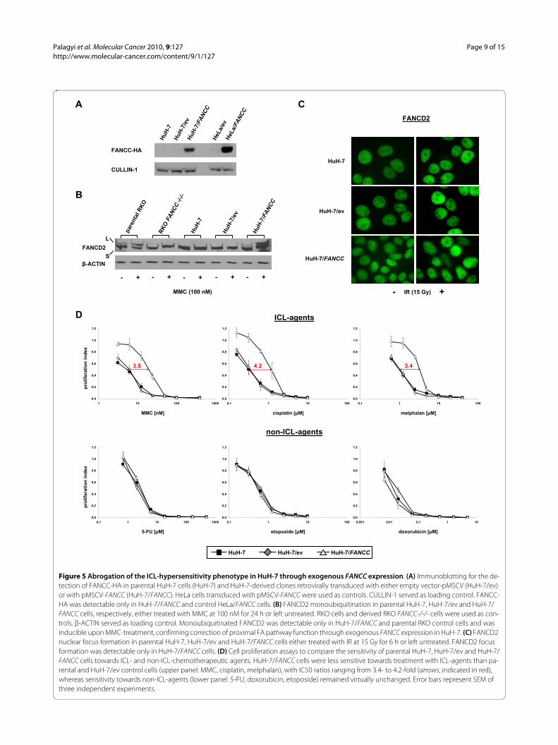
ICL-hypersensitivity of HuH-7 cells is abrogated upon exogenous FANCC expressionHuH-7 cells were stably transduced using retroviral con-structs containing either full length FANCC-cDNA(HuH-7/FANCC), tagged with recombinant influenzavirus hemagglutinin (HA-tag), or empty vector (HuH-7/ev). Consecutively, exogenous FANCC expression wasconfirmed (Fig. 5A). HuH-7/FANCC cells, but not paren-tal HuH-7 or HuH-7/ev control cells, displayed restoredproximal FA-pathway function as demonstrated by con-stitutive and MMC-inducible [41] FANCD2 monoubiq-uitination (Fig. 5B) as well as proficiency in FANCD2nuclear focus formation (Fig. 5C). Most importantly, sen-sitivity towards ICL-agents was significantly reduced inHuH-7/FANCC cells as compared to parental HuH-7 andHuH-7/ev control cells (Fig. 5D, upper panel), whereassensitivity towards non-ICL-chemotherapeuticsremained virtually unchanged (Fig. 5D, lower panel).
No evidence for inactivation of FANCC, FANCG or FANCF in 18 HCC tissue specimensTo assess the prevalence of proximal FA pathway inacti-vation in HCC, 18 surgical HCC tissue specimens wereanalyzed for inactivation of FANCC, FANCF or FANCGthrough screening for genetic mutations, epigeneticsilencing through CpG hypermethylation or lack ofmRNA-expression. On the genetic level, direct sequenc-ing of the complete coding sequences of FANCC, FANCFand FANCG in 18 HCC samples identified a single notpreviously reported, heterozygous, synonymous FANCCsequence variant (c.813G > A) and two heterozygous,non-synonymous FANCG variants (c.20C > T, p.S7F;c.643C > A, p.Q215K) (Fig. 6A). No other sequence vari-ants, especially no inactivating point mutations, smalldeletions, insertions, large intragenic deletions or com-plete homozygous gene deletions, were found. On theepigenetic level, HpaII-restriction assays yielded no evi-dence for FANCC-, FANCF- or FANCG-silencingthrough CpG hypermethylation (Fig. 6B) and consis-tently, FANCC-, FANCF- and FANCG-mRNA was detect-able in all 18 HCC samples (Fig. 6C).
DiscussionWe report here the identification and functional charac-terization of an inactivating nonsense FANCC mutation
in the HCC cell line HuH-7. This cell line was establishedfrom a well-differentiated HCC of a 57 year-old Japanesemale patient [42], displays an aneuploid phenotype withan average number of 60 chromosomes, and is negativefor HBV and HCV [43,44]. To our knowledge, this is thefirst evidence of genetic inactivation of the proximal FApathway in a GI tumor entity other than pancreatic can-cer.
The identified FANCC nonsense mutation c.553C > T,p.R185X in HuH-7 represents a known FA mutation, firstdescribed by Gibson et al. [45]. Interestingly, non-splicesite nonsense mutations can cause exon-skippingthrough aberrant splicing [46], and accordingly, thec.553C > T mutation has been reported to cause partialtranscriptional skipping of exon 6 of FANCC in an FApatient [45], a mechanism confirmed for HuH-7 in ourstudy.
Unfortunately, no matched non-malignant tissue isavailable for HuH-7, precluding definitive genomic copynumber analyses in regard to whether the identifiedFANCC mutation represents a homo- or hemizygousmutation in this cell line. However, according to copynumber analyses by the Sanger Institute (Cambridge, UK)using high-density single nucleotide polymorphism(SNP) arrays (SNP 6.0) [47], HuH-7 harbors three nearlyidentical copies of the chromosomal arm 9q, whereFANCC is located at 9q22.3, as evidenced by virtuallyexclusive homozygosity of all SNPs assessed on 9q.According to proposed evaluation models for the identifi-cation of LOH events where no matching normal tissue isavailable, these data are strongly suggestive (although notdefinitely evidentiary) of allelic loss of one copy of chro-mosome 9q including the non-mutated FANCC allele inthe original tumor (or its precursor cells), followed byrepeated duplication of the remaining chromosome 9q,including the mutated FANCC allele, later on [48-50].Typical recurrent numerical chromosomal aberrations inHCC include losses on 1p, 4q, 8p, 13q, 16q, and 17p andgains on 1q, 8q, 16p and 20q [51]. Although chromosome9 is rarely clonally altered on the cytogenetic level inHCC, LOH has been reported for several regions onchromosome 9 including the loci of the FANCC (9q22.3)and the FANCG (9p13) genes [52].
FA pathway defects in tumors require bi-allelic inacti-vation of one of at least 13 FA genes. On the one hand,these bi-allelic mutations could both be inherited, asapplies to tumors occurring in FA-patients. On the otherhand, mono-allelic germline mutations of distal FA path-way genes, such as FANCD1, FANCN or FANCJ, conferlow to medium penetrance susceptibility for breast/ovar-ian cancer [12,13,53] and, as applies to FANCD1 andFANCN, also for pancreatic cancer [15,54-57]. In addi-tion, inherited mono-allelic mutations of proximal FApathway genes have been associated with the predisposi-

Palagyi et al. Molecular Cancer 2010, 9:127http://www.molecular-cancer.com/content/9/1/127
Page 8 of 15
Figure 4 Hypersensitivity of HuH-7 cells specifically towards ICL-agents. (A) Cell proliferation assays to compare the sensitivity of five HCC cell lines towards ICL- and non-ICL-chemotherapeutic agents. HuH-7 cells were hypersensitive towards treatment with any of three ICL-agents as com-pared to four FA pathway-proficient HCC lines (upper panel: MMC, cisplatin, melphalan), with IC50 ratios ranging from 3.4- to 18.6-fold (arrows, indi-cated in red), but not towards any of three non-ICL-agents (lower panel: 5-FU, doxorubicin, etoposide). Error bars represent SEM of three independent experiments. (B) Flow cytometric cell cycle profiles of five HCC cell lines 48 h after treatment with MMC at the indicated concentrations. A pronounced G2 arrest (defined here as a cell fraction of >40% in G2 [36]) at low-dose MMC (25 nM) was observed only in HuH-7 cells. (C) Graphic representation of the cell cycle distributions from a representative experiment displaying the fraction of cells in G1, S or G2 at the indicated MMC concentrations for the indicated HCC cell lines. Arrows indicate the lowest dose at which a G2 arrest of more than 40% of the cells was observed.
0,0
0,2
0,4
0,6
0,8
1,0
1,2
0,1 1 10 100
0,0
0,2
0,4
0,6
0,8
1,0
1,2
0,01 0,1 1 100,0
0,2
0,4
0,6
0,8
1,0
1,2
1 10 100 1000
0,0
0,2
0,4
0,6
0,8
1,0
1,2
0,01 0,1 1 100,0
0,2
0,4
0,6
0,8
1,0
1,2
0,001 0,01 0,1 1 10
0,0
0,2
0,4
0,6
0,8
1,0
1,2
0,1 1 10 100 1000
0%
20%
40%
60%
80%
100%
0 25 50 100 200
A
4.5 18.6 8.0
5-FU [�M] etoposide [�M] doxorubicin [�M]
MMC [nM] cisplatin [�M] melphalan [�M]
pro
lifer
atio
n in
dex
ICL-agentsH
uH
-7C
han
gP
LC
PR
F5
Hep
3BH
epG
2
B
- +MMC (25 nM)
C
Cel
ls in
G2/
MC
ells
in G
2/M
Cel
ls in
G2/
M
0%
20%
40%
60%
80%
100%
0 25 50 100 200
0%
20%
40%
60%
80%
100%
0 25 50 100 200
0%
20%
40%
60%
80%
100%
0 25 50 100 200
0%
20%
40%
60%
80%
100%
0 25 50 100 200
HuH-7
Chang
PLCPRF5
Hep3B
HepG2
MMC [nM] MMC [nM]
MMC [nM]MMC [nM]
MMC [nM]
G2/MG1 S
cell
cou
nt
DNA content
cell
cou
nt
in %
cell
cou
nt
in %
cell
cou
nt
in %
cell
cou
nt
in %
cell
cou
nt
in %
non-ICL-agents
3.4 7.7
pro
lifer
atio
n in
dex
HepG2Hep3BHuH-7 Chang PLCPRF5
Palagyi et al. Molecular Cancer 2010, 9:127http://www.molecular-cancer.com/content/9/1/127
Page 9 of 15
Figure 5 Abrogation of the ICL-hypersensitivity phenotype in HuH-7 through exogenous FANCC expression. (A) Immunoblotting for the de-tection of FANCC-HA in parental HuH-7 cells (HuH-7) and HuH-7-derived clones retrovirally transduced with either empty vector-pMSCV (HuH-7/ev) or with pMSCV-FANCC (HuH-7/FANCC). HeLa cells transduced with pMSCV-FANCC were used as controls. CULLIN-1 served as loading control. FANCC-HA was detectable only in HuH-7/FANCC and control HeLa/FANCC cells. (B) FANCD2 monoubiquitination in parental HuH-7, HuH-7/ev and HuH-7/FANCC cells, respectively, either treated with MMC at 100 nM for 24 h or left untreated. RKO cells and derived RKO FANCC-/-/- cells were used as con-trols. β-ACTIN served as loading control. Monoubiquitinated FANCD2 was detectable only in HuH-7/FANCC and parental RKO control cells and was inducible upon MMC-treatment, confirming correction of proximal FA pathway function through exogenous FANCC expression in HuH-7. (C) FANCD2 nuclear focus formation in parental HuH-7, HuH-7/ev and HuH-7/FANCC cells either treated with IR at 15 Gy for 6 h or left untreated. FANCD2 focus formation was detectable only in HuH-7/FANCC cells. (D) Cell proliferation assays to compare the sensitivity of parental HuH-7, HuH-7/ev and HuH-7/FANCC cells towards ICL- and non-ICL-chemotherapeutic agents. HuH-7/FANCC cells were less sensitive towards treatment with ICL-agents than pa-rental and HuH-7/ev control cells (upper panel: MMC, cisplatin, melphalan), with IC50 ratios ranging from 3.4- to 4.2-fold (arrows, indicated in red), whereas sensitivity towards non-ICL-agents (lower panel: 5-FU, doxorubicin, etoposide) remained virtually unchanged. Error bars represent SEM of three independent experiments.
HuH-7
FANCD2
IR (15 Gy)
HuH-7/ev
HuH-7/FANCC
- +
C
par
enta
l RK
O
RK
OFA
NC
C-/-/-
HuH
-7
HuH
-7/e
v
HuH
-7/F
AN
CC
A
- +
MMC (100 nM)
FANCD2
L
S
�-ACTIN
FANCC-HA
- + - + - + - +
0,0
0,2
0,4
0,6
0,8
1,0
1,2
0,1 1 10 100
0,0
0,2
0,4
0,6
0,8
1,0
1,2
0,1 1 10 100 1000
0,0
0,2
0,4
0,6
0,8
1,0
1,2
0,001 0,01 0,1 1 10
0,0
0,2
0,4
0,6
0,8
1,0
1,2
1 10 100 1000
0,0
0,2
0,4
0,6
0,8
1,0
1,2
0,1 1 10 100
0,0
0,2
0,4
0,6
0,8
1,0
1,2
0,1 1 10 100
D
5-FU [µM] etoposide [µM] doxorubicin [µM]
MMC [nM] cisplatin [µM] melphalan [µM]
pro
life
rati
on
in
dex
ICL-agents
HuH-7 HuH-7/ev HuH-7/FANCC
non-ICL-agents
pro
life
rati
on
in
dex
3.5 3.44.2
B
CULLIN-1
HuH
-7
HuH
-7/e
vH
uH
-7/F
AN
CC
HeL
a/ev
HeL
a/FA
NC
C

Palagyi et al. Molecular Cancer 2010, 9:127http://www.molecular-cancer.com/content/9/1/127
Page 10 of 15
tion for or the accelerated development of certain tumors[21,54,55,58]. In particular, germline mutations ofFANCC have been described in pancreatic cancer, associ-ated with LOH in the tumor [21,22]. However, germlineand somatic changes in FANCC and FANCG may havecomparatively low penetrance for pancreatic cancer [55],which is supported by studies investigating germlinemutations of upstream FA pathway genes in sporadic, yetFA-typical tumors among the general population [59].Nevertheless, as the FANCC mutation in HuH-7 reportedin our study represents an established FA mutation andwas therefore most likely present in the germline of thepatient in mono-allelic form, our data might indicate anincreased risk for the development of HCC in individualsof the general population harboring this or other FANCCmutations.
The occurrence of an FA-associated FANCC mutationin HCC could also denote a tissue-specific susceptibilityfor the development of HCC in FA patients; The majorityof solid malignancies seen in FA patients consists of head
and neck or gynaecologic carcinomas (5.3%), as reportedin a large meta-analysis of 1300 cases [60], but also 2.8%of all FA patients developed liver tumors. These tumorsmanifested at a significantly younger age than other solidmalignancies (median age: 13 years for liver tumors ascompared to 26 years for other solid malignancies). Infact, the cumulative probability of liver tumors in FApatients has been estimated to be 46% by age 50 [60].However, the significance of these data regarding apotential liver-specific cancer susceptibility of FApatients is complicated by the observation that many livertumors do not proceed to malignancy during the life spanof FA patients [60,61]. In addition, there appears to be astrong association between androgen therapy, which isfrequently used for the treatment of bone marrow failurein FA, and the occurrence of liver tumors [60-63]. Never-theless, HCC represented the majority (58%) of tumors of36 FA patients with androgen-related liver tumors in onestudy [61]. Thus, the association of FA with HCC couldbe attributable both to the primary tumorigenic effects of
Figure 6 Screening of 18 HCC tissues for inactivation of FANCC, FANCF or FANCG. (A) Chromatograms displaying the identified heterozygous sequence variants (arrows) in FANCC and FANCG. (B) PCR-based HpaII-restriction assays to determine methylation status of FANCC, FANCF and FANCG in 18 HCC tissues. The corresponding CpG island-containing regions were PCR-amplified before and after digestion using the indicated enzymes: U = undigested, H = HpaII digestion, M = MspI digestion. Universal methylated and unmethylated human DNA (hDNA) was used as controls. No hyper-methylation of FANCC, FANCF or FANCG was detected in any of the 18 HCC samples. (C) RT-PCR to assess mRNA-expression of FANCC, FANCF and FANCG in 18 HCC tissues. All samples expressed FANCC-, FANCF- and FANCG-mRNA.
FANCC c.813G>A
unknown synonymous SNP
FANCG c.20C>T, p.S7F
known non-synonymous SNP
FANCG c.643C>A, p.Q215K
unknown non-synonymous SNP
Sample 198
Sample 152
Sample 240
152
U
meth
. h
DN
A
un
me
th.
hD
NA
U
U
UU U U U U U U
U U U U U U U U U
H H
H H H H H H H H H
H H H H H H H H H
M M
M M M M M M M M M
M M M M M M M M M
179 188 195 198 217 224 234 240
345 5106 6191 6426 7156 7221 7451 7766 7966
FANCF
152
U
meth
. h
DN
A
un
me
th.
hD
NA
U
U
UU U U U U U U
U U U U U U U U U
H H
H H H H H H H H H
H H H H H H H H H
M M
M M M M M M M M M
M M M M M M M M M
179 188 195 198 217 224 234 240
345 5106 6191 6426 7156 7221 7451 7766 7966
FANCG
152
U
meth
. h
DN
A
un
me
th.
hD
NA
U
U
UU U U U U U U
U U U U U U U U U
H H
H H H H H H H H H
H H H H H H H H H
M M
M M M M M M M M M
M M M M M M M M M
179 188 195 198 217 224 234 240
345 5106 6191 6426 7156 7221 7451 7766 7966
FANCCA
FANCF
FANCG
FANCC
152
179
188
195
198
217
224
234
240
345
5106
6191
6426
7156
7221
7451
7766
7966
B
C

Palagyi et al. Molecular Cancer 2010, 9:127http://www.molecular-cancer.com/content/9/1/127
Page 11 of 15
FA pathway inactivation in hepatocytes, as well as topotential secondary, amplifying or accelerating effects ofandrogen therapy in FA patients.
We demonstrated that HCC cells having an inactivatedFA pathway display the classic cellular FA phenotype,including a specific hypersensitivity towards ICL-agents,illustrated in HuH-7 by a pronounced cell cycle arrest inG2 upon treatment with MMC at low concentrations anda strongly decreased proliferation rate as compared to apanel of non-isogenic HCC lines. Importantly, this ICL-hypersensitivity phenotype was reversed using anisogenic HuH-7 model of exogenous FANCC expression,confirming that ICL-hypersensitivity in these cells wasattributable specifically to inactivation of FANCC. Ofnote however, IC50-ratios between FANCC-deficient andFANCC-proficient cells were partly smaller in theisogenic model than could have been expected from ourresults using the non-isogenic model. This observationcould be attributable to FA pathway-independent ICL-sensitivity differences among the non-isogenic HCC celllines, but could also provide further support for our pre-vious hypothesis that constitutive exogenous FANCCover-expression does not completely substitute for physi-ologically regulated, endogenous FANCC expression[37,38].
It is well established that systemic chemotherapy lackseffectiveness in unselected HCC patients [64,65] andHCC are therefore considered largely chemoresistant, atleast partially explaining the poor prognosis of this tumorentity [66]. Accordingly, guidelines are currently lackingalso regarding the choice of chemotherapeutic agent touse in transarterial chemoembolization (TACE), one ofthe major treatment modalities for non-surgical patientsat advanced HCC stages [67]. Our data indicate that non-FA patients having a FA-deficient HCC might predictablybenefit from treatment using ICL-agents. Consequently,assessment of FA pathway function in HCC could facili-tate individualized therapeutic approaches, using geno-type-based patient stratification in regard to bothsystemic chemotherapy and TACE.
To get an estimate of the prevalence of FANCC inacti-vation in HCC, we sequenced cDNA derived from 18 sur-gical HCC tissue specimens to screen for genetic FANCCinactivation. We further screened these samples forhypermethylation of the FANCC promoter region and forlack of FANCC mRNA expression, as epigenetic FANCCinactivation has previously been reported in acute leu-kaemia and breast cancer [68,69]. Additionally, weincluded FANCG and FANCF in these analyses, asFANCG represents another proximal FA gene that hasbeen described to be mutated in GI cancer, specifically inpancreatic cancer [20,22], while FANCF has beenreported to be epigenetically inactivated in various tumor
types [23-25,27]. On the genetic level, we found no fur-ther inactivating alterations, especially no evidence forcomplete homozygous gene deletions, inactivating pointmutations, small deletions or insertions, in FANCC,FANCG or FANCF, respectively. The detected FANCGvariant c.20C > T, p.S7F has been reported in an FApatient of the complementation group G in addition topathogenic FANCG mutations [70]. There is no informa-tion available on the nature of the c.643C > A, p.Q215Kvariant in FANCG. However, LOH or a second sequencevariant was not detected in that tumor either. Addition-ally, since only two to three overlapping PCR reactionswere used to amplify the complete coding sequences ofFANCC, FANCG and FANCF, respectively, most poten-tial large intragenic deletions would have been detected.However, this mechanism of proximal FA gene inactiva-tion occurs almost exclusively in FANCA, whereas itappears to be extremely rare in FANCC and has not at allbeen described in FANCG [20,71-73]. On the epigeneticlevel, we found no evidence for hypermethylation ofFANCC, FANCG or FANCF in any of the 18 samples.Consistently, FANCC, FANCG and FANCF wereexpressed in all samples on the mRNA level.
Our negative screening results for proximal FA pathwayinactivation in HCC were not unexpected, as a hypotheti-cal high prevalence should have become evident earlierduring clinical trials - manifesting as a selective chemo-sensitivity of the majority of HCC towards ICL-agents.Nevertheless, the lack of FA mutations in 18 HCC doesnot rule out rare occurrences of proximal FA pathwayinactivation in HCC and is consistent with previousreports on the low prevalence of proximal FA pathwayinactivation in various tumor entities among the generalpopulation [20-22,26,29,68]. Future studies applying ahigher sample number are required to definitely deter-mine the prevalence of FA pathway inactivation in HCC.
ConclusionsIn summary, we identified a HCC cell line harboring aninactivating mutation of the FANCC gene, specificallycausing proximal FA pathway inactivation and the classiccellular ICL-hypersensitivity phenotype. Assessment ofFA pathway function in HCC could thus represent a novelapproach to identify small subgroups of HCC patientsexpected to predictably benefit from individualized treat-ment protocols using ICL-agents. However, as proximalFA pathway inactivation occurs at low frequency in HCCand other tumors among the general population, thedevelopment of rapid, economic and clinically applicablescreening tests for proximal FA pathway inactivation,particularly in primary tumor tissues, remains an indis-pensable requirement to facilitate future clinical studies[38].

Palagyi et al. Molecular Cancer 2010, 9:127http://www.molecular-cancer.com/content/9/1/127
Page 12 of 15
Materials and methodsCell lines, culture conditions and HCC tissue samplesCell lines PL3, PL5, PL8, PL11 and PL45 were kindly pro-vided by S.E. Kern (Johns Hopkins University, Baltimore,Maryland). Cell lines AZ-521, CoGa-5, CoGa-5L, Coga-12, HCA-7, Isreco 1, MKN7, MKN45, NCl-N87 andL3.6pl were kindly donated by C.J. Bruns, M. Gerhard,F.T. Kolligs and M. Ogris (Technical University and Lud-wig-Maximilians-University, Munich, Germany). Theremaining cell lines were purchased from the EuropeanCollection of Cell Cultures (Sigma-Aldrich, Munich, Ger-many) or the American Type Culture Collection (LGCStandards, Wesel, Germany). RKO cells harboring anengineered disruption of the FANCG or FANCC genehave been described [36]. Cells were grown in DMEMsupplemented with 10% fetal calf serum, L-glutamine andpenicillin/streptomycin (PAA, Cölbe, Germany). The 18HCC tissue samples were a kind gift of the Human Tissueand Cell Research-Trust (HTCR, Regensburg, Germany)and originated from HCCs that where surgically removedat the Ludwig-Maximilians-University (Munich, Ger-many).
ImmunoblottingCells were treated with MMC (Sigma) at 100 nM for 24 hor left untreated. Consecutively, cells were lysed and pro-tein extracts boiled and loaded on 6% polyacrylamidegels. After electrophoresis, proteins were transferred toPVDF membranes, which were blocked for 1 h in TBS-Triton X-100/2% milk before the primary antibody wasapplied overnight at 4°C (1:1000; anti-FANCD2 H-300,Santa Cruz Biotechnology, Heidelberg, Germany; anti-FANCC ab5065, Abcam, Cambridge, UK). HA-taggedFANCC was detected using an anti-HA antibody (1:1000;12CA5, Santa Cruz). Antibodies directed against anti-RAD50 (1:5000; 13B3, GeneTex, San Antonio, TX), anti-p84 (1:1000; ab487, Abcam), anti-CUL1 (1:1000; 2H4C9,Invitrogen, Karlsruhe, Germany) or anti-β-ACTIN(1:10.000; AC-15, Sigma) served as loading controls. Themembranes were washed and stained with anti-rabbit oranti-mouse HRP-conjugated antibodies (1:2000 to1:10.000; GE Healthcare, Freiburg, Germany). Enhancedchemo-luminescence was elicited using SuperSignalWest Pico substrate (Thermo Scientific, Schwerte, Ger-many) according to the manufacturer's instructions.
Nuclear focus formation assaysExperiments were performed as described before [74]. Inbrief, cells were grown on coverslips until ~80% conflu-ency and were exposed to ionizing γ-radiation (IR) at 15Gy using a cesium-137 irradiator. After incubation for 6h, the cells were fixed using 3.7% formaldehyde and -20°Cmethanol. The cells were permeabilized using Triton X-100 and incubated in blocking buffer (PBS +2% bovine
serum albumin +0.5% Triton X-100) for 30 min. Consec-utively, the cells were labelled using antibodies againstFANCD2 (FI-17) or RAD51 (H-92) (Santa Cruz), respec-tively, for 2 h at room temperature. After washing, Alexa488 goat anti-mouse or anti-rabbit antibody (Invitrogen)was applied for 1.5 h. Nuclei were counterstained usingHoechst 33342 (Roche Diagnostics, Mannheim, Ger-many), mounted and analyzed using a fluorescencemicroscope and Axiovision Software (Carl Zeiss AG;Oberkochen, Germany). The settings were kept identicalfor all samples.
Gene complementation studiesFor FA complementation group determination, the cellline HuH-7 was transduced with retroviral constructscontaining full-length cDNAs of FANCA, FANCB,FANCC, FANCE, FANCF, FANCG or FANCL and ana-lyzed for cell cycle arrest upon treatment with MMC andfor restoration of FANCD2 monoubiquitination, as previ-ously described [26]. For isogenic studies, HuH-7 cellswere stably transduced with either HA-tagged pMSCV-FANCC (HuH-7/FANCC) or the corresponding pMSCVempty control vector (HuH-7/ev).
gDNA sequencingHigh molecular weight genomic DNA was preparedusing a salting-out technique. Amplification of theFANCC exons was performed using published primersets [75]. PCR products were purified using the GFX PCRDNA and Gel Band Purification kit (GE Healthcare).DNA sequencing of PCR products was performed usingABI-PRISM big-dye terminator chemistry on the ABI 310instrument (Applied Biosystems, Darmstadt, Germany).
mRNA expression analyses and cDNA sequencingTotal RNA was extracted from surgical HCC tissues orcell lines using the NucleoSpin RNAII-Kit (Macherey-Nagel, Düren, Germany). RNA was reverse-transcribedusing SuperScriptII reverse transcriptase (Invitrogen)and cDNA synthesized. Aberrant splicing of FANCC wasdetermined by RT-PCR using the following primer pairs:Fwd 5'-ACTGCCCAAACTGCTGAAG-3' and Rev 5'-GTTCAGACGCTAATGATAAAACCA-3' (spanning thefirst non-coding exon to coding exon 5), Fwd 5'-TTCTG-GACAATCAAAACTTAACTCC-3' and Rev 5'-GCT-GCTGCTTCTGGACATT-3' (spanning the coding exons3 to 13), Fwd 5'-GTAGTCTG CCTCTGGCTTCG-3' andRev 5'-TTGAGGAGAAGGTGCCTGAT-3' (spanning thecoding exons 7 to 13). Expression of FANCC, FANCF andFANCG mRNA was validated by RT-PCR as describedpreviously [76]. For sequencing of the complete codingregions of FANCC, FANCF and FANCG, respectively, thecorresponding cDNAs were amplified in two (FANCF) orthree (FANCC and FANCG) overlapping PCR reactions(primer sets available on request). Amplified products

Palagyi et al. Molecular Cancer 2010, 9:127http://www.molecular-cancer.com/content/9/1/127
Page 13 of 15
were sequenced using an ABI-Prism 3100-AvantSequencer (Applied Biosystems) and sequence changesconfirmed at the genomic level by gDNA sequencing.Reference genomic and cDNA sequences of the FA genesare available in the Fanconi Anemia Mutation Database[77].
Cell proliferation assaysThe assays were performed over a broad range of concen-trations covering 100% to 0% cell survival. 1,500-2,000cells/well were plated in 96-well plates, allowed to adhere,and treated. Following incubation for 6 d, the cells werewashed, lysed in 100 μl H2O, and 0.5% Picogreen (Molec-ular Probes, Invitrogen) was added. Fluorescence wasmeasured (Cytofluor Series 4000, Applied Biosystems)and growth inhibition calculated as compared to theuntreated control samples. Three independent experi-ments were performed per agent, with each data pointreflecting triplicate wells. Error bars represent standarderror of the mean (SEM) from three experiments.
Cell cycle analysesCells were seeded in 12-well plates and treated in dupli-cate for 48 h using various MMC concentrations (rangingfrom 25 to 200 nM) or were left untreated. The cells werefixed, stained with propidium iodide, and the DNA con-tent per cell was measured using flow cytometry (FACS-Calibur, Becton Dickinson, Heidelberg, Germany). Thedata were analyzed using CELLQuest Pro software (Bec-ton Dickinson). Alternatively, unfixed cells were stainedwith DAPI at a final concentration of 1 μg/ml in a buffercontaining 154 mM NaCl, 1 mM CaCl2, 0.5 mM MgCl2,0.1 M TRIS, 0.2% BSA, and 0.1% NP40 for 30 min in thedark. Univariate flow histograms were recorded on ananalytical, triple-laser equipped flow cytometer (LSRII,Becton Dickinson) using UV excitation of the DAPI dye.The resulting cell cycle distributions reflecting cellularDNA content were quantified using the MPLUS AV soft-ware package (Phoenix Flow Systems, San Diego, CA).
HpaII restriction enzyme methylation analysisPCR-based HpaII-restriction assays were performed asdescribed previously [23]. Transcriptional silencingthrough CpG hypermethylation was analyzed using pub-lished primer sets for FANCF [23] and FANCC [69],respectively. Primers used for FANCG were 5'-GAGTG-CAATGGCACGATG-3' (forward) and 5'-GCATGCTG-GGAGTCGTAGTA-3' (reverse). CpGenome universalmethylated or unmethylated DNA (Chemicon - Milli-pore, Schwalbach am Taunus, Germany), respectively,were used as controls.
List of abbreviationsFA: Fanconi anemia; GI: gastrointestinal, HCC: hepato-cellular carcinoma; ICL: interstrand-crosslink; IC50:
inhibitory concentration 50%; LOH: loss of heterozygos-ity; MMC: mitomycin C; SNP: single nucleotide polymor-phism; RT-PCR: reverse-transcription PCR; TACE:transarterial chemoembolization
Competing interestsThe authors declare that they have no competing interests.
Authors' contributionsAP was involved in study design, acquisition, analysis and interpretation ofmost of the data and drafted the manuscript. KN and DS carried out the com-plementation studies and parts of the genomic sequencing. UP, AZ, BT and DMperformed and interpreted most of the immunoblotting assays. UP and AZ fur-ther performed and interpreted drug sensitivity assays and AZ additionally per-formed parts of the genomic sequencing. AR, EDT and AZ performed andinterpreted the FACS analyses. SO performed immunoblotting, immunofluo-rescence and interpreted the nuclear focus formation assays. BT and FB engi-neered the stably transduced HuH-7 pMSCV lines. GUD provided and analyzedcholangiocellular carcinoma cell lines. WET provided the HCC tumor speci-mens. HH provided the retroviral constructs. EDT, CS, BG and DS participated inthe conception and the design of the study or provided important intellectualcontent. EG conceived the project, coordinated the experiments, directed theanalysis and interpretation of the data and wrote the final manuscript. Allauthors read and approved the final manuscript.
AcknowledgementsTissue samples were obtained and experimental procedures were performed according to the guidelines of the charitable state controlled foundation HTCR, with the informed patient's consent [78]. We kindly acknowledge expert tech-nical assistance with flow cytometry by Richard Friedl, Würzburg. This work was supported by grants to EG (DFG Ga762/3-1, Bavarian Academy of Sciences and Arts, Förderprogramm für Forschung und Lehre, Friedrich-Baur-Foundation, K.L. Weigand Fund) and to EDT (DFG To605/2-1).
Author Details1Department of Medicine II, Campus Grosshadern, Ludwig-Maximilians-University, Marchioninistrasse 15, 81377 Munich, Germany, 2Department of Human Genetics, Biocenter, Julius-Maximilians-University, Am Hubland, 97074 Würzburg, Germany, 3Department of Medicine III, Klinikum rechts der Isar, Technical University Munich, Ismaningerstrasse 22, 81675 Munich, Germany, 4Department of Surgery, Campus Grosshadern, Ludwig-Maximilians-University, Marchioninistrasse 15, 81377 Munich, Germany and 5Department of Pediatric Oncology, Hematology and Clinical Immunology, Heinrich Heine University, Moorenstrasse 5, 40225 Düsseldorf, Germany
References1. D'Andrea AD, Grompe M: The Fanconi anaemia/BRCA pathway. Nat Rev
Cancer 2003, 3:23-34.2. Wang W: Emergence of a DNA-damage response network consisting of
Fanconi anaemia and BRCA proteins. Nat Rev Genet 2007, 8:735-748.3. Gallmeier E, Hucl T, Calhoun ES, Cunningham SC, Bunz F, Brody JR, Kern
SE: Gene-Specific Selection Against Experimental Fanconi Anemia Gene Inactivation in Human Cancer. Cancer Biol Ther 2007, 6:654-660.
4. Meetei AR, Medhurst AL, Ling C, Xue Y, Singh TR, Bier P, Steltenpool J, Stone S, Dokal I, Mathew CG, et al.: A human ortholog of archaeal DNA repair protein Hef is defective in Fanconi anemia complementation group M. Nat Genet 2005, 37:958-963.
5. Levitus M, Joenje H, de Winter JP: The Fanconi anemia pathway of genomic maintenance. Cell Oncol 2006, 28:3-29.
6. Dorsman JC, Levitus M, Rockx D, Rooimans MA, Oostra AB, Haitjema A, Bakker ST, Steltenpool J, Schuler D, Mohan S, et al.: Identification of the Fanconi anemia complementation group I gene, FANCI. Cell Oncol 2007, 29:211-218.
7. Sims AE, Spiteri E, Sims RJ III, Arita AG, Lach FP, Landers T, Wurm M, Freund M, Neveling K, Hanenberg H, et al.: FANCI is a second monoubiquitinated member of the Fanconi anemia pathway. Nat Struct Mol Biol 2007, 14:564-567.
Received: 14 October 2009 Accepted: 28 May 2010 Published: 28 May 2010This article is available from: http://www.molecular-cancer.com/content/9/1/127© 2010 Palagyi et al; licensee BioMed Central Ltd. This is an Open Access article distributed under the terms of the Creative Commons Attribution License (http://creativecommons.org/licenses/by/2.0), which permits unrestricted use, distribution, and reproduction in any medium, provided the original work is properly cited.Molecular Cancer 2010, 9:127

Palagyi et al. Molecular Cancer 2010, 9:127http://www.molecular-cancer.com/content/9/1/127
Page 14 of 15
8. Smogorzewska A, Matsuoka S, Vinciguerra P, McDonald ER III, Hurov KE, Luo J, Ballif BA, Gygi SP, Hofmann K, D'Andrea AD, Elledge SJ: Identification of the FANCI protein, a monoubiquitinated FANCD2 paralog required for DNA repair. Cell 2007, 129:289-301.
9. Wong AK, Pero R, Ormonde PA, Tavtigian SV, Bartel PL: RAD51 interacts with the evolutionarily conserved BRC motifs in the human breast cancer susceptibility gene brca2. J Biol Chem 1997, 272:31941-31944.
10. Yuan SS, Lee SY, Chen G, Song M, Tomlinson GE, Lee EY: BRCA2 is required for ionizing radiation-induced assembly of Rad51 complex in vivo. Cancer Res 1999, 59:3547-3551.
11. Godthelp BC, Wiegant WW, Waisfisz Q, Medhurst AL, Arwert F, Joenje H, Zdzienicka MZ: Inducibility of nuclear Rad51 foci after DNA damage distinguishes all Fanconi anemia complementation groups from D1/BRCA2. Mutat Res 2006, 594:39-48.
12. Reid S, Schindler D, Hanenberg H, Barker K, Hanks S, Kalb R, Neveling K, Kelly P, Seal S, Freund M, et al.: Biallelic mutations in PALB2 cause Fanconi anemia subtype FA-N and predispose to childhood cancer. Nat Genet 2007, 39:162-164.
13. Fackenthal JD, Olopade OI: Breast cancer risk associated with BRCA1 and BRCA2 in diverse populations. Nat Rev Cancer 2007, 7:937-948.
14. Pal T, Permuth-Wey J, Betts JA, Krischer JP, Fiorica J, Arango H, LaPolla J, Hoffman M, Martino MA, Wakeley K, et al.: BRCA1 and BRCA2 mutations account for a large proportion of ovarian carcinoma cases. Cancer 2005, 104:2807-2816.
15. Murphy KM, Brune KA, Griffin C, Sollenberger JE, Petersen GM, Bansal R, Hruban RH, Kern SE: Evaluation of candidate genes MAP2K4, MADH4, ACVR1B, and BRCA2 in familial pancreatic cancer: deleterious BRCA2 mutations in 17%. Cancer Res 2002, 62:3789-3793.
16. King MC, Marks JH, Mandell JB: Breast and ovarian cancer risks due to inherited mutations in BRCA1 and BRCA2. Science 2003, 302:643-646.
17. Goggins M, Schutte M, Lu J, Moskaluk CA, Weinstein CL, Petersen GM, Yeo CJ, Jackson CE, Lynch HT, Hruban RH, Kern SE: Germline BRCA2 gene mutations in patients with apparently sporadic pancreatic carcinomas. Cancer Res 1996, 56:5360-5364.
18. Friedenson B: BRCA1 and BRCA2 pathways and the risk of cancers other than breast or ovarian. MedGenMed 2005, 7:60.
19. Heijden MS van der, Brody JR, Dezentje DA, Gallmeier E, Cunningham SC, Swartz MJ, DeMarzo AM, Offerhaus GJ, Isacoff WH, Hruban RH, Kern SE: In vivo therapeutic responses contingent on Fanconi anemia/BRCA2 status of the tumor. Clin Cancer Res 2005, 11:7508-7515.
20. Heijden MS van der, Brody JR, Gallmeier E, Cunningham SC, Dezentje DA, Shen D, Hruban RH, Kern SE: Functional defects in the fanconi anemia pathway in pancreatic cancer cells. Am J Pathol 2004, 165:651-657.
21. Couch FJ, Johnson MR, Rabe K, Boardman L, McWilliams R, de Andrade M, Petersen G: Germ line Fanconi anemia complementation group C mutations and pancreatic cancer. Cancer Res 2005, 65:383-386.
22. Heijden MS van der, Yeo CJ, Hruban RH, Kern SE: Fanconi anemia gene mutations in young-onset pancreatic cancer. Cancer Res 2003, 63:2585-2588.
23. Taniguchi T, Tischkowitz M, Ameziane N, Hodgson SV, Mathew CG, Joenje H, Mok SC, D'Andrea AD: Disruption of the Fanconi anemia-BRCA pathway in cisplatin-sensitive ovarian tumors. Nat Med 2003, 9:568-574.
24. Tischkowitz M, Ameziane N, Waisfisz Q, De Winter JP, Harris R, Taniguchi T, D'Andrea A, Hodgson SV, Mathew CG, Joenje H: Bi-allelic silencing of the Fanconi anaemia gene FANCF in acute myeloid leukaemia. Br J Haematol 2003, 123:469-471.
25. Narayan G, Arias-Pulido H, Nandula SV, Basso K, Sugirtharaj DD, Vargas H, Mansukhani M, Villella J, Meyer L, Schneider A, et al.: Promoter hypermethylation of FANCF: disruption of Fanconi Anemia-BRCA pathway in cervical cancer. Cancer Res 2004, 64:2994-2997.
26. Neveling K, Kalb R, Florl AR, Herterich S, Friedl R, Hoehn H, Hader C, Hartmann FH, Nanda I, Steinlein C, et al.: Disruption of the FA/BRCA pathway in bladder cancer. Cytogenet Genome Res 2007, 118:166-176.
27. Marsit CJ, Liu M, Nelson HH, Posner M, Suzuki M, Kelsey KT: Inactivation of the Fanconi anemia/BRCA pathway in lung and oral cancers: implications for treatment and survival. Oncogene 2004, 23:1000-1004.
28. Gallmeier E, Kern SE: Genetic Pathways of Pancreatic Tumorigenesis. In "Disease of the Pancreas: Current Surgical Therapy": Genetic Pathways of Pancreatic Tumorigenesis Edited by: Beger H, Cameron JL, Matsuno S. Heidelberg: Springer Verlag; 2007.
29. Ameziane N, Chen F, Leemans CR, Brakenhoff RH, Joenje H: No evidence for FANCF gene silencing in head-and-neck squamous cell carcinomas. Cell Oncol 2009, 31:53-56.
30. Hahn SA, Hoque AT, Moskaluk CA, da Costa LT, Schutte M, Rozenblum E, Seymour AB, Weinstein CL, Yeo CJ, Hruban RH, Kern SE: Homozygous deletion map at 18q21.1 in pancreatic cancer. Cancer Res 1996, 56:490-494.
31. Bryant HE, Schultz N, Thomas HD, Parker KM, Flower D, Lopez E, Kyle S, Meuth M, Curtin NJ, Helleday T: Specific killing of BRCA2-deficient tumours with inhibitors of poly(ADP-ribose) polymerase. Nature 2005, 434:913-917.
32. Farmer H, McCabe N, Lord CJ, Tutt AN, Johnson DA, Richardson TB, Santarosa M, Dillon KJ, Hickson I, Knights C, et al.: Targeting the DNA repair defect in BRCA mutant cells as a therapeutic strategy. Nature 2005, 434:917-921.
33. McCabe N, Lord CJ, Tutt AN, Martin NM, Smith GC, Ashworth A: BRCA2-Deficient CAPAN-1 Cells are Extremely Sensitive to the Inhibition of Poly (ADP-Ribose) Polymerase: An Issue of Potency. Cancer Biol Ther 2005, 4:934-936.
34. Gallmeier E, Kern SE: Absence of specific cell killing of the BRCA2-deficient human cancer cell line CAPAN1 by poly(ADP-ribose) polymerase inhibition. Cancer Biol Ther 2005, 4:703-706.
35. Ashwell S, Zabludoff S: DNA damage detection and repair pathways--recent advances with inhibitors of checkpoint kinases in cancer therapy. Clin Cancer Res 2008, 14:4032-4037.
36. Gallmeier E, Calhoun ES, Rago C, Brody JR, Cunningham SC, Hucl T, Gorospe M, Kohli M, Lengauer C, Kern SE: Targeted disruption of FANCC and FANCG in human cancer provides a preclinical model for specific therapeutic options. Gastroenterology 2006, 130:2145-2154.
37. Gallmeier E, Hucl T, Brody JR, Dezentje DA, Tahir K, Kasparkova J, Brabec V, Bachman KE, Kern SE: High-throughput screening identifies novel agents eliciting hypersensitivity in fanconi pathway-deficient cancer cells. Cancer Res 2007, 67:2169-2177.
38. Gallmeier E, Kern SE: Targeting Fanconi Anemia/BRCA2 Pathway Defects in Cancer: The Significance of Preclinical Pharmacogenomic Models. Clin Cancer Res 2007, 13:4-10.
39. Heijden MS Van Der, Brody JR, Kern SE: Functional screen of the fanconi anemia pathway in cancer cells by fancd2 immunoblot. Cancer Biol Ther 2004, 3:534-537. Epub 2004 Jun 2018
40. Hucl T, Gallmeier E, Kern SE: Distinguishing rational from irrational applications of pharmacogenetic synergies from the bench to clinical trials. Cell Cycle 2007, 6:1336-1341.
41. Andreassen PR, D'Andrea AD, Taniguchi T: ATR couples FANCD2 monoubiquitination to the DNA-damage response. Genes Dev 2004, 18:1958-1963.
42. Nakabayashi H, Taketa K, Miyano K, Yamane T, Sato J: Growth of human hepatoma cells lines with differentiated functions in chemically defined medium. Cancer Res 1982, 42:3858-3863.
43. Japanese Collection of Research Bioresources [http://cellbank.nibio.go.jp/]
44. Lohmann V, Korner F, Koch J, Herian U, Theilmann L, Bartenschlager R: Replication of subgenomic hepatitis C virus RNAs in a hepatoma cell line. Science 1999, 285:110-113.
45. Gibson RA, Hajianpour A, Murer-Orlando M, Buchwald M, Mathew CG: A nonsense mutation and exon skipping in the Fanconi anaemia group C gene. Hum Mol Genet 1993, 2:797-799.
46. Valentine CR: The association of nonsense codons with exon skipping. Mutat Res 1998, 411:87-117.
47. The Sanger Institute Cancer Cell Line Project [http://www.sanger.ac.uk/genetics/CGP/CellLines]
48. Calhoun ES, Gallmeier E, Cunningham SC, Eshleman JR, Hruban RH, Kern SE: Copy-number methods dramatically underestimate loss of heterozygosity in cancer. Genes Chromosomes Cancer 2006, 45(11):1070-1.
49. Calhoun ES, Hucl T, Gallmeier E, West KM, Arking DE, Maitra A, Iacobuzio-Donahue CA, Chakravarti A, Hruban RH, Kern SE: Identifying Allelic Loss and Homozygous Deletions in Pancreatic Cancer without Matched Normals Using High-Density Single-Nucleotide Polymorphism Arrays. Cancer Res 2006, 66:7920-7928.
50. Greenman CD, Bignell G, Butler A, Edkins S, Hinton J, Beare D, Swamy S, Santarius T, Chen L, Widaa S, et al.: PICNIC: an algorithm to predict
http://www.ncbi.nlm.nih.gov/entrez/query.fcgi?cmd=Retrieve&db=PubMed&dopt=Abstract&list_uids=9405383
http://www.ncbi.nlm.nih.gov/entrez/query.fcgi?cmd=Retrieve&db=PubMed&dopt=Abstract&list_uids=8968085
http://www.ncbi.nlm.nih.gov/entrez/query.fcgi?cmd=Retrieve&db=PubMed&dopt=Abstract&list_uids=8564959
http://www.ncbi.nlm.nih.gov/entrez/query.fcgi?cmd=Retrieve&db=PubMed&dopt=Abstract&list_uids=6286115
http://www.ncbi.nlm.nih.gov/entrez/query.fcgi?cmd=Retrieve&db=PubMed&dopt=Abstract&list_uids=7689011

Palagyi et al. Molecular Cancer 2010, 9:127http://www.molecular-cancer.com/content/9/1/127
Page 15 of 15
absolute allelic copy number variation with microarray cancer data. Biostatistics 11:164-175.
51. Wong CM, Ng IO: Molecular pathogenesis of hepatocellular carcinoma. Liver Int 2008, 28:160-174.
52. Liew CT, Li HM, Lo KW, Leow CK, Lau WY, Hin LY, Lim BK, Lai PB, Chan JY, Wang XQ, et al.: Frequent allelic loss on chromosome 9 in hepatocellular carcinoma. Int J Cancer 1999, 81:319-324.
53. Seal S, Thompson D, Renwick A, Elliott A, Kelly P, Barfoot R, Chagtai T, Jayatilake H, Ahmed M, Spanova K, et al.: Truncating mutations in the Fanconi anemia J gene BRIP1 are low-penetrance breast cancer susceptibility alleles. Nat Genet 2006, 38:1239-1241.
54. Rogers CD, Couch FJ, Brune K, Martin ST, Philips J, Murphy KM, Petersen G, Yeo CJ, Hruban RH, Goggins M: Genetics of the FANCA gene in familial pancreatic cancer. J Med Genet 2004, 41:e126.
55. Rogers CD, Heijden MS van der, Brune K, Yeo CJ, Hruban RH, Kern SE, Goggins M: The genetics of FANCC and FANCG in familial pancreatic cancer. Cancer Biol Ther 2004, 3:167-169.
56. Jones S, Hruban RH, Kamiyama M, Borges M, Zhang X, Parsons DW, Lin JC, Palmisano E, Brune K, Jaffee EM, et al.: Exomic sequencing identifies PALB2 as a pancreatic cancer susceptibility gene. Science 2009, 324:217.
57. Gallmeier E, Hucl T, Winter JM, Kern SE: No mutations identified in the Fanconi Anemia gene BRIP1 (BACH1, FANCJ). NOGO 2005, 10:1.
58. Berwick M, Satagopan JM, Ben-Porat L, Carlson A, Mah K, Henry R, Diotti R, Milton K, Pujara K, Landers T, et al.: Genetic heterogeneity among Fanconi anemia heterozygotes and risk of cancer. Cancer Res 2007, 67:9591-9596.
59. Neveling K, Kalb R, Schindler D: Cancer in Fanconi anemia and Fanconi anemia genes in cancer. In Fanconi Anemia - A Paradigmatic disease for the Understanding of Cancer and Aging Edited by: Schindler D, Hoehn H. Basel: Karger; 2007:59-78.
60. Alter BP: Cancer in Fanconi anemia, 1927-2001. Cancer 2003, 97:425-440.
61. Velazquez I, Alter BP: Androgens and liver tumors: Fanconi's anemia and non-Fanconi's conditions. Am J Hematol 2004, 77:257-267.
62. Johnson FL, Lerner KG, Siegel M, Feagler JR, Majerus PW, Hartmann JR, Thomas ED: Association of androgenic-anabolic steroid therapy with development of hepatocellular carcinoma. Lancet 1972, 2:1273-1276.
63. Sweeney EC, Evans DJ: Hepatic lesions in patients treated with synthetic anabolic steriods. J Clin Pathol 1976, 29:626-633.
64. Llovet JM, Bruix J: Systematic review of randomized trials for unresectable hepatocellular carcinoma: Chemoembolization improves survival. Hepatology 2003, 37:429-442.
65. Lopez PM, Villanueva A, Llovet JM: Systematic review: evidence-based management of hepatocellular carcinoma--an updated analysis of randomized controlled trials. Aliment Pharmacol Ther 2006, 23:1535-1547.
66. Bruix J, Sherman M, Llovet JM, Beaugrand M, Lencioni R, Burroughs AK, Christensen E, Pagliaro L, Colombo M, Rodes J: Clinical management of hepatocellular carcinoma. Conclusions of the Barcelona-2000 EASL conference. European Association for the Study of the Liver. J Hepatol 2001, 35:421-430.
67. El-Serag HB, Marrero JA, Rudolph L, Reddy KR: Diagnosis and treatment of hepatocellular carcinoma. Gastroenterology 2008, 134:1752-1763.
68. Hess CJ, Ameziane N, Schuurhuis GJ, Errami A, Denkers F, Kaspers GJ, Cloos J, Joenje H, Reinhardt D, Ossenkoppele GJ, et al.: Hypermethylation of the FANCC and FANCL promoter regions in sporadic acute leukaemia. Cell Oncol 2008, 30:299-306.
69. Sinha SM, Singh RD, Alam ND, Roy AD, Roychoudhury SD, Panda CD: Alterations in candidate genes PHF2, FANCC, PTCH1 and XPA at chromosomal 9q22.3 region: Pathological significance in early- and late-onset breast carcinoma. Mol Cancer 2008, 7:84.
70. Demuth I, Wlodarski M, Tipping AJ, Morgan NV, de Winter JP, Thiel M, Grasl S, Schindler D, D'Andrea AD, Altay C, et al.: Spectrum of mutations in the Fanconi anaemia group G gene, FANCG/XRCC9. Eur J Hum Genet 2000, 8:861-868.
71. Morgan NV, Tipping AJ, Joenje H, Mathew CG: High frequency of large intragenic deletions in the Fanconi anemia group A gene. Am J Hum Genet 1999, 65:1330-1341.
72. Rischewski JR, Gross M, Hanenberg H, Michael K, Schneppenheim R, Schindler D: Mutation Spectrum of FANCC in Europe and DHLPC-Based
Identification of a Large Deletion. In Fifteenth Annual Fanconi Anemia Research Fund Scientific Symposium Houston, TX; 2003.
73. Neveling K, Endt D, Hoehn H, Schindler D: Genotype-phenotype correlations in Fanconi anemia. Mutat Res 2009, 668:73-91.
74. Gallmeier E, Winter JM, Cunningham SC, Kahn SR, Kern SE: Novel genotoxicity assays identify norethindrone to activate p53 and phosphorylate H2AX. Carcinogenesis 2005, 26:1811-1820.
75. Gibson RA, Buchwald M, Roberts RG, Mathew CG: Characterisation of the exon structure of the Fanconi anaemia group C gene by vectorette PCR. Hum Mol Genet 1993, 2:35-38.
76. Snyder ER, Ricker JL, Chen Z, Waes CV: Variation in cisplatinum sensitivity is not associated with Fanconi Anemia/BRCA pathway inactivation in head and neck squamous cell carcinoma cell lines. Cancer Lett 2007, 245:75-80.
77. The Fanconi Anemia Mutation Database [http://www.rockefeller.edu/fanconi/mutate]
78. Thasler WE, Weiss TS, Schillhorn K, Stoll PT, Irrgang B, Jauch KW: Charitable State-Controlled Foundation Human Tissue and Cell Research: Ethic and Legal Aspects in the Supply of Surgically Removed Human Tissue For Research in the Academic and Commercial Sector in Germany. Cell Tissue Bank 2003, 4:49-56.
doi: 10.1186/1476-4598-9-127Cite this article as: Palagyi et al., Genetic inactivation of the Fanconi anemia gene FANCC identified in the hepatocellular carcinoma cell line HuH-7 con-fers sensitivity towards DNA-interstrand crosslinking agents Molecular Cancer 2010, 9:127
http://www.ncbi.nlm.nih.gov/entrez/query.fcgi?cmd=Retrieve&db=PubMed&dopt=Abstract&list_uids=4117807