Heterogeneous activation of the Fanconi anemia pathway by patient ...
Upload
phamkhuongCategory
view
231download
3
Chapter 7Endocrine Disorders in Fanconi AnemiaSusan R. Rose, MD, Anna Petryk, MD, and Constantine A. Stratakis, MD
Introduction
Many children and adults with Fanconi anemia have endocrine problems. These include short stature, growth hormone (GH) deficiency, hypothyroidism, pubertal delay or related abnormalities, osteopenia or osteoporo-sis, and abnormal glucose or insulin metabolism (lead-ing to impaired glucose tolerance or diabetes mellitus). Both the basic underlying condition of FA and its treat-ment (hematopoietic cell transplantation [HSCT]) may have effects on the endocrine systems of FA patients.
For these reasons, a thorough baseline and annual endocrine evaluation should be performed in every per-son with FA. The endocrine clinical care team should include a dietician, pediatric endocrinologist (with experience in growth and puberty), and a reproductive endocrinologist (with experience in children and fertil-ity) or a pediatric gynecologist.
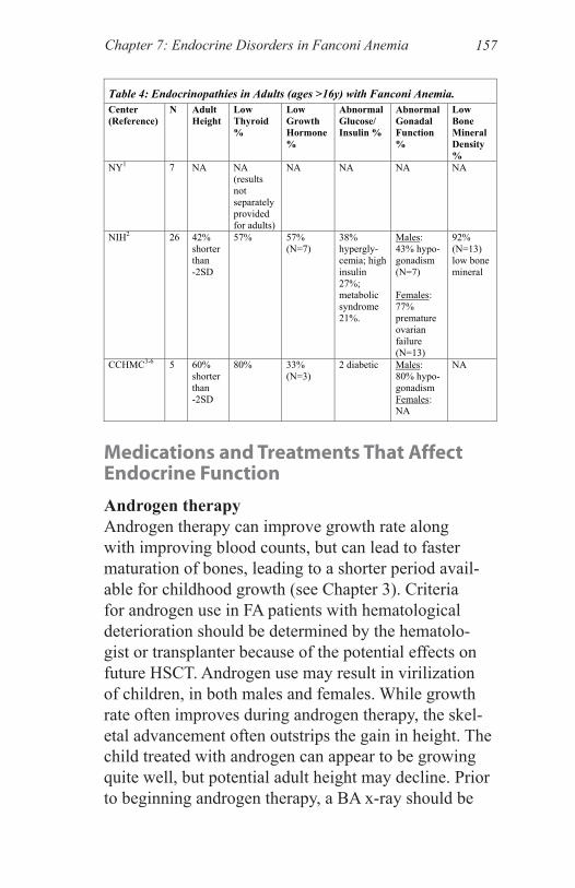
Until recently, only one medical journal article existed on endocrine function in FA patients (New York group [NY]1), but now three additional centers have each published articles on the subject (National Institutes of Health [NIH]2; Cincinnati Children’s Hospital Medical Center [CCHMC]3-6; and the University of Minnesota [U. of M.].7,8) Over 80% of FA individuals in all reports have had at least one abnormal endocrine test result (Tables 3 and 4).
Height
Height is a major clinical focus of the pediatric endocri-nologist, the parent, and the child with FA. The average height for persons with FA is -2.1 standard deviation units (SD). SD units are a way of expressing divergence from average. For instance, 2.5% of the normal popula-tion have heights more than 2 SD above average, and 2.5% of the normal population have heights shorter than 2 SD (-2 SD) below average. The average height in FA of -2.1 SD is about equivalent to 150 cm (4 feet, 11 inches) in women and 161 cm (5 feet, 3.5 inches) in men (Table 1). About half of FA patients are shorter than the 2.5 percentile or -2 SD, including some children with FA who are extremely small for their age (average height of the short group, -3.5 SD).1-6 However, almost half of individuals with FA have height within the normal range. Thus, some FA children have no problems related to height, with about 10% having heights above the average for the general population.
Effect of hormone deficiencies on heightFA patients with hormone deficiencies have a shorter height. Those with growth hormone (GH) insufficiency have an average height of -2.7 SD, and those with hypothyroidism -3.0 SD, while those with no demon-strable endocrinopathy have an average height of -2.0 SD.1 FA children who have untreated GH defi-ciency or hypothyroidism will achieve an even shorter adult height. However, short stature in FA cannot fully be explained on the basis of endocrinopathy alone. Occasionally, significant short stature is seen in FA even with no detectable hormone deficiencies. As a result, hormonal replacement therapy is unlikely to normalize growth completely.
135Chapter 7: Endocrine Disorders in Fanconi Anemia
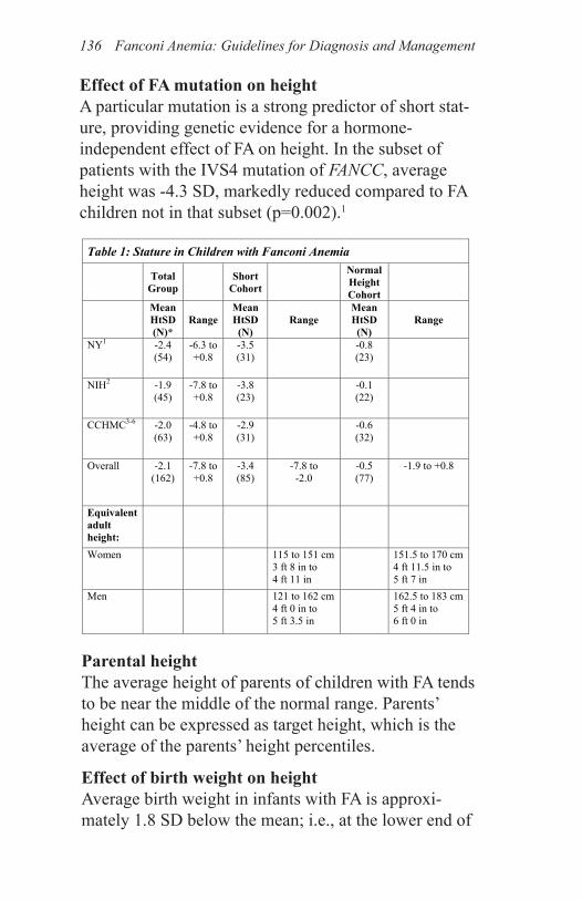
Effect of FA mutation on heightA particular mutation is a strong predictor of short stat-ure, providing genetic evidence for a hormone-independent effect of FA on height. In the subset of patients with the IVS4 mutation of FANCC, average height was -4.3 SD, markedly reduced compared to FA children not in that subset (p=0.002).1
Parental heightThe average height of parents of children with FA tends to be near the middle of the normal range. Parents’ height can be expressed as target height, which is the average of the parents’ height percentiles.
Effect of birth weight on heightAverage birth weight in infants with FA is approxi-mately 1.8 SD below the mean; i.e., at the lower end of
Table 1: Stature in Children with Fanconi Anemia
Total Group Short
Cohort Normal Height Cohort
Mean HtSD (N)*
Range Mean HtSD (N)
Range Mean HtSD (N)
Range
NY1 -2.4 (54)
-6.3 to +0.8
-3.5 (31)
-0.8 (23)
NIH2 -1.9 (45)
-7.8 to +0.8
-3.8 (23)
-0.1 (22)
CCHMC3-6 -2.0 (63)
-4.8 to +0.8
-2.9 (31)
-0.6 (32)
Overall -2.1 (162)
-7.8 to +0.8
-3.4 (85)
-7.8 to -2.0
-0.5 (77)
-1.9 to +0.8
Equivalent adult height:
Women 115 to 151 cm 3 ft 8 in to 4 ft 11 in
151.5 to 170 cm 4 ft 11.5 in to 5 ft 7 in
Men 121 to 162 cm 4 ft 0 in to 5 ft 3.5 in
162.5 to 183 cm 5 ft 4 in to 6 ft 0 in
*Number of standard Deviations from the Mean Height for Age
Fanconi Anemia: Guidelines for Diagnosis and Management136
the normal range. Approximately 40% of FA children are born small for gestational age (SGA). In the general population, about 90% of children who were born SGA gradually catch up to the normal range for height. How-ever, in the CCHMC patient group, 80% of the children with FA who were born SGA had height below -2 SD, while only 20% had normal height.
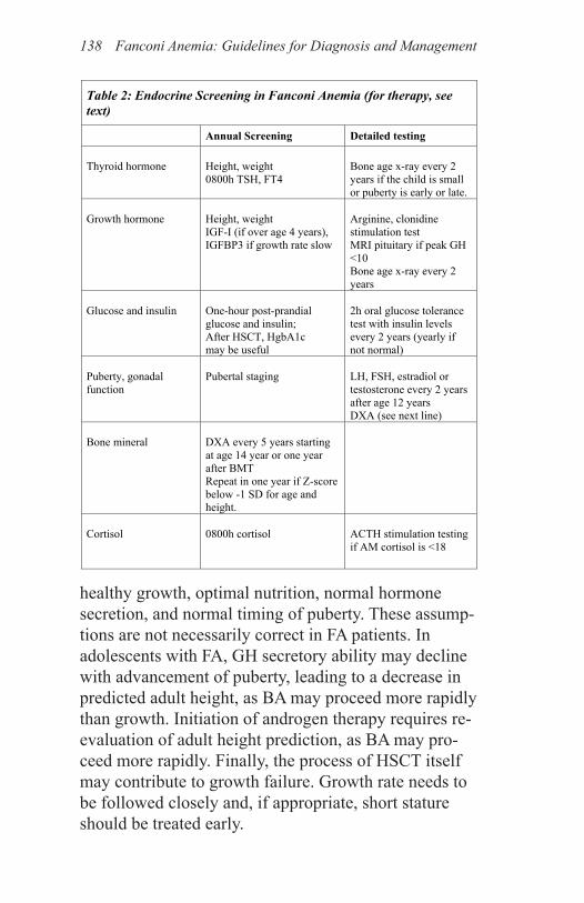
Clinical observations of growth rateGrowth in children with FA should be followed clinical-ly. Accurate height by stadiometer measurement is im-portant, and height should be plotted on a growth chart. Not all individuals with FA experience growth failure. When growth velocity and stature decline below expec-tations relative to the family background, appropriate endocrine evaluation should be performed. Nutritional and medical causes for poor growth should be identified in children as early as possible (Table 2).
Estimates of adult height based on bone age (BA) mea-surements may lead to over-optimistic height predic-tions. Androgens and corticosteroids may alter growth, advance BA or impair adult height. Adult height pre-dictions should be re-evaluated after a decrease in the growth velocity or following initiation of androgen therapy and after HSCT.9,10
Bone maturationHeight prediction algorithms make use of information about the amount of delay in bone maturation. Height measurement and a radiograph of the patient’s left hand and wrist should be obtained every two years in short children or every year in a child receiving androgen therapy.
If BA is like that of a younger child, height predic-tion may suggest that adult height will be normal. This expectation assumes that there will be continued
137Chapter 7: Endocrine Disorders in Fanconi Anemia
healthy growth, optimal nutrition, normal hormone secretion, and normal timing of puberty. These assump-tions are not necessarily correct in FA patients. In adolescents with FA, GH secretory ability may decline with advancement of puberty, leading to a decrease in predicted adult height, as BA may proceed more rapidly than growth. Initiation of androgen therapy requires re-evaluation of adult height prediction, as BA may pro-ceed more rapidly. Finally, the process of HSCT itself may contribute to growth failure. Growth rate needs to be followed closely and, if appropriate, short stature should be treated early.
Table 2: Endocrine Screening in Fanconi Anemia (for therapy, see text)
Annual Screening Detailed testing Thyroid hormone
Height, weight 0800h TSH, FT4
Bone age x-ray every 2 years if the child is small or puberty is early or late.
Growth hormone
Height, weight IGF-I (if over age 4 years), IGFBP3 if growth rate slow
Arginine, clonidine stimulation test MRI pituitary if peak GH <10 Bone age x-ray every 2 years
Glucose and insulin
One-hour post-prandial glucose and insulin; After HSCT, HgbA1c may be useful
2h oral glucose tolerance test with insulin levels every 2 years (yearly if not normal)
Puberty, gonadal function
Pubertal staging
LH, FSH, estradiol or testosterone every 2 years after age 12 years DXA (see next line)
Bone mineral
DXA every 5 years starting at age 14 year or one year after BMT Repeat in one year if Z-score below -1 SD for age and height.
Cortisol
0800h cortisol
ACTH stimulation testing if AM cortisol is <18
Fanconi Anemia: Guidelines for Diagnosis and Management138
Weight and Nutrition
As mentioned above, the average birth weight in infants with FA is approximately 1.8 SD below the mean; i.e., at the lower end of the normal range. Approximately 40% of FA children are born small for gestational age.
Nutritional and gastroenterological problems are common in at least a quarter of FA patients and may contribute to poor linear growth. In children with FA, weight is generally below average for the general population (-1.5 SD), but is significantly better than the height, indicating that inadequate caloric intake is not sufficient to explain the height deficit. About one-quarter of children with FA have low weight for height (sometimes called failure to thrive), while about
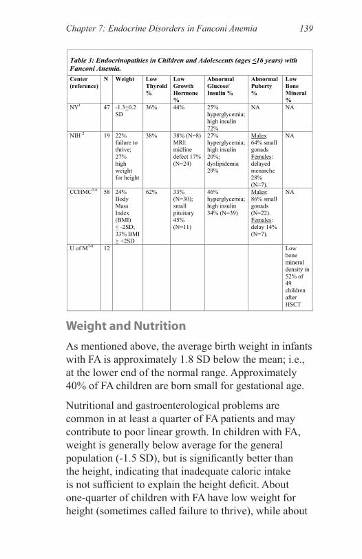
Table 3: Endocrinopathies in Children and Adolescents (ages <16 years) with Fanconi Anemia. Center (reference)
N Weight Low Thyroid %
Low Growth Hormone %
Abnormal Glucose/ Insulin %
Abnormal Puberty %
Low Bone Mineral %
NY1 47 -1.3+0.2 SD
36% 44% 25% hyperglycemia; high insulin 72%
NA NA
NIH 2 19 22% failure to thrive; 27% high weight for height
38% 38% (N=8) MRI: midline defect 17% (N=24)
27% hyperglycemia; high insulin 20%; dyslipidemia 29%
Males: 64% small gonads Females: delayed menarche 28% (N=7).
NA
CCHMC3-6 58 24% Body Mass Index (BMI) < -2SD; 33% BMI > +2SD
62% 33% (N=30); small pituitary 45% (N=11)
46% hyperglycemia; high insulin 34% (N=39)
Males: 86% small gonads (N=22). Females: delay 14% (N=7).
NA
U of M7-8 12 Low bone mineral density in 52% of 49 children after HSCT
139Chapter 7: Endocrine Disorders in Fanconi Anemia
one-quarter to one-third are relatively overweight for their height (Table 3).1-6 Of note, this frequency of rela-tive overweight is similar to trends being observed in the general population.
Some children may have a smaller than expected appe-tite; others may experience malabsorption. In addition, illness can raise caloric requirements or metabolism. Children who secrete inadequate insulin do not make use of all of the calories that they eat, and many have elevated blood sugars and lose glucose in their urine. Thus, insulin deficiency can contribute to poor weight gain in childhood.
Alternatively, excess weight gain or glucocorticoid ther-apy (which can occur during stem cell transplant) can worsen glucose intolerance by making the body require higher insulin levels from a sluggish pancreas. As a result, persons with FA may develop overt diabetes.
Healthy dietary intake should be encouraged, including sufficient calcium and vitamin D and avoiding concen-trated sweets, in order to normalize serum glucose and to optimize growth and bone mineral.
Hypothyroidism
Many children with FA have serum thyroid hormone levels that are not fully normal, such as borderline low thyroxine (T4) or free T4 (FT4) or borderline high thy-roid stimulating hormone (TSH) (especially in diagnos-tic samples drawn first thing in the morning) (Tables 3 and 4).1,2,5 This combination of test results is consistent with mild hypothyroidism. Mild hypothyroidism can occur either because of limited thyroid production by the thyroid gland (primary hypothyroidism) or because of limited TSH production centrally by the pituitary (central hypothyroidism). Forty to 63% of individuals
Fanconi Anemia: Guidelines for Diagnosis and Management140
with FA have thyroid function tests that are borderline for primary hypothyroidism.
One study described reduced thyroid hormone binding in persons with FA.1 Although reduced thyroid hormone binding is not usually clinically significant, it can make total T4 levels appear low and lead to a false diagnosis of hypothyroidism. Thyroid hormone binding globulin (TBG)-bound T4 (but not other bound forms) was low-est in individuals receiving androgen therapy.1
Thyroid treatment was compared to placebo in short children with FA who have TSH >3 mU/L or FT4 in the lowest third of the normal range, whether or not TSH is high.5 Eight children with FA were treated for seven months with thyroid hormone, compared to seven months with placebo. Children grew significantly better on thyroid hormone than during placebo, and parents felt that their children did better during the thyroid hormone phase.5 Thus, it is likely that treatment of such children with thyroid hormone will improve their growth.
There is controversy about use of TSH >3 mU/L as a diagnostic criterion for mild hypothyroidism. Among endocrinologists who treat adults, this criterion has been acknowledged as defining the upper limit of TSH in healthy individuals, but treatment is not usu-ally started unless TSH is persistently 10 mU/L or higher.11-13 Among pediatric endocrinologists, some use a similar approach, while others feel that mild TSH elevation provides an opportunity to treat a short child to achieve potential benefit.14 Although the thyroid treatment study was conducted in a small number of FA children, children with FA who have small stature and borderline thyroid function tests may benefit from thyroid hormone therapy.5
141Chapter 7: Endocrine Disorders in Fanconi Anemia
Thyroid function evaluation and treatmentThyroid function should be evaluated by obtaining an early morning (e.g., 8 am) blood sample for free T4 and a TSH level. All FA patients should undergo screening for hypothyroidism every 6 to 12 months. Abnormal thyroid hormone binding is common; thus, a low total T4 may be misleading. TSH levels above 3 mU/L may indicate mild hypothyroidism.5
Hypothyroidism should be treated promptly. Thyroid hormone treatment can improve growth rate. Replace-ment therapy for hypothyroidism is according to usual dosing for age. Thyroid hormone replacement treatment should be initiated as in non-FA patients—based on low thyroid hormone levels, especially if the TSH is above 3 mU/L at 8 am or FT4 is 1.0 ng/dL or below. An initial dose in a child age 4 to 13 years can be 3 mcg/kg daily, with monthly levels and dose adjustment until the treat-ment target is achieved. Initial dose in adults is about 1.5 mcg/kg or 100 mcg/m2. Target for thyroid hormone therapy should be TSH of 0.5 to 2 mU/L in the case of primary hypothyroidism. If the problem is a hypotha-lamic-pituitary deficiency (low night TSH secretion or central hypothyroidism), target for therapy is a free T4 just above the middle of the normal range.
Growth Hormone Deficiency
Growth hormone deficiency (GHD) has been described in case reports of a few patients with FA.15-19 About 54% of the subjects tested (under age 20 years) failed to produce GH in response to clonidine stimulation, while 72% failed to raise GH levels in response to arginine stimulation.1 Timing of peak GH response to stimula-tion tests was somewhat delayed. GH secretory dynam-ics are often not normal in FA children during sponta-neous overnight GH secretion studies. Taken together,
Fanconi Anemia: Guidelines for Diagnosis and Management142
these test results are consistent with a “hypoactive hypothalamus,” leading to “partial” GH deficiency or, alternatively, neurosecretory GH deficiency. GH and insulin-like growth factor I (IGF-I) values are not as severely affected as is height.
Magnetic resonance imaging (MRI) of the brain and pituitary suggest that the pituitary is small in children with FA compared to that of age-matched children without FA.3 In addition, four of seven FA patients with GHD in a study at the National Institutes of Health had midline brain anomalies on MRI, and one had pituitary stalk interruption syndrome (PSIS).2 Five patients with FA and PSIS were previously reported, suggesting that PSIS is a diagnostic marker of GHD and severe growth failure.18
If growth rate remains slow during thyroid therapy and with improved glucose control, further endocrine evalu-ation is indicated. Growth velocity may worsen with time and, therefore, longitudinal follow-up is necessary. Screening can be performed by drawing a blood sample for IGF-I and IGFBP3. If both are normal, growth hormone stimulation testing is not necessary. If IGF-I and IGFBP3 values are below -1 SDS for age, evalua-tion should include standard GH stimulation testing and thyroid function. Evaluation of GH secretory ability in a slowly growing child can be done utilizing clonidine (150 mcg/m2, maximum dose 300 mcg) and arginine (0.5 gm/kg, maximum dose 20 gm). GH peak is consid-ered normal if 10mcg/L or greater. If GH peak is nor-mal, GH therapy is not indicated in FA children before HSCT even if they meet other FDA-approved indica-tions for GH therapy, such as SGA birth or “idiopathic short stature.” Such treatment may be considered in a patient who has had HSCT.
143Chapter 7: Endocrine Disorders in Fanconi Anemia
Treatment for growth hormone deficiencySimilar to hypothyroidism, GH deficiency can cause poor growth and can be treated with exogenous GH treatment. Physicians should counsel FA families as to the risks and benefits of therapy. Therapy should be discontinued immediately if routine hematologi-cal examination reveals clonal hematopoietic stem cell proliferation.
A short child with FA should be treated with GH if GH deficiency has been unequivocally documented. Treat-ment with GH prior to HSCT, or in the absence of GH deficiency, is controversial. There is not full consensus among endocrinologists familiar with FA regarding safety of GH therapy prior to or after HSCT. There may be better growth response to GH therapy before use of glucocorticoid therapy than during glucocorticoid ther-apy. If GH therapy is used in FA patients, doses should probably be titrated to achieve IGF-I concentrations in the mid-normal range for age. As a separate issue, cur-rent recommendations are to temporarily discontinue GH therapy during critical illness.20
The issue of use of GH therapy in FA subjects raises medical dilemmas. FA patients are at an increased risk of malignancy, including head and neck and gynecolog-ical cancers, and are at ~800-fold greater risk of devel-oping acute myelogenous leukemia (AML).21-23 Thus, FA patients have a lifelong cancer risk even if their bone marrow has been corrected by HSCT. It is not known whether GH therapy could increase these risks.
The incidence of leukemia development in GH-treated patients who do not have predisposing risk factors is not thought to be different from that of the general population.24-27 Indeed, some authors have postulated that patients who developed leukemia after GH therapy
Fanconi Anemia: Guidelines for Diagnosis and Management144

may have been FA patients with short stature and no other abnormalities.23 The limited available data do not suggest that GH-treated FA persons are at higher risk of AML (or other malignancies) than are FA patients not treated with GH.
Patient registries have provided useful safety and efficacy data on the use of GH in the general popula-tion and in cancer survivors, but include few subjects with the diagnosis of FA.28-34 Growth rate during GH therapy in FA patients is improved, but not to the extent seen in the non-FA patient. Families should be coun-seled regarding predicted adult heights, the effects on growth rate of the available treatment modalities, and the potential risks and benefits of GH treatment—with the tempering statement that there is a paucity of clini-cal data regarding long-term safety of GH therapy in FA patients.
Abnormal Glucose or Insulin Metabolism
Glucose elevation/delayed insulin secretionDiabetes mellitus occurs more commonly in FA patients than in the general population.35 In the general popula-tion, children with diabetes usually have insulin defi-ciency. However, in children with FA, insulin levels are usually elevated. Insulin elevation could occur because of resistance to insulin or because of delayed insulin secretion. In FA, there is a relatively high incidence of impaired glucose tolerance, elevated insulin levels, and overt diabetes mellitus. Both androgen therapy and HSCT have been associated with insulin resistance.30,37 However, glucose intolerance and limited rapid insulin response to food predate either treatment and are inher-ent features of FA.4
145Chapter 7: Endocrine Disorders in Fanconi Anemia
Using the World Health Organization criteria, approxi-mately 8% of persons with FA were diabetic, while an additional 27 to 46% had impaired glucose tolerance (Tables 3 and 4).1,2,4 In addition, up to 72% of persons with FA showed elevated insulin levels by one to two hours after eating. Insulin levels were low at 10 to 45 minutes after oral glucose, suggesting slow initial insulin secretion, while high insulin levels occurred by 60 to 120 minutes.4 Similar impaired first-phase insulin secretion has been found at the University of Minnesota after HSCT in children with FA.7 Although the elevated insulin levels could suggest the possibility of insulin resistance, analysis was more consistent with beta cell dysfunction and sluggish insulin release.4
Evaluation for abnormal glucose or insulin metabolismAll FA patients should be tested for abnormalities of glucose and insulin homeostasis upon diagnosis and, thereafter, at least annually. Screening of glucose toler-ance can be performed with one post-prandial glucose and insulin. The practice of obtaining only serum glucose values or relying on fasting values should be avoided. Measuring glucose values only will fail to identify those subjects in the “early, pre-diabetic” stage. This, therefore, mandates measuring insulin levels. Fasting glucose and insulin levels are often normal, while post-prandial levels may be elevated. The aim of identifying both glucose and insulin level abnormali-ties is to intervene optimally. Glycosylated hemoglobin (HbA1c) and fructosamine levels may be deceptively normal and are of no use in FA patients prior to HSCT. HgbA1c may be more useful after HSCT.
More detailed annual evaluation should consist of a two-hour oral glucose tolerance test (OGTT,
Fanconi Anemia: Guidelines for Diagnosis and Management146
1.75 gm/kg, maximum dose 75 gm), with serum sam-ples for both glucose and insulin levels obtained every 30 minutes. FA patients with normal OGTT response should be assessed every two years and annually in those with mildly abnormal tests. The prevalence of diabetes mellitus in this population is age and disease progression-dependent, and the majority of FA patients may be at risk.
Beta-cell injury in the FA population may result from damage from excessive reactive oxygen species (ROS). β-cells are susceptible to oxidative damage by ROS in FA persons because of low levels of anti-oxidant enzyme mRNA, protein, and activity of superoxide dismutases, catalase and glutathione peroxidase.38,39
FA proteins are directly involved with the machinery of cellular defense and the modulation of oxidative stress.40 Thus, it is possible that impaired early insulin secretion in patients with FA may result from damage from enhanced ROS action on the β-cell.
Medications known to alter glucose metabolism in FASeveral medications used in the treatment of FA are known to alter glucose metabolism, most importantly androgens and corticosteroids. Androgen treatment can lead to significant rise in both glucose and insulin levels.1 Glucocorticoids may also alter glycemic con-trol. The guidelines regarding glucocorticoid use in FA should be the same as in any other subject; i.e., use the minimum necessary. Pharmacological doses of cortico-steroids (as used in transplant patients) commonly lead to hyperglycemia that should be treated in a standard fashion with insulin.
Diabetes treatment: insulin and glucoseFA individuals with low insulin levels and glucose above 200 at 30 minutes after oral glucose load may
147Chapter 7: Endocrine Disorders in Fanconi Anemia
need treatment using appropriate insulin regimens involving long-acting basal insulin and short-acting insulin at the time of meals, as in the general popula-tion. A serum insulin concentration less than or equal to 44 mcIU/mL (300 pmol/L) at 30 min on the OGTT would be below 2 SD of values seen in normal individ-uals. In persons with normal fasting glucose but abnor-mal oral glucose tolerance test results (impaired glucose tolerance) or patients with deficient early insulin secre-tion, administration of short-acting insulin with meals may be more beneficial than metformin. Since most children with FA have normal fasting glucose, it is not necessary to treat them with long-acting basal insulin.
If post-prandial glucose is consistently above 180 mg/dL, insulin therapy at mealtime (carbohydrate coverage) using short-acting insulin may be consid-ered. A good starting dose is short-acting insulin, 0.5 unit for each 15g of carbohydrate, just before eating. Both child and parent need to know how to check blood sugar. Blood sugars should be checked two hours after the start of each meal initially. If blood sugar after eating remains over 180mg/dL, insulin dose should be increased to 1 unit for 25g of carbohydrate, then 1 unit for 20g, and so on. The goal for insulin therapy is a post-prandial glucose of 90 to 150 mg/dL without hypoglycemia. The American Diabetes Association guidelines for blood glucose control in type 1 diabetes suggest the following finger stick glucose ranges: for toddlers and preschoolers, 100-180 mg/dL before meals and 110-200mg/dL at bedtime; for school age (6-12 years), 90-180 before meals and 100-180 at bedtime; and for adolescents and young adults (13-19 years), 90-130 before meals and 90-150 at bedtime.
Fanconi Anemia: Guidelines for Diagnosis and Management148
Insulin therapy during HSCTDuring HSCT, most children with FA require insulin therapy. A regimen similar to that described above may be utilized while the child is able to eat. Addi-tional basal insulin may be required using a long-acting insulin (24 h duration), starting at 0.3 unit per kilogram once daily. If the child is not eating, long-acting insulin can be used alone or short-acting insulin can be given in an intravenous infusion or in the hyperalimentation.
Isolated hyperinsulinemiaSome practitioners have begun treating otherwise nor-mal children and adolescents with FA who have isolated hyperinsulinemia (i.e., without glucose impairment) with oral agents, such as metformin. In cases where the FA individual is overweight, metformin may be the better first choice. Such use in FA should be accompa-nied by increased surveillance, as potential risk for side effects is not known.
DietAll persons diagnosed with FA—regardless of OGTT results—should be placed on a healthful diet that avoids concentrated sweets and excessive sugar intake, follow-ing the guidelines of the American Diabetes Associa-tion. It should be emphasized that this recommendation applies only to concentrated sweets (e.g., juices, soda, and candy) and not all forms of carbohydrate. It is important to ensure adequate caloric consumption and regular exercise.
Dyslipidemia, Obesity, and Metabolic Syndrome
Of the 29 patients with FA who have had lipid studies reported, 55% had dyslipidemia: elevated LDL in 21%, low HDL in 31%, and elevated triglycerides in 10%.2
149Chapter 7: Endocrine Disorders in Fanconi Anemia
Abnormal lipid profile was associated with glucose intolerance and was observed in 40% of patients with hyperglycemia or insulin resistance. Of those with diabetes, 75% were overweight or obese. FA adults who had diabetes were overweight or obese compared with those without these metabolic abnormalities. Meta-bolic syndrome (overweight/obesity, dyslipidemia, and insulin resistance) was diagnosed in 21% of the adults with FA, while 12 of 24 children tested had at least one metabolic abnormality: four with insulin resistance; one with diabetes; and seven with dyslipidemia.
Cortisol Sufficiency
Cortisol sufficiency should be evaluated in young FA children who have poor growth and who require major surgery because of the possibility of central hypotha-lamic dysfunction, even in the absence of a detectible midline central nervous system defect.3,15 If cortisol and ACTH adequacy have not been assessed, hydrocor-tisone stress dosing (50 to 100 mg/m2 intravenous or intramuscular) should be provided for major surgical procedures.
Most FA patients have had normal circadian cortisol levels and normal responses to ACTH administration testing.
Multiple Hormonal Deficiencies
If multiple hormonal deficiencies are found and hypo-thalamic or other central causes are suspected, a brain MRI should be obtained with emphasis on the pituitary-hypothalamic area.
Fanconi Anemia: Guidelines for Diagnosis and Management150
Puberty
Early onset of pubertyChildren and adolescents with FA often have abnor-mally timed puberty (see Chapter 6). If puberty starts too early, it may progress quickly, thus limiting the number of years that a child can grow.
In the boy or girl with FA who has early onset of puberty at a short height (or before age 11), therapy to delay puberty can allow additional time to grow. Use of Lupron Depot therapy can be used to delay puberty for four years to achieve an average of a 4 to 5 cm increase in adult height.41
Delayed pubertyMore commonly, boys and girls with FA appear to enter puberty late. Delay can be defined as no signs of puberty in a 12- to 13-year-old girl or a 13- to 14-year-old boy, or no menstrual period in a 14- to 16-year-old girl or by three years after onset of breast buds. Testing of the pubertal axis should be undertaken when delayed pubertal development is noted.
While clinically well-recognized, the cause of delayed puberty in FA is not at all understood. There may be blunted and/or prolonged gonadotropin (primarily luteinizing hormone [LH]) responses to stimulation, suggesting hypothalamic-pituitary dysregulation.
Delayed onset of puberty and pubertal progression should be followed by at least annual physical examina-tion to evaluate stage of puberty. Pubertal hormone con-centrations (LH, FSH, estradiol or testosterone) can be useful in adolescent children who are not progressing normally or in adults with symptoms of hypogonadism.
151Chapter 7: Endocrine Disorders in Fanconi Anemia
Treatment for delayed puberty in boys In a boy with no signs of puberty by age 14, low dose testosterone therapy can be initiated, again taking into account height and ability to grow. Injectable testoster-one can be given at 45 mg per m2 monthly for three to six months, with observation for pubertal progression during the following six months.
For long-term low-dose therapy in a short adolescent boy with diagnosed gonadal deficiency, one alternative to injections is topical testosterone gel with an initial dose of 1.25g daily, titrated to achieve reasonable serum levels for age and height. Initial goal might be a testos-terone of 50 ng/dL, similar to levels observed in a boy in early puberty.
For mid-pubertal boys, a level of 100-200 ng/dL might be appropriate; for later puberty, a level of 300-350 ng/dL; and for adults a level of 450 to 800 ng/dL is appropri-ate. Low-dose testosterone dosing can be maintained for several years while the boy grows taller.
If the boy is short for his age, rapid increase in testos-terone dose should be avoided. When acceptable adult height is achieved, adult testosterone dosing can be used for sex steroid replacement.
Not every adult man with FA will require testosterone therapy. The decision to treat should be based on clini-cal evidence of rate of puberty, energy level, physician examination, and laboratory results (testosterone, LH, FSH). Testosterone production or therapy in an adult affects not only bone mineral, but also energy, stamina, muscle development, self esteem, and ability to be appropriately assertive.
Fanconi Anemia: Guidelines for Diagnosis and Management152
Treatment for delayed puberty in girls In a girl with FA who has no signs of puberty by age 13, low-dose estrogen therapy should be started under the care of the pediatric endocrinologist or adolescent gyne-cologist, taking into account height and ability to grow42 (see Chapter 6). The reason to use estrogen therapy in a girl with delay is for medical benefit: to decrease cardiac risk factors, increase BMD, optimize growth rate, and to optimize breast development. One possible low-dose approach is to use a half tablet of conjugated estrogen daily (Premarin, 0.3 mg tablets).
In FA girls who are tall enough to expect to reach adequate adult height, the estrogen dose should be slowly increased every 6 months or so until the full replacement dose of Premarin, 1.25 mg daily, is being taken. Progesterone (Provera, 10 mg by mouth daily for 10 days) should be added when breakthrough bleeding occurs or after two years of estrogen replacement.
In FA girls who are short for age, rapid increase in estrogen dose should be avoided. Low-dose estrogen therapy can be continued for several years while the girl grows taller. When spotting occurs, oral progesterone can be given for ten days, then low-dose estrogen can be resumed. When acceptable adult height is achieved, birth control pills can be used for estrogen and proges-terone replacement. Standard birth control pills provide a simple way to give estrogen and progesterone in a monthly pattern. Daily doses of estrogen in these pills range from 20 to 50 mcg of estradiol. The lower end of this range is adequate for the medical benefits (and for birth control), especially in a small adult.
Therapy is not indicated if a girl has normal pubertal development or is having normal menstrual cycles unless there is evidence of ovarian hormone deficiency.
153Chapter 7: Endocrine Disorders in Fanconi Anemia
Genital Tract Abnormalities
Developmental anomalies of the genital tract are more frequent in FA patients than in the general population. Boys may be born with cryptorchidism and hypospa-dias. Many boys with FA have small testes for their age and pubertal status, most likely reflecting reduced Sertoli cell mass and spermatogenesis. Girls with FA may be at higher risk for a unicornuate uterus or hemi-uteri.43 Chemotherapy and radiation therapy, usually given in preparation for HSCT, may also result in gonadal failure.
Fertility
Fertility in FA patients is often impaired, with males often being infertile and females having premature menopause and a significantly abbreviated window of fertility. Infertility in men with FA is at least in part due to a reduced sperm count; on the other hand, in some rare cases fertility has been documented.43 A specific endocrine basis for the decreased fertility has not yet been identified except for after HSCT, when it can be due to radiation and chemotherapy.
Cryopreservation of embryos or spermCryopreservation of embryos or sperm is being inves-tigated as a reproductive option. Contraception should always be utilized when pregnancy is not desired (see Chapter 6).
Bone Mineral Density
Evaluation of bone mineral density (BMD) in FA has been reported in two studies. All but one of 13 adults with FA were found to have osteopenia or osteoporosis, compared to normal for gender and age.2 In 49 chil-dren (12 with FA), 52% had reduced BMD at 1 year
Fanconi Anemia: Guidelines for Diagnosis and Management154
after HSCT.8 The effects of HSCT on BMD in children with FA were similar to the bone mineral effects in other children after HSCT. On average, BMD Z-score declined 0.5 SD units during the first six months after HSCT.8 Longer follow-up will be worthwhile to observe whether BMD declines further or recovers over a longer period of time. These findings point out the importance of intake of adequate dietary calcium and vitamin D.
Dual energy absorptiometry (DXA) evaluationDXA evaluation of bone mineral density should be performed every five years, beginning at about one year after HSCT and/or beginning at about age 14 if there has been no HSCT. Subsequent yearly studies are indi-cated if bone mineral is low for height age.
Interpretation of DXA resultsOf note, interpretation of BMD studies by DXA will overestimate the incidence of osteopenia in persons with small stature. A DXA estimate of BMD is two-dimensional, and bone thickness is influenced by statu-ral height and, thus, bone size. Interpretation should be adjusted for bone size (such as adjustment for height age or bone age). DXA results should be expressed as Z-score for age (not T-score, which compares BMD to DXA results in young adults).
Bone therapyAmong other dietary recommendations, it is important to maintain adequate dietary intake of calcium and vitamin D to offer opportunity for normal growth and normal mineralization of bone.
• Elemental calcium recommendations: 500 mg daily in a young child; 1,000 to 1,500 mg in an adolescent; and 1,500 mg in an adult.
155Chapter 7: Endocrine Disorders in Fanconi Anemia
• Vitamin D recommendations: 400 units daily in a young child; 800 to 1,000 units in an adoles-cent or adult.
More aggressive intervention with calcium and vitamin D replacement is indicated if BMD is low for height.
Bisphosphonates (oral or intravenous) have been used safely in children with Osteogenesis Imperfecta and in growing children with osteopenia and fractures. Treat-ment with bisphosphonates may be considered if the child has sustained two or more low-impact fractures and a DXA result is lower than -1.5 SD (after adjust-ment for height age). Oral bisphosphonates may worsen esophageal reflux. Treatment for any hormone defi-ciency, especially for pubertal delay, can be beneficial for bone mineralization.
Adults with Fanconi Anemia
Adults with FA must be monitored for a variety of endocrinopathies, including hypogonadism, thyroid function, diabetes mellitus, hyperlipidemia, reduced fertility, and bone mineral density.
Endocrine functionEndocrine function in FA adults has not been well described. Each of the publications of endocrine func-tion in FA patients has included some adults (Table 4). The total number of adults with FA who have had endocrine results reported has been small.1,2,4,5 Early intervention and therapy may improve quality of life. Treatment of endocrine issues in adults with FA should be monitored by endocrinologists who care for adults, with attention to thyroid status, glucose tolerance, lipid abnormality, gonadal function, and bone mineral density.
Fanconi Anemia: Guidelines for Diagnosis and Management156
Medications and Treatments That Affect Endocrine Function
Androgen therapyAndrogen therapy can improve growth rate along with improving blood counts, but can lead to faster maturation of bones, leading to a shorter period avail-able for childhood growth (see Chapter 3). Criteria for androgen use in FA patients with hematological deterioration should be determined by the hematolo-gist or transplanter because of the potential effects on future HSCT. Androgen use may result in virilization of children, in both males and females. While growth rate often improves during androgen therapy, the skel-etal advancement often outstrips the gain in height. The child treated with androgen can appear to be growing quite well, but potential adult height may decline. Prior to beginning androgen therapy, a BA x-ray should be
Table 4: Endocrinopathies in Adults (ages >16y) with Fanconi Anemia. Center (Reference)
N Adult Height
Low Thyroid %
Low Growth Hormone%
Abnormal Glucose/ Insulin %
Abnormal Gonadal Function %
Low Bone Mineral Density %
NY1 7 NA NA (results not separately provided for adults)
NA NA NA NA
NIH2 26 42% shorter than -2SD
57% 57% (N=7)
38% hypergly-cemia; high insulin 27%; metabolic syndrome 21%.
Males: 43% hypo-gonadism (N=7) Females: 77% premature ovarian failure (N=13)
92% (N=13) low bone mineral
CCHMC3-6 5 60% shorter than -2SD
80% 33% (N=3)
2 diabetic Males: 80% hypo-gonadism Females: NA
NA
157Chapter 7: Endocrine Disorders in Fanconi Anemia
performed. The impact of androgen therapy on height and maturation should be discussed with the family. During androgen therapy, the BA should be reassessed every six months.
Multiple transfusion therapyMultiple transfusion therapy can affect endocrine function through the mechanism of iron overload (see Chapter 3). Iron deposition in endocrine glands can affect testicular function, contribute to development of diabetes, and can lead to primary hypothyroidism, hypoparathyroidism or pituitary dysfunction.
Hematological cell transplantTransplantation is associated with a state of illness. Illness is not an optimal time to assess any hormone concentrations, as thyroid levels, growth, gonadal func-tion, nutrition, and glucose regulation are often altered in illness (see Chapter 9 and 10). For example:
• Cytoxan has a known dose-related effect on gonadal function in both males and females.
• Glucocorticoids can lead to elevation in appe-tite, weight gain, insulin resistance, and hyper-glycemia, even to the extent of requiring insulin therapy.
• Metaclopromide raises prolactin. This can lead to galactorrhea or fluid from the breasts, and alteration of thyroid function or pubertal devel-opment.
• Anticonvulsant therapy can lead to alteration of thyroid function or thyroid dose requirement. Some anticonvulsants, such as Valproate, can lead to weight gain and altered ovarian function.
Fanconi Anemia: Guidelines for Diagnosis and Management158
After HSCT, additional hormone alterations may be evident as a consequence of the transplantation process. HSCT regimens may result in transient or persistent growth failure due to health alteration, GH deficiency, primary hypothyroidism, gonadal failure or other hor-mone deficiencies. Most common late effects of HSCT are primary hypothyroidism, partial gonadal failure, and osteopenia. Persons with FA appear to have underlying risk for these endocrinopathies even before HSCT, as well as for hyperglycemia.
Summary
Patients with FA are usually shorter than the general population and shorter than their parent-derived target heights, with average height being just below the third percentile. However, many FA patients are not short. Children with FA frequently have reduced GH secre-tion, hypothyroidism, and abnormal glucose homeosta-sis with deficient beta cell secretion of insulin and/or insulin resistance, which may further compromise their growth. In addition, puberty and fertility may not be normal, and bone mineral accrual may be delayed.
Etiology of endocrinopathies in FA is unclear. Hypo-thyroidism is generally accompanied by elevated TSH levels and, therefore, appears to be of thyroidal (i.e., peripheral) origin, although hypothalamic-pituitary dysregulation leading to abnormal central TSH release may also be present. Hyperglycemia/hyperinsulinemia is generally considered to be related to pancreatic beta cell dysfunction. In contrast, GH insufficiency is prob-ably of hypothalamic or pituitary (i.e., central) origin. Thus, there is not currently a single unifying cause for all of these endocrinopathies. It is possible that endo-crine secretory cells are damaged by excessive reactive oxygen species, with inadequate repair mechanisms in FA.
159Chapter 7: Endocrine Disorders in Fanconi Anemia
With the recent increased success of hematopoietic stem cell transplantation in curing the hematological com-plications of Fanconi anemia, the long-term effects of FA are now of greater importance.8,11 With the prospect of a significantly extended lifespan, we must prepare to treat the previously considered “secondary problems” of non-hematological conditions and to address quality of life issues, such as adult height. The endocrinolo-gist should be involved in initiating and supervising endocrine therapy. Treatment regimens must always be specific for the individual subject. Individuals with FA should be followed for the most common endocrine abnormalities, including growth failure, insulin insuf-ficiency/hyperinsulinemia/glucose intolerance, and hypothyroidism.
Attention to endocrine issues may permit the individual with FA to have the energy and stamina to better enjoy his or her life. Overall treatment philosophy is to ensure an optimal quality of life.
References1. Wajnrajch MP, Gertner JM, Huma Z, et al. Evaluation of growth and hormonal status in patients referred to the International Fanconi Anemia Registry. Pediatrics 2001; 107: 744-754.
2. Giri N, Batista DL, Alter BP, Stratakis CA. Endocrine abnor-malities in patients with Fanconi anemia. Journal of Clinical Endocrinology and Metabolism 2007; 92: 2624-2631.
3. Sherafat-Kazemzadeh R, Mehta SN, Care MM, Kim MO, Wil-liams DA, Rose SR. Small pituitary size in children with Fanconi anemia. Pediatric Blood and Cancer 2007; 49: 166-170.
4. Elder D, D’Alessio DA, Eyal O, et al. Abnormalities in glucose tolerance are common in children with Fanconi anemia and associ-ated with impaired insulin secretion. Pediatric Blood and Cancer 2008; 51: 256-260.
Fanconi Anemia: Guidelines for Diagnosis and Management160
5. Eyal O, Blum S, Mueller R, Smith F, Rose SR. Improved growth velocity during thyroid hormone therapy in children with Fanconi anemia who have borderline thyroid function. Pediatric Blood and Cancer 2008; 51: 652-656.
6. Wadwa RP, Rose SR, Eaton ME, and the Fanconi Anemia Comprehensive Care Center. Endocrine effects of Fanconi anemia. Pediatric Research 2003; 52: 437A.
7. Polgreen LE, MacMillan ML, Wagner JE, Moran A, Petryk A. Impaired first-phase insulin secretion in children with Fanconi anemia after hematopoietic cell transplantation. 2008; (submitted).
8. Petryk A, Bergemann TL, Polga KM, et al. Prospective study of changes in bone mineral density and turnover in children after hematopoietic cell transplantation. Journal of Clinical Endocrinol-ogy and Metabolism 2006; 91: 899-905.
9. Gluckman E, Auerbach AD, Horowitz MM, et al. Bone marrow transplantation for Fanconi anemia. Blood 1995; 86: 2856-2862.
10. Wagner JE, Davies SM, Auerbach AD. Hematopoietic cell transplantation in the treatment of Fanconi anemia. Forman SJ, Thomas, ED, eds. Hematopoietic Cell Transplantation. Malden, MA: Blackwell Science, Inc: 1998.
11. Spencer CA, Hollowell JG, Kararosyan M, Braverman LE. National Health and Nutrition Examination Survey III thyroid-stimulating hormone (TSH)-thyroperoxidase antibody relationships demonstrate that TSH upper reference limits may be skewed by occult thyroid dysfunction. Journal of Clinical Endocrinology and Metabolism 2007; 92: 4236-4240.
12. Baloch Z, Carayon P, Conte-Devolx B, et al. Guidelines Com-mittee, National Academy of Clinical Biochemistry. Laboratory medicine practice guidelines. Laboratory support for the diagnosis and monitoring of thyroid disease. Thyroid 2003; 13: 3-126.
13. Wartofsky L, Dickey RA. The evidence for a narrower thyro-tropin reference range is compelling. Journal of Clinical Endocri-nology and Metabolism 2005; 90: 5483-5488.
14. Rose SR. TSH above 3 is not usually normal. The Endocri-nologist 2006; 16: July-August.
161Chapter 7: Endocrine Disorders in Fanconi Anemia
15. Aynsley-Green A, Zachmann M, Werder EA, Illig R, Prader A. Endocrine studies in Fanconi’s anemia. Archives of Disease in Childhood 1978; 53: 126-131.
16. Zachmann M, Illig R, Prader A. Fanconi’s anemia with iso-lated growth hormone deficiency. Journal of Pediatrics 1972; 80: 159-160.
17. Nordon UZ, Humbert JR, MacGillivray MH, Fitzpatrick JE. Fanconi’s anemia with growth hormone deficiency. American Journal of Diseases of Children 1979; 133: 291-293.
18. Dupuis-Girod S, Gluckman E, Souberbielle JC, Brauner R. Growth hormone deficiency caused by pituitary stalk interruption in Fanconi’s anemia. Journal of Pediatrics 2001; 138: 129-133.
19. Potchedly C, Collipp PJ, Wolman SR, Suwansirikul S, Rez-vani I. Fanconi’s anemia with growth hormone deficiency. Journal of Pediatrics 1971; 79: 93-96.
20. Wilson TA, Rose SR, Cohen P, et al. Update of guidelines for the use of growth hormone in children: the Lawson Wilkins Pedi-atric Endocrinology Society Drug and Therapeutics Committee. Journal of Pediatrics 2003; 143: 415-421.
21. Swift M. Fanconi’s anaemia in the genetics of neoplasia. Nature 1971; 230: 370.
22. Auerbach AD, Allen RG. Leukemia and preleukemia in Fanconi anemia patients: a review of the literature and report of the International Fanconi Anemia Registry. Cancer Genetics and Cytogenetics 1991; 51: 1-12.
23. Butturini A, Gale RP, Verlander PC, et al. Hematologic abnormalities in Fanconi anemia: an International Fanconi Anemia Registry study. Blood 1994; 84: 1650-1655.
24. Stahnke N. Leukemia in growth-hormone-treated patients: an update, 1992. Hormone Research 1992; 38: 56-62.
25. Blethen SL, Allen DB, Graves D, et al. Safety of recombinant deoxyribonucleic acid-derived growth hormone: The National Cooperative Growth Study Experience. Journal of Clinical Endo-crinology and Metabolism 1996; 81: 1704-1710.
Fanconi Anemia: Guidelines for Diagnosis and Management162
26. Taback SP, Dean HJ. Mortality in Canadian children with growth hormone (GH) deficiency receiving GH therapy 1967-1992. The Canadian Growth Hormone Advisory Committee. Journal of Clinical Endocrinology and Metabolism 1996; 81: 1693-1696.
27. MacGillivray MH, Blethen SL, Buchlis JG, et al. Current dosing of growth hormone in children with growth hormone defi-ciency: how physiologic? Pediatrics 1998; 102: 527-530.
28. Darendeliler F, Karagiannis G, Wilton P. Headache, idiopathic intracranial hypertension and slipped capital femoral epiphysis during growth hormone treatment: a safety update from the KIGS database. Hormone Research 2007; 68 Suppl 5: 41-47.
29. Kemp SF, Kuntze J, Attie KM, et al. Efficacy and safety results of long-term growth hormone treatment of idiopathic short stature. Journal of Clinical Endocrinology and Metabolism 2005; 90: 5247-5253.
30. Quigley CA, Gill AM, Crowe BJ, et al. Safety of growth hor-mone treatment in pediatric patients with idiopathic short stature. Journal of Clinical Endocrinology and Metabolism 2005; 90: 5188-5196.
31. Bowlby DA, Rapaport R. Safety and efficacy of growth hormone therapy in childhood. Pediatric Endocrinology Reviews 2004; 2 Suppl 1: 68-77.
32. Harris M, Hofman PL, Cutfield WS. Growth hormone treat-ment in children: review of safety and efficacy. Paediatric Drugs 2004; 6: 93-106.
33. Gibney J, Johannsson G. Safety of growth hormone replace-ment therapy in adults. Expert Opinion on Drug Safety 2004; 3: 305-316.
34. Ergun-Longmire B, Mertens AC, Mitby P, et al. Growth hormone treatment and risk of second neoplasms in the childhood cancer survivor. Journal of Clinical Endocrinology and Metabo-lism 2006; 91: 3494-3498.
35. Morrell D, Chase CL, Kupper LL, Swift M. Diabetes mellitus in ataxia-telangiectasia, Fanconi, xeroderma pigmentosum, com-mon variable immune deficiency, and severe combined immune deficiency families. Diabetes 1986; 35: 143-147.
163Chapter 7: Endocrine Disorders in Fanconi Anemia
36. Woodard TL, Burghen GA, Kitabchi AE, Wilimas JA. Glucose intolerance and insulin resistance in aplastic anemia treated with oxymetholone. Journal of Clinical Endocrinology and Metabolism 1981; 53: 905-908.
37. Wilson DM, Frane JW, Sherman B, et al. Carbohydrate and lipid metabolism in Turner syndrome: effect of therapy with growth hormone, oxandrolone and a combination of both. Journal of Pediatrics 1988; 112: 210-217.
38. Lenzen S, Drinkgern J, Tiedge M. Low antioxidant enzyme gene expression in pancreatic islets compared with various other mouse tissues. Free Radical Biology and Medicine 1996; 20: 463-466.
39. Sigfrid LA, Cunningham JM, Beeharry N, et al. Antioxidant enzyme activity and mRNA expression in the islets of Langerhans from the BB/S rat model of type 1 diabetes and an insulin-produc-ing cell line. Journal of Molecular Medicine (Berlin, Germany) 2004; 82: 325-335.
40. Pagano G, Youssoufian H. Fanconi anaemia proteins: major roles in cell protection against oxidative damage. Bioessays 2003; 25: 589-595.
41. Yanovski JA, Rose SR, Municchi G, et al. Treatment with a luteinizing hormone-releasing hormone agonist in adolescents with short stature. New England Journal of Medicine 2003; 348: 908-17.
42. Rose SR. Induction of puberty in female hypogonadism. The Endocrinologist 1996; 6: 439-442.
43. Alter BP, Frissora CL, Halpérin DS, et al. Fanconi’s anaemia and pregnancy. British Journal of Haematology 1991; 77: 410-418.
Fanconi Anemia: Guidelines for Diagnosis and Management164