12. Papil Edem & Atrofi Optik
22
PAPIL EDEM & ATROFI OPTIK Dr. Mandiri Nindiasari, SpM, MSc
Transcript of 12. Papil Edem & Atrofi Optik
PAPIL EDEM & ATROFI OPTIK
Dr. Mandiri Nindiasari, SpM, MSc
Papilledema
• Bilateral optic disk edema secondary to increased intracranial pressure
Epidemiology
• Epidemiologic data (1950s): 60% papilledema brain tumors.
• neuroradiology ↑ papilledema incidence↓
Etiology
• increased intracranial pressure and impeded axonal plasma narrowed lamina cribrosa nerve fiber edema.
• severe papilledema can occur within a few hours of increased intracranial pressure, such as in acute intracranial hemorrhage.
• Papilledema is a conditional, unspecific sign of increased intracranial pressure that does not provide conclusive evidence of the cause or location of a process.
• ± 60% increased intracranial pressure with papilledema is caused by an intracranial tumor
• 40% other causes, such as hydrocephalus, meningitis, brain abscess, encephalitis, malignant hypertension, or intracranial hemorrhages.
• referred to a neurologist, neurosurgeon, or internist for diagnosis of the underlying causes.
Symptoms and diagnostic considerations
• Early functional impairments can include reversible obscurations.
• Perimetry testing may reveal an increase in the size of the blind spot.
• Central visual field defects and concentric narrowing of the visual field are late functional impairments that occur with existing complex atrophy of the optic nerve.
Differential diagnosis
• Pseudopapilledema• optic disk drusen• abnormalities of the optic disk without
functional impairment• optic disk edema with hypertension• optic neuritis.
Treatment
• treating the underlying disorder. • intracranial pressure ↓ papilledema ↓
within a few weeks.• Usually complex atrophy of the optic nerve
will remain

Normal optic nerve
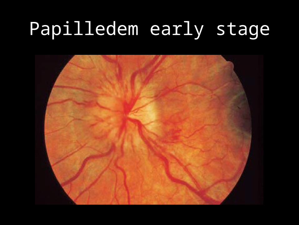
Papilledem early stage
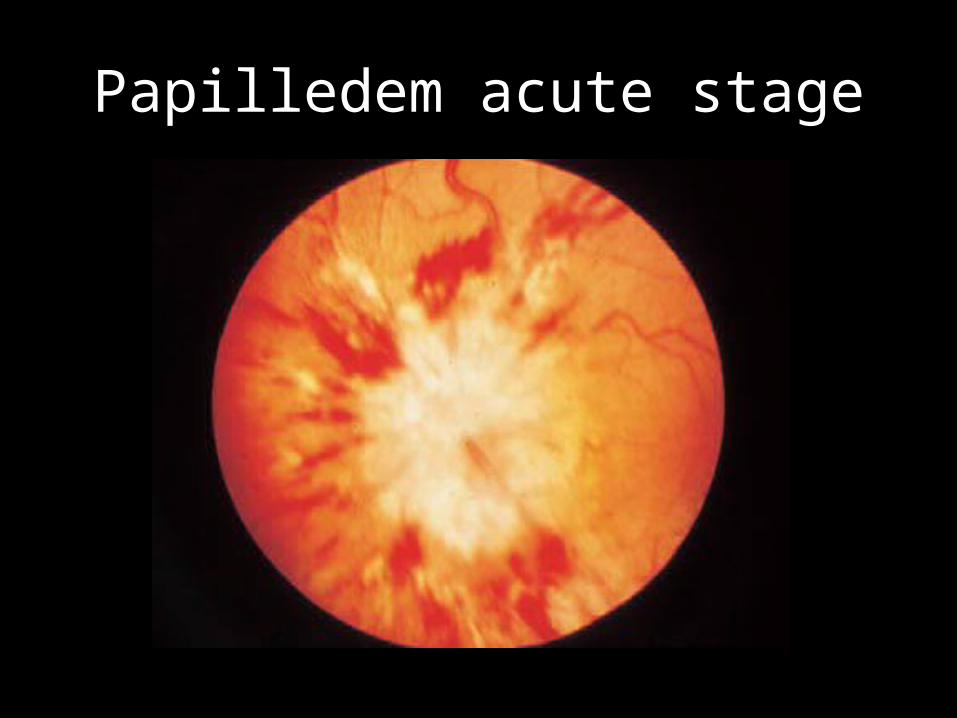
Papilledem acute stage
Atrophy of the Optic Nerve
• Irreversible loss of axons in the region of the third neuron (from the retinal layer of ganglion cells to the lateral geniculate body).
Morphology and pathologic classification
• on the basis of ophthalmoscopic findings:– Primary atrophy of the optic nerve.– Secondary atrophy of the optic nerve.– Glaucomatous atrophy of the optic nerve.
Etiology
Primary atrophy of the optic nerve.• Ascending atrophy (> 2-4 weeks):
– Usually vascular, such as central retinal artery occlusion or anterior ischemic optic neuropathy.
• Descending atrophy (> 4-6 weeks):– Compressive, such as from an orbital or intracranial mass or
hydrocephalus.– Traumatic, such as avulsion, compression of the optic nerve
in a fracture, or hematoma in the optic nerve sheath.– Inflammatory, such as retrobulbar optic neuritis,
arachnoiditis of the optic chiasm, or syphilis.
• Toxic:– Chronic abuse of low-grade tobacco and alcohol in
tobacco and alcohol amblyopia.– Lead, arsenic, or thallium.– Methyl alcohol.– Medications, such as ethambutol,
chloramphenicol, gentamicin, isoniazid, vincristine, penicillamine, etc.
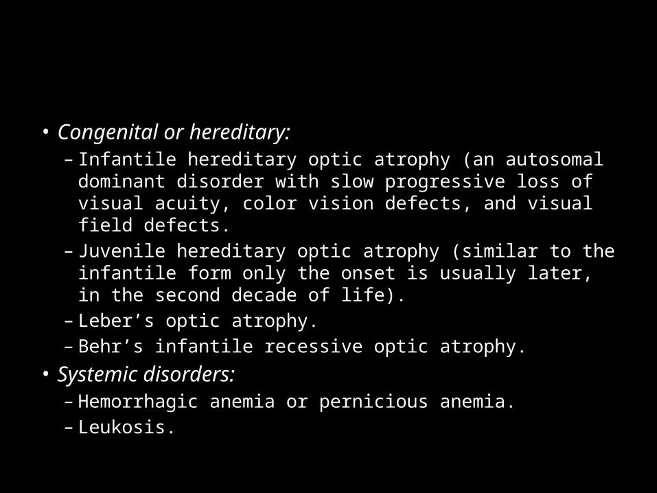
• Congenital or hereditary:– Infantile hereditary optic atrophy (an autosomal dominant
disorder with slow progressive loss of visual acuity, color vision defects, and visual field defects.
– Juvenile hereditary optic atrophy (similar to the infantile form only the onset is usually later, in the second decade of life).
– Leber’s optic atrophy.– Behr’s infantile recessive optic atrophy.
• Systemic disorders:– Hemorrhagic anemia or pernicious anemia.– Leukosis.
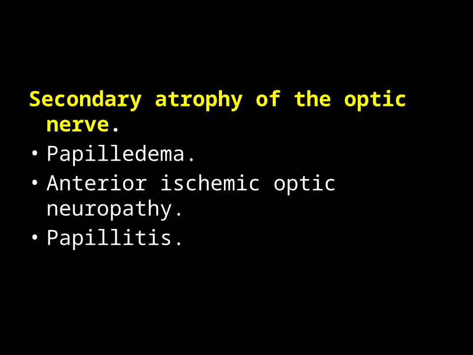
Secondary atrophy of the optic nerve.• Papilledema.• Anterior ischemic optic neuropathy.• Papillitis.
Symptoms
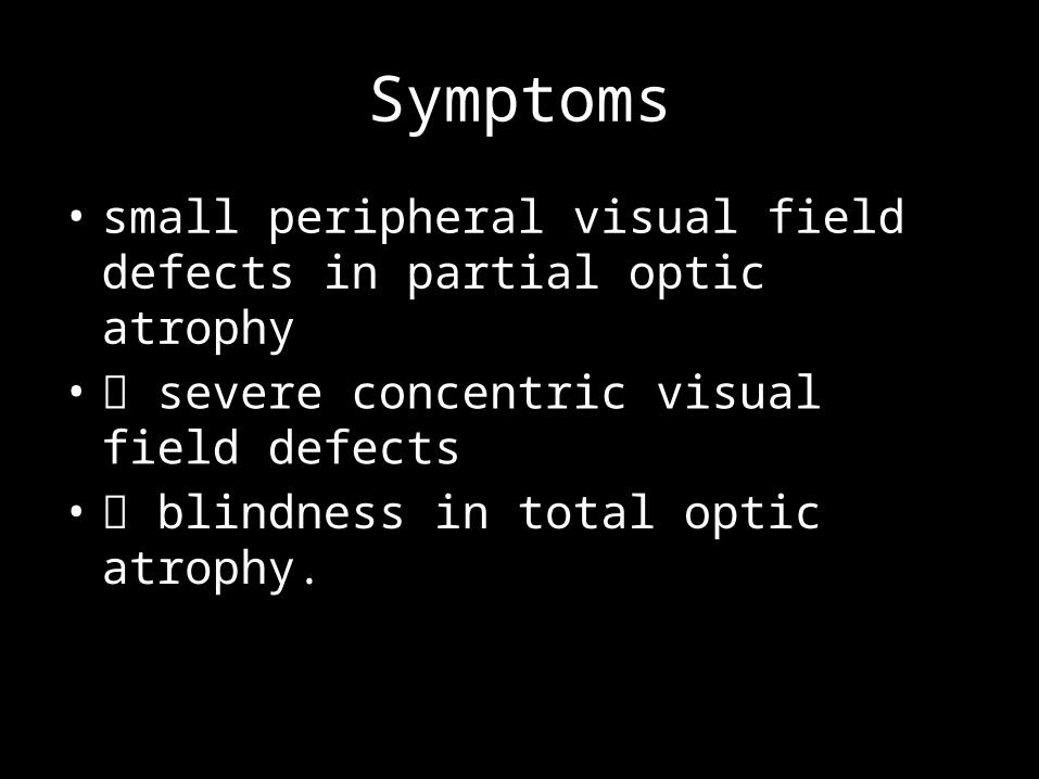
• small peripheral visual field defects in partial optic atrophy
• severe concentric visual field defects • blindness in total optic atrophy.

Diagnostic considerations
• History• Ophthalmoscopy• perimetry testing• Color vision testing• visual evoked potential
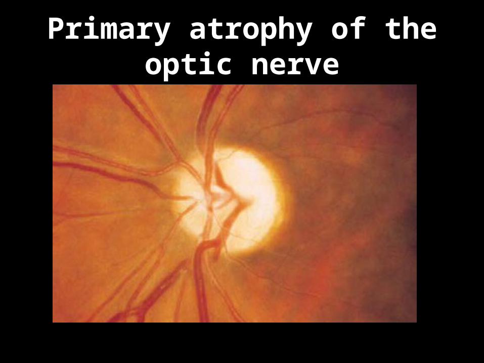
Primary atrophy of the optic nerve
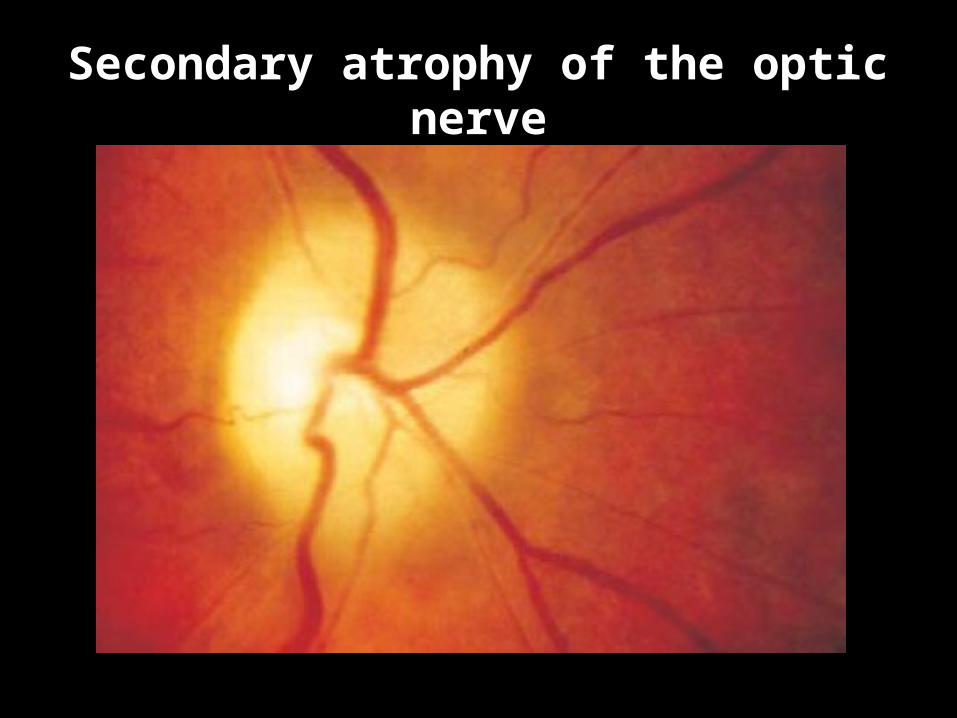
Secondary atrophy of the optic nerve
Treatment
• The disorder involves irreversible damage to the nerve fibers.
• no effective treatment is available.
Prognosis
• Early identification and timely management of a treatable cause such as a tumor or pernicious anemia can arrest the progression of the disorder.
• Where this is not the case, the prognosis for vision is poor.