Ultrasound Doppler Assessment of Liver Vasculature Post ...
1
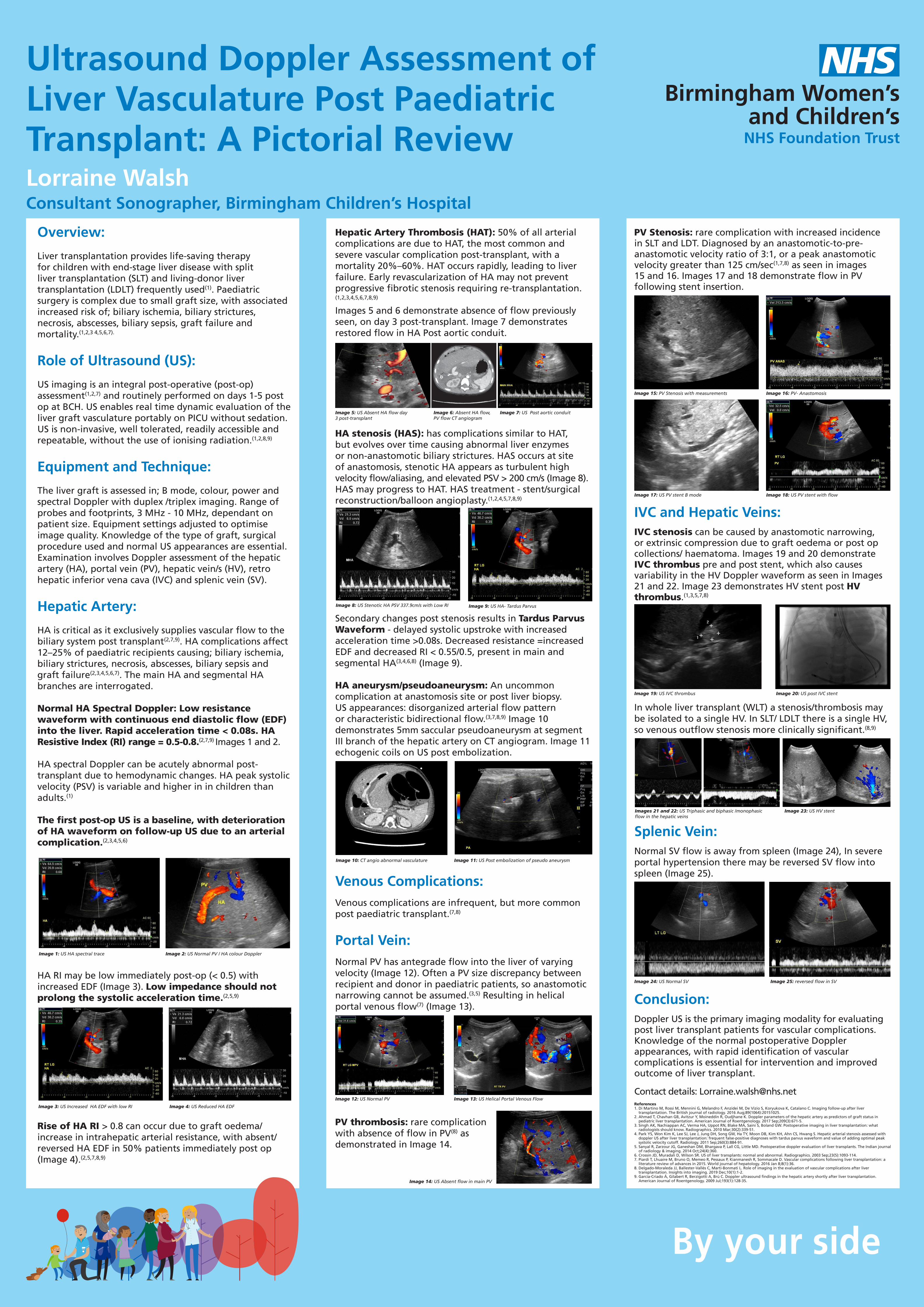
Ultrasound Doppler Assessment of Liver Vasculature Post Paediatric Transplant: A Pictorial Review Lorraine Walsh Overview: Liver transplantation provides life-saving therapy for children with end-stage liver disease with split liver transplantation (SLT) and living-donor liver transplantation (LDLT) frequently used (1) . Paediatric surgery is complex due to small graft size, with associated increased risk of; biliary ischemia, biliary strictures, necrosis, abscesses, biliary sepsis, graft failure and mortality. (1,2,3 4,5,6,7). Role of Ultrasound (US): US imaging is an integral post-operative (post-op) assessment (1,2,7) and routinely performed on days 1-5 post op at BCH. US enables real time dynamic evaluation of the liver graft vasculature portably on PICU without sedation. US is non-invasive, well tolerated, readily accessible and repeatable, without the use of ionising radiation. (1,2,8,9) Equipment and Technique: The liver graft is assessed in; B mode, colour, power and spectral Doppler with duplex /triplex imaging. Range of probes and footprints, 3 MHz - 10 MHz, dependant on patient size. Equipment settings adjusted to optimise image quality. Knowledge of the type of graft, surgical procedure used and normal US appearances are essential. Examination involves Doppler assessment of the hepatic artery (HA), portal vein (PV), hepatic vein/s (HV), retro hepatic inferior vena cava (IVC) and splenic vein (SV). Hepatic Artery: HA is critical as it exclusively supplies vascular flow to the biliary system post transplant (2,7,9) . HA complications affect 12–25% of paediatric recipients causing; biliary ischemia, biliary strictures, necrosis, abscesses, biliary sepsis and graft failure (2,3,4,5,6,7) . The main HA and segmental HA branches are interrogated. Normal HA Spectral Doppler: Low resistance waveform with continuous end diastolic flow (EDF) into the liver. Rapid acceleration time < 0.08s. HA Resistive Index (RI) range = 0.5-0.8. (2,7,9) Images 1 and 2. HA spectral Doppler can be acutely abnormal post- transplant due to hemodynamic changes. HA peak systolic velocity (PSV) is variable and higher in in children than adults. (1) The first post-op US is a baseline, with deterioration of HA waveform on follow-up US due to an arterial complication. (2,3,4,5,6) HA RI may be low immediately post-op (< 0.5) with increased EDF (Image 3). Low impedance should not prolong the systolic acceleration time. (2,5,9) Rise of HA RI > 0.8 can occur due to graft oedema/ increase in intrahepatic arterial resistance, with absent/ reversed HA EDF in 50% patients immediately post op (Image 4). (2,5,7,8,9) Consultant Sonographer, Birmingham Children’s Hospital Hepatic Artery Thrombosis (HAT): 50% of all arterial complications are due to HAT, the most common and severe vascular complication post-transplant, with a mortality 20%–60%. HAT occurs rapidly, leading to liver failure. Early revascularization of HA may not prevent progressive fibrotic stenosis requiring re-transplantation. (1,2,3,4,5,6,7,8,9) Images 5 and 6 demonstrate absence of flow previously seen, on day 3 post-transplant. Image 7 demonstrates restored flow in HA Post aortic conduit. HA stenosis (HAS): has complications similar to HAT, but evolves over time causing abnormal liver enzymes or non-anastomotic biliary strictures. HAS occurs at site of anastomosis, stenotic HA appears as turbulent high velocity flow/aliasing, and elevated PSV > 200 cm/s (Image 8). HAS may progress to HAT. HAS treatment - stent/surgical reconstruction/balloon angioplasty. (1,2,4,5,7,8,9) Secondary changes post stenosis results in Tardus Parvus Waveform - delayed systolic upstroke with increased acceleration time >0.08s. Decreased resistance =increased EDF and decreased RI < 0.55/0.5, present in main and segmental HA (3,4,6,8) (Image 9). HA aneurysm/pseudoaneurysm: An uncommon complication at anastomosis site or post liver biopsy. US appearances: disorganized arterial flow pattern or characteristic bidirectional flow. (3,7,8,9) Image 10 demonstrates 5mm saccular pseudoaneurysm at segment III branch of the hepatic artery on CT angiogram. Image 11 echogenic coils on US post embolization. Venous Complications: Venous complications are infrequent, but more common post paediatric transplant. (7,8) Portal Vein: Normal PV has antegrade flow into the liver of varying velocity (Image 12). Often a PV size discrepancy between recipient and donor in paediatric patients, so anastomotic narrowing cannot be assumed. (3,5) Resulting in helical portal venous flow (7) (Image 13). PV thrombosis: rare complication with absence of flow in PV (8) as demonstrated in Image 14. PV Stenosis: rare complication with increased incidence in SLT and LDT. Diagnosed by an anastomotic-to-pre- anastomotic velocity ratio of 3:1, or a peak anastomotic velocity greater than 125 cm/sec (1,7,8) as seen in images 15 and 16. Images 17 and 18 demonstrate flow in PV following stent insertion. IVC and Hepatic Veins: IVC stenosis can be caused by anastomotic narrowing, or extrinsic compression due to graft oedema or post op collections/ haematoma. Images 19 and 20 demonstrate IVC thrombus pre and post stent, which also causes variability in the HV Doppler waveform as seen in Images 21 and 22. Image 23 demonstrates HV stent post HV thrombus. (1,3,5,7,8) In whole liver transplant (WLT) a stenosis/thrombosis may be isolated to a single HV. In SLT/ LDLT there is a single HV, so venous outflow stenosis more clinically significant. (8,9) Splenic Vein: Normal SV flow is away from spleen (Image 24), In severe portal hypertension there may be reversed SV flow into spleen (Image 25). Conclusion: Doppler US is the primary imaging modality for evaluating post liver transplant patients for vascular complications. Knowledge of the normal postoperative Doppler appearances, with rapid identification of vascular complications is essential for intervention and improved outcome of liver transplant. Contact details: [email protected] References 1. Di Martino M, Rossi M, Mennini G, Melandro F, Anzidei M, De Vizio S, Koryukova K, Catalano C. Imaging follow-up after liver transplantation. The British journal of radiology. 2016 Aug;89(1064):20151025. 2. Ahmad T, Chavhan GB, Avitzur Y, Moineddin R, Oudjhane K. Doppler parameters of the hepatic artery as predictors of graft status in pediatric liver transplantation. American Journal of Roentgenology. 2017 Sep;209(3):671-5. 3. Singh AK, Nachiappan AC, Verma HA, Uppot RN, Blake MA, Saini S, Boland GW. Postoperative imaging in liver transplantation: what radiologists should know. Radiographics. 2010 Mar;30(2):339-51. 4. Park YS, Won Kim K, Lee SJ, Lee J, Jung DH, Song GW, Ha TY, Moon DB, Kim KH, Ahn CS, Hwang S. Hepatic arterial stenosis assessed with doppler US after liver transplantation: frequent false-positive diagnoses with tardus parvus waveform and value of adding optimal peak systolic velocity cutoff. Radiology. 2011 Sep;260(3):884-91. 5. Sanyal R, Zarzour JG, Ganeshan DM, Bhargava P, Lall CG, Little MD. Postoperative doppler evaluation of liver transplants. The Indian journal of radiology & imaging. 2014 Oct;24(4):360. 6. Crossin JD, Muradali D, Wilson SR. US of liver transplants: normal and abnormal. Radiographics. 2003 Sep;23(5):1093-114. 7. Piardi T, Lhuaire M, Bruno O, Memeo R, Pessaux P, Kianmanesh R, Sommacale D. Vascular complications following liver transplantation: a literature review of advances in 2015. World journal of hepatology. 2016 Jan 8;8(1):36. 8. Delgado-Moraleda JJ, Ballester-Vallés C, Marti-Bonmati L. Role of imaging in the evaluation of vascular complications after liver transplantation. Insights into imaging. 2019 Dec;10(1):1-2. 9. García-Criado Á, Gilabert R, Berzigotti A, Brú C. Doppler ultrasound findings in the hepatic artery shortly after liver transplantation. American Journal of Roentgenology. 2009 Jul;193(1):128-35. Image 1: US HA spectral trace Image 2: US Normal PV / HA colour Doppler Image 3: US Increased HA EDF with low RI Image 4: US Reduced HA EDF Image 8: US Stenotic HA PSV 337.9cm/s with Low RI Image 9: US HA- Tardus Parvus Image 10: CT angio abnormal vasculature Image 11: US Post embolization of pseudo aneurysm Image 12: US Normal PV Image 13: US Helical Portal Venous Flow Image 14: US Absent flow in main PV Image 15: PV Stenosis with measurements Image 16: PV- Anastomosis Image 17: US PV stent B mode Image 18: US PV stent with flow Image 19: US IVC thrombus Image 20: US post IVC stent Images 21 and 22: US Triphasic and biphasic /monophasic flow in the hepatic veins Image 23: US HV stent Image 24: US Normal SV Image 25: reversed flow in SV Image 5: US Absent HA flow day 3 post-transplant Image 6: Absent HA flow, PV flow CT angiogram Image 7: US Post aortic conduit
Transcript of Ultrasound Doppler Assessment of Liver Vasculature Post ...
Ultrasound Doppler Assessment of Liver Vasculature Post Paediatric Transplant: A Pictorial ReviewLorraine Walsh
Overview:
Liver transplantation provides life-saving therapy for children with end-stage liver disease with split liver transplantation (SLT) and living-donor liver transplantation (LDLT) frequently used(1). Paediatric surgery is complex due to small graft size, with associated increased risk of; biliary ischemia, biliary strictures, necrosis, abscesses, biliary sepsis, graft failure and mortality.(1,2,3 4,5,6,7).
Role of Ultrasound (US):
US imaging is an integral post-operative (post-op) assessment(1,2,7) and routinely performed on days 1-5 post op at BCH. US enables real time dynamic evaluation of the liver graft vasculature portably on PICU without sedation. US is non-invasive, well tolerated, readily accessible and repeatable, without the use of ionising radiation.(1,2,8,9)
Equipment and Technique:
The liver graft is assessed in; B mode, colour, power and spectral Doppler with duplex /triplex imaging. Range of probes and footprints, 3 MHz - 10 MHz, dependant on patient size. Equipment settings adjusted to optimise image quality. Knowledge of the type of graft, surgical procedure used and normal US appearances are essential. Examination involves Doppler assessment of the hepatic artery (HA), portal vein (PV), hepatic vein/s (HV), retro hepatic inferior vena cava (IVC) and splenic vein (SV).
Hepatic Artery:
HA is critical as it exclusively supplies vascular flow to the biliary system post transplant(2,7,9). HA complications affect 12–25% of paediatric recipients causing; biliary ischemia, biliary strictures, necrosis, abscesses, biliary sepsis and graft failure(2,3,4,5,6,7). The main HA and segmental HA branches are interrogated.
Normal HA Spectral Doppler: Low resistance waveform with continuous end diastolic flow (EDF) into the liver. Rapid acceleration time < 0.08s. HA Resistive Index (RI) range = 0.5-0.8.(2,7,9) Images 1 and 2.
HA spectral Doppler can be acutely abnormal post-transplant due to hemodynamic changes. HA peak systolic velocity (PSV) is variable and higher in in children than adults.(1)
The first post-op US is a baseline, with deterioration of HA waveform on follow-up US due to an arterial complication.(2,3,4,5,6)
HA RI may be low immediately post-op (< 0.5) with increased EDF (Image 3). Low impedance should not prolong the systolic acceleration time.(2,5,9)
Rise of HA RI > 0.8 can occur due to graft oedema/ increase in intrahepatic arterial resistance, with absent/ reversed HA EDF in 50% patients immediately post op (Image 4).(2,5,7,8,9)
Consultant Sonographer, Birmingham Children’s Hospital
Hepatic Artery Thrombosis (HAT): 50% of all arterial complications are due to HAT, the most common and severe vascular complication post-transplant, with a mortality 20%–60%. HAT occurs rapidly, leading to liver failure. Early revascularization of HA may not prevent progressive fibrotic stenosis requiring re-transplantation. (1,2,3,4,5,6,7,8,9)
Images 5 and 6 demonstrate absence of flow previously seen, on day 3 post-transplant. Image 7 demonstrates restored flow in HA Post aortic conduit.
HA stenosis (HAS): has complications similar to HAT, but evolves over time causing abnormal liver enzymes or non-anastomotic biliary strictures. HAS occurs at site of anastomosis, stenotic HA appears as turbulent high velocity flow/aliasing, and elevated PSV > 200 cm/s (Image 8). HAS may progress to HAT. HAS treatment - stent/surgical reconstruction/balloon angioplasty.(1,2,4,5,7,8,9)
Secondary changes post stenosis results in Tardus Parvus Waveform - delayed systolic upstroke with increased acceleration time >0.08s. Decreased resistance =increased EDF and decreased RI < 0.55/0.5, present in main and segmental HA(3,4,6,8) (Image 9).
HA aneurysm/pseudoaneurysm: An uncommon complication at anastomosis site or post liver biopsy. US appearances: disorganized arterial flow pattern or characteristic bidirectional flow.(3,7,8,9) Image 10 demonstrates 5mm saccular pseudoaneurysm at segment III branch of the hepatic artery on CT angiogram. Image 11 echogenic coils on US post embolization.
Venous Complications: Venous complications are infrequent, but more common post paediatric transplant.(7,8)
Portal Vein:Normal PV has antegrade flow into the liver of varying velocity (Image 12). Often a PV size discrepancy between recipient and donor in paediatric patients, so anastomotic narrowing cannot be assumed.(3,5) Resulting in helical portal venous flow(7) (Image 13).
PV thrombosis: rare complication with absence of flow in PV(8) as demonstrated in Image 14.
PV Stenosis: rare complication with increased incidence in SLT and LDT. Diagnosed by an anastomotic-to-pre-anastomotic velocity ratio of 3:1, or a peak anastomotic velocity greater than 125 cm/sec(1,7,8) as seen in images 15 and 16. Images 17 and 18 demonstrate flow in PV following stent insertion.
IVC and Hepatic Veins: IVC stenosis can be caused by anastomotic narrowing, or extrinsic compression due to graft oedema or post op collections/ haematoma. Images 19 and 20 demonstrate IVC thrombus pre and post stent, which also causes variability in the HV Doppler waveform as seen in Images 21 and 22. Image 23 demonstrates HV stent post HV thrombus.(1,3,5,7,8)
In whole liver transplant (WLT) a stenosis/thrombosis may be isolated to a single HV. In SLT/ LDLT there is a single HV, so venous outflow stenosis more clinically significant.(8,9)
Splenic Vein:Normal SV flow is away from spleen (Image 24), In severe portal hypertension there may be reversed SV flow into spleen (Image 25).
Conclusion:Doppler US is the primary imaging modality for evaluating post liver transplant patients for vascular complications. Knowledge of the normal postoperative Doppler appearances, with rapid identification of vascular complications is essential for intervention and improved outcome of liver transplant.
Contact details: [email protected] 1. Di Martino M, Rossi M, Mennini G, Melandro F, Anzidei M, De Vizio S, Koryukova K, Catalano C. Imaging follow-up after liver
transplantation. The British journal of radiology. 2016 Aug;89(1064):20151025.2. Ahmad T, Chavhan GB, Avitzur Y, Moineddin R, Oudjhane K. Doppler parameters of the hepatic artery as predictors of graft status in
pediatric liver transplantation. American Journal of Roentgenology. 2017 Sep;209(3):671-5.3. Singh AK, Nachiappan AC, Verma HA, Uppot RN, Blake MA, Saini S, Boland GW. Postoperative imaging in liver transplantation: what
radiologists should know. Radiographics. 2010 Mar;30(2):339-51.4. Park YS, Won Kim K, Lee SJ, Lee J, Jung DH, Song GW, Ha TY, Moon DB, Kim KH, Ahn CS, Hwang S. Hepatic arterial stenosis assessed with
doppler US after liver transplantation: frequent false-positive diagnoses with tardus parvus waveform and value of adding optimal peak systolic velocity cutoff. Radiology. 2011 Sep;260(3):884-91.
5. Sanyal R, Zarzour JG, Ganeshan DM, Bhargava P, Lall CG, Little MD. Postoperative doppler evaluation of liver transplants. The Indian journal of radiology & imaging. 2014 Oct;24(4):360.
6. Crossin JD, Muradali D, Wilson SR. US of liver transplants: normal and abnormal. Radiographics. 2003 Sep;23(5):1093-114.7. Piardi T, Lhuaire M, Bruno O, Memeo R, Pessaux P, Kianmanesh R, Sommacale D. Vascular complications following liver transplantation: a
literature review of advances in 2015. World journal of hepatology. 2016 Jan 8;8(1):36.8. Delgado-Moraleda JJ, Ballester-Vallés C, Marti-Bonmati L. Role of imaging in the evaluation of vascular complications after liver
transplantation. Insights into imaging. 2019 Dec;10(1):1-2.9. García-Criado Á, Gilabert R, Berzigotti A, Brú C. Doppler ultrasound findings in the hepatic artery shortly after liver transplantation.
American Journal of Roentgenology. 2009 Jul;193(1):128-35.
Image 1: US HA spectral trace Image 2: US Normal PV / HA colour Doppler
Image 3: US Increased HA EDF with low RI Image 4: US Reduced HA EDF
Image 8: US Stenotic HA PSV 337.9cm/s with Low RI Image 9: US HA- Tardus Parvus
Image 10: CT angio abnormal vasculature Image 11: US Post embolization of pseudo aneurysm
Image 12: US Normal PV Image 13: US Helical Portal Venous Flow
Image 14: US Absent flow in main PV
Image 15: PV Stenosis with measurements Image 16: PV- Anastomosis
Image 17: US PV stent B mode Image 18: US PV stent with flow
Image 19: US IVC thrombus Image 20: US post IVC stent
Images 21 and 22: US Triphasic and biphasic /monophasic flow in the hepatic veins
Image 23: US HV stent
Image 24: US Normal SV Image 25: reversed flow in SV
Image 5: US Absent HA flow day 3 post-transplant
Image 6: Absent HA flow, PV flow CT angiogram
Image 7: US Post aortic conduit