Salting out is a method of separating proteins based on the principle that proteins are less soluble...
16
LABORATORY TECHNIQUES
-
Upload
myles-francis -
Category
Documents
-
view
220 -
download
0
Transcript of Salting out is a method of separating proteins based on the principle that proteins are less soluble...

LABORATORYTECHNIQUES
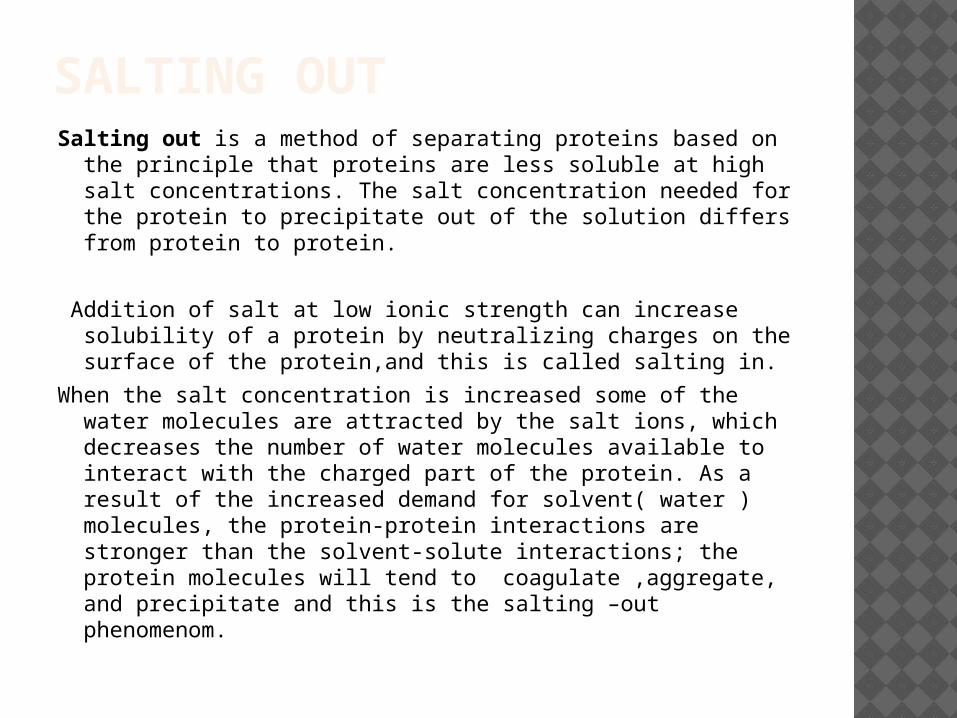
SALTING OUTSalting out is a method of separating proteins based on the
principle that proteins are less soluble at high salt concentrations. The salt concentration needed for the protein to precipitate out of the solution differs from protein to protein.
Addition of salt at low ionic strength can increase solubility of a protein by neutralizing charges on the surface of the protein,and this is called salting in.
When the salt concentration is increased some of the water molecules are attracted by the salt ions, which decreases the number of water molecules available to interact with the charged part of the protein. As a result of the increased demand for solvent( water ) molecules, the protein-protein interactions are stronger than the solvent-solute interactions; the protein molecules will tend to coagulate ,aggregate, and precipitate and this is the salting –out phenomenom.
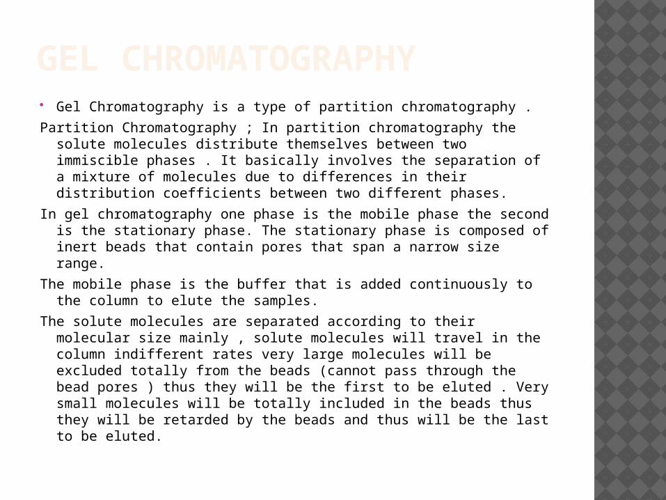
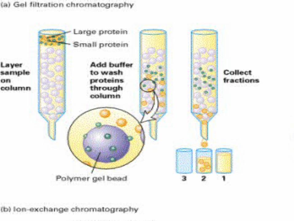
GEL CHROMATOGRAPHY Gel Chromatography is a type of partition chromatography .
Partition Chromatography ; In partition chromatography the solute molecules distribute themselves between two immiscible phases . It basically involves the separation of a mixture of molecules due to differences in their distribution coefficients between two different phases.
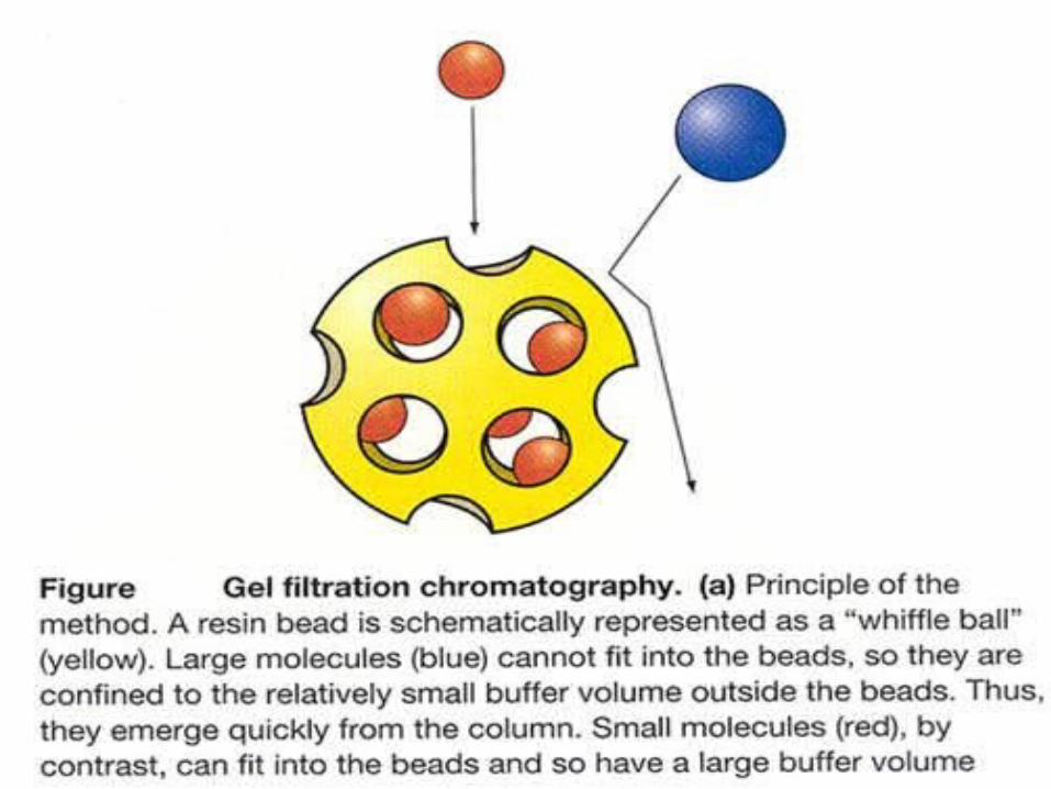
In gel chromatography one phase is the mobile phase the second is the stationary phase. The stationary phase is composed of inert beads that contain pores that span a narrow size range.
The mobile phase is the buffer that is added continuously to the column to elute the samples.
The solute molecules are separated according to their molecular size mainly , solute molecules will travel in the column indifferent rates very large molecules will be excluded totally from the beads (cannot pass through the bead pores ) thus they will be the first to be eluted . Very small molecules will be totally included in the beads thus they will be retarded by the beads and thus will be the last to be eluted.
GEL CHROMATOGRAPHY Thus the solute molecules will be eluted in the following
order large molecules first followed by smaller molecules then the smallest will eluted last.
Example; Consider the separation of the following mixture
-Glutamate dehydrogenase ( molecular weight 290000) .
-Lactate dehydrogenase (molecular weight 140000).
-Serum albumin (molecular weight 67000).
-Ovalbumin (molecular weight 43000).
-Cytochrome C (molecular weight 12400).
With Bio-gel-P-150.fractionation range (15000-150000).
When the protein mixture is loaded on the column , the first protein to be eluted is Glutamate dehydrogenase which is totally excluded from the beads since its MW is above the upper fractionation limit.
GEL CHROMATOGRAPHY Cytochrome C is the last to be eluted from the column
since its MW is below the lower fractionation limit so it will enter the pores of the beads and thus be retarded in the column and be eluted last.The rest of the protein molecules will be eluted in the order of decreasing MW.
So the order of elution is ;
1- Glutamate dehdrogenase.
2-Lactae dehydrogenase.
3-Serum albumin.
4-Ovalbumin.
5-Cytochrome C.
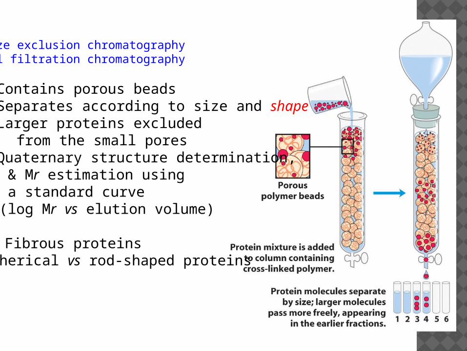
Size exclusion chromatographyGel filtration chromatography
Contains porous beads Separates according to size and shape· Larger proteins excluded from the small pores Quaternary structure determination, & Mr estimation using a standard curve (log Mr vs elution volume)
Ⓠ Fibrous proteinsSpherical vs rod-shaped proteins
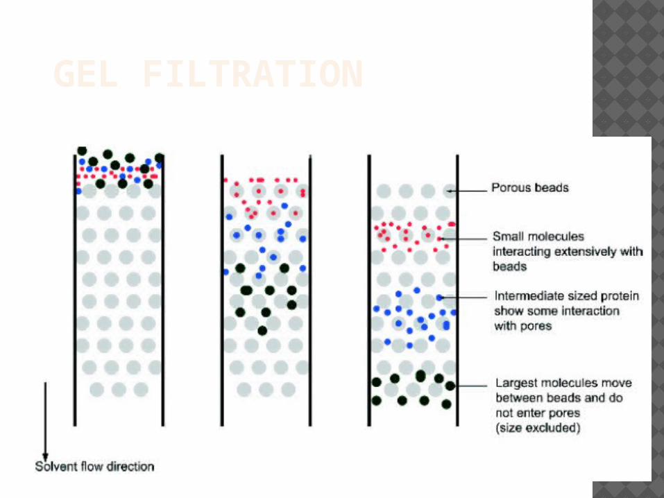
GEL FILTRATION
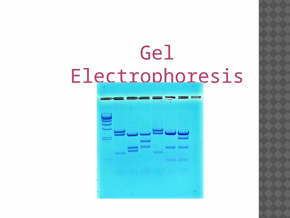
Gel Electrophoresis
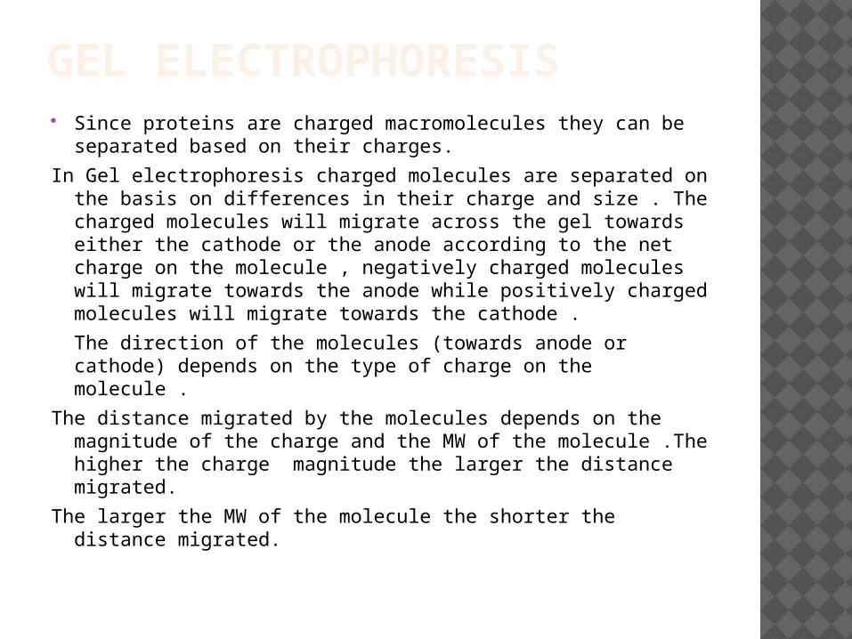
GEL ELECTROPHORESIS Since proteins are charged macromolecules they can be
separated based on their charges.
In Gel electrophoresis charged molecules are separated on the basis on differences in their charge and size . The charged molecules will migrate across the gel towards either the cathode or the anode according to the net charge on the molecule , negatively charged molecules will migrate towards the anode while positively charged molecules will migrate towards the cathode .
The direction of the molecules (towards anode or cathode) depends on the type of charge on the molecule .
The distance migrated by the molecules depends on the magnitude of the charge and the MW of the molecule .The higher the charge magnitude the larger the distance migrated.
The larger the MW of the molecule the shorter the distance migrated.
passage of molecules according to their size and shape.
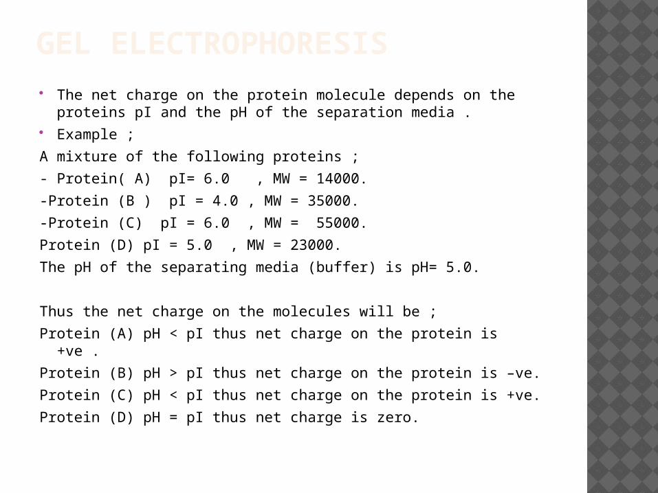
GEL ELECTROPHORESIS The net charge on the protein molecule depends on the
proteins pI and the pH of the separation media . Example ;
A mixture of the following proteins ;
- Protein( A) pI= 6.0 , MW = 14000.
-Protein (B ) pI = 4.0 , MW = 35000.
-Protein (C) pI = 6.0 , MW = 55000.
Protein (D) pI = 5.0 , MW = 23000.
The pH of the separating media (buffer) is pH= 5.0.
Thus the net charge on the molecules will be ;
Protein (A) pH < pI thus net charge on the protein is +ve .
Protein (B) pH > pI thus net charge on the protein is –ve.
Protein (C) pH < pI thus net charge on the protein is +ve.
Protein (D) pH = pI thus net charge is zero.
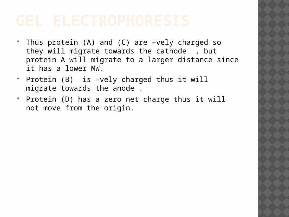
GEL ELECTROPHORESIS Thus protein (A) and (C) are +vely charged so they will
migrate towards the cathode , but protein A will migrate to a larger distance since it has a lower MW.
Protein (B) is –vely charged thus it will migrate towards the anode .
Protein (D) has a zero net charge thus it will not move from the origin.
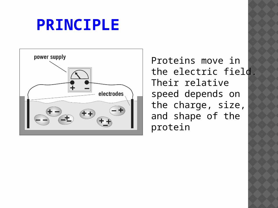
PRINCIPLE
Proteins move in the electric field. Their relative speed depends on the charge, size, and shape of the protein
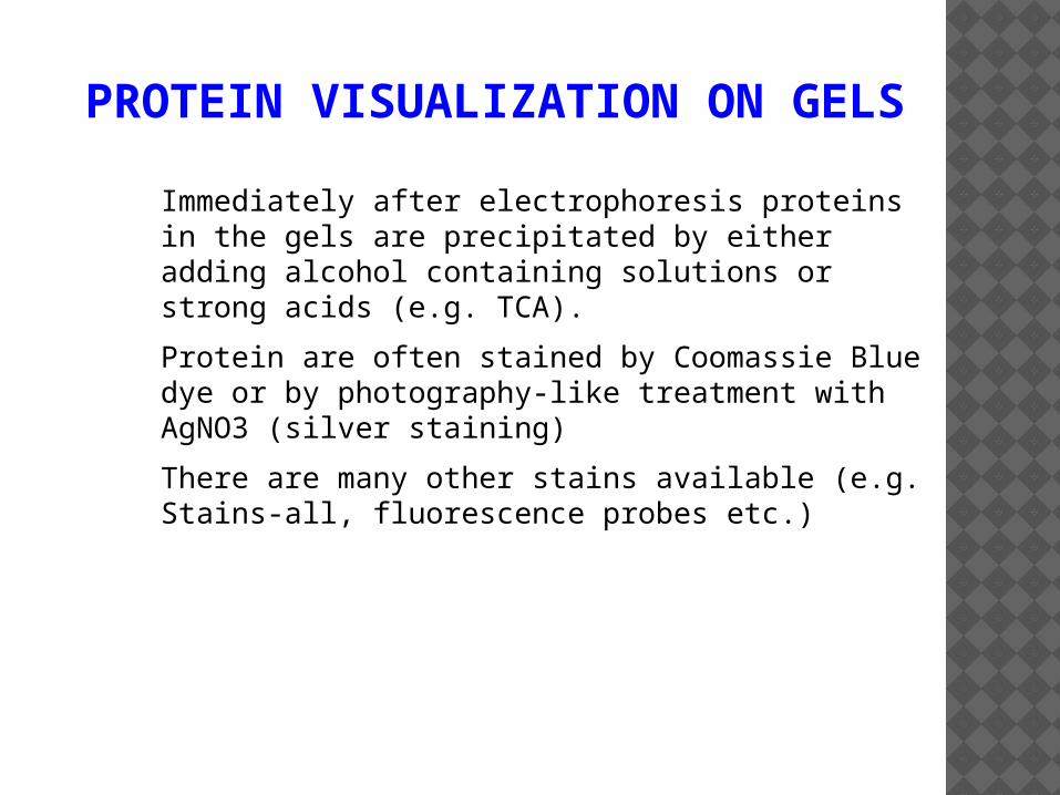
PROTEIN VISUALIZATION ON GELS
Immediately after electrophoresis proteins in the gels are precipitated by either adding alcohol containing solutions or strong acids (e.g. TCA).
Protein are often stained by Coomassie Blue dye or by photography-like treatment with AgNO3 (silver staining)
There are many other stains available (e.g. Stains-all, fluorescence probes etc.)
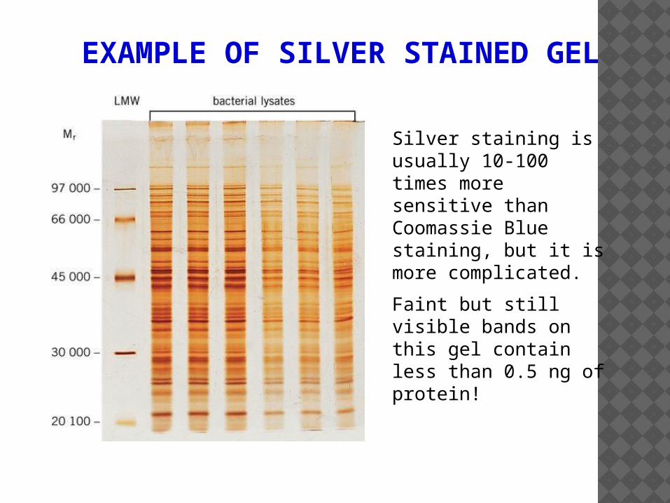
EXAMPLE OF SILVER STAINED GEL
Silver staining is usually 10-100 times more sensitive than Coomassie Blue staining, but it is more complicated.
Faint but still visible bands on this gel contain less than 0.5 ng of protein!
![Salting in and Salting out of proteins and Dialysis BCH 333 [practical]](https://static.fdocuments.net/doc/165x107/56649ef55503460f94c087a8/salting-in-and-salting-out-of-proteins-and-dialysis-bch-333-practical.jpg)