Classification OTHERS Xerostomia Sialorrhea Mucocele Mucous retention Ranula.
59
Classification • OTHERS • Xerostomia • Sialorrhea • Mucocele • Mucous retention • Ranula
-
Upload
jessie-sanders -
Category
Documents
-
view
249 -
download
2
Transcript of Classification OTHERS Xerostomia Sialorrhea Mucocele Mucous retention Ranula.
Classification
• OTHERS
• Xerostomia• Sialorrhea• Mucocele• Mucous retention• Ranula
XEROSTOMIA
Xerostomia
• Xerostomia (dry mouth)
• Is not a disease but a symptom caused by many factors.
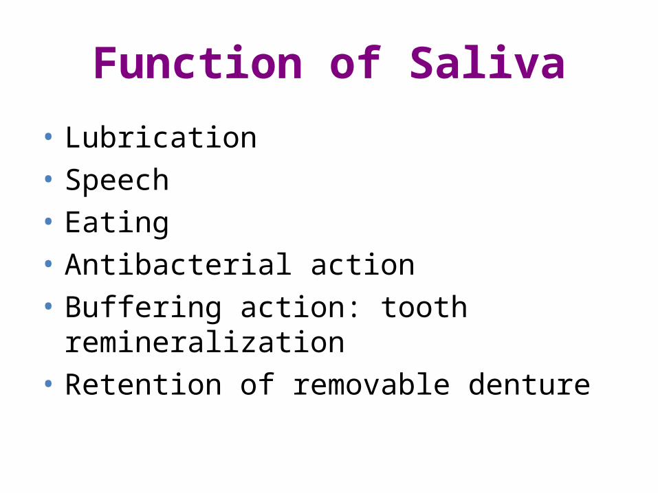
Function of Saliva
• Lubrication• Speech• Eating• Antibacterial action• Buffering action: tooth remineralization• Retention of removable denture
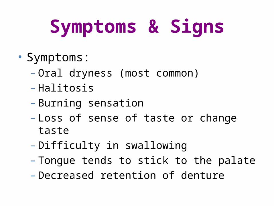
Symptoms & Signs
• Symptoms:– Oral dryness (most common)– Halitosis– Burning sensation – Loss of sense of taste or change taste– Difficulty in swallowing – Tongue tends to stick to the palate – Decreased retention of denture
Symptoms & Signs
–Signs– Angular cheilitis– Rampant caries: cervical or cusp tip– Periodontitis– Candidiasis – Saliva pool disappear– glossitis
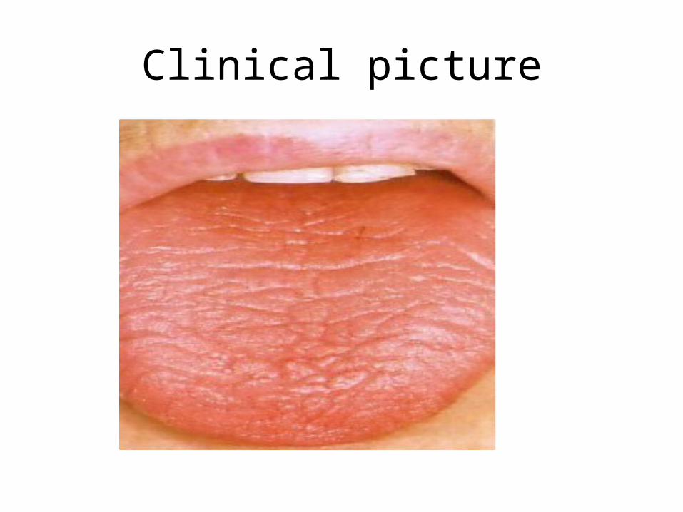
Clinical picture
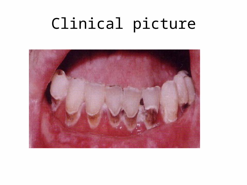
Clinical picture

Etiology (Causes)
• Developmental• Water/Metabolite loss• Iatrogenic• Systemic Diseases• Local factors
Developmental
• Salivary gland aplasia
Water/Metabolite loss
• Impaired fluid in take• Blood loss (Hemorrhage)• Vomiting / Diarrhea

Iatrogenic
• Medication• Radiation therapy
Iatrogenic (Medication)
• Anti histamine (Diphenhydramine,chlorpheniramine)• Anti depressant (Amitriptyline)• Anti hypertensive
(Reserpine,Methyldopa,furosemide,CCB, heloperidol,chlorothiazide)
• Anti cholinergic (Atropine,Scopolamine)
Systemic Diseases
• Sjogrens Syndrome• Diabetes mellitus• Diabetes insipidus• Sarcoidosis• HIV infection• Psychogenic disorder
Local factors
• Aging• Foods• Emotions• Stress• Mouth breathing

Foods:
• alcohol,• coffee,• coco cola, • Smoke.
Diagnosis• History taking• Clinical examination• Investigations– Salivary flow rate (Sialometry)– Salivary scintiscanning– Sialochemical analysis & laboratory values – Labial biopsy– Sialography
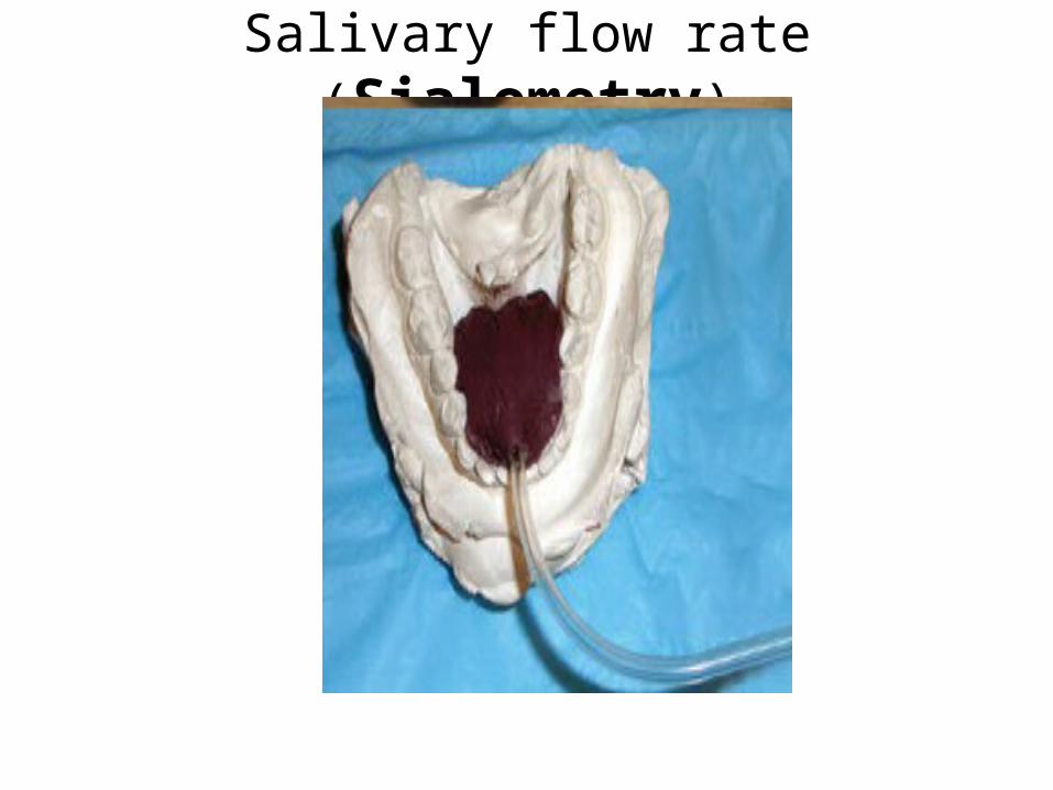
Salivary flow rate (Sialometry)
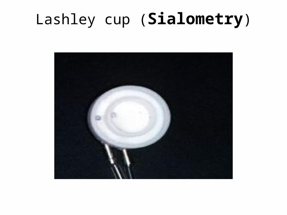
Lashley cup (Sialometry)
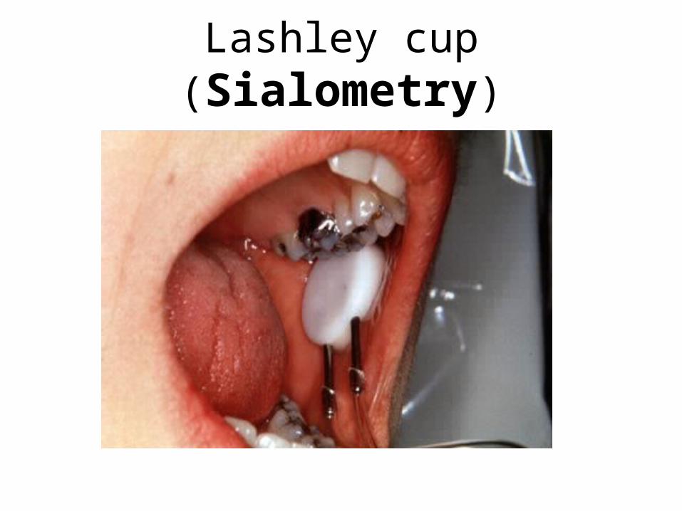
Lashley cup (Sialometry)
Management
• Dietary & environmental considerations• Preventive Dental Care Measures • Saliva stimulatants• Saliva substitutes

SIALORRHEASIALORRHEAPtyalismPtyalismDroolingDrooling
SIALORRHEA
• Excess Saliva• The condition in which there is increased
Salivary flow
Causes• Ill fitting Denture• New Denture Wearer• Apthous Ulcers• GIT Diseases• Rabies bites• Metal poisoning• Stroke• Hemiplagia--paralysis patient• Sour or Spicy Foods
Causes
• Drugs (antipsychotic, Cholinergic drugs)• Mentally retard Patients• Recent surgery• Neuromuscular problems• Large tongue (Macroglossia - Downs
syndrome)
Clinical Features
• Drooling of Saliva• Soiling of cloths• Ulcers around the corners of mouth• Choking of saliva during speech• Perioral infections• Chin and Neck infection• Respiratory problems
Diagnosis
• History Normal 14 months of age• Examination resection of mandible, mental retard, GIT
disorders, Drugs• Investigation Sialometry
Management
• Identify and Remove the Cause
• Non Medical
• Medical • Surgical

Management
• Non Medical or Physical
1-Self motivation 2-Habit Breaking 3-Physiotherapy 4- Radiotherapy
Management
• Medical
1- Glycoprrolate tablet 1 to 2 mg two times a day 2- Scopolamine patches 1.5mg once day
Management
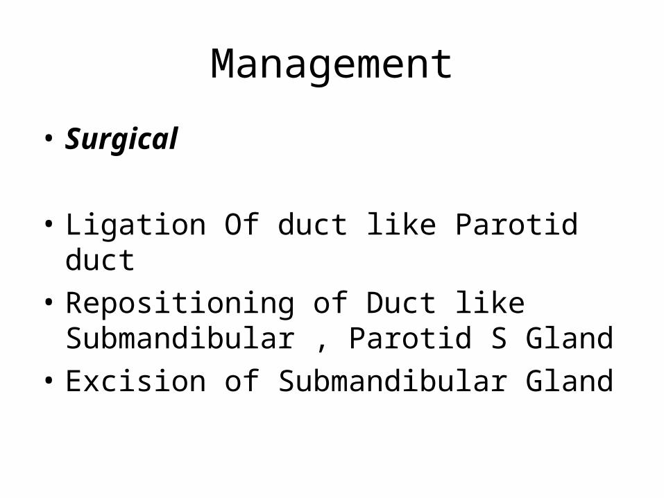
• Surgical
• Ligation Of duct like Parotid duct• Repositioning of Duct like Submandibular ,
Parotid S Gland• Excision of Submandibular Gland

Mucocele
MUCOCELE
• It is a tissue swelling composed of pooled mucus that escapes into the connective tissue from several excretory ducts
Mucocele
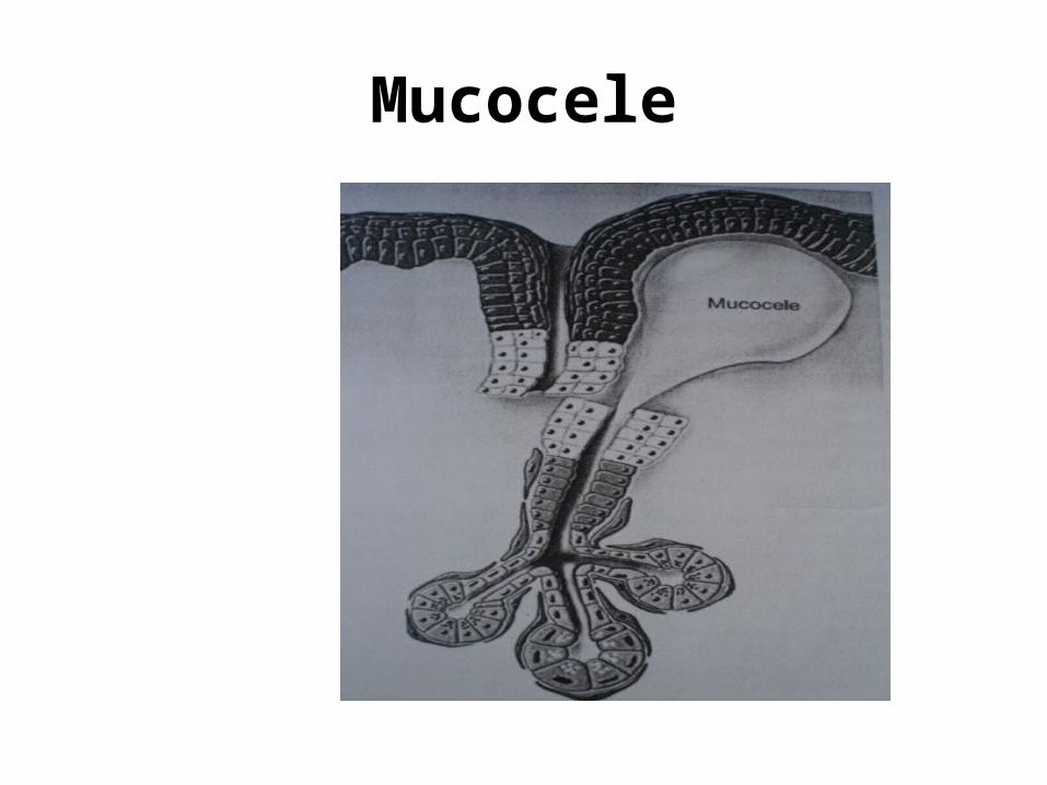
• When salivary duct is severed the acinar cells will continue to secrete saliva into the severed duct.
• At the site of the cut/severance the secretory product escape into the connective tissue forming a pool of mucus that distends the surrounding tissue
Mucocele
Mucocele
ETIOLOGY:• Minor glands of the lip are most prone to
severance as a result of injury or biting the mucosa.
• Intra oral minor salivary gland can also be effected as result of some irritation as well.
CLINICAL FEATURES
• Mostly encountered in children and young adults.• Two third of the Mucocele occur in the 3rd decade of
life.• Both males and females are effected equally.• SITE: mucosal surface of the lower lip buccal mucosa floor the mouth ventral surface of the tongue and palate• Clinical appearance of the Mucocele depends on its
location within the submucosa
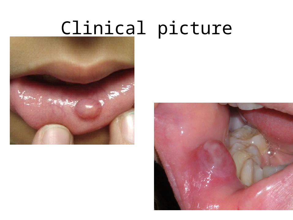
CLINICAL FEATURES
• More superficial zones of mucous extravasations presents a fluctuant mass with bluish translucent appearance.
• Patient usually feels the Mucocele and the fluctuation in its size
• Pain is quite rare .• Initially the Mucocele are well circumscribed
but with repeated trauma they become nodular ,more diffuse and firm on palpation.
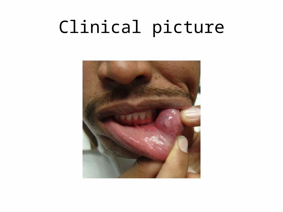
Clinical picture
Clinical picture
HISTOPATHOLOGY:
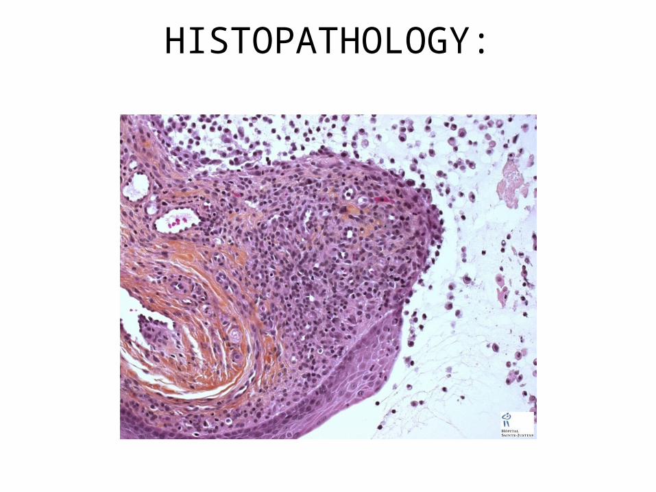
• Underlying pool of mucin distends the surface epithelium.• The mucin is walled of by the rim of granulation tissue or in
long standing cases by condensed collagen.• An epithelial lining is lacking• The mucinous material basophilic or acidophillic and
contains neutrophils and large oval foam cells the histocytes .
• The base of the mucocele will reveal feeder duct.• Long standing mucoceles will show acinar degeneration
with fibrosis and minimal inflammation
HISTOPATHOLOGY:
TREATMENT:
• Minor salivary gland mucocele will not resolve on its own it must be surgically excised.
• To minimize the chances of recurrence the feeder gland should also be removed.
Mucus retention cyst
Mucus retention cyst
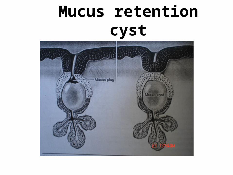
• It is a swelling caused by an obstruction of a salivary gland excretory duct resulting in an epithelial lining cavity containing mucus.
• Mucus retention cyst is sometimes also referred as Sialocyst.
Mucus retention cyst
• The mucus retention cyst is lined by epithelium and rarely occur in the major salivary gland, when they do occur they are multiple i.e. poly cystic disease of the parotid gland
Mucus retention cyst
Mucus retention cystCLINICAL FEATURES:• Encountered in adults from 3rd -5th decade.• The lesion is painless and fluctuant and bluish in
appearance.
SITE: Parotid cysts are located in the superficial lobe as
fluctuant well defined mass. Floor of the mouth is the most common place. -Lip -Buccal mucosa
Mucus retention cystHISTOPATHOLOGY:• The epithelium of the cyst is stratified cuboidal or
columnar duct like epithelium.• The cytoplasm in the of these cells is either clear or
eosinophlic and my show some features mucous differentiation
• 70% of these cyst are unilocular rest of the 30% have multilocular pattern.
Mucus retention cyst
TREATMENT:• Simple excision is the treatment of choice
with caution of rupturing the cystic sacs.• Recurrence is rare.

Ranula
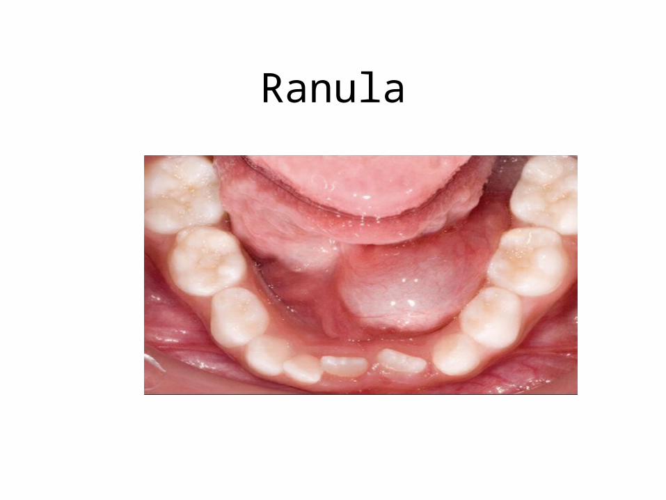
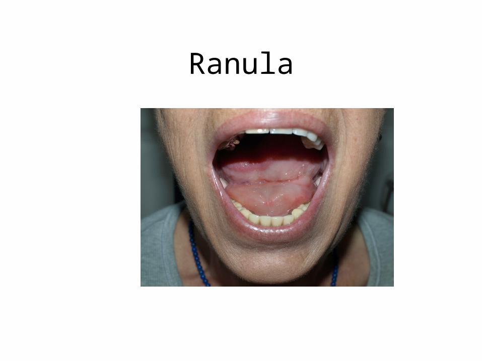
Ranula• Is a term used for mucoceles that occur in the
floor of the mouth.• The name is derived form the word rana,
because the swelling may resemble the translucent underbelly of the frog.
Ranula
Ranula
Ranula
• Although the source is usually the sublingual gland, – may also arise from the submandibular duct– or possibly the minor salivary glands in the
floor of the mouth.
Ranula
• Presents as a blue dome shaped swelling in the floor of mouth.
• They tend to be larger than mucoceles & can cover floor of the mouth & elevate tongue.
• Located lateral to the midline, helping to distinguish it from a midline dermoid cyst.
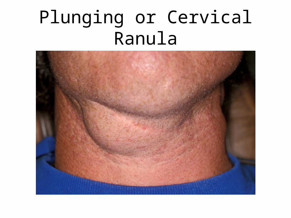
Plunging or Cervical Ranula
• Occurs when spilled mucin dissects through the mylohyoid muscle and produces swelling in the neck.
• Concomitant floor of the mouth swelling may or may not be visible.
Plunging or Cervical Ranula
Ranula Treatment
• Marsupialization• Sublingual gland removal via intraoral
approach