Cholestasis in infancy: Definition, Practical Approach and ... in infancy.pdf · Cholestasis in...
12
<I \.'JK SCIENCE " I REVIEW ARTICLE I Cholestasis in infancy: Definition, Practical ,., Approach and Management B. R. Thapa Introduction Neonatal cholestasis syndrome (NCS) comprises of heterogenous group of hepatobiliary disorders responsible for cholestasis during neonatal life. The cholestasis due to some disorders can extend beyond neonatal period hence, this is appropriate to call this syndrome as cholestasis of infancy. Most of the disorders have linkage with insults during antenatal, natal and postnatal period indicating intrauterine or postnatal events. Main causes of NCS are, infection, metabolic disorders and extrahepatic biliary obstructions those cover large number of conditions (l). The present scenario about the spectrum of illnesses is changing fast. Premature babies are now being looked after in neonatal specialised care units, the causes of cholestasis due to sepsis, total parenteral nutrition (TPN), drugs etc, are emerging as another large group (2). But this remains a challenge to evaluate and manage NCS because offast change in the diagnostic and therapeutic approach world over. It becomes mandatory to define the cause ofNCS that needs battery of tests to carry out. This is also important to do various tests simultaneously to differentiate between neonatal hepatitis (NH) and extrahepatic biliary atresia (EHBA) to avoid delay in Definition and Classification Jaundice is a very common symptom encountered during neonatal life. Approach to jaundice during neonatal period and infancy is given in figure 1. This is also called hyperbilirubinemia when defined by raised serum bilirubin> 2mg/dl. Based upon the composition of serum bilirubin, this is divided into unconjugated and conjugated hyperbilirubinemia. Unconjugated hyperbilirubinemia is very common during first few weeks of life and the unconjugated bilirubin constitutes 80% of total serum bilirubin level. This is characterised by icterus, normal coloured urine and yellow normal coloured stools. Most of the times it is attributed to physiological jaundice or breast milkjaundice. Whereas conjugated hyperbilirubinemia is defined when the conjugated fraction of bilirubin is more than 20% of the total serum bilirubin or when conjugated bilirubin is more than 1.5 mg/dl in neonatal period and is labelled as cholestasis. This is also associated with retention of bile salts in the blood. Cholestasis is also defined as pathological stage or reduced bile formation or flow and clinical cirteria to define are; passage of high coloured urine that stains the diaper yellow and pale/ white/acholic or intermittent pale yellow or yellow stools.
Transcript of Cholestasis in infancy: Definition, Practical Approach and ... in infancy.pdf · Cholestasis in...

<I~ \.'JK SCIENCE
"
IREVIEW ARTICLE ICholestasis in infancy: Definition, Practical ,.,
Approach and Management
B. R. Thapa
Introduction
Neonatal cholestasis syndrome (NCS) comprises of
heterogenous group of hepatobiliary disorders
responsible for cholestasis during neonatal life. The
cholestasis due to some disorders can extend beyond
neonatal period hence, this is appropriate to call this
syndrome as cholestasis of infancy. Most ofthe disorders
have linkage with insults during antenatal, natal and
postnatal period indicating intrauterine or postnatal
events. Main causes of NCS are, infection, metabolic
disorders and extrahepatic biliary obstructions those
cover large number of conditions (l).
The present scenario about the spectrum of illnesses
is changing fast. Premature babies are now being looked
after in neonatal specialised care units, the causes of
cholestasis due to sepsis, total parenteral nutrition (TPN),
drugs etc, are emerging as another large group (2). But
this remains a challenge to evaluate and manage NCS
because offast change in the diagnostic and therapeutic
approach world over. It becomes mandatory to define
the cause ofNCS that needs battery oftests to carry out.
This is also important to do various tests simultaneously
to differentiate between neonatal hepatitis (NH) and
extrahepatic biliary atresia (EHBA) to avoid delay in
Definition and Classification
Jaundice is a very common symptom encountered
during neonatal life. Approach to jaundice during
neonatal period and infancy is given in figure 1. This is
also called hyperbilirubinemia when defined by raised
serum bilirubin> 2mg/dl. Based upon the composition
ofserum bilirubin, this is divided into unconjugated and
conjugated hyperbilirubinemia. Unconjugated
hyperbilirubinemia is very common during first few
weeks of life and the unconjugated bilirubin constitutes
80% of total serum bilirubin level. This is characterised
by icterus, normal coloured urine and yellow normal
coloured stools. Most of the times it is attributed to
physiological jaundice or breast milkjaundice.
Whereas conjugated hyperbilirubinemia is defined
when the conjugated fraction of bilirubin is more than
20% of the total serum bilirubin or when conjugated
bilirubin is more than 1.5 mg/dl in neonatal period and
is labelled as cholestasis. This is also associated with
retention of bile salts in the blood. Cholestasis is also
defined as pathological stage or reduced bile formation
or flow and clinical cirteria to define are; passage ofhigh
coloured urine that stains the diaper yellow and pale/
white/acholic or intermittent pale yellow or yellow stools.

~T~JK SCIENCE------------_\~~~'.,----------------~~~r
Figure-I. JAUNDICE DURING INFANCY
I Jaundice1
recognised during early life, but irritability is a common
feature. After 6 months of life itch_ing is quite apparent.
These clinical pointers are very important to differentiate
between cholestasis and unconjugated
hyperbilirubinemia. Histopathological definition of
chole ta i i the a earance of bile within the elements
of the liver, responsible for secondary cell injury.
Various terlns have been used in literature to
describe cholestasis as neonatal cholestasis (NC),
neonatal cholestasis syndrome (NCS), cholestasis of
infancy, neonatal hepatitis (NH) and EHBA. Problem is
that all the pathological conditions causing cholestasis
do not start in neonatal period but may appear after one
month of life or may last long and extend upto 6 months
of life or even during second 6 Inonth~ of life. So this
may be appropriate to call this as cholestasis of infancy
rather than NCS but this has been retained in the text.
I Hyperbilirubinemia IS. Bilirubin> 2mg/dl
A
re
111
cholestasis: neonatal hepatitis and paucity of intrahepatic
bile ducts (PIBD). For better understanding, cholestasis
can also be classified into neonatal hepatitis and
obstructive cholangiopathy or obstructive cholestasis.
Obstruction could be at level ofextrahepatic biliary tree
and or intrahepatic biliary tree also called as paucity of
Intra11epatlcbl e ducts (l,_ ,4).
Factors predisposing of cholestasis during neonatalperiod
New born infants are more prone to develop
cholestasis because of immaturity of excretory function,
inborn errors manifesting in early life and inherent
susceptibility to various viral, septic and toxic insults.
The immature liver cells are associated with peculiar kind
of pathological response to different kind of noxious
agents during neonatal life and infancy.
The excretory function is further cOlnpromised by
the ineffective enterohepatic circulation of bile. There
is gradual maturation of hepatocytes in respect to
handling of bilirubin, excretion of bile, synthetic
functions and enzymes system during infancy. The
maturation of these functions tnay be equivalent to
adulthood by age of 4-6 months. Some biochemical
1l1arkers of cholestasis like alkaline phosphatase and
glutatnyl transpeptidase are raised during early life.
Serum unconjugated bilirubin, bile concentration are
normally in higher concentration in blood again
suggesting that there are clearance problems in neonatal
liver. Due to these reasons the neonatal hepatobiliary
systeln is affected by various infective, Inetabolic and
obstructive causes faster as compared to older children
and adults (1,5).
Etiology
I
I
Unconjugated
• S. Bilirubin> 80%• Urine colour normal• Stool colour normal
Extrahepatic
• Clay coloured
stools or intermittent
I
I
ICholectasis :....-------1
Conjugated
• S. Bilirubin> 20%• Urine high coloured• Diaper staining• Stool clay or normal coloured
Intrahepatic
• Clay or normal
coloured stools
It is important to divide cholestasis into 2 groups
which are well defined now and are intrahepatic
cholestasis and extrahepatic cholestasis_ based ~pon the
nature and site of pathological lesions. Intrahepatic
cholestasis covers two important groups hepatocellular
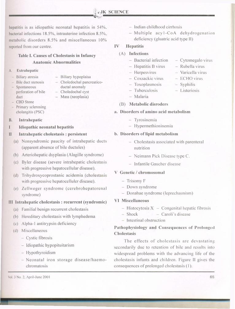
Various causes of cholestasis are given in table I
(5). The spectruln of cholestasis of infancy seen in our
centre is like this: neonatal hepatitis 620/0, extra-hepatic
biliary atresia (EHBA) 30%, choledochal cyst 60/0 and
paucity of intrahepatic ducts 20/0. Etiology of neonatal
64 Vol. 3 No.2, April-lune 2001
______________~':.;~ SCIENCE
Table I. Causes of Cholestasis in Infancy
Anatomic Abnormalities
- Biliary hypoplaisa- Choledochal pancreatico-
ductal anomalyCholedochal cyst
- Mass (neoplasia)
V Gcnetic / chromosomal
Cytomegalo virus
- Rubella virus
Varicella virus
ECHO virus
- Syphilis
- Listeriosis
Bacterial infection
Hepatitis B virus
Herpesvirus
Coxsackie virus
Toxoplasmosis
Tuberculosis
Malaria
Trisomy F
- Down syndrome
Donahue syndrome (leprechaun ism)
VI Miscellaneous
Neimann Pick Disease type C.
Infanti Ie Gaucher disease
b. Disorders of lipid metabolism
- Cholestasis associated with parenteral
nutrition
Tyrosinemia
Hypermeth ion inem ia
(B) Metabolic disroders
a. Disorders of amino acid mctabolism
- Indian childhood cirrhosis
Multiple acy I-CoA dehydrogenation
deficiency (gluatric acid type II)
IV Hepatitis
(A) Infections
- Histocytosis X - Congenital hepatic fibrosis
- Shock - Caroli's disease
Intestinal obstruction
Pathophysiology and Consequenccs of P"olongl't1
Cholestasis
The effects of cholestasis are devastating
secondarily due to retention of bile and results into
widespread problems with thc advancing life of the
cholestasis infants and childrcn. rigure II gives the
consequences of prolonged cholestasis (I).
- Cystic fibrosis
- Idiopathic hypopituitarism
- Hypothyroidism
- Neonatal iron storage disease/haemo-
chromatosis
hepatitis is as idiopathic neonatal hepatitis in 54%,
bacterial infections 18.5%, intrauterine infection 8.5%,
metabolic disorders 8.5% and miscellaneous 10%
reported from our centre.
A. Extrahepatic
- Biliary atresia- Bile duct stenosis- Spontaneous
perforation of bi Ieduct
- CBO Stone- Primary sclerosing
cholangitis (PSC)
B. Intrahepatic
I Idiopathic neonatal hepatitis
II Intrahepatic cholestasis : persistent
(a) Nonsyndromic paucity of intrahepatic ducts
(apparent absence of bile ductules)
(b) Arteriohepatic dsyplasia (Alagille syndrome)
(c) Byler disease (severe intrahepatic cholestasis
with progressive hepatocellular disease).
(d) Trihydroxycoprostanic acidemia (cholestasis
w.ith progressive hepatocelluler disease).
(e) Zellweger syndrome (cerebrohepatorenal
syndrome)
III Intrahepatic cholestasis : recurrent (sYlldromic)
(a) Familial benign recurrent cholestasis
(b) Hereditary cholestasis with lymphedema
(c) Alpha-I antitrypsin deficiency
(d) Miscellaneous
Vol. 3 NO.2. I\pril-June 200 I 65

Figure II - Consequences of Chronic Cholestasis
f
o
c
Reduction in biledelivery to gut
tDecreased Intraluminalbile salt concentration
tM
- I .ljalabsorpti\. h
a nutritIOn Fat Dlarr oeaGrowth Soluble Calcium lossretardation Vitamin
deficiencyA-Night blindnessD-MetaboliC bone diseaseK-HypoprothrombinemiaE-NeuromuscuJar degeneration
have increased unconjugated bilirubin also. This i~
possibly due to associated hemolysis or due to the
hepatocellular injury leading to compromised
conjugation. Recently, delta bilirubin or bili-protein
has been fractionated. The presence of large quantity
of this bilirubin denotes prolonged cholestasis. The
estimation of this in the cord blood or in the newborn
may suggest the intrauterine insult.
Retention of bile salts is responsible for
pruritus possibly that is a serious symptom of
cholestasis. It is very difficult to manage. Initially
this may not be prominent but after the age of6
months cholestatic baby starts scratching. This rna)
become unremitting feature later on.Retention ofbile
salts also results into injury to various biological
membranes of the body. In Iiver, production of
secondary bile acid like lithocholic acid hastens
Portal Hypertension
Bile retentionRegurgitation Biliary Cirrhosis
Bile 'dd, --=::::::::: Pm"tu, .. ./ IHep'tutux",ty / / t
Cholesterol----+_ XanthomatosisBilirubin _ JaundiceCopper _ Hepatotoxicity
- '~______________\J~!K SCIENCE
~11>,*1f
The retention of bile salts and conjugated
hyperbilirubinemia are the hallmarks of NCS. In
hepatocellular cholestasis (Neonatal hepatitis) the
conjugated bilirubin effluxes directly from the
hepatocytes by diffusion or vesicular exocytosis,
whereas in case ofobstructive cholestasis the bile and
conjugated bilirubin from canalicular and ductular
spaces effluxes back through weakened tight junctions
and goes into the blood. It is loosely bound to albumin,
hence gets excreted into urine. The excess ofexcretion
of conjugated bilirubin and bile salts are responsible
for dark coloured urine that stain the diaper in
cholestatic children. At the same time less of bile
production and less of excretion into the biliary tree
resulting negligible bile into the intestine. The
enterohepatic circulation is also effected considerably.
In cholestatic infants, it has been seen that they
66 Vol. 3 No_ 2, April-June 2001

'~'~·~ll\~JK SCIENCE
-----------------~~ti~f
hepatocytic membrance injury and enhances
hepatic fibrosis. Red blood cells may get
hemolysed resulting hemolytic anemia. In
respiratory tract, injury to mucus membrane leads
to asthma like picture. Nasal bleeds are also very
common.
Hyperlipidemia IS characteristic of
cholestasis. Metabolic degradation and excretion
of cholesterol are affected. It hampers the
function of hepatocytic and canalicular
membranes and cholestasis increases. Deposition
of cholesterol in the skin leads to formation of
xanthomas on the body.
Major clinical effects of cholestasis are poor
growth of the infants. This is due to
malabsorption, poor nutrient utilisation,
hormonal disturbances and secondary tissue
injury. Malabsorption is due to lack of bile into
the small bowel resulting inefficient absorption
of fats and fat soluble vitamins. There is loss of
significant calories in the stools in the children.
Simultaneously there is loss of calcium due to
Ca++soap formation with fats which are lost in
the stools. In presence- of vitamin D deficiency,
this leads to development of rickets later on. In
chronic cholestasis, during late infancy and early
childhood, features of vitamin E deficiency in
form of neuropathy and hemolysis develop. If
untreated, can lead to crippling neuromuscular
weakness 'and patient becomes bed ridden .
. Vitamin D deficiency results into rickets and
os teo pen ia . Vi tam inA de fi c ienc y Ie ads to
blindness and hyperkeratotic skin. Vitamin K
Vol. 3 No.2, April-June 200 I
deficiency is responsible for coagulopathy and
bleeding and may have linkage with reduced brain
development (1,3,4,5).
Approach to Cholestasis to differentiate Neonatal
Hepatitis and EHBA.
In NCS, it is mandatory to differentiate b~tween
neonatal hepatitis and EHBA. Neonatal hepatitis
warrants medical treatment whereas obstructive
cholestasis largely EHBA needs only surgical
treatment and is effective if done within 60 days of
life. There should not be any kind of delay to find the
underlying cause responsible for cholestasis.
Clinically, one should be able to pick up cases
where there is high index of suspicion of EHBA. If
the baby is passing pale or acholic stools from very
beginning or starts after few weeks of life one should
.act very fast to make the diagnosis of EHBA'.There is
no point in wasting tinle. In EHBA, fibrosis sets in as
early as 4 weeks of life. EHBA babies are usually tenn
born and have good weight. 20% of these babies may
have associated congenital malformations. Liver
function tests at times may not help to differentiate.
In case ofobstructive cholestasis'l alkaline phosphatase
and Gamma GT Inay be very high in comparison to
neonatal hepatitis. Liver enzylnes ·like ALT and AST
are nearly normal in EHBA but are always rasied in
NH. One should keep in mind that in case of severe
cholestasis there is overlapping picture. In EHBA,
prolonged PTI usually reponds to vitarn in K
adtninistration.
The approach to cholestasis to differentiate
between obstructive cholestas· " ( d hepatocellular
cholestasis is given in algorithn .
67

>
-----------------l:;~ SCIENCE
ALGORITHM-I TO DIFFERENTIATE INTRAHEPATIC AND EXTRAHEPATIC CHOLESTASIS
I Ultrasound
II
Choledochal cyst
Surgery
INo choledochal cyst
IGallstones
(TPN. drugs. hemolytic cause)
,...---1-No CBO stone CBO obstruction
IERCP
Conservative
HEPATOBILIARY SCINTIGRAPHY
IPIBO
IObserve
Neonatal hepatitis
IObserve
EHBA
Intestinal excretion
I (rules °T EHBA)
MRCP Intrahepatic cholestasls
LIr- I N_e_o_n_a_ta..Jl hepatitis
LIVER BIOPSYI
INo intestinal excretion
IEquivocal finding
__1__-
Age < 6 wks Age> 6 wks
IBRID,\ scan ERCP
1-disualizationofPOfNO CBO
Rcpe~1t bx I(10-11 D) -
B"'", (t." \ EHBA
defined U II L\~ c()fwtalrulcd olll Ikpatitis
IEHBAcontirmcd
KASAl'S procedure
Mechanical obstruction Biliary tree patent
sludge. stone cleared JL W,dge Biopsy
Close and observe
6X Vol. 3 10. 2. April-June 2001

""-h<1 <",JK SCIENCE
--------------~'!l!\l:f~------------------
Ultrasound
Ultrasound done by an experienced person is a good
modality to see the status of liver parenchyma, dilated
intrahepatic or extrahepatic biliary tree anfl presence of
gallbladder. Conditions like choledochal cyst, bile plug
syndrome, common bile duct (CBD) stone and Caroli's
disease can be picked up with great accuracy. The
presence or absence of gallbladder in light of normal
intrahepatic radicles and non-visualisation ofCBD does
110t rule out EHBA. Earlier, the presence of gallbladder
was considered to be in favour of NH but in severe
cholestasis GB may not be seen. Absence ofgall bladder
has been correlated with EHBA with low sensitivity and
specificity (60-70%). But there is significant overlap.
Even in severe cholestasis gallbladder may not be defined
because of less production of bile and it may be
hypoplastic. Inspite of problems with US examination,
it is mandatory to rule out other obstructive lesions
as mentioned earlier. Moreover, this is a non-invasive
test (6).
Scintigraphy
HIDA scan is now becoming as investigation going
out offashion like it happened earlier with Rose Bangle
excretion test. In severe cholestasisdue to NH there may
not be excretion ofdye even after adequate priming with
phenobarbitone for 5 days. In case where excretion of
dye is seen, this favours the diagnosis ofNH but does
not rule out severe cholestasis due to NH and EHBA.
However, EHBA is ruled out ifdye is seen in duodenum.
[n absence of exeretion of dye, it creates confusion
whether one is dealing with EHBA or severe
hepatocellular cholestasis. The sensitivity is very high
to pick up severe cholestasis whereas specificity to pick
up EHBA is very low i. e. 60-70%. In some centres this
investigation is not done routinely since there is wastage
of 6-7 days period.
Vol. 3 No.2. April-June 200 I
Magnetic Resonance Cholangiopancreatography
(MRCP)
This IS newer modality but again has similar
problems as we have seen in case of US. False positivity
rate is very high but certainly where it defines gallbladder
and CBD, it rules out EHBA.
Liver Biopsy
This is mandatory to do liver biopsy and
histopathology report can be available within 2-3 days.
But for interpretation, there is need ofexpert pathologist
who is familiar with developing neonatal liver and then
reaction ofvarious toxic factors like infections, metabolic
and obstructive insults. General pathologist can not do
the justice. In best hand, histopathology can differentiate
N Hand EHBA up to the tune of 95%. But in 5% cases
there can be overlap problems to label.
EHBA is characterised by presence ofproliferation
of interlobular ducts, plugged with bile casts and portal
tracts show fibrosis. This liver parenchyma may be
normal and may show intrahepatocytic or canalicular
cholestasis. But in advanced cases after 2 months of life,
there may be full fledged changes of secondary cirrhosis.
If the biopsy is done early between 4-6 weeks of life, the
changes classical ofEHBA may be less prominent, hence
repeat biopsy after 10-14 days is warranted to be sure
about diagnosis of EHBA.
In neonatal hepatitis there is marked parenchymal
injury suggesting focal necrosis, ballooning
degeneration, gaint cell transformation, inflammatory
infilterate, pseudoacinar formation and portal tract may
show mild portal triaditis. There is no fibrosis until the
disease is chronic.
Diagnosis ofPIBD can be made on histology if the
ratio ofpresence of bile ducts to portal tracts is less than
0.4-0.6. But liver biospy should contain minimum 5
69

______________~t"(',JK SCIENCE
portal tracts to make the diagnosis of PIBD in a biopsy
specimen.
Ifthe results ofbiospy are equivocal (5%) and age
is less than 6 weeks, BRIDA scan and or repeat liver
biopsy after 10-14 days should be done. Even if the
diagnosis is not estabilished by liver biopsy and the age
is more than 6 weeks ERCP is indicated.
Endoscopic Retrograde Cholangiopancreatography
(ERCP)
Problem with ERCP is technical failure and non
ayailability of small diameter ERCP scopes in most of
centres.
Percutaneous Transhepatic Cholangiography (pTC)
This is done by injecting dye in dilated intrahepatic
biliary radicles (IHBR) and then visulisation of
extrahepatic biliary tree in antegrade manner. This is
not routinely done since the IHBR are not dilated in
EHBA.
Laparoscopy
Laparoscopic visualisation ofhepatobiliary area has
not been popularised in children but some experts are
attempting it.
Duodenal Intubation
Aspiration of duodenal fluids for 24 hours to see
for bile is procedure in Japanese centres whereas it has
been accepted by others. This can be done during
scintigraphy to define the radioactivity in stomach and
duodenal fluids.
Intraoperative cholangiography (IOC) or
Peroperative cholangiography (POC).
Explorative laparotomy is indicated in very small
percentage of cases where diagnosis is not established
with above modalities. In presence of gall bladder IOC
showing dye in duodenum a~d after clamping the CBD
70
showing dye in intrahepatic radicles rules out EHBA.
There is advantage oftaking wedge biopsy ofliver. Incase
there is EHBA, Kasai's postoenterostomy can be done
simultaneously (6,7,8). The results of investigations to
pick up EHBA in a prospective study done by Lai el. al.
(7) are given in Table II.
Table II
Investigations
* LFT Alkaline phosphatase, Gamma GT, OT/PT
Diagnostic accuracy (Lai et ai, 1994)
.:. Presistent clay 60-80%coloured stools
.:. DoudenaI juice 90-92%
.:. Ultrasound 78-80%
.:. Hepatic scan (HIDA) 80-91%
.:. Liver Biopsy 92-97%
.:. ERCP 90%
.:. Final diagnosis 94-97%
.:. JOC 100%
Approach to Neonatal Hepatitis
Neonatal hepatitis is most important cause ofNCS
(60-70%). It is mandatory to record detail history
regarding antenatal, natal and postnatal events, family
history, exposure to various drugs, maturity ofthe baby,
neonatal sepsis, intrauterine infections, various metabolic
and genetic disorders. Thorough clinical examination is
warranted. The clue to the etiological diagnosis should
come from good clinical evaluation of the case. This
should guide the clinician to decide which way to
investigate. Infections in our set up are very important
cause of neonatal hepatitis. These include bacterial, viral.
protozoal and spirochaetal infections (algorithm H). If
there is direct clue to some metabolic or a genetic
disorder, the investigations should be done accordingl).
If the infections have been ruled out reasonably, the next
choice is to do metabolic work up. High index of
VoL 3 No.2, April-June 2001

~~JK SCIENCE
-----------------~I"----------------------..t{
suspicion to think of these disorders should be based
upon certain clinical pointers like family history of
previous sib, death due to similar disoder, repeated
hypoglycemia, seizures, vomiting, failure to thrive,
cataract etc. Preliminary metabolic work up includes
urine for reducing substances viz. galactose or fructose,
serum alpha-I anti-trypsin level, thyroid function tests,
serum aminoacids, urine aminoacid screening, eye
examination, urinary succinyl acetone, serum ferritin etc.
Based upon the suspected diagnosis the specific enzyme
estimation/genetic work up should be done.
Inspite of elaborate work, in 30-40% cases of
neonatal hepatitis, the etiology can not be defined. This
group is labelled as idiopathic neonatal hepatitis or gaint
cell hepatitis. Liver histology shows marked giant cell
reaction. Liver biopsy also gives clue towards metabolic
disorders at times (3,5,9).
Treatment
Neonatal Hepatitis
Infections constitute major causative agents for
neonatal hepatitis in developing countries. Bacterial
infections must be treated very effectively. Urinary tract
infection remains hidden infection in neonatal period and
it should be diagnosed and treated energetically. Viral
infections persay don't require any specific therapy in
this age group but protozoal infections like malaria and
toxoplasmosis and congenital syphilis as mentioned in
the algorithm II should be treated effectively.
ALGORITHM-II APPROACH TO NEONATAL HEPATITIS
Cholestasiscontinues
ICongenitalSyphilis
ITreat withPenicillin
~~Cholestasisresolves
Negative
Protozoal*Toxoplasmosis*Malaria*Listeriosis
Cholestasiscontinues
Positive
INFECTION
I
Cholestasisresolves
Negative Treataccordingly
I
IViral
*CMV*Rubella*Herpes*Hepatitis B*AIDS
nPositiveif any
Iobserve/treataccordingly
Cholestasis----I ~onsider metabolic work up 1 ----'continues .
Infectionlikely
I
Consider changeof antibiotics
I
ICholestasiscontinues
Bacterial
Treat withantibiotics
ICholestasisresolves
Infectionfound
I
ICholestasisresolves
Vol. 3 No.2, April-June 200 I 71

<i\1C,JK~S~C~I;.EN~C~E;.. _
---------------~Jrf
Obstructive Cholestasis
Table III EHBA : Age of Presentation at (n-36) PGI
Treatment of various metabolic disorders should
be started at the earliest. The offending agent should be
withdrawn promptly for example in case ofgalactosemia,
milk should be stopped immediately to avoid effect on
the developing brain. This is the commonest metabolic
disorder encountered in our centre. In case of
fructosemia, fructose containing food items must be
withdrawn immediately. Treatment of various
endocrinologic and metabolic disorders should be done
accordingly. Genetic counselling and need of the
antenatal diagnosis should be stressed in the affected
families (10).
a
m
th
M
P
Liver Transplantation
This shows that diagnosis ofEHBA should be done
at the earliest and surgery should be performed within
60 days oflife. Best time is 4-6 weeks of life.
[nspite of advancement in surgical skills
the outcome is not encouraging. Even after
portoenterostomy, 1/3 cases deteriorate in perioperative
period and first year of surgery and may require liver
transplanation, 1/3 develop complications of liver disease
during first decade of life and require liver transplantation
whereas 1/3 survive beyond 10 years of life with
abnormal liver functions.
One year survjval repol1ed is varying from 30-71%.
The highest survival rate is reported by Japanese workers.
One year survival in our country is 25-30%. This shows
that surgery is not fool proof treatment and needs liver
transplantation. Good prognostic factors of EHBA
surgery are : surgery done under 60 days of Iife, minimal
or no histology defect, good bile flow after surgery and
absence of cholangitis in immediate post-operative or
first year of Iife and availabil ity of surgical expertise. In
our set up, late presentation of the cases (Table Ill) and
cholangitis are the main deterimental reasons for the
bleak outcome of these cases.
Choledochal cyst during infancy is also very
important cause of cholestasis (6%) and needs surgical
treatment ( 11,13).
Liver transplantation has revolutionised the
outcome of EHBA worldover (14). The indications are:
failed Kasai's procedure, progressive liver disease inspite
of successfu I Kasai's procedure and late presentation of
EHBA (unoperated). Ten years survival is 85-90% in
various centres. In our country, this has not picked up
because of lack of awareness, poverty, ignorance, over
population and absence ofcadaveric liver transplantation
progra111me.
} 83.4%
Numberr (%)
6 (16.6)
8 (22.2)
22 (61.2)
13 (36.1)
9 (25)
2-3
>3
1-2
3-6
6-12
Extrahepatic Biliary Atresia (EHBA)
EHBA is a big challenge world-over, but it is more
alarming problem in developing countries. This
constitutes 30% of the NCS seen at our centre (11). The
late presentation of the disease is responsible for
development ofcirrhosis. This is a stage when it becomes
untreatable and death is inevitable within 2 years oflife.
The age of presentation of EHBA cases reporting at our
centre is given in table Ill. Bile flow can be established
in 80-90% cases after Kasai's procedure
(portoenterostoI11Y) if done within 60 days of life (12).
With the advancing age the bile flow decreases. If the
surgery is done within 2-3 months, the bile flow can be
cstabl ished in 40-45% cases whereas if surgery is done
after 3 months ofage, the bile flow can be established in
10-20% of cases only.
Age in months
72 Vol. 3 No.2. April-June 2001

A______________~~:~~K SCIENCE
Medical Treatment of Cholestasis
Chronic cholestasis is responsible for various life
threatening consequences which need prolonged therapy.
Pruritus
Pruritus is a most distressing symptom. It leads to
miserble life in term oflack ofsleep, emotional problems
and children become mentally reackoned. Various
treatment modalities in form of use of cholestyramine,
phenobarbitone, rifampicin, terfenadine, UCDA and
phototherapy have been tried with variable resu Its.
UDCA seems to be promising as it is one of choleretic
drug. Some times untreatable and unremitted pruritus
becomes sole indication for liver transplantation (15).
Malnutrition
Malnutrition is very common and is due to obvious
reasons mentioned in the Fig. IV. Breast feeding should
be encouraged in these babies. If anorexia is a prominent
feature nasogastric feeding is indicated. The diet should
have 200 calories/kg and protein 1-2 g/kg body
weighLThe diet should be constituted by MCT, 2-3%
calories from PUFA, carbohydrates (glucose polymers),
minerals, trace elements and vitamins. MCT rich
available diets are coconut oil, Simyl MCT, Pregestimil
and Portagen. All vitamins should be given in double
the daily requirement.
Vitamin A
Vitamin A should be given 2500-5000 IU/day.
Monitor the vitamin level. Ifblood level is less than30ug!
dl increase the oral dose by 10 folds or 50,000IU 1M
monthly should be given.
Vitamin D
Daily 400-1200 IU ofvitamin D are recommended.
This can be given in form of 40,000 IU 1M monthly. 25
hydoxycholecalciferol 5-7 Ilg/kg can be given dai Iy.
Vol. 3 No.2. April-June 200 I
Monitor serum calcium, phosphate and alkaline
phosphatase and X-ray wrist at 2 months interval.
Vitamin E
Vitamin E deficiency is now recognised very
oftenly since the age of the children with cholestasis is
increasing. The dose of vitamin E recommended is
15-200 mg daily. Serum monitoring is mandatory. If
levels are on lower side, then higher dose should be given.
Six monthly neurological and yearly eye examination
are required.
Vitamin K
In case of prolonged cholestasis with steatorrhea
vitamin K 5mg 1M monthly should be given. Treatment
of other compl ication of Iiver disease Iike ascites, pOtial
hypertension, variceal bleed and encephalopathy should
be done accordingly (10).
To summarise the scenario of NCS In India is
disappointing at present because of late referral of the
. cases to centres where facilities to diagnose metabolic
diseases and EHBA are available. This has been shown
in table III. More than 80%of EHBA cases come for
treatment when they have already crossed 2 months of
age as shown in table III when it is difficult to establish
the bile flow. The mistake is at various levels statiing
from undergraduate training to postgraduate training in
pediatric medicine, unawareness about the seriousness
of the problem, wastage of time on making diagnosis
and treatment. These are not correctly practiced in
peripheral hospital/medical colleges hence delay in
referral of these cases. The algorithm III shows reasons
for later referral of EHBA in India. There is lack of
surgical expetiise also to handle EHBA. We don't have
the facilities of liver transplanation in our country. Cost
factor and non-availability of cadaveric liver are main
hinderances. In this context parents can help by giving
piece of their liver to own produced baby. Hence, there
73

.\' .... JK SCIENCE
------------~~----------------is need of "Yellow Alert" nation wide to detect these
cholestatic babies and early referral to well equipped
tertiary care centre where facilities to handle these babies
are available. There is need for anlndian efforts to detect
cholestatic babies, to make early diagnosis and prompt
treatment to avoid occurence of end stage liver disease
given in algorithm IV.
References
I. Whitington PI. Chronic cholestasis ofinfancy. PedCIiIl.~~
Am 1996; 43: 1-26.
2. Klein S. Nealon WHo Hepatobiliary abnormalities associatewith total parenteral nutrition. Semin Liver Di~' 1988.8237-46.
3. Shah HA. Spivak W. Neonatal cholestasis: Ncw approachto diagnostic evaluation and therapy. Ped Clin .\'or.11994: 41 : 943-66.
Algorithm-III
• Physiological Jaundice: Phototherapy
• Breast Milk Jaundice: Herbal Medicine
• Lack of facilities
6. Paltiel 111. Imaging of Nconatal cholestasis. SemUltrasound. CT and AiRI 1994 : 15 : 290-305.
7. Lai MW. Chang MH. Hsu SC. Ilsu I-Ie. Cheng TS. KaoelLce CY. Differcntail diagnosis ofcxtrahcpatic biliar) aIr.from neonatal hepatitis a prospective study. J PediaGastroenterol NutI' 1994. 18 : 121-217.
4. McEvoy CF, Suehy RJ. Biliary tract diseasc in children.Pe('lin Nor Am 1996 : 43 : 75-98.
5. Balisteri WF. Neonatal cholestasis. J Pediatr 1985 : 106171-83.
8. Mowat A, Pascharopoulos. William R. Extrahepatic bilia~
atresia versus neonatal hepatitis. Arch Dis (,hild 1976: il763-70·
All Doctors I+'
Doctors to treat Jaundice
LATE REFERRRAL OF EHBA IN INDIAHealthy Baby
Ignorance of parents .. 1 ~ ILocal Doctor or G.P...........
Late referral to Tertiary Care Centre
t
9. Motala e. Ireland .10. I-lill !D. Bowie MD. Cholestati.disorders of infancy-etiology and outcomc. J Trap Pedla'1990: 36: 218-22
Algorithm-IV
NEED FOR AN INDIAN EFFORTCHOLESTASIS OF INFANCY
(High coloured urine, diaper staining, clay coloured stool& conjugated S. bilirubin> 20%)
~Significant Hepatobiliary Disease
....------::-+':..-.., ,.. r-+:....-----.Urgent Early Prevent
Diagnostic Specific Morbidity
Evaluation Therapy Mortality
• Prompt referral to Tertiary care centrePediatric Gastroenterology, Hepatology &Nutrition
• Era of LIVER TRANSPLANTATION
10. Thapa BR. Role of nutrition in liver and biliary disorde~
In : Nutrition in children. Developing country concern:Sachdev I-1PS. Choudhary P. (eds.) First edition. Cambridg.Press. Kashmere gate New Delhi. 1994: pp 376-89.
11. Chhabra M. Poddar U. Thapa SR. Singh K. Spectul11 0
cholestasis of infancy. Ind.J Gastroenterol 1996 : Ii(Suppl. I) A 73.
12. Kasai M, Kimura S. Asakura Y et al. Surgicaltreatmentobiliary atresia J Pediatr Surg 1968 : 3 : 665-75 .
13. Poddar U. Thapa BR. Chhabra M. Rao KLN. Mitra SK. Sin.K. Choledochal cysts in inlilllts and children Ind Pedial1998: 35: 613-18.
14. WhitinglOn PF. Balisteri WF. Livcr transplantation I
pediatrics: Indication, contraindicalions and prctransplanmanagcmenl. J Pediatr 1991 : 118 : 169-77.
15. l3ergasa NY. Jones EA. Management ofpruri(us ofchoieslasisPotential role of opiate antagonists. Am J Gastroen/ero1991: 86: 1404-12.
74 Vol. 3 No.2, April-June 2001


![[2015] post lt cholestasis](https://static.fdocuments.net/doc/165x107/58ee0ee21a28ab92198b4665/2015-post-lt-cholestasis.jpg)