Chapter 8: The Appendicular Skeleton. The Appendicular Skeleton Figure 8–1.
8/8/2019 Ch 8 - Appendicular Skeleton
http://slidepdf.com/reader/full/ch-8-appendicular-skeleton 1/27
Chapter 8:The Appendicular Skeleton
8/8/2019 Ch 8 - Appendicular Skeleton
http://slidepdf.com/reader/full/ch-8-appendicular-skeleton 2/27
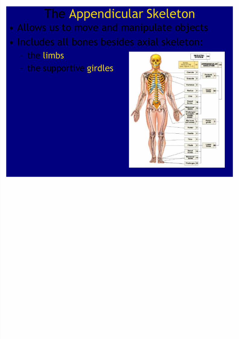
The Appendicular Skeleton Allows us to move and manipulate objects
Includes all bones besides axial skeleton:² the limbs
² the supportive girdles

8/8/2019 Ch 8 - Appendicular Skeleton
http://slidepdf.com/reader/full/ch-8-appendicular-skeleton 3/27
The Pectoral Girdle
Figure 8²2a
Also called the shoulder
girdle Connects the arms to the
body
Positions the shoulders
Provides a base for armmovement
Consists of:
² 2 clavicles
² 2 scapulae
Connects with the axial skeleton only at the
manubrium

8/8/2019 Ch 8 - Appendicular Skeleton
http://slidepdf.com/reader/full/ch-8-appendicular-skeleton 4/27
The Clavicles
Figure 8²2b, c
Also called collarbones
Long, S-shaped bones
Originate at the manubrium (sternal end)
Articulate with the scapulae (acromial end)
8/8/2019 Ch 8 - Appendicular Skeleton
http://slidepdf.com/reader/full/ch-8-appendicular-skeleton 5/27
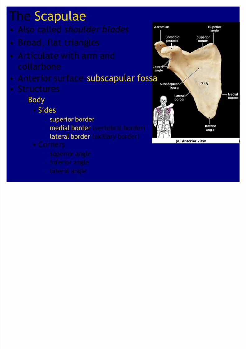
The Scapulae Also called shoulder blades
Broad, flat triangles Articulate with arm and
collarbone
Anterior surface-subscapular fossa
Structures² Body
Sides² superior border
² medial border (vertebral border)² lateral border (axillary border)
Corners² superior angle
² inferior angle
² lateral angle

8/8/2019 Ch 8 - Appendicular Skeleton
http://slidepdf.com/reader/full/ch-8-appendicular-skeleton 6/27
² Head
Holds glenoid cavity
² Which articulates with humerus to form shoulder joint
Coracoid process-anterior, smaller Acromion-posterior, larger

8/8/2019 Ch 8 - Appendicular Skeleton
http://slidepdf.com/reader/full/ch-8-appendicular-skeleton 7/27
² Scapular spine:
ridge across posterior surface of body
Separates 2 regions:
² supraspinous fossa
² infraspinous fossa
8/8/2019 Ch 8 - Appendicular Skeleton
http://slidepdf.com/reader/full/ch-8-appendicular-skeleton 8/27
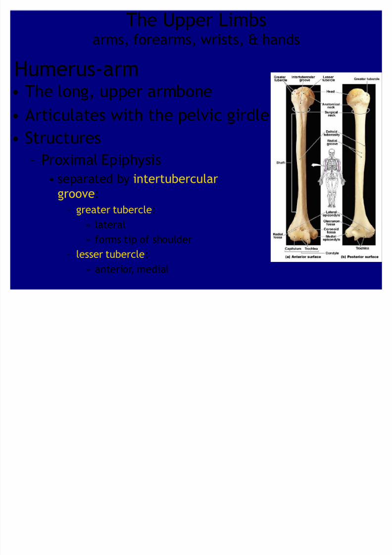
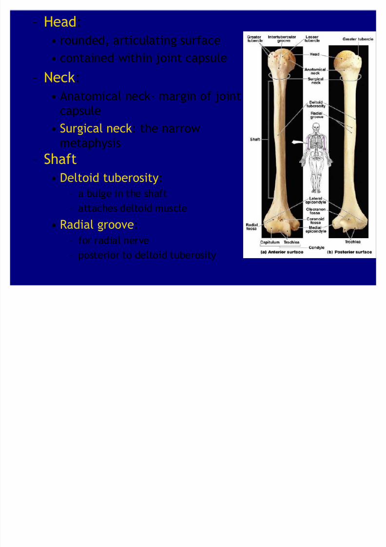
The Upper Limbsarms, forearms, wrists, & hands
Humerus-arm The long, upper armbone
Articulates with the pelvic girdle
Structures² Proximal Epiphysis
separated by intertubercular
groove:
² greater tubercle:
» lateral
» forms tip of shoulder
² lesser tubercle:
» anterior, medial
8/8/2019 Ch 8 - Appendicular Skeleton
http://slidepdf.com/reader/full/ch-8-appendicular-skeleton 9/27
² Head:
rounded, articulating surface
contained within joint capsule
² Neck:
Anatomical neck- margin of joint
capsule
Surgical neck: the narrow
metaphysis
² Shaft
Deltoid tuberosity:
² a bulge in the shaft
² attaches deltoid muscle
Radial groove:
² for radial nerve
² posterior to deltoid tuberosity

8/8/2019 Ch 8 - Appendicular Skeleton
http://slidepdf.com/reader/full/ch-8-appendicular-skeleton 10/27
² Distal Epiphysis
Medial and lateral epicondyles:
² for muscle attachment
Condyle of the humerus:
² Trochlea:
» coronoid fossa & olecranon fossa
» articulates with ulna
² Capitulum:» radial fossa
» articulates with radius
8/8/2019 Ch 8 - Appendicular Skeleton
http://slidepdf.com/reader/full/ch-8-appendicular-skeleton 11/27
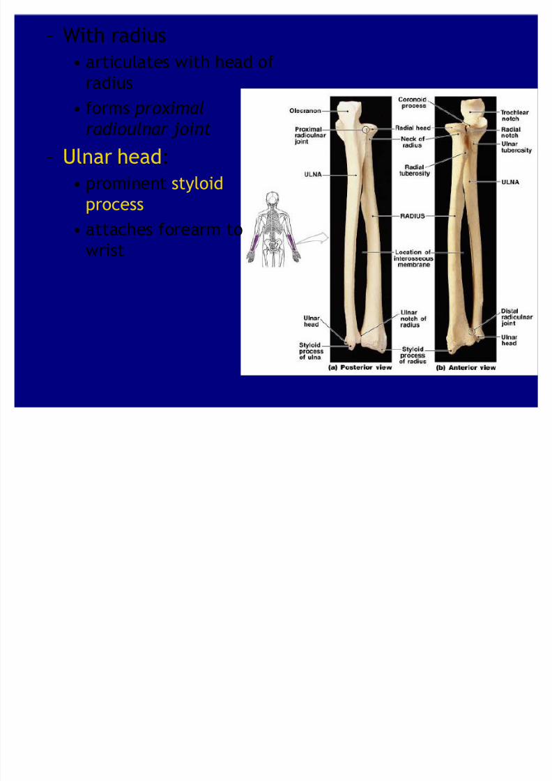
² With radius
articulates with head of
radius
forms pr ox imal
radioulnar joint
² Ulnar head:
prominent styloid
process
attaches forearm to
wrist

8/8/2019 Ch 8 - Appendicular Skeleton
http://slidepdf.com/reader/full/ch-8-appendicular-skeleton 12/27
The Forearm-antebrachium Consists of 2 long bones: ulna & radius
Ulna Articulations
² With humerus
Forearm e xt
ended :olecranon (elbow
point) enters
olecranon fossa
Forearm fle x ed :
coronoid processenters coronoid fossa
8/8/2019 Ch 8 - Appendicular Skeleton
http://slidepdf.com/reader/full/ch-8-appendicular-skeleton 13/27
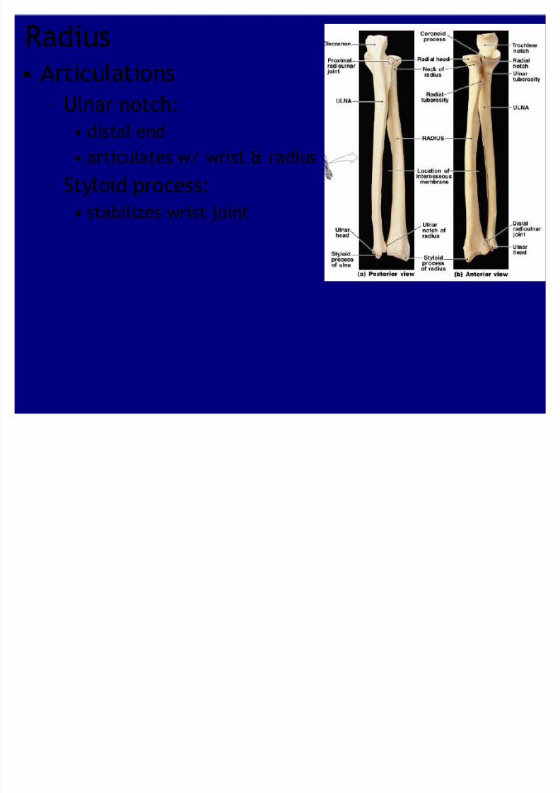
Radius Articulations
² Ulnar notch: distal end
articulates w/ wrist & radius
² Styloid process: stabilizes wrist joint
8/8/2019 Ch 8 - Appendicular Skeleton
http://slidepdf.com/reader/full/ch-8-appendicular-skeleton 14/27
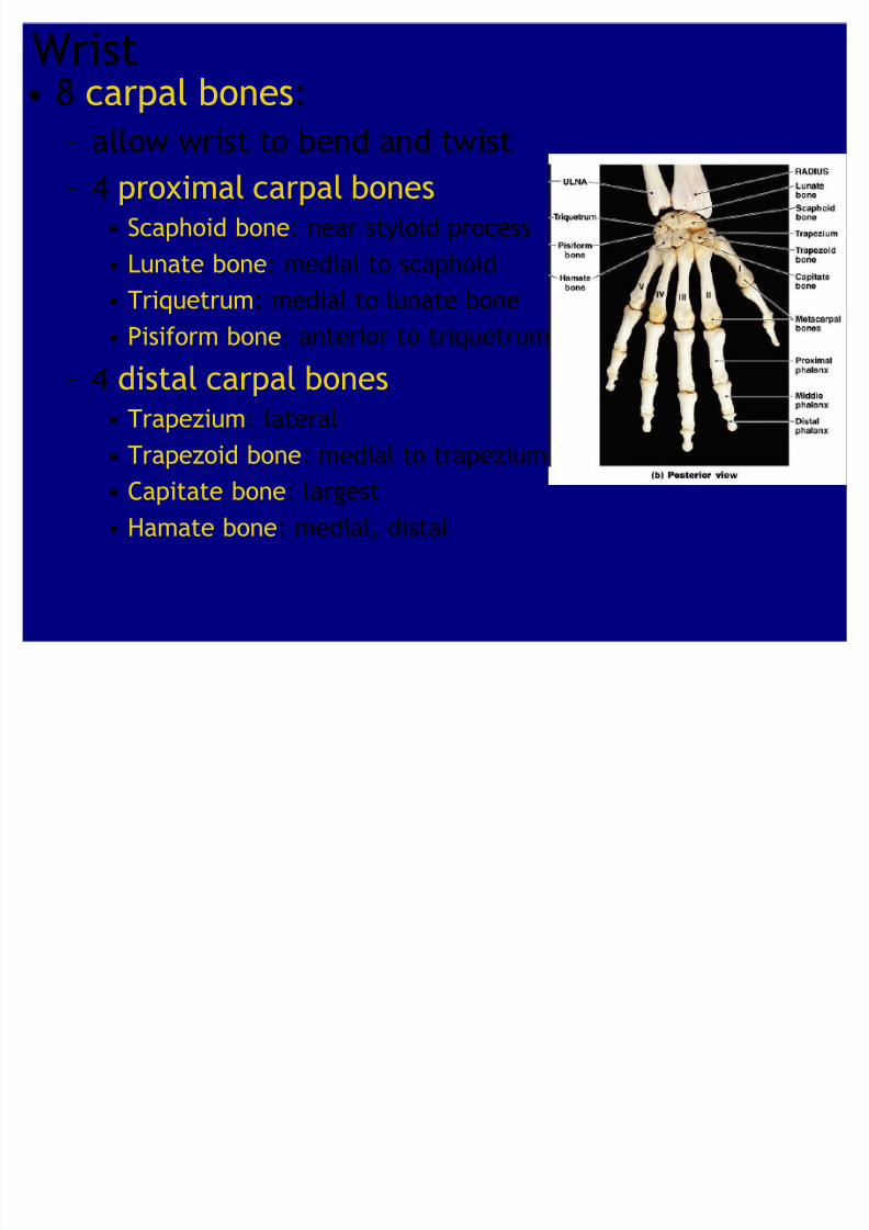
Wrist 8 carpal bones:
² allow wrist to bend and twist² 4 proximal carpal bones
Scaphoid bone: near styloid process
Lunate bone: medial to scaphoid
Triquetrum: medial to lunate bone Pisiform bone: anterior to triquetrum
² 4 distal carpal bones Trapezium: lateral
Trapezoid bone: medial to trapezium Capitate bone: largest
Hamate bone: medial, distal

8/8/2019 Ch 8 - Appendicular Skeleton
http://slidepdf.com/reader/full/ch-8-appendicular-skeleton 15/27
5 Metacarpal Bones
² long bones of the hand² Numbered I²V from lateral
(thumb) to medial
² Articulate with proximal
phalanges
Phalanges
² Pollex (thumb): 2 phalanges (proximal,
distal)
² Fingers:
3 phalanges (proximal,middle, distal)
Hands
8/8/2019 Ch 8 - Appendicular Skeleton
http://slidepdf.com/reader/full/ch-8-appendicular-skeleton 16/27
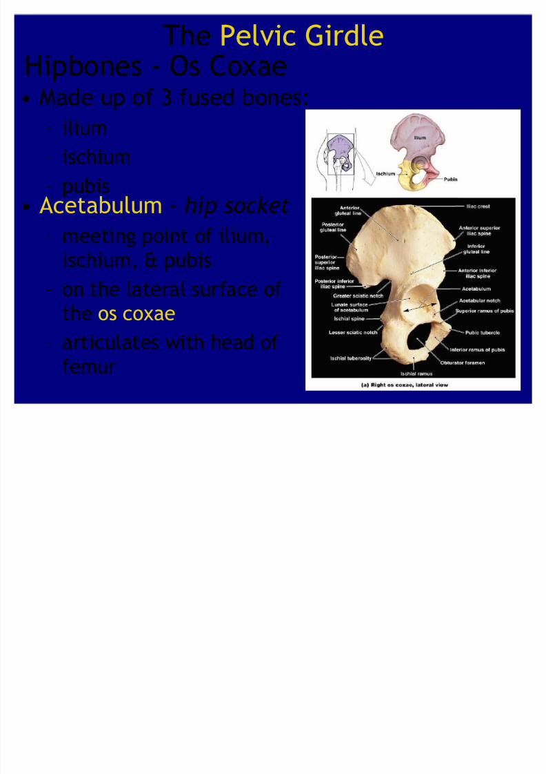
The Pelvic GirdleHipbones - Os Coxae
Made up of 3 fused bones:² ilium
² ischium
² pubis Acetabulum - hi p socket
² meeting point of ilium,
ischium, & pubis
² on the lateral surface ofthe os coxae
² articulates with head of
femur
8/8/2019 Ch 8 - Appendicular Skeleton
http://slidepdf.com/reader/full/ch-8-appendicular-skeleton 17/27
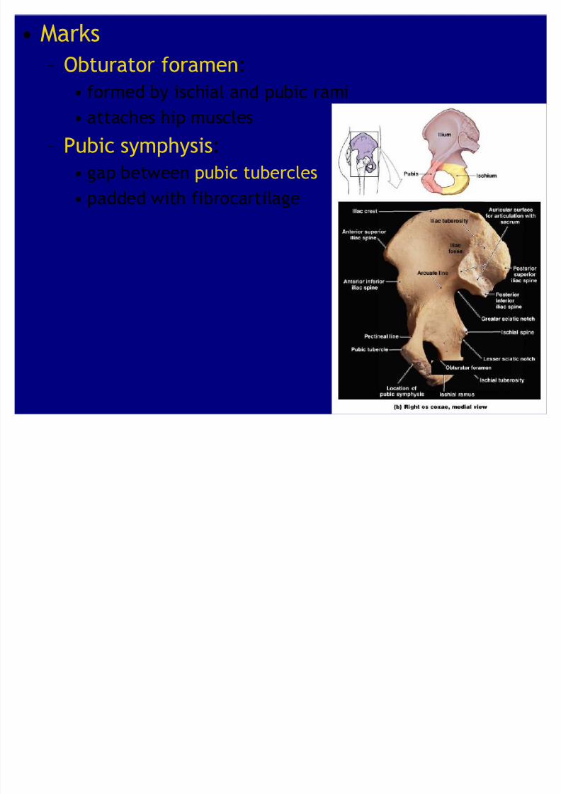
Marks
² Obturator foramen:
formed by ischial and pubic rami attaches hip muscles
² Pubic symphysis:
gap between pubic tubercles
padded with fibrocartilage
8/8/2019 Ch 8 - Appendicular Skeleton
http://slidepdf.com/reader/full/ch-8-appendicular-skeleton 18/27
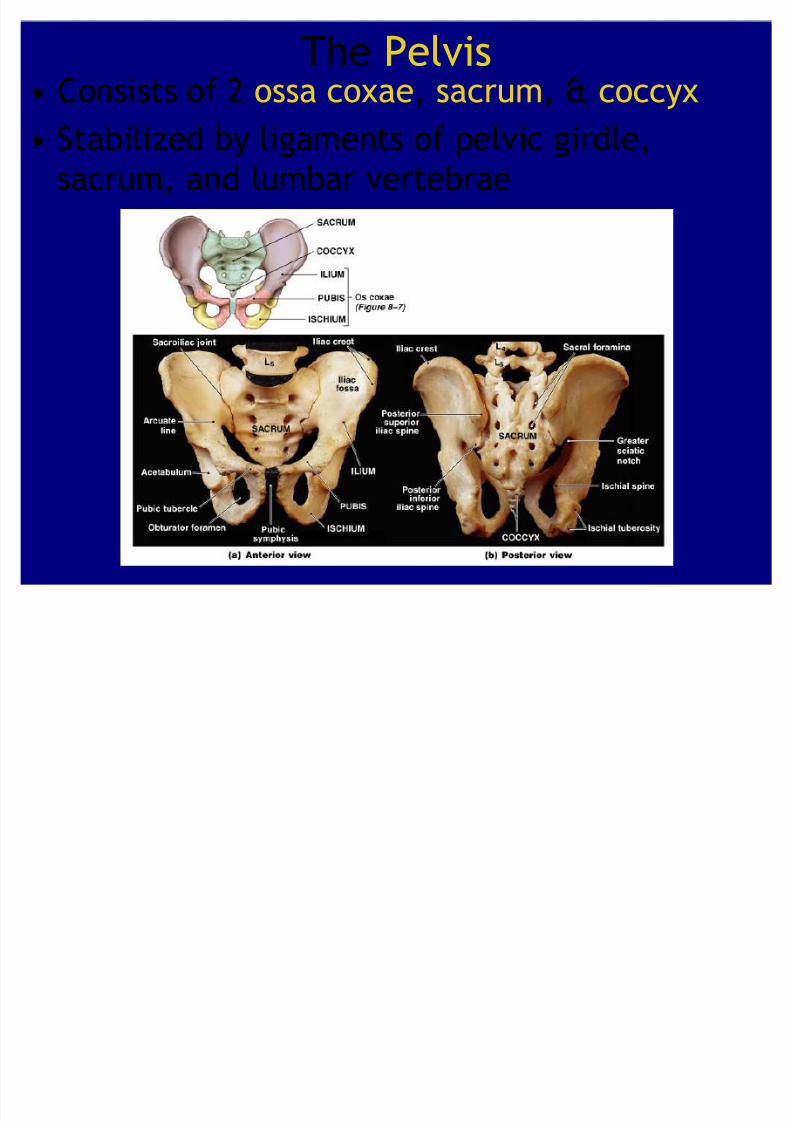
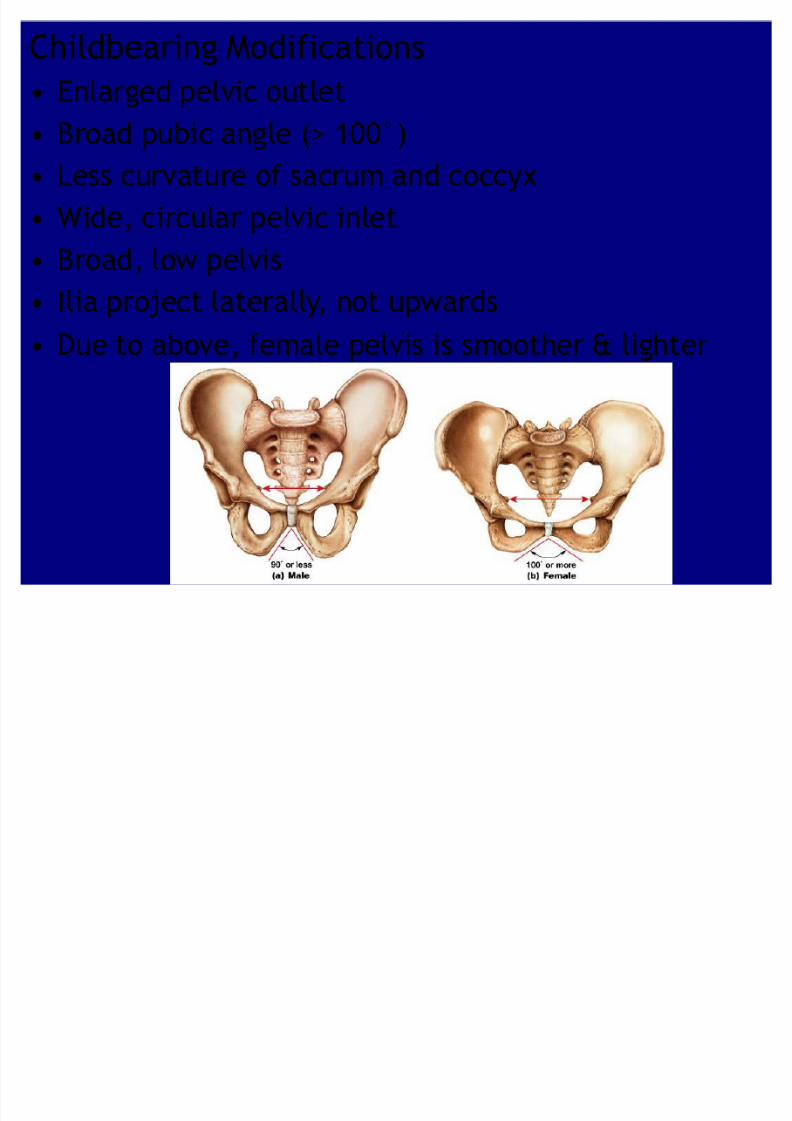
The Pelvis Consists of 2 ossa coxae, sacrum, & coccyx
Stabilized by ligaments of pelvic girdle,sacrum, and lumbar vertebrae
8/8/2019 Ch 8 - Appendicular Skeleton
http://slidepdf.com/reader/full/ch-8-appendicular-skeleton 19/27
8/8/2019 Ch 8 - Appendicular Skeleton
http://slidepdf.com/reader/full/ch-8-appendicular-skeleton 20/27
8/8/2019 Ch 8 - Appendicular Skeleton
http://slidepdf.com/reader/full/ch-8-appendicular-skeleton 21/27
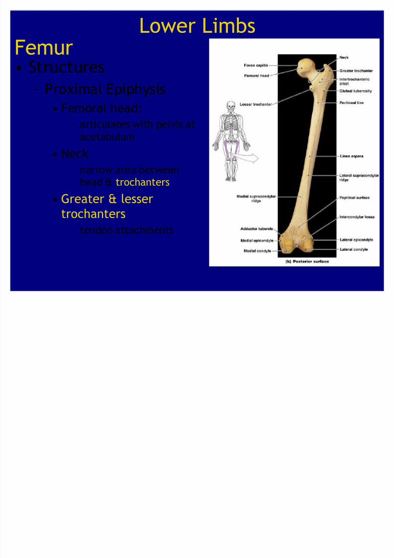
Lower LimbsFemur
Structures² Proximal Epiphysis
Femoral head:
² articulates with pelvis at
acetabulum Neck
² narrow area between
head & trochanters
Greater & lesser
trochanters
² tendon attachments
8/8/2019 Ch 8 - Appendicular Skeleton
http://slidepdf.com/reader/full/ch-8-appendicular-skeleton 22/27
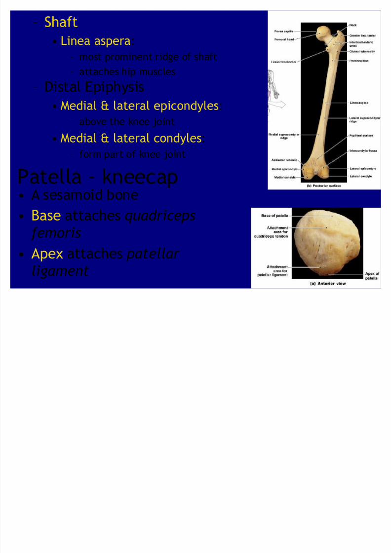
² Shaft
Linea aspera:
² most prominent ridge of shaft
² attaches hip muscles
² Distal Epiphysis
Medial & lateral epicondyles:
² above the knee joint
Medial & lateral condyles:² form part of knee joint
Patella - kneecap A sesamoid bone
Base attaches q uadrice ps
femoris
Apex attaches patellar
ligament

8/8/2019 Ch 8 - Appendicular Skeleton
http://slidepdf.com/reader/full/ch-8-appendicular-skeleton 23/27
Tibia
Figure 8²13
Structures
² Proximal Epiphysis Medial and lateral tibial
condyles
articulate with medial & lateral
condyles of femur Tibial tuberosity: attaches
patellar ligament
² Shaft
Anterior margin: sharp ridge ofshinbone² Distal Epiphysis
Medial malleolus: medial projection at ankle
8/8/2019 Ch 8 - Appendicular Skeleton
http://slidepdf.com/reader/full/ch-8-appendicular-skeleton 24/27
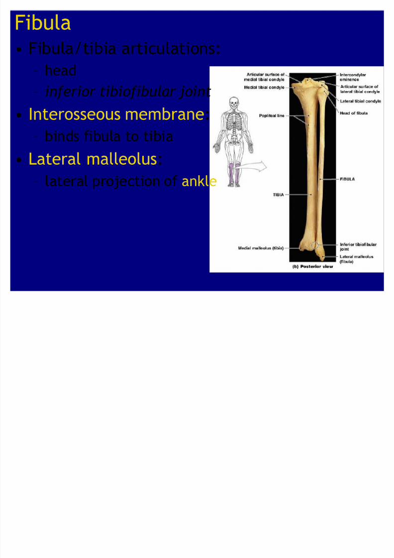
Fibula Fibula/tibia articulations:
² head² in ferior tibio fibular joint
Interosseous membrane:
² binds fibula to tibia Lateral malleolus:
² lateral projection of ankle

8/8/2019 Ch 8 - Appendicular Skeleton
http://slidepdf.com/reader/full/ch-8-appendicular-skeleton 25/27
Ankle - tarsus consists of 7 tarsal bones
Figure 8²14a
² Talus carries weight from tibia
across trochlea
² Calcaneus (heel bone):
transfers weight from talus to
ground & attaches Achilles
tendon
² Cuboid bone: articulates with
calcaneus² Navicular bone: articulates with talus and
3 cuneiform bones
² Medial cuneiform
² Intermediate cuneiform
² Lateral cuneiform
8/8/2019 Ch 8 - Appendicular Skeleton
http://slidepdf.com/reader/full/ch-8-appendicular-skeleton 26/27
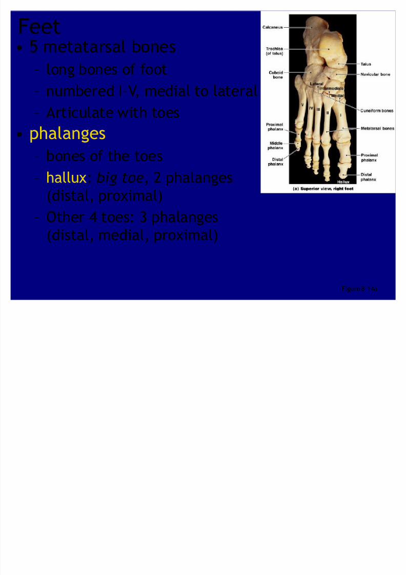
Figure 8²14a
5 metatarsal bones
² long bones of foot² numbered I²V, medial to lateral
² Articulate with toes
phalanges
² bones of the toes
² hallux: big toe, 2 phalanges
(distal, proximal)
² Other 4 toes: 3 phalanges(distal, medial, proximal)
Feet
8/8/2019 Ch 8 - Appendicular Skeleton
http://slidepdf.com/reader/full/ch-8-appendicular-skeleton 27/27
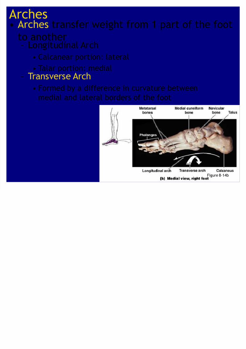
Arches Arches transfer weight from 1 part of the foot
to another
Figure 8²14b
² Longitudinal Arch Calcanear portion: lateral
Talar portion: medial² Transverse Arch
Formed by a difference in curvature betweenmedial and lateral borders of the foot