Annals 2/Gilmour - Bryozoa
19
Paper in: Patrick N. Wyse Jackson & Mary E. Spencer Jones (eds) (2011) Annals of Bryozoology 3: aspects of the history of research on bryozoans. International Bryozoology Association, Dublin, pp. viii+225.
Transcript of Annals 2/Gilmour - Bryozoa
Paperin:PatrickN.WyseJackson&MaryE.SpencerJones(eds)(2011)AnnalsofBryozoology3:aspectsofthehistoryofresearchonbryozoans.InternationalBryozoologyAssociation,Dublin,pp.viii+225.
17THE HISTORY OF ENTOPROCT RESEARCH
The history of Entoproct researchand why it continues
Judith FuchsDepartment of Zoology, University of Gothenburg, Box 463, 40530 Göteborg, Sweden
1. Introduction2. Early findings3. Description of new genera4. A natural group called Entoprocta5. Controversy over entoproct systematics6. Dawning of a new age7. A new generation of entoproct researchers8. Fossils and Australian entoprocts9. Turning point – a new phylum, molecular tools and more morphology10. Conclusion
1. Introduction
The history of entoproct research encompasses a period of more than 250 years. Theobservations on the taxon were naturally influenced and restricted by the technicalinstruments and methods that were available at certain times. Early researchers weremuch challenged by investigating the miniature entoprocts with only a magnifying glassor a simple light microscope. First details of entoproct morphology were discovered whenlight microscopes became an established tool. Basic studies about entoproct reproduction,life history, and anatomy were made in the years between 1880 and 1960. The use ofsectioning techniques in combination with scanning electron microscopy was notestablished in entoproct research until the late 1960s, when ultrastructural details ofcertain body parts as tentacles, digestive tract, or attachment structure were uncovered. Inthe 1990s, molecular studies were initiated to evaluate lophotrochozoan and metazoanrelationships; however, only few studies were designed to resolve entoproct evolution.Entoproct research at the beginning of the 21st century can be characterized by (1)investigations of the anatomy (e.g., neuromuscular system) by applied electron microscopyand immunocytochemistry combined with confocal microscopy, (2) the first cladisticanalyses including entoproct morphological characters, and (3) on-going research of theentoproct molecular body plan.
18 ANNALS OF BRYOZOOLOGY 3
It is probably not an overstatement to remark that Entoprocta are amongst the mostenigmatic animals on our planet. Questions concerning the systematics of the taxon areas old as its first discovery and they have continuously been applied to entoproct researchthroughout the centuries. The question now is “how far have we come?” and instead ofgiving an answer at this point, the following review will shed some light on this issue.
2. Early findings
To determine when research on entoprocts began, one has to go back to the literatureof the late 18th century. In John Ellis’ work Natural history of the Corallines and othermarine productions of the like kind published in 1755, it is intriguing to read the author’sdedication to the Princess Dowager of Wales, which includes the following:
The minutest works of creation are not always the least wonderful: and though the element, ofwhich these are natives, prohibits an exact enquiry, yet enough of order, contrivance, andregularity, appear in their structure, to convince us, they are the workmanship of that Almightypower, whom, exalted as you are in dignity of sentiment, as well as station, you think it yourhighest honour to reverence and obey.
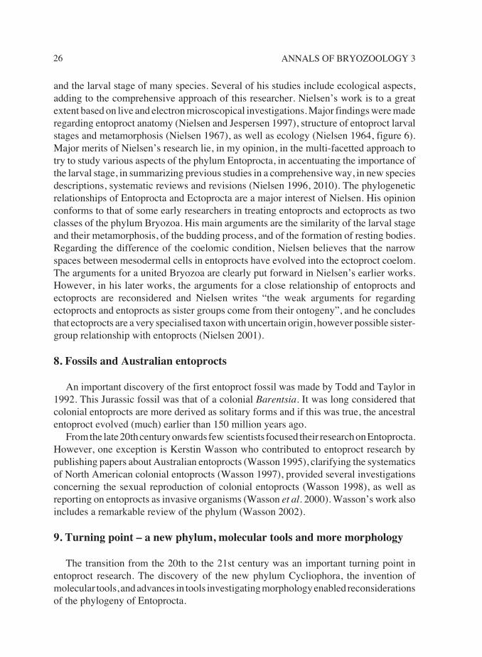
Although the idea of the Almighty has by now rather disappeared from the minds ofmodern biologists, many would probably agree that they share with Ellis and other earlybiologists the sudden emotional excitement when the astonishing complexity of a livingorganism is observed. Modern zoologists also share with early biologists that this initialexcitement subsequently leads to a more detailed observation of the organism and theparts it is made of. Although the short description and drawings of the “polyps of a red
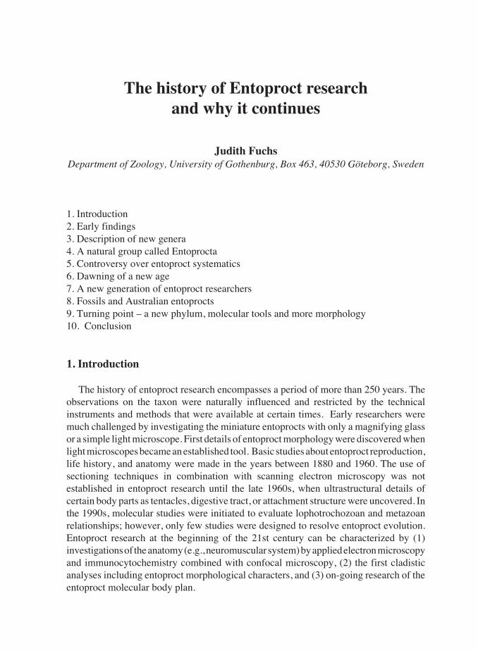
Figure 1. First drawings of a colonial entoproct growing on a hydroid stem (from Ellis, 1755).
19THE HISTORY OF ENTOPROCT RESEARCH
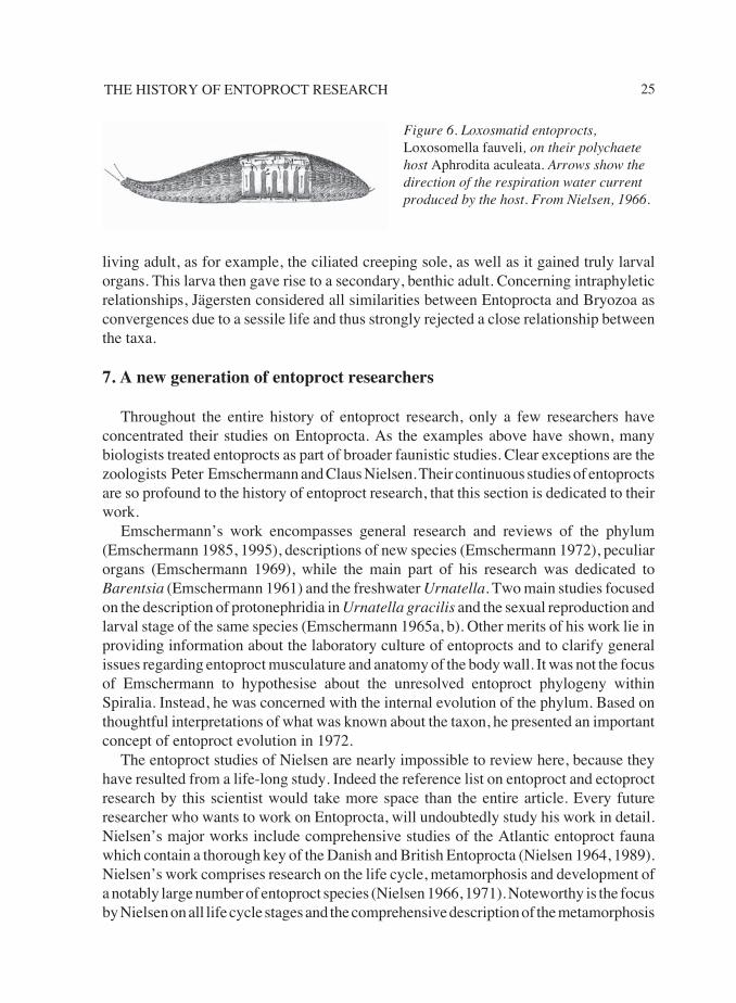
colour and a particular kind” in Ellis’s book are not much more than a passing comment,they are considered the earliest report of an entoproct in literature (Figure 1). It was laterconfirmed that Ellis’s organism matches that of the colonial entoproct genus Pedicellina.
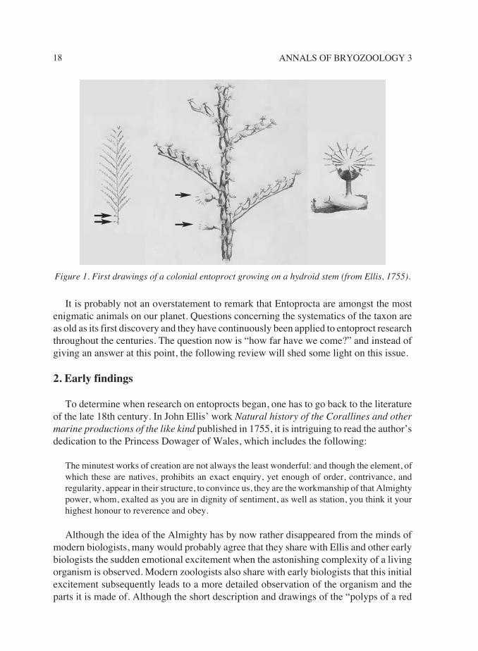
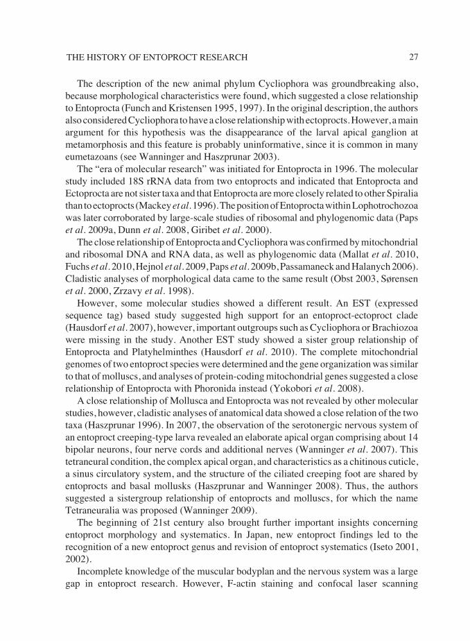
Some years later, Pallas (1774, 1778) published the first recognizable, and somewhatlonger description of a Pedicellina species together with a drawing in his “Novel andstrange animal species”. However, Pallas himself identified the entoproct species as therotifer Brachionus cernuus, a kind of a strange, marine polyp, related to hydroids (Figure2A).
In the following years, several biologists made entoproct findings in nature, however,little focus was placed on their thorough description. Observing these small creatures indetail was still impossible due to the lack of modern light microscopes. In Bosc’s “Naturalhistory of worms” from 1830, a drawing and description of a Hydre jaune appears, whichlater also was suggested to be an entoproct, and Lister in 1834 devoted an entire page ofdescription to a zoophyte (clearly an entoproct) and noted its characteristic bendingmotion. Lister referred to Ellis’s description and presented a more detailed drawing.Milne-Edwards connected the descriptions of Ellis, Lister, and Bosc, and stated that“Hydre jaune” (Hydra coronata in Fleming, 1828) is a polype and certainly not a Hydraand he proposed a new genus for it, which he named “Lusie (Lusia)” (Deshayes and Milne-Edwards 1836).
The problem in these early years of entoproct research was how to determine thetaxonomic position (and name) of these strange animals in respect to where they shouldbe placed in the tree of life. This dilemma is reflected in the work of Lister for example,who wrote “the species seemed to be intermediate between such animals of Flustra as Ihad met with, and the pedunculated compound Ascidia...”. A zoophyte, closely resemblingthat described by Lister, was observed by Sharpey, and in 1836 he described the feedingmechanism of this zoophyte which he illustrated (Figure 2B).
The first entoproct genus, Pedicellina, was erected by Sars in 1835, who described twospecies P. echinata (now considered to be P. cernua) and P. gracilis (now Barentsiagracilis). The genus name he derived from the shape and movement of the animals, whichreminded the author of the pincer-like appendages pedicellariae of some echinoderms.Sars stated in his description, that the general form and position of the digestive tract ofPedicellina was similar to that of Vorticella, and the tentacles resembled those of otherpolyps. For him it seemed that Pedicellina was the link between Infusoria (now mostly
Figure 2. Pedicellina. A. Drawing by Pallas (1778). B. Drawings from Sharpey (1836),showing a colony and the calyx of a specimen to describe the feeding mechanism.

20 ANNALS OF BRYOZOOLOGY 3
Protista) and the polyps. In the following year, Wiegmann stated that the description ofPedicellina would define them as a naked bryozoan, and not Vorticella (Wiegmann 1836).
Generally, in these early years of biological research, a systematic understanding ofinvertebrate animals was being developed and this was a chaotic, but fruitful process.Within this changing hierarchy, the position of Pedicellina (the name Entoprocta was notyet invented) was unclear and the insecurity regarding the position of these strangeanimals was expressed by almost all researchers. An example which illustrates thedynamic development of the invertebrate system in the early 19th century is that Johnstonin the first edition of his book British Zoophytes (Johnston 1838) mentioned the“entoproct” described by Lister (1834, see above) in the division Zoophyta Ascidioida andthe family Vesiculariadae. In the second edition of this book published nine years later in1847, the same entoproct can be found instead in the class Polyzoa (together withectoprocts) and within its own family Pedicellinae. Note that the name Polyzoa was firstcoined in 1830 by John Vaughan Thompson who included only ectoproct species.
3. Description of new genera
Van Beneden (1845) gave the first detailed description of an entoproct. He investigatedthe species Pedicellina belgica (now Barentsia gracilis) and although he was not able tosee the nervous system, he could observe the general hermaphroditism in entoprocts, andthe cleavage and development of the larva of this species. The author stated that the naturalrelationship of Pedicellina was neither in the Vorticelles nor in the ascidians, but in theBryozoa instead. Note that the name Bryozoa was introduced in 1831 by Ehrenberg whodid not include entoprocts.
Subsequently observations of “Pedicellina” (Pedicellina and Barentsia according torecent taxonomy) were made in different regions. When a larval stage of a solitaryentoproct was found, it was erroneously described as a “new animal form” under the nameCyclopelma longociliatum (Busch 1851).
A new “entoproct” genus (the name Entoprocta was still not invented) was describedfor the first time from a freshwater habitat near Philadelphia by Leidy in 1851, when heintroduced the freshwater species Urnatella gracilis. He described the characteristiccolonies of Urnatella comprising erect, articulated stalks growing from a commonattachment base, without seeing the actual soft body parts, which he observed in laterstudies. However, Leidy suspected Urnatella belonged to the Polyzoa. Allman (1856)understood the necessity of searching for homologies instead of analogies, in order to findthe allies of entoprocts. He explained similarities of entoprocts to other Polyzoa and alsomentioned their uniqueness. Allman included Urnatella and Pedicellina in the Polyzoa(which he decided contained the two orders Phylactolaemata and the Gymnolaemata).Urnatella was tentatively included in the Gymnolaemata and Pedicellina in thePhylactolaemata. He realised the similarities between Urnatella and Pedicellina butdecided the taxonomic position of Urnatella had to be provisional due to the lack ofsufficient anatomical details of the species.
21THE HISTORY OF ENTOPROCT RESEARCH
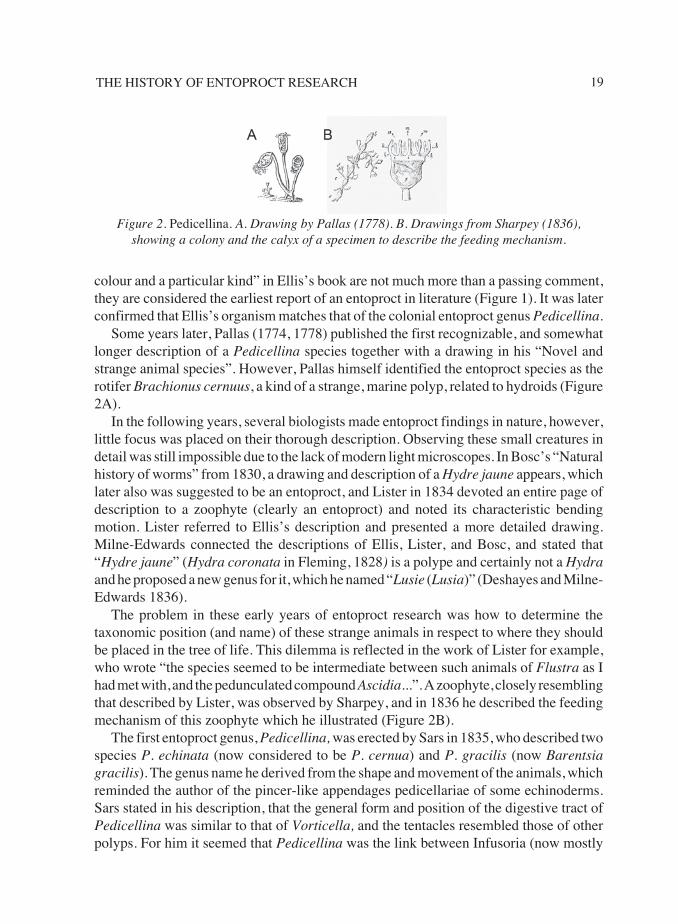
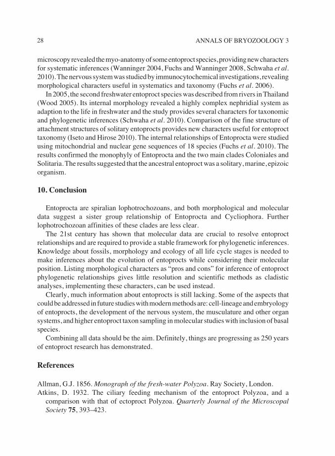
The findings of the 19th century weresignificantly expanded with theobservation of a third entoproct genus,the solitary Loxosoma, which we knownow, constitutes the most species-richgroup of Entoprocta, and most species arefound in epi-symbiotic relationships withother invertebrates. Nevertheless, in theoriginal description (Norman 1861), theauthor did not realize Loxosoma was anindependent organism but described themas posterior, club-shaped appendages ofthe “new echinoderm Strephenterusclaviger” instead. (According to thedescription, the echinoderm is asipunculid, a common host for somesolitary entoprocts).
Later publications on entoprocts dealtwith the description of new species,observations of different life cycle stagesof Loxosoma, and included observations
of the developmental biology of entoprocts (e.g., Uljanin 1869). One of the early drawingsof a Loxosoma species is shown in Figure 3.
4. A natural group called Entoprocta
An important step in the history of entoproct research was made with the publicationsof Nitsche (1869, 1875). Nitsche reviewed much of the previous work and his ownpublications are augmented with useful anatomical illustrations. Through previous andhis own studies Nitsche concluded that the genera Urnatella, Pedicellina, and Loxosomawere a natural grouping. He split them off from Bryozoa and proposed the nameEntoprocta, leaving the taxon Ectoprocta as a separate group (Bryozoa sensu stricto). Thefamily Entoprocta was defined by Nitsche (1869) as “Mouth and anus lie inside thetentacle crown, the anterior part of the body wall is not retractable, thus a tentacle sheathis lacking. The tentacles are arranged in a bilateral manner, not retractable but can just beinfolded.”
In the next few decades researchers investigated entoprocts with much focus onembryonic development and the larval stage, and important observations of budding inentoprocts were made (Nitsche 1875). Barrois and Hatschek investigated the entire group
Figure 3. One of the first drawings of asolitary Loxosoma by Claparède (1863).
22 ANNALS OF BRYOZOOLOGY 3
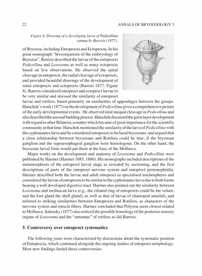
of Bryozoa, including Entoprocta and Ectoprocta. In hisgreat monograph “Investigations of the embryology ofBryozoa”, Barrois described the larvae of the entoproctsPedicellina and Loxosoma as well as many ectoproctsbased on live observations. He observed the spiralcleavage in entoprocts, the radial cleavage of ectoprocts,and provided beautiful drawings of the development ofsome entoprocts and ectoprocts (Barrois 1877: Figure4). Barrois considered entoproct and ectoproct larvae tobe very similar and stressed the similarity of entoproctlarvae and rotifers, based primarily on similarities of appendages between the groups.Hatschek’s work (1877) on the development of Pedicellina gives a comprehensive pictureof the early developmental events. He observed total inequal cleavage in Pedicellina andalso described the asexual budding process. Hatschek discussed the germ layer developmentwith regard to other Bilateria, a matter which became of great importance for the scientificcommunity at that time. Hatschek mentioned the similarity of the larva of Pedicellina withthe cyphonautes larva and he considered entoprocts to be basal bryozoans, and argued thata close relationship between bryozoans and Rotifera could be true, if the bryozoanganglion and the supraesophageal ganglion were homologous. On the other hand, thebryozoan larval form would put them at the base of the Mollusca.
Major works on the development and anatomy of Loxosoma and Pedicellina werepublished by Harmer (Harmer 1885, 1886). His monographs included descriptions of themetamorphosis of the entoproct larval stage as revealed by sectioning, and the firstdescriptions of parts of the entoproct nervous system and entoproct protonephridia.Harmer described both the larvae and adult entoproct as specialised trochospheres andconsidered the larvae of entoprocts to be similar to the cyphonautes larva due to both formsbearing a well developed digestive tract. Harmer also pointed out the similarity betweenLoxosoma and molluscan larva (e.g., the ciliated ring of entoprocts could be the velum,and the foot gland the shell gland), as well as that of larvae of chaetopod annelids, andreferred to striking similarities between Entoprocta and Rotifera, as characters of thenervous system and muscle fibres. Harmer concluded that Polyzoa seem closest relatedto Mollusca. Salensky (1877) also noticed the possible homology of the posterior sensoryorgans of Loxosoma and the “antennae” of rotifers as did Barrois.
5. Controversy over entoproct systematics
The following years were characterized by discussions about the systematic positionof Entoprocta, which continued alongside the ongoing studies of entoproct morphology.Most new findings fueled these controversies.
Figure 4. Drawing of a developing larva of Pedicellinacernua by Barrois (1877).
23THE HISTORY OF ENTOPROCT RESEARCH
In 1888, Hatschek was the first to propose that the Entoprocta were a separate classwithin the lower worms (Cladus Scolecida; a group also containing Rotifera and flatworms sensu lato). Bryozoa (Ectoprocta) were grouped with Brachiopoda and Phoronidain the clade Tentaculata (= Molluscoidea). Hatschek based his view on the similarity ofthe entoproct larva and the trochophora of Mollusca and Annelida. Stiasny (1905), whodescribed the nephridia of Loxosoma and Pedicellina in detail, agreed for the moment withHatschek’s classification, since the structure of entoproct nephridia would strictlyseparate entoprocts from ectoprocts (note: entoprocts have protonephridia, while ectoproctshave no excretory organs). The three main arguments that were put forward at that timein favour of a diphyletic origin of Entoprocta and Ectoprocta, were (1) the position of theanus inside versus outside the tentacle crown, (2) the presence or absence of a body cavity,and (3) protonephridia versus metanephridia. Note that the three arguments are valid untiltoday, except that it has been shown that ectoprocts lack metanephridia.
Hatschek’s opinion, however, was not adopted by several other researchers such asEhlers (1890), Prouho (1832), Davenport (1893), Seeliger (1906), or Cwicklitzer (1909).Their arguments were based on morphological similarities between Entoprocta andEctoprocta. Davenport refuted the three main arguments separating entoprocts fromectoprocts (see above) by (1) the closure of the tentacular corona between mouth and anuswould be late in development, (2) the conditions would be continuous between ento- andectoprocts (spaces in the entoproct parenchym would represent the ectoproct coelom), and(3) “the existence of an excretory tubule” in entoprocts would “at the presence not evenbe probable”. The authors Seeliger and Cwicklitzer focused on the investigation of thelarval stage. Seeliger (1906) concluded that entoprocts were the basal bryozoans. Hisarguments for this statement were the similar budding processes in entoprocts andectoprocts and the similar anatomy of the larvae, especially of the larval nervous systems.
6. Dawning of a new age
In the years between 1920 and 1970, entoproct research reached a new level due tomajor findings regarding early embryology, larval stage, feeding mechanisms, regenerationand budding in entoprocts. Entoprocta were clearly recognized as unique taxon andprevious work was reviewed and re-evaluated. New hypothesys regarding entoproctrelationships were developed. Regarding entoproct taxonomy, three families wererecognized in this period, the solitary Loxosomatidae, the colonial Pedicellinidae (includingPedicellina, Barentsia and some other genera) and the Urnatellidae.
In the early 1920s, a new name for entoprocts, Calyssozoa (meaning “calyx-animals”),was proposed in the “New classification of animals”, to distinguish them better fromBryozoa sensu stricto (= Polyzoa) (Clark 1921). Nevertheless, the similar name Calycozoawas already used for cnidarian stauromedusae and Cori (1929) chose a new name“Kamptozoa” for entoprocts. The latter name refers to the characteristic bending movementof the animals (from the Greek ‘campto’, I bend). Cori argued for a separation ofentoprocts from bryozoans, with the former following the main body plan of lesser worms
24 ANNALS OF BRYOZOOLOGY 3
(Scolecida) on one hand, and Bryozoa sensu stricto (= Ectoprocta)following the common plan of Coelomata on the other hand.Several reasons for this view were listed by Cori (1936) andcontain acoeli versus coelomate body plan, protonephridia versusunknown state, the ganglion being subesophageal as againstsupraesophageal. One main reason that Cori did not regard Entoprocta as ancestral togymnolaemate bryozoans, as proposed earlier, was that in such a scenario, one wouldexpect to find transitional forms between a sessile acoelomate and a sessile coelomate.Obviously, such a state is not represented. He saw the entoprocts rather, as the mostdeveloped form of Scolecida, and did not exclude the relationship of entoprocts to rotifers.Cori’s two main publications on Entoprocta in 1929 and 1936 are of special value sincethey comprise an elaborate review of previous works combined with the author’s ownstudies. The works cover detailed aspects of the history of research, entoproct reproduction,development, anatomy, regeneration, ecology, taxonomy and phylogenetic perspective.
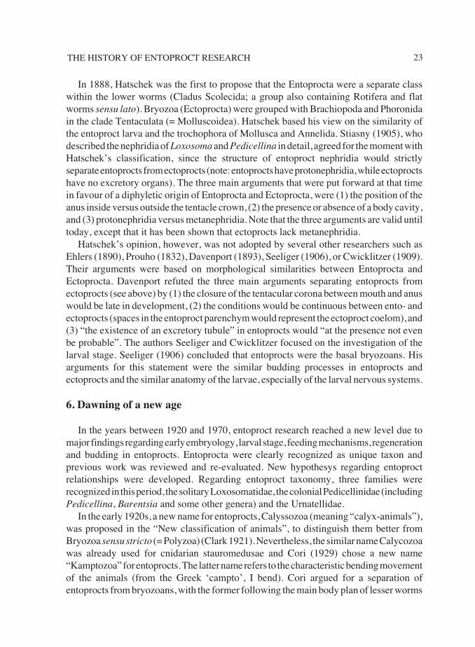
Further studies provided additional support for Entoprocta/Kamptozoa independenceof Bryozoa. The feeding mechanism of entoprocts differs broadly from that of Ectoprocta(Atkins 1932). The intestine in larval and adult Pedicellina does not suggest a closerelationship of entoprocts and ectoprocts Becker (1937). In 1939, Marcus was able toobserve the cleavage and embryology of Pedicellina cernua and his results of the cell-lineage and cell-fate remain the only source of entoproct cell-lineage data until today(Figure 5).
Entoprocts are certainly not missing in Hyman’s great work of invertebrate animals(Hyman 1951). She summarized knowledge about the phylum Entoprocta and rejected thepossibility of a close relationship between entoprocts and ectoprocts due to severalreasons; the condition of no coelom versus coelomic cavity was considered by her to bethe most decisive. Affinities of entoprocts were instead drawn with the pseudocoelomategroups and the closest relatives were thought to be the Rotifera. The latter shared withentoprocts, similarities as glands in a stalk, protonephridia, similarity of parts of thedigestive tract, and Hyman considered the antennae of rotifers homologous with the pairof preoral organs in entoproct larvae and loxosomatid adults, as proposed by earlierinvestigators.
Prenant and Bobin (1956), however, treated entoprocts and ectoprocts as two classesof Bryozoa in their book Faune de France. However, their most useful contribution wasto key the entoprocts in a detailed form with useful drawings and descriptions.
Jägersten (1964, 1972) developed new hypotheses of metazoan evolution. The essenceof his work was to focus on the entire life cycle of animals and his aim was to get moreinsights in metazoan evolutionary relationships through implementing their comprehensivelife-histories. Regarding entoprocts, Jägersten considered their evolution, includingpedomorphosis. The entoproct larval stage retained some features of an ancestral free-
Figure 5. Lateral view of 56-cell stage of P. cernua;cell-lineage study of Marcus (1939).
25THE HISTORY OF ENTOPROCT RESEARCH
living adult, as for example, the ciliated creeping sole, as well as it gained truly larvalorgans. This larva then gave rise to a secondary, benthic adult. Concerning intraphyleticrelationships, Jägersten considered all similarities between Entoprocta and Bryozoa asconvergences due to a sessile life and thus strongly rejected a close relationship betweenthe taxa.
7. A new generation of entoproct researchers
Throughout the entire history of entoproct research, only a few researchers haveconcentrated their studies on Entoprocta. As the examples above have shown, manybiologists treated entoprocts as part of broader faunistic studies. Clear exceptions are thezoologists Peter Emschermann and Claus Nielsen. Their continuous studies of entoproctsare so profound to the history of entoproct research, that this section is dedicated to theirwork.
Emschermann’s work encompasses general research and reviews of the phylum(Emschermann 1985, 1995), descriptions of new species (Emschermann 1972), peculiarorgans (Emschermann 1969), while the main part of his research was dedicated toBarentsia (Emschermann 1961) and the freshwater Urnatella. Two main studies focusedon the description of protonephridia in Urnatella gracilis and the sexual reproduction andlarval stage of the same species (Emschermann 1965a, b). Other merits of his work lie inproviding information about the laboratory culture of entoprocts and to clarify generalissues regarding entoproct musculature and anatomy of the body wall. It was not the focusof Emschermann to hypothesise about the unresolved entoproct phylogeny withinSpiralia. Instead, he was concerned with the internal evolution of the phylum. Based onthoughtful interpretations of what was known about the taxon, he presented an importantconcept of entoproct evolution in 1972.
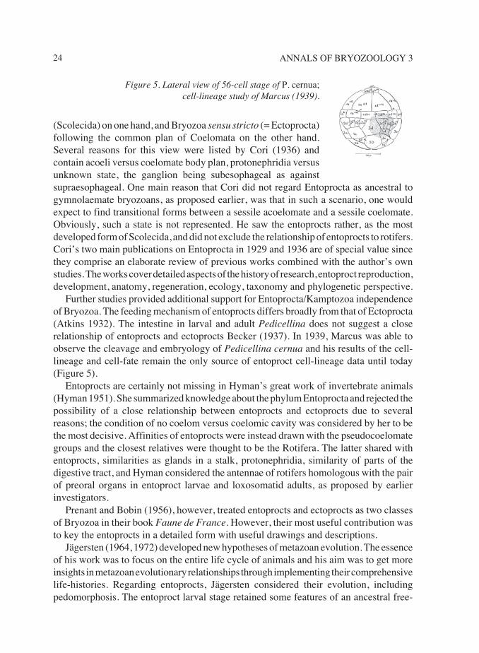
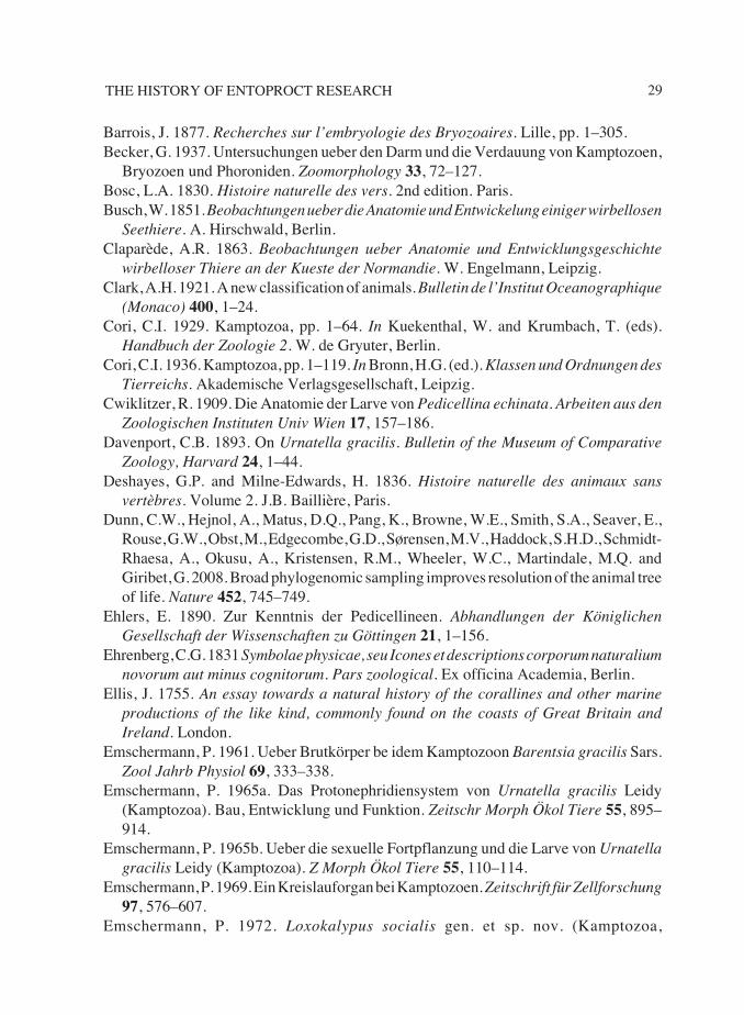
The entoproct studies of Nielsen are nearly impossible to review here, because theyhave resulted from a life-long study. Indeed the reference list on entoproct and ectoproctresearch by this scientist would take more space than the entire article. Every futureresearcher who wants to work on Entoprocta, will undoubtedly study his work in detail.Nielsen’s major works include comprehensive studies of the Atlantic entoproct faunawhich contain a thorough key of the Danish and British Entoprocta (Nielsen 1964, 1989).Nielsen’s work comprises research on the life cycle, metamorphosis and development ofa notably large number of entoproct species (Nielsen 1966, 1971). Noteworthy is the focusby Nielsen on all life cycle stages and the comprehensive description of the metamorphosis
Figure 6. Loxosmatid entoprocts,Loxosomella fauveli, on their polychaetehost Aphrodita aculeata. Arrows show thedirection of the respiration water currentproduced by the host. From Nielsen, 1966.
26 ANNALS OF BRYOZOOLOGY 3
and the larval stage of many species. Several of his studies include ecological aspects,adding to the comprehensive approach of this researcher. Nielsen’s work is to a greatextent based on live and electron microscopical investigations. Major findings were maderegarding entoproct anatomy (Nielsen and Jespersen 1997), structure of entoproct larvalstages and metamorphosis (Nielsen 1967), as well as ecology (Nielsen 1964, figure 6).Major merits of Nielsen’s research lie, in my opinion, in the multi-facetted approach totry to study various aspects of the phylum Entoprocta, in accentuating the importance ofthe larval stage, in summarizing previous studies in a comprehensive way, in new speciesdescriptions, systematic reviews and revisions (Nielsen 1996, 2010). The phylogeneticrelationships of Entoprocta and Ectoprocta are a major interest of Nielsen. His opinionconforms to that of some early researchers in treating entoprocts and ectoprocts as twoclasses of the phylum Bryozoa. His main arguments are the similarity of the larval stageand their metamorphosis, of the budding process, and of the formation of resting bodies.Regarding the difference of the coelomic condition, Nielsen believes that the narrowspaces between mesodermal cells in entoprocts have evolved into the ectoproct coelom.The arguments for a united Bryozoa are clearly put forward in Nielsen’s earlier works.However, in his later works, the arguments for a close relationship of entoprocts andectoprocts are reconsidered and Nielsen writes “the weak arguments for regardingectoprocts and entoprocts as sister groups come from their ontogeny”, and he concludesthat ectoprocts are a very specialised taxon with uncertain origin, however possible sister-group relationship with entoprocts (Nielsen 2001).
8. Fossils and Australian entoprocts
An important discovery of the first entoproct fossil was made by Todd and Taylor in1992. This Jurassic fossil was that of a colonial Barentsia. It was long considered thatcolonial entoprocts are more derived as solitary forms and if this was true, the ancestralentoproct evolved (much) earlier than 150 million years ago.
From the late 20th century onwards few scientists focused their research on Entoprocta.However, one exception is Kerstin Wasson who contributed to entoproct research bypublishing papers about Australian entoprocts (Wasson 1995), clarifying the systematicsof North American colonial entoprocts (Wasson 1997), provided several investigationsconcerning the sexual reproduction of colonial entoprocts (Wasson 1998), as well asreporting on entoprocts as invasive organisms (Wasson et al. 2000). Wasson’s work alsoincludes a remarkable review of the phylum (Wasson 2002).
9. Turning point – a new phylum, molecular tools and more morphology
The transition from the 20th to the 21st century was an important turning point inentoproct research. The discovery of the new phylum Cycliophora, the invention ofmolecular tools, and advances in tools investigating morphology enabled reconsiderationsof the phylogeny of Entoprocta.
27THE HISTORY OF ENTOPROCT RESEARCH
The description of the new animal phylum Cycliophora was groundbreaking also,because morphological characteristics were found, which suggested a close relationshipto Entoprocta (Funch and Kristensen 1995, 1997). In the original description, the authorsalso considered Cycliophora to have a close relationship with ectoprocts. However, a mainargument for this hypothesis was the disappearance of the larval apical ganglion atmetamorphosis and this feature is probably uninformative, since it is common in manyeumetazoans (see Wanninger and Haszprunar 2003).
The “era of molecular research” was initiated for Entoprocta in 1996. The molecularstudy included 18S rRNA data from two entoprocts and indicated that Entoprocta andEctoprocta are not sister taxa and that Entoprocta are more closely related to other Spiraliathan to ectoprocts (Mackey et al. 1996). The position of Entoprocta within Lophotrochozoawas later corroborated by large-scale studies of ribosomal and phylogenomic data (Papset al. 2009a, Dunn et al. 2008, Giribet et al. 2000).
The close relationship of Entoprocta and Cycliophora was confirmed by mitochondrialand ribosomal DNA and RNA data, as well as phylogenomic data (Mallat et al. 2010,Fuchs et al. 2010, Hejnol et al. 2009, Paps et al. 2009b, Passamaneck and Halanych 2006).Cladistic analyses of morphological data came to the same result (Obst 2003, Sørensenet al. 2000, Zrzavy et al. 1998).
However, some molecular studies showed a different result. An EST (expressedsequence tag) based study suggested high support for an entoproct-ectoproct clade(Hausdorf et al. 2007), however, important outgroups such as Cycliophora or Brachiozoawere missing in the study. Another EST study showed a sister group relationship ofEntoprocta and Platyhelminthes (Hausdorf et al. 2010). The complete mitochondrialgenomes of two entoproct species were determined and the gene organization was similarto that of molluscs, and analyses of protein-coding mitochondrial genes suggested a closerelationship of Entoprocta with Phoronida instead (Yokobori et al. 2008).
A close relationship of Mollusca and Entoprocta was not revealed by other molecularstudies, however, cladistic analyses of anatomical data showed a close relation of the twotaxa (Haszprunar 1996). In 2007, the observation of the serotonergic nervous system ofan entoproct creeping-type larva revealed an elaborate apical organ comprising about 14bipolar neurons, four nerve cords and additional nerves (Wanninger et al. 2007). Thistetraneural condition, the complex apical organ, and characteristics as a chitinous cuticle,a sinus circulatory system, and the structure of the ciliated creeping foot are shared byentoprocts and basal mollusks (Haszprunar and Wanninger 2008). Thus, the authorssuggested a sistergroup relationship of entoprocts and molluscs, for which the nameTetraneuralia was proposed (Wanninger 2009).
The beginning of 21st century also brought further important insights concerningentoproct morphology and systematics. In Japan, new entoproct findings led to therecognition of a new entoproct genus and revision of entoproct systematics (Iseto 2001,2002).
Incomplete knowledge of the muscular bodyplan and the nervous system was a largegap in entoproct research. However, F-actin staining and confocal laser scanning
28 ANNALS OF BRYOZOOLOGY 3
microscopy revealed the myo-anatomy of some entoproct species, providing new charactersfor systematic inferences (Wanninger 2004, Fuchs and Wanninger 2008, Schwaha et al.2010). The nervous system was studied by immunocytochemical investigations, revealingmorphological characters useful in systematics and taxonomy (Fuchs et al. 2006).
In 2005, the second freshwater entoproct species was described from rivers in Thailand(Wood 2005). Its internal morphology revealed a highly complex nephridial system asadaption to the life in freshwater and the study provides several characters for taxonomicand phylogenetic inferences (Schwaha et al. 2010). Comparison of the fine structure ofattachment structures of solitary entoprocts provides new characters useful for entoprocttaxonomy (Iseto and Hirose 2010). The internal relationships of Entoprocta were studiedusing mitochondrial and nuclear gene sequences of 18 species (Fuchs et al. 2010). Theresults confirmed the monophyly of Entoprocta and the two main clades Coloniales andSolitaria. The results suggested that the ancestral entoproct was a solitary, marine, epizoicorganism.
10. Conclusion
Entoprocta are spiralian lophotrochozoans, and both morphological and moleculardata suggest a sister group relationship of Entoprocta and Cycliophora. Furtherlophotrochozoan affinities of these clades are less clear.
The 21st century has shown that molecular data are crucial to resolve entoproctrelationships and are required to provide a stable framework for phylogenetic inferences.Knowledge about fossils, morphology and ecology of all life cycle stages is needed tomake inferences about the evolution of entoprocts while considering their molecularposition. Listing morphological characters as “pros and cons” for inference of entoproctphylogenetic relationships gives little resolution and scientific methods as cladisticanalyses, implementing these characters, can be used instead.
Clearly, much information about entoprocts is still lacking. Some of the aspects thatcould be addressed in future studies with modern methods are: cell-lineage and embryologyof entoprocts, the development of the nervous system, the musculature and other organsystems, and higher entoproct taxon sampling in molecular studies with inclusion of basalspecies.
Combining all data should be the aim. Definitely, things are progressing as 250 yearsof entoproct research has demonstrated.
References
Allman, G.J. 1856. Monograph of the fresh-water Polyzoa. Ray Society, London.Atkins, D. 1932. The ciliary feeding mechanism of the entoproct Polyzoa, and a
comparison with that of ectoproct Polyzoa. Quarterly Journal of the MicroscopalSociety 75, 393–423.
29THE HISTORY OF ENTOPROCT RESEARCH
Barrois, J. 1877. Recherches sur l’embryologie des Bryozoaires. Lille, pp. 1–305.Becker, G. 1937. Untersuchungen ueber den Darm und die Verdauung von Kamptozoen,
Bryozoen und Phoroniden. Zoomorphology 33, 72–127.Bosc, L.A. 1830. Histoire naturelle des vers. 2nd edition. Paris.Busch, W. 1851. Beobachtungen ueber die Anatomie und Entwickelung einiger wirbellosen
Seethiere. A. Hirschwald, Berlin.Claparède, A.R. 1863. Beobachtungen ueber Anatomie und Entwicklungsgeschichte
wirbelloser Thiere an der Kueste der Normandie. W. Engelmann, Leipzig.Clark, A.H. 1921. A new classification of animals. Bulletin de l’Institut Oceanographique
(Monaco) 400, 1–24.Cori, C.I. 1929. Kamptozoa, pp. 1–64. In Kuekenthal, W. and Krumbach, T. (eds).
Handbuch der Zoologie 2. W. de Gryuter, Berlin.Cori, C.I. 1936. Kamptozoa, pp. 1–119. In Bronn, H.G. (ed.). Klassen und Ordnungen des
Tierreichs. Akademische Verlagsgesellschaft, Leipzig.Cwiklitzer, R. 1909. Die Anatomie der Larve von Pedicellina echinata. Arbeiten aus den
Zoologischen Instituten Univ Wien 17, 157–186.Davenport, C.B. 1893. On Urnatella gracilis. Bulletin of the Museum of Comparative
Zoology, Harvard 24, 1–44.Deshayes, G.P. and Milne-Edwards, H. 1836. Histoire naturelle des animaux sans
vertèbres. Volume 2. J.B. Baillière, Paris.Dunn, C.W., Hejnol, A., Matus, D.Q., Pang, K., Browne, W.E., Smith, S.A., Seaver, E.,
Rouse, G.W., Obst, M., Edgecombe, G.D., Sørensen, M.V., Haddock, S.H.D., Schmidt-Rhaesa, A., Okusu, A., Kristensen, R.M., Wheeler, W.C., Martindale, M.Q. andGiribet, G. 2008. Broad phylogenomic sampling improves resolution of the animal treeof life. Nature 452, 745–749.
Ehlers, E. 1890. Zur Kenntnis der Pedicellineen. Abhandlungen der KöniglichenGesellschaft der Wissenschaften zu Göttingen 21, 1–156.
Ehrenberg, C.G. 1831 Symbolae physicae, seu Icones et descriptions corporum naturaliumnovorum aut minus cognitorum. Pars zoological. Ex officina Academia, Berlin.
Ellis, J. 1755. An essay towards a natural history of the corallines and other marineproductions of the like kind, commonly found on the coasts of Great Britain andIreland. London.
Emschermann, P. 1961. Ueber Brutkörper be idem Kamptozoon Barentsia gracilis Sars.Zool Jahrb Physiol 69, 333–338.
Emschermann, P. 1965a. Das Protonephridiensystem von Urnatella gracilis Leidy(Kamptozoa). Bau, Entwicklung und Funktion. Zeitschr Morph Ökol Tiere 55, 895–914.
Emschermann, P. 1965b. Ueber die sexuelle Fortpflanzung und die Larve von Urnatellagracilis Leidy (Kamptozoa). Z Morph Ökol Tiere 55, 110–114.
Emschermann, P. 1969. Ein Kreislauforgan bei Kamptozoen. Zeitschrift für Zellforschung97, 576–607.
Emschermann, P. 1972. Loxokalypus socialis gen. et sp. nov. (Kamptozoa,

30 ANNALS OF BRYOZOOLOGY 3
Loxokalypodidae fam. Nov.), ein neuer Kamptozoentyp aus dem nördlichen PazifischenOzean. Ein Vorschlag zur Neufassung der Kamptozoensystematik. Marine BiologyBerlin 12, 237–254.
Emschermann, P. 1985. Cladus Kamptozoa = Entoprocta, Kelchwuermer, Nicktiere, pp.576–586. In Wurmbach, H. and Siewing, R. (eds). Lehrbuch der Zoologie. Volume 2,Systematik. Gustav Fischer, Stuttgart.
Emschermann, P. 1995. Kamptozoa, pp. 111–142. In Schwoerbel, J. and Zwick, P. (eds).Suesswasserfauna von Mitteleuropa. Gustav Fischer Verlag, Stuttgart.
Fleming, J. 1828. A history of British animals. J. Duncan, London.Fuchs, J., Bright, M., Funch, P. and Wanninger, A. 2006. Immunocytochemistry of the
neuromuscular systems of Loxosmella vivipara and L. parguerensis (Entoprocta:Loxosmatidae). Journal of Morphology 267, 866–883.
Fuchs, J., Iseto, T., Hirose, M., Sundberg, P. and Obst, M. 2010. The first internalmolecular phylogeny of the phylum Entoprocta (Kamptozoa). Molecular Phylogeneticsand Evolution 56, 370–379.
Fuchs. J. and Wanninger, A. 2008. Reconstruction of the neuromuscular system of theswimming-type larva of Loxosomella atkinsae (Entoprocta) as inferred by fluorescencelabelling and confocal microscopy. Organisms Diversity and Evolution 8, 325–335.
Funch, P. and Kristensen, R.M. 1995. Cycliophora is a new phylum with affinities toEntoprocta and Ectoprocta. Nature 378, 711–714.
Funch, P. and Kristensen, R.M. 1997. Cycliophora, pp. 409–474. In Harrison, F.W. andWoollacott, R.M. (eds). Microscopic Anatomy of Invertebrates. Volume 13,Lophophorates, Entoprocta and Cycliophora. Wiley-Liss, New York.
Giribet, G., Distel, D.L., Polz, M., Sterrer, W. and Wheeler, W.C. 2000. Triploblasticrelationships with emphasis on the acoelomates and the position of Gnathostomulida,Cycliophora, Plathelminthes, and Chaetognatha: a combined approach of 18S rDNAsequences and morphology. Systematic Biology 49, 539–562.
Harmer, S.F. 1885. On the structure and development of Loxosoma. Quarterly Journal ofMicroscopical Science 25, 261–337.
Harmer, S.F. 1886. On the life-history of Pedicellina. Quarterly Journal of MicroscopicalScience 27, 239–263.
Haszprunar, G. 1996. The Mollusca: coelomate turbellarians or mesenchymate annelids?,pp. 1–28. In Taylor, J.D. (ed.) Origin and evolutionary radiation of the Mollusca.Oxford University Press, Oxford.
Haszprunar, G. and Wanninger, A. 2008. On the fine structure of the creeping larva ofLoxosomella murmanica: additional evidence for a clade of Kamptozoa (Entoprocta)and Mollusca. Acta Zoologica 89, 137–148.
Hatschek, B. 1877. Embryonalentwicklung und Knospung der Pedicellina echinata. Zwiss Zool 29, 502–549.
Hatschek, B. 1888. Lehrbuch der Zoologie. Eine morphologische Uebersicht desThierreiches zur Einfuehrung in das Studium dieser Wissenschaft. Fischer, Jena.
31THE HISTORY OF ENTOPROCT RESEARCH
Hejnol, A., Obst, M., Stamatakis, A., Ott, M., Rouse, G.W., Edgecombe, G.D., Martinez,P., Bagunà, J., Bailly, X., Jondelius, U., Wiens, M., Müller, W.E.G., Seaver, E.,Wheeler, W.C., Martindale, M.Q., Giribet, G. and Dunn, C.W. 2009. Assessing theroot of bilaterian animals with scalable phylogenomic methods. Proceedings of theRoyal Society B 276, 4261–4270.
Hausdorf, B., Helmkampf, M., Meyer, A., Witek, A., Herlyn, H., Bruchhaus, I., Hankeln,T., Struck, T.H. and Lieb, B. 2007. Spiralian phylogenomics supports the resurrectionof Bryozoa comprising Ectoprocta and Entoprocta. Molecular Biology and Evolution24, 2723–2729.
Hausdorf, B., Helmkampf, M., Nesnidal, M.P. and Bruchhaus, I. 2010. Phylogeneticrelationships within the lophophorate lineages (Ectoptocta, Brachiopoda, Phoronida).Molecular Phylogenetics and Evolution 55, 1121–1127.
Hyman, L. 1951. Acanthocephala, Aschelminthes, and Entoprocta. The pseudocoelomateBilateria. The invertebrates. Volume 3. McGraw-Hill, New York.
Iseto, T. 2001. Three new Loxosomella (Entoprocta: Loxosmatidae) from coral reef shorein Okinawa, Ryukyu Archipelago, Japan. Zoological Science 18, 879–887.
Iseto, T. 2002. Loxocorone, a new genus of the family Loxosomatidae (Entoprocta:Solitaria), with descriptions of two new Loxomitra (sensu stricto) and a new Loxocoronefrom Okinawa, the Ryukyu Archipelago, Japan. Zoological Science 19, 359–367.
Iseto, T. and Hirose, E. 2010. Comparative morphology of the foot structure of four generaof Loxosomatidae (Entoprocta): implications for foot functions and taxonomy. Journalof Morphology 271, 1185–1196.
Jägersten, G. 1964. On the morphology and reproduction of entoproct larvae. ZoologiskaBidrag Uppsala 36, 295–314.
Jägersten, G. 1972. Evolution of the metazoan life cycle. A comprehensive theory.Academic Press, New York.
Johnston, G 1838. A history of the British zoophytes. 1st edition. Lizars, Edinburgh.Johnston, G 1847. A history of the British zoophytes. 2nd edition. Volume 1. Van Voorst,
London.Leidy, J. 1851. On some American fresh-water Polyzoa. Proceedings of the Academy of
Natural Sciences, Philadelphia 5, 320–322.Lister, J.J. 1834. Some observations on the structure and functions of tubular and cellular
Polypi, and of Ascidiae. Philosophical Transactions of the Royal Society, London 124,365–388.
Mackey, L.J., Winnepenninckx, B., De Wachter, R., Backeljau, T., Emschermann, P. andGarey, J.R. 1996. 18S rRNA suggests that Entoprocta are protostomes, unrelated toEctoprocta. Journal of Molecular Evolution 42, 552–559.
Mallatt, J., Craig, C.W. and Yoder, M.J. 2010. Nearly complete rRNA genes assembledfrom across the metazoan animals: effects of more taxa, a structure-based alignment,and paired-sites evolutionary models on phylogeny reconstruction. MolecularPhylogenetics and Evolution 55, 1–17.
Marcus, E. 1939. Briozoários marinhos brasileiros 3. Boletins da Faculdade de Filosofia,
32 ANNALS OF BRYOZOOLOGY 3
Ciências e Letras Univ S Paulo Zool 3, 111–299. [13, 11–353 in Annals 1 (p. 358)]Nielsen, C. 1964. Studies on Danish Entoprocta. Ophelia 1, 1–76.Nielsen, C. 1966. On the life-cycle of some Loxosomatidae (Entoprocta). Ophelia 3, 221–
247.Nielsen, C. 1967. Metamorphosis of the larva of Loxosomella murmanica (Nilus)
(Entoprota). Ophelia 4, 85–89.Nielsen, C. 1971. Entoproct life–cycles and entoproct/ectoproct relationship. Ophelia 9,
209–341.Nielsen, C. 1989. Entoprocts. Synopsis of the British Fauna, New Series. Brill Academic
Publ., Leiden.Nielsen, C. 1996. Three new species of Loxosoma (Entoprocta) from Phuket, Thailnd,
with a review of the genus. Zoologica Scripta 25, 61–75.Nielsen, C. 2001. Animals Evolution. Interrelationships of the living phyla. 2nd edition.
Oxford University Press.Nielsen, C. 2010. A review of the taxa of solitary entoprocts (Loxosomatidae). Zootaxa
2395, 45–56.Nielsen, C. and Jespersen, Å. 1997. Entoprocta, pp. 13–43. In Harrison, F.W. (ed.)
Microscopic anatomy of invertebrates. Volume 13. Wiley-Liss, New York.Nitsche, H. 1869. Beiträge zur Kenntnis der Bryozoen. Z wiss Zool 20, 1–36.Nitsche, H. 1875. Beiträge zur Kenntnis der Bryozoen. V. Ueber die Knospung der
Bryozoen. Z wiss Zool 25 Suppl, 343–402.Norman, A.M. 1861. On an echinoderm new to science, from Ireland. Annals and
Magazine of Natural History 6, 112–114.Obst M 2003. Cycliophoran relationships revisited. Cladistics 19, 159–160.Paps, J., Baguñà, J. and Riutort, M. 2009a. Bilaterian phylogeny: a broad sampling of 13
nuclear genes provides a new lophotrochozoan phylogeny and supports a paraphyleticbasal Acoelomorpha. Molecular Biology and Evolution 26, 2397–2406.
Paps, J., Baguñà, J. and Riutort, M. 2009b. Lophotrochozoa internal phylogeny: newinsights from an up-to-date analysis of nuclear ribosomal genes. Proceedings of theRoyal Society, London B 276, 1245–1254.
Pallas, P.S. 1774. Spicilegia Zoologica. Quibus novae imprimis et obscurae animaliumspecies. Volume 1. G.A. Lange, Berlin.
Pallas, P.S. 1778. Naturgeschichte merkwuerdiger Thiere. Volume 10. G.A. Lange,Berlin.
Passamaneck, Y. and Halanych, K.M. 2006. Lophotrochozoan phylogeny assessed withLSU and SSU data: evidence of lophophorate polyphyly. Molecular Phylogenetics andEvolution 40, 20–28.
Prenant, M. and Bobin, G. 1956. Bryozoaires. Pt. 1. Entoproctes, Phylactolèmes,Ctènostomes. Faune de France 60, 1–398.
Prouho, H. 1892. Contribution a l’histoire des bryozoaires. Arch Zool Exp Gèn (2. Ser) 10,557–656.
Salensky, M. 1877. Études sur les bryozoaires entoproctes. Ann Sci Nat (Zool) 6, 1–60.
33THE HISTORY OF ENTOPROCT RESEARCH
Sars M 1835. Beskrivelser og iagtagelser over nogle maerkelige eller nye i havet ved denBergenske kyst levende dyr af polypernes, acalephernes, radiaternes, annelidernes ogmolluskernes Classer. T. Hallager, Bergen.
Schwaha, T., Wood, T.S. and Wanninger, A 2010. Trapped in freshwater: the internalanatomy of the entoproct Loxosomatoides sirindhornae. Frontiers in Zoology 7, 7.
Seeliger, O. 1906. Ueber die Larven und Verwandtschaftsbeziehungen der Bryozoen. Zwiss Zool 84, 1–78.
Sharpey, W. 1836. Cilia, pp. 608–638. In Todd, R.B. (ed.) Cyclopaedia of anatomy andphysiology. Volume 1. London.
Sørensen, M.V., Funch, P., Willerslev, E., Hansen, A.J. and Olesen, J. 2000. On thephylogeny of the Metazoa in the light of Cycliophora and Micrognathozoa. ZoologischerAnzeiger 239, 297–318.
Stiasny, G. 1905. Beitrag zur Kenntnis des Exkretionsapparates der Entoprocta. Arb ZoolInst Univ Wien 15, 183–196.
Thompson, J.V. 1830. On Polyzoa, a new animal discovered as an inhabitant of someZoophites- with a description of the newly instituted genera of Pedicellaria andVesicularia, and their species. Zoological researchs and illustrations. Volume 4,Memoir 5. J. Hennessy, Cork, pp. 89–102.
Todd, J.A. and Taylor, P.D. 1992. The first fossil entoproct. Naturwissenschaften 79,311–314.
Uljanin, B. 1869. Zur Anatomie und Entwickulngsgeschichte der Pedicellina. Bull SocImp Nat Moscou 42, 425–440.
Van Beneden, P.J. 1845. Recherches sur l’anatomie, la physiologie et le développementdes Bryozoaires qui habitent la Côte d’Ostende. Histoire naturelle du genre Pedicellina.Nouv Mem Acad Roy Sci Bel Let Brux 19, 1–31.
Wanninger, A. 2004. Myo-anatomy of juvenile and adult loxosomatid Entoprocta and theuse of muscular body plans for pylogenetic inferences. Journal of Morphology 261,249–257.
Wanninger, A. 2009. Shaping the things to come: ontogeny of lophotrochozoanneuromuscular systems and the Tetraneuralia concept. Biological Bulletin 216, 293–306.
Wanninger, A., Fuchs, J. and Haszprunar, G. 2007. Anatomy of the serotonergic nervoussystem of an entoproct creeping-type larva and its phylogenetic implications.Invertebrate Biology 126, 268–278.
Wanninger, A. and Haszprunar, G. 2003. The development of the serotonergic andFMRF-amidergic nervous system in Antalis entalis (Mollusca, Scaphopoda).Zoomorphology 122, 77–85.
Wasson, K. 1995. The kamptozoon Pedicellina whiteleggii Johnston and Walker, 1927and other pedicellinids in Australia and Zew Zealand. Records of the South AustralianMuseum 28, 131–141.
Wasson, K. 1997. Systematic revision of colonial kamptozoans (entoprocts) of the Pacificcoast of North America. Zoological Journal of the Linnean Society 121, 1–63.
34 ANNALS OF BRYOZOOLOGY 3
Wasson, K. 1998. Sexual reproduction of the colonial kamptozoan Barentsia hildegardae.Invertebrate Biology 117, 123–128.
Wasson, K. 2002. A review of the invertebrate phylum Kamptozoa (Entoprocta) andsynopsis of kamptozoan diversity in Australia and New Zealand. Tranactions of theRoyal Society of South Australia 126, 1–20.
Wasson, K, Van Holle, B., Toft, J. and Ruiz, G. 2000. Detecting invasions of marineorganisms: kamptozoan case histories. Biological Invasions 2, 59–74.
Wood, T.S. 2005. Loxosomatoides sirindhornae, new species, a freshwater kamptozoanfrom Thailand (Entoprocta). Hydrobiologia 544, 27–31.
Wiegmann, A.F. 1836. Archiv fuer Naturgeschichte. Jahrgang 2, Volume 1. NicolaischeBuchhandlung, Berlin.
Yokobori, S., Iseto, T., Asakawa, S., Sasaki, T., Shimizu, N., Yamagishi, A., Oshima, T.and Hirose, E. 2008. Complete nucleotide sequences of mitochondrial genomes of twosolitary entoprocts, Loxocorone allax and Loxosomella aloxiata: implications forlophotrochozoan phylogeny. Molecular Phylogenetics and Evolution 47, 612–628.
Zrzavy, J., Mihulka, S., Kepka, P., Bezdek, A. and Tietz, D. 1998. Phylogeny of theMetazoa based on morphological and 18S ribosomal DNA evidence. Cladistics 14,249–285.