Developmental and Wound-, Cold-, Desiccation-, Ultraviolet- B ...

An ATP Binding Cassette Transporter Is Required for CuticularWax Deposition and Desiccation Tolerance in the MossPhyscomitrella patensW
Gregory J. Buda,a William J. Barnes,a Eric A. Fich,a Sungjin Park,a Trevor H. Yeats,a,1 Lingxia Zhao,b
David S. Domozych,c and Jocelyn K.C. Rosea,2
a Department of Plant Biology, Cornell University, Ithaca, New York 14853b Plant Biotechnology Research Center, School of Agriculture and Biology, Shanghai Jiao Tong University, Shanghai 200240, ChinacDepartment of Biology and Skidmore Microscopy Imaging Center, Skidmore College, Saratoga Springs, New York 12866
ORCID ID: 0000-0003-1881-9631 (J.K.C.R.).
The plant cuticle is thought to be a critical evolutionary adaptation that allowed the first plants to colonize land, because of itskey roles in regulating plant water status and providing protection from biotic and abiotic stresses. Much has been learnedabout cuticle composition and structure through genetic and biochemical studies of angiosperms, as well as underlyinggenetic pathways, but little is known about the cuticles of early diverging plant lineages. Here, we demonstrate that the mossPhyscomitrella patens, an extant relative of the earliest terrestrial plants, has a cuticle that is analogous in both structure andchemical composition to those of angiosperms. To test whether the underlying cuticle biosynthetic pathways were alsoshared among distant plant lineages, we generated a genetic knockout of the moss ATP binding cassette subfamily G (ABCG)transporter Pp-ABCG7, a putative ortholog of Arabidopsis thaliana ABCG transporters involved in cuticle precursortrafficking. We show that this mutant is severely deficient in cuticular wax accumulation and has a reduced tolerance ofdesiccation stress compared with the wild type. This work provides evidence that the cuticle was an adaptive feature presentin the first terrestrial plants and that the genes involved in their formation have been functionally conserved for over 450million years.
INTRODUCTION
The primary interface between a land plant and its aerial envi-ronment is provided by the cuticle, a specialized cell wallcomponent produced by epidermal cells that consists of a ma-trix of the polyester cutin, coated and infiltrated with waxes.Cutin is comprised mainly of hydroxy fatty acids, diacids, andglycerol, whereas waxes can include very-long-chain fatty acids(VLCFAs), primary and secondary fatty alcohols, fatty ketonesand aldehydes, alkanes, and wax esters, as well as other non-aliphatic components, such as triterpenoids and flavonoids(Samuels et al., 2008; Javelle et al., 2011). Cutin and waxes areintegrated into the outer portion of the polysaccharide cell wall,forming a lipid-impregnated zone known as the cuticular layer(CL), and are also layered on top of the CL to create a hydro-phobic surface devoid of polysaccharides called the cuticleproper (CP; Jeffree, 2006).
The cuticle has many functions, perhaps the most critical ofwhich is limiting plant desiccation and regulating water ex-change with the surrounding environment (Javelle et al., 2011;
Yeats and Rose, 2013). In addition, it serves to separate adja-cent organs during their development, acts as a defensive bar-rier against pests and pathogens (Barthlott and Neinhuis, 1997;Reina-Pinto and Yephremov, 2009), and provides protectionfrom potentially damaging UV radiation (Shepherd and WynneGriffiths, 2006).Collectively, these protective roles have led to the suggestion
that the development of a cuticle was a key evolutionary ad-aptation that allowed the first land plants to colonize terrestrialhabitats, ;450 million years ago (Wood, 2005; Rensing et al.,2008). The transition to life on land would have necessitated theability to tolerate a range of abiotic and biotic stresses thatwould not have been present in exclusively aquatic environ-ments, and the development of a cuticle was likely instrumentalin this regard. Numerous reports have described a variety of waxand cutin compositions and cuticle architectural designs acrossdiverse plant taxa (Holloway, 1982; Jetter et al., 2006), and,more recently, many of the genes involved in cuticle precursorbiosynthesis, export and assembly have been identified (Javelleet al., 2011; Beisson et al., 2012; Yeats et al., 2012; Yeats andRose, 2013). However, detailed studies of cuticle biology linkingthe underlying molecular genetic pathways with composition,structure, and physiological function have mostly focused onexperimental model flowering plants, such as Arabidopsisthaliana (Javelle et al., 2011), maize (Zea mays; Javelle et al.,2011; Post-Beittenmiller, 1998), and tomato (Solanum lyco-persicum; Leide et al., 2007; Isaacson et al., 2009; Matas et al.,2011). Consequently, there are many unanswered questionsthat are key to understanding the similarities and differences
1Current address: Energy Biosciences Institute, University of California,Berkeley, CA 94720.2 Address correspondence to [email protected] author responsible for distribution of materials integral to the findingspresented in this article in accordance with the policy described in theInstructions for Authors (www.plantcell.org) is: Jocelyn K.C. Rose([email protected]).W Online version contains Web-only data.www.plantcell.org/cgi/doi/10.1105/tpc.113.117648
This article is a Plant Cell Advance Online Publication. The date of its first appearance online is the official date of publication. The article has been
edited and the authors have corrected proofs, but minor changes could be made before the final version is published. Posting this version online
reduces the time to publication by several weeks.
The Plant Cell Preview, www.aspb.org ã 2013 American Society of Plant Biologists. All rights reserved. 1 of 14

between cuticles of diverse and evolutionarily distant plant lin-eages. For example, is there a common ancestral form of thecuticle in earlier diverging taxa and, if so, are the molecularpathways underlying its biosynthesis and function similar tothose of flowering plants? These questions are important forunderstanding the evolutionary origins of plant cuticles andrepresent the major focus of this current study.
The bryophytes, comprising the mosses, liverworts, andhornworts, are the extant relatives of the earliest diverging landplants (Rensing et al., 2008). They typically colonize moisthabitats and collectively exhibit a range of adaptive strategies toresist or tolerate desiccating conditions, including an abscisicacid–responsive pathway to counter dehydration, accumulationof compatible solutes to retain water, production of rehydrins toprevent cellular damage following rehydration of dry tissues(Charron and Quatrano, 2009), and late embryogenesis abun-dant protein synthesis (Minami et al., 2005). In addition, there aremany reports of cuticles both in early land plants from the fossilrecord (Jeffree, 2006) and in extant bryophytes (Schönherr andZiegler, 1975; Caldicott and Eglinton, 1976; Proctor, 1979; Haas,1982; Sack and Paolillo, 1983; Clayton-Greene et al., 1985;Nissinen and Sewón, 1994; Neinhuis and Jetter, 1995; Budkeet al., 2011), though studies of the latter have highlightedchemical composition or architecture, but rarely both.
The moss Physcomitrella patens has emerged as a valuablemodel system to address questions related to bryophyte bi-ology, plant development, and evolution. In addition, P. patenshas been used extensively for bryophyte genetic studies due toits amenability to genetic modification via homologous re-combination (Hohe et al., 2004; Cove et al., 2009). Moreover,following the publication of its genome sequence and the cre-ation of associated functional genomics tools (Rensing et al.,2008), its utility has grown rapidly as a system in which to in-vestigate the evolution of plant gene pathways. For these rea-sons, this moss represents a potentially excellent system inwhich to conduct an investigation of the structure, biosynthesis,and function of a putative bryophyte cuticle or equivalentstructure. Wyatt et al. (2008) provided preliminary evidence thatP. patens has a hydrophobic surface layer, but no additionalcharacterization has been described to date. We report here thatP. patens indeed has a cuticle that is similar in both compositionand structure to those of flowering plants and that it shares atleast one important cuticle biosynthetic molecular pathway withlater diverging plant lineages. In addition, we address the hy-pothesis that such pathways were likely critical for the earlycolonization of land through studies of the growth and de-velopment of a transgenic P. patens line with altered cuticlecomposition, with particular focus on the effects of exposure todesiccating conditions.
RESULTS
The Phyllids of P. patens Are Covered by a Thin andStructurally Simple Cuticle
The dominant phase of the moss life cycle is haploid, unlikeflowering plants, and development begins with the germination
of a single haploid spore into a network of filamentous pro-tonema (Cove et al., 2006). Protonemal cells can subsequentlydifferentiate to form buds that develop into leafy gametophores.The gametophores of P. patens are comprised of leaf-likephyllids, a single cell layer thick, emanating from a central axis(Figures 1A and 1B). After a fertilization event, a sporophytedevelops at the apex of a female gametophore shoot, anchoredwithin the gametophyte tissue (Figure 1A). To study the cuticlearchitecture of P. patens, both gametophores and sporophyteswere prepared for transmission electron microscopy (TEM) us-ing osmium tetraoxide postfixation to generate image contrastand highlight the lipid components of the cells. A thin cuticle(;50 nm) was observed on both abaxial and adaxial surfaces ofthe phyllids (Figure 1C) that exhibited similar structural featuresto those recently described in the moss Funaria hygrometrica(Budke et al., 2011). This cuticle can be classified as structuraltype six, defined by an amorphous CP consisting of an outerosmiophilic layer and an inner electron-lucent layer (Holloway,1982). The CL is slightly more osmiophilic than the underlyingpolysaccharide cell wall, with which it is intercalated at the in-terface (Figure 1C). No apparent structural differences wereobserved between the abaxial and adaxial phyllid cuticles.Particular attention was paid in preparing the sample to preventthe detachment of the outermost osmiophilic layer of the CP,and only regions of the cell wall where this layer was still at-tached were selected for imaging. The cuticle of the sporophytecapsule was more substantial (;400 nm), with a highly ex-panded reticulate region underlying the amorphous and elec-tron-lucent CP (Figures 1D and 1E). The outer periclinal cell wallsof the capsule epidermal layer were also much thicker thanthose of the phyllid epidermis (Figure 1D).After analyzing the lamellar structure of the cuticle in trans-
verse sections, phyllids were prepared for scanning electronmicroscopy to determine whether epicuticular wax crystalsdecorated the surface of the CP, as can often been seen inflowering plants. The gametophores were air dried for severalhours prior to sputter coating as this decreased the number ofcells that ruptured when the vacuum was applied. Areas withdense, irregularly shaped wax platelets were observed on manyphyllids (Figures 2A and 2B), but the presence and density ofwax crystals were highly variable between gametophores andphyllids, and even within regions of a single phyllid. Most phyllidepidermal cells exhibited a smooth surface, suggesting anamorphous epicuticular wax film, but when observed, the waxcrystals were predominantly in areas over the anticlinal cellwalls, as opposed to the center of the cell. Developmental dif-ferences in wax accumulation were also noted as wax crystalswere more prevalent in older phyllids taken from the base of thegametophores and were not observed on younger phyllids closeto the shoot apex.
P. patens Cutin Analysis
Protocols used to evaluate the cuticle composition of Arabi-dopsis (Li-Beisson et al., 2010) were adapted to investigate thechemical constituents of the P. patens cuticle. Ground mosscolonies were extensively delipidated with a series of organicsolvents, and the remaining plant residue was depolymerized by
2 of 14 The Plant Cell

methanolysis to release methyl esters of any cutin monomersprior to analysis using gas chromatography–mass spectrometry(GC-MS). Cutin monomer identity was assigned based on ref-erence to known mass pectra and retention index (Figure 3). Themost abundant aliphatic monomer was 5-hydroxytetradecane dioicacid, and substantial amounts of 10,16-dihydroxyhexadecanoicacid were detected, together with lesser amounts of 16-hydroxyhexadecanoic acid. No substituted C18 fatty acids ordiacids were detected, although a C18 triol was identified inminor quantities. Large amounts of phenolic cutin monomers,specifically m- and p-coumaric acid and caffeic acid, werealso identified, although no ferulic acid appeared to be present.
Several peaks corresponded to ions resulting from presumeda-cleavage of cutin monomers containing midchain hydroxylgroups. However, the structures from which these were de-rived could not be conclusively identified, and they weretherefore designated as unidentified. Methyl esters of fattyacids were not considered further as they were likely derivedfrom wall-bound lipids, a term that is commonly used to refer tononcuticular lipids that remain in cell wall extracts followingcutin depolymerization treatments. Because of the complexityof the sample, we cannot exclude the possibility that othermonomers comprising a small percentage of the cutin matrixwere present but were not identified or detected due to theirlow abundance and/or unfamiliar fragmentation patterns.
P. patens Cuticular Wax Analysis
The composition of waxes was determined by chloroform ex-traction using a single large biological pool containing manythousands of individual moss plants, since the less abundant
Figure 1. P. patens Morphology and Cuticle Structure.
(A) P. patens gametophore with several phyllids (P) and an immaturesporophyte (S) growing from the apex.(B) P. patens phyllid shown in transverse section, corresponding to thewhite line in (A), consisting of a single-cell-layer-thick lamina attached toa central midrib (M). Cell walls are stained red and intracellular contentsblue. OP, outer periclinal wall.(C) Transmission electron micrograph of the phyllid outer periclinal cellwall, showing the layers of the cuticle (CL), polysaccharide cell wall(PCW), and plasma membrane (PM).(D) Transmission electron micrograph of the sporophyte capsule outerpericlinal cell wall (cuticle [C]).(E) Expanded view of the box in (D).Bars = 750 mm in (A), 20 mm in (B), and 200 nm in (C) to (E).
Figure 2. P. patens Epicuticular Wax.
(A) Scanning electron micrograph of phyllid epidermal cells denselycovered in wax platelets.(B) Higher magnification scanning electron micrograph showing mor-phology of wax platelets (WP; arrow).Bars = 10 mm in (A) and 2 mm in (B).
Structure and Function of Moss Cuticles 3 of 14

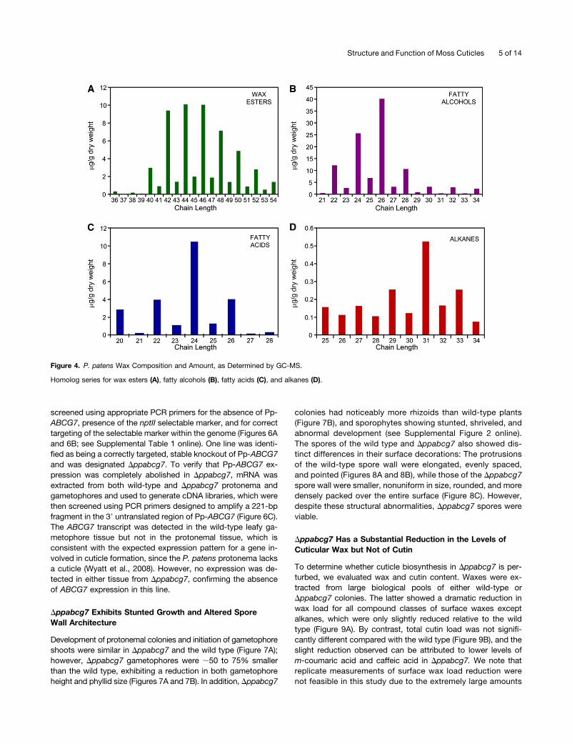
waxes were consistently below the detection limit in smallersample sizes. This ensured that the extraction was consistentacross all plants but meant that any minor compositional dif-ferences between moss colonies or gametophores were notconsidered. A prefractionation step using thin-layer chroma-tography (TLC) was included in the analysis to improve theidentification and quantification of wax compounds and to reducedeterioration of peak quality during the subsequent GC-MSanalysis that occurred when attempting to analyze derivatizedcrude wax extracts. The major compound classes present wereprimary fatty alcohols and long-chain wax esters, with minoramounts of VLCFAs and alkanes (Figures 4A to 4D), whilesecondary fatty alcohols, ketones, or fatty aldehydes werenot detected. The additional TLC bands most likely resultedfrom intracellular lipid contamination and corresponded to triacylglycerols and steryl esters, which are present in large amounts ingametophyte tissues (Huang et al., 2009).
The distribution of carbon chain length within each wax com-pound class followed the expected pattern with long-chain waxesters, primary fatty alcohols, and VLCFAs exhibiting a strong biastoward chain lengths with an even number of carbons (Figures 4Ato 4C). Conversely, the alkanes, arising from decarbonylation ofVLCFAs during their biosynthesis, exhibited an odd chain lengthbias (Figure 4D). Alkanes shorter than C25 were not considered inthe survey as they did not exhibit the odd chain length prefer-ence characteristic of cuticular alkanes and likely correspondedto contaminants introduced during prefractionation from sourcessuch as plasticware.
Functional Analysis of ABCG7, a Putative Moss WaxTransporter Gene
After determining that P. patens has a cuticle that is both struc-turally and compositionally similar to those of flowering plants, weaddressed the hypothesis that the basic underlying moleculargenetic pathways involved in cuticle formation have also beenclosely conserved throughout plant evolution. Since Arabi-dopsis has the most comprehensive set of characterized cuticle-associated genes, we used an extensive selection of these tosearch the P. patens genome sequence for putative orthologs.Many candidate genes were identified with a high degree ofpredicted protein sequence similarity to those known in Arabi-dopsis to be involved in various aspects of wax and cutin bio-synthesis and cuticle formation.One of these, ABCG7 (Rensing et al., 2008), is predicted to be
a 10-exon gene, spanning 3544 bp, and encoding an ABC halftransporter in the G subfamily, with 63% amino acid identity and78% similarity with the Arabidopsis protein At-ABCG11 (alsonamed White Brown Complex11 [WBC11]) and slightly lowersequence homology to At-ABCG12 (also named WBC12 andEceriferum 5; 52% identity and 72% similarity). A phylogeneticanalysis of all predicted ATP binding cassette subfamily G (ABCG)half transporters from Arabidopsis and P. patens revealed thatPp-ABCG7 falls into a well-supported clade containing all ofthe Arabidopsis ABCG half transporters that have previouslybeen shown to be involved in cuticle formation (Figure 5; seeSupplemental Data Set 1 online). Of these, At-ABCG11 andAt-ABCG12 are two well characterized members that are re-quired for the trafficking of wax and cutin precursors through theplasma membrane and whose loss-of-function mutants showsevere abnormal cuticle phenotypes (Pighin et al., 2004; Birdet al., 2007; Luo et al., 2007).The subcellular localization of Pp-ABCG7 was evaluated
by transient coexpression in onion (Allium cepa) epidermalcells of full-length Pp-ABCG7 fused to red fluorescent pro-tein (RFP) at the C terminus, together with the plasma mem-brane resident Arabidopsis aquaporin At-PIP2a fused to greenfluorescent protein (GFP). Confocal imaging revealed thatPpABCG7-RFP and AtPIP2a-GFP showed overlapping expres-sion in plasmolyzed transformed onion cells (see SupplementalFigure 1 online), indicating that Pp-ABCG7 localizes to the plasmamembrane, consistent with its putative function as an ABCGhalf transporter.To establish whether Pp-ABCG7 has similar functions in
trafficking cuticle components, a targeted ABCG7 knockoutmutant was generated. Approximately 1.1 kb of flanking se-quence both upstream and downstream of the gene was clonedinto the pTN80 vector on either side of the nptII G418 resistancemarker (Figure 6A). These 59 and 39 flanking regions served totarget the knockout cassette to the Pp-ABCG7 gene and al-lowed replacement with the selectable marker. Wild-type mossprotoplasts were transformed as previously described (Coveet al., 2009; Saavedra et al., 2011) via polyethylene glycol–mediated transformation. Transformed protoplasts were re-generated and the resulting moss colonies were subjected to tworounds of selection with the antibiotic G418 to yield a collec-tion of potential stable knockout lines. Each of these lines was
Figure 3. P. patens Cutin Monomer Composition and Amount, as De-termined by GC-MS.
Error bars indicate SE; n = 4.
4 of 14 The Plant Cell
screened using appropriate PCR primers for the absence of Pp-ABCG7, presence of the nptII selectable marker, and for correcttargeting of the selectable marker within the genome (Figures 6Aand 6B; see Supplemental Table 1 online). One line was identi-fied as being a correctly targeted, stable knockout of Pp-ABCG7and was designated Dppabcg7. To verify that Pp-ABCG7 ex-pression was completely abolished in Dppabcg7, mRNA wasextracted from both wild-type and Dppabcg7 protonema andgametophores and used to generate cDNA libraries, which werethen screened using PCR primers designed to amplify a 221-bpfragment in the 39 untranslated region of Pp-ABCG7 (Figure 6C).The ABCG7 transcript was detected in the wild-type leafy ga-metophore tissue but not in the protonemal tissue, which isconsistent with the expected expression pattern for a gene in-volved in cuticle formation, since the P. patens protonema lacksa cuticle (Wyatt et al., 2008). However, no expression was de-tected in either tissue from Dppabcg7, confirming the absenceof ABCG7 expression in this line.
Dppabcg7 Exhibits Stunted Growth and Altered SporeWall Architecture
Development of protonemal colonies and initiation of gametophoreshoots were similar in Dppabcg7 and the wild type (Figure 7A);however, Dppabcg7 gametophores were ;50 to 75% smallerthan the wild type, exhibiting a reduction in both gametophoreheight and phyllid size (Figures 7A and 7B). In addition, Dppabcg7
colonies had noticeably more rhizoids than wild-type plants(Figure 7B), and sporophytes showing stunted, shriveled, andabnormal development (see Supplemental Figure 2 online).The spores of the wild type and Dppabcg7 also showed dis-tinct differences in their surface decorations: The protrusionsof the wild-type spore wall were elongated, evenly spaced,and pointed (Figures 8A and 8B), while those of the Dppabcg7spore wall were smaller, nonuniform in size, rounded, and moredensely packed over the entire surface (Figure 8C). However,despite these structural abnormalities, Dppabcg7 spores wereviable.
Dppabcg7 Has a Substantial Reduction in the Levels ofCuticular Wax but Not of Cutin
To determine whether cuticle biosynthesis in Dppabcg7 is per-turbed, we evaluated wax and cutin content. Waxes were ex-tracted from large biological pools of either wild-type orDppabcg7 colonies. The latter showed a dramatic reduction inwax load for all compound classes of surface waxes exceptalkanes, which were only slightly reduced relative to the wildtype (Figure 9A). By contrast, total cutin load was not signifi-cantly different compared with the wild type (Figure 9B), and theslight reduction observed can be attributed to lower levels ofm-coumaric acid and caffeic acid in Dppabcg7. We note thatreplicate measurements of surface wax load reduction werenot feasible in this study due to the extremely large amounts
Figure 4. P. patens Wax Composition and Amount, as Determined by GC-MS.
Homolog series for wax esters (A), fatty alcohols (B), fatty acids (C), and alkanes (D).
Structure and Function of Moss Cuticles 5 of 14

of moss material needed. However, the lack of a statisticallysignificant difference in levels of cutin extracted from thesame pooled biological samples, for which replicate analyseswere performed (Figure 9B), indicates that the differences inwaxes were specific and not due to a general reduction in
cuticle material or some other artifact induced during samplepreparation.We prepared samples of Dppabcg7 gametophores to examine
cuticle structure using TEM and scanning electron microscopy. Nonoticeable phenotypic differences were observed in the Dppabcg7
Figure 5. Unrooted Neighbor Joining Protein Phylogeny of Predicted ABCG Half Transporters from Arabidopsis and P. patens.
Arabidopsis subfamily members are shown in red and P. patens subfamily members in black. Those marked by an asterisk are not supported by EST evidence.The clade highlighted by a green background contains all of the Arabidopsis ABCG half transporters known to be involved in cuticle precursor export (bold), as wellas the putative moss wax transporter Pp-ABCG7. Bootstrap values (1000 replicates) are given for each node. Bar indicates 0.1 amino acid substitutions per site.
6 of 14 The Plant Cell

cuticle, either in the phyllid or in the sporophyte. Scanning electronmicroscopy analysis suggested a decrease in the abundance ofepicuticular wax crystals on the phyllids, but because their pres-ence was so variable in wild-type phyllids, it was not possible toconclude that there was a reduction in Dppabcg7. However, whileareas containing small crystals were observed in Dppabcg7, re-gions with wax crystals as large or populous as those seen in thewild type (Figures 2A and 2B) were not apparent.
Pp-ABCG7 Does Not Functionally Complement a Mutationin the Homologous Arabidopsis Gene ABCG12
The substantial reduction in wax levels in Dppabcg7 is remi-niscent of a similarly large reduction in waxes reported in theArabidopsis abcg12 (wbc12) mutant (Pighin et al., 2004), whichhas perturbed expression of ABCG12, a closely related homologof Pp-ABCG7 (Figure 5). To investigate whether Pp-ABCG7 isa functional ortholog of At-ABCG12, as the mutant phenotypesuggests, transformants of the At-abcg12 mutant expressingPp-ABCG7 driven by the constitutive 35S promoter were gen-erated (see Supplemental Figure 3A online) and the cuticles ofthe transgenic plants evaluated to determine whether the mutanthad been rescued. In wild-type Arabidopsis, the 29C alkane ac-counts for over 50% of the wax load in mature stems (Li-Beissonet al., 2010). This was used as a diagnostic compound to determinewhether the complementation was successful. Wax was extractedfrom the inflorescence stems of T2 plants, some of which carriedthe transgene and some of which did not, as well as from wild-typeColumbia-0 plants and the amount of 29C alkane in each line was
determined (see Supplemental Figure 3B online)). The amount of 29Calkane in At-abcg12mutant plants was substantially reduced relativeto the wild type but was equally low in the Pro35S:PpABCG7 lines,indicating that complementation of the mutation did not occur.
Dppabcg7 Has a Reduced Tolerance of Desiccation Stress
Given the substantially reduced wax load in Dppabcg7, we hypoth-esized that this might result in reduced vitality under desiccatingconditions. Six-week-old colonies of both the wild type andDppabcg7 were exposed to atmospheres of different RH and theirgrowthmonitored bymeasuring the fresh weight of the moss on eachplate every 3 d over a 21-d period. At 100% RH, both the wild typeand Dppabcg7 grew at the same rate (Figure 10A), while at 91% RH,wild-type colonies maintained their original fresh weight for the du-ration of the experiment but showed no significant growth. However,Dppabcg7 colonies exposed to 91% RH maintained their originalfresh weight for only 12 d, before rapidly losing weight and eventuallydying (Figure 10B). At 86% RH treatment, both lines steadily lostweight until they were highly desiccated and necrotic (Figure 10C).
DISCUSSION
P. patens Has a Cuticle That Is Architecturally andCompositionally Similar to Those of Later DivergingLand Plants
TEM imaging of the P. patens phyllid cell walls revealed a thincuticle evenly coating the whole phyllid surface, approximately the
Figure 6. Generation of the Dppabcg7 Knockout Mutant.
(A) Schematic diagram depicting the targeted replacement of Pp-ABCG7 with the nptII selectable marker by homologous recombination to yielda stable P. patens knockout mutant. (FR, flanking region).(B) PCR screening of wild-type and Dppabcg7 genomic DNA with the PCR primer pairs shown in (A) showing the presence (Yes) or absence (No) of anamplified product. Primer pairs specific to Pp-ABCG7 generate a PCR product in the wild type but not in Dppabcg7.(C) PCR screening of wild-type and Dppabcg7 cDNA with Pp-ABCG7 transcript-specific primers, showing that Pp-ABCG7 expression is abolished inDppabcg7 (P, protonema; G, gametophore). Primers specific to P. patens actin (Pp-ACT2) were used as a control for cDNA quality.
Structure and Function of Moss Cuticles 7 of 14

same thickness as that of the Arabidopsis leaf cuticle (Figure 1C).Flowering plants are covered by a waterproof cuticle, and gasexchange for photosynthesis is facilitated by stomata that connectthe outside environment with internal air spaces. However, bryo-phytes, other than a few select examples (Sack and Paolillo, 1983;Beerling and Franks, 2009), do not have stomata, and in the caseof P. patens, the photosynthetic phyllids are only a single cell layerthick (Figures 1A and 1B). Therefore, a cuticle is likely necessary inpreventing rapid water loss from phyllid cells, but it must at thesame time be sufficiently permeable to allow gas exchange, as noalternate internal routes exist.
Despite the variability of epicuticular wax crystal density(Figure 2) on the phyllids, the predominance of crystals on olderphyllids near the base of the gametophores suggests a devel-opmental program for wax synthesis. The substantially thickercuticle of the sporophyte capsule (Figures 1D and 1E) may reflectthe fact that the moss sporophyte is nutritionally depended on thefemale gametophores to which it is anchored, so a cuticle that issufficiently permeable to allow gas exchange for photosynthesisis presumably not as important. Furthermore, the water status ofthe sporophyte capsule must be carefully controlled to ensurespore dispersal under the appropriate conditions (Duckett et al.,2009), which would be facilitated by the presence of a morerobust cuticle. Taken together, these results suggest that theelaboration of the cuticle biosynthetic program in the sporophytegeneration may have served as the foundation for producing alarger and more complex cuticle in the sporophyte-dominant,later-diverging land plants.
Biochemical analysis resulted in the identification of the majorP. patens cutin aliphatic monomers as 5-hydroxytetradecanedioicacid (C14), 10,16-dihydroxyhexadecanoic acid (C16), and 16-hydroxyhexadecanoic acid (C16) (Figure 3), revealing clear similar-ities with the cutin components of diverse plant taxa. Shorter chainlength hydroxyalkane diacids (C14-C16) are substantial componentsof the cutin matrix in a variety of plant species, including Gnetumgnemon (Hunneman and Eglinton, 1972) and Limonia acidissima(Das and Thakur, 1989), as well as minor components of Sphagnummoss (Caldicott and Eglinton, 1976), Ginkgo (Hunneman andEglinton, 1972), and coffee (Coffea arabica; Holloway et al., 1972).In some cases, a particular monomer can comprise >80% of thecutin precursor population, such as 10,16-dihydroxyhexadecanoicacid in tomato fruit cutin (Isaacson et al., 2009). We also foundminor amounts of 1,9,18-octadecanetriol (C18). Alkanetriols havebeen shown to be present in large amounts in the cutins of earlierdiverging land plants, such as Psilotum (Caldicott et al., 1975),suggesting that the incorporation of these monomers into cutinsis ubiquitous in land plants.
Figure 7. Stunted Growth Phenotype of Dppabcg7.
(A) Development of wild-type and Dppabcg7 colonies over a 21-d timeperiod showing stunted growth of the Dppabcg7 gametophores.(B) Close-up view of wild-type and Dppabcg7 colonies, showing stuntedgrowth of gametophores in the mutant line.Bars = 5 mm in (A) and 2 mm in (B).
Figure 8. P. patens Spore Phenotypic Analysis.
Scanning electron micrographs of spore surface decorations in the wildtype ([A] and [B]) and Dppabcg7 (C), showing irregular and roundedprotrusions in the mutant. Bars = 10 mm in (A) and 5 mm in (B) and (C).
8 of 14 The Plant Cell

A particularly notable result was the high levels of aromaticcutin monomers in P. patens cutin, with m- and p-coumaric acidand caffeic acid (Figure 3), which together account for >50% ofthe cutin monomers. There has been considerable debate as towhether these aromatic acids are actually incorporated into thecutin matrix (Holloway, 1982), but an Arabidopsis acyl-transferasewas recently identified that esterifies ferulic acid to v-hydroxyfatty acids and a loss of function of the corresponding generesults in the absence of ferulate in leaf cutin (Rautengartenet al., 2012). However, this deficiency does not apparently af-fect permeability of the cuticle to water, suggesting that thesearomatic monomers do not significantly contribute to reducingtranspirational water loss. The presence of such high amountsin moss cutin might therefore be taken to indicate other func-tions, such as defense against pathogens or protection fromUV radiation, as suggested by Clarke and Robinson (2008), andwarrants further investigation. We also note that the polymersuberin typically contains high amounts of aromatic monomersderived from cinnamic acid (Beisson et al., 2012) and suggestthat the first pathways responsible for cutin synthesis in earlydiverging plants may have been the progenitors of suberin bio-synthetic pathways in later diverging lineages.
The cutin polymer provides the major structural framework ofthe cuticle, but the wax that fills and covers this matrix is thought
to contribute greatly to its sealing and protective properties,depending on both amount and composition (Yeats and Rose,2013). Nonuniform deposition of waxes within the intra- andepicuticular regions of the cuticle is often observed and, to-gether with the overall wax composition, leads to a diversity ofepicuticular wax crystals and films (Jeffree, 2006). Analysis ofP. patens waxes revealed four major compound classes: fatty al-cohols, long-chain wax esters, VLCFAs, and alkanes (Figures 4Ato 4D). In some plants, a particular compound class can account
Figure 9. Cuticle Composition Analysis.
(A) Cuticular wax levels in wild-type and Dppabcg7 moss.(B) Cutin monomer levels in the wild type and Dppabcg7. Error bars in-dicate SE; n = 4.
Figure 10. Response of Wild-Type and Mutant Moss Colonies to Des-iccation Stress.
Fresh weight of wild-type and Dppabcg7 moss colonies at 100% RH (A),91% RH (B), and 86% RH (C) was measured over a 21-d period. As-terisks indicate statistically significant differences between wild-typeand transgenic lines (P < 0.05, Student’s t test). Error bars indicate SE;3 # n # 5.
Structure and Function of Moss Cuticles 9 of 14

for most of the cuticular wax, as with the fern Pteridium aquili-num, where wax esters comprise >90% of the surface wax load(Baker and Gaskin, 1987). In other species, such as Arabidopsis,the surface wax is composed of comparable amounts of severalcompound classes (Li-Beisson et al., 2010). It is clear that thebryophytes have the capacity to synthesize the full complementof wax compound classes mentioned, even though not all ofthem were found in P. patens. For example, the cuticular wax ofthe moss Pogonatum urnigerum has substantial amounts ofsecondary alcohols and related species produce long-chain al-dehydes (Neinhuis and Jetter, 1995). Taken together, we sug-gest that the biosynthetic pathways needed to synthesize thediverse suite of cuticular waxes that have been identified in landplant lineages were likely present in the first land plant taxa, priorto the divergence of vascular plants. Moreover, specific evolu-tionary pressures have refined cuticular wax compositions re-sulting in the observed compositional and structural diversity.
The Cuticle Phenotype of Dppabcg7 Suggests DeepConservation of Cuticle-Associated Gene Pathwaysin Land Plants
Having establishing that the P. patens cuticle is architectur-ally and compositionally comparable to those of many later di-verging plant lineages, we hypothesized that the underlyinggenetic pathways that regulate cuticle synthesis and assemblyare likely also shared among land plants. To test this, we usedthe sequences of well-studied cuticle-associated genes inflowering plants to identify putative orthologs in the P. patensgenome and indeed found that most of the gene families knownto be involved in cuticle synthesis, trafficking, and assemblywere represented. Among these genes was a putative ortholog(Pp-ABCG7) of the Arabidopsis ABCG transporters ABCG11(WBC11) and ABCG12 (WBC12/CER5), which encode halftransporters that form homo- or heterodimers and mediate thetransport of waxes and cutin precursors across the plasmamembrane (Kang et al., 2011). The Arabidopsis abcg12 (wbc12)mutant has a 50% decrease in surface wax load and lipid in-clusions in the vacuoles of the epidermal cells (Pighin et al.,2004), while the abcg11 (wbc11) mutant phenotype is moresevere, with a substantial reduction in levels of both cutin andwax, as well as severe dwarfing, a permeable cuticle, andpostgenital organ fusions (Bird et al., 2007; Luo et al., 2007). Atargeted knockout mutant of Pp-ABCG7 (Dppabcg7) alsoshowed a dwarf phenotype (Figure 7), accompanied by a sub-stantial reduction in surface wax, while cutin levels were notaffected (Figure 9). Furthermore, Dppabcg7 had a reducedability to tolerate low humidity conditions compared with thewild type (Figure 10B). The 91% RH conditions at which thereduced growth phenotype was apparent was selected becausea study by Koster et al. (2010) indicated that wild-type P. patensis able to survive at this RH, but not lower. These conditionswere therefore chosen as a tipping point at which impairedviability resulting from a defective cuticle might be more evident,as indeed proved to be the case.
The fact that no organ fusion phenotype or altered cuticle ar-chitecture was observed in Dppabcg7 further suggests thatABCG7 is responsible for wax transport, as such phenotypes are
more typically associated with cutin deficiency (Javelle et al.,2011). In addition, water loss through the cuticle is thought to becontrolled mainly by wax composition and amount and is notcorrelated with cutin content (Burghardt and Riederer, 2006;Isaacson et al., 2009). We conclude that the physiological phe-notypes of Dppabcg7 are a consequence of its decreased cu-ticular wax content. The At-abcg12 (At-wbc12) mutant showslipidic inclusion bodies within the epidermal cells, presumablycomprised of cuticular waxes that are unable to be transportedout of the cell (Pighin et al., 2004). These were not observed in theepidermal cells of the Pp-abcg7 mutant. It is possible that themoss cells have a feedback mechanism that prevents suchwaxes from accumulating in the cytoplasm or vacuole. Alterna-tively, the fact that Pp-abcg7 was cultured under conditions ap-proaching 100% humidity may have reduced the need for it toproduce copious amounts of wax.The reduction in the amount of cuticular wax, but not cutin, in
Dppabcg7, together with the length of the predicted gene se-quence, suggests that Pp-ABCG7, like At-ABCG12 (At-WBC12),encodes a wax half transporter. In Arabidopsis, cuticle-associatedhalf transporters dimerize in differing combinations to traffic dif-ferent compound classes, and it is therefore likely that Pp-ABCG7also forms either a homodimer or a heterodimer with another halftransporter to traffic cuticular waxes. Due to these and otherprotein–protein interactions that may be necessary for the activityof this class of transporter, it is perhaps not surprising thatPp-ABCG7 does not complement the At-abcg12 mutant (seeSupplemental Figure 3 online), since the precise protein con-formations necessary for such interactions would likely not beconserved over the ;450 million years since the divergence ofthese species. Alternatively, constitutive expression of Pp-ABCG7in the At-abcg12 mutant driven by the 35S promoter may notbe appropriate for its cell type–specific function, and use of anArabidopsis epidermal-specific promoter might be sufficient forcomplementation. Another notable phenotype of Dppabcg7 wasthe abnormal surface decorations of the spore walls (Figure 8).The Arabidopsis half transporter ABCG26 is required for exineformation in pollen and is believed to be responsible for the exportof sporopollenin precursors in tapetal cells (Kang et al., 2011). Ourresults suggest that ABCG7 may serve multiple functions inmoss, depending on where it is expressed, or possibly with whichother half transporter it dimerizes, and could play a role in bothcuticle and spore wall formation.
Conclusions
The evolutionary origins of the molecular pathways that mediatecuticle biosynthesis and formation remain unclear, but it is likelythat they were derived from those responsible for membranelipid biosynthesis in the algal ancestors of the first land plants.An expansion of these new pathways in the first terrestrial plantsinto the complex cuticle biosynthetic machinery seen in angio-sperms has been hypothesized for many years, but, until now,no concrete evidence has been presented. This study linkscuticle formation in a bryophyte with one of its correspond-ing genetic pathways. The substantial reduction in cuticularwax in the Dppabcg7 moss line suggests that the role of ABCGtransporters in trafficking cuticle components has been conserved
10 of 14 The Plant Cell
since the earliest divergence of terrestrial plants. Taken togetherwith the strong similarities in the chemical composition andstructure between the P. patens cuticle and those of other di-verse plant taxa, we conclude that at least some of the mech-anisms required to synthesize a specialized lipidic cell wall havebeen conserved through plant evolution and were likely instru-mental in allowing the first terrestrial plants to survive in desic-cating environments.
METHODS
Plant Material and Cultivation
Wild-type Physcomitrella patens (Gransden 2008 strain, obtained from theCold Spring Harbor Laboratory, NY) and mutant strains were cultured onBCDAT or BCD media (Nishiyama et al., 2000) supplemented with vanco-mycin (50 µg/mL) in 9-mm Petri plates overlaid with 9-mm-diameter cello-phane discs (AA Packaging). Subcultures were performed by homogenizingprotonemal material in water using a Power Gen 125 homogenizer (FischerScientific) or by placing small pieces of young protonemal tissue (<1 mm indiameter) on BCDAT plates to induce protonemal growth. To induce ga-metophore development, cellophane discs containing protonemal colonieswere transferred to BCDmedium without ammonium tartrate. Cultures weregrown at 23°C under 120 µmolm22 s21 light with a long-day cycle (16 h light/8 h dark). Sporophyteswere induced fromgametophytes cultured in jiffy potsas described by Cove et al. (2009).
Light Microscopy
The phyllid transverse section depicted in Figure 1B was obtained fromwild-type moss material fixed, embedded, and cryosectioned as de-scribed by Buda et al. (2009). A 6-µm cryosection was melted ontoa VistaVision Histobond (VWR) slide, dried, and stained using safranin O(0.1% in 50% ethanol) with an alcian blue counterstain (0.1% in water).The slide was mounted in water and viewed using an AxioImager A1microscope with an Achroplan 320/0.45 objective and AxioCam Mrccolor video camera (Zeiss) using differential interference contrast optics.
TEM
P. patens gametophores and sporophytes were fixed using conventionalchemical protocols. Samples were fixed with 1% glutaraldehyde in 0.1 MSorensen’s phosphate buffer, postfixed in 1% osmium tetroxide, dehy-drated in acetone, and embedded in Spurr’s Low Viscosity Resin (EMS).Sections (60 to 80 nm) were cut using a diamond knife on a Reichert Om-U2 ultramicrotome (MOC), stained with uranyl acetate (1% in water) andlead citrate (0.1% in water), and viewed on a Libra 120 transmissionelectron microscope (Zeiss) at 120 kV, equipped with a Cantega G-2camera (Olympus).
Scanning Electron Microscopy
Wild-type and Dppabcg7 gametophores (at least 8 weeks old) were airdried overnight in a laminar flow hood, and individual phyllids werecarefully dissected. These were sputter coated with gold-palladium for90 s at 2.2 kV, and epicuticular wax crystals were imaged on a JEOL 6480variable-pressure scanning electron microscope (JEOL) with secondaryelectrons at 15 kV with a spot size of 55. The wild-type moss sporesdepicted in Figure 8Awere placed on nitrocellulose, plunge frozen in liquidnitrogen, and viewed on a JEOL 6480 variable-pressure scanning electronmicroscope with backscattered electrons under variable-pressure modewith 30 Pa of vacuum and a spot size of 60. Spores shown in Figure 8B
and 8C were sputter coated for 50 s and visualized with secondaryelectrons at 10 to 12 kV and a spot size of 60.
Wax Analysis
Plates containing colonies of wild-type and Dppabcg7 moss grown for3 weeks on BCDAT medium, followed by 7 to 10 weeks on BCD medium,were opened for 2 h to reduce surface moisture. Colonies were thensubmerged for 30 s in a beaker of chloroform containing 150 µg each ofmethyl heptadecanoate, nonadecane, and heptadecanoic acid as internalstandards. The extract was dried over anhydrous sodium sulfate, passedthrough filter paper, and concentrated using a rotary evaporator. Mosswax was prefractionated on preparative 20 3 20-cm TLC plates(Whatman; 250-µmcoating, silica gel 60), using a mobile phase of 80:18:1hexane:diethyl ether:glacial acetic acid and employing the sandwichtechnique, which consisted of a glass plate fastened to the TLC plate witha 1- to 2-mm space between it and the silica gel to allow for increasedsaturation of the atmosphere adjacent to the sample. Bands were visu-alized under a UV lamp after misting with primuline (0.005% in 80:20acetone:water), scraped from the plate, and eluted from the silica withchloroform. The band extract corresponding to fatty acids and primaryalcohols was derivatized using equal parts of bis-N,O-(trimethylsilyl)tri-fluoroacetamide and pyridine for 15 min at 100°C. All extracts were driedunder a gentle stream of nitrogen and resuspended in chloroform. Waxcompound identification was performed by GC-MS as described byWang et al. (2011), using a 6890 gas chromatograph (Agilent) coupled toa GCMate II mass spectrometer (JEOL). Individual waxes were quantifiedbased on peak integration of the total ion chromatogram, except for waxesters, which were quantified using gas chromatography–flame ionizationdetection on a model 6850 gas chromatograph (Agilent) due to dis-crimination of the GC-MS against compounds in this molecular massrange. All waxes were quantified based on their corresponding internalstandard, with the exception of primary alcohols, which were quantifiedusing the heptadecanoic acid standard. AMDIS 32 GC-MS Analysissoftware (v 2.69) was used for peak identification and TSSPro GC-MSdata reduction software (v 3.0) for manual peak integration.
Cutin Analysis
The same wild-type and Dppabcg7 tissue that was extracted for waxanalysis (see above) was oven-dried at 50°C for several days, and 4 g ofdried tissue from each line was powdered in a pestle andmortar with liquidnitrogen and distributed among four tubes. The material was exhaustivelydelipidated as outlined by Li-Beisson et al. (2010) with the followingmodifications. All extraction steps were performed twice for 1 h eachusing a rotary shaker set to 300 rpm, and an additional two extractionswith acetone were included after the last step. Base-catalyzed de-polymerization, extraction, and subsequent trimethylsilyl derivatization ofthe samples were performed as described above. Extracts were re-suspended in chloroform, and cutin monomers were identified byGC-MS.Individual monomers were quantified using v-pentadecalactone as aninternal standard with the software listed above.
Transient Expression and Subcellular Localization of Pp-ABCG7 inOnion Cells
The full-length coding sequence Pp-ABCG7 containing the att recom-bination sites (forward, 59-GGGGACAAGTTTGTACAAAAAAGC-AGGCTTCATGGCTTCTTCTAATTGTTTACATGC-39, and reverse,59-GGGGACCACTTTGTACAAGAAAGCTGGGTCATCAATTGAAGTGAA-ATTAAAATTGTCA-39) was synthesized (Life Technologies) and clonedinto the pDONR221 vector by overnight incubation in the presence ofGateway BP clonase (Invitrogen) at 25°C, according to themanufacturer’s
Structure and Function of Moss Cuticles 11 of 14

instructions. The positive entry clones were then introduced into thepSITE-4NA (RFP tag in the C terminus) destination vector (Chakrabartyet al., 2007). Plasma membrane labeling was based on the full-lengthcoding region of At-PIP2A, a plasma membrane aquaporin, acquiredfrom The Arabidopsis Information Resource Stock Center (ABRC stocknumbers: CD3-1003; Cutler et al., 2000; Nelson et al., 2007) fused to theN terminus of GFP. Approximately 1 mg each of the PpABCG7-RFP andAtPIP2A-GFP plasmids was mixed and used to bombard onion (Alliumcepa) epidermal cells as described by Yamane et al. (2005). The tissuewas incubated for 16 to 24 h at 24°C in the dark, the epidermal layer waspeeled, transferred to glass slides, and mounted in a 30% Suc solutionto induce plasmolysis, and the cells were observed using a Leica TCS-SP5confocal scanning laser microscope (Leica Microsystems).
ABCG Half Transporter Phylogenetic Analysis
Amino acid sequences of all known ABCG half transporters from Arabidopsisthaliana andP. patenswere obtained fromGenBank (http://www.ncbi.nlm.nih.gov) and Cosmoss (http://cosmoss.org) databases, respectively. Sequenceswere aligned using ClustalW (Thompson et al., 1994), and an unrootedneighbor joining phylogenetic tree was generated using MEGA5 software(Tamura et al., 2007) with bootstrap values assigned to each node (1000replicates).
Dppabcg7 Knockout Cassette Construction
To selectively disruptABCG7, the annotated gene sequence and surroundinggenomic sequence were obtained from the latest version of the P. patensgenome using the BLAST tool in the Cosmoss database. Primer pairPpABCG7-5FR (see Supplemental Table 1 online) incorporating the HindIIIandEcoRI restriction siteswas used to amplify the 59 flanking region (1157 bp)of ABCG7. Similarly, primer pair PpABCG7-3FR (see Supplemental Table 1online) incorporating theBamHI andSacI restriction sites was used to amplifythe 39 flanking region (1143 bp) of the target gene using ExTaq PCR (Takara).These flanking regions were subsequently cloned into the pTN80 vector(obtained from Mitsuyasu Hasebe at the National Institute for Basic Biology,Okazaki, Japan) on either side of the nptII selectable marker. The Pp-ABCG7knockout cassette was then replicated in Escherichia coli, isolated, andlinearized using the BssHII restriction enzyme prior to moss transformation.
Transformation to Generate Dppabcg7
Polyethylene glycol–mediated protoplast transformation was performedas outlined by Cove et al. (2009) and Saavedra et al. (2011). Coloniessurviving the first round of selection on BCDAT medium supplementedwith G418 were maintained for 1 week on BCDAT without antibiotics andthen subjected to a second round of selection on BCDAT with G418 for1 week. Several surviving colonies were propagated and screened usingPCR primers specific to both Pp-ABCG7 and the nptII resistance marker(Figures 6A and 6B; see Supplemental Table 1 online) to verify correcttargeting of the knockout cassette.
Expression Analysis
Total mRNA was isolated from moss protonema (2 weeks old) andgametophores (8 weeks old) using the Dynabead mRNA direct kit (Invi-trogen Dynal) following the manufacturer’s instructions. One hundred mi-crograms of purifiedmRNAwas used for subsequent cDNA synthesis usingSuperscript II (Invitrogen) and according to the manufacturer’s instructions.The gene-specificPCRprimer pair PpABCG7-3FR (seeSupplemental Table1 online) was used to amplify a 221-bp fragment at the 39 end of the maturemRNA of ABCG7. The protonema and gametophore cDNA libraries of thewild-type and Dppabcg7 lines were screened with these primers via PCR(94°C for 5 min; 94°C for 30 s, 54°C for 45 s, and 72°C for 30 s for 38 cycles)
to determine presence or absence of gene expression. PCR primersdesigned to the 39 end of Act2 mRNA (primer pair PpACT2; seeSupplemental Table 1 online) were used as a positive control for geneexpression between tissue types and genetic lines (Huang et al.,2009).
Physiological Assays
For the physiological studies of wild-type and Dppabcg7 moss grownunder different RHs, cultures were initiated using three pieces of pro-tonemal tissue (<1 mm in diameter) placed on a plate of BCDAT mediasupplemented with vancomycin (50 µg/µL) and overlaid with cellophane(AA Packaging). After 3 weeks, the 9-mmcellophane discswere transferredto BCD media for an additional 3 weeks to induce robust gametophoregrowth. Wild-type and Dppabcg7 cultures were weighed (cellophaneand moss colonies only), placed onto plates containing BCD media,and exposed to different RH conditions. The plates were left openinside sealed chambers (Pyrex dishes) floating on solutions of satu-rated magnesium sulfate or potassium chloride to obtain RHs of 91 or86%, respectively (Koster et al., 2010). Other moss cultures were sealed inplates containing BCD media supplemented with vancomycin (50 µg/µL)to generate close to 100% RH. The fresh weight of each sample(cellophane and moss colonies) was recorded every 3 d for 3 weeks.After 3 weeks, the final weight was recorded and the mass of the cel-lophane was subtracted from each previous measurement of the sampleto obtain fresh weight values. Any plates with substantial microbialcontamination were discarded and were not included in subsequentstatistical analyses.
Complementation of At-abcg12 with PPABCG7
To construct the plasmid vector for complementation analysis, thePp-ABCG7 coding sequence (as described above) was cloned intothe pDONR Gateway shuttle vector and then subcloned in frame with thePro35S:CPMV 59-UTR in the destination vector pEAQ-HT-DEST1. Thevector construct was transformed into 3-week-old abcg12 T-DNA in-sertional mutant Arabidopsis plants (Salk 036776) by Agrobacteriumtumefaciens–mediated transformation (GV2260 strain) as previously de-scribed (Clough and Bent, 1998). To identify transformants, the T1 seedswere sown on one-quarter Murashige and Skoog plates containing100 mg/L kanamycin and screened by PCR using Pp-ABCG7–specificprimers (primer pair PpABCG7-S; see Supplemental Table 1 online). Off-spring from a transformed line (a segregating T2 population) were grownand genotyped by PCR for the presence of the ABCG7 transgene. In-florescence stems were collected from five transgenic and three non-transgenic T2 plants, as well as from three wild-type plants, grownconcurrently. The stems, from which leaves, flowers, and siliques wereremoved, were cut into 3-cm sections, with eight to 15 sectionscollected per plant, and sections from each plant were swirled in30 mL of chloroform, containing 100 mL of tetracosane as an internalstandard, for 30 s. The diameters of the stem sections were measured attheir center to calculate surface area. Wax extracts were dried overanhydrous sodium sulfate, filtered, and then concentrated to a volumeof 4 mL. The level of C29 alkane in these samples was quantified asdescribed above.
Accession Numbers
Sequence data from this article can be found in the GenBank/EMBL datalibraries under the following accession numbers: XP_001775949 (Pp-ABCG7 genomic sequence), XP_001775897 (Pp-ABCG7mRNA sequence),and XM_001775900 (Pp-ACT2 mRNA sequence).
12 of 14 The Plant Cell

Supplemental Data
The following materials are available in the online version of this article.
Supplemental Figure 1. Localization of Pp-ABCG7 Fused to an RFPMarker Protein Using Confocal Scanning Laser Microscopy, followingTransient Expression in Onion Epidermal Cells.
Supplemental Figure 2. Abnormal Sporophyte Phenotype of Dppabcg7.
Supplemental Figure 3. Overexpression of Pp-ABCG7 in the Arabi-dopsis abcg12 Mutant.
Supplemental Table 1. List of PCR Primers, Referenced in Methods.
Supplemental Data Set 1. Protein Alignment of All Known ABCG HalfTransporters from Arabidopsis and P. patens.
ACKNOWLEDGMENTS
We thank Antonio Matas for helpful discussion and for assisting with thestatistical analysis, Eliel Ruiz-May for assistance with confocal microscopy,and Iben Sørensen for careful reading of the article. This work wassupported by grants from the National Science Foundation (Plant GenomeProgram, DBI-0606595), by the U.S.–Israel Binational Agricultural Researchand Development Fund (IS-4234-09), and by the Agriculture and FoodResearch Initiative Competitive Grants Program (2011-67013-19399) fromthe USDA National Institute of Food and Agriculture. G.J.B. and T.H.Y. werepartly supported by a National Institutes of Health chemistry/biologyinterface training grant (T32 GM008500).
AUTHOR CONTRIBUTIONS
G.J.B. designed and performed the research, analyzed data, and cowrotethe article. W.J.B., E.A.F., S.P., T.H.Y., L.Z., and D.S.D. performed theresearch and analyzed data. J.K.C.R. designed the research, analyzeddata, and cowrote the article.
Received August 19, 2013; revised September 11, 2013; acceptedSeptember 25, 2013; published October 25, 2013.
REFERENCES
Baker, E.A., and Gaskin, R.E. (1987). Composition of leaf epicuticularwaxes of Pteridium subspecies. Phytochemistry 26: 2847–2848.
Barthlott, W., and Neinhuis, C. (1997). Purity of the sacred lotus, orescape from contamination in biological surfaces. Planta 202: 1–8.
Beerling, D.J., and Franks, P.J. (2009). Evolution of stomatal functionin ‘lower’ land plants. New Phytol. 183: 921–925.
Beisson, F., Li-Beisson, Y., and Pollard, M. (2012). Solving the puzzles ofcutin and suberin polymer biosynthesis. Curr. Opin. Plant Biol. 15: 329–337.
Bird, D., Beisson, F., Brigham, A., Shin, J., Greer, S., Jetter, R., Kunst, L.,Wu, X., Yephremov, A., and Samuels, L. (2007). Characterization ofArabidopsis ABCG11/WBC11, an ATP binding cassette (ABC) transporterthat is required for cuticular lipid secretion. Plant J. 52: 485–498.
Buda, G.J., Isaacson, T., Matas, A.J., Paolillo, D.J., and Rose, J.K.C. (2009). Three-dimensional imaging of plant cuticle architectureusing confocal scanning laser microscopy. Plant J. 60: 378–385.
Budke, J.M., Goffinet, B., and Jones, C.S. (2011). A hundred-year-oldquestion: Is the moss calyptra covered by a cuticle? A case study ofFunaria hygrometrica. Ann. Bot. (Lond.) 107: 1279–1286.
Burghardt, M., and Riederer, M. (2006). Cuticular transpiration. InBiology of the Plant Cuticle, M. Riederer and C. Muller, eds (Oxford,UK: Blackwell Publishing), pp. 292–311.
Caldicott, A.B., and Eglinton, G. (1976). Cutin acids from bryophytes: Anv-1 hydroxy alkanoic acid in two liverwort species. Phytochemistry 15:1139–1143.
Caldicott, A.B., Simoneit, B.R.T., and Eglinton, G. (1975). Alkanetriols inpsilotophyte cutins. Phytochemistry 14: 2223–2228.
Chakrabarty, R., Banerjee, R., Chung, S.M., Farman, M., Citovsky, V.,Hogenhout, S.A., Tzfira, T., and Goodin, M. (2007). PSITE vectors forstable integration or transient expression of autofluorescent proteinfusions in plants: Probing Nicotiana benthamiana-virus interactions. Mol.Plant Microbe Interact. 20: 740–750.
Charron, A.J., and Quatrano, R.S. (2009). Between a rock and a dryplace: The water-stressed moss. Mol. Plant 2: 478–486.
Clarke, L.J., and Robinson, S.A. (2008). Cell wall-bound ultraviolet-screening compounds explain the high ultraviolet tolerance of theAntarctic moss, Ceratodon purpureus. New Phytol. 179: 776–783.
Clayton-Greene, K.A., Collins, N.J., Green, T.G.A., and Proctor,M.C.F. (1985). Surface wax, structure and function in leaves ofPolytrichaceae. J. Bryol. 13: 549–562.
Clough, S.J., and Bent, A.F. (1998). Floral dip: A simplified method forAgrobacterium-mediated transformation of Arabidopsis thaliana.Plant J. 16: 735–743.
Cove, D., Bezanilla, M., Harries, P., and Quatrano, R. (2006).Mosses as model systems for the study of metabolism anddevelopment. Annu. Rev. Plant Biol. 57: 497–520.
Cove, D.J., Perroud, P., Charron, A.J., McDaniel, S.F., Khandelwal,A., and Quatrano, R.S. (2009). The moss Physcomitrella patens: Anovel model system for plant development and genomic studies. InEmerging Model Organisms, Vol. 1, (Cold Spring Harbor, NY: ColdSpring Harbor Laboratory Press), pp. 69–104.
Cutler, S.R., Ehrhardt, D.W., Griffitts, J.S., and Somerville, C.R.(2000). Random GFP:cDNA fusions enable visualization of subcellularstructures in cells of Arabidopsis at a high frequency. Proc. Natl. Acad.Sci. USA 97: 3718–3723.
Das, S., and Thakur, S. (1989). Constituent acids of Limoniaacidissima leaf cutin. Phytochemistry 28: 509–511.
Duckett, J.G., Pressel, S., P’ng, K.M.Y., and Renzaglia, K.S. (2009).Exploding a myth: The capsule dehiscence mechanism and the functionof pseudostomata in Sphagnum. New Phytol. 183: 1053–1063.
Haas, K. (1982). Surface wax of Andreaea and Pogonatum species.Phytochemistry 21: 657–659.
Hohe, A., Egener, T., Lucht, J.M., Holtorf, H., Reinhard, C.,Schween, G., and Reski, R. (2004). An improved and highlystandardised transformation procedure allows efficient productionof single and multiple targeted gene-knockouts in a moss, Physcomitrellapatens. Curr. Genet. 44: 339–347.
Holloway, P.J. (1982). The chemical constitution of plant cutins. InThe Plant Cuticle, D.F. Cutler, K.L. Alvin, and C.E. Price, eds (London,UK: Academic Press), pp. 45–85.
Holloway, P.J., Deas, A.H.B., and Kabaara, A.M. (1972). Composition ofcutin from coffee leaves. Phytochemistry 11: 1443–1447.
Hunneman, D.H., and Eglinton, G. (1972). The constituent acids ofgymnosperm cutins. Phytochemistry 11: 1989–2001.
Huang, C.Y., Chung, C.I., Lin, Y.C., Hsing, Y.I., and Huang, A.H.C.(2009). Oil bodies and oleosins in Physcomitrella possess characteristicsrepresentative of early trends in evolution. Plant Physiol. 150: 1192–1203.
Isaacson, T., Kosma, D.K., Matas, A.J., Buda, G.J., He, Y., Yu, B.,Pravitasari, A., Batteas, J.D., Stark, R.E., Jenks, M.A., and Rose,J.K.C. (2009). Cutin deficiency in the tomato fruit cuticle consistentlyaffects resistance to microbial infection and biomechanical properties,but not transpirational water loss. Plant J. 60: 363–377.
Javelle, M., Vernoud, V., Rogowsky, P.M., and Ingram, G.C. (2011).Epidermis: The formation and functions of a fundamental planttissue. New Phytol. 189: 17–39.
Structure and Function of Moss Cuticles 13 of 14

Jeffree, C.E. (2006). The fine structure of the plant cuticle. In Biologyof the Plant Cuticle, M. Riederer and C. Muller, eds (Oxford, UK:Blackwell Publishing), pp. 11–125.
Jetter, R., Kunst, L., and Samuels, A.L. (2006). Composition of plantcuticular waxes. In Biology of the Plant cuticle, M. Riederer and C.Muller, eds (Oxford, UK: Blackwell Publishing), pp. 145–181.
Kang, J., Park, J., Choi, H., Burla, B., Kretzschmar, T., Lee, Y., andMartinoia, E. (2011). Plant ABC transporters. The ArabidopsisBook 9: e0153, doi/10.1199/tab.0153.
Koster, K.L., Balsamo, R.A., Espinoza, C., and Oliver, M.J. (2010).Desiccation sensitivity and tolerance in the moss Physcomitrella patens:Assessing limits and damage. Plant Growth Regul. 62: 293–302.
Leide, J., Hildebrandt, U., Reussing, K., Riederer, M., and Vogg, G.(2007). The developmental pattern of tomato fruit wax accumulationand its impact on cuticular transpiration barrier properties: Effectsof a deficiency in a b-ketoacyl-coenzyme A synthase (LeCER6).Plant Physiol. 144: 1667–1679.
Li-Beisson, Y., et al. (2010). Acyl-lipid metabolism. The ArabidopsisBook 8: e0133, doi/10.1199/tab.0133.
Luo, B., Xue, X.Y., Hu, W.L., Wang, L.J., and Chen, X.Y. (2007). AnABC transporter gene of Arabidopsis thaliana, AtWBC11, is involvedin cuticle development and prevention of organ fusion. Plant CellPhysiol. 48: 1790–1802.
Matas, A.J., et al. (2011). Tissue- and cell-type specific transcriptomeprofiling of expanding tomato fruit provides insights into metabolicand regulatory specialization and cuticle formation. Plant Cell 23:3893–3910.
Minami, A., Nagao, M., Ikegami, K., Koshiba, T., Arakawa, K., Fujikawa,S., and Takezawa, D. (2005). Cold acclimation in bryophytes: low-temperature-induced freezing tolerance in Physcomitrella patensis associated with increases in expression levels of stress-relatedgenes but not with increase in level of endogenous abscisic acid.Planta 220: 414–423.
Neinhuis, C., and Jetter, R. (1995). Ultrastructure and chemistry ofepicuticular wax crystals in Polytrichales sporophytes. J. Bryol. 18:399–406.
Nelson, B.K., Cai, X., and Nebenführ, A. (2007). A multicolored set ofin vivo organelle markers for co-localization studies in Arabidopsisand other plants. Plant J. 51: 1126–1136.
Nishiyama, T., Hiwatashi, Y., Sakakibara, I., Kato, M., and Hasebe,M. (2000). Tagged mutagenesis and gene-trap in the moss,Physcomitrella patens by shuttle mutagenesis. DNA Res. 7: 9–17.
Nissinen, R., and Sewón, P. (1994). Hydrocarbons of Polytrichumcommune. Phytochemistry 37: 179–182.
Pighin, J.A., Zheng, H., Balakshin, L.J., Goodman, I.P., Western,T.L., Jetter, R., Kunst, L., and Samuels, A.L. (2004). Plant cuticularlipid export requires an ABC transporter. Science 306: 702–704.
Post-Beittenmiller, D. (1998). The cloned Eceriferum genes of Arabidopsisand the corresponding Glossy genes in maize. Plant Physiol. Biochem.36: 157–166.
Proctor, M.C.F. (1979). Surface wax on the leaves of some mosses. J.Bryol. 10: 531–538.
Rautengarten, C., Ebert, B., Ouellet, M., Nafisi, M., Baidoo, E.E.K.,Benke, P., Stranne, M., Mukhopadhyay, A., Keasling, J.D., Sakuragi,Y., and Scheller, H.V. (2012). Arabidopsis Deficient in Cutin Ferulateencodes a transferase required for feruloylation of v-hydroxy fatty acidsin cutin polyester. Plant Physiol. 158: 654–665.
Reina-Pinto, J.J., and Yephremov, A. (2009). Surface lipids and plantdefenses. Plant Physiol. Biochem. 47: 540–549.
Rensing, S.A., et al. (2008). The Physcomitrella genome revealsevolutionary insights into the conquest of land by plants. Science319: 64–69.
Saavedra, L., Balbi, V., Lerche, J., Mikami, K., Heilmann, I., andSommarin, M. (2011). PIPKs are essential for rhizoid elongation andcaulonemal cell development in the moss Physcomitrella patens.Plant J. 67: 635–647.
Sack, F.D., and Paolillo, D.J. (1983). Stomatal pore and cuticleformation in Funaria. Protoplasma 116: 1–13.
Samuels, L., Kunst, L., and Jetter, R. (2008). Sealing plant surfaces:Cuticular wax formation by epidermal cells. Annu. Rev. Plant Biol.59: 683–707.
Schönherr, J., and Ziegler, H. (1975). Hydrophobic cuticular ledgesprevent water entering the air pores of liverwort thalli. Planta 124:51–60.
Shepherd, T., and Wynne Griffiths, D. (2006). The effects of stress onplant cuticular waxes. New Phytol. 171: 469–499.
Tamura, K., Dudley, J., Nei, M., and Kumar, S. (2007). MEGA4:Molecular Evolutionary Genetics Analysis (MEGA) software version4.0. Mol. Biol. Evol. 24: 1596–1599.
Thompson, J.D., Higgins, D.G., and Gibson, T.J. (1994). CLUSTAL W:Improving the sensitivity of progressive multiple sequence alignmentthrough sequence weighting, position-specific gap penalties and weightmatrix choice. Nucleic Acids Res. 22: 4673–4680.
Wang, Z., Guhling, O., Yao, R., Li, F., Yeats, T.H., Rose, J.K.C., andJetter, R. (2011). Two oxidosqualene cyclases responsible forbiosynthesis of tomato fruit cuticular triterpenoids. Plant Physiol.155: 540–552.
Wood, A.J. (2005). Eco-physiological adaptations to limited waterenvironments. In Plant Abiotic Stress, M.A. Jenks and P.M. Hase-gawa, eds (Oxford, UK: Blackwell Publishing), pp. 1–13.
Wyatt, H.D.M., Ashton, N.W., and Dahms, T.E.S. (2008). Cell wallarchitecture of Physcomitrella patens is revealed by atomic forcemicroscopy. Botany 86: 385–397.
Yamane, H., Lee, S.-J., Kim, B.-D., Tao, R., and Rose, J.K.C. (2005).A coupled yeast signal sequence trap and transient plantexpression strategy to identify genes encoding secreted proteinsfrom peach pistils. J. Exp. Bot. 56: 2229–2238.
Yeats, T.H., Buda, G.J., Wang, Z., Chehanovsky, N., Moyle, L.C.,Jetter, R., Schaffer, A.A., and Rose, J.K.C. (2012). The fruitcuticles of wild tomato species exhibit architectural and chemicaldiversity, providing a new model for studying the evolution of cuticlefunction. Plant J. 69: 655–666.
Yeats, T.H., and Rose, J.K.C. (2013). The formation and function ofplant cuticles. Plant Physiol. 163: 5–20.
14 of 14 The Plant Cell

DOI 10.1105/tpc.113.117648; originally published online October 25, 2013;Plant CellS. Domozych and Jocelyn K.C. Rose
Gregory J. Buda, William J. Barnes, Eric A. Fich, Sungjin Park, Trevor H. Yeats, Lingxia Zhao, DavidPhyscomitrella patensTolerance in the Moss
An ATP Binding Cassette Transporter Is Required for Cuticular Wax Deposition and Desiccation
This information is current as of June 6, 2018
Supplemental Data /content/suppl/2013/10/11/tpc.113.117648.DC1.html
Permissions https://www.copyright.com/ccc/openurl.do?sid=pd_hw1532298X&issn=1532298X&WT.mc_id=pd_hw1532298X
eTOCs http://www.plantcell.org/cgi/alerts/ctmain
Sign up for eTOCs at:
CiteTrack Alerts http://www.plantcell.org/cgi/alerts/ctmain
Sign up for CiteTrack Alerts at:
Subscription Information http://www.aspb.org/publications/subscriptions.cfm
is available at:Plant Physiology and The Plant CellSubscription Information for
ADVANCING THE SCIENCE OF PLANT BIOLOGY © American Society of Plant Biologists