![TERAPIA GENICA [Autoguardado]](https://static.fdocuments.net/doc/165x107/55cf9dec550346d033afdb53/terapia-genica-autoguardado.jpg)
An RbAp48-like gene regulates adult stem cells in...
9
690 Research Article Introduction Freshwater planarians (Platyhelminthes) are able to regenerate complete organisms in only two weeks independently from the amputation site and also from a very tiny fragment of their body (Morgan, 1898). This extraordinary regeneration capability derives from the presence of a heterogeneous population of pluripotent adult stem cells, called neoblasts, which are maintained throughout the animal life (Baguñà et al., 1989; Rossi et al., 2008). The presence of neoblasts, coupled with the successful application of molecular, cellular and genomic approaches, including the possibility of applying RNA interference (RNAi) to in vivo functional studies, makes planarians a sound model system for in vivo studies of pluripotent adult stem cells and regeneration. Neoblasts are small cells (5-10 m diameter), with a large nucleus surrounded by a scanty rim of undifferentiated cytoplasm. They are spread throughout the parenchyma (a mesenchymal tissue) and also accumulate in clusters along the midline and the dorso-lateral parenchyma. These stem cells are absent only in the proximal part of the head and in the pharynx (Orii et al., 2005; Salvetti et al., 2000). Neoblasts give rise to the regenerative blastema and, in intact animals, are involved in tissue homeostasis, continuously replacing specialized cells lost during physiological cell turnover (Rossi et al., 2008). Neoblasts are the only proliferating cells in asexual planarians and are destroyed a few days after treatment with lethal doses of X-rays, whereas differentiated cells are unaffected at the same time after treatment with such X-ray doses (Wolff and Dubois, 1948). Taking advantage of this possibility, we previously compared the transcriptional profile of planarians deprived of stem cells with that of ‘wild-type’ animals by microarray analysis. We identified a neoblast signature composed of 44 neoblast-specific genes that principally belong to chromatin-modelling and post-transcriptional regulation functional categories (Rossi et al., 2007). Among the chromatin-modelling factors, we found a planarian homologue of mammalian RbAp48, initially identified as a retinoblastoma (Rb)- binding protein (Qian et al., 1993), which is a WD40 repeat protein that is conserved in animals and plants. RbAp48 and its homologue RbAp46 are components of several complexes involved in chromatin metabolism, including NURD (nucleosome remodelling histone deacetylase complex), CAF-1, histone acetyltransferases and histone-deacetylase (HDAC)-containing complexes (Parthun et al., 1996; Philpott et al., 2000; Qian et al., 1993; Taunton et al., 1996; Verreault et al., 1996). Because all these complexes have histones as their substrates, the general hypothesis is that RbAp46 and RbAp48 connect histones with these complexes (Loyola and Almouzni, 2004). Depending on subunit composition, these protein complexes are capable of affecting all stages of chromatin metabolism. Consistent with this role, developmental studies in plants have suggested that RbAp48 proteins might play a role in maintaining epigenetic changes throughout cell division (Hennig et al., 2003). Moreover, these factors also play a role in the regulation of Ras-regulated pathways in Caenorhabditis elegans, Saccharomyces cerevisiae and mammalians (Lu and Horvitz, 1998; Qian et al., 1993; Qian et al., 1995; Ruggieri et al., 1989; Scuto et al., 2007). The Ras pathway is involved in several important cellular processes, including proliferation, apoptosis, cytoskeletal organization and differentiation (Bar-Sagi and Hall, 2000; Cox and Der, 2003; Downward, 2003; Hancock, 2003; Shields et al., 2000). Another complex containing RbAp46 and RbAp48 factors is polycomb repressive complex 2 (PRC2), a histone- methyltransferase-containing complex whose activity is related to epigenetic gene regulation in stem cells. Although several reports An RbAp48-like gene regulates adult stem cells in planarians Lucia Bonuccelli 1 , Leonardo Rossi 1 , Annalisa Lena 1 , Vittoria Scarcelli 1 , Giuseppe Rainaldi 2 , Monica Evangelista 2 , Paola Iacopetti 1 , Vittorio Gremigni 1 and Alessandra Salvetti 1, * 1 Dipartimento di Morfologia Umana e Biologia Applicata, Università di Pisa, Pisa, Italy 2 Istituto di Fisiologia Clinica, Laboratorio di Terapia Genica e Molecolare, CNR, Pisa, Italy *Author for correspondence ([email protected]) Accepted 21 November 2009 Journal of Cell Science 123, 690-698 © 2010. Published by The Company of Biologists Ltd doi:10.1242/jcs.053900 Summary Retinoblastoma-associated proteins 46 and 48 (RbAp46 and RbAp48) are factors that are components of different chromatin-modelling complexes, such as polycomb repressive complex 2, the activity of which is related to epigenetic gene regulation in stem cells. To date, no direct findings are available on the in vivo role of RbAp48 in stem-cell biology. We recently identified DjRbAp48 – a planarian (Dugesia japonica) homologue of human RBAP48 – expression of which is restricted to the neoblasts, the adult stem cells of planarians. In vivo silencing of DjRbAp48 induces lethality and inability to regenerate, even though neoblasts proliferate and accumulate after wounding. Despite a partial reduction in neoblast number, we were always able to detect a significant number of these cells in DjRbAp48 RNAi animals. Parallel to the decrease in neoblasts, a reduction in the number of differentiated cells and the presence of apoptotic- like neoblasts were detectable in RNAi animals. These findings suggest that DjRbAp48 is not involved in neoblast maintenance, but rather in the regulation of differentiation of stem-cell progeny. We discuss our data, taking into account the possibility that DjRbAp48 might control the expression of genes necessary for cell differentiation by influencing chromatin architecture. Key words: Retinoblastoma-associated protein 48, Neoblast, RNAi, Differentiation, Invertebrate Journal of Cell Science
-
Upload
vuongtuyen -
Category
Documents
-
view
214 -
download
0
Transcript of An RbAp48-like gene regulates adult stem cells in...
690 Research Article
IntroductionFreshwater planarians (Platyhelminthes) are able to regeneratecomplete organisms in only two weeks independently from theamputation site and also from a very tiny fragment of their body(Morgan, 1898). This extraordinary regeneration capability derivesfrom the presence of a heterogeneous population of pluripotent adultstem cells, called neoblasts, which are maintained throughout theanimal life (Baguñà et al., 1989; Rossi et al., 2008). The presenceof neoblasts, coupled with the successful application of molecular,cellular and genomic approaches, including the possibility ofapplying RNA interference (RNAi) to in vivo functional studies,makes planarians a sound model system for in vivo studies ofpluripotent adult stem cells and regeneration.
Neoblasts are small cells (5-10 m diameter), with a large nucleussurrounded by a scanty rim of undifferentiated cytoplasm. They arespread throughout the parenchyma (a mesenchymal tissue) and alsoaccumulate in clusters along the midline and the dorso-lateralparenchyma. These stem cells are absent only in the proximal partof the head and in the pharynx (Orii et al., 2005; Salvetti et al.,2000). Neoblasts give rise to the regenerative blastema and, in intactanimals, are involved in tissue homeostasis, continuously replacingspecialized cells lost during physiological cell turnover (Rossi etal., 2008). Neoblasts are the only proliferating cells in asexualplanarians and are destroyed a few days after treatment with lethaldoses of X-rays, whereas differentiated cells are unaffected at thesame time after treatment with such X-ray doses (Wolff andDubois, 1948).
Taking advantage of this possibility, we previously compared thetranscriptional profile of planarians deprived of stem cells with thatof ‘wild-type’ animals by microarray analysis. We identified aneoblast signature composed of 44 neoblast-specific genes that
principally belong to chromatin-modelling and post-transcriptionalregulation functional categories (Rossi et al., 2007). Among thechromatin-modelling factors, we found a planarian homologue ofmammalian RbAp48, initially identified as a retinoblastoma (Rb)-binding protein (Qian et al., 1993), which is a WD40 repeat proteinthat is conserved in animals and plants. RbAp48 and its homologueRbAp46 are components of several complexes involved inchromatin metabolism, including NURD (nucleosome remodellinghistone deacetylase complex), CAF-1, histone acetyltransferases andhistone-deacetylase (HDAC)-containing complexes (Parthun et al.,1996; Philpott et al., 2000; Qian et al., 1993; Taunton et al., 1996;Verreault et al., 1996). Because all these complexes have histonesas their substrates, the general hypothesis is that RbAp46 andRbAp48 connect histones with these complexes (Loyola andAlmouzni, 2004). Depending on subunit composition, these proteincomplexes are capable of affecting all stages of chromatinmetabolism. Consistent with this role, developmental studies inplants have suggested that RbAp48 proteins might play a role inmaintaining epigenetic changes throughout cell division (Henniget al., 2003). Moreover, these factors also play a role in theregulation of Ras-regulated pathways in Caenorhabditis elegans,Saccharomyces cerevisiae and mammalians (Lu and Horvitz, 1998;Qian et al., 1993; Qian et al., 1995; Ruggieri et al., 1989; Scuto etal., 2007). The Ras pathway is involved in several important cellularprocesses, including proliferation, apoptosis, cytoskeletalorganization and differentiation (Bar-Sagi and Hall, 2000; Cox andDer, 2003; Downward, 2003; Hancock, 2003; Shields et al., 2000).Another complex containing RbAp46 and RbAp48 factorsis polycomb repressive complex 2 (PRC2), a histone-methyltransferase-containing complex whose activity is related toepigenetic gene regulation in stem cells. Although several reports
An RbAp48-like gene regulates adult stem cells inplanariansLucia Bonuccelli1, Leonardo Rossi1, Annalisa Lena1, Vittoria Scarcelli1, Giuseppe Rainaldi2,Monica Evangelista2, Paola Iacopetti1, Vittorio Gremigni1 and Alessandra Salvetti1,*1Dipartimento di Morfologia Umana e Biologia Applicata, Università di Pisa, Pisa, Italy2Istituto di Fisiologia Clinica, Laboratorio di Terapia Genica e Molecolare, CNR, Pisa, Italy*Author for correspondence ([email protected])
Accepted 21 November 2009Journal of Cell Science 123, 690-698 © 2010. Published by The Company of Biologists Ltddoi:10.1242/jcs.053900
SummaryRetinoblastoma-associated proteins 46 and 48 (RbAp46 and RbAp48) are factors that are components of different chromatin-modellingcomplexes, such as polycomb repressive complex 2, the activity of which is related to epigenetic gene regulation in stem cells. Todate, no direct findings are available on the in vivo role of RbAp48 in stem-cell biology. We recently identified DjRbAp48 – a planarian(Dugesia japonica) homologue of human RBAP48 – expression of which is restricted to the neoblasts, the adult stem cells of planarians.In vivo silencing of DjRbAp48 induces lethality and inability to regenerate, even though neoblasts proliferate and accumulate afterwounding. Despite a partial reduction in neoblast number, we were always able to detect a significant number of these cells in DjRbAp48RNAi animals. Parallel to the decrease in neoblasts, a reduction in the number of differentiated cells and the presence of apoptotic-like neoblasts were detectable in RNAi animals. These findings suggest that DjRbAp48 is not involved in neoblast maintenance, butrather in the regulation of differentiation of stem-cell progeny. We discuss our data, taking into account the possibility that DjRbAp48might control the expression of genes necessary for cell differentiation by influencing chromatin architecture.
Key words: Retinoblastoma-associated protein 48, Neoblast, RNAi, Differentiation, Invertebrate
Jour
nal o
f Cel
l Sci
ence
691DjRbAp48 controls cell differentiation
implicate PRC2 in the maintenance of stem cells (Azuara et al.,2006; Boyer et al., 2006a; Boyer et al., 2006b; Kamminga et al.,2006; Lee et al., 2006), recent findings indicate that PRC2 isdispensable for embryonic stem-cell pluripotency and that it mightbe important for differentiation and maintenance of multipotencyin later progenitor cells (Chamberlain et al., 2008). Despite thesedata concerning the role of PRC2 in stem cells, no direct findingsare available to date on the in vivo role of RbAp48 factors in stem-cell biology.
In this paper, we investigate the functional role of planarian(Dugesia japonica) DjRbAp48 by RNAi analysis. We found thatDjRbAp48 RNAi is lethal in both regenerating and intact planarians.Although neoblasts are activated and accumulate below theepithelium after wounding, the regeneration capability is affectedby DjRbAp48 silencing. Interestingly, we observed the presence ofapoptotic-like neoblasts below the wound of RNAi animals. IntactDjRbAp48-silenced animals also show the presence of apoptosis,together with a progressive reduction in the number of neoblastsand differentiated cells. We propose that DjRbAp48 is involved inneoblast differentiation and discuss this hypothesis on the basis ofour findings.
ResultsDjRbAp48 is a WD40-like gene expressed in neoblastsWe investigated the function of the gene corresponding to the ESTclone Gi32900731, which is similar to the retinoblastoma-bindingprotein Rbp4, previously identified in a neoblast signature (Rossiet al., 2007). First, we completed the sequence of this clone andnamed the full-length gene DjRbAp48. DjRbAp48 encodes aputative protein that contains typical multiple WD40 repeats in itsC-terminal region (residues 210 to 249; 260 to 299; 306 to 345;350 to 389; 407 to 446); this allows us to consider it a new memberof the WD40 protein family (Neer et al., 1994). The WD40 proteinmost closely related (BLASTX E-value: 0.0) to DjRbAp48 is Daniorerio retinoblastoma-binding protein 4 (Rbbp-4).
DjRbAp48 is expressed in neoblasts and its expression patternis very similar to that of the neoblast molecular marker DjMCM2in intact planarians (Rossi et al., 2007). During regeneration,DjRbAp48 transcripts are located close to the wound epithelium 1day after transection and are detected in the blastema region after2-3 days (supplementary material Fig. S1).
DjRbAp48 role during regenerationTo investigate the function of DjRbAp48 in neoblast biology, weanalyzed the effect of DjRbAp48 RNAi during regeneration.Planarians amputated 7 days after the first dsRNA injection wereable to form a blastema on the second day of regeneration but,starting 15 days after transection, they began to show reducedmotility and several defects, such as curling and tissue regression(30/30); 40 days after transection, all RNAi animals were dead (datanot shown). Interestingly, planarians amputated 20 days after thefirst dsRNA injection never formed a blastema (data not shown).
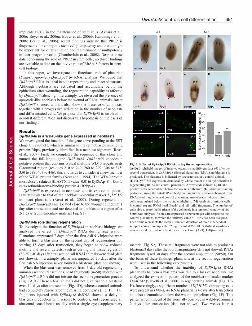
When the blastema was removed from 3-day-old regeneratinganimals (second transection), head fragments (n50) injected withDjRbAp48 dsRNA did not initiate the second regeneration process(Fig. 1A,B). These RNAi animals did not give rise to a blastemaeven 14 days after transection (Fig. 1D), whereas control animalshad completely regenerated the missing body parts (Fig. 1C). Tailfragments injected with DjRbAp48 dsRNA showed a delay inblastema production with respect to controls, and regenerated anabnormal, small head, usually with a single eye (supplementary
material Fig. S2). These tail fragments were not able to produce ablastema 3 days after the fourth amputation (data not shown). RNAifragments lysed 30 days after the second amputation (50/50). Onthe basis of these findings, planarians at the second regenerationwere used in the following experiments.
To understand whether the inability of DjRbAp48 RNAiplanarians to form a blastema was due to a loss of neoblasts, weanalyzed the expression pattern of the neoblast molecular markerDjMCM2 (Salvetti et al., 2000) in regenerating animals (Fig. 1E-H). Interestingly, a significant number of DjMCM2-expressing cellswere present in DjRbAp48 RNAi planarians 4 days after transectionand also accumulated below the wound epithelium (Fig. 1F). Thispattern is reminiscent of that normally observed in wild-type animals2 days after transection (data not shown). Two weeks later, a
Fig. 1. Effect of DjRbAp48 RNAi during tissue regeneration.(A-D)Brightfield images of injected organisms at different days (d) after thesecond transection. In DjRbAp48-silenced planarians (RNAi), no blastema isproduced. The blastema is indicated by two asterisks in a control animal.(E-H)DjMCM2 expression visualized by whole-mount in situ hybridization inregenerating RNAi and control planarians. Arrowheads indicate DjMCM2-positive cells accumulated below the wound epithelium. (I-L)Immunostainingperformed using the anti-H3P antibody on longitudinal sections obtained fromRNAi head fragments and control planarians. Arrowheads indicate mitoticcells accumulated below the wound epithelium. (M)Analysis of mitotic cellsin control (c) and RNAi head (heads) and tail (tails) fragments. The number ofcells able to enter the M-phase of the cell cycle in a temporal window of sixhours was analyzed. Values are expressed as percentages with respect to thecontrol planarians, to which the arbitrary value of 100% has been assigned.Each value represents the mean ± standard deviation of three independentsamples counted in duplicate. **Significant at P<0.01. Statistical significancewas assessed by Student’s t-test. Scale bars: 1 mm (A-H); 150m (I-L).
Jour
nal o
f Cel
l Sci
ence
692
reduction in the number of DjMCM2-positive cells was observedin the parenchyma, but significant accumulation of positive cellswas still detected below the wound epithelium (Fig. 1G,H).
Because a significant number of neoblasts were present inDjRbAp48 RNAi planarians that are unable to produce aregenerative blastema, we wondered whether such neoblasts wereable to proliferate. To this aim, we used the mitotic cell markeranti-phosphohistone H3 antibody (anti-H3P) (Newmark andSánchez Alvarado, 2000; Wei et al., 1998) to count the number ofcells able to enter the M-phase of the cell cycle. Although mitoticcells were detected both in the parenchyma and below the woundepithelium of amputated DjRbAp48-silenced planarians 4 days aftercutting (Fig. 1I,J), the number of mitotic cells was reduced withrespect to controls (Fig. 1M). At 22 days after transection, the mitoticneoblasts spread all over the parenchyma were strongly reduced innumber in RNAi animals in comparison to controls, but severalproliferating neoblasts still accumulated below the woundepithelium (Fig. 1K,L). We also investigated whether DjRbAp48RNAi might affect tissue differentiation during the regeneration ofthe head in tail-injected fragments able to produce a small blastema.To this aim, we analyzed the expression of DjSyt (Tazaki et al.,1999), DjAHNAK (Rossi et al., 2007) and Djinx1 (Nogi and Levin,2005), molecular markers of the nervous system, epidermis and gut,respectively, by whole-mount in situ hybridization. Although the
Journal of Cell Science 123 (5)
RNAi tail fragments showed a significant number of neoblasts,RNAi animals regenerated a smaller nervous system in comparisonto controls (supplementary material Fig. S2). In addition, wedetected defects in gut regeneration (i.e. failure in regenerating theanterior gut branch), as well as reduced epithelial cell density, afterDjRbAp48 silencing (supplementary material Fig. S2).
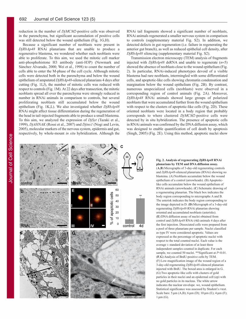
Transmission electron microscopy (TEM) analysis of fragmentsinjected with DjRbAp48 dsRNA and unable to regenerate (n5)showed the absence of neoblasts close to the wound epithelium (Fig.2). In particular, RNAi-induced phenotypes devoid of a visibleblastema had rare neoblasts, intermingled with some differentiatedcells, and apoptotic-like cells showing chromatin condensation andmargination below the wound epithelium (Fig. 2B). By contrast,numerous unspecialized cells (neoblasts) were observed in acorresponding region of control animals (Fig. 2A). Moreover,DjRbAp48 RNAi animals showed large groups of polarizedneoblasts that were accumulated further from the wound epitheliumwith respect to the clusters of apoptotic-like cells (Fig. 2D). Theseoriented neoblasts were located in a body region that probablycorresponds to where clustered DjMCM2-positive cells weredetected by in situ hybridization. The presence of apoptotic cellsin RNAi animals was confirmed by the DNA diffusion assay, whichwas designed to enable quantification of cell death by apoptosis(Singh, 2005) (Fig. 2E). Using this method, apoptotic nuclei show
Fig. 2. Analysis of regenerating DjRbAp48 RNAiplanarians by TEM and DNA diffusion assay.(A,B)Micrographs of 3-day-old regenerating controlsand DjRbAp48-silenced planarians (RNAi) showing noblastema. (A)Neoblasts accumulate below the woundepithelium of a control (arrowheads). (B)Apoptotic-like cells accumulate below the wound epithelium ofRNAi animals (arrowheads). (C)Schematic drawing ofa regenerating planarian. The black box indicates thebody region corresponding to micrographs A and B.The asterisk indicates the body region corresponding tothe image depicted in D. (D)Micrograph of a 3-day-oldregenerating DjRbAp48-RNAi planarian showingoriented and accumulated neoblasts (asterisks).(E)DNA diffusion assay of nuclei obtained fromcontrol and DjRbAp48 RNAi (4d) animals 4 days afterthe first injection. Dissociated cells were prepared froma pool of three planarians per sample. Nuclei classifiedas type IV were considered apoptotic. Values areexpressed as the percentage of apoptotic nuclei withrespect to the total counted nuclei. Each value is theaverage ± standard deviation of at least threeindependent samples counted in duplicate. For eachsample, we counted 50 nuclei. **Significant at P<0.01.(F,G)Analysis of BrdU-positive cells by TEM.(F)Low-magnification image of the wound region of a3-day-old regenerating DjRbAp48-silenced planarianinjected with BrdU. The boxed area is enlarged in G.(G)Two apoptotic-like cells with clusters of goldparticles in their nuclei and an epidermal cell (ep) withno gold particles in its nucleus. The white arrowindicates the nuclear envelope. we, wound epithelium.Statistical significance was assessed by Student’s t-test.Scale bars: 5m (A,B); 6m (D); 10m (E); 4m (F);1m (G).
Jour
nal o
f Cel
l Sci
ence
693DjRbAp48 controls cell differentiation
a halo of granular DNA with hazy outer boundaries. Apoptotic cellscan be distinguished from necrotic ones, as the latter exhibitunusually large, homogeneous nuclei with clearly definedboundaries (Singh, 2000).
To understand whether the clusters of apoptotic-like cells thataccumulated below the wound region of regenerating RNAi animalswere dying neoblasts, we injected BrdU into RNAi animals and thenanalyzed BrdU incorporation by TEM analysis. As neoblasts are theonly proliferating cells in asexual planarians, we expected to findBrdU in neoblasts, in neoblast differentiating progeny and in somedifferentiated cells. We detected gold particles in the nuclei ofclustered apoptotic-like cells located below the wound epithelium,but not in the nuclei of epithelial cells 3 and 6 days after RNAi (Fig.2F), a time sufficient for epithelial cell differentiation (Newmarkand Sánchez Alvarado, 2000). Fig. 2G shows the nuclear envelopeand the chromatin structures of a BrdU-positive cell.
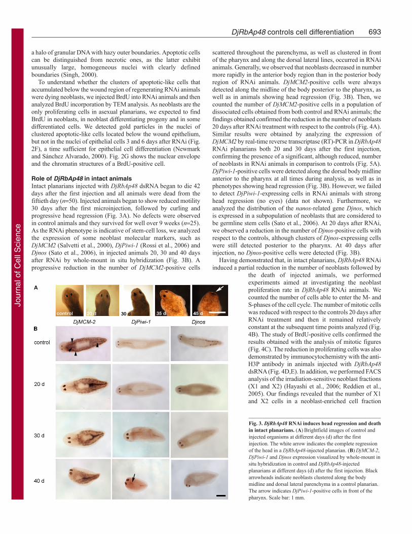
Role of DjRbAp48 in intact animalsIntact planarians injected with DjRbAp48 dsRNA began to die 42days after the first injection and all animals were dead from thefiftieth day (n50). Injected animals began to show reduced motility30 days after the first microinjection, followed by curling andprogressive head regression (Fig. 3A). No defects were observedin control animals and they survived for well over 9 weeks (n25).As the RNAi phenotype is indicative of stem-cell loss, we analyzedthe expression of some neoblast molecular markers, such asDjMCM2 (Salvetti et al., 2000), DjPiwi-1 (Rossi et al., 2006) andDjnos (Sato et al., 2006), in injected animals 20, 30 and 40 daysafter RNAi by whole-mount in situ hybridization (Fig. 3B). Aprogressive reduction in the number of DjMCM2-positive cells
scattered throughout the parenchyma, as well as clustered in frontof the pharynx and along the dorsal lateral lines, occurred in RNAianimals. Generally, we observed that neoblasts decreased in numbermore rapidly in the anterior body region than in the posterior bodyregion of RNAi animals. DjMCM2-positive cells were alwaysdetected along the midline of the body posterior to the pharynx, aswell as in animals showing head regression (Fig. 3B). Then, wecounted the number of DjMCM2-positive cells in a population ofdissociated cells obtained from both control and RNAi animals; thefindings obtained confirmed the reduction in the number of neoblasts20 days after RNAi treatment with respect to the controls (Fig. 4A).Similar results were obtained by analyzing the expression ofDjMCM2 by real-time reverse transcriptase (RT)-PCR in DjRbAp48RNAi planarians both 20 and 30 days after the first injection,confirming the presence of a significant, although reduced, numberof neoblasts in RNAi animals in comparison to controls (Fig. 5A).DjPiwi-1-positive cells were detected along the dorsal body midlineanterior to the pharynx at all times during analysis, as well as inphenotypes showing head regression (Fig. 3B). However, we failedto detect DjPiwi-1-expressing cells in RNAi animals with stronghead regression (no eyes) (data not shown). Furthermore, weanalyzed the distribution of the nanos-related gene Djnos, whichis expressed in a subpopulation of neoblasts that are considered tobe germline stem cells (Sato et al., 2006). At 20 days after RNAi,we observed a reduction in the number of Djnos-positive cells withrespect to the controls, although clusters of Djnos-expressing cellswere still detected posterior to the pharynx. At 40 days afterinjection, no Djnos-positive cells were detected (Fig. 3B).
Having demonstrated that, in intact planarians, DjRbAp48 RNAiinduced a partial reduction in the number of neoblasts followed by
the death of injected animals, we performedexperiments aimed at investigating the neoblastproliferation rate in DjRbAp48 RNAi animals. Wecounted the number of cells able to enter the M- andS-phases of the cell cycle. The number of mitotic cellswas reduced with respect to the controls 20 days afterRNAi treatment and then it remained relativelyconstant at the subsequent time points analyzed (Fig.4B). The study of BrdU-positive cells confirmed theresults obtained with the analysis of mitotic figures(Fig. 4C). The reduction in proliferating cells was alsodemonstrated by immunocytochemistry with the anti-H3P antibody in animals injected with DjRbAp48dsRNA (Fig. 4D,E). In addition, we performed FACSanalysis of the irradiation-sensitive neoblast fractions(X1 and X2) (Hayashi et al., 2006; Reddien et al.,2005). Our findings revealed that the number of X1and X2 cells in a neoblast-enriched cell fraction
Fig. 3. DjRbAp48 RNAi induces head regression and deathin intact planarians. (A)Brightfield images of control andinjected organisms at different days (d) after the firstinjection. The white arrow indicates the complete regressionof the head in a DjRbAp48-injected planarian. (B)DjMCM-2,DjPiwi-1 and Djnos expression visualized by whole-mount insitu hybridization in control and DjRbAp48-injectedplanarians at different days (d) after the first injection. Blackarrowheads indicate neoblasts clustered along the bodymidline and dorsal lateral parenchyma in a control planarian.The arrow indicates DjPiwi-1-positive cells in front of thepharynx. Scale bar: 1 mm.
Jour
nal o
f Cel
l Sci
ence

694
obtained from RNAi animals showing no head regression wasreduced with respect to controls (Fig. 4F). FACS analysis alsorevealed that Xis cells, representing the X-ray-insensitive cellfraction, were only slightly reduced in number (10%). When weanalyzed the number of total cells in a cell fraction enriched indifferentiated cells (cell diameter >25 m) obtained from controlsand RNAi animals, we found a stronger reduction (30%) indifferentiated (Xis) cells upon RNAi (Fig. 4F). To further verifythe reduction in the number of differentiated cells after RNAi, we
Journal of Cell Science 123 (5)
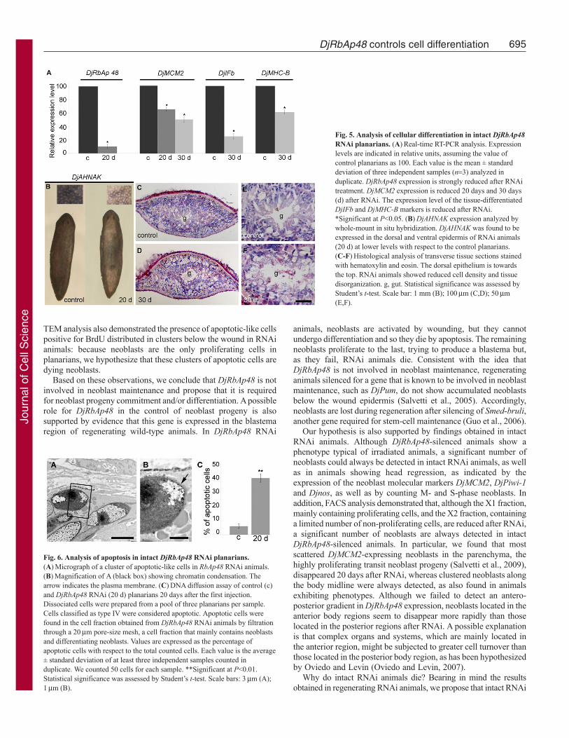
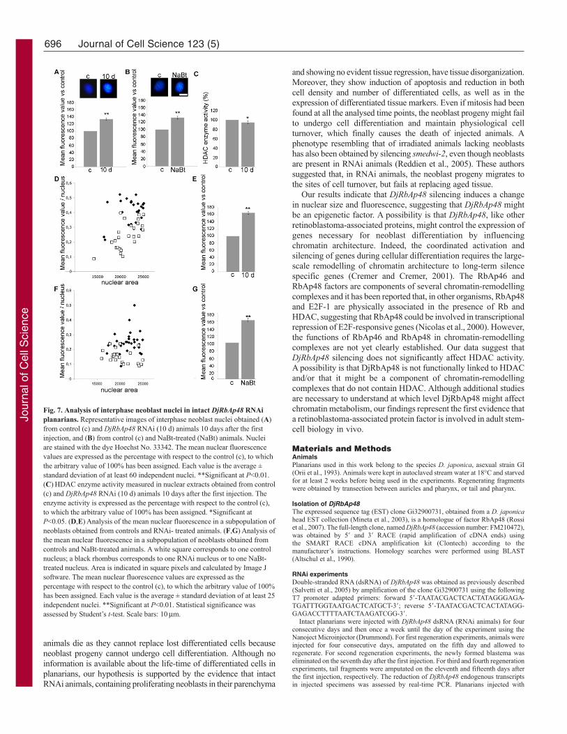
analysed the expression of some markers of differentiated cells byreal-time RT-PCR [DjIFb (Tazaki et al., 2002); DjMHC-B(Kobayashi et al., 1998)] and whole-mount in situ hybridization(DjAHNAK). The results demonstrated that these markers arereduced in RNAi animals with respect to the controls (Fig. 5A,B).Histological analyses of DjRbAp48-injected animals, showing nogross morphological tissue regression, revealed a loss of tissueorganization and a reduction in cell density (Fig. 5C-F). In addition,ultrastructural analysis showed the presence of apoptotic-like cellsin animals 20 and 30 days after RNAi, but not in controls (Fig.6A,B). These findings were confirmed by a DNA diffusion assay,which indicated that a significant number of apoptotic cells werepresent in neoblast-enriched cell fractions, but not in a suspensionenriched in differentiated cells, obtained from animals killed 20 daysafter DjRbAp48 RNAi with respect to the control planarians (Fig.6C).
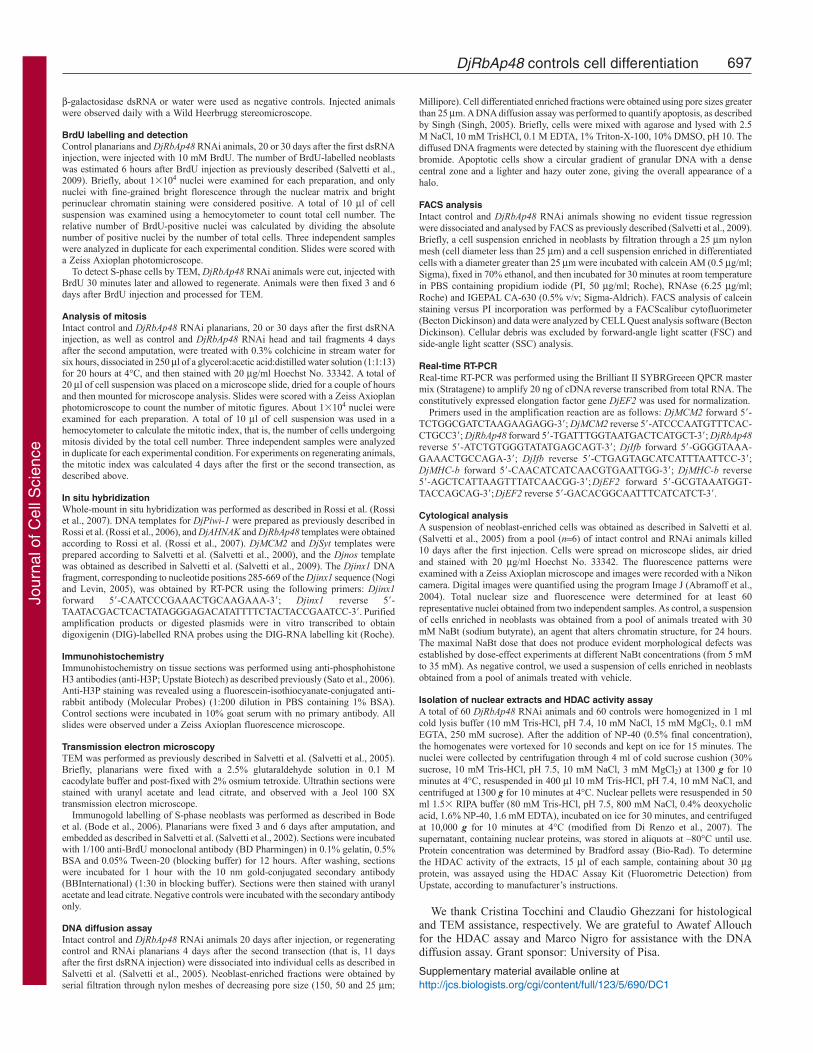
DjRbAp48 RNAi effect on chromatin architectureBecause RbAp46 and RbAp48 factors are involved in chromatinmetabolism at different levels, we wondered whether DjRbAp48silencing induced changes in neoblast chromatin. To this aim, weanalysed the size and the amount of fluorescence emitted byHoechst-stained neoblast nuclei obtained from animals killed 10days after the first dsRNA injection, a time at which planarians donot show phenotypes (Fig. 7). The size of neoblast interphase nucleiwas greater (10%) in RNAi animals compared to controls (data notshown) and the mean nuclear fluorescence of neoblasts in RNAianimals was 35% higher with respect to control interphase nuclei(Fig. 7A). Interestingly, a total nuclear fluorescence 65% higherthan that of the controls was detected when the analysis wasperformed on a selected population of neoblasts with a nuclear areabelow 25,000 square pixels (Fig. 7D,E). Similarly, the treatment ofplanarians with NaBt, an agent known to be capable of alteringchromatin structure (Davie, 2003), induced an increase in the totalfluorescence of neoblast nuclei compared to the untreated controls(Fig. 7B,F,G). To test the effect of DjRbAp48 RNAi on HDACenzyme activity, we prepared nuclear extracts from RNAi animalsand controls and performed an enzymatic activity assay. Fig. 7Cshows that HDAC activity was not significantly affected byDjRbAp48 silencing.
DiscussionA sequence of events produces a complete and functionalregenerated planarian. After wound closure, stem cells proliferate,migrate and accumulate, thus generating a blastema. The neoblastprogeny within the blastema differentiate and organize, and the lostbody parts are then restored.
DjRbAp48 is expressed in neoblasts (Rossi et al., 2007) andanimals silenced for its expression are not able to produce ablastema, as is the case for X-ray-treated animals devoid ofneoblasts as a consequence of irradiation (Rossi et al., 2008).However, the loss of neoblasts is not the primary cause ofregeneration failure in DjRbAp48 RNAi animals. Indeed, we foundthat neoblasts still respond to the wound by proliferating andaccumulating under the epithelium of DjRbAp48-silenced animalsthat are unable to generate a functional blastema.
Interestingly, regenerating DjRbAp48 RNAi animals have ahigher number of apoptotic cells with respect to control planarians.TEM analysis shows the presence of clusters of apoptotic-like cellswith a high nucleus:cytoplasm ratio close to the wound, in a regioncorresponding to where the blastema is formed in control animals.
Fig. 4. Analysis of proliferating cells in intact DjRbAp48 RNAi planarians.(A)Representative images of cells expressing the DjMCM-2 marker. DjMCM-2-positive cells were counted in control (c) and RNAi animals (20 d). Valuesare expressed as percentages with respect to the control, to which the arbitraryvalue of 100% has been assigned. Each value is the mean ± standard deviationof three independent samples counted in duplicate. *Significant at P<0.05.(B,C)Analysis of mitotic and S-phase cells in control (c) and RNAi animals(20 d, 30 d). The number of cells able to enter the M- or S-phases of the cellcycle in a temporal window of 6 h was analyzed. Values are expressed aspercentages with respect to the control planarians, to which the arbitrary valueof 100% has been given. Each value is the mean ± standard deviation of atleast three independent samples counted in duplicate. **Significant at P<0.01.(D,E)Distribution of mitotic cells by immunofluorescence using anti-H3P ontransverse sections obtained from the body region anterior to the pharynx. Thedorsal epithelium is towards the top. The asterisks indicate the nervous system.(F)FACS profile of PI- and calcein-labelled cells. The analysis was performedin a neoblast-enriched population of cells (diameter <25m) and in apopulation of cells enriched in differentiated cells (diameter >25m) obtainedfrom control (c) and DjRbAp48 RNAi (30 d) planarians 30 days after the firstinjection. X1 (red) and X2 (blue) populations are irradiation sensitive, Xis(black) is an X-ray-insensitive cell fraction. Statistical significance wasassessed by Student’s t-test. Scale bars: 10m (A); 200m (D,E).
Jour
nal o
f Cel
l Sci
ence
695DjRbAp48 controls cell differentiation
TEM analysis also demonstrated the presence of apoptotic-like cellspositive for BrdU distributed in clusters below the wound in RNAianimals: because neoblasts are the only proliferating cells inplanarians, we hypothesize that these clusters of apoptotic cells aredying neoblasts.
Based on these observations, we conclude that DjRbAp48 is notinvolved in neoblast maintenance and propose that it is requiredfor neoblast progeny commitment and/or differentiation. A possiblerole for DjRbAp48 in the control of neoblast progeny is alsosupported by evidence that this gene is expressed in the blastemaregion of regenerating wild-type animals. In DjRbAp48 RNAi
animals, neoblasts are activated by wounding, but they cannotundergo differentiation and so they die by apoptosis. The remainingneoblasts proliferate to the last, trying to produce a blastema but,as they fail, RNAi animals die. Consistent with the idea thatDjRbAp48 is not involved in neoblast maintenance, regeneratinganimals silenced for a gene that is known to be involved in neoblastmaintenance, such as DjPum, do not show accumulated neoblastsbelow the wound epidermis (Salvetti et al., 2005). Accordingly,neoblasts are lost during regeneration after silencing of Smed-bruli,another gene required for stem-cell maintenance (Guo et al., 2006).
Our hypothesis is also supported by findings obtained in intactRNAi animals. Although DjRbAp48-silenced animals show aphenotype typical of irradiated animals, a significant number ofneoblasts could always be detected in intact RNAi animals, as wellas in animals showing head regression, as indicated by theexpression of the neoblast molecular markers DjMCM2, DjPiwi-1and Djnos, as well as by counting M- and S-phase neoblasts. Inaddition, FACS analysis demonstrated that, although the X1 fraction,mainly containing proliferating cells, and the X2 fraction, containinga limited number of non-proliferating cells, are reduced after RNAi,a significant number of neoblasts are always detected in intactDjRbAp48-silenced animals. In particular, we found that mostscattered DjMCM2-expressing neoblasts in the parenchyma, thehighly proliferating transit neoblast progeny (Salvetti et al., 2009),disappeared 20 days after RNAi, whereas clustered neoblasts alongthe body midline were always detected, as also found in animalsexhibiting phenotypes. Although we failed to detect an antero-posterior gradient in DjRbAp48 expression, neoblasts located in theanterior body regions seem to disappear more rapidly than thoselocated in the posterior regions after RNAi. A possible explanationis that complex organs and systems, which are mainly located inthe anterior region, might be subjected to greater cell turnover thanthose located in the posterior body region, as has been hypothesizedby Oviedo and Levin (Oviedo and Levin, 2007).
Why do intact RNAi animals die? Bearing in mind the resultsobtained in regenerating RNAi animals, we propose that intact RNAi
Fig. 5. Analysis of cellular differentiation in intact DjRbAp48RNAi planarians. (A)Real-time RT-PCR analysis. Expressionlevels are indicated in relative units, assuming the value ofcontrol planarians as 100. Each value is the mean ± standarddeviation of three independent samples (n3) analyzed induplicate. DjRbAp48 expression is strongly reduced after RNAitreatment. DjMCM2 expression is reduced 20 days and 30 days(d) after RNAi. The expression level of the tissue-differentiatedDjIFb and DjMHC-B markers is reduced after RNAi.*Significant at P<0.05. (B)DjAHNAK expression analyzed bywhole-mount in situ hybridization. DjAHNAK was found to beexpressed in the dorsal and ventral epidermis of RNAi animals(20 d) at lower levels with respect to the control planarians.(C-F)Histological analysis of transverse tissue sections stainedwith hematoxylin and eosin. The dorsal epithelium is towardsthe top. RNAi animals showed reduced cell density and tissuedisorganization. g, gut. Statistical significance was assessed byStudent’s t-test. Scale bar: 1 mm (B); 100m (C,D); 50m(E,F).
Fig. 6. Analysis of apoptosis in intact DjRbAp48 RNAi planarians.(A)Micrograph of a cluster of apoptotic-like cells in RbAp48 RNAi animals.(B)Magnification of A (black box) showing chromatin condensation. Thearrow indicates the plasma membrane. (C)DNA diffusion assay of control (c)and DjRbAp48 RNAi (20 d) planarians 20 days after the first injection.Dissociated cells were prepared from a pool of three planarians per sample.Cells classified as type IV were considered apoptotic. Apoptotic cells werefound in the cell fraction obtained from DjRbAp48 RNAi animals by filtrationthrough a 20m pore-size mesh, a cell fraction that mainly contains neoblastsand differentiating neoblasts. Values are expressed as the percentage ofapoptotic cells with respect to the total counted cells. Each value is the average± standard deviation of at least three independent samples counted induplicate. We counted 50 cells for each sample. **Significant at P<0.01.Statistical significance was assessed by Student’s t-test. Scale bars: 3m (A);1m (B).
Jour
nal o
f Cel
l Sci
ence
696
animals die as they cannot replace lost differentiated cells becauseneoblast progeny cannot undergo cell differentiation. Although noinformation is available about the life-time of differentiated cells inplanarians, our hypothesis is supported by the evidence that intactRNAi animals, containing proliferating neoblasts in their parenchyma
Journal of Cell Science 123 (5)
and showing no evident tissue regression, have tissue disorganization.Moreover, they show induction of apoptosis and reduction in bothcell density and number of differentiated cells, as well as in theexpression of differentiated tissue markers. Even if mitosis had beenfound at all the analysed time points, the neoblast progeny might failto undergo cell differentiation and maintain physiological cellturnover, which finally causes the death of injected animals. Aphenotype resembling that of irradiated animals lacking neoblastshas also been obtained by silencing smedwi-2, even though neoblastsare present in RNAi animals (Reddien et al., 2005). These authorssuggested that, in RNAi animals, the neoblast progeny migrates tothe sites of cell turnover, but fails at replacing aged tissue.
Our results indicate that DjRbAp48 silencing induces a changein nuclear size and fluorescence, suggesting that DjRbAp48 mightbe an epigenetic factor. A possibility is that DjRbAp48, like otherretinoblastoma-associated proteins, might control the expression ofgenes necessary for neoblast differentiation by influencingchromatin architecture. Indeed, the coordinated activation andsilencing of genes during cellular differentiation requires the large-scale remodelling of chromatin architecture to long-term silencespecific genes (Cremer and Cremer, 2001). The RbAp46 andRbAp48 factors are components of several chromatin-remodellingcomplexes and it has been reported that, in other organisms, RbAp48and E2F-1 are physically associated in the presence of Rb andHDAC, suggesting that RbAp48 could be involved in transcriptionalrepression of E2F-responsive genes (Nicolas et al., 2000). However,the functions of RbAp46 and RbAp48 in chromatin-remodellingcomplexes are not yet clearly established. Our data suggest thatDjRbAp48 silencing does not significantly affect HDAC activity.A possibility is that DjRbAp48 is not functionally linked to HDACand/or that it might be a component of chromatin-remodellingcomplexes that do not contain HDAC. Although additional studiesare necessary to understand at which level DjRbAp48 might affectchromatin metabolism, our findings represent the first evidence thata retinoblastoma-associated protein factor is involved in adult stem-cell biology in vivo.
Materials and MethodsAnimalsPlanarians used in this work belong to the species D. japonica, asexual strain GI(Orii et al., 1993). Animals were kept in autoclaved stream water at 18°C and starvedfor at least 2 weeks before being used in the experiments. Regenerating fragmentswere obtained by transection between auricles and pharynx, or tail and pharynx.
Isolation of DjRbAp48The expressed sequence tag (EST) clone Gi32900731, obtained from a D. japonicahead EST collection (Mineta et al., 2003), is a homologue of factor RbAp48 (Rossiet al., 2007). The full-length clone, named DjRbAp48 (accession number: FM210472),was obtained by 5� and 3� RACE (rapid amplification of cDNA ends) usingthe SMART RACE cDNA amplification kit (Clontech) according to themanufacturer’s instructions. Homology searches were performed using BLAST(Altschul et al., 1990).
RNAi experimentsDouble-stranded RNA (dsRNA) of DjRbAp48 was obtained as previously described(Salvetti et al., 2005) by amplification of the clone Gi32900731 using the followingT7 promoter adapted primers: forward 5�-TAATACGACTCACT ATAGGGAGA -TGATTTGGTAATGACTCATGCT-3�; reverse 5�-TAATACGA CTCACTATAGG -GA GACCTTTTAATCTAAGATCGG-3�.
Intact planarians were injected with DjRbAp48 dsRNA (RNAi animals) for fourconsecutive days and then once a week until the day of the experiment using theNanoject Microinjector (Drummond). For first regeneration experiments, animals wereinjected for four consecutive days, amputated on the fifth day and allowed toregenerate. For second regeneration experiments, the newly formed blastema waseliminated on the seventh day after the first injection. For third and fourth regenerationexperiments, tail fragments were amputated on the eleventh and fifteenth days afterthe first injection, respectively. The reduction of DjRbAp48 endogenous transcriptsin injected specimens was assessed by real-time PCR. Planarians injected with
Fig. 7. Analysis of interphase neoblast nuclei in intact DjRbAp48 RNAiplanarians. Representative images of interphase neoblast nuclei obtained (A)from control (c) and DjRbAp48 RNAi (10 d) animals 10 days after the firstinjection, and (B) from control (c) and NaBt-treated (NaBt) animals. Nucleiare stained with the dye Hoechst No. 33342. The mean nuclear fluorescencevalues are expressed as the percentage with respect to the control (c), to whichthe arbitrary value of 100% has been assigned. Each value is the average ±standard deviation of at least 60 independent nuclei. **Significant at P<0.01.(C)HDAC enzyme activity measured in nuclear extracts obtained from control(c) and DjRbAp48 RNAi (10 d) animals 10 days after the first injection. Theenzyme activity is expressed as the percentage with respect to the control (c),to which the arbitrary value of 100% has been assigned. *Significant atP<0.05. (D,E)Analysis of the mean nuclear fluorescence in a subpopulation ofneoblasts obtained from controls and RNAi- treated animals. (F,G)Analysis ofthe mean nuclear fluorescence in a subpopulation of neoblasts obtained fromcontrols and NaBt-treated animals. A white square corresponds to one controlnucleus; a black rhombus corresponds to one RNAi nucleus or to one NaBt-treated nucleus. Area is indicated in square pixels and calculated by Image Jsoftware. The mean nuclear fluorescence values are expressed as thepercentage with respect to the control (c), to which the arbitrary value of 100%has been assigned. Each value is the average ± standard deviation of at least 25independent nuclei. **Significant at P<0.01. Statistical significance wasassessed by Student’s t-test. Scale bars: 10m.
Jour
nal o
f Cel
l Sci
ence
697DjRbAp48 controls cell differentiation
b-galactosidase dsRNA or water were used as negative controls. Injected animalswere observed daily with a Wild Heerbrugg stereomicroscope.
BrdU labelling and detectionControl planarians and DjRbAp48 RNAi animals, 20 or 30 days after the first dsRNAinjection, were injected with 10 mM BrdU. The number of BrdU-labelled neoblastswas estimated 6 hours after BrdU injection as previously described (Salvetti et al.,2009). Briefly, about 1�104 nuclei were examined for each preparation, and onlynuclei with fine-grained bright florescence through the nuclear matrix and brightperinuclear chromatin staining were considered positive. A total of 10 l of cellsuspension was examined using a hemocytometer to count total cell number. Therelative number of BrdU-positive nuclei was calculated by dividing the absolutenumber of positive nuclei by the number of total cells. Three independent sampleswere analyzed in duplicate for each experimental condition. Slides were scored witha Zeiss Axioplan photomicroscope.
To detect S-phase cells by TEM, DjRbAp48 RNAi animals were cut, injected withBrdU 30 minutes later and allowed to regenerate. Animals were then fixed 3 and 6days after BrdU injection and processed for TEM.
Analysis of mitosisIntact control and DjRbAp48 RNAi planarians, 20 or 30 days after the first dsRNAinjection, as well as control and DjRbAp48 RNAi head and tail fragments 4 daysafter the second amputation, were treated with 0.3% colchicine in stream water forsix hours, dissociated in 250 l of a glycerol:acetic acid:distilled water solution (1:1:13)for 20 hours at 4°C, and then stained with 20 g/ml Hoechst No. 33342. A total of20 l of cell suspension was placed on a microscope slide, dried for a couple of hoursand then mounted for microscope analysis. Slides were scored with a Zeiss Axioplanphotomicroscope to count the number of mitotic figures. About 1�104 nuclei wereexamined for each preparation. A total of 10 l of cell suspension was used in ahemocytometer to calculate the mitotic index, that is, the number of cells undergoingmitosis divided by the total cell number. Three independent samples were analyzedin duplicate for each experimental condition. For experiments on regenerating animals,the mitotic index was calculated 4 days after the first or the second transection, asdescribed above.
In situ hybridizationWhole-mount in situ hybridization was performed as described in Rossi et al. (Rossiet al., 2007). DNA templates for DjPiwi-1 were prepared as previously described inRossi et al. (Rossi et al., 2006), and DjAHNAK and DjRbAp48 templates were obtainedaccording to Rossi et al. (Rossi et al., 2007). DjMCM2 and DjSyt templates wereprepared according to Salvetti et al. (Salvetti et al., 2000), and the Djnos templatewas obtained as described in Salvetti et al. (Salvetti et al., 2009). The Djinx1 DNAfragment, corresponding to nucleotide positions 285-669 of the Djinx1 sequence (Nogiand Levin, 2005), was obtained by RT-PCR using the following primers: Djinx1forward 5�-CAATCCCGAAACTGCAAGAAA-3�; Djinx1 reverse 5�-TAATACGACTCACTATAGGGAGACATATTTTCTACTACCGAATCC-3�. Purifiedamplification products or digested plasmids were in vitro transcribed to obtaindigoxigenin (DIG)-labelled RNA probes using the DIG-RNA labelling kit (Roche).
ImmunohistochemistryImmunohistochemistry on tissue sections was performed using anti-phosphohistoneH3 antibodies (anti-H3P; Upstate Biotech) as described previously (Sato et al., 2006).Anti-H3P staining was revealed using a fluorescein-isothiocyanate-conjugated anti-rabbit antibody (Molecular Probes) (1:200 dilution in PBS containing 1% BSA).Control sections were incubated in 10% goat serum with no primary antibody. Allslides were observed under a Zeiss Axioplan fluorescence microscope.
Transmission electron microscopyTEM was performed as previously described in Salvetti et al. (Salvetti et al., 2005).Briefly, planarians were fixed with a 2.5% glutaraldehyde solution in 0.1 Mcacodylate buffer and post-fixed with 2% osmium tetroxide. Ultrathin sections werestained with uranyl acetate and lead citrate, and observed with a Jeol 100 SXtransmission electron microscope.
Immunogold labelling of S-phase neoblasts was performed as described in Bodeet al. (Bode et al., 2006). Planarians were fixed 3 and 6 days after amputation, andembedded as described in Salvetti et al. (Salvetti et al., 2002). Sections were incubatedwith 1/100 anti-BrdU monoclonal antibody (BD Pharmingen) in 0.1% gelatin, 0.5%BSA and 0.05% Tween-20 (blocking buffer) for 12 hours. After washing, sectionswere incubated for 1 hour with the 10 nm gold-conjugated secondary antibody(BBInternational) (1:30 in blocking buffer). Sections were then stained with uranylacetate and lead citrate. Negative controls were incubated with the secondary antibodyonly.
DNA diffusion assayIntact control and DjRbAp48 RNAi animals 20 days after injection, or regeneratingcontrol and RNAi planarians 4 days after the second transection (that is, 11 daysafter the first dsRNA injection) were dissociated into individual cells as described inSalvetti et al. (Salvetti et al., 2005). Neoblast-enriched fractions were obtained byserial filtration through nylon meshes of decreasing pore size (150, 50 and 25 m;
Millipore). Cell differentiated enriched fractions were obtained using pore sizes greaterthan 25 m. A DNA diffusion assay was performed to quantify apoptosis, as describedby Singh (Singh, 2005). Briefly, cells were mixed with agarose and lysed with 2.5M NaCl, 10 mM TrisHCl, 0.1 M EDTA, 1% Triton-X-100, 10% DMSO, pH 10. Thediffused DNA fragments were detected by staining with the fluorescent dye ethidiumbromide. Apoptotic cells show a circular gradient of granular DNA with a densecentral zone and a lighter and hazy outer zone, giving the overall appearance of ahalo.
FACS analysisIntact control and DjRbAp48 RNAi animals showing no evident tissue regressionwere dissociated and analysed by FACS as previously described (Salvetti et al., 2009).Briefly, a cell suspension enriched in neoblasts by filtration through a 25 m nylonmesh (cell diameter less than 25 m) and a cell suspension enriched in differentiatedcells with a diameter greater than 25 m were incubated with calcein AM (0.5 g/ml;Sigma), fixed in 70% ethanol, and then incubated for 30 minutes at room temperaturein PBS containing propidium iodide (PI, 50 g/ml; Roche), RNAse (6.25 g/ml;Roche) and IGEPAL CA-630 (0.5% v/v; Sigma-Aldrich). FACS analysis of calceinstaining versus PI incorporation was performed by a FACScalibur cytofluorimeter(Becton Dickinson) and data were analyzed by CELL Quest analysis software (BectonDickinson). Cellular debris was excluded by forward-angle light scatter (FSC) andside-angle light scatter (SSC) analysis.
Real-time RT-PCRReal-time RT-PCR was performed using the Brilliant II SYBRGreeen QPCR mastermix (Stratagene) to amplify 20 ng of cDNA reverse transcribed from total RNA. Theconstitutively expressed elongation factor gene DjEF2 was used for normalization.
Primers used in the amplification reaction are as follows: DjMCM2 forward 5�-TCTGGCGATCTAAGAAGAGG-3�; DjMCM2 reverse 5�-ATCCCAATGTTTCAC-CTGCC3�; DjRbAp48 forward 5�-TGATTTGGTAATGACTCATGCT-3�; DjRbAp48reverse 5�-ATCTGTGGGTATATGAGCAGT-3�; DjIfb forward 5�-GGGGTAAA-GAAACTGCCAGA-3�; DjIfb reverse 5�-CTGAGTAGCATCATTTAATTCC-3�;DjMHC-b forward 5�-CAACATCATCAACGTGAATTGG-3�; DjMHC-b reverse5�-AGCTCATTAAGTTTATCAACGG-3�;DjEF2 forward 5�-GCGTAAATGGT-TACCAGCAG-3�;DjEF2 reverse 5�-GACACGGCAATTTCATCATCT-3�.
Cytological analysisA suspension of neoblast-enriched cells was obtained as described in Salvetti et al.(Salvetti et al., 2005) from a pool (n6) of intact control and RNAi animals killed10 days after the first injection. Cells were spread on microscope slides, air driedand stained with 20 g/ml Hoechst No. 33342. The fluorescence patterns wereexamined with a Zeiss Axioplan microscope and images were recorded with a Nikoncamera. Digital images were quantified using the program Image J (Abramoff et al.,2004). Total nuclear size and fluorescence were determined for at least 60representative nuclei obtained from two independent samples. As control, a suspensionof cells enriched in neoblasts was obtained from a pool of animals treated with 30mM NaBt (sodium butyrate), an agent that alters chromatin structure, for 24 hours.The maximal NaBt dose that does not produce evident morphological defects wasestablished by dose-effect experiments at different NaBt concentrations (from 5 mMto 35 mM). As negative control, we used a suspension of cells enriched in neoblastsobtained from a pool of animals treated with vehicle.
Isolation of nuclear extracts and HDAC activity assayA total of 60 DjRbAp48 RNAi animals and 60 controls were homogenized in 1 mlcold lysis buffer (10 mM Tris-HCl, pH 7.4, 10 mM NaCl, 15 mM MgCl2, 0.1 mMEGTA, 250 mM sucrose). After the addition of NP-40 (0.5% final concentration),the homogenates were vortexed for 10 seconds and kept on ice for 15 minutes. Thenuclei were collected by centrifugation through 4 ml of cold sucrose cushion (30%sucrose, 10 mM Tris-HCl, pH 7.5, 10 mM NaCl, 3 mM MgCl2) at 1300 g for 10minutes at 4°C, resuspended in 400 l 10 mM Tris-HCl, pH 7.4, 10 mM NaCl, andcentrifuged at 1300 g for 10 minutes at 4°C. Nuclear pellets were resuspended in 50ml 1.5� RIPA buffer (80 mM Tris-HCl, pH 7.5, 800 mM NaCl, 0.4% deoxycholicacid, 1.6% NP-40, 1.6 mM EDTA), incubated on ice for 30 minutes, and centrifugedat 10,000 g for 10 minutes at 4°C (modified from Di Renzo et al., 2007). Thesupernatant, containing nuclear proteins, was stored in aliquots at –80°C until use.Protein concentration was determined by Bradford assay (Bio-Rad). To determinethe HDAC activity of the extracts, 15 l of each sample, containing about 30 gprotein, was assayed using the HDAC Assay Kit (Fluorometric Detection) fromUpstate, according to manufacturer’s instructions.
We thank Cristina Tocchini and Claudio Ghezzani for histologicaland TEM assistance, respectively. We are grateful to Awatef Allouchfor the HDAC assay and Marco Nigro for assistance with the DNAdiffusion assay. Grant sponsor: University of Pisa.
Supplementary material available online athttp://jcs.biologists.org/cgi/content/full/123/5/690/DC1
Jour
nal o
f Cel
l Sci
ence
698 Journal of Cell Science 123 (5)
ReferencesAbramoff, M. D., Magelhaes, P. J. and Ram, S. J. (2004). Image processing with ImageJ.
Biophotonics International 11, 36-42.Altschul, S. F., Gish, W., Miller, W., Myers, E. W. and Lipman, D. J. (1990). Basic
local alignment search tool. J. Mol. Biol. 215, 403-410.Azuara, V., Perry, P., Sauer, S., Spivakov, M., Jørgensen, H. F., John, R. M., Gouti,
M., Casanova, M., Warnes, G., Merkenschlager, M. et al. (2006). Chromatinsignatures of pluripotent cell lines. Nat. Cell Biol. 8, 532-538.
Baguna, J., Salo, E. and Auladell, C. (1989). Regeneration and pattern formation inplanarians: III. Evidence that neoblasts are totipotent stem cells and the source of blastemacells. Development 107, 77-86.
Bar-Sagi, D. and Hall, A. (2000). Ras and Rho GTPases: a family reunion. Cell 103, 227-238.
Bode, A., Salvenmoser, W., Nimeth, K., Mahlknecht, M., Adamski, Z., Rieger, R. M.,Peter, R. and Ladurner, P. (2006). Immunogold-labeled S-phase neoblasts, total neoblastnumber, their distribution, and evidence for arrested neoblasts in Macrostomum lignano(Platyhelminthes, Rhabditophora). Cell Tissue Res. 325, 577-587.
Boyer, L. A., Mathur, D. and Jaenisch, R. (2006a). Molecular control of pluripotency.Curr. Opin. Genet. Dev. 16, 455-462.
Boyer, L. A., Plath, K., Zeitlinger, J., Brambrink, T., Medeiros, L. A., Lee, T. I., Levine,S. S., Wernig, M., Tajonar, A., Ray, M. K. et al. (2006b). Polycomb complexes repressdevelopmental regulators in murine embryonic stem cells. Nature 441, 349-353.
Chamberlain, S. J., Yee, D. and Magnuson, T. (2008). Polycomb repressive complex 2is dispensable for maintenance of embryonic stem cell pluripotency. Stem Cells 26, 1496-1505.
Cox, A. D. and Der, C. J. (2003). The dark side of Ras: regulation of apoptosis. Oncogene22, 8999-9006.
Cremer, T. and Cremer, C. (2001). Chromosome territories, nuclear architecture and generegulation in mammalian cells. Nat. Rev. Genet. 2, 292-301.
Davie, J. R. (2003). Inhibition of histone deacetylase activity by butyrate. J. Nutr. 133,2485-2493.
Di Renzo, F., Cappelletti, G., Broccia, M. L., Giovini, E. and Menegola, E. (2007).Boric acid inhibits histone deacetylases: a suggested mechanism to explain boric acid-related teratogenicity. Toxicol. Appl. Pharmacol. 220, 178-185.
Downward, J. (2003). Targeting RAS signalling pathways in cancer therapy. Nat. Rev.Cancer 3, 11-22.
Guo, T., Peter, A. H. and Newmark, P. A. (2006). A bruno-like gene is required for stemcell maintenance. Dev. Cell 11, 159-169.
Hancock, J. F. (2003). Ras proteins: different signals from different locations. Nat. Rev.Mol. Cell. Biol. 4, 373-384.
Hayashi, T., Asami, M., Higuchi, S., Shibata, N. and Agata, K. (2006). Isolation ofplanarian X-ray-sensitive stem cells by fluorescence-activated cell sorting. Dev. GrowthDiffer. 48, 371-380.
Hennig, L., Taranto, P., Walser, M., Schönrock, N. and Gruissem, W. (2003).Arabidopsis MSI1 is required for epigenetic maintenance of reproductive development.Development 130, 2555-2565.
Kamminga, L. M., Bystrykh, L. V., de Boer, A., Houwer, S., Douma, J., Weersing, E.,Dontje, B. and de Haan, G. (2006). The Polycomb group gene Ezh2 preventshematopoietic stem cell exhaustion. Blood 107, 2170-2179.
Kobayashi, C., Kobayashi, S., Orii, H., Watanabe, K. and Agata, K. (1998). Identificationof two distinct muscles in the planarian Dugesia japonica by their expression of myosinchain genes. Zool. Sci. 15, 861-869.
Lee, T. I., Jenner, R. G., Boyer, L. A., Guenther, M. G., Levine, S. S., Kumar, R. M.,Chevalier, B., Johnstone, S. E., Cole, M. F., Isono, K. et al. (2006). Control ofdevelopmental regulators by Polycomb in human embryonic stem cells. Cell 125, 301-313.
Loyola, A. and Almouzni, G. (2004). Histone chaperones, a supporting role in the limelight.Biochim. Biophys. Acta. 1677, 3-11.
Lu, X. and Horvitz, H. R. (1998). lin-35 and lin-53, two genes that antagonize a C. elegansRas pathway, encode proteins similar to Rb and its binding protein RbAp48. Cell 95,981-991.
Mineta, K., Nakazawa, M., Cebria, F., Ikeo, K., Agata, K. and Gojobori, T. (2003).Origin and evolutionary process of the CNS elucidated by comparative genomics analysisof planarian ESTs. Proc. Natl. Acad. Sci. USA 100, 7666-7671.
Morgan, T. H. (1898). Experimental studies of the regeneration of Planaria maculata.Arch. Entwm. Org. 7, 364-397.
Neer, E. J., Schmidt, C. J., Nambudripad, R. and Smith, T. F. (1994). The ancientregulatory-protein family of WD-repeat proteins. Nature 371, 297-300.
Newmark, P. A. and Sánchez Alvarado, A. (2000). Bromodeoxyuridine specifically labelsthe regenerative stem cells of planarians. Dev. Biol. 220, 142-153.
Nicolas, E., Morales, V., Magnaghi-Jaulin, L., Harel-Bellan, A., Richard-Foy, H. andTrouche, D. (2000). RbAp48 belongs to the histone deacetylase complex that associateswith the retinoblastoma protein. J. Biol. Chem. 275, 9797-9804.
Nogi, T. and Levin, M. (2005). Characterization of innexin gene expression and functionalroles of gap-junctional communication in planarian regeneration. Dev. Biol. 15, 314-335.
Orii, H., Agata, K. and Watanabe, K. (1993). POU-domain genes in planarian Dugesiajaponica: the structure and expression. Biochem. Biophys. Res. Commun. 192, 1395-1402.
Orii, H., Sakurai, T. and Watanabe, K. (2005). Distribution of the stem cells (neoblasts)in the planarian Dugesia japonica. Dev. Genes Evol. 215, 143-157.
Oviedo, N. J. and Levin, M. (2007). smedinx-11 is a planarian stem cell gap junctiongene required for regeneration and homeostasis. Development 134, 3121-3131.
Parthun, M. R., Widom, J. and Gottschling, D. E. (1996). The major cytoplasmic histoneacetyltransferase in yeast: links to chromatin replication and histone metabolism. Cell87, 85-94.
Philpott, A., Krude, T. and Laskey, R. (2000). Nuclear chaperones. Semin. Cell Dev.Biol. 11, 7-14.
Qian, Y. W. and Lee, E. Y. H. P. (1995). Dual retinoblastoma-binding proteins with propertiesrelated to a negative regulator of Ras in yeast. J. Biol. Chem. 270, 25507-25513.
Qian, Y. W., Wang, Y. C., Hollingsworth, R. E., Jr, Jones, D., Ling, N. and Lee, E. Y.(1993). A retinoblastoma-binding protein related to a negative regulator of Ras in yeast.Nature 364, 648-652.
Reddien, P. W., Oviedo, N. J., Jennings, J. R., Jenkin, J. C. and Sánchez Alvarado,A. (2005). SMEDWI-2 is a PIWI-like protein that regulates planarian stem cells. Science310, 1327-1330.
Rossi, L., Salvetti, A., Lena, A., Batistoni, R., Deri, P., Pugliesi, C., Loreti, E. andGremigni, V. (2006). DjPiwi-1, a member of the PAZ-Piwi gene family, defines asubpopulation of planarian stem cells. Dev. Genes Evol. 216, 335-346.
Rossi, L., Salvetti, A., Marincola, F. M., Lena, A., Deri, P., Mannini, L., Batistoni, R.,Wang, E. and Gremigni, V. (2007). Deciphering the molecular machinery of stem cells:a look at the neoblast gene expression profile. Genome Biol. 8, R62.
Rossi, L., Salvetti, A., Batistoni, R., Deri, P. and Gremigni, V. (2008). Planarians, a taleof stem cells. Cell Mol. Life Sci. 65, 16-23.
Ruggieri, R., Tanaka, K., Nakafuku, M., Kaziro, Y., Toh-e, A. and Matsumoto, K.(1989). MSI1, a negative regulator of the RAS-cAMP pathway in Saccharomycescerevisiae. Proc. Natl. Acad. Sci. USA 86, 8778-8782.
Salvetti, A., Rossi, L., Deri, P. and Batistoni, R. (2000). An MCM2-related gene isexpressed in proliferating cells of intact and regenerating planarians. Dev. Dyn. 218,603-614.
Salvetti, A., Lena, A., Rossi, L., Deri, P., Cecchettini, A., Batistoni, R. and Gremigni,V. (2002). Characterization of DeY1, a novel Y-box gene specifically expressed indifferentiating male germ cells of planarians. Gene Expr. Patterns 2, 195-200.
Salvetti, A., Rossi, L., Lena, A., Batistoni, R., Deri, P., Rainaldi, G., Locci, M. T.,Evangelista, M. and Gremigni, V. (2005). DjPum, a homologue of DrosophilaPumilio, is essential to planarian stem cell maintenance. Development 132, 1863-1874.
Salvetti, A., Rossi, L., Bonuccelli, L., Lena, A., Pugliesi, C., Rainaldi, G., Evangelista,M. and Gremigni, V. (2009). Adult stem cell plasticity: neoblast repopulation in non-lethally irradiated planarians. Dev. Biol. 328, 305-314.
Sato, K., Shibata, N., Orii, H., Amikura, R., Sakurai, T., Agata, K., Kobayashi, S. andWatanabe, K. (2006). Identification and origin of the germline stem cells as revealedby the expression of nanos-related gene in planarians. Dev. Growth Differ. 48, 615-628.
Scuto, A., Zhang, H., Zhao, H., Rivera, M., Yeatman, T. J., Jove, R. and Torres-Roca,J. F. (2007). RbAp48 regulates cytoskeletal organization and morphology by increasingK-Ras activity and signaling through mitogen-activated protein kinase. Cancer Res. 67,10317-10324.
Shields, J. M., Pruitt, K., McFall, A., Shaub, A. and Der, C. J. (2000). UnderstandingRas: “it ain’t over ‘til it’s over.” Trends Cell Biol. 10, 147-154.
Singh, N. P. (2000). A simple method for accurate estimation of apoptotic cells. Exp. CellRes. 256, 328-337.
Singh, N. P. (2005). Apoptosis assessment by the DNA diffusion assay. Methods Mol. Med.111, 55-67.
Taunton, J., Hassig, C. A. and Schreiber, S. L. (1996). A mammalian histone deacetylaserelated to the yeast transcriptional regulator Rpd3p. Science 272, 408-411.
Tazaki, A., Gaudieri, S., Ikeo, K., Gojobori, T., Watanabe, K. and Agata, K. (1999).Neural network in planarian revealed by an antibody against planarian synaptogaminhomologue. Biochem. Biophys. Res. Commun. 260, 426-432.
Tazaki, A., Kato, K., Orii, H., Agata, K. and Watanabe, K. (2002). The body marginof the planarian Dugesia japonica: characterization by the expression of an intermediatefilament gene. Dev. Genes Evol. 212, 365-373.
Verreault, A., Kaufman, P. D., Kobayashi, R. and Stillman, B. (1996). Nucleosomeassembly by a complex of CAF-1 and acetylated histones H3/H4. Cell 87, 95-104.
Wei, Y., Mizzen, C. A., Cook, R. G., Gorovsky, M. A. and Allis, C. D. (1998).Phosphorylation of histone H3 at serine 10 is correlated with chromosome condensationduring mitosis and meiosis in Tetrahymena. Proc. Natl. Acad. Sci. USA 95, 7480-7484.
Wolff, E. and Dubois, F. (1948). Sur la migration des cellules de régénération chez lesplanaires. Rev. Suisse Zool. 55, 218-227.
Jour
nal o
f Cel
l Sci
ence