
1 s2.0-s0272884213017057-main
6
CERAMICS INTERNATIONAL Available online at www.sciencedirect.com Ceramics International 40 (2014) 7425–7430 Food-directed synthesis of cerium oxide nanoparticles and their neurotoxicity effects Majid Darroudi a,b,n , Seyed Javad Hoseini c , Reza Kazemi Oskuee c,d , Hasan Ali Hosseini e , Leila Gholami b , Sina Gerayli f a Nuclear Medicine Research Center, Mashhad University of Medical Sciences, Mashhad, Iran b Department of Modern Sciences and Technologies, School of Medicine, Mashhad University of Medical Sciences, Mashhad, Iran c Department of Medical Biotechnology, School of Medicine, Mashhad University of Medical Sciences, Mashhad, Iran d Targeted Drug Delivery Research Center, Mashhad University of Medical Sciences, Mashhad, Iran e Chemistry Department, Payame Noor University, 19395-4697 Tehran, Iran f Department of Biology, Faculty of Sciences, Ferdowsi University of Mashhad, Mashhad, Iran Received 10 December 2013; received in revised form 21 December 2013; accepted 21 December 2013 Available online 14 January 2014 Abstract The use of food-directed and natural products for the synthesis of different nanoparticles (e.g., metal and metal oxide) is of enormous interest to modern nanoscience and nanotechnology. We have developed a facile and green chemistry method with bio-directed, and low cost materials for the synthesis of cerium oxide nanoparticles (CeO 2 -NPs) using honey. In this method, the conversion of cerium cations into CeO 2 -NPs was achieved via a sol–gel process in aqueous honey solutions. The synthesized CeO 2 -NPs were characterized by the following title: UV–vis spectroscopy, field emission scanning electron microscopy (FESEM), Fourier transform infrared spectroscopy (FT-IR), thermogravimetric (TGA– DTA) analysis, Energy dispersive spectrum (EDS), and powder X-ray diffraction (PXRD). Spherical CeO 2 -NPs were synthesized at different calcination temperatures and FESEM imaging along with its corresponding particles size distribution indicated the formation of nanoparticles in size of about 23 nm. The PXRD analysis revealed fluorite cubic structure for CeO 2 -NPs with preferential orientation at (111) reflection plane. In vitro cytotoxicity studies on neuro2A cells, a dose dependent toxicity with non-toxic effect of a concentration below about 25 mg/mL was illustrated. The synthesis of CeO 2 -NPs in aqueous honey solutions was found to be comparable to those obtained from conventional reduction methods that uses hazardous materials proving to be an excellent alternative for the preparation of CeO 2 -NPs, using food and bio-derived materials. & 2013 Elsevier Ltd and Techna Group S.r.l. All rights reserved. Keywords: A. Sol–gel processes; B. Electron microscopy; D. CeO 2 1. Introduction Cerium oxide nanoparticles (CeO 2 -NPs), as important rare- earth oxide materials, have attracted enormous interest in recent years due to its unique physicochemical properties that are significantly different from those of bulk materials [1]. Nanocrystalline CeO 2 -NPs have been considered as a key nanoscaled material for applications in catalysts [2], hydrogen storage materials [3], fuel cells [4], polishing materials [5], gas sensors [6], optical devices [7], ultraviolet absorbers [8], and biomedical science fields [9,10]. Crystalline CeO 2 -NPs have been synthesized by means of a variety of routes and techniques, including solution precipitation [11], sonochemical [12], hydrothermal [13], solvothermal [14], ball milling [15], thermal decomposition [16], spray pyrolysis [17], thermal hydrolysis [18], and sol–gel method [19–21]. However, the utility of the mentioned methods suffers several drawbacks like the use of high temperature and pressure, toxic reagents, long reaction time, requirement of external additives as a specific stabilizer, base and promoter during the reaction which limits www.elsevier.com/locate/ceramint 0272-8842/$ - see front matter & 2013 Elsevier Ltd and Techna Group S.r.l. All rights reserved. http://dx.doi.org/10.1016/j.ceramint.2013.12.089 n Corresponding author at: Nuclear Medicine Research Center, Mashhad University of Medical Sciences, Mashhad, Iran. Tel.: þ98 511 800 2286; fax: þ98 511 800 2287. E-mail addresses: [email protected], [email protected] (M. Darroudi).
-
Upload
normarieli-passalacqua -
Category
Documents
-
view
59 -
download
0
Transcript of 1 s2.0-s0272884213017057-main
CERAMICSINTERNATIONAL
Available online at www.sciencedirect.com
0272-8842/$ - sehttp://dx.doi.org/
nCorrespondinUniversity of Mfax: þ98 511 80
E-mail addredarroudim@mum
Ceramics International 40 (2014) 7425–7430www.elsevier.com/locate/ceramint
Food-directed synthesis of cerium oxide nanoparticles and theirneurotoxicity effects
Majid Darroudia,b,n, Seyed Javad Hoseinic, Reza Kazemi Oskueec,d, Hasan Ali Hosseinie,Leila Gholamib, Sina Geraylif
aNuclear Medicine Research Center, Mashhad University of Medical Sciences, Mashhad, IranbDepartment of Modern Sciences and Technologies, School of Medicine, Mashhad University of Medical Sciences, Mashhad, Iran
cDepartment of Medical Biotechnology, School of Medicine, Mashhad University of Medical Sciences, Mashhad, IrandTargeted Drug Delivery Research Center, Mashhad University of Medical Sciences, Mashhad, Iran
eChemistry Department, Payame Noor University, 19395-4697 Tehran, IranfDepartment of Biology, Faculty of Sciences, Ferdowsi University of Mashhad, Mashhad, Iran
Received 10 December 2013; received in revised form 21 December 2013; accepted 21 December 2013Available online 14 January 2014
Abstract
The use of food-directed and natural products for the synthesis of different nanoparticles (e.g., metal and metal oxide) is of enormous interest tomodern nanoscience and nanotechnology. We have developed a facile and green chemistry method with bio-directed, and low cost materials forthe synthesis of cerium oxide nanoparticles (CeO2-NPs) using honey. In this method, the conversion of cerium cations into CeO2-NPs wasachieved via a sol–gel process in aqueous honey solutions. The synthesized CeO2-NPs were characterized by the following title: UV–visspectroscopy, field emission scanning electron microscopy (FESEM), Fourier transform infrared spectroscopy (FT-IR), thermogravimetric (TGA–DTA) analysis, Energy dispersive spectrum (EDS), and powder X-ray diffraction (PXRD). Spherical CeO2-NPs were synthesized at differentcalcination temperatures and FESEM imaging along with its corresponding particles size distribution indicated the formation of nanoparticles insize of about 23 nm. The PXRD analysis revealed fluorite cubic structure for CeO2-NPs with preferential orientation at (111) reflection plane. Invitro cytotoxicity studies on neuro2A cells, a dose dependent toxicity with non-toxic effect of a concentration below about 25 mg/mL wasillustrated. The synthesis of CeO2-NPs in aqueous honey solutions was found to be comparable to those obtained from conventional reductionmethods that uses hazardous materials proving to be an excellent alternative for the preparation of CeO2-NPs, using food and bio-derivedmaterials.& 2013 Elsevier Ltd and Techna Group S.r.l. All rights reserved.
Keywords: A. Sol–gel processes; B. Electron microscopy; D. CeO2
1. Introduction
Cerium oxide nanoparticles (CeO2-NPs), as important rare-earth oxide materials, have attracted enormous interest inrecent years due to its unique physicochemical properties thatare significantly different from those of bulk materials [1].Nanocrystalline CeO2-NPs have been considered as a key
e front matter & 2013 Elsevier Ltd and Techna Group S.r.l. All ri10.1016/j.ceramint.2013.12.089
g author at: Nuclear Medicine Research Center, Mashhadedical Sciences, Mashhad, Iran. Tel.: þ98 511 800 2286;0 2287.sses: [email protected],s.ac.ir (M. Darroudi).
nanoscaled material for applications in catalysts [2], hydrogenstorage materials [3], fuel cells [4], polishing materials [5], gassensors [6], optical devices [7], ultraviolet absorbers [8], andbiomedical science fields [9,10]. Crystalline CeO2-NPs havebeen synthesized by means of a variety of routes andtechniques, including solution precipitation [11], sonochemical[12], hydrothermal [13], solvothermal [14], ball milling [15],thermal decomposition [16], spray pyrolysis [17], thermalhydrolysis [18], and sol–gel method [19–21]. However, theutility of the mentioned methods suffers several drawbacks likethe use of high temperature and pressure, toxic reagents, longreaction time, requirement of external additives as a specificstabilizer, base and promoter during the reaction which limits
ghts reserved.
M. Darroudi et al. / Ceramics International 40 (2014) 7425–74307426
the purity of the final product. Therefore, seeking a facile routefor low-cost, large-scale, controlled growth of CeO2-NPs atatmospheric pressure and lower temperatures is essential. Thesol–gel method has gained quite an interest among researcherssince it offers controlled consolidation, shape modulation,patterning of the nanostructures and a low processing tem-perature [22,23]. Recently, biomaterials have been used in thesynthesis of CeO2-NPs [10,20,21], due to their quality of beingbiodegradable and bioabsorbable with degradation productsthat are non-toxic.
Currently, honey has been used in the field of nanotechnol-ogy to apply green chemistry rules and environmentally benignsynthesis of nanoparticles [24–26]. Honey is a sweet viscousfluid which is produced from honeybees, and is mainlycomposed of carbohydrates, enzymes, vitamins, minerals andantioxidants [27]. Honey mediated biological synthesis has lotsof advantages over other types of biological methods, includ-ing avoidance of elaborate processes such as drying plantmaterials and the maintenance of cell cultures [26]. Here, wedemonstrated a sol–gel approach for the synthesis of ultrafineCeO2-NPs that has the advantages of being a simple process,bio-directed, easy to scale-up and of low cost. Cerium nitratewas used as the cerium source at different calcinationtemperatures and the synthesized samples were then character-ized through FESEM, PXRD, TGA–DTA, FT-IR, EDS, andUV–vis spectroscopy.
2. Materials and methods
2.1. Materials and reagents
All the materials used were of analytical grade and wereused without any purification. Cerium (III) nitrate hexahydratewas purchased from Fluka (Germany) and honey was pur-chased from the local market (Ghayour-Mobarhan Honey Co.,Mashhad – Iran). All glassware used in the laboratoryexperiments were cleaned with fresh solutions of HNO3/HCl(3:1, v/v), washed thoroughly with doubly distilled water, anddried before use. Double distilled water was used in allexperiments.
2.2. Synthesis of CeO2-NPs
To prepare 5.0 g of CeO2-NPs, 12.6 g of Ce(NO3)3 � 6H2Owas dissolved in 30 ml of distilled water and then stirred for30 min. Meanwhile, 25 g of honey was dissolved in 50 ml ofdistilled water and stirred for 15 min at room temperature toachieve a clear honey solution. Afterwards, the cerium nitratesolution was added to the honey solution, and the containerwas place in an oil bath with a temperature at 60 1C. Throughstirring for 6 h, a light yellow color resin was obtained anddivided into 4 parts to be individually heated at a rate of 4 1C/min up to the respective temperatures of 200 (C1), 400 (C2),600 (C3), and 800 1C (C4), then the products was maintainedfor 2 h at the specified temperatures in air to obtain CeO2-NPs.
2.3. Characterization of CeO2-NPs
The prepared CeO2-NPs were characterized by PXRD (Philips,X0pert, Cu Kα), FTIR (ST-IR\ST-SIR spectrometer), TGA (Q600),UV–vis (Evolution 300s Thermo Fisher Scientific), EDX (Leo1450VP), and FESEM (Carl Zeiss Supra 55VP).
2.4. Evaluation of neurotoxicity effect
The cytotoxicity of CeO2-NPs was evaluated by the methodusing 3-(4,5-dimethylthiazol-2-yl)-2,5-diphenyltetrazoliumbromide (MTT) assay [28]. Briefly, neuro2A cells were seededat a density of 1� 104 cells per well in 96-well plates andincubated for 24 h. Thereafter, the cells were treated withvarious concentrations of nanoparticles in the presence of 10%FBS. The calcined CeO2-NPs (C3) was suspended in a stocksolution at 5 μg/ml in a solution of dimethyl sulfoxide(DMSO)/double distilled water. After 24 h of incubation,20 μl of 5 mg/ml MTT in the PBS buffer was added to eachwell, and the cells were further incubated for 4 h at 37 1C. Themedium containing unreacted dye was discarded, and 100 μl ofDMSO was added to dissolve the formazan crystal formed bylive cells. Optical absorbance was measured at 590 nm(reference wavelength 630 nm) using a microplate reader(Statfax-2100, Awareness Technology, USA), and cell viabi-lity was expressed as a percent relative to untreated controlcells. Values of metabolic activity are presented as mean7SDof triplicates.
3. Results and discussion
In this section, we shall discuss the results of the synthesizedCeO2-NPs in aqueous honey solutions. In this study, weattempted the fabrication of CeO2-NPs by using the sol–gelmethod and honey as a greener capping and/or stabilizingagent. The extensive number of carbohydrates, enzymes, andvitamins containing hydroxyl and amine groups in the honeymatrix structure can facilitate the complexation of ceriumcations (Ce3þ ) to an initial molecular matrix. This structureenables honey to coat and stabilize cerium species and finallyCeO2-NPs while inhibiting their excessive aggregation orcrystal growth. As it is illustrated in Fig. 1, due to theincreased calcination temperature, the color of sol–gel derivedCeO2-NPs changed from black to lemon and finally to white.The thermogravimetric and derivative analysis (TGA/DTA)
curves of the as-prepared gel in an aqueous honey environmentare presented in Fig. 2. The heating process was started atabout 20 1C, and then increased up to 800 1C along with atemperature rate change of 10 1C/min. The first weight lossbetween 20 and 160 1C is an initial mass loss of 37.4% (bendsof Ed1 and Ed2) attributable to the loss of adsorbed water andcrystal water, since gradual dehydration of cerium hydroxidetakes place in this temperature range. The second stage appearsat 160–355 1C (48.5%) is attributed to the decomposition ofchemically bound groups, which corresponds to bends of Ed3and Ed4. In this case, it represents decomposition of honeymolecules to other organic compound(s). Further mass loss of

Fig. 1. Lab photo of honey-based synthesized CeO2-NPs at different calcina-tion temperatures.
Fig. 2. TGA–DTA curves of initial resin from 20 to 800 1C.
Fig. 3. UV–vis spectrum and band gap estimation (inset) of C3.
Fig. 4. PXRD patterns of honey-based synthesized CeO2-NPs in air atdifferent temperatures.
M. Darroudi et al. / Ceramics International 40 (2014) 7425–7430 7427
8.2% (Ed5) takes place in the range of 355–400 1C, associatedwith further oxidation of cerium components [29]. The totalmass loss of the sample is 94.1%. No weight loss between 400and 800 1C was detected on the TGA curve, which indicatesthe formation of CeO2-NPs as the stable product.
The typical UV–vis absorption peak of the C3 is representedin Fig. 3. The C3 was dispersed in water with concentration of0.1 wt% and had the solution used to perform the UV–vismeasurement. The spectrum revealed a characteristic absorp-tion peak at wavelength of 314 nm for C3, which can beassigned to the intrinsic band-gap absorption of CeO2-NPs dueto the electron transitions from the valence band to theconduction band. In other words, the absorption of the chargetransfer transition from O2p to Ce4f in CeO2-NPs produces theband at approximately 300 nm [30,31]. A common way toobtain the band gap of the materials with a direct band gapfrom the absorbance spectra is to get the first derivative of theabsorbance with respect to the photon energies. The band gapcan be estimated from the maximum in the derivative spectrumat the lower energy sides [32,33]. The derivative of theabsorbance of the CeO2-NPs illustrated in the inset of Fig. 3,which indicates a band-gap of 3.5 eV for the C3 and is higher
than the bulk experimental value (3.19 eV) [34]. For nanopar-ticles with particle sizes down to a few nanometers, the band-gap value is modified because of the quantum confinementeffect [35]. The good absorption of the CeO2-NPs in the UVregion proves the applicability of this product in such medicalapplication such as sun-screen protectors or antiseptic inointments.The CeO2-NP is a kind of typical calcium fluoride (CaF2)
structure with space group Fm3m. Fig. 4 portraits the PXRDpatterns of calcined CeO2-NPs in different temperatures thatwere synthesized in the honey substrate. The same crystalline
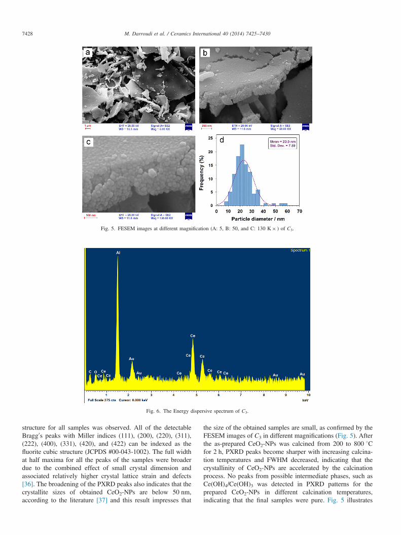
Fig. 5. FESEM images at different magnification (A: 5, B: 50, and C: 130 K� ) of C3.
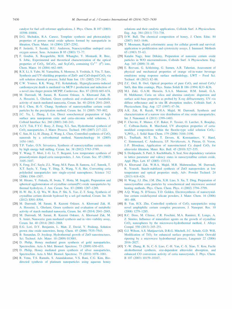
Fig. 6. The Energy dispersive spectrum of C3.
M. Darroudi et al. / Ceramics International 40 (2014) 7425–74307428
structure for all samples was observed. All of the detectableBragg0s peaks with Miller indices (111), (200), (220), (311),(222), (400), (331), (420), and (422) can be indexed as thefluorite cubic structure (JCPDS #00-043-1002). The full widthat half maxima for all the peaks of the samples were broaderdue to the combined effect of small crystal dimension andassociated relatively higher crystal lattice strain and defects[36]. The broadening of the PXRD peaks also indicates that thecrystallite sizes of obtained CeO2-NPs are below 50 nm,according to the literature [37] and this result impresses that
the size of the obtained samples are small, as confirmed by theFESEM images of C3 in different magnifications (Fig. 5). Afterthe as-prepared CeO2-NPs was calcined from 200 to 800 1Cfor 2 h, PXRD peaks become sharper with increasing calcina-tion temperatures and FWHM decreased, indicating that thecrystallinity of CeO2-NPs are accelerated by the calcinationprocess. No peaks from possible intermediate phases, such asCe(OH)4/Ce(OH)3 was detected in PXRD patterns for theprepared CeO2-NPs in different calcination temperatures,indicating that the final samples were pure. Fig. 5 illustrates

Fig. 7. The FT-IR spectrum of C3.
Fig. 8. Cell viability of neuro2A cells measured by the MTT assay for C3.
M. Darroudi et al. / Ceramics International 40 (2014) 7425–7430 7429
the FESEM images of C3 in high magnifications. It is evidentfrom the images that obtained CeO2-NPs were small in size(�23 nm) and uniform in shape. The elemental compositionof the C3 was investigated by energy dispersive spectroscopy(EDS) taken during FESEM analyses and is displayed inFig. 6. The EDS result shows cerium and oxygen atoms in thespecimen in a ratio close to 1:2 indicating the presence ofCeO2 in the sample.
Fig. 7 shows FT-IR spectral features of CeO2-NPs (C3). Strongintense bands at 3408, 2922, 2850, 2372, 2343 cm–1 and below700 cm–1 were observed. The intense bands at 3408 and1611 cm–1 correspond to the ν(O–H) mode of (H-bonded) watermolecules and δ(OH), respectively [38]. Residual water andhydroxyl groups are usually detected in the as-prepared CeO2-NPs and further heat treatment is necessary for their elimination.The FT-IR spectra of CeO2-NPs in the 1300–400 cm–1 regionhave shown vibrations at 516 cm–1, which is a characteristicphonon mode for cubic cerium oxide [39]. The peak correspond-ing to the Ce–O stretching is observed at 420 cm–1 and the bandsat 1054 and 1350 cm–1 are due to ν(Ce–O–Ce) vibration [40].The band at 1078 cm–1 is assigned to the first overtone of thefundamental vibration [39] at �520 cm–1. The FT-IR spectrumof the CeO2-NPs also exhibits a band below 700 cm–1 which isdue to the δ(Ce–O–C) mode [41]. The hydroxylation and
deprotonation of metal ions can be accelerated by raising thesolution temperature or pressure [42]. A sharp band at 1384 cm–1
is indicative of N¼O stretching vibration. This peak indicatestraces of nitrate [43].The results of in vitro cytotoxicity studies, ranging from 0 to
100 μg/mL, after 24 h of incubation with different concentra-tions of nanoparticles, are represented in Fig. 8. As the resultsshowed, for concentration above 25 μg/mL the metabolicactivity was decreased in a concentration dependent mannermeaning that metabolic activity started to decrease from 25 μg/mL and reached its maximal decreasing in 100 μg/mL.
4. Conclusion
A food-directed, facile, eco-friendly and economicallyfeasible synthetic method has been applied for the synthesisof CeO2-NPs via sol–gel route by using aqueous honeysolutions. From PXRD results, it was observed that all thecalcinations of CeO2-NPs at different temperatures exhibitedthe high purity with the calcium fluoride (CaF2) structure. Thetypical band gap was estimated from UV–vis spectrum andwas obtained to be about 3.5 eV and is higher than the bulkexperimental value. This method is interesting to apply andextend the green chemistry rules in the preparation ofnanoparticles such as simple and low cost synthesis in anormal atmosphere without any special physical conditions. Itis expected that these nanoparticles have the potential applica-tions in different fields especially medicinal applications suchas cosmetics, sun-screen protectors, and as antiseptic inointments.
References
[1] X. Jiao, H. Song, H. Zhao, W. Bai, L. Zhang, Y. Lv, Well-redispersedceria nanoparticles: promising peroxidase mimetics for H2O2 and glucosedetection, Anal. Methods 4 (2012) 3261–3267.
[2] Q. Fu, H. Saltsburg, M. Flytzani-Stephanopoulos, Active nonmetallic Auand Pt species on ceria-based water-gas shift catalysts, Science 301(2003) 935–938.
[3] K. Sohlberg, S.T. Pantelides, S.F. Pennycook, Interactions of hydrogenwith CeO2, J. Am. Chem. Soc. 123 (2001) 6609–6611.
[4] G. Jacobs, L. Williams, U. Graham, D. Sparks, B.H. Davis, Low-temperature water-gas shift: in-situ DRIFTS – reaction study of a Pt/CeO2
M. Darroudi et al. / Ceramics International 40 (2014) 7425–74307430
catalyst for fuel cell reformer applications, J. Phys. Chem. B 107 (2003)10398–10404.
[5] D.G. Shchukin, R.A. Caruso, Template synthesis and photocatalyticproperties of porous metal oxide spheres formed by nanoparticle infiltration, Chem. Mater. 16 (2004) 2287–2292.
[6] P. Jasinski, T. Suzuki, H.U. Anderson, Nanocrystalline undoped ceriaoxygen sensor, Sens. Actuators B 95 (2003) 73–77.
[7] F. Goubin, X. Rocquefelte, M.H. Whangbo, Y. Montardi, R. Brec,S. Jobic, Experimental and theoretical characterization of the opticalproperties of CeO2, SrCeO3, and Sr2CeO4 containing Ce4þ (f0) ions,Chem. Mater. 16 (2004) 662–669.
[8] R.X. Li, S. Yabe, M. Yamashita, S. Momose, S. Yoshida, S. Yin, T. Sato,Synthesis and UV-shielding properties of ZnO- and CaO-doped CeO2 viasoft solution chemical process, Solid State Ion. 151 (2002) 235–241.
[9] C.W. Younce, K.K. Wang, P.E. Kolattukudy, Hyperglycaemia-inducedcardiomyocyte death is mediated via MCP-1 production and induction ofa novel zinc-finger protein MCPIP, Cardiovasc. Res. 87 (2010) 665–674.
[10] M. Darroudi, M. Sarani, R. Kazemi Oskuee, A. Khorsand Zak, H.A. Hosseini, L. Gholami, Green synthesis and evaluation of metabolicactivity of starch mediated nanoceria, Ceram. Int. 40 (2014) 2041–2045.
[11] H.-I. Chen, H.-Y. Chang, Synthesis of nanocrystalline cerium oxideparticles by the precipitation method, Ceram. Int. 31 (2005) 795–802.
[12] J.C. Yu, L. Zhang, J. Lin, Direct sonochemical preparation of highsurface area nanoporous ceria and ceria–zirconia solid solutions, J.Colloid Interface Sci. 260 (2003) 240–243.
[13] A.I.Y. Tok, F.Y.C. Boey, Z. Dong, X.L. Sun, Hydrothermal synthesis ofCeO2 nanoparticles, J. Mater. Process. Technol. 190 (2007) 217–222.
[14] C. Sun, H. Li, H. Zhang, Z. Wang, L. Chen, Controlled synthesis of CeO2
nanorods by a solvothermal method, Nanotechnology 16 (2005)1454–1463.
[15] T.P. Yadav, O.N. Srivastava, Synthesis of nanocrystalline cerium oxideby high energy ball milling, Ceram. Int. 38 (2012) 5783–5789.
[16] Y. Wang, T. Mori, J.-G. Li, T. Ikegami, Low temperature synthesis ofpraseodymium doped ceria nanopowders, J. Am. Ceram. Soc. 85 (2002)3105–3107.
[17] X. Feng, D.C. Sayle, Z.L. Wang, M.S. Paras, B. Santora, A.C. Sutorik, T.X.T. Sayle, Y. Yang, Y. Ding, X. Wang, Y.-S. Her, Converting ceriapolyhedral nanoparticles into single-crystal nanospheres, Science 312(2006) 1504–1507.
[18] M. Hirano, Y. Fukuda, H. Iwata, Y. Hotta, M. Inagaki, Preparation andspherical agglomeration of crystalline cerium(IV) oxide nanoparticles bythermal hydrolysis, J. Am. Ceram. Soc. 83 (2000) 1287–1289.
[19] H.-W. He, X.-Q. Wu, W. Ren, P. Shi, X. Yao, Z.-T. Song, Synthesis ofcrystalline cerium dioxide hydrosol by a sol–gel method, Ceram. Int. 38(2012) S501–S504.
[20] M. Darroudi, M. Sarani, R. Kazemi Oskuee, A. Khorsand Zak, H.A. Hosseini, L. Gholami, Green synthesis and evaluation of metabolicactivity of starch mediated nanoceria, Ceram. Int. 40 (2014) 2041–2045.
[21] M. Darroudi, M. Sarani, R. Kazemi Oskuee, A. Khorsand Zak, M.S. Amiri, Nanoceria: gum mediated synthesis and in vitro viability assay,Ceram. Int. 40 (2014) 2863–2868.
[22] E.G. Lori, D.Y. Benjamin, L. Matt, Z. David, Y. Peidong, Solutiongrown zinc oxide nanowires, Inorg. Chem. 45 (2006) 7535–7543.
[23] B. Sunandan, D. Joydeep, Hydrothermal growth of ZnO nanostructures,Sci. Technol. Adv. Mater. 10 (2009) 013001.
[24] D. Philip, Honey mediated green synthesis of gold nanoparticles,Spectrochim. Acta A Mol. Biomol. Spectrosc. 73 (2009) 650–653.
[25] D. Philip, Honey mediated green synthesis of silver nanoparticles,Spectrochim. Acta A Mol. Biomol. Spectrosc. 75 (2010) 1078–1081.
[26] R. Venu, T.S. Ramulu, S. Anandakumar, V.S. Rani, C.G. Kim, Bio-directed synthesis of platinum nanoparticles using aqueous honey
solutions and their catalytic applications, Colloids Surf. A Physicochem.Eng. Asp. 384 (2011) 733–738.
[27] D.W. Ball, The chemical composition of honey, J. Chem. Educ. 84(2007) 1643–1646.
[28] T. Mosmann, Rapid colorimetric assay for cellular growth and survival:application to proliferation and cytotoxicity assays, J. Immunol. Methods65 (1983) 55–63.
[29] Katalin Nagy, Imre Dékány, Preparation of nanosize cerium oxideparticles in W/O microemulsions, Colloids Surf. A Physicochem. Eng.Asp. 345 (2009) 31–40.
[30] E. Rezvani, G. Schleining, G. Sumen, A.R. Taherian, Assessment ofphysical and mechanical properties of orange oil-in-water beverageemulsions using response surface methodology, LWT – Food Sci.Technol. 48 (2012) 82–88.
[31] Z.C. Orel, B. Orel, Optical properties of pure CeO2 and mixed CeO2/SnO2 thin film coatings, Phys. Status Solidi B 186 (1994) K33–K36.
[32] M.I. Zaki, G.A.M. Hussein, S.A.A. Mansour, H.M. Ismail, G.A.H. Mekhemer, Ceria on silica and alumina catalysts: dispersion andsurface acid–base properties as probed by X-ray diffractometry, UV–visdiffuse reflectance and in situ IR absorption studies, Colloids Surf. APhysicochem. Eng. Asp. 127 (1997) 47–56.
[33] A.K. Zak, R. Razali, W.H.A. Majid, M. Darroudi, Synthesis andcharacterization of a narrow size distribution of zinc oxide nanoparticles,Int. J. Nanomed. 6 (2011) 1399–1403.
[34] F. Chevire, F. Munoz, C.F. Baker, F. Tessier, O. Larcher, S. Boujday,C. Colbeau-Justin, R. Marchand, UV absorption properties of ceria-modified compositions within the fluorite-type solid solution CeO2–
Y6WO12, J. Solid State Chem. 179 (2006) 3184–3190.[35] L. Truffault, M.-T. Ta, T. Devers, K. Konstantinov, V. Harel,
C. Simmonard, C. Andreazza, I.P. Nevirkovets, A. Pineau, O. Veron,J.-P. Blondeau, Application of nanostructured Ca doped CeO2 forultraviolet filtration, Mater. Res. Bull. 45 (2010) 527–535.
[36] S. Deshpande, S. Patil, S. Kuchibhatla, S. Seal, Size dependency variationin lattice parameter and valency states in nanocrystalline cerium oxide,Appl. Phys. Lett. 87 (2005) 133113.
[37] A. Khorsand Zak, W.H.A. Majid, M.R. Mahmoudian, M. Darroudi,R. Yousefi, Starch-stabilized synthesis of ZnO nanopowders at lowtemperature and optical properties study, Adv. Powder Technol. 24(2013) 618–624.
[38] H. Wang, J.J. Zhu, J.M. Zhu, X.H. Liao, S. Xu, T. Ding, Preparation ofnanocrystalline ceria particles by sonochemical and microwave assistedheating methods, Phys. Chem. Chem. Phys. 4 (2002) 3794–3799.
[39] A.Q. Wang, N. D0Souza, T.D. Golden, Electrosynthesis of nanocrystal-line cerium oxide/layered silicate powders, J. Mater. Chem. 16 (2006)481–488.
[40] B. Yan, H.X. Zhu, Controlled synthesis of CeO2 nanoparticles usingnovel amphiphilic cerium complex precursors, J. Nanopart. Res. 10(2008) 1279–1285.
[41] R.C. Deus, M. Cilense, C.R. Foschini, M.A. Ramirez, E. Longo, A.Z. Simões, Influence of mineralizer agents on the growth of crystallineCeO2 nanospheres by the microwave-hydrothermal method, J. AlloysCompd. 550 (2013) 245–251.
[42] G.J. Wilson, A.S. Matijasevich, D.R.G. Mitchell, J.C. Schulz, G.D. Will,Modification of TiO2 for enhanced surface properties: finite Ostwaldripening by a microwave hydrothermal process, Langmuir 22 (2006)2016–2027.
[43] Y.-W. Zhang, R. Si, C.-S. Liao, C.-H. Yan, C.-X. Xiao, Y. Kou, Facilealcohothermal synthesis, size-dependent ultraviolet absorption, andenhanced CO conversion activity of ceria nanocrystals, J. Phys. Chem.B 107 (2003) 10159–10167.