Zahedan rhabdovirus, a novel virus detected in ticks from Iran et al... · virus European X, Perch...
9
RESEARCH Open Access Zahedan rhabdovirus, a novel virus detected in ticks from Iran Meik Dilcher 1 , Oumar Faye 2 , Ousmane Faye 2 , Franziska Weber 1 , Andrea Koch 1 , Chinikar Sadegh 3 , Manfred Weidmann 4* and Amadou Alpha Sall 2 Abstract Background: Rhabdoviridae infect a wide range of vertebrates, invertebrates and plants. Their transmission can occur via various arthropod vectors. In recent years, a number of novel rhabdoviruses have been identified from various animal species, but so far only few tick-transmitted rhabdoviruses have been described. Methods: We isolated a novel rhabdovirus, provisionally named Zahedan rhabdovirus (ZARV), from Hyalomma anatolicum anatolicum ticks collected in Iran. The full-length genome was determined using 454 next-generation sequencing and the phylogenetic relationship to other rhabdoviruses was analyzed. Inoculation experiments in mammalian Vero cells and mice were conducted and a specific PCR assay was developed. Results: The complete genome of ZARV has a size of 11,230 nucleotides (nt) with the typical genomic organization of Rhabdoviridae. Phylogenetic analysis confirms that ZARV is closely related to Moussa virus (MOUV) from West Africa and Long Island tick rhabdovirus (LITRV) from the U.S., all forming a new monophyletic clade, provisionally designated Zamolirhabdovirus, within the Dimarhabdovirus supergroup. The glycoprotein (G) contains 12 conserved cysteins which are specific for animal rhabdoviruses infecting fish and mammals. In addition, ZARV is able to infect mammalian Vero cells and is lethal for mice when inoculated intracerebrally or subcutaneously. The developed PCR assay can be used to specifically detect ZARV. Conclusion: The novel tick-transmitted rhabdovirus ZARV is closely related to MOUV and LITRV. All three viruses seem to form a new monophyletic clade. ZARV might be pathogenic for mammals, since it can infect Vero cells, is lethal for mice and its glycoprotein contains 12 conserved cysteins only found in animal rhabdoviruses. The mammalian host of ZARV remains to be identified. Keywords: Zahedan rhabodovirus, ZARV, Tick-transmitted, Hyalomma anatolicum, Iran Background Rhabdoviruses are non-segmented, negative-sense RNA viruses with 11 to 15 kilobasepair (Kb) genomes coding for at least five transcription units: the nucleoprotein (N), phosphoprotein (P), matrix protein (M), glycopro- tein (G) and the RNA-dependent RNA-Polymerase (L) each flanked by conserved transcription initiation and termination/polyadenylation signals and a short inter- genic region. In the genomes of some genera of the Rhabdoviridae additional genes are interposed between the five canonical protein genes. Some rhabdoviruses contain alternative or overlapping ORFs within the five main coding regions that may encode for additional accessory proteins, most without known function and homology to any other known proteins [1]. The Rhabdoviridae include species infecting a wide range of vertebrates, invertebrates and plants [2]. To date the complete genomes of more than 80 rhabdoviruses have been identified which can be phylogenetically grouped into eleven genera (Lyssavirus, Vesiculovirus, Ephemovirus, Novirhabdovirus, Cytorhabdovirus, Nucleor- habdovirus, Perhabdovirus, Sigmavirus, Sprivivirus, Tibrovirus and Tupavirus) and at least two additional pro- posed genera (Hart park group and Almpivar group [3] not yet officially recognized by the ICTV. The advent of next generation sequencing has led to the determination of numerous novel rhabdovirus * Correspondence: [email protected] 4 Institute of Aquaculture, University of Stirling, Stirling FK9 4LA, UK Full list of author information is available at the end of the article © 2015 Dilcher et al. Open Access This article is distributed under the terms of the Creative Commons Attribution 4.0 International License (http://creativecommons.org/licenses/by/4.0/), which permits unrestricted use, distribution, and reproduction in any medium, provided you give appropriate credit to the original author(s) and the source, provide a link to the Creative Commons license, and indicate if changes were made. The Creative Commons Public Domain Dedication waiver (http://creativecommons.org/publicdomain/zero/1.0/) applies to the data made available in this article, unless otherwise stated. Dilcher et al. Virology Journal (2015) 12:183 DOI 10.1186/s12985-015-0410-5
Transcript of Zahedan rhabdovirus, a novel virus detected in ticks from Iran et al... · virus European X, Perch...

RESEARCH Open Access
Zahedan rhabdovirus, a novel virusdetected in ticks from IranMeik Dilcher1, Oumar Faye2, Ousmane Faye2, Franziska Weber1, Andrea Koch1, Chinikar Sadegh3,Manfred Weidmann4* and Amadou Alpha Sall2
Abstract
Background: Rhabdoviridae infect a wide range of vertebrates, invertebrates and plants. Their transmission canoccur via various arthropod vectors. In recent years, a number of novel rhabdoviruses have been identified fromvarious animal species, but so far only few tick-transmitted rhabdoviruses have been described.
Methods: We isolated a novel rhabdovirus, provisionally named Zahedan rhabdovirus (ZARV), from Hyalommaanatolicum anatolicum ticks collected in Iran. The full-length genome was determined using 454 next-generationsequencing and the phylogenetic relationship to other rhabdoviruses was analyzed. Inoculation experiments inmammalian Vero cells and mice were conducted and a specific PCR assay was developed.
Results: The complete genome of ZARV has a size of 11,230 nucleotides (nt) with the typical genomic organizationof Rhabdoviridae. Phylogenetic analysis confirms that ZARV is closely related to Moussa virus (MOUV) from WestAfrica and Long Island tick rhabdovirus (LITRV) from the U.S., all forming a new monophyletic clade, provisionallydesignated Zamolirhabdovirus, within the Dimarhabdovirus supergroup. The glycoprotein (G) contains 12 conservedcysteins which are specific for animal rhabdoviruses infecting fish and mammals. In addition, ZARV is able to infectmammalian Vero cells and is lethal for mice when inoculated intracerebrally or subcutaneously. The developed PCRassay can be used to specifically detect ZARV.
Conclusion: The novel tick-transmitted rhabdovirus ZARV is closely related to MOUV and LITRV. All three virusesseem to form a new monophyletic clade. ZARV might be pathogenic for mammals, since it can infect Vero cells, islethal for mice and its glycoprotein contains 12 conserved cysteins only found in animal rhabdoviruses. Themammalian host of ZARV remains to be identified.
Keywords: Zahedan rhabodovirus, ZARV, Tick-transmitted, Hyalomma anatolicum, Iran
BackgroundRhabdoviruses are non-segmented, negative-sense RNAviruses with 11 to 15 kilobasepair (Kb) genomes codingfor at least five transcription units: the nucleoprotein(N), phosphoprotein (P), matrix protein (M), glycopro-tein (G) and the RNA-dependent RNA-Polymerase (L)each flanked by conserved transcription initiation andtermination/polyadenylation signals and a short inter-genic region. In the genomes of some genera of theRhabdoviridae additional genes are interposed betweenthe five canonical protein genes. Some rhabdovirusescontain alternative or overlapping ORFs within the five
main coding regions that may encode for additionalaccessory proteins, most without known function andhomology to any other known proteins [1].The Rhabdoviridae include species infecting a wide
range of vertebrates, invertebrates and plants [2]. To datethe complete genomes of more than 80 rhabdoviruseshave been identified which can be phylogeneticallygrouped into eleven genera (Lyssavirus, Vesiculovirus,Ephemovirus, Novirhabdovirus, Cytorhabdovirus, Nucleor-habdovirus, Perhabdovirus, Sigmavirus, Sprivivirus,Tibrovirus and Tupavirus) and at least two additional pro-posed genera (Hart park group and Almpivar group [3]not yet officially recognized by the ICTV.The advent of next generation sequencing has led to
the determination of numerous novel rhabdovirus* Correspondence: [email protected] of Aquaculture, University of Stirling, Stirling FK9 4LA, UKFull list of author information is available at the end of the article
© 2015 Dilcher et al. Open Access This article is distributed under the terms of the Creative Commons Attribution 4.0International License (http://creativecommons.org/licenses/by/4.0/), which permits unrestricted use, distribution, andreproduction in any medium, provided you give appropriate credit to the original author(s) and the source, provide a link tothe Creative Commons license, and indicate if changes were made. The Creative Commons Public Domain Dedication waiver(http://creativecommons.org/publicdomain/zero/1.0/) applies to the data made available in this article, unless otherwise stated.
Dilcher et al. Virology Journal (2015) 12:183 DOI 10.1186/s12985-015-0410-5
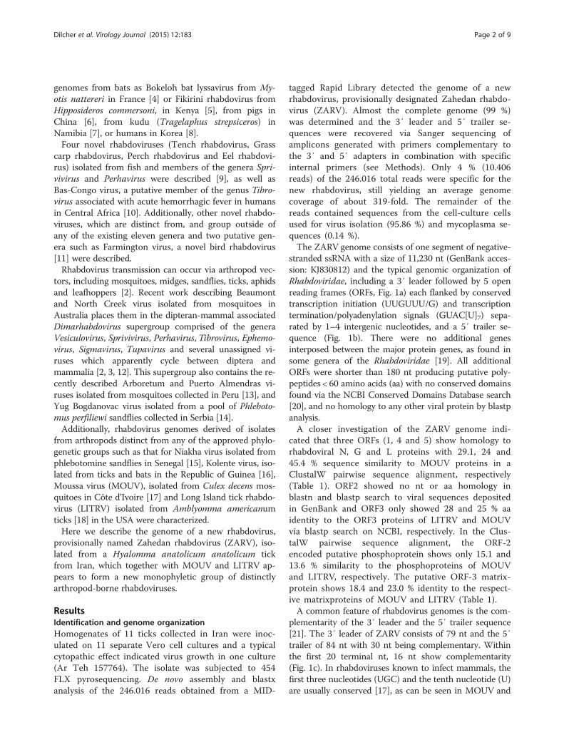
genomes from bats as Bokeloh bat lyssavirus from My-otis nattereri in France [4] or Fikirini rhabdovirus fromHipposideros commersoni, in Kenya [5], from pigs inChina [6], from kudu (Tragelaphus strepsiceros) inNamibia [7], or humans in Korea [8].Four novel rhabdoviruses (Tench rhabdovirus, Grass
carp rhabdovirus, Perch rhabdovirus and Eel rhabdovi-rus) isolated from fish and members of the genera Spri-vivirus and Perhavirus were described [9], as well asBas-Congo virus, a putative member of the genus Tibro-virus associated with acute hemorrhagic fever in humansin Central Africa [10]. Additionally, other novel rhabdo-viruses, which are distinct from, and group outside ofany of the existing eleven genera and two putative gen-era such as Farmington virus, a novel bird rhabdovirus[11] were described.Rhabdovirus transmission can occur via arthropod vec-
tors, including mosquitoes, midges, sandflies, ticks, aphidsand leafhoppers [2]. Recent work describing Beaumontand North Creek virus isolated from mosquitoes inAustralia places them in the dipteran-mammal associatedDimarhabdovirus supergroup comprised of the generaVesiculovirus, Sprivivirus, Perhavirus, Tibrovirus, Ephemo-virus, Sigmavirus, Tupavirus and several unassigned vi-ruses which apparently cycle between diptera andmammalia [2, 3, 12]. This supergroup also contains the re-cently described Arboretum and Puerto Almendras vi-ruses isolated from mosquitoes collected in Peru [13], andYug Bogdanovac virus isolated from a pool of Phleboto-mus perfiliewi sandflies collected in Serbia [14].Additionally, rhabdovirus genomes derived of isolates
from arthropods distinct from any of the approved phylo-genetic groups such as that for Niakha virus isolated fromphlebotomine sandflies in Senegal [15], Kolente virus, iso-lated from ticks and bats in the Republic of Guinea [16],Moussa virus (MOUV), isolated from Culex decens mos-quitoes in Côte d’Ivoire [17] and Long Island tick rhabdo-virus (LITRV) isolated from Amblyomma americanumticks [18] in the USA were characterized.Here we describe the genome of a new rhabdovirus,
provisionally named Zahedan rhabdovirus (ZARV), iso-lated from a Hyalomma anatolicum anatolicum tickfrom Iran, which together with MOUV and LITRV ap-pears to form a new monophyletic group of distinctlyarthropod-borne rhabdoviruses.
ResultsIdentification and genome organizationHomogenates of 11 ticks collected in Iran were inoc-ulated on 11 separate Vero cell cultures and a typicalcytopathic effect indicated virus growth in one culture(Ar Teh 157764). The isolate was subjected to 454FLX pyrosequencing. De novo assembly and blastxanalysis of the 246.016 reads obtained from a MID-
tagged Rapid Library detected the genome of a newrhabdovirus, provisionally designated Zahedan rhabdo-virus (ZARV). Almost the complete genome (99 %)was determined and the 3′ leader and 5′ trailer se-quences were recovered via Sanger sequencing ofamplicons generated with primers complementary tothe 3′ and 5′ adapters in combination with specificinternal primers (see Methods). Only 4 % (10.406reads) of the 246.016 total reads were specific for thenew rhabdovirus, still yielding an average genomecoverage of about 319-fold. The remainder of thereads contained sequences from the cell-culture cellsused for virus isolation (95.86 %) and mycoplasma se-quences (0.14 %).The ZARV genome consists of one segment of negative-
stranded ssRNA with a size of 11,230 nt (GenBank acces-sion: KJ830812) and the typical genomic organization ofRhabdoviridae, including a 3′ leader followed by 5 openreading frames (ORFs, Fig. 1a) each flanked by conservedtranscription initiation (UUGUUU/G) and transcriptiontermination/polyadenylation signals (GUAC[U]7) sepa-rated by 1–4 intergenic nucleotides, and a 5′ trailer se-quence (Fig. 1b). There were no additional genesinterposed between the major protein genes, as found insome genera of the Rhabdoviridae [19]. All additionalORFs were shorter than 180 nt producing putative poly-peptides < 60 amino acids (aa) with no conserved domainsfound via the NCBI Conserved Domains Database search[20], and no homology to any other viral protein by blastpanalysis.A closer investigation of the ZARV genome indi-
cated that three ORFs (1, 4 and 5) show homology torhabdoviral N, G and L proteins with 29.1, 24 and45.4 % sequence similarity to MOUV proteins in aClustalW pairwise sequence alignment, respectively(Table 1). ORF2 showed no nt or aa homology inblastn and blastp search to viral sequences depositedin GenBank and ORF3 only showed 28 and 25 % aaidentity to the ORF3 proteins of LITRV and MOUVvia blastp search on NCBI, respectively. In the Clus-talW pairwise sequence alignment, the ORF-2encoded putative phosphoprotein shows only 15.1 and13.6 % similarity to the phosphoproteins of MOUVand LITRV, respectively. The putative ORF-3 matrix-protein shows 18.4 and 23.0 % identity to the respect-ive matrixproteins of MOUV and LITRV (Table 1).A common feature of rhabdovirus genomes is the com-
plementarity of the 3′ leader and the 5′ trailer sequence[21]. The 3′ leader of ZARV consists of 79 nt and the 5′trailer of 84 nt with 30 nt being complementary. Withinthe first 20 terminal nt, 16 nt show complementarity(Fig. 1c). In rhabdoviruses known to infect mammals, thefirst three nucleotides (UGC) and the tenth nucleotide (U)are usually conserved [17], as can be seen in MOUV and
Dilcher et al. Virology Journal (2015) 12:183 Page 2 of 9
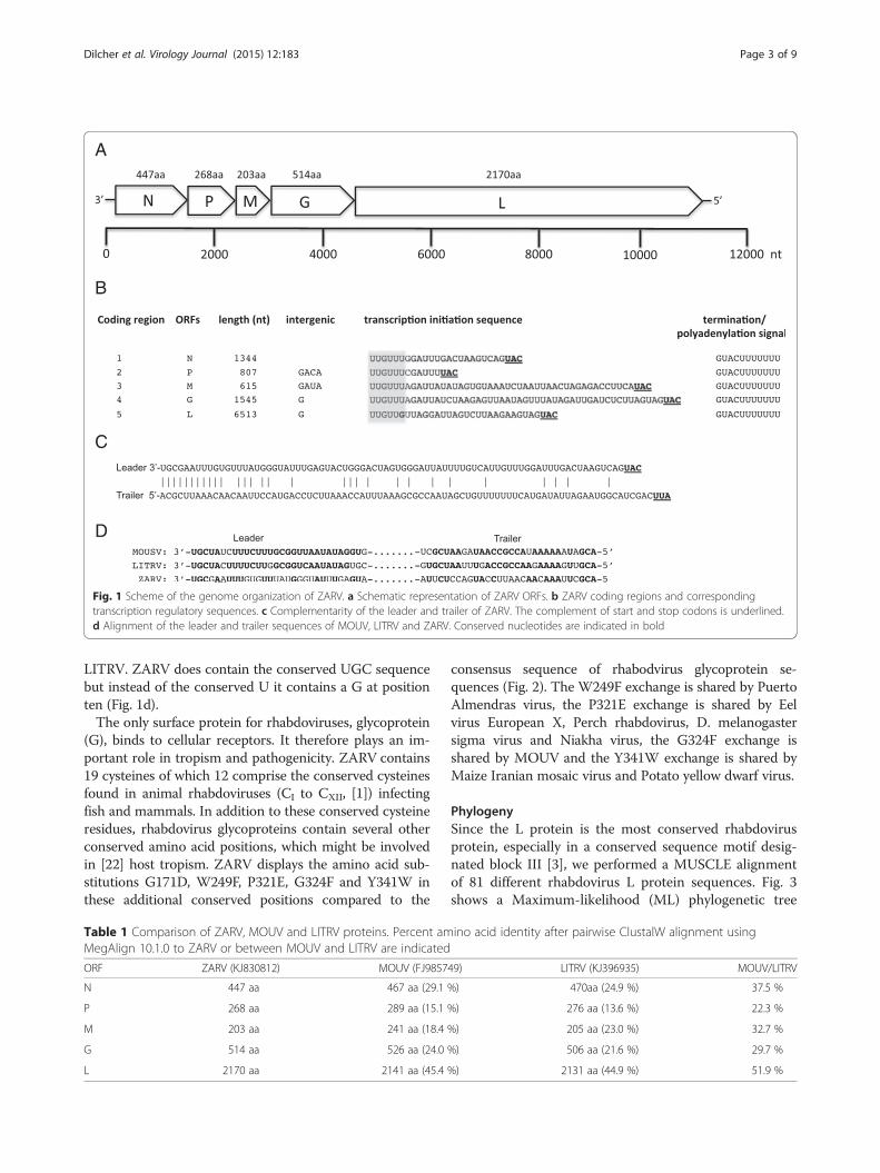
LITRV. ZARV does contain the conserved UGC sequencebut instead of the conserved U it contains a G at positionten (Fig. 1d).The only surface protein for rhabdoviruses, glycoprotein
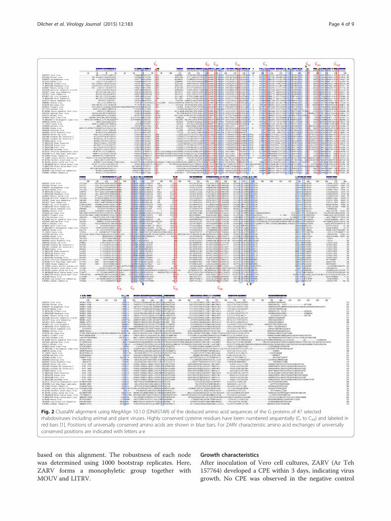
(G), binds to cellular receptors. It therefore plays an im-portant role in tropism and pathogenicity. ZARV contains19 cysteines of which 12 comprise the conserved cysteinesfound in animal rhabdoviruses (CI to CXII, [1]) infectingfish and mammals. In addition to these conserved cysteineresidues, rhabdovirus glycoproteins contain several otherconserved amino acid positions, which might be involvedin [22] host tropism. ZARV displays the amino acid sub-stitutions G171D, W249F, P321E, G324F and Y341W inthese additional conserved positions compared to the
consensus sequence of rhabodvirus glycoprotein se-quences (Fig. 2). The W249F exchange is shared by PuertoAlmendras virus, the P321E exchange is shared by Eelvirus European X, Perch rhabdovirus, D. melanogastersigma virus and Niakha virus, the G324F exchange isshared by MOUV and the Y341W exchange is shared byMaize Iranian mosaic virus and Potato yellow dwarf virus.
PhylogenySince the L protein is the most conserved rhabdovirusprotein, especially in a conserved sequence motif desig-nated block III [3], we performed a MUSCLE alignmentof 81 different rhabdovirus L protein sequences. Fig. 3shows a Maximum-likelihood (ML) phylogenetic tree
A
B
C
D
Fig. 1 Scheme of the genome organization of ZARV. a Schematic representation of ZARV ORFs. b ZARV coding regions and correspondingtranscription regulatory sequences. c Complementarity of the leader and trailer of ZARV. The complement of start and stop codons is underlined.d Alignment of the leader and trailer sequences of MOUV, LITRV and ZARV. Conserved nucleotides are indicated in bold
Table 1 Comparison of ZARV, MOUV and LITRV proteins. Percent amino acid identity after pairwise ClustalW alignment usingMegAlign 10.1.0 to ZARV or between MOUV and LITRV are indicated
ORF ZARV (KJ830812) MOUV (FJ985749) LITRV (KJ396935) MOUV/LITRV
N 447 aa 467 aa (29.1 %) 470aa (24.9 %) 37.5 %
P 268 aa 289 aa (15.1 %) 276 aa (13.6 %) 22.3 %
M 203 aa 241 aa (18.4 %) 205 aa (23.0 %) 32.7 %
G 514 aa 526 aa (24.0 %) 506 aa (21.6 %) 29.7 %
L 2170 aa 2141 aa (45.4 %) 2131 aa (44.9 %) 51.9 %
Dilcher et al. Virology Journal (2015) 12:183 Page 3 of 9
based on this alignment. The robustness of each nodewas determined using 1000 bootstrap replicates. Here,ZARV forms a monophyletic group together withMOUV and LITRV.
Growth characteristicsAfter inoculation of Vero cell cultures, ZARV (Ar Teh157764) developed a CPE within 3 days, indicating virusgrowth. No CPE was observed in the negative control
Fig. 2 ClustalW alignment using MegAlign 10.1.0 (DNASTAR) of the deduced amino acid sequences of the G proteins of 47 selectedrhabdoviruses including animal and plant viruses. Highly conserved cysteine residues have been numbered sequentially (CI to CXII) and labeled inred bars [1]. Positions of universally conserved amino acids are shown in blue bars. For ZARV characteristic amino acid exchanges of universallyconserved positions are indicated with letters a-e
Dilcher et al. Virology Journal (2015) 12:183 Page 4 of 9
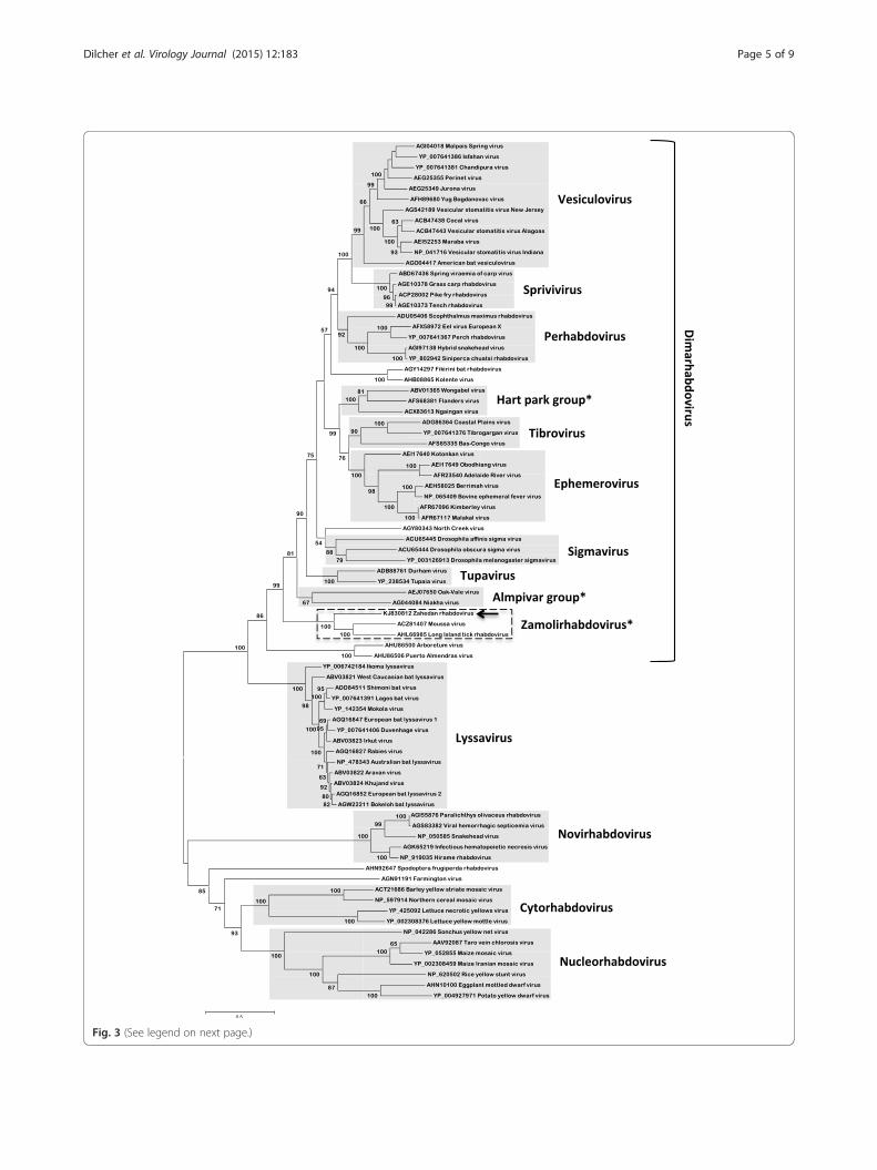
Fig. 3 (See legend on next page.)
Dilcher et al. Virology Journal (2015) 12:183 Page 5 of 9

culture (Fig. 4). The ZARV inoculum was tested negativefor mycoplasma sequences using PCR (data not shown).In addition, newborn (1–2 day old) mice were inocu-
lated with 20 μl Vero-grown ZARV containing approxi-mately 2x107.6 lethal dose 50% (LD50) per ml byintracerebral, intraperitoneal and subcutaneous route.The animals were observed daily for illness. Animalswere tested for ZARV infection by PCR. The intracere-bral inoculation route showed infection of the animals at4 and 6 days postinoculation (8/10 = 80 %). No illnesswas observed after subcutaneaous and intraperitonealinoclulation. The intracerebral inoculation route showedsuch severe symptoms that the mice had to be eutha-nized at 4 and 6 days postinoculation (8/10 mice =80 %). No disease was observed after subcutaneous andintraperitoneal route inoculation.
Development of a classical PCR assayWe developed a conventional RT-PCR assay detectingan amplicon within the ORF2/putative phosphoproteingene with primers ZARV-UP and ZARV-DP, which canbe used to specifically identify ZARV. This assay wasused to retest the supernatants of all eleven virus isola-tion attempts (all samples were collected at the sametime in the same area of Iran), confirming the ZARVpositive supernatant but not detecting any other positivesupernatants (data not shown). In addition, this assay wasused to test ZARV-RNA=Ar Teh 157764 [Zahedan/Iran2001] and in parallel 24 African rhabdovirus RNA’s from
viruses isolated in the years 1959 until 2013: MOSSURIL[Mozambique 1959], RV401 (AND401) [Senegal 1964],ANB373 [Central African Republic 1970], SA262037[Senegal 2013], SA194858 [Senegal 2008], SA217694[Senegal 2011], SA206776 [Senegal 2010], ARD111213[Senegal 1995], ARD129179 [Senegal 1997], ARD111757[Senegal 1995], MOULIN [n.d.], CHAND13 [n.d.],AND42443 [Senegal 1985], LG6 [n.d.], YM55 [Cameroune1965], ASH763 [Senegal 1965], ANB439 [Central AfricanRepublic 1970], MOKOLA [Cameroune 1973], ANB1094[Central African Republic 1970], ANB4289 [CentralAfrican Republic 1973], ARD129178 [Senegal 1997],ARD1275 [n.d.], RV5314 [Senegal 1968], ANK6909[Guinea 1985]. Only the ZARV-RNA produced a specificband at 807 bp (data not shown). Furthermore, all thebrain tissues of the deceased mice tested positive with thisRT-PCR.
DiscussionPhylogenetic analysis of the genome sequence groupsZARV into a monophyletic clade with LITRV anothertick-borne virus from the U.S.A. and MOUV amosquito-borne virus from Côte d’Ivoire.Although G proteins of rhabdoviruses from different
genera only share very low amino acid sequence identity,alignment of the sequences reveals remarkable conserva-tion of cysteine residues, glycosylation sites and themajor antigenic domains [1]. Since the glycoproteinsinteract with specific cell-surface receptors of the hostcells sequence analysis might give indications about theputative animal host and the host surface-receptor usageof the respective rhabdovirus.Twelve of nineteen cysteines found in the ZARV G se-
quence are typically conserved in animal rhabdoviruses(CI to CXII, [1]) indicating that the new tick-transmittedrhabdovirus does not seem to be a pure tick virus(Fig. 2). These 12 cysteines are also conserved in MOUVand LITRV. Some of these conserved cysteines are notpresent in Niakha virus (lacking CVIII and CX) isolatedfrom phlebotomine sandflies or Drosophila melanogastersigmavirus (lacking CVII) both without any known verte-brate host, or in plant rhabdoviruses like Maize Iranianmosaic virus (lacking CII, CIII, CIV, CVI, CVII, CIX, CX). Inaddition, the mosquito-borne rhabdoviruses Arboretumvirus and Puerto Almendras virus are lacking CI, CVI
and CXI. It needs to be further analyzed if these substitu-tions play a role in host tropism or if one can predict a
(See figure on previous page.)Fig. 3 Maximum-likelihood (ML) phylogenetic tree based on 81 complete rhabdovirus L protein amino acid sequences aligned via MUSCLE andcalculated using MEGA 5.2.2. Bootstrap values (>50 %) are given in percent. The scale bar indicates number of substitutions per site. * Generaproposed but not yet formally approved by the International Committee on Taxonomy of Viruses (ICTV). The arrowhead indicates Zahedanrhabdovirus (ZARV) and the dashed box indicates the proposed new rhabdovirus genotype Zamolirhabdovirus
Fig. 4 ZARV plaque assay: A1-2: negative control (cell culturemedium). A3-D6: serial 10-fold dilutions in duplex wells down to 10−11 of ZARV culture supernatant on Vero cell culture monolayers.The ZARV inoculum as well as the Vero cells was previouslyconfirmed by PCR to be negative for mycoplasma sequences (datanot shown)
Dilcher et al. Virology Journal (2015) 12:183 Page 6 of 9

potential host based on the amino acid composition ofthese conserved positions.Interestingly, ZARV shows the highest aa similarity in
N, P, G and L proteins to the mosquito-borne MOUV,with 29.1, 15.1, 24.0 and 45.4 % identity, respectively(Table 1). Only the M protein shows a higher similarity(23.0 %) to the tick-transmitted LITRV. But in general,MOUV and LITRV are more closely related and ZARVseems to represent an ancestral relative of both viruses,which is supported by high bootstrap values. This alsofeatures in the termination/polyadenylation sequenceGAAC[U]7 which is identical for MOUV and LITRV, butslightly different for ZARV which uses GUAC[U]7.The virus was cultured in cell culture and in mouse
brain where it grew to high titres of 2x107.6 LD50 typicalfor rhabdoviruses. Here it led to severe symptoms indi-cating pathogenicity in 80 % of the mice, so that theyhad to be euthanized 6 or 8 days postinoculation. Wedeveloped a conventional RT-PCR assay, which can beused to specifically identify ZARV.In summary it can be said that although the tick-borne
rhabdovirus subclade into which ZARV groups is phylo-genetically distinct from the major rhabdovirus groups,the overall pattern of conserved amino acids of the Gproteins of ZARV, MOUV and LITRV support their as-sociation to the (Dimarhabdovirus supergroup. Since allthree viruses form a monophyletic group we suggest tocombine them in a new genus designated Zamolirhab-dovirus (Zahedan rhabdovirus, Moussa virus, Long Is-land tick rhabdovirus). It remains to be seen ifmammalian hosts can be identified for this new rhabdo-virus genus.
Materials and methodsZARV was isolated on the 21st of December 2001 at theInstitute Pasteur de Dakar, Senegal from a Hyalommaanatolicum anatolicum tick collected on the 27th of Oc-tober 2001 in Zahedan/Iran. In this attempt, 11 homoge-nized ticks (150 μl each) were inoculated separately ontoconfluent Vero cell culture monolayers (25 cm2) in Lei-bovitz 15 growth medium (L-15) supplemented with 5 %fetal bovine serum (both GibcoBRL, Grand Island, NY,USA), and 10 mM each of Penicillin and Streptomycin(Sigma, Gmbh, Germany).At 1 hpi, cells were washed twice with PBS and incu-
bated with fresh medium. At 7 dpi, supernatant of in-fected cells was harvested and used for sub-passaging.After appearance of a CPE in one of the cultures (3 dpi)passage 6 was harvested and 140 μl of supernatant wasused for RNA extraction via the QIAamp viral RNAmini kit (Qiagen), omitting the addition of carrier RNA.After DNase digestion (Turbo-DNA-Free-Kit, Ambion)and Pellet-Paint precipitation (Pellet Paint NF Co-Precipitant, Novagen), the purified RNA was used for
amplification via the TransPlex Whole TranscriptomeAmplification Kit (WTA2) from Sigma-Aldrich [23].After purification via the QIAquick PCR purification kit(Qiagen), an additional size exclusion step via AmpureXP-beads (Agencourt) was used to remove fragmentsshorter than 350 bp. 300 ng of the whole genome ampli-fied dsDNA was used for Roche Rapid Library Prepar-ation employing MID barcodes starting with the EndRepair step [24]. The MID-tagged Rapid Library was se-quenced in a pool of 11 MID-tagged libraries. For deter-mining the 3′ leader region, a 3′-FLAC adapter wasligated to the 3′-OH group [25]. A PCR with a comple-mentary primer to the FLAC adapter in combinationwith an internal primer and sanger sequencing of thePCR product yielded the 3′ leader. To determine the 5′trailer, first an RNA-5′-polyphosphatase treatment (Epi-center Biotechnologies) was carried out to remove twophosphate groups of the 5′-triphosphate. Next a 5′-RACE adapter (FirstChoice RLM-RACE kit, Ambion)was ligated to the 5′-monophosphate and a PCR with an5′-RACE-outer primer in combination with an internalprimer and sanger sequencing of the amplicon closedthe gap on the 5′ trailer. Assembly of the genome wasdone with the GS De Novo Assembler (Newbler) version2.6 in combination with the SeqMan Pro Software ver-sion 10.1.0. Phylogenetic analysis was performed usingcomplete L protein sequences downloaded from Gen-Bank and aligned to the L protein sequence of ZARV viaClustalW using MegAlign 10.1.0 (DNASTAR) andMUSCLE using MEGA 5.2.2 [26]. A maximum likeli-hood tree was generated using the Jones-Taylor-Thornton model with MEGA 5.2.2. Statistical significanceof the tree topologies was evaluated by 1000 bootstrapreplicates.
Mouse experimentsMice used in this study were reared at the Institute Pas-teur in Dakar animal laboratory accredited by WHO asCollaborating Centre for Arboviruses and/or HemorrhagicFever Reference and Research and were used for routinevirus isolation in suckling mice for surveillance, diagnos-tics and research as authorized by the senegalese nationalethical comittee.A serial dilution (10-1 to 10-11) of the Ar Teh 157764
strain was prepared in maintenance medium (L-15, with3 % serum). Twenty microliter of each dilution of thevirus was inoculated intracerebrally, intraperitoneal andsubcutaneous into 10 BALB/c mouse pups (1–2 daysold) provided by the Pasteur Institute Dakar facilities.The animals were observed for 21 days and ill mice weretested for ZARV infection using the PCR developed. Theinfectivity titer which is the reciprocal of the highest di-lution showing 50 % mortality in the inoculated mice
Dilcher et al. Virology Journal (2015) 12:183 Page 7 of 9
and expressed as LD50 /ml was calculated by using a for-mula similar to that described by Karber.
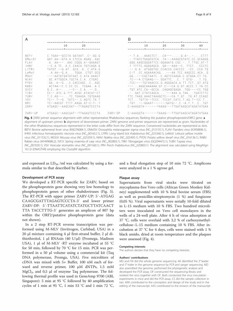
Development of PCR assayWe developed a RT-PCR specific for ZARV, based onthe phosphoprotein gene showing very low homology tophosphoprotein genes of other rhabdoviruses (Fig. 5).The RT-PCR with upper primer ZARV-UP: 5′-ATGAGCAAGCGATTTAGAGTCCCTA-3′ and lower primerZARV-DP: 5′-TTAATTCATATCTATGCTTATCAACTTTA TACCTTTG-3′ generates an amplicon of 807 bpwithin the ORF2/putative phosphoprotein gene (datanot shown).In a 2 step RT-PCR reverse transcription was per-
formed using M-MLV (Invitrogen, Carlsbad, USA) in a20 μl mixture containing 4 μl first-strand buffer, 2 μl di-thiothreitol, 1 μl RNAsin (40 U/μl) (Promega, MadisonUSA), 1 μl of M-MLV -RT enzyme incubated at 55 °Cfor 50 min, followed by 70 °C for 15 min. PCR was per-formed in a 50 μl volume using a commercial kit (TaqDNA polymerase, Promga, USA). Five microliters ofcDNA was mixed with 5× Buffer, 100 nM each of for-ward and reverse primer, 100 μM dNTPs, 1.5 mMMgCl2, and 0,5 μl of enzyme Taq polymerase. The fol-lowing thermal profile was used in GeneAmp 9700 (ABI,Singapore): 5 min at 95 °C followed by 40 amplificationcycles of 1 min at 95 °C, 1 min 53 °C and 1 min 72 °C,
and a final elongation step of 10 min 72 °C. Ampliconswere analyzed in a 1 % agrose gel.
Plaque assaySupernatants from viral stocks were titrated onmycoplasma-free Vero cells (African Green Monkey Kid-ney) supplemented with 10 % fetal bovine serum (FBS)as well as penicillin-streptomycin (1 %) and fungizone(0,05 %). Viral supernatants were serially 10-fold dilutedin L-15 medium with 10 % FBS. Two hundred microli-ters were inoculated on Vero cell monolayers in thewells of a 24-well plate. After 4 h of virus adsorption at37 °C, cells were overlaid with 3,2 % of carboxymethyl-cellulose–L-15 medium containing 10 % FBS. After in-cubation at 37 °C for 4 days, cells were stained with 1 %black amido, dried at room temperature and the plaqueswere assessed (Fig. 4).
Competing interestsThe authors declare that they have no competing interests.
Authors’ contributionsMD and FW did the whole genome sequencing. AK identified the 3′ leaderand 5′ trailer in the genome sequence by PCR and sanger sequencing. MDalso assembled the genome, performed the phylogenetic analysis anddeveloped the PCR assay. OF constructed the sequencing library andisolated the virus together with OF. Both conducted the virus inoculationexperiments in mice and did the PCR assay. CS did the sample collection inIran. MW contributed to the conception and design of the study and in theediting of the manuscript. AAS contributed to the revision of the manuscript
A B
Fig. 5 ZARV primer sequence alignment with other representative Rhabdovirus sequences, flanking the putative phosphoprotein/ORF2 gene. aalignment of upstream primer. b alignment of downstream primer. ZARV genome and primer sequences are represented as given. Nucleotides ofthe other Rhabdovirus sequences represented in the letter code differ from the ZARV sequence. Consereved nucleotides are represented as dots.BEFV: Bovine ephemeral fever virus (KM276084.1), DMelSV: Drosophila melanogaster sigma virus (NC_013135.1), FLAV: Flanders virus (KJ958896.1),IHNV: Infectious hematopoietic necrosis virus (NC_001652.1), LITRV: Long Island tick rhabdovirus (NC_025340.1), LeMoV: Lettuce yellow mottlevirus (NC_011532.1), MOUV: Moussa virus (NC_025359.1), NIAV: Niakha virus (NC_025405.1), PYDV: Potato yellow dwarf virus (NC_016136.1), RABV:Rabies virus (AF499686.2), SVCV: Spring viraemia of carp virus (NC_002803.1), TIBV: Tibrogargan virus (GQ294472.1), TUBV: Tupaia virus(NC_007020.1), VSV: Vesicular stomatitis virus (NC_001560.1), PRV: Perch rhabdovirus (NC_020803.1). The alignment was calculated using MegAlign10.1.0 (DNASTAR) employing the ClustalW algorithm
Dilcher et al. Virology Journal (2015) 12:183 Page 8 of 9
and the design of the study. All authors read and approved the finalmanuscript.
AcknowledgementsThis project was funded by the CCH Fever Network (Collaborative Project)supported by the European Commission under the Health Cooperation WorkProgram of the 7thFramework Program (Grant agreement no. 260427). We are indebted toMarie-Francoise Saron who coordinated the Pasteur Institute ACIP project(2001), which collected the ticks and we are also thankful to the CDC andthe veterinary organization of Iran. We thank Frank T. Hufert for his support.
Author details1Department of Virology, Univerity Medical Center Göttingen, Kreuzbergring57, 37075 Göttingen, Germany. 2Institute Pasteur de Dakar, 36 AvenuePasteur, BP 220 Dakar, Senegal. 3Pasteur Institute of Iran, 69 Pasteur Avenue,Tehran, Iran. 4Institute of Aquaculture, University of Stirling, Stirling FK9 4LA,UK.
Received: 1 April 2015 Accepted: 26 October 2015
References1. Walker PJ, Kongsuwan K. Deduced structural model for animal rhabdovirus
glycoproteins. J General Virol. 1999;80(Pt 5):1211–20.2. Kuzmin IV, Novella IS, Dietzgen RG, Padhi A, Rupprecht CE. The
rhabdoviruses: biodiversity, phylogenetics, and evolution. Infect Genet Evol.2009;9(4):541–53.
3. Bourhy H, Cowley JA, Larrous F, Holmes EC, Walker PJ. Phylogeneticrelationships among rhabdoviruses inferred using the L polymerase gene.J General Virol. 2005;86(Pt 10):2849–58.
4. Picard-Meyer E, Servat A, Robardet E, Moinet M, Borel C, Cliquet F. Isolationof Bokeloh bat lyssavirus in Myotis nattereri in France. Arch Virol.2013;158(11):2333–40.
5. Kading RC, Gilbert AT, Mossel EC, Crabtree MB, Kuzmin IV, Niezgoda M, et al.Isolation and molecular characterization of Fikirini rhabdovirus, a novel virusfrom a Kenyan bat. J General Virol. 2013;94(Pt 11):2393–8.
6. Luo Y, Zhang Y, Liu X, Yang Y, Yang X, Zheng Z, et al. Characterization of awild rabies virus isolate of porcine origin in China. Infect Genet Evol.2013;17:147–52.
7. Scott TP, Fischer M, Khaiseb S, Freuling C, Hoper D, Hoffmann B, et al.Complete genome and molecular epidemiological data infer themaintenance of rabies among kudu (tragelaphus strepsiceros) in Namibia.Plos One. 2013;8(3):e58739.
8. Park JS, Kim CK, Kim SY, Ju YR. Molecular characterization of KGH, the firsthuman isolate of rabies virus in Korea. Virus Genes. 2013;46(2):231–41.
9. Stone DM, Kerr RC, Hughes M, Radford AD, Darby AC. Characterisation ofthe genomes of four putative vesiculoviruses: tench rhabdovirus, grass carprhabdovirus, perch rhabdovirus and eel rhabdovirus European X. Arch Virol.2013;158(11):2371–7.
10. Grard G, Fair JN, Lee D, Slikas E, Steffen I, Muyembe JJ, et al. A novelrhabdovirus associated with acute hemorrhagic fever in central africa. PlosPathog. 2012;8(9):e1002924.
11. Palacios G, Forrester NL, Savji N, da Rosa APAT, Guzman H, DeToy K, et al.Characterization of Farmington virus, a novel virus from birds that isdistantly related to members of the family Rhabdoviridae. Virol J.2013;10:219.
12. Coffey LL, Page BL, Greninger AL, Herring BL, Russell RC, Doggett SL, et al.Enhanced arbovirus surveillance with deep sequencing: Identification ofnovel rhabdoviruses and bunyaviruses in Australian mosquitoes. Virology.2014;448:146–58.
13. Vasilakis N, Castro-Llanos F, Widen SG, Aguilar PV, Guzman H, Guevara C,et al. Arboretum and Puerto Almendras viruses: two novel rhabdovirusesisolated from mosquitoes in Peru. J General Virol. 2014;95(Pt 4):787–92.
14. Pfeffer M, Dilcher M, Tesh RB, Hufert FT, Weidmann M. Geneticcharacterization of Yug Bogdanovac virus. Virus Genes. 2013;46(1):201–2.
15. Vasilakis N, Widen S, Mayer SV, Seymour R, Wood TG, Popov V, et al. Niakhavirus: a novel member of the family Rhabdoviridae isolated fromphlebotomine sandflies in Senegal. Virology. 2013;444(1–2):80–9.
16. Ghedin E, Rogers MB, Widen SG, Guzman H, da Rosa AP T, Wood TG, et al.Kolente virus, a rhabdovirus species isolated from ticks and bats in theRepublic of Guinea. J General Virol. 2013;94(Pt 12):2609–15.
17. Quan PL, Junglen S, Tashmukhamedova A, Conlan S, Hutchison SK, Kurth A,et al. Moussa virus: a new member of the Rhabdoviridae family isolatedfrom Culex decens mosquitoes in Cote d’Ivoire. Virus Res. 2010;147(1):17–24.
18. Tokarz R, Sameroff S, Leon MS, Jain K, Lipkin WI. Genome characterization ofLong Island tick rhabdovirus, a new virus identified in Amblyommaamericanum ticks. Virol J. 2014;11:26.
19. Walker PJ, Dietzgen RG, Joubert DA, Blasdell KR. Rhabdovirus accessorygenes. Virus Res. 2011;162(1–2):110–25.
20. Marchler-Bauer A, Lu S, Anderson JB, Chitsaz F, Derbyshire MK, DeWeese-Scott C, et al. CDD: a conserved domain database for the functionalannotation of proteins. Nucleic Acids Res. 2011;39(Database issue):D225–9.
21. Whelan SP, Barr JN, Wertz GW. Transcription and replication ofnonsegmented negative-strand RNA viruses. Curr Top Microbiol Immunol.2004;283:61–119.
22. Dietzgen RG, Calisher CH, Kurath G, Kuzmin IV, Rodriguez LL MSD, Tesh RB,et al. Familiy rhabdoviridae. In: King AMQ, Adams MJ, Carstens EB, LefkowitzEJ, editors. Virus taxonomy, classification and nomenclature of viruses, ninthreport of the international committee on taxonomy of viruses, internationalunion of microbiological societies, virology division. London, UK: Elsevier;2012. p. 686–713.
23. Dilcher M, Hasib L, Lechner M, Wieseke N, Middendorf M, Marz M, et al.Genetic characterization of Tribec virus and Kemerovo virus, two tick-transmitted human-pathogenic Orbiviruses. Virology. 2012;423(1):68–76.
24. Dilcher M, Koch A, Hasib L, Dobler G, Hufert FT, Weidmann M. Geneticcharacterization of Erve virus, a European Nairovirus distantly related toCrimean-Congo hemorrhagic fever virus. Virus Genes. 2012;45(3):426–32.
25. Maan S, Rao S, Maan NS, Anthony SJ, Attoui H, Samuel AR, et al. RapidcDNA synthesis and sequencing techniques for the genetic study ofbluetongue and other dsRNA viruses. J Virol Methods. 2007;143(2):132–9.
26. Tamura K, Peterson D, Peterson N, Stecher G, Nei M, Kumar S. MEGA5:molecular evolutionary genetics analysis using maximum likelihood,evolutionary distance, and maximum parsimony methods. Mol Biol Evol.2011;28(10):2731–9.
Submit your next manuscript to BioMed Centraland take full advantage of:
• Convenient online submission
• Thorough peer review
• No space constraints or color figure charges
• Immediate publication on acceptance
• Inclusion in PubMed, CAS, Scopus and Google Scholar
• Research which is freely available for redistribution
Submit your manuscript at www.biomedcentral.com/submit
Dilcher et al. Virology Journal (2015) 12:183 Page 9 of 9