WHITE PAPER SYBR Safe DNA Gel Stain SYBR Safe...
26
SYBR Safe DNA Gel Stain: assessment of mutagenicity and environmental safety Compiled by Thermo Fisher Scientific from the results of four independent testing services SYBR Safe DNA Gel Stain was only weakly mutagenic (and only with S9 metabolic activation) in the standard Ames bacterial mutation assay. Compared to ethidium bromide, SYBR Safe DNA Gel Stain caused fewer mutations in the standard Ames test, as measured in several different strains of Salmonella typhimurium. Weakly positive results for SYBR Safe DNA Gel Stain in the test occurred in four of out seven strains, and only with activation by a mammalian S9 fraction. SYBR Safe DNA Gel Stain does not induce transformation in primary cultures of Syrian hamster embryo (SHE) cells when compared with solvent alone, calling into question the weakly mutagenic results from the Ames test. In contrast, ethidium bromide tests positive in the SHE cell assay, consistent with its known activity as a strong mutagen. In addition, SYBR Safe DNA Gel Stain does not cause mutations in mouse lymphoma cells at the TK locus, nor does it induce chromosomal aberrations in cultured human peripheral blood lymphocytes, with or without S9 metabolic activation, using standardized tests against appropriate controls. Overview This report provides safety data, generated by four independent testing services, that highlight the increased safety of using Invitrogen ™ SYBR ™ Safe DNA Gel Stain, compared with widely used conventional nucleic acid stains such as ethidium bromide. SYBR Safe DNA Gel Stain is nontoxic, noncarcinogenic, and classified as nonhazardous waste. This means that using SYBR Safe DNA Gel Stain not only helps to reduce exposure to hazardous chemicals in the lab but also avoids generation of hazardous waste and the associated management and disposal costs. Executive summary SYBR Safe DNA Gel Stain was subjected to a battery of tests to assess health and environmental impacts associated with its use and disposal. SYBR Safe DNA Gel Stain in 0.5X TBE did not show any toxicity or mortality in an acute oral toxicity study: the rat LD50 was >5,000 mg/kg (U.S. EPA OPPTS 870.1100 method). A single dose oral administration of SYBR Safe DNA Gel Stain in 0.5X TBE, at a limit dose of 5000 mg/kg to rats, produced no mortalities or toxic signs. In aquatic toxicity tests with fathead minnows (CA Title 22 acute screening methodology), SYBR Safe DNA Gel Stain in 0.5X TBE was not toxic (LC 50 >750 mg/L). SYBR Safe DNA Gel Stain is not classified as hazardous waste under US federal regulations (Resource Conservation and Recovery Act, RCRA). SYBR Safe stain also meets the requirements of the Clean Water Act and the National Pollutant Discharge Elimination System (NPDES) regulations. WHITE PAPER SYBR Safe DNA Gel Stain
Transcript of WHITE PAPER SYBR Safe DNA Gel Stain SYBR Safe...
SYBR Safe DNA Gel Stain: assessment of mutagenicity and environmental safetyCompiled by Thermo Fisher Scientific from the results of four independent testing services
SYBR Safe DNA Gel Stain was only weakly mutagenic (and only with S9 metabolic activation) in the standard Ames bacterial mutation assay. Compared to ethidium bromide, SYBR Safe DNA Gel Stain caused fewer mutations in the standard Ames test, as measured in several different strains of Salmonella typhimurium. Weakly positive results for SYBR Safe DNA Gel Stain in the test occurred in four of out seven strains, and only with activation by a mammalian S9 fraction. SYBR Safe DNA Gel Stain does not induce transformation in primary cultures of Syrian hamster embryo (SHE) cells when compared with solvent alone, calling into question the weakly mutagenic results from the Ames test. In contrast, ethidium bromide tests positive in the SHE cell assay, consistent with its known activity as a strong mutagen. In addition, SYBR Safe DNA Gel Stain does not cause mutations in mouse lymphoma cells at the TK locus, nor does it induce chromosomal aberrations in cultured human peripheral blood lymphocytes, with or without S9 metabolic activation, using standardized tests against appropriate controls.
OverviewThis report provides safety data, generated by four independent testing services, that highlight the increased safety of using Invitrogen™ SYBR™ Safe DNA Gel Stain, compared with widely used conventional nucleic acid stains such as ethidium bromide. SYBR Safe DNA Gel Stain is nontoxic, noncarcinogenic, and classified as nonhazardous waste. This means that using SYBR Safe DNA Gel Stain not only helps to reduce exposure to hazardous chemicals in the lab but also avoids generation of hazardous waste and the associated management and disposal costs.
Executive summary SYBR Safe DNA Gel Stain was subjected to a battery of tests to assess health and environmental impacts associated with its use and disposal. SYBR Safe DNA Gel Stain in 0.5X TBE did not show any toxicity or mortality in an acute oral toxicity study: the rat LD50 was >5,000 mg/kg (U.S. EPA OPPTS 870.1100 method). A single dose oral administration of SYBR Safe DNA Gel Stain in 0.5X TBE, at a limit dose of 5000 mg/kg to rats, produced no mortalities or toxic signs. In aquatic toxicity tests with fathead minnows (CA Title 22 acute screening methodology), SYBR Safe DNA Gel Stain in 0.5X TBE was not toxic (LC50 >750 mg/L). SYBR Safe DNA Gel Stain is not classified as hazardous waste under US federal regulations (Resource Conservation and Recovery Act, RCRA). SYBR Safe stain also meets the requirements of the Clean Water Act and the National Pollutant Discharge Elimination System (NPDES) regulations.
WHITE PAPER SYBR Safe DNA Gel Stain
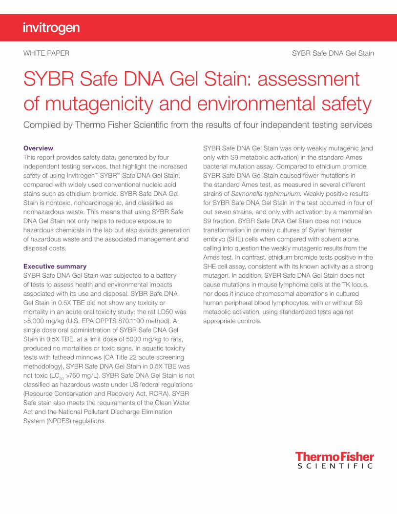
Summary of results
Test Result
Mammalian genotoxicity
SHE transformation assayChromosomal aberration assay
NegativeNegative
Bacterial mutagenicity
Ames assay: (–) S9 Mix (+) S9 Mix
Negative Weakly positive
Acute oral toxicity
Limit dose of 5,000 mg/kg No mortality or toxic signs
Ignitability
Not ignitable
Reactivity
Not detected
Corrosivity
pHCorrositex™ test
pH = 8.25: noncorrosiveCategory 2 noncorrosive
Pollutant discharge
Clean Water ActNPDES regulations
Meets requirements Meets requirements
Acute aquatic toxicity
Aquatic toxicity testNot classified as hazardous or toxic to aquatic life
Geno-toxicological testing forward mutation 4
Transformation 4
Chromosomal aberration 5
Mutagenicity 7
S9 metabolic activation system 8
Test system 9
Preparation of overnight cultures 10
Confirmation of tester strain genotype 10
Experimental design 11
Procedures 11
Data evaluation 11
Acute oral toxicity 13
Physicochemical testing for waste characterization 15
Corrosivity—Corrositex test 15
Pollutant discharge 16
Appendix I: data tables 17
Contents
4
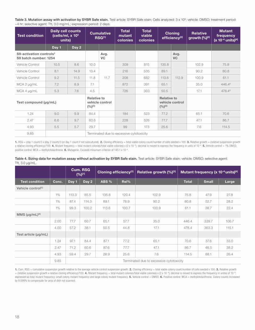
Under activation conditions, treatments from 9.85 to 625 μg/mL were terminated due to excessive cytotoxicity. The remaining treatments were used for the mutation assay. Treatment at 1.24 μg/mL induced weak cytotoxicity, treatment at 2.47 μg/mL induced moderate cytotoxicity, and treatment at 4.93 μg/mL induced excessive cytotoxicity (relative total growth of 65.1%, 47.1%, and 7.6% respectively). In the mutation assay, no increases in the mutant frequency were observed that exceeded the minimum criterion of 145.1 x 10⁻⁶. Results of the mutation assay with SYBR Safe stain under activation conditions are shown in Appendix I, Tables 3 and 4.
All tests were performed by independent and accredited laboratories using established methodologies.
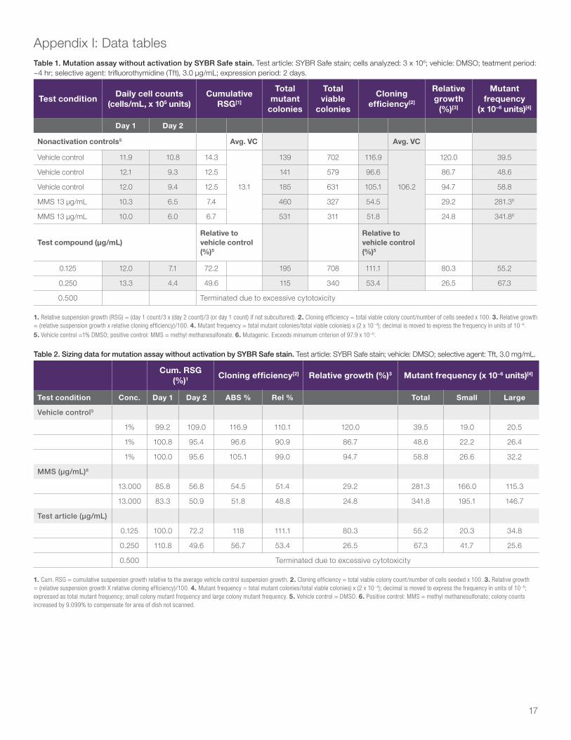
ConclusionSYBR Safe stain was evaluated as negative with and without metabolic activation in the L5178Y TK+/– mouse lymphoma forward mutation screening assay. (see Appendix I, Tables 1-4)
Transformation
We also investigated the ability of SYBR Safe stain to induce an increase in morphological transformation of cultured Syrian hamster embryo (SHE) cells, relative to vehicle control cultures, following a 7-day exposure period. Cryopreserved SHE cell stock prepared from embryo cells obtained from first-time pregnant Syrian golden hamsters at 13 to 13.5 days gestation was used for this assay.
Test article handlingThe test article, SYBR Safe stain, was stored at room temperature with desiccant. Dimethylsulfoxide (DMSO, CAS No. 67-68-5, Acros Organics, Lot No. A017190501) was used as a vehicle control. The test article formed a red transparent solution in DMSO at 5 mg/mL. Orange transparent solutions were obtained when the test article was dosed into media at concentrations of 3.33 and 10.0 μg/mL. At concentrations of 1.00 μg/mL and lower, the test article formed transparent solutions with normal media color. At a concentration of 10.0 μg/mL, the test article did not have significant effect on pH and osmolality of the culture medium. Both pH and osmolality values were within acceptable ranges.
Geno-toxicological testing forward mutation
Test article handling The test article formed a transparent orange solution in DMSO at 62.5 mg/mL and formed an opaque suspension at higher concentrations. In treatment medium, SYBR Safe stain formed a precipitate at ≥ 78.5 μg/mL at treatment termination.
Dose range–finding studyA preliminary dose range–finding assay was initiated with ten treatments at 1.24, 2.47, 4.93, 9.85, 19.7, 39.3, 78.5, 157, 313, and 625 μg/mL. Cells were exposed to the test article for 4 hr in the presence and absence of rat liver S9 metabolic activation. Since cytotoxicity was observed on Day 1 with and without activation, the study was continued to determine if a good toxicity range could be obtained for a mutation assay using the cells from the dose range–finding assay.
On Day 2, under non-activation conditions, all treatments were terminated due to excessive cytotoxicity. Because a good range of cytotoxicity was not achieved for the non-activation assay, the dose range–finding assay was terminated prior to cloning and a mutation assay was initiated.
ResultsIn the mutation assay without activation, eight treatments at 0.0313, 0.0625, 0.125, 0.250, 0.500, 0.750, 1.00, and 1.50 μg/mL were initiated, and three treatments at 0.125, 0.250, and 0.500 μg/mL were analyzed for mutant induction (Appendix I, Tables 1 and 2). SYBR Safe stain induced no cytotoxicity at 0.125 μg/mL and moderately high cytotoxicity at 0.250 μg/mL (80.3% and 26.5% relative total growth, respectively). A small increase in concentration from 0.250 to 0.500 μg/mL was excessively cytotoxic. The minimum criterion for a positive response in this non-activation assay was 97.9 x 10⁻⁶. No increases in the mutant frequency were observed that exceeded the minimum criterion. Results of the mutation assay with SYBR Safe stain under non-activation conditions are shown in Appendix I, Tables 1 and 2.
5
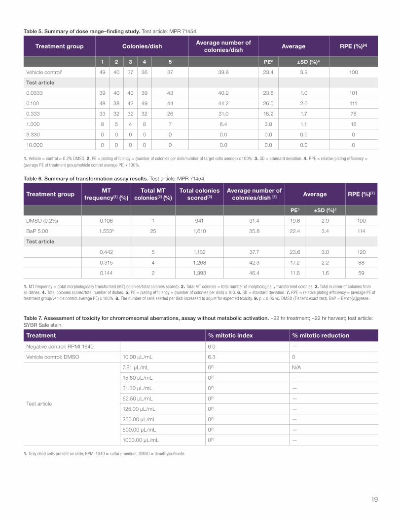
Dose range–finding studyA preliminary dose range–finding study was initiated with six treatments from 0.0333 μg/mL to 10.0 μg/mL (Appendix I, Table 5). Non-cytotoxicity was observed at a test article concentration up to 0.100 μg/mL. The test article was moderately cytotoxic at 0.333 μg/mL and was excessively cytotoxic at higher doses. Based on the results, three doses ranging from 0.200 to 0.700 μg/mL were tested in the initial trial of the transformation assay.
ResultsThe following three doses were tested in the initial trial of the transformation assay: 0.200, 0.400, and 0.700 μg/mL.
However, this trial failed due to higher-than-expected cytotoxicity. A second trial was performed with the following concentrations: 0.0500, 0.150, and 0.300 μg/mL. Results of this trial are summarized by Appendix I, Table 6. SYBR Safe stain was essentially non-cytotoxic at 0.0500 µg/mL (120% RPE), slightly cytotoxic at 0.150 μg/mL (88% RPE), and moderately cytotoxic at 0.300 μg/mL (59% RPE). None of the three treatment groups induced a significant increase in the frequency of morphological transformation compared to the concurrent vehicle control. In addition, a significant increase of the morphological transformational frequency was obtained from the positive control treatment with benzyo[α]pyrene at 5.0 μg/mL. The test article was therefore evaluated as negative in the screening SHE cell transformation assay under 7-day exposure conditions of this study.
ConclusionThe test article, SYBR Safe stain, tested in the SHE cell cultures with a 7-day exposure, was evaluated as negative in the screening SHE cell transformation assay under 7-day exposure conditions of this study (see Appendix I, Tables 5 and 6).
Chromosomal aberration
AbstractThe ability of SYBR Safe stain to induce chromosomal aberrations in cultured human peripheral blood lymphocytes with and without exogenous metabolic activation was investigated. The assay was initiated both in the presence and absence of an exogenous metabolic activation system of mammalian microsomal enzymes derived from Aroclor™ 1254–induced rat liver (S9 homogenate) [1].
Most known chemical clastogens (chromosome-breaking agents) require a period of DNA synthesis to convert initial DNA damage into chromosome alterations that become visible at mitosis [2]. The lymphocytes in blood do not usually divide, but they were stimulated to divide in culture by exposure to phytohemagglutinin (PHA-M). At predetermined intervals after exposure to the test article, the lymphocytes were treated with the metaphase-arresting Colcemid™ reagent, then were harvested and stained, and metaphase cells were analyzed microscopically for the presence of chromosomal aberrations.
Many mutagenic chemicals do not act directly on DNA but do so after being converted to active intermediates by enzymes found in the liver. Human lymphocytes have only a limited capacity to metabolize some test articles, so an exogenous metabolic activation system (rat liver S9 homogenate) was included with a series of treatments to enhance the degree of conversion and the ability of the assay to detect clastogenic, metabolic intermediates.
This study evaluated structural chromosomal aberrations (defined as structural chromosome damage expressed as breakage followed by reunion of both sister chromatids at an identical site). Numerical aberrations (a change in the number of chromosomes from the modal number of 46 for human cells) were not determined. However, the occurrence of polyploidy or endoreduplication, which was scored, may indicate that the test article has the potential to induce numerical aberrations [3,4].
6
The in vitro metabolic activation system consisted of S9 homogenate and an energy-producing system (NADP plus isocitric acid) [5]. Various hepatic P450 isoenzyme levels were increased by treatment of the rats with Aroclor 1254 (single concentration of 500 mg/kg) and sacrificed 5 days later (Molecular Toxicology, Inc., Lot No. 1393). The S9 fraction, prepared in potassium chloride, was retained frozen at ≤–60°C until use. Aliquots of S9 homogenate were thawed immediately before use and added to the other components to form the activation system described as follows:
S9 activation system
Component Concentration in cultures
NADP (sodium salt) 1.5 mg/mL (1.8 mM)
Isocitric acid 2.7 mg/mL (10.5 mM)
Homogenate (S9 fraction) 15.0 μL/mL* (1.5%)
* This concentration of rat S9, obtained from Molecular Toxicology, Inc., Boone, NC, has consistently caused cyclophosphamide to be highly clastogenic.
Human venous blood from a healthy adult donor (nonsmoker without a history of radiotherapy, chemotherapy, or drug usage, and lacking current viral infections) was drawn into sterile, heparinized blood collection tubes. Whole blood cultures were initiated in 15 mL centrifuge tubes by adding approximately 0.3 mL of fresh heparinized blood to a sufficient volume of culture medium so that the final volume was 5 mL in the assay without metabolic activation after the addition of the test article in its chosen vehicle, or was 5 mL in the assay with metabolic activation after the addition of the test article in its chosen vehicle and the S9 mix. Cultures were initiated in 15 mL tubes and were incubated with loose caps at 37°C ±2°C in a humidified atmosphere of 5% ±1.5% CO2 in air. The medium was RPMI 1640 supplemented with approximately 20% heat-inactivated fetal bovine serum (FBS), penicillin (100 units/mL), streptomycin (100 µg/mL), L-glutamine (2 mM), and 2% phytohemagglutinin M (PHA-M). Single cultures were used for each dose of the test article.
The dose range–finding assay was conducted with a ~3 hr treatment in the presence of S9 and a ~22 hr treatment in the absence of S9. All cultures were harvested ~22 hr from the initiation of treatment. This harvest time corresponds to 1.5 times the cell cycle time of approximately 15 hr [6]. If a dose level with adequate toxicity for a valid high dose was available from the dose range–finding assay, the
chromosomal aberrations assay was not conducted and chromosomal aberrations were evaluated from the dose selected for analysis.
The chromosomal aberrations assay was conducted for those test articles and exposure conditions where a valid high dose was not available from the dose range–finding assay. This assay was also conducted with a ~3 hr treatment in the presence of S9 and a ~22 hr treatment in the absence of S9. All cultures were harvested ~22 hr from the initiation of the treatment.
Test article handlingThe dosing solutions were prepared in (Acros Organics, Lot No. A01719051). The test article was solubilized in DMSO at stock concentrations100-fold higher than the dose in tissue culture medium. Lower doses were obtained by serial dilutions of the stocks with DMSO. A dose volume of 10.0 μL/mL was used. The 100 mg/mL stock of SYBR Safe stain was a dark, orangish-red, transparent solution. A summary of the treatment time is given below.
Summary of range-finding/chromosomal aberrations assay treatment schedule (approximate)
Activation condition
Test article added
WashColcemid
addedHarvest started
–S9 0 hr – 20 hr 22 hr
+S9 0 hr 3 20 hr 22 hr
Dose range–finding assay In the dose range–finding assay, concentrations of 7.81, 15.6, 31.3, 62.5, 125, 250, 500, and 1,000 μg/mL were tested with and without S9.
Results In the assay without S9, excessive toxicity was observed at all doses tested (Appendix I, Table 7). Based on these data, a chromosomal aberration assay was conducted testing concentrations of 0.500, 1.00, 2.00, 4.00, 6.00, 8.00, and 10.0 μg/mL. In this trial, toxicity was observed at ≥2.00 μg/mL (Appendix I, Table 8). Structural chromosomal aberrations were evaluated at 1.00 μg/mL (Appendix I, Table 9). No significant increase in the number of cells with structural aberrations, polyploidy, or endoreduplication was observed.
7
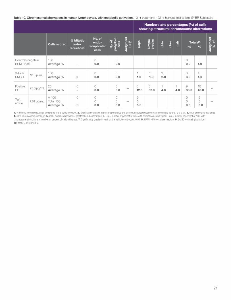
In the assay with S9, excessive toxicity was observed at ≥15.6 μg/mL (Appendix I, Table 10). Structural chromosomal aberrations were evaluated at 7.81 μg/mL (Appendix I, Table 11). No significant increase in the number of cells with structural aberrations, polyploidy, or endoreduplication was observed.
All tests were performed by independent and accredited laboratories using established methodologies.
ConclusionSYBR Safe stain was considered negative for inducing structural aberrations, both with and without metabolic activation.
References1. Evans, H.J., Cytological Methods for Detecting
Chemical Mutagens. Chemical Mutagens, Principles and Methods for their Detection, Hollaender, A. (ed), Vol 4, pp.1-29, Plenum Press: New York and London (1976).
2. Evans, H.J., Chromosomal aberrations produced by ionizing radiation. International Review of Cytology, 13:221-321 (1962).
3. OECD Guideline 473. In vitro Mammalian Chromosomal Aberration Test, updated and adopted July 21, 1997.
4. Thakur, A.J., Berry, K.J., and Mielke, P.Q., Jr., A FORTRAN program for testing trend and homogeneity in proportions. Computer Programs in Biomedicine, 19:229-233 (1985).
5. Maron, D.M., and Ames, B.N., Revised methods for the Salmonella mutagenicity test. Mutation Research, 113:173-215 (1983).
6. Galloway, S.M., Aardema, M.J., Ishidate, M., Jr., Ivett, J.L., Kirkland, D.J., Morita, T., Mosesso, P., and Sofuni, T., Report from working group on in vitro tests for chromosomal aberrations. Mutation Research, 312(3):241-261 (1994). (See Appendix I, Tables 7-11)
MutagenicityAbstractThe objective of this study was to evaluate the test article, SYBR Safe stain, for the ability to induce reverse mutations either in the presence or absence of mammalian microsomal enzymes at the histidine locus in the genome of several strains of S. typhimurium.
The doses tested in the mutagenicity assay were selected based on the results of a previous study. The tester strains used in the mutagenicity assay were S. typhimurium tester strains TA97a, TA98, TA100, TA102, TA1535, TA1537, and TA1538. The assay was conducted with seven doses of test article in both the presence and absence of S9 mix, along with concurrent vehicle and positive controls using three plates per dose. The doses tested with all the tester strains in the presence of S9 mix were 0.100, 0.333, 1.00, 3.33, 10.0, 25.0, and 50.0 μg per plate. The doses tested with all strains in the absence of S9 mix were 0.0100, 0.0333, 0.100, 0.333, 1.00, 3.33, and 10.0 μg per plate.
The results of the Salmonella/Mammalian-Microsome Reverse Mutation Assay indicate that under the conditions of this study, the test article, SYBR Safe stain, did cause positive increases in the mean number of revertants per plate with tester strains TA97a, TA98, TA102, and TA1538 in the presence of S9 mix. No positive increases were observed with any of the other tester strain/activation condition combinations.
ObjectiveThe objective of this study was to evaluate the test article and its metabolites for their ability to induce reverse mutations either in the presence or absence of mammalian microsomal enzymes at the histidine locus in the genome of several strains of S. typhimurium. The assay design was based on OECD Guideline 471, updated and adopted 21 July 1997.
Study timetableExperimental start date 08 August 2003 Study initiation date 31 July 2003 Study start date 08 August 2003 Study end date 12 September 2003 Experimental end date 12 September 2003 Study completion date At finalization
Test article handlingThe test article was supplied by the sponsor as a red flocculent solid on 19 March 2003 and was identified as follows:
Test article Lot number Storage Purity (%)Expiration
date
SYBR Safe stain MPB031403 Room temperature with desiccant
>95 Not provided
Information on the identity, strength, purity, stability, uniformity, or other characteristics that define the test and control articles is on file with the sponsor or the respective manufacturer(s).
8
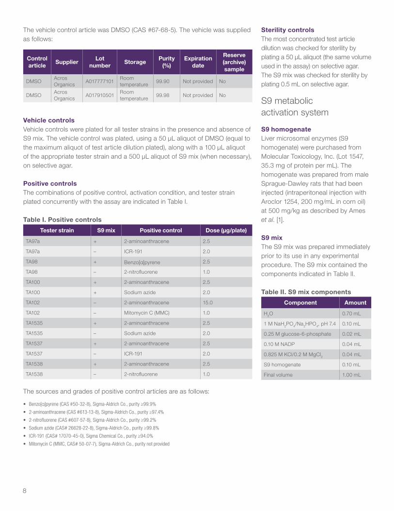
The vehicle control article was DMSO (CAS #67-68-5). The vehicle was supplied as follows:
Control article
SupplierLot
numberStorage
Purity (%)
Expiration date
Reserve (archive) sample
DMSOAcros Organics
A017777101Room temperature
99.90 Not provided No
DMSOAcros Organics
A017910501Room temperature
99.98 Not provided No
Vehicle controlsVehicle controls were plated for all tester strains in the presence and absence of S9 mix. The vehicle control was plated, using a 50 μL aliquot of DMSO (equal to the maximum aliquot of test article dilution plated), along with a 100 μL aliquot of the appropriate tester strain and a 500 μL aliquot of S9 mix (when necessary), on selective agar.
Positive controlsThe combinations of positive control, activation condition, and tester strain plated concurrently with the assay are indicated in Table I.
Table I. Positive controls
Tester strain S9 mix Positive control Dose (μg/plate)
TA97a + 2-aminoanthracene 2.5
TA97a – ICR-191 2.0
TA98 + Benzo[α]pyrene 2.5
TA98 – 2-nitrofluorene 1.0
TA100 + 2-aminoanthracene 2.5
TA100 + Sodium azide 2.0
TA102 – 2-aminoanthracene 15.0
TA102 – Mitomycin C (MMC) 1.0
TA1535 + 2-aminoanthracene 2.5
TA1535 – Sodium azide 2.0
TA1537 + 2-aminoanthracene 2.5
TA1537 – ICR-191 2.0
TA1538 + 2-aminoanthracene 2.5
TA1538 – 2-nitrofluorene 1.0
The sources and grades of positive control articles are as follows:
• Benzo[α]pyrene (CAS #50-32-8), Sigma-Aldrich Co., purity ≥99.9%
• 2-aminoanthracene (CAS #613-13-8), Sigma-Aldrich Co., purity ≥97.4%
• 2-nitrofluorene (CAS #607-57-8), Sigma-Aldrich Co., purity ≥99.2%
• Sodium azide (CAS# 26628-22-8), Sigma-Aldrich Co., purity ≥99.8%
• ICR-191 (CAS# 17070-45-0), Sigma Chemical Co., purity ≥94.0%
• Mitomycin C (MMC, CAS# 50-07-7), Sigma-Aldrich Co., purity not provided
Sterility controlsThe most concentrated test article dilution was checked for sterility by plating a 50 μL aliquot (the same volume used in the assay) on selective agar. The S9 mix was checked for sterility by plating 0.5 mL on selective agar.
S9 metabolic activation system
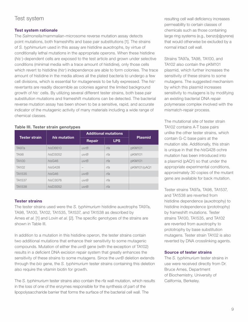
S9 homogenateLiver microsomal enzymes (S9 homogenate) were purchased from Molecular Toxicology, Inc. (Lot 1547, 35.3 mg of protein per mL). The homogenate was prepared from male Sprague-Dawley rats that had been injected (intraperitoneal injection with Aroclor 1254, 200 mg/mL in corn oil) at 500 mg/kg as described by Ames et al. [1].
S9 mixThe S9 mix was prepared immediately prior to its use in any experimental procedure. The S9 mix contained the components indicated in Table II.
Table II. S9 mix components
Component Amount
H2O 0.70 mL
1 M NaH2PO4/Na2HPO4, pH 7.4 0.10 mL
0.25 M glucose-6-phosphate 0.02 mL
0.10 M NADP 0.04 mL
0.825 M KCl/0.2 M MgCl2 0.04 mL
S9 homogenate 0.10 mL
Final volume 1.00 mL
9
Test system
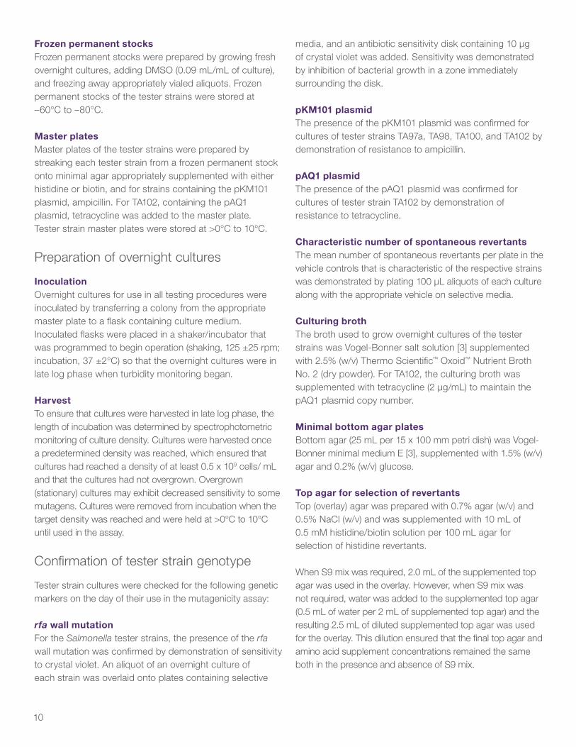
Test system rationaleThe Salmonella/mammalian-microsome reverse mutation assay detects point mutations, both frameshifts and base pair substitutions [1]. The strains of S. typhimurium used in this assay are histidine auxotrophs, by virtue of conditionally lethal mutations in the appropriate operons. When these histidine (his–)-dependent cells are exposed to the test article and grown under selective conditions (minimal media with a trace amount of histidine), only those cells which revert to histidine (his+) independence are able to form colonies. The trace amount of histidine in the media allows all the plated bacteria to undergo a few cell divisions, which is essential for mutagenesis to be fully expressed. The his+ revertants are readily discernible as colonies against the limited background growth of his– cells. By utilizing several different tester strains, both base pair substitution mutations and frameshift mutations can be detected. The bacterial reverse mutation assay has been shown to be a sensitive, rapid, and accurate indicator of the mutagenic activity of many materials including a wide range of chemical classes.
Table III. Tester strain genotypes
Tester strain his mutationAdditional mutations
PlasmidRepair LPS
TA97a hisD6610 uvrB rfa pKM101
TA98 hisD3052 uvrB rfa pKM101
TA100 hisG46 uvrB rfa pKM101
TA102 hisG428 – rfa pKM101/pAQ1
TA1535 hisG46 uvrB rfa
TA1537 hisC3076 uvrB rfa
TA1538 hisD3052 uvrB rfa
Tester strainsThe tester strains used were the S. typhimurium histidine auxotrophs TA97a, TA98, TA100, TA102, TA1535, TA1537, and TA1538 as described by Ames et al. [1] and Lovin et al. [2]. The specific genotypes of the strains are shown in Table III.
In addition to a mutation in this histidine operon, the tester strains contain two additional mutations that enhance their sensitivity to some mutagenic compounds. Mutation of either the uvrB gene (with the exception of TA102) results in a deficient DNA excision repair system that greatly enhances the sensitivity of these strains to some mutagens. Since the uvrB deletion extends through the bio gene, the S. typhimurium tester strains containing this deletion also require the vitamin biotin for growth.
The S. typhimurium tester strains also contain the rfa wall mutation, which results in the loss of one of the enzymes responsible for the synthesis of part of the lipopolysaccharide barrier that forms the surface of the bacterial cell wall. The
resulting cell wall deficiency increases permeability to certain classes of chemicals such as those containing large ring systems (e.g., benzo[α]pyrene) that would otherwise be excluded by a normal intact cell wall.
Strains TA97a, TA98, TA100, and TA102 also contain the pKM101 plasmid, which further increases the sensitivity of these strains to some mutagens. The suggested mechanism by which this plasmid increases sensitivity to mutagens is by modifying an existing bacterial DNA repair polymerase complex involved with the mismatch-repair process.
The mutational site of tester strain TA102 contains A-T base pairs unlike the other tester strains, which contain G-C base pairs at the mutation site. Additionally, this strain is unique in that the hisG428 ochre mutation has been introduced into a plasmid (pAQ1) so that under the appropriate experimental conditions, approximately 30 copies of the mutant gene are available for back mutation.
Tester strains TA97a, TA98, TA1537, and TA1538 are reverted from histidine dependence (auxotrophy) to histidine independence (prototrophy) by frameshift mutations. Tester strains TA100, TA1535, and TA102 are reverted from auxotrophy to prototrophy by base substitution mutagens. Tester strain TA102 is also reverted by DNA crosslinking agents.
Source of tester strainsThe S. typhimurium tester strains in use were received directly from Dr. Bruce Ames, Department of Biochemistry, University of California, Berkeley.
10
Frozen permanent stocksFrozen permanent stocks were prepared by growing fresh overnight cultures, adding DMSO (0.09 mL/mL of culture), and freezing away appropriately vialed aliquots. Frozen permanent stocks of the tester strains were stored at –60°C to –80°C.
Master platesMaster plates of the tester strains were prepared by streaking each tester strain from a frozen permanent stock onto minimal agar appropriately supplemented with either histidine or biotin, and for strains containing the pKM101 plasmid, ampicillin. For TA102, containing the pAQ1 plasmid, tetracycline was added to the master plate. Tester strain master plates were stored at >0°C to 10°C.
Preparation of overnight cultures
InoculationOvernight cultures for use in all testing procedures were inoculated by transferring a colony from the appropriate master plate to a flask containing culture medium. Inoculated flasks were placed in a shaker/incubator that was programmed to begin operation (shaking, 125 ±25 rpm; incubation, 37 ±2°C) so that the overnight cultures were in late log phase when turbidity monitoring began.
HarvestTo ensure that cultures were harvested in late log phase, the length of incubation was determined by spectrophotometric monitoring of culture density. Cultures were harvested once a predetermined density was reached, which ensured that cultures had reached a density of at least 0.5 x 10⁹ cells/ mL and that the cultures had not overgrown. Overgrown (stationary) cultures may exhibit decreased sensitivity to some mutagens. Cultures were removed from incubation when the target density was reached and were held at >0°C to 10°C until used in the assay.
Confirmation of tester strain genotype
Tester strain cultures were checked for the following genetic markers on the day of their use in the mutagenicity assay:
rfa wall mutationFor the Salmonella tester strains, the presence of the rfa wall mutation was confirmed by demonstration of sensitivity to crystal violet. An aliquot of an overnight culture of each strain was overlaid onto plates containing selective
media, and an antibiotic sensitivity disk containing 10 μg of crystal violet was added. Sensitivity was demonstrated by inhibition of bacterial growth in a zone immediately surrounding the disk.
pKM101 plasmidThe presence of the pKM101 plasmid was confirmed for cultures of tester strains TA97a, TA98, TA100, and TA102 by demonstration of resistance to ampicillin.
pAQ1 plasmidThe presence of the pAQ1 plasmid was confirmed for cultures of tester strain TA102 by demonstration of resistance to tetracycline.
Characteristic number of spontaneous revertantsThe mean number of spontaneous revertants per plate in the vehicle controls that is characteristic of the respective strains was demonstrated by plating 100 μL aliquots of each culture along with the appropriate vehicle on selective media.
Culturing brothThe broth used to grow overnight cultures of the tester strains was Vogel-Bonner salt solution [3] supplemented with 2.5% (w/v) Thermo Scientific™ Oxoid™ Nutrient Broth No. 2 (dry powder). For TA102, the culturing broth was supplemented with tetracycline (2 μg/mL) to maintain the pAQ1 plasmid copy number.
Minimal bottom agar platesBottom agar (25 mL per 15 x 100 mm petri dish) was Vogel-Bonner minimal medium E [3], supplemented with 1.5% (w/v) agar and 0.2% (w/v) glucose.
Top agar for selection of revertantsTop (overlay) agar was prepared with 0.7% agar (w/v) and 0.5% NaCl (w/v) and was supplemented with 10 mL of 0.5 mM histidine/biotin solution per 100 mL agar for selection of histidine revertants.
When S9 mix was required, 2.0 mL of the supplemented top agar was used in the overlay. However, when S9 mix was not required, water was added to the supplemented top agar (0.5 mL of water per 2 mL of supplemented top agar) and the resulting 2.5 mL of diluted supplemented top agar was used for the overlay. This dilution ensured that the final top agar and amino acid supplement concentrations remained the same both in the presence and absence of S9 mix.
11
Test article dispositionThe remaining test article was appropriately discarded after issuance of the audited draft report. The disposal of the remaining test article was documented in the study file.
Experimental design
Mutagenicity assay designThe assay was performed using tester strains TA97a, TA98, TA100, TA102, TA1535, TA1537, and TA1538 both in the presence and absence of S9 mix along with the appropriate vehicle and positive controls.
Frequency and route of administrationThe tester strains were exposed to the test article via the plate incorporation methodology originally described by Ames et al. [1] and Maron and Ames [4]. This methodology has been shown to detect a wide range of classes of chemical mutagens. In the plate incorporation methodology, the test article, the tester strain, and the S9 mix (where appropriate) were combined in molten agar, which was overlaid onto a minimal agar plate. Following incubation, revertant colonies were counted. All doses of the test article, the vehicle controls, and the positive controls were plated in triplicate.
Procedures
Plating proceduresEach plate was labeled with a code that identified the test article, test phase, tester strain, activation condition, and dose level. The S9 mix and dilutions of the test article were prepared immediately prior to their use. When S9 mix was not required, 100 μL of tester strain and 50 μL of control or test article dilution were added to 2.5 mL of molten selective top agar (maintained at 45 ±2°C). When S9 mix was required, 500 μL of S9 mix, 100 μL of tester strain, and 50 μL of control or test article dilution were added to 2.0 mL of molten selective top agar. After the required components had been added, the mixture was vortexed and overlaid onto the surface of 25 mL of minimal bottom agar contained in a 15 x 100 mm petri dish. After the overlay solidified, the plates were inverted and incubated for 52 ±4 hr at 37 ±2°C. Positive control articles were plated using a 50 μL plating aliquot.
Scoring the platesPlates that were not evaluated immediately following the incubation period were held at >0°C to 10°C until such
time that colony counting and bacterial background lawn evaluation could take place.
Bacterial background lawn evaluationThe condition of the bacterial background lawn was evaluated both macroscopically and microscopically (using a dissecting microscope) for indications of cytotoxicity and test article precipitate. Evidence of cytotoxicity was scored relative to the vehicle control plate and was recorded along with the revertant counts for all plates at that dose level. Lawns were scored as normal (N), reduced (R), obscured by precipitate (O), macroscopic precipitate present (P), absent (A), or enhanced (E); contaminated plates (C) were also noted.
Counting revertant coloniesRevertant colonies were counted by automated colony counter or by hand.
Data evaluation
Data presentationFor all replicate platings, the mean revertants per plate and the standard deviation were calculated. The results of these calculations are presented in tabular form in Appendix I, Data Tables section of this report. The historical control data are presented after the data tables.
Assay acceptance criteriaBefore assay data were evaluated, the criteria for a valid assay had to be met. The following criteria were used to determine a valid assay:
Tester strain integrityrfa wall mutation To demonstrate the presence of the rfa wall mutation, S. typhimurium tester strain cultures exhibited sensitivity to crystal violet.
pKM101 plasmid To demonstrate the presence of the pKM101 plasmid, cultures of tester strains TA97a, TA98, TA100, and TA102 exhibited resistance to ampicillin.
pAQ1 plasmid The presence of the pAQ1 plasmid was confirmed for cultures of tester strain TA102 by demonstration of resistance to tetracycline.
12
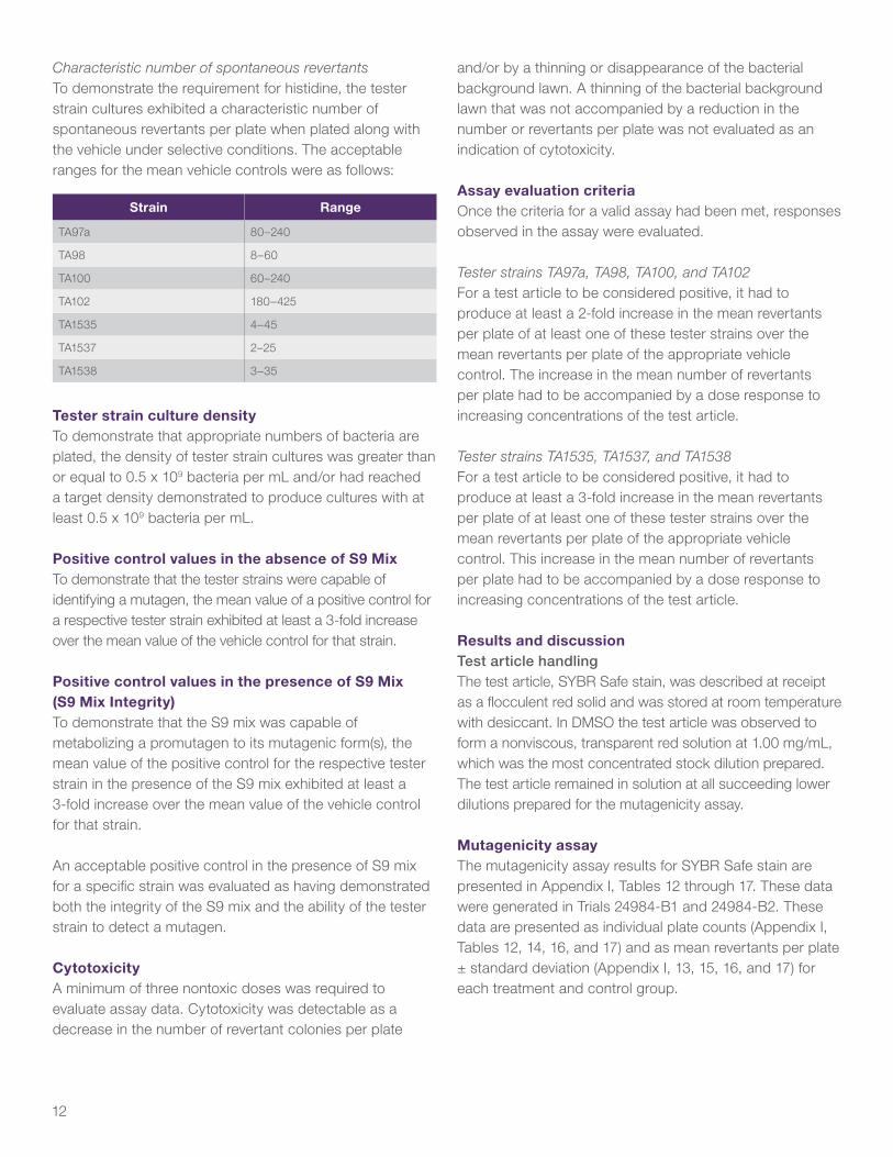
Characteristic number of spontaneous revertants To demonstrate the requirement for histidine, the tester strain cultures exhibited a characteristic number of spontaneous revertants per plate when plated along with the vehicle under selective conditions. The acceptable ranges for the mean vehicle controls were as follows:
Strain Range
TA97a 80–240
TA98 8–60
TA100 60–240
TA102 180–425
TA1535 4–45
TA1537 2–25
TA1538 3–35
Tester strain culture densityTo demonstrate that appropriate numbers of bacteria are plated, the density of tester strain cultures was greater than or equal to 0.5 x 10⁹ bacteria per mL and/or had reached a target density demonstrated to produce cultures with at least 0.5 x 10⁹ bacteria per mL.
Positive control values in the absence of S9 MixTo demonstrate that the tester strains were capable of identifying a mutagen, the mean value of a positive control for a respective tester strain exhibited at least a 3-fold increase over the mean value of the vehicle control for that strain.
Positive control values in the presence of S9 Mix (S9 Mix Integrity)To demonstrate that the S9 mix was capable of metabolizing a promutagen to its mutagenic form(s), the mean value of the positive control for the respective tester strain in the presence of the S9 mix exhibited at least a 3-fold increase over the mean value of the vehicle control for that strain.
An acceptable positive control in the presence of S9 mix for a specific strain was evaluated as having demonstrated both the integrity of the S9 mix and the ability of the tester strain to detect a mutagen.
CytotoxicityA minimum of three nontoxic doses was required to evaluate assay data. Cytotoxicity was detectable as a decrease in the number of revertant colonies per plate
and/or by a thinning or disappearance of the bacterial background lawn. A thinning of the bacterial background lawn that was not accompanied by a reduction in the number or revertants per plate was not evaluated as an indication of cytotoxicity.
Assay evaluation criteriaOnce the criteria for a valid assay had been met, responses observed in the assay were evaluated.
Tester strains TA97a, TA98, TA100, and TA102 For a test article to be considered positive, it had to produce at least a 2-fold increase in the mean revertants per plate of at least one of these tester strains over the mean revertants per plate of the appropriate vehicle control. The increase in the mean number of revertants per plate had to be accompanied by a dose response to increasing concentrations of the test article.
Tester strains TA1535, TA1537, and TA1538 For a test article to be considered positive, it had to produce at least a 3-fold increase in the mean revertants per plate of at least one of these tester strains over the mean revertants per plate of the appropriate vehicle control. This increase in the mean number of revertants per plate had to be accompanied by a dose response to increasing concentrations of the test article.
Results and discussionTest article handlingThe test article, SYBR Safe stain, was described at receipt as a flocculent red solid and was stored at room temperature with desiccant. In DMSO the test article was observed to form a nonviscous, transparent red solution at 1.00 mg/mL, which was the most concentrated stock dilution prepared. The test article remained in solution at all succeeding lower dilutions prepared for the mutagenicity assay.
Mutagenicity assayThe mutagenicity assay results for SYBR Safe stain are presented in Appendix I, Tables 12 through 17. These data were generated in Trials 24984-B1 and 24984-B2. These data are presented as individual plate counts (Appendix I, Tables 12, 14, 16, and 17) and as mean revertants per plate ± standard deviation (Appendix I, 13, 15, 16, and 17) for each treatment and control group.
13
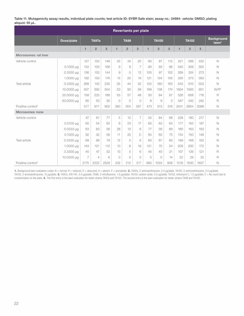
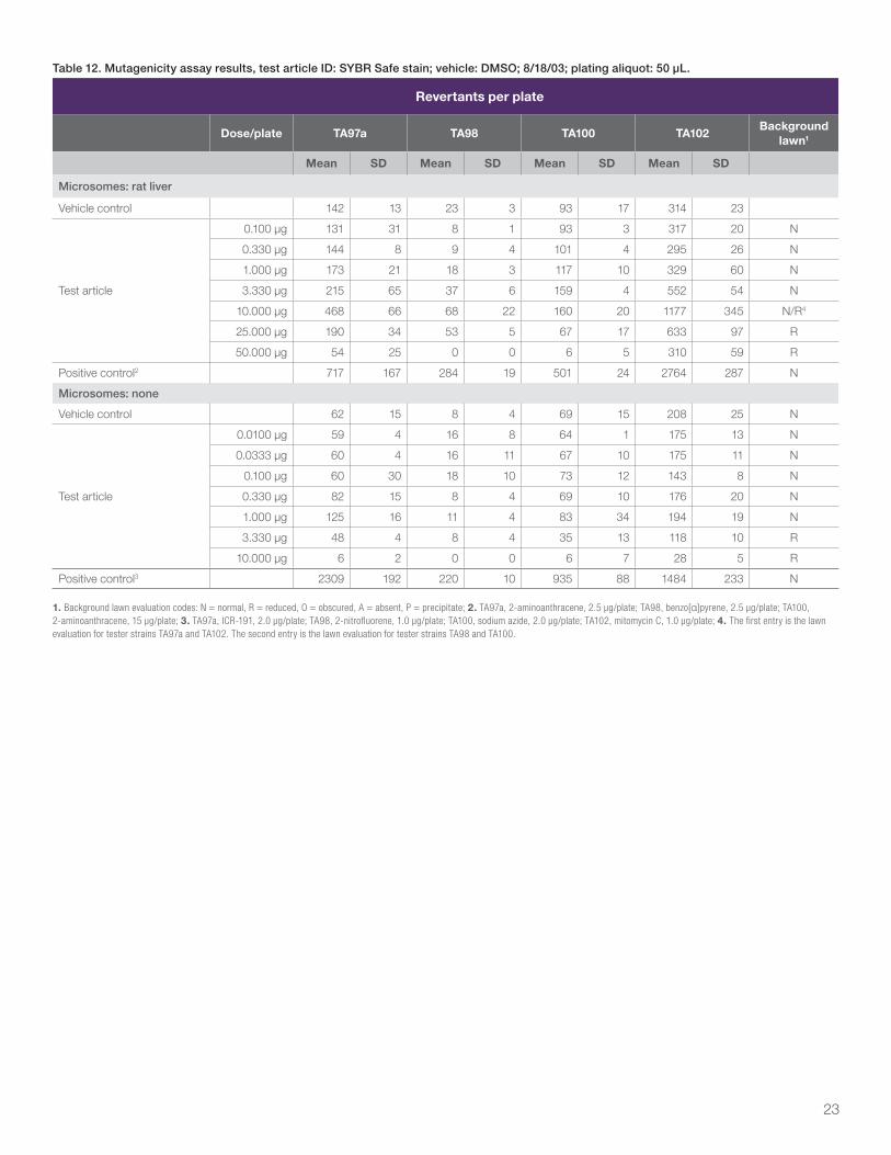
In the mutagenicity assay, Trial 24984-B1 (Appendix I, Tables 12–15), positive increases in the mean number of revertants per plate were observed in the presence of S9 mix with tester strains TA97a (3.3-fold), TA98 (3.0-fold), and TA102 (3.8-fold). An increase was also observed with tester strain TA98 (2.3-fold) in the absence of S9 mix, however this increase was not clearly dose-related and all observed values were within the acceptable vehicle control range for this strain. The observed increase appeared to be a result of a lower than routinely observed TA98 mean vehicle control value and was not considered to be biologically relevant. No positive increases were observed with any of the remaining tester strain/activation condition combinations.
In this trial, the mean vehicle control value for tester strain TA97a in the absence of S9 mix was not within the acceptable range specified in the protocol. For this reason, the data generated with TA97a in the absence of S9 mix in trial 24984-B1 were not used to evaluate the test article. In addition, the mean positive control value for tester stain TA1538 in the absence of S9 mix did not exhibit at least a 3-fold increase over the mean vehicle control value. Also, indications of toxicity were observed with all tester strains in the presence of S9 mix in this trial except TA1538. For these reasons, the data generated with TA1538 in both the presence and absence of S9 mix in trial 24984-B1 were not used to evaluate the test article. The test article was re-tested with tester strain TA97a in the absence of S9 mix and TA1538 in both the presence and absence of S9 mix in Trial 24984-B2.
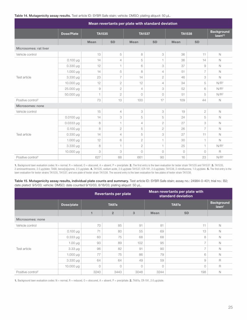
In the repeat mutagenicity assay, Trial 24984-B2 (Appendix I, Tables 16 and 17), all data were acceptable and a 3.7-fold positive increase in the mean number of revertants per plate was observed with tester strain TA1538 in the presence of S9 mix. No positive increases were observed with tester strains TA97a and TA1538 in the absence of S9 mix.
All criteria for a valid study were met. All tests were performed by independent and accredited laboratories using established methodologies [5].
ConclusionThe results of the Salmonella/Mammalian-Microsome Reverse Mutation Assay indicate that under the conditions of this study, the test article, SYBR Safe stain, did cause
positive increases in the mean number of revertants per plate with tester strains TA97a, TA98, TA102, and TA1538 in the presence of S9 mix. No positive increases were observed with any of the other tester strain/activation condition combinations.
References1. Ames, B.N., McCann, J., and Yamasaki, E., “Methods for detecting carcinogens and
mutagens with the Salmonella/Mammalian-Microsome Mutagenicity Test.” Mutation Research, 31:347-364 (1975).
2. Levin, D.M., Hollstein, M., Christian, M/F., Schwiers, E.A., and Ames B.N., “A new Salmonella tester strain (TA102) with A-T base pairs at the site of mutation detects oxidative mutagens.” Proc. Natl. Acad. Sci. USA 79:7445-7449 (1982).
3. Vogel, H.J and Bonner, D.M., “Acetylornithinase of E. coli: Partial purification and some properties.” J. Biol. Chem., 218:97-106 (1956).
4. Maron, D.M. and Ames B., “Revised Methods for the Salmonella Mutagenicity Test.” Mutation Research, 113:173-215 (1983).
5. OECD Guideline 471, Bacterial Reverse Mutation Test, updated and adopted 21 July 1997. (see Appendix I, Tables 12-17)
Acute oral toxicity
SummaryA single oral administration of SYBR Safe DNA Gel Stain in 0.5X TBE at a limit dose of 5,000 mg/kg to three female rats produced no mortalities or toxic signs.
IntroductionThis procedure is designed to determine the acute oral toxicity of the material under test.
A Limit Screen test was performed using three female Sprague-Dawley rats, which received an oral limit dose of 5,000 mg/kg of the test article. The animals were observed for mortality, weight change, and toxic signs for a two-week period.
All three rats survived for 2 weeks after the dose administration. Further study was not performed.
Test article identificationName: SYBR Safe DNA Gel Stain in 0.5X TBE Total quantity received for testing: 1 L Expiration date: January 05, 2005 Sterility status: nonsterile Physical description: pale pink liquid Total quantity used for this study: 10 g Lot number: 52E16-1 Storage condition: room temperature
14
ProtocolThis test was conducted according to Protocol Number X4H165G, which incorporates by reference Northview Standard Operating Procedure 16D-05 and is on file at Northview Pacific Laboratories, Inc. There were no amendments to or deviations from the protocol.
Data dispositionRaw data and the final report from this study are archived at Northview Pacific Laboratories, Inc., 551 Linus Pauling Drive, Hercules, CA, 94547, under Northview Report Number X4H165G.
Justification for test systemRats are the species required by the Federal Insecticide, Fungicide, and Rodenticide Act (FIFRA), Toxic Substances Control Act (TSCA), and Health Effects Guideline OPPTS 870.1100—Acute Oral Toxicity, December 2002.
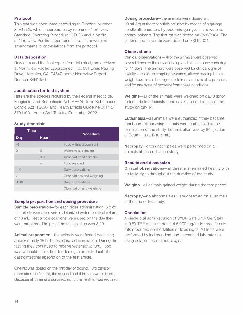
Study timetable
TimeProcedure
Day Hour
–1 Food withheld overnight
0 0 Weighing and dosing
0–4 Observation of animals
4 Food restored
1–6 Daily observations
7 Observations and weighing
8–13 Daily observations
14 Observation and weighing
Sample preparation and dosing procedureSample preparation—for each dose administration, 5 g of test article was dissolved in deionized water to a final volume of 10 mL. Test article solutions were used on the day they were prepared. The pH of the test solution was 8.29.
Animal preparation—the animals were fasted beginning approximately 18 hr before dose administration. During the fasting they continued to receive water ad libitum. Food was withheld until 4 hr after dosing in order to facilitate gastrointestinal absorption of the test article.
One rat was dosed on the first day of dosing. Two days or more after the first rat, the second and third rats were dosed. Because all three rats survived, no further testing was required.
Dosing procedure—the animals were dosed with 10 mL/kg of the test article solution by means of a gavage needle attached to a hypodermic syringe. There were no control animals. The first rat was dosed on 8/25/2004. The second and third rats were dosed on 8/31/2004.
ObservationsClinical observations—all of the animals were observed several times on the day of dosing and at least once each day for 14 days. The animals were observed for clinical signs of toxicity such as unkempt appearance, altered feeding habits, weight loss, and other signs of distress or physical depression, and for any signs of recovery from these conditions.
Weights—all of the animals were weighed on day 0 (prior to test article administration), day 7, and at the end of the study on day 14.
Euthanasia—all animals were euthanized if they became moribund. All surviving animals were euthanized at the termination of the study. Euthanization was by IP injection of Beuthanasia-D (0.5 mL).
Necropsy—gross necropsies were performed on all animals at the end of the study.
Results and discussionClinical observations—all three rats remained healthy with no toxic signs throughout the duration of the study.
Weights—all animals gained weight during the test period.
Necropsy—no abnormalities were observed on all animals at the end of the study.
ConclusionA single oral administration of SYBR Safe DNA Gel Stain in 0.5X TBE at a limit dose of 5,000 mg/kg to three female rats produced no mortalities or toxic signs. All tests were performed by independent and accredited laboratories using established methodologies.

15
Physicochemical testing for waste characterization
Ignitability: >212°F (not classified as ignitable) Reactivity: not detected (for both cyanide and sulfide—mg/L) Corrosivity: pH = 8.25 (not corrosive) Corrositex test method: >60 minutes; classified as Category 2 noncorrosive Bioassay/toxicity test results: LC50 >500 mg/L; not classified as hazardous or toxic to aquatic life. Kow (Conducted in 2015 by Intertek): 1.02
Sample and bioassay information:
Test type: CCR Title 22 Acute Screening Test conditions: static Test species: Pimephales promelas Common name: fathead minnow Organism supplier: Thomas Fish Co. Number per tank: 10 Mean length (mm): 27.0 Mean weight (g): 0.250 Range (mm): 24–31 Loading rate (g/L): 0.31 Dilution water: charcoal-filtered tap water Test solution volume (L): 8.0 Sample receipt date: 9/3/2003 Test dates: 9/4/2003–9/8/2003
Summary test results
Treatment Rep.Initial count
Final countPercent survival
Average survival
Control A 10 10 100
B 10 10 100 100
250 mg/L A 10 10 100
B 10 10 100 100
500 mg/L A 10 10 100
B 10 10 100 100
750 mg/L A 10 10 100
B 10 10 100 100
Note: LC50
= the test concentration that produces a 50% lethal effect on the test organisms. LC
50 value >500 mg/L is classified as not hazardous under CCR Title 22, Acute Aquatic Toxicity.
Corrosivity—Corrositex test
Completion date: September 10, 2003
Executive summaryA single sample provided by AMEC Earth and Environmental, Inc., was evaluated with the Corrositex test method to determine its corrosive potential and to designate its packing group classification. The results of this study may be summarized as follows:
Sample description: MPR71454 Mean Corrositex time (minutes): >60 Packing group: NC
Study objectiveA single sample provided by AMEC Earth and Environmental, Inc. was evaluated with the Corrositex test method to determine its corrosive potential and to designate its packing group classification. To achieve this objective, the sample was subjected to a three-step testing process as described under materials and methods.
BackgroundThe Corrositex test is a standardized and reproducible method that can be employed to determine the potential corrosivity and determine the packing group classification of specified categories of chemical compounds under the hazardous materials transportation regulations administered by the US Department of Transportation (DOT) and international dangerous goods codes. The Corrositex test predicts the in vivo corrosive potential of a chemical compound or mixture by using as an endpoint the time it takes for the chemical to permeate through or destroy a synthetic biobarrier. When the chemical has passed through this biobarrier, a visual change is produced in a proprietary chemical detection system (CDS).
Materials and methods The Corrositex test is performed in three steps. First, a qualification test is done to ensure that the test sample and the CDS reagent are compatible.
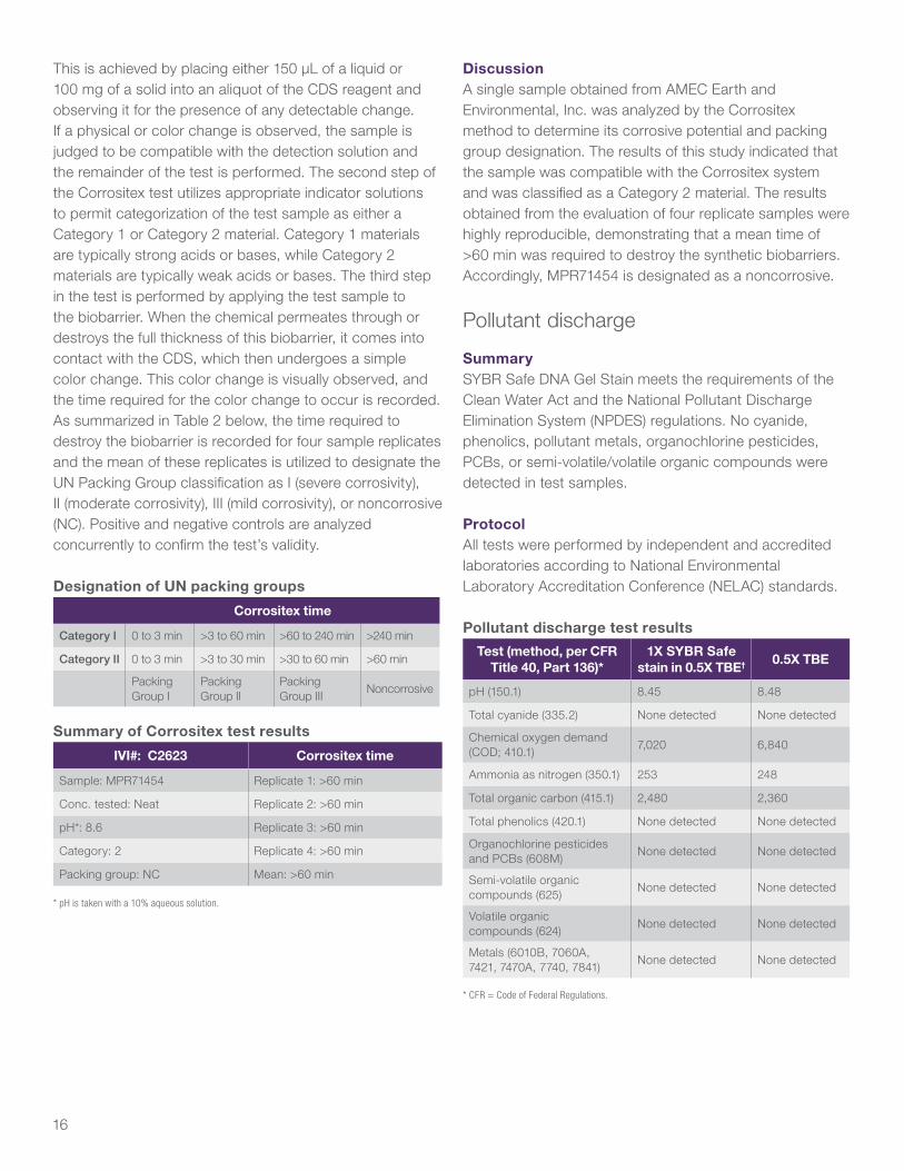
16
This is achieved by placing either 150 μL of a liquid or 100 mg of a solid into an aliquot of the CDS reagent and observing it for the presence of any detectable change. If a physical or color change is observed, the sample is judged to be compatible with the detection solution and the remainder of the test is performed. The second step of the Corrositex test utilizes appropriate indicator solutions to permit categorization of the test sample as either a Category 1 or Category 2 material. Category 1 materials are typically strong acids or bases, while Category 2 materials are typically weak acids or bases. The third step in the test is performed by applying the test sample to the biobarrier. When the chemical permeates through or destroys the full thickness of this biobarrier, it comes into contact with the CDS, which then undergoes a simple color change. This color change is visually observed, and the time required for the color change to occur is recorded. As summarized in Table 2 below, the time required to destroy the biobarrier is recorded for four sample replicates and the mean of these replicates is utilized to designate the UN Packing Group classification as I (severe corrosivity), II (moderate corrosivity), III (mild corrosivity), or noncorrosive (NC). Positive and negative controls are analyzed concurrently to confirm the test’s validity.
Designation of UN packing groups
Corrositex time
Category I 0 to 3 min >3 to 60 min >60 to 240 min >240 min
Category II 0 to 3 min >3 to 30 min >30 to 60 min >60 min
Packing Group I
Packing Group II
Packing Group III
Noncorrosive
Summary of Corrositex test results
IVI#: C2623 Corrositex time
Sample: MPR71454 Replicate 1: >60 min
Conc. tested: Neat Replicate 2: >60 min
pH*: 8.6 Replicate 3: >60 min
Category: 2 Replicate 4: >60 min
Packing group: NC Mean: >60 min
* pH is taken with a 10% aqueous solution.
DiscussionA single sample obtained from AMEC Earth and Environmental, Inc. was analyzed by the Corrositex method to determine its corrosive potential and packing group designation. The results of this study indicated that the sample was compatible with the Corrositex system and was classified as a Category 2 material. The results obtained from the evaluation of four replicate samples were highly reproducible, demonstrating that a mean time of >60 min was required to destroy the synthetic biobarriers. Accordingly, MPR71454 is designated as a noncorrosive.
Pollutant discharge
SummarySYBR Safe DNA Gel Stain meets the requirements of the Clean Water Act and the National Pollutant Discharge Elimination System (NPDES) regulations. No cyanide, phenolics, pollutant metals, organochlorine pesticides, PCBs, or semi-volatile/volatile organic compounds were detected in test samples.
ProtocolAll tests were performed by independent and accredited laboratories according to National Environmental Laboratory Accreditation Conference (NELAC) standards.
Pollutant discharge test results
Test (method, per CFR Title 40, Part 136)*
1X SYBR Safe stain in 0.5X TBE† 0.5X TBE
pH (150.1) 8.45 8.48
Total cyanide (335.2) None detected None detected
Chemical oxygen demand (COD; 410.1)
7,020 6,840
Ammonia as nitrogen (350.1) 253 248
Total organic carbon (415.1) 2,480 2,360
Total phenolics (420.1) None detected None detected
Organochlorine pesticides and PCBs (608M)
None detected None detected
Semi-volatile organic compounds (625)
None detected None detected
Volatile organic compounds (624)
None detected None detected
Metals (6010B, 7060A, 7421, 7470A, 7740, 7841)
None detected None detected
* CFR = Code of Federal Regulations.
17
Appendix I: Data tablesTable 1. Mutation assay without activation by SYBR Safe stain. Test article: SYBR Safe stain; cells analyzed: 3 x 10⁶; vehicle: DMSO; teatment period: ~4 hr; selective agent: trifluorothymidine (Tft), 3.0 µg/mL; expression period: 2 days.
Test conditionDaily cell counts
(cells/mL, x 105 units)Cumulative
RSG[1]
Total mutant
colonies
Total viable
colonies
Cloning efficiency[2]
Relative growth
(%)[3]
Mutant frequency
(x 10–6 units)[4]
Day 1 Day 2
Nonactivation controls5 Avg. VC Avg. VC
Vehicle control 11.9 10.8 14.3
13.1
139 702 116.9
106.2
120.0 39.5
Vehicle control 12.1 9.3 12.5 141 579 96.6 86.7 48.6
Vehicle control 12.0 9.4 12.5 185 631 105.1 94.7 58.8
MMS 13 μg/mL 10.3 6.5 7.4 460 327 54.5 29.2 281.36
MMS 13 μg/mL 10.0 6.0 6.7 531 311 51.8 24.8 341.86
Test compound (μg/mL)Relative to vehicle control (%)5
Relative to vehicle control (%)5
0.125 12.0 7.1 72.2 195 708 111.1 80.3 55.2
0.250 13.3 4.4 49.6 115 340 53.4 26.5 67.3
0.500 Terminated due to excessive cytotoxicity
1. Relative suspension growth (RSG) = (day 1 count/3 x (day 2 count)/3 (or day 1 count) if not subcultured). 2. Cloning efficiency = total viable colony count/number of cells seeded x 100. 3. Relative growth = (relative suspension growth x relative cloning efficiency)/100. 4. Mutant frequency = total mutant colonies/total viable colonies) x (2 x 10⁻⁴); decimal is moved to express the frequency in units of 10–6.
5. Vehicle control =1% DMSO; positive control: MMS = methyl methanesulfonate. 6. Mutagenic. Exceeds minumum criterion of 97.9 x 10⁻⁶.
Table 2. Sizing data for mutation assay without activation by SYBR Safe stain. Test article: SYBR Safe stain; vehicle: DMSO; selective agent: Tft, 3.0 mg/mL.
Cum. RSG (%)1 Cloning efficiency[2] Relative growth (%)3 Mutant frequency (x 10–6 units)[4]
Test condition Conc. Day 1 Day 2 ABS % Rel % Total Small Large
Vehicle control5
1% 99.2 109.0 116.9 110.1 120.0 39.5 19.0 20.5
1% 100.8 95.4 96.6 90.9 86.7 48.6 22.2 26.4
1% 100.0 95.6 105.1 99.0 94.7 58.8 26.6 32.2
MMS (μg/mL)6
13.000 85.8 56.8 54.5 51.4 29.2 281.3 166.0 115.3
13.000 83.3 50.9 51.8 48.8 24.8 341.8 195.1 146.7
Test article (μg/mL)
0.125 100.0 72.2 118 111.1 80.3 55.2 20.3 34.8
0.250 110.8 49.6 56.7 53.4 26.5 67.3 41.7 25.6
0.500 Terminated due to excessive cytotoxicity
1. Cum. RSG = cumulative suspension growth relative to the average vehicle control suspension growth. 2. Cloning efficiency = total viable colony count/number of cells seeded x 100. 3. Relative growth = (relative suspension growth X relative cloning efficiency)/100. 4. Mutant frequency = total mutant colonies/total viable colonies) x (2 x 10⁻⁴); decimal is moved to express the frequency in units of 10–6; expressed as total mutant frequency; small colony mutant frequency and large colony mutant frequency. 5. Vehicle control = DMSO. 6. Positive control: MMS = methyl methanesulfonate; colony counts increased by 9.099% to compensate for area of dish not scanned.
18
Table 3. Mutation assay with activation by SYBR Safe stain. Test article: SYBR Safe stain; Cells analyzed: 3 x 10⁶; vehicle: DMSO; treatment period: ~4 hr; selective agent: Tft, 3.0 mg/mL; expression period: 2 days.
Test conditionDaily cell counts (cells/ml, x 105
units)
Cumulative RSG[1]
Total mutant
colonies
Total viable
colonies
Cloning efficiency[2]
Relative growth (%)[3]
Mutant frequency
(x 10–6 units)[4]
Day 1 Day 2
S9-activation controls5
S9 batch number: 1254Avg. VC
Avg. VC
Vehicle Control 10.5 8.6 10.0
11.7
309 815 135.8
112.9
102.9 75.8
Vehicle Control 8.1 14.9 13.4 216 535 89.1 90.2 80.8
Vehicle Control 9.2 11.5 11.8 208 682 113.6 100.9 61.1
MCA 2 μg/mL 7.2 8.9 7.1 872 391 65.1 35.0 446.46
MCA 4 μg/mL 5.3 7.6 4.5 726 303 50.5 17.1 478.46
Test compound (μg/mL)Relative to vehicle control (%)[5]
Relative to vehicle control (%)[5]
1.24 9.0 9.9 84.4 184 523 77.2 65.1 70.6
2.47 6.6 9.7 60.6 228 526 77.7 47.1 86.7
4.93 5.5 5.7 29.7 99 173 25.6 7.6 114.5
9.85 Terminated due to excessive cytotoxicity
1. RSG = (day 1 count/3 x (day 2 count)/3 (or day 1 count if not subcultured). 2. Cloning efficiency = total viable colony count/number of cells seeded x 100. 3. Relative growth = (relative suspension growth x relative cloning efficiency)/100. 4. Mutant frequency = total mutant colonies/total viable colonies) x (2 x 10⁻⁴); decimal is moved to express the frequency in units of 10–6. 5. Vehicle control = 1% DMSO,
positive control: MCA = methylcholanthrene. 6. Mutagenic. Exceeds minumum criterion of 145.1 x 10⁻⁶.
Table 4. Sizing data for mutation assay without activation by SYBR Safe stain. Test article: SYBR Safe stain; vehicle: DMSO; selective agent: Tft, 3.0 μg/mL.
Cum. RSG (%)[1] Cloning efficiency[2] Relative growth (%)[3] Mutant frequency (x 10–6 units)[4]
Test condition Conc. Day 1 Day 2 ABS % Rel% Total Small Large
Vehicle control[5]
1% 113.3 85.5 135.8 120.4 102.9 75.8 47.9 27.8
1% 87.4 114.3 89.1 78.9 90.2 80.8 52.7 28.2
1% 99.3 100.2 113.6 100.7 100.9 61.1 38.7 22.4
MMS (μg/mL)[6]
2.00 77.7 60.7 65.1 57.7 35.0 446.4 339.7 106.7
4.00 57.2 38.1 50.5 44.8 17.1 478.4 363.3 115.1
Test article (μg/mL)
1.24 97.1 84.4 87.1 77.2 65.1 70.6 37.6 33.0
2.47 71.2 60.6 87.6 77.7 47.1 86.7 48.5 38.2
4.93 59.4 29.7 28.9 25.6 7.6 114.5 88.1 26.4
9.85 Terminated due to excessive cytotoxicity
1. Cum. RSG = cumulative suspension growth relative to the average vehicle control suspension growth. 2. Cloning efficiency = total viable colony count/number of cells seeded x 100. 3. Relative growth = (relative suspension growth x relative cloning efficiency)/100. 4. Mutant frequency = total mutant colonies/total viable colonies) x (2 x 10⁻⁴); decimal is moved to express the frequency in unites of 10–6; expressed as total mutant frequency; small colony mutant frequency and large colony mutant frequency. 5. Vehicle control = DMSO. 6. Positive control: MCA = methylcholanthrene. Colony counts increased by 9.099% to compensate for area of dish not scanned.
19
Table 5. Summary of dose range–finding study. Test article: MPR 71454.
Treatment group Colonies/dishAverage number of
colonies/dishAverage RPE (%)[4]
1 2 3 4 5 PE2 ±SD (%)3
Vehicle control1 49 40 37 36 37 39.8 23.4 3.2 100
Test article
0.0333 39 40 40 39 43 40.2 23.6 1.0 101
0.100 48 38 42 49 44 44.2 26.0 2.6 111
0.333 33 32 32 32 26 31.0 18.2 1.7 78
1.000 8 5 4 8 7 6.4 3.8 1.1 16
3.330 0 0 0 0 0 0.0 0.0 0.0 0
10.000 0 0 0 0 0 0.0 0.0 0.0 0
1. Vehicle = control = 0.2% DMSO. 2. PE = plating efficiency = (number of colonies per dish/number of target cells seeded) x 100%. 3. SD = standard deviation. 4. RPE = relative plating efficiency =
(average PE of treatment group/vehicle control average PE) x 100%.
Table 6. Summary of transformation assay results. Test article: MPR 71454.
Treatment groupMT
frequency[1] (%)Total MT
colonies[2] (%)Total colonies
scored[3]
Average number of colonies/dish [4] Average RPE (%)[7]
PE5 ±SD (%)6
DMSO (0.2%) 0.106 1 941 31.4 19.6 2.9 100
BaP 5.00 1.5539 25 1,610 35.8 22.4 3.4 114
Test article
0.442 5 1,132 37.7 23.6 3.0 120
0.315 4 1,268 42.3 17.2 2.2 88
0.144 2 1,393 46.4 11.6 1.6 59
1. MT frequency = (total morphologically transformed (MT) colonies/total colonies scored). 2. Total MT colonies = total number of morphologically transformed colonies. 3. Total number of colonies from all dishes. 4. Total colonies scored/total number of dishes. 5. PE = plating efficiency = (number of colonies per dish) x 100. 6. SD = standard deviation. 7. RPE = relative plating efficiency = (average PE of treatment group/vehicle control average PE) x 100%. 8. The number of cells seeded per dish increased to adjust for expected toxicity. 9. p ≤ 0.05 vs. DMSO (Fisher’s exact test). BaP = Benzo[α]pyrene.
Table 7. Assessment of toxicity for chromomsomal aberrations, assay without metabolic activation. ~22 hr treatment; ~22 hr harvest; test article: SYBR Safe stain.
Treatment % mitotic index % mitotic reduction
Negative control: RPMI 1640 6.0 —
Vehicle control: DMSO 10.00 μL/mL 6.3 0
Test article
7.81 μL/mL 0[1] N/A
15.60 μL/mL 0[1] —
31.30 μL/mL 0[1] —
62.50 μL/mL 0[1] —
125.00 μL/mL 0[1] —
250.00 μL/mL 0[1] —
500.00 μL/mL 0[1] —
1000.00 μL/mL 0[1] —
1. Only dead cells present on slide; RPMI 1640 = culture medium; DMSO = dimethylsulfoxide.
20
Table 8. Chromosomal aberrations in human lymphocytes, without metabolic activation. ~22 hr treatment; ~22 hr harvest; lab no.: Cy041103; test article: SYBR Safe stain.
Numbers and percentages (%) of cells showing structural chromosome aberrations
Cells scored
% Mitotic index
reduction[1]
No. of endo-
reduplicated cells
No
. of
po
lyp
loid
ce
lls
Jud
gm
ent
(+/-
)[2]
Gap
s
Sim
ple
b
reak
s
chte
[3]
chre
[4]
mab
5
Totals–g +g
Jud
gm
ent
(+/–
)[7]
Controls Negative: RPMI 1640[8]
A 100Total 100Average % –
0 0 0.0
0 0 0.0
111.0
0 0 0.0
1 1 1.0
Vehicle: DMSO[9] 10.000 μl/mL
A 100Total 100Average % 0
0 0 0.0
0 0 0.0
555.0
0 0 0.0
5 5 5.0
Positive: MMC[10] 0.200 μg/mL
A 25Total 25Average %
0
–
0 0 0.0
0 0.0
—6
624.0
936.0
8832.0
14 14 56.0
17 17 68.0
+
Test article
1.000 μg/mLA 100Total 100Average %
0
53
0 0 0.0
0 0.0
—8
88.0
55.0
0 5 5.0
13 13 13.0
—
1. % Mitotic index reduction as compared to the vehicle control. 2. Significantly greater in % polyploidy and % endoreduplication than the vehicle control, p ≤ 0.01. 3. chte: chromatid exchange. 4. chre: chromosome exchange. 5. mab: multiple aberrations, greater than 4 aberrations. 6. –g = number or percent of cells with chromosome aberrations, +g = number or percent of cells with chromosome aberrations + number or percent of cells with gaps. 7. Significantly greater in –g than the vehicle control; p ≤ 0.01. 8. RPMI 1640 = culture medium. 9. DMSO = dimethylsulfoxide. 10. MMC = mitomycin C.
Table 9. Assessment of toxicity for chromosomal aberrations, assay with metabolic activiation. ~22 hr treatment; ~22 hr harvest; test article: SYBR Safe stain.
Treatment % Mitotic index % Mitotic reduction
Negative control: RPMI 1640 5.5 —
Vehicle control: DMSO 10.000 μg/mL 4.5 0
Test article
7.81 μg/mL 1.7 62
15.6 μg/mL 0.2 96
31.3 μg/mL 0.0 100
62.5 μg/mL 0[1] —
125 μg/mL 0[1] —
250 μg/mL 0[1] —
500 μg/mL 0[1] —
1,000 μg/mL 0[1] —
1. Only dead cells present on slide. RPMI 1640 = culture medium. DMSO = dimethylsulfoxide.
21
Table 10. Chromosomal aberrations in human lymphocytes, with metabolic activation. ~3 hr treatment; ~22 hr harvest; test article: SYBR Safe stain.
Numbers and percentages (%) of cells showing structural chromosome aberrations
Cells scored% Mitotic
index reduction[1]
No. ofendo-
reduplicated cells
No.
of
po
lyp
loid
ce
lls
Jud
gm
ent
(+/-
)2
Gap
s
Sim
ple
b
reak
s
chte
chre
mab Totals[3]
–g +g
Jud
gm
ent
(+/–
)[4]
Controls negative: RPMI 1640
100Average %
–
0 0.0
0 0.0
0 0.0
0 1.0
Vehicle: DMSO
10.0 μl/mL100Average % 0
0 0.0
0 0.0
1 1.0
1 1.0
2 2.0
3 3.0
4 4.0
Positive: CP
25.0 μg/mL25Average %
0–
0 0.0
0 0.0
— 3 12.0
8 32.0
1 4.0
1 4.0
9 36.0
10 40.0
+
Test article
7.81 μg/mLA 100Total 100Average %
0
62
0 0 0.0
0 0 0.0
— 5 5 5.0
0 0 0.0
5 5 5.0
—
1. % Mitotic index reduction as compared to the vehicle control. 2. Significantly greater in percent polyploidy and percent endoreduplication than the vehicle control, p ≤ 0.01. 3. chte: chromatid exchange. 4. chre: chromosome exchange. 5. mab: multiple aberrations, greater than 4 aberrations. 6. –g = number or percent of cells with chromosome aberrations, +g = number or percent of cells with chromosome aberrations + number or percent of cells with gaps. 7. Significantly greater in –g than the vehicle control; p ≤ 0.01. 8. RPMI 1640 = culture medium. 9. DMSO = dimethylsulfoxide. 10. MMC = mitomycin C.
22
Table 11. Mutagenicity assay results, individual plate counts; test article ID: SYBR Safe stain; assay no.: 24984- vehicle: DMSO; plating aliquot: 50 μL.
Revertants per plate
Dose/plate TA97a TA98 TA100 TA102Background
lawn1
1 2 3 1 2 3 1 2 3 1 2 3
Microsomes: rat liver
Vehicle control 127 150 148 25 24 20 80 87 112 321 288 332 N
Test article
0.1000 μg 122 105 166 9 9 7 90 93 96 340 309 302 N
0.3330 μg 136 152 144 9 5 12 105 97 102 289 324 273 N
1.0000 μg 192 150 176 15 20 19 121 124 105 320 273 393 N
3.3300 μg 268 142 235 35 44 32 155 160 163 543 610 503 N
10.0000 μg 507 392 504 53 93 58 169 138 174 1564 1065 901 N/R4
25.0000 μg 158 225 186 55 57 48 50 84 67 526 658 716 R
50.0000 μg 80 53 30 0 0 0 8 9 0 347 342 242 R
Positive control2 577 671 902 280 304 267 473 513 516 2541 2664 3088 N
Microsomes: none
Vehicle control 47 61 77 5 12 7 55 84 68 228 180 217 N
Test article
0.0100 μg 60 54 62 8 23 17 65 63 63 177 162 187 N
0.0333 μg 63 60 56 28 13 6 77 58 66 180 163 183 N
0.1000 μg 92 32 56 11 25 C 84 60 75 134 150 146 N
0.3330 μg 69 98 79 12 5 6 60 67 80 199 168 162 N
1.0000 μg 143 121 112 10 8 16 121 75 54 209 200 172 N
3.3300 μg 45 47 52 13 5 6 45 40 21 107 126 121 R
10.0000 μg 7 4 6 0 0 0 5 0 14 22 29 32 R
Positive control3 2175 2222 2529 232 212 217 865 1033 906 1216 1630 1607 N
1. Background lawn evaluation codes: N = normal, R = reduced, O = obscured, A = absent, P = precipitate. 2. TA97a, 2-aminoanthracene, 2.5 μg/plate. TA100, 2-aminoanthracene, 2.5 μg/plate; TA102, 2-aminoanthracene, 15 μg/plate. 3. TA97a, ICR-191, 2.0 μg/plate; TA98, 2-nitrofluorene, 1.0 μg/plate; TA100, sodium azide, 2.0 μg/plate; TA102, mitomycin C, 1.0 μg/plate; C = No count due to contamination on the plate. 4. The first entry is the lawn evaluation for tester strains TA97a and TA102. The second entry is the lawn evaluation for tester strains TA98 and TA100.
23
Table 12. Mutagenicity assay results, test article ID: SYBR Safe stain; vehicle: DMSO; 8/18/03; plating aliquot: 50 μL.
Revertants per plate
Dose/plate TA97a TA98 TA100 TA102Background
lawn1
Mean SD Mean SD Mean SD Mean SD
Microsomes: rat liver
Vehicle control 142 13 23 3 93 17 314 23
Test article
0.100 μg 131 31 8 1 93 3 317 20 N
0.330 μg 144 8 9 4 101 4 295 26 N
1.000 μg 173 21 18 3 117 10 329 60 N
3.330 μg 215 65 37 6 159 4 552 54 N
10.000 μg 468 66 68 22 160 20 1177 345 N/R4
25.000 μg 190 34 53 5 67 17 633 97 R
50.000 μg 54 25 0 0 6 5 310 59 R
Positive control2 717 167 284 19 501 24 2764 287 N
Microsomes: none
Vehicle control 62 15 8 4 69 15 208 25 N
Test article
0.0100 μg 59 4 16 8 64 1 175 13 N
0.0333 μg 60 4 16 11 67 10 175 11 N
0.100 μg 60 30 18 10 73 12 143 8 N
0.330 μg 82 15 8 4 69 10 176 20 N
1.000 μg 125 16 11 4 83 34 194 19 N
3.330 μg 48 4 8 4 35 13 118 10 R
10.000 μg 6 2 0 0 6 7 28 5 R
Positive control3 2309 192 220 10 935 88 1484 233 N
1. Background lawn evaluation codes: N = normal, R = reduced, O = obscured, A = absent, P = precipitate; 2. TA97a, 2-aminoanthracene, 2.5 μg/plate; TA98, benzo[α]pyrene, 2.5 μg/plate; TA100, 2-aminoanthracene, 15 μg/plate; 3. TA97a, ICR-191, 2.0 μg/plate; TA98, 2-nitrofluorene, 1.0 μg/plate; TA100, sodium azide, 2.0 μg/plate; TA102, mitomycin C, 1.0 μg/plate; 4. The first entry is the lawn evaluation for tester strains TA97a and TA102. The second entry is the lawn evaluation for tester strains TA98 and TA100.

24
Table 13. Mutagenicity assay results, individual plate counts. Test article id: SYBR safe stain; vehicle: DMSO. plating aliquot: 50 μL.
Revertants/plate
Dose/plate TA1535 TA1537 TA1538Background
lawn1
1 2 3 1 2 3 1 2 3
Microsomes: rat liver
Vehicle control 8 13 17 9 5 10 29 30 48 N
Test article
0.100 μg 10 17 15 5 6 5 22 49 36 N
0.333 μg 11 12 13 9 3 5 44 40 27 N
1.000 μg 9 19 13 3 10 11 59 47 48 N
3.330 μg 31 17 21 12 16 13 44 49 46 N
10.000 μg 14 11 12 17 9 10 30 37 C2 N/R4
25.000 μg 7 9 11 1 5 7 47 50 59 N/R4
50.000 μg 4 0 0 0 0 0 56 49 47 N/R4
Positive control3 59 79 82 111 108 80 126 143 59 N
Microsomes: none
Vehicle control C2 12 17 3 5 0 18 19 21 N
Test article
0.0100 μg 14 16 11 5 10 1 27 19 27 N
0.0333 μg 8 7 9 5 C2 2 29 28 23 N
0.100 μg 8 6 10 5 3 6 21 31 C2 N
0.333 μg 18 13 11 4 2 8 21 20 39 N
1.000 μg 17 5 14 2 1 2 29 31 29 N
3.330 μg 7 8 C2 3 2 1 26 24 24 N/R4
10.000 μg 3 6 0 0 0 0 0 0 0 R
Positive control5 526 666 689 635 587 761 C2 0 32 N/R6
1. Background lawn evaluation codes: N = normal, R = reduced, O = obscured, A = absent, P = precipitate. 2. C = No count due to contamination on the plate. 3. TA1535, 2-aminoanthracene, 2.5 μg/plate; TA1538, 2-aminoanthracene, 2.5 µg/plate. 4. The first entry is the lawn evaluation for tester strain TA1538; the second entry is the lawn evaluation for tester strains TA1535 and TA1537. 5. TA1535, sodium azide, 2.0 µg/plate; TA1537, CR-191, 2.0 µg/plate; TA1538, 2-nitrofluorene; 1.0 µg/plate. 6. The first entry is the lawn evaluation for tester strains TA1535, TA1537, and one plate of tester strain TA1538; the second entry is the lawn evaluation for two plates of tester strain TA1538.
25
Table 14. Mutagenicity assay results. Test article ID: SYBR Safe stain; vehicle: DMSO; plating aliquot: 50 μL.
Mean revertants per plate with standard deviation
Dose/Plate TA1535 TA1537 TA1538Background
lawn[1]
Mean SD Mean SD Mean SD
Microsomes: rat liver
Vehicle control 13 5 8 3 36 11 N
Test article
0.100 μg 14 4 5 1 36 14 N
0.330 μg 12 1 6 3 37 9 N
1.000 μg 14 5 8 4 51 7 N
3.330 μg 23 7 14 2 46 3 N
10.000 μg 12 2 12 4 34 5 N/R2
25.000 μg 9 2 4 3 52 6 N/R2
50.000 μg 1 2 0 0 51 5 N/R2
Positive control3 73 13 100 17 109 44 N
Microsomes: none
Vehicle control 15 4 3 3 19 2 N
Test article
0.0100 μg 14 3 5 5 24 5 N
0.0333 μg 8 1 4 2 27 3 N
0.100 μg 8 2 5 2 26 7 N
0.330 μg 14 4 5 3 27 11 N
1.000 μg 12 6 2 1 30 1 N
3.330 μg 8 1 2 1 25 1 N/R2
10.000 μg 3 3 0 0 0 0 R
Positive control4 627 88 661 90 16 23 N/R5
1. Background lawn evaluation codes: N = normal, R = reduced, O = obscured, A = absent, P = precipitate. 2. The first entry is the lawn evaluation for tester strain TA1535 and TA1537. 3. TA1535, 2-aminoanthracene, 2.5 μg/plate; TA98, benzo[α]pyrene, 2.5 μg/plate. 4. TA1535, sodium azide, 2.0 μg/plate TA1537, ICR-191, 2.0 μg/plate; TA1538, 2-nitrofluorene, 1.0 μg/plate. 5. The first entry is the lawn evaluation for tester strains TA1535, TA1537, and one plate of tester strain TA1538. The second entry is the lawn evaluation for two plates of tester strain TA1538.
Table 15. Mutagenicity assay results, individual plate counts and summary. Test article ID: SYBR Safe stain; assay no.: 24984-0-401; trial no.: B2; date plated: 9/5/03; vehicle: DMSO; date counted 9/10/03, 8/18/03; plating aliquot: 50 μL.
Revertants per plateMean revertants per plate with
standard deviation
Dose/plate TA97a TA97aBackground
lawn1
1 2 3 Mean SD
Microsomes: none
Vehicle control 70 85 91 81 11 N
Test article
0.100 μg 71 80 55 69 13 N
0.333 μg 60 75 68 68 8 N
1.00 μg 93 89 102 95 7 N
3.33 μg 96 82 91 90 7 N
1.000 μg 77 75 86 79 6 N
3.330 μg 64 64 49 59 9 R
10.000 μg 0 0 0 0 0 R
Positive control2 3240 3443 3048 3244 198 N
1. Background lawn evaluation codes: N = normal, R = reduced, O = obscured, A = absent, P = precipitate. 2. TA97a, CR-191, 2.0 μg/plate.
Find out more at thermofisher.com/sybrsafe
For Research Use Only. Not for use in diagnostic procedures. © 2016 Thermo Fisher Scientific Inc. All rights reserved. All trademarks are the property of Thermo Fisher Scientific and its subsidiaries unless otherwise specified. Aroclor is a trademark of Restek Corporation. Colcemid is a trademark of Ciba-Geigy Corporation. Corrositex is a trademark of InVitro International corporation. COL02288 1016
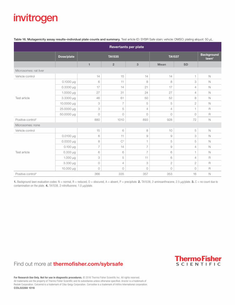
Table 16. Mutagenicity assay results–individual plate counts and summary. Test article ID: SYBR Safe stain; vehicle: DMSO; plating aliquot: 50 μL.
Revertants per plate
Dose/plate TA1535 TA1537Background
lawn1
1 2 3 Mean SD
Microsomes: rat liver
Vehicle control 14 15 14 14 1 N
Test article
0.1000 μg 6 11 8 8 3 N
0.3330 μg 17 14 21 17 4 N
1.0000 μg 27 31 24 27 4 N
3.3300 μg 46 61 50 52 8 N
10.0000 μg 3 7 5 5 2 N
25.0000 μg 3 5 4 4 1 R
50.0000 μg 0 0 0 0 0 R
Positive control2 880 1010 893 928 72 N
Microsomes: none
Vehicle control 15 6 8 10 5 N
Test article
0.0100 μg 6 11 9 9 3 N
0.0333 μg 8 C3 1 5 5 N
0.100 μg 7 14 7 9 4 N
0.333 μg 6 6 7 6 1 N
1.000 μg 3 5 11 6 4 R
3.330 μg 0 4 3 2 2 R
10.000 μg 0 0 0 0 0 R
Positive control4 366 335 357 353 16 N
1. Background lawn evaluation codes: N = normal, R = reduced, O = obscured, A = absent, P = precipitate. 2. TA1538, 2-aminoanthracene, 2.5 µg/plate. 3. C = no count due to contamination on the plate. 4. TA1538, 2-nitrofluorene, 1.0 µg/plate.