Basic Biotechnologypbge.faculty.ucdavis.edu/.../sites/408/2016/10/Restriction-Enzyme-Analysis.pdfto...
16
Basic Biotechnology Kit RESTRICTION ENZYME ANALYSIS Partnership for Biotechnology and Genomics Education Barbara Soots Linda Curro Education Coordinator University of California Davis Assistant Education Coordinator University of California Davis 530 752‐6552 530 752‐6613 530 754‐4410 (fax) 530 754‐4410 (fax) [email protected] [email protected] This program is made possible through the generous support of:
Transcript of Basic Biotechnologypbge.faculty.ucdavis.edu/.../sites/408/2016/10/Restriction-Enzyme-Analysis.pdfto...

Basic Biotechnology Kit RESTRICTION ENZYME ANALYSIS
Partnership for Biotechnology and Genomics Education Barbara Soots Linda Curro
Education Coordinator
University of California Davis
Assistant Education Coordinator
University of California Davis
530 752‐6552 530 752‐6613
530 754‐4410 (fax) 530 754‐4410 (fax)
[email protected] [email protected]
This program is made possible through the generous support of:

Biotechnology in the Classroom 2009 2 updated 9/23/2009
Teacher Information
RESTRICTION ENZYME ANALYSIS Lambda DNA (SYBR Safe® Staining) Introduction
Restriction mapping is a cornerstone of modern‐day biotechnology. Restriction enzymes cleave DNA at known sites, making possible detailed analysis of specific sections of an organism’s genome.
During this three‐day exercise, students will digest samples of λ DNA with three different restriction enzymes and one unknown. These DNA samples will be electrophoresed and the resulting fragments photographed under an ultraviolet transilluminator.
Attached readings and activities help explain the process of restriction enzyme analysis and how it is used in the DNA fingerprinting procedure.
Objectives
1. Understand the mechanics of a restriction digest.
2. Use a micropipet, electrophorese an agarose gel.
3. Compare the λ DNA bands on a gel to a λ DNA restriction map.
4. Understand the importance of restriction enzymes.
Materials
For Each Lab Group four microtubes microtube rack 20 μl micropipet and sterile tips waterproof pen beaker or foam cup with crushed ice * 20 μl of 0.1 μg/μl lambda (λ) DNA 25 μl 2X restriction buffer 4 μl BamHI restriction enzyme 4 μl EcoRI restriction enzyme 4 μl HindIII restriction enzyme
10 μl distilled water * gloves safety glasses * 500 ml beaker (day 2) * colored lab tape (day 2) electrophoresis chamber (day 2) power supply (day 2) 10 μl loading dye (day 2) 0.7% agarose gel with SYBR Safe®
Common Materials
microcentrifuge carboy with TBE solution (1X) 37°C water bath w/ floating rack cooler with crushed ice * freezer (non frost‐free, if possible) * digital camera with hood attachment UV transilluminator unknown DNA digest sample (instructor only)
distilled water * * Materials provided by instructor
Advance Preparation
DAY 1
1. Dilute the concentrated TBE (10X) solution to a final concentration of (1X) and store in a plastic carboy. If you saved the solution from the Gel Electrophoresis with Dyes activity, reuse it for this laboratory. Figure on about 300 ml per group. For instance, if your class has 12 groups, prepare 360 ml 10X TBE + 3,240 ml distilled water, for a total of 3,600 ml 1X TBE.
2. Prepare aliquots of the following for each
team: use 0.5 ml microtubes for enzymes
a) 20 μl of 0.1 μg/μl λ DNA b) 25 μl of 2X restriction buffer c) 4 μl of BamHI restriction enzyme

Biotechnology in the Classroom 2009 3 updated 9/23/2009
d) 4 μl of EcoRI restriction enzyme
e) 4 μl of HindIII restriction enzyme
f) 10 μl of distilled water 3. Obtain enough crushed ice and ice
containers for each lab group. Styrofoam cups work well.
4. Fill a water bath with distilled water and
adjust it to 37°C. DAY 2:
1. Pre‐pour enough agarose gels for each lab group. a) Wear gloves. b) Microwave the agarose bottle (cap
loosened) until the gel liquefies. Be sure to use a microwave not designated for food usage. Alternately, you may melt the agarose in a waterbath. This may take an hour or so.
c) Cover your workspace with lab paper. d) Make sure the sides of the gel tray are
up. If necessary, use lab tape to seal the ends.
e) Place the plastic comb in the slots close to the end of the tray.
f) With a disposable pipette run a thin line of gel around the periphery of each tray.
g) Pour approximately 25 ml of agarose into each gel tray.
h) Let cool until solidified (approximately 15 minutes) before removing comb.
i) If storing overnight, place trays in a container with TBE solution so they do not dry out. Each gel may also be sealed in a zip‐loc bag with a little TBE buffer.
2. Aliquot 10 μl of loading dye for each
lab group.
3. Remove the microtubes from the freezer just before students are ready to use them. The small amounts will defrost quickly!
DAY 3:
1. Remove student gels from the refrigerator. 2. Prepare the digital camera/hood setup to
take photographs on the UV illuminator.
Teaching Tips
USE OF MICROPIPETTORS:
See description in Micropipet Technique Laboratory. GEL DISPOSAL
Used gels, tips, and tubes may be disposed of in the regular garbage. Waste TBE may be poured down the sink. Please do not return waste TBE to us in the clean buffer carboys. USE OF SYBR SAFE®:
SYBR Safe® is a fluorescent dye used to stain DNA. For this experiment the stain is added directly to the agarose and the gels will be able to be visualized directly after electrophoresis.
SYBR Safe® DNA gel stain was designed to replace ethidium bromide—a known mutagen. SYBR Safe® DNA gel stain is less mutagenic, less toxic, more environmentally friendly, and it works with traditional gel staining methods. Gels made with the dye may be disposed in the regular trash and waste buffer solution may be poured down the drain.
Regardless, we still practice the same safety precautions as we would do with ethidium bromide. Gel boxes and gels should ALWAYS be handled with gloves.
Please see enclosed safety binder for more information about SYBR® Safe.
ALTERNATE STAINING TECHNIQUES
Methylene Blue may also be used to stain DNA gels. In this instance, a white light, not UV,

Biotechnology in the Classroom 2009 4 updated 9/23/2009
illumination system is used. SYBR Safe® is not added to the agarose and the concentration of lambda DNA is much higher (0.4 μg/μl λ DNA) due to the lower sensitivity of the methylene blue stain. Use the Restriction Enzyme Analysis of Lambda DNA (methylene blue stain) laboratory protocol USE OF POWER SUPPLIES:
See description in Gel Electrophoresis of Dyes Laboratory
USE OF THE UV TRANSILLUMINATOR:
Ultraviolet (UV) radiation can cause severe eye and skin damage
Do not turn on the UV light without the protective cover down
Do not use the transilluminator if the UV blocking cover becomes cracked or broken
When students are ready to look at their gels, view them only through the UV blocking cover. Do not attempt to operate the transilluminator with the UV blocking cover raised. Any attempt to override the safety interlock and view the gel directly could result in serious eye and skin damage.
With a paper towel, remove any excess liquid in the agar plate.
With the power switch OFF, place the agar plate under the protective glass cover. Close the blocking cover and turn the power switch ON. In most cases, the DNA is visible even with the room lights on; however, it may be necessary to turn off the room lights or block out some of the ambient light. View the agar plate briefly, then switch OFF the power and remove sample.
ENZYMES:
Restriction enzymes easily and rapidly degrade unless kept frozen. Loss of activity will occur if the enzymes are subjected to warm temperatures for any length of time.
When aliquoting student reagents, be careful to keep them on ice. Also, be sure to use good
sterile technique when you prepare student aliquots.
It is best to store enzymes in a non‐frost‐free freezer since they maintain a more stable temperature. If you must use a frost‐free freezer, store the enzymes in a foam container.
2X UNIVERSAL RESTRICTION BUFFER:
Restriction enzymes like the ones we use in this lab operate under different conditions of salt and pH. For optimal activity, a different buffer would be used for each enzyme. To simplify procedures, however, we use a universal buffer‐‐ a compromise between the conditions preferred by various enzymes.
LAMBDA (λ) DNA: Because it is fairly inexpensive and readily available, purified DNA from bacteriophage λ is most suitable for demonstrating the concept of DNA restriction. Most λ DNA is 48,502 base pairs in length. The linear fragments migrate through an agarose gel at a distance that is inversely proportional to the log10 value of their molecular weight. In this exercise, molecular weight is represented by the number of base pairs.

Biotechnology in the Classroom 2009 5 updated 9/23/2009
UNKNOWN λ DNA DIGEST SAMPLE:
The instructor will add 5 μl of this sample (with loading dye) to each groups’ gel just prior to electrophoresis. The unknown is a double digest of λ DNA with EcoR1 and HindIII.
This sample may act in two ways, depending on your classroom needs.
First, it can be a “known” marker for the students so they can gauge the size of their generated bands (see diagram at left).
Alternately, it may be an “unknown” sample and they will need to figure out
which of the enzymes were used to generate the bands.
Another option might be to run the lab as a forensic mystery (similar to the paternity example in the student paper activity section). In that case, the unknown would act as the child. The mother would be the EcoR1 digest and the students would need to determine if the father was the HindIII or BamH1 digest.
LOADING DYE:
This dye is added to the digest sample in each well. Although you cannot see the progress of the DNA down the gel, you can watch the dye move toward the positive electrode. It will appear as a blue band, eventually resolving into two bands of color.
The faster‐moving, purplish band is bromophenol blue dye that migrates at roughly the same rate as a 500 base pair fragment of DNA. The slower‐moving aqua band is xylene cyanol, nearly equivalent to a 4000 base pair fragment.
Best separation for analysis of DNA is achieved when the bromophenol blue band migrates 4‐7 cm or more from the origin. However, be
careful not to let the bromophenol blue band run off the end!
USE OF THE MICROCENTRIFUGE:
During aliquoting and movement to and from freezer or refrigerator and ice bucket, reagent aliquots often become spread in a film around the sides or caps of the microtubes. To pool the reagents, spin them briefly in a microcentrifuge just prior to use.
NEVER run with the lid open.
NEVER put hands in the rotor area unless the rotor is completely stopped.
ALWAYS make sure the tubes are loaded in the rotor symmetrically; each tube should be balanced by another tube directly opposite it.
USE OF THE CAMERA/TRANSILLUMINATOR
Follow these instructions when taking a picture of your gel electrophoresis results:
1. Only the teacher should use the digital camera! Do not handle any gels before using the camera and do not wear gloves while taking photographs.
2. Have a student wearing gloves load their gel on the transilluminator.
3. Make sure the hood completely covers the UV transilluminator platform.
4. Turn on the UV light. 5. Take a picture with the digital camera. 6. Turn off the UV light and have the student
remove the gel in preparation for the next group.
7. After entire class has photographed gels, download to your computer to view results
.

Biotechnology in the Classroom 2009 6 updated 9/23/2009
STUDENT GEL ACTIVITY ANSWER:
DNA FINGERPRINTING ACTIVITY ANSWERS
Part A: Forensic Science‐A Paternity Case
Sample 5 Part B: Forensic Science‐A Rape Case
1. a) The women were assaulted by different men. b) Suspect X attacked victim A, and suspect Y attacked victim B.
2. Since the bands in Well 3 match with the bands in Well 5, and the bands in Well 4 match with
the bands in Well 6, we know that the semen samples came from the suspects. Wells 1 and 2 are important to determine that the samples in Wells 3 and 4 are not blood from the victims, because the bands in Wells 1 & 3 and Wells 2 & 4 do not match up, and are in fact semen from the suspects.
Base Pair Length A 5,505 L 3,530 B 16,841 M 23,130 C 5,626 N 2,027 D 6,527 O 2,322 E 7,233 P 9,416 F 6,770 Q 564 G 21,226 R 125 H 4,878 S 6,557 I 5,643 T 4,336.1 J 7,421 λ 48,502 K 5,804
λ
{
BamHI EcoRI HindIII (uncut λ DNA) Control
B
Will appear as
} May appear as one band
These smaller fragments are not likely to be seen {
G M
E
C A
FD
J
K I
H
L
P
S
T
O
N
Q
R
These smaller fragments are not likely to be seen

Biotechnology in the Classroom 2009 7 updated 9/23/2009
Laboratory RESTRICTION ENZYME ANALYSIS Lambda DNA (SYBR Safe® Staining) Background Reading
In this lab, you will use special chemicals called restriction enzymes that act as chemical scissors to cut DNA into pieces. Restriction enzymes make two incisions, one through each of the phosphate backbones of the double helix without damaging the bases. The chemical bonds that the enzymes cleave can be reformed by other enzymes known as ligases, so that fragments carved from different chromosomes or genes can be spliced together, provided their ends are complementary. Many of the procedures of molecular biology and genetic engineering rely on restriction enzymes.
The term restriction comes from the fact that these enzymes were discovered in E. coli strains that appeared to be restricting the infection by certain bacteriophages. Restriction enzymes therefore are believed to be a mechanism evolved by bacteria to resist viral attack and to help in the removal of viral sequences.
The DNA you will be using today comes from one of the most studied viruses, bacteriophage λ, a virus that preys on bacterial cells. Since viruses have a relatively simple genome, scientists have studied their DNA and used this information to test theories and develop concepts that apply to the genetics of living organisms. The size of DNA fragments is measured in base pairs or kilobase pairs (1000 base pairs). The DNA of bacteriophage λ is approximately 48,514 base pairs in length. Compare this to the human genome of approximately 3 billion base pairs!
Once the λ DNA has been cut, the pieces are electrophoresed. Because DNA is a negatively charged molecule, the fragments move toward the positive electrode. Fragments travel through the gel according to their molecular weight ‐‐ with the smallest fragments moving the greatest distance. For more information on electrophoresis, review the Gel Electrophoresis of Dyes lab.
When the gel is photographed under UV light, a special stain in the agarose will make the DNA fluoresce. The fragments appear as a pattern of bands. In this protocol, we will compare our banding pattern with a predicted result generated on the first day of the lab.
Readings that your teacher will hand out provide more information on this process.
Objectives
1. Understand the mechanics of a restriction digest. 2. Use a micropipet; electrophorese an agarose gel.
3. Compare the λ DNA bands on a gel to a λ DNA restriction map. 4. Understand the importance of restriction enzymes.
Ideal Gel Results

Biotechnology in the Classroom 2009 8 updated 9/23/2009
Materials
For Each Lab Group four microtubes microtube rack 20 μl micropipet and sterile tips waterproof pen beaker or foam cup with crushed ice * 20 μl of 0.1 μg/μl lambda (λ) DNA 25 μl 2X restriction buffer 4 μl BamHI restriction enzyme 4 μl EcoRI restriction enzyme 4 μl HindIII restriction enzyme 10 μl distilled water * gloves safety glasses * 500 ml beaker (day 2) * colored lab tape (day 2) electrophoresis chamber (day 2) power supply (day 2) 10 μl loading dye (day 2) 0.7% agarose gel with SYBR Safe®
Common Materials
microcentrifuge carboy with TBE solution (1X) 37°C water bath w/ floating rack cooler with crushed ice * freezer (non frost‐free, if possible) * Digital camera with hood attachment UV transilluminator unknown DNA digest sample (instructor only)
distilled water * * Materials provided by instructor
Precautions
While the SYBR Safe® stain found in the agarose gel is a safer alternative to ethidium bromide, it still should not be touched. Gel boxes, trays, and gels should always be handled with gloves. Always leave gel in its tray.
When using the microcentrifuge, always make sure the tubes are loaded in the rotor symmetrically; each tube should be balanced by another tube directly opposite it. Never run the centrifuge with the lid open or the rotor missing. Never put hands in the rotor area unless the rotor is completely stopped.
Ultraviolet (UV) radiation can cause severe eye and skin damage! The camera hood is strictly for photographic purposes. Do not attempt to use the camera hood to view samples on the transilluminator. Make sure that the hood is seated correctly and there are no raised edges that would allow UV light to leak out.
If you choose to view the gel prior to making a photograph, be sure the UV blocking cover is down before turning the power on.
Biotechnology in the Classroom 2009 9 updated 9/23/2009
Procedure
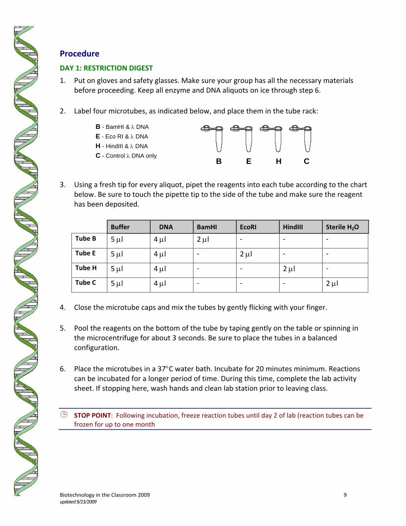
DAY 1: RESTRICTION DIGEST
1. Put on gloves and safety glasses. Make sure your group has all the necessary materials before proceeding. Keep all enzyme and DNA aliquots on ice through step 6.
2. Label four microtubes, as indicated below, and place them in the tube rack:
3. Using a fresh tip for every aliquot, pipet the reagents into each tube according to the chart
below. Be sure to touch the pipette tip to the side of the tube and make sure the reagent has been deposited.
Buffer � DNA BamHI EcoRI HindIII Sterile H2O
Tube B 5 μl 4 μl 2 μl ‐ ‐ ‐
Tube E 5 μl 4 μl ‐ 2 μl ‐ ‐
Tube H 5 μl 4 μl ‐ ‐ 2 μl ‐
Tube C 5 μl 4 μl ‐ ‐ ‐ 2 μl
4. Close the microtube caps and mix the tubes by gently flicking with your finger. 5. Pool the reagents on the bottom of the tube by taping gently on the table or spinning in
the microcentrifuge for about 3 seconds. Be sure to place the tubes in a balanced configuration.
6. Place the microtubes in a 37°C water bath. Incubate for 20 minutes minimum. Reactions
can be incubated for a longer period of time. During this time, complete the lab activity sheet. If stopping here, wash hands and clean lab station prior to leaving class.
STOP POINT: Following incubation, freeze reaction tubes until day 2 of lab (reaction tubes can be frozen for up to one month
B E H C
B - BamHI & λ DNA E - Eco RI & λ DNA H - HindIII & λ DNA C - Control λ DNA only

Biotechnology in the Classroom 2009 10 updated 9/23/2009
DAY 2: ELECTROPHORESIS
1. Put on gloves and safety glasses. Make sure your group has all the necessary materials before proceeding. Collect your Day 1 reactant tubes from the freezer. Keep tubes on ice until needed.
2. Obtain a pre‐poured gel from your instructor. If your tray has gates, move them to the
down position. If tape is present, gently remove it. 3. Place the gel tray in the gel box with the wells at the negative (black) end of the box. 4. Obtain 250 ml of TBE (1X) solution from the plastic carboy and add it to the sides of the box
until the wells are just barely submerged (about 2 mm under). Do not add buffer solution directly on top of wells. Return any leftover TBE solution to the carboy.
5. Add 2 μl of loading dye to each of the microtubes. Make sure to change pipette tips for
each sample. Put the ejected tips and tube of loading dye into a waste container. 6. Close the microtube caps and spin the tubes in the microcentrifuge for about 3 seconds or
tap gently on the table to pool the reagents. 7. Load 10 μl of each sample into separate wells (see diagram below). Make sure to change
pipet tips for each sample! _ + 8. Your instructor will add 5 μl of an unknown DNA sample to one of the empty wells. 9. Clean up spilled buffer or any other liquid surrounding the gel box.
empty well for
unknown sample
empty well
Tube B DNA & BamHI
Tube E DNA & EcoRI
Tube H DNA & HindIII
Tube C DNA no enzyme (control)

Biotechnology in the Classroom 2009 11 updated 9/23/2009
10. Make sure that the power supply is unplugged and switched off before proceeding. Carefully place the lid on the gel box (first making sure it is clean and dry).
11. Connect the red (positive) patch cord to the red terminal on the power supply. Similarly,
connect the black cord to the black terminal. Each power supply will run two gel boxes. Notice what channel you have plugged your box into.
12. Wait until the group that is sharing your power supply has completed steps 1‐11 before
proceeding.
IMPORTANT! Have your teacher look over all of your connections before plugging in the power supply.
13. Plug in the power supply and turn on the machine on. The run light will illuminate,
signifying that power is running to the cell. 14. Turn the display select switch to the appropriate channel for your group. Turn the voltage
indicator knob to 100V. Make sure both groups set their voltage. 15. Electrophorese for 30‐45 minutes. Longer running times will produce better results. If you
are able to run the gel longer, be sure to turn the power supply off when the purple dye from the loading dye (bromophenol blue) is about 2‐3 centimeters from the end of the gel.
STOP POINT: Following electrophoresis, wrap the moist gel tray in plastic wrap or place in a zip‐loc bag with TBE buffer and refrigerate until day 3 of lab. Do not touch the gel without gloves.

Biotechnology in the Classroom 2009 12 updated 9/23/2009
DAY 3: PHOTOGRAPHING GELS
1. Put on gloves and safety glasses. Retrieve your gel tray from the refrigerator. Wipe off any excess moisture from the tray.
Steps 2‐7 must take place under the supervision of your instructor.
2. Make sure the power to the UV transilluminator is OFF. Lift the UV blocking cover and place gel tray on the black platform. Close the lid completely before turning machine on. UV radiation can cause severe eye and skin damage.
3. Briefly observe the number, placement and relative brightness of the DNA bands. Turn the
transilluminator OFF. 4. With the transilluminator OFF, have your teacher place the hooded digital camera over the
black platform. Make sure that the hood is seated correctly and there are no raised edges that would allow UV light to leak out.
5. Turn ON the transilluminator and have your instructor photograph the gel. Only the
instructor should handle the digital camera with UNGLOVED hands, while you handle the gel with GLOVED hands.
6. Turn OFF the transilluminator. 7. Remove your gel and allow the next group to photograph their gel. Once all groups are
complete, your instructor may download the digital images to their computer.
8. Follow your teacher’s instructions for clean‐up. Discard all gels into trash. Do not touch the
gel! Gel boxes and plates must be rinsed in distilled or deionized water and air dried. Please take great care with the equipment so many more students may enjoy these experiments.
9. Wash hands and clean lab station.

Biotechnology in the Classroom 2009 13 updated 9/23/2009
Student Activity
RESTRICTION ENZYME ANALYSIS Lambda DNA (SYBR Safe® Staining) On the following page you will find a partial restriction map of the linear λ genome. The enzymes we are using in this laboratory cleave � DNA in the areas noted on the map. Each fragment is assigned a letter. Calculate the base pair length of each fragment and write it down in the space provided below. Next, arrange your λ DNA fragment letters on the “paper gel” as you would expect them to appear after electrophoresis. Be careful to keep the fragments from each restriction enzyme in their proper lane. Also include where the uncut λ sample will be. The first fragment has been done for you.
Base Pair Length(fragment size in bp)
A 5505 B C D E F G H I J K L M N O P Q R S T
- 50,000 bp 24,000 bp 20,000 bp 16,000 bp 10,000 bp 9000 bp 8000 bp 7000 bp 6000 bp 5000 bp 4000 bp 3000 bp 2000 bp 1000 bp +
BamHI EcoRI HindIII Control (uncut λ DNA)
A

Biotechnology in the Classroom 2009 14 updated 9/23/2009
Restriction Maps of the Linear Lambda Genome
(DNA Fragments in base pairs)
λ DNA
BAMHI CLEAVAGE SITES
ECORI CLEAVAGE SITES
HINDIII CLEAVAGE SITES
M N O P Q R S T
0 10,000 20,000 30,000 40,000 48,502
G H I J K L
21,226 26,104 31,747 39,168 44,972 48,502
A B C D E F
5,505 22,346 27,972 34,499 41,732 48,502
25,157 37,459
23,130 27,479 36,895 37,584 44,141 48,502

Biotechnology in the Classroom 2009 15 updated 9/23/2009
Student Activity
RESTRICTION ENZYME ANALYSIS DNA Fingerprinting Part A: Forensic Science‐‐ A Paternity Case
Assume that you are a molecular biologist involved in analyzing a paternity dispute. In order to determine with accuracy which of the men listed contributed genetic material to the child, you took blood samples from the mother, child and each potential father. Then you ran the samples on an agarose gel electrophoresis, adding an appropriate restriction enzyme to each of the samples. Results from the restriction enzyme digest are as follows:
1. Which sample contains blood from the child’s actual father? ____________________
Well 1: mother Well 2: child Well 3: father 1 Well 4: father 2 Well 5: father 3
1 2 3 4 5

Biotechnology in the Classroom 2009 16 updated 9/23/2009
Part B: Forensic Science‐‐ A Rape Case
You are a molecular biologist involved in forensic medicine. Two young women have recently been raped within a span of two weeks and you have been given blood and semen samples from the crime scenes.
You ran an agarose gel electrophoresis with the samples in the following wells, applying the restriction enzyme, EcoRI to each sample.
Well 1: blood from victim A
Well 2: blood from victim B
Well 3: semen collected on Victim A
Well 4: semen collected on Victim B
Well 5: blood from suspect X
Well 6: blood from suspect Y
Well 7: blood from suspect Z
1. Examine the results above and determine:
a) whether both women were, in fact, assaulted by the same man b) which, if any, of the accused is involved in the crime
2. Write a brief summary of your findings. Note: In actuality, gel electrophoresis is combined with another technique called Southern Blotting in order to obtain a DNA fingerprint. Southern Blots involve the use of radioactively labeled DNA probes which attach to fragments of DNA which have been made single‐stranded.