Clinical Study An Association between BK Virus Replication ...
Upload
nguyenquynhCategory
view
224download
2
From DEPARTMENT OF MICROBIOLOGY,
TUMOR AND CELL BIOLOGY
Karolinska Institutet, Stockholm, Sweden
VIRUS-HOST INTERACTIONS: ENTRY AND REPLICATION OF
CRIMEAN-CONGO HEMORRHAGIC FEVER VIRUS
Cecilia Andersson
Stockholm 2013

All previously published papers were reproduced with permission from the publisher.
Published by Karolinska Institutet.
Printed by Åtta.45 tryckeri AB
© Cecilia Andersson, 2013
ISBN 978-91-7549-533-0
i
Virus-Host interactions: Entry and replication of Crimean-Congo hemorrhagic fever virus AKADEMISK AVHANDLING som för avläggande av medicine doktorsexamen (Ph.D.) vid Karolinska Institutet offentligen försvaras i förläsningsalen Jacob Berzelius, Berzelius väg 3, Solna Fredagen den 16 maj 2014, kl 9:30
av
Cecilia Andersson
Huvudhandledare:
Docent Ali Mirazimi
Karolinska Institutet
Institutionen för mikrobiologi,
tumör- och cellbiologi
Bihandledare:
Professor Mats Nilsson
Stockholms universitet
Institutionen för biokemi och
biofysik
Fakultetsopponent:
Dr. Pierre-Yves Lozach
Heidelberg University
Department of Infectious Diseases
Betygsnämnd:
Professor Michael Lindberg
Linnéuniversitetet
Institutionen för kemi och biomedicin
Professor Francesca Chiodi
Karolinska Institutet
Institutionen för mikrobiologi,
tumör- och cellbiologi
Docent Marianne Jansson
Lunds universitet
Avdelningen för medicinsk
mikrobiologi

ii

iii
“When I was 5years old, my mother always told me
that happiness was the key to life. When I went to school, they
asked me what I wanted to be when I grew up. I wrote ‘happy’.
They told me I didn’t understand the assignment, and I told them
they didn’t understand life”
John Lennon
To my family

ii
i
ABSTRACT
Crimean-Congo hemorrhagic fever (CCHF) is a severe acute human disease with
potential lethal outcome caused by a virus, Crimean-Congo hemorrhagic fever
virus (CCHFV). Not much is known regarding how CCHFV infects cells,
replicates and why it cause vascular dysfunction. To better understand CCHFV-
pathogenesis increased knowledge regarding these issues is needed.
Viruses have to enter a host cell in order to replicate its genome and here we
show that CCHFV entry occur by clathrin-mediated endocytosis and is pH-
dependent.
A new in situ detection technique was establihed to visualize individual CCHFV
cRNA and vRNA transcripts. Potential colocalization with the viral nucleocapsid
protein (NP) was also investigated. cRNA was found to be more concentrated to
particular regions within the cytoplasm and co-localized with CCHFV NP. While
vRNA was detected throughout the cytoplasma not colocalizating with CCHFV
NP.
It is not known if the vascular leakage observed in CCHF is due to direct virus
infection or is immune-mediated. A new in vitro model system was therefore
established and it was found that CCHFV has a preference for basolateral entry.
However and surprisingly, using CCHFV-infected monocyte-derived dendritic
cells (moDCs) or their supernatant, a preference for apical entry was observed.
This indicate that the change in entry site could be due to soluble factors from
the moDCs. Neither direct infection nor addition of CCHFV-infected moDCs
affected the cellular permeability of the human polarized epithelial cell layer,
indicating that other factors are most likely are causing the vascular leakage.
Taken together, we established several new in vitro model systems to study
CCHFV’s interaction with host cells. We also demonstrated the entry pathway for
CCHFV into cells. These data and tools will hopefully facilitate and promote
research on virus-host interactions which in turn may result in the development of
new antivirals and/or vaccines against CCHFV.

ii
iii
LIST OF SCIENTIFIC PAPERS
I. Simon M, Johansson C, Mirazimi A.
Crimean-Congo hemorrhagic fever virus entry and replication is
clathrin, pH and cholesterol dependent. Journal of General Virology. 2009 Jan; 90 (Pt 1):210-5
II. Andersson C*, Henriksson S*, Magnusson KE, Nilsson M, Mirazimi A.
In situ rolling circle amplification detection of Crimean Congo
hemorrhagic fever virus (CCHFV) complementary and viral RNA. Virology. 2012. May 10; 426 (2): 87-92
III. Andersson C, Mirazimi A.
An in vitro assay to study the molecular pathogenesis of Crimean-Congo
hemorrhagic fever virus
Manuscript
*contributed equally
iv
RELATED SCIENTIFIC PAPERS
Simon M, Johansson C, Lundkvist A, Mirazimi A.
Microtubule-dependent and microtubule-independent steps in
Crimean-Congo hemorrhagic fever virus replication cycle.
Virology, 2009 Mar 15; 385 (2): 313-22
Connolly-Andersen AM, Moll G, Andersson C, Åkerstöm S, Karlberg
H, Douagi I, Mirazimi A.
Crimean-Congo hemorrhagic fever virus activates endothelial
cells.
Journal of Virology. 2011 Aug; 85(15): 7766-74
v
CONTENTS
1 Introduction .................................................................................................. 1
1.1 History ................................................................................................ 1
2 CCHF-Virus ................................................................................................. 2
2.1 Molecular characteristics.................................................................... 2
2.2 Replication .......................................................................................... 4
2.3 Occurrence .......................................................................................... 5
2.4 Transmission ....................................................................................... 6
3 CCHF- The Disease ..................................................................................... 9
3.1 Treatment .......................................................................................... 10
3.2 Animal models.................................................................................. 11
3.3 Pathology .......................................................................................... 12
4 Immune responses towards CCHFV infection .......................................... 13
4.1 Innate immune system ...................................................................... 13
4.2 Macrophages and dendritic cells ...................................................... 14
4.3 Adaptive immune system ................................................................. 16
4.4 Cytokines .......................................................................................... 16
4.5 Proposed pathogenesis ..................................................................... 17
5 Polarized cells ............................................................................................. 20
5.1 Polarized cells and virus infection ................................................... 21
6 Endocytosis ................................................................................................ 23
6.1 Clathrin mediated endocytosis ......................................................... 24
6.2 Caveolae and lipid raft entry ............................................................ 26
6.3 Clathrin and Caveolae independent endocytosis ............................. 27
vi
6.4 Macropinocytosis and phagocytosis ................................................ 28
6.5 Bunyaviridae entry and receptors .................................................... 28
7 In situ detection of nucleic acid ................................................................. 30
7.1 ISH and FISH ................................................................................... 30
7.2 Padlock probes .................................................................................. 31
7.2.1 Rolling circle amplification ................................................. 31
7.2.2 RNA detection in situ using padlock probes and RCA ...... 33
8 Prelimininary results .................................................................................. 35
9 Results and discussion................................................................................ 38
9.1 CCHFV entry (Paper I) .................................................................... 38
9.2 In situ detection of CCHFV RNA (Paper II) ................................... 39
9.3 In vitro pathogenesis model (Paper III) ........................................... 41
10 Concluding remarks ................................................................................... 44
11 Populärvetenskaplig sammanfattning ........................................................ 46
12 Acknowledgements .................................................................................... 48
13 References .................................................................................................. 51
vii
LIST OF ABBREVIATIONS
APC
BSL-4
CCHF
Antigen-presenting cell
Biosafety level 4
Crimean-Congo hemorrhagic fever
CCHFV
CME
cRNA
Crimean-Congo hemorrhagic fever virus
Clathrin-mediated endocytosis
Complementary ribonucleic acid
DC
DIC
DNA
ER
FISH
IFN
ISH
LNA
moDC
MHC
mRNA
NP
NK
OLA
PCR
Dendritic cell
Disseminated intravascular coagulation
Deoxyribonucleic acid
Endoplasmic reticulum
Fluorescence in situ hybridization
Interferon
In situ hybridization
Locked nucleic acid
Monocyte-derived dendritic cell
Major histocompatibility complex
Messanger ribonucleic acid
Nucleocapsid protein
Natural killer
Oligonucleotide ligation assay
Polymerase chain reaction
RCA
RCP
Rolling circle amplification
Rolling circle product
viii
RdRp
RNA
vRNA
RNA-dependent RNA polymerase
Ribonucleic acid
Viral ribonucleic acid
1
1 INTRODUCTION
Crimean-Congo hemorrhagic fever (CCHF) is exclusively a human disease with
a high case fatality rate, of up to 30%. The virus causing the disease, Crimean-
Congo hemorrhagic fever virus (CCHFV), is spread by ticks and humans
contract the virus following tick bites, the handling of infected livestock or
caring for a patient in the acute phase of the disease. There is currently no
commercially available vaccine and no specific treatment.
1.1 HISTORY
A human disease with bleeding from numerous sites and where the causing agent
was believed to be a small tough tick or louse was first described in 1100 ad [1].
A similar disease has also been known in the Termez region of Uzbekistan under
3 names indicating blood taking, nose bleeding and black death [1]. But the first
clinical description of CCHF was made during an outbreak in Crimea in 1944,
when over 200 military personnel developed severe disease with bleeding from
various sites while helping restore abandoned farmland [1]. The following years
there were other outbreaks of what was then referred to as Crimean hemorrhagic
fever in other parts of Eastern Europe [2]. The viral nature of the disease was
determined by Chumakov, who gave serum from hemorrhagic patients to
psychiatric patients. He also determined ticks as the vector by giving a solution
of crushed Hyalomma ticks to healthy volunteers and in both studies disease was
manifested [1].
In 1969, the Soviet reference strain for Crimean hemorrhagic fever was found to
be antigenically identical to several strains isolated in Congo where it was known
to cause a similar disease [3]. After some dispute over the name, Crimean-Congo
hemorrhagic fever was finally accepted [1].
2
2 CCHF-VIRUS
CCHFV belongs to the Bunyaviridae family and is classified within the
Nairovirus genus. The Bunyaviridae family is a large family containing more
than 350 viruses divided into 5 genera, Orthobunyavirus, Nairovirus,
Phlebovirus, Hantavirus and Tospovirus [4]. With the exception of
Tospoviruses all other members are transmitted by animals, rodents for
Hantavirus and arthropods (mosquitos, tick or sandfly) for the others.
Bunyaviridae viruses are enveloped and circular. All members have a genome
consisting of 3 single stranded RNA segments of negative sense, the large (L),
medium (M) and the small (S) segment, encoding 4 structural proteins [5],
several members also encode nonstructural proteins.
Nairoviruses are spread by ticks and have a very large L segment compared to
other Bunyaviridae members, nearly twice the size [6]. The genus consists of 35
viruses, but only 5 viruses are known to cause human disease. Apart from
CCHFV, Farallon and Erve virus may cause human disease with headache, fever
and neurological disorder while Dugbe and Nairo sheep disease virus can cause
human disease but mainly cause disease in sheep and goats [7].
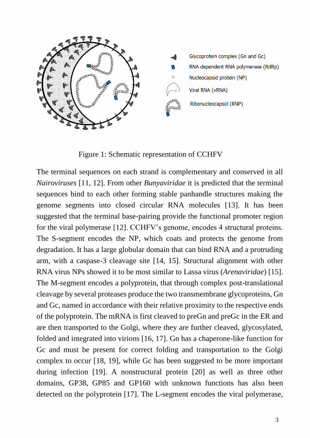
2.1 MOLECULAR CHARACTERISTICS
The CCHF virus particle is spherical and between 90-100nm in diameter [8, 9].
It has an outer cell-derived lipid envelope membrane, through which the
glycoproteins, Gn and Gc, protrude. Like the other Bunyaviridae members,
CCHFV has a negative single strand genome divided on three segments, the
large (L), medium (M) and small (S). Together with the nucleocapsid protein
(NP) each genome strand form individual ribonucleocapsids [5, 10]. Each viral
particle also contains RNA-dependent RNA polymerase (RdRp), necessary to
initiate transcription and genome replication.
3
Figure 1: Schematic representation of CCHFV
The terminal sequences on each strand is complementary and conserved in all
Nairoviruses [11, 12]. From other Bunyaviridae it is predicted that the terminal
sequences bind to each other forming stable panhandle structures making the
genome segments into closed circular RNA molecules [13]. It has been
suggested that the terminal base-pairing provide the functional promoter region
for the viral polymerase [12]. CCHFV’s genome, encodes 4 structural proteins.
The S-segment encodes the NP, which coats and protects the genome from
degradation. It has a large globular domain that can bind RNA and a protruding
arm, with a caspase-3 cleavage site [14, 15]. Structural alignment with other
RNA virus NPs showed it to be most similar to Lassa virus (Arenaviridae) [15].
The M-segment encodes a polyprotein, that through complex post-translational
cleavage by several proteases produce the two transmembrane glycoproteins, Gn
and Gc, named in accordance with their relative proximity to the respective ends
of the polyprotein. The mRNA is first cleaved to preGn and preGc in the ER and
are then transported to the Golgi, where they are further cleaved, glycosylated,
folded and integrated into virions [16, 17]. Gn has a chaperone-like function for
Gc and must be present for correct folding and transportation to the Golgi
complex to occur [18, 19], while Gc has been suggested to be more important
during infection [19]. A nonstructural protein [20] as well as three other
domains, GP38, GP85 and GP160 with unknown functions has also been
detected on the polyprotein [17]. The L-segment encodes the viral polymerase,
4
RdRp, necessary for viral replication and mRNA synthesis. It has an ovarian
tumor protease domain near its N-terminus that has been shown to remove
ubiquitin from cellular proteins [21].
2.2 REPLICATION
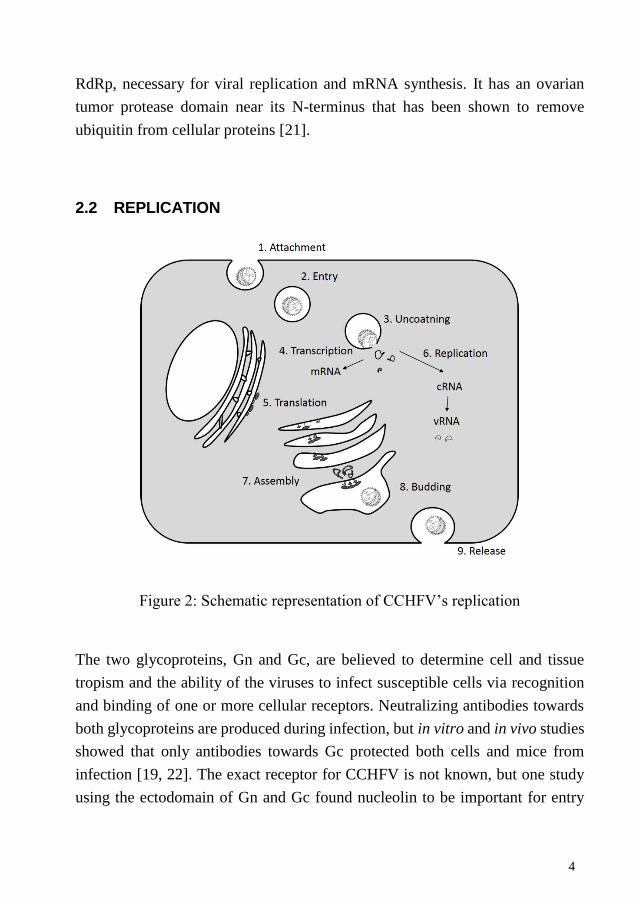
Figure 2: Schematic representation of CCHFV’s replication
The two glycoproteins, Gn and Gc, are believed to determine cell and tissue
tropism and the ability of the viruses to infect susceptible cells via recognition
and binding of one or more cellular receptors. Neutralizing antibodies towards
both glycoproteins are produced during infection, but in vitro and in vivo studies
showed that only antibodies towards Gc protected both cells and mice from
infection [19, 22]. The exact receptor for CCHFV is not known, but one study
using the ectodomain of Gn and Gc found nucleolin to be important for entry
5
[23]. Following attachment, CCHFV enters through clathrin-mediated
endocytosis (CME) in a pH-dependent manner with fusion likely to occur in the
early endosomes [24, Paper I]. Viral replication occurs in the cytoplasm where
the negative stranded genome interacts with the viral RdRp for the synthesis of
the positive stranded messaganger RNA (mRNA) and full-length
complementary RNA (cRNA). The mRNA is used for transcription of the viral
proteins and the cRNA is used as template for the synthesis of new genomic viral
RNA (vRNA). Newly synthesized vRNA binds to the NP and is then
incorporated with the glycoproteins in the Golgi complex (Donets Chumakov
1977), after which the virus is released by exocytosis. Host-cell microtubule are
needed during replication, assembly and egress [25] while actin is important for
transporting NP to the site of assembly [26].
2.3 OCCURRENCE
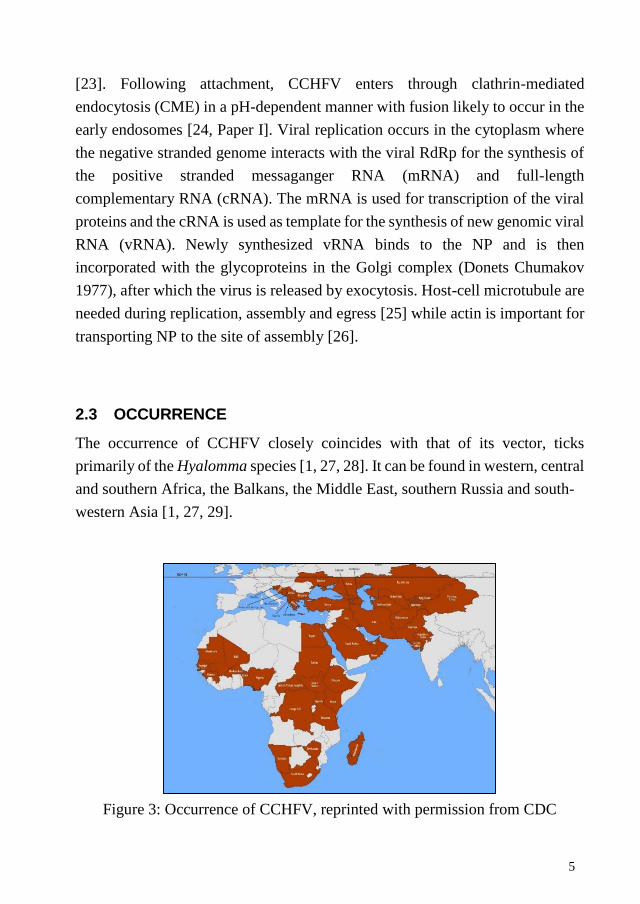
The occurrence of CCHFV closely coincides with that of its vector, ticks
primarily of the Hyalomma species [1, 27, 28]. It can be found in western, central
and southern Africa, the Balkans, the Middle East, southern Russia and south-
western Asia [1, 27, 29].
Figure 3: Occurrence of CCHFV, reprinted with permission from CDC
6
In Europe it is mainly found in the Balkans, Russia and countries of the former
Soviet Union. Previously there had only been two antibody based reports of
CCHFV from Western Europe, both on bats, one from the border of France and
Spain and one from Portugal [1, 30]. But a recent report found CCHFV positive
ticks in Spain [31]. Another recent report found CCHFV positive ticks on a
migratory bird travelling from Africa to southern Europe [32]. Imported cases of
human CCHF have occurred to France [33] and the United Kingdom [34], but
without further transmission. During the last few year most cases of CCHF have
been reported from Turkey. Although the virus was known to circulate there
before, the first clinical cases of CCHF in Turkey didn’t occur until 2002 [35].
However, since then numerous cases have been confirmed, between 2002 and
2012, 6864 cases were reported [36].
The increase in cases is most likely a combination of better awareness within the
health care system, effective molecular methods for virus detection and virus
spread. The spread can both be explained by natural bird migration, where birds
are carrying infected ticks [32], and by the trade of virus infected and/or tick-
infested livestock to previously unaffected areas where permissive ticks are
available [1, 37, 38]. The number of human cases also increase when previously
abandoned farm land is recultivated in areas where the virus is already
circulating [2].
2.4 TRANSMISSION
CCHFV is spread and maintained by ticks. Human are exposed to the virus
following tick bites, the handling of viremic or tick-infected livestock or through
person-to-person (nosocomial) transmission. Humans who are mainly at risk of
contracting the virus therefore include agricultural and slaughterhouse workers
as well as hospital staff caring for infected patients.
7
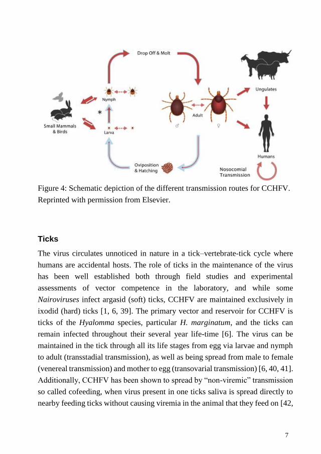
Figure 4: Schematic depiction of the different transmission routes for CCHFV.
Reprinted with permission from Elsevier.
Ticks
The virus circulates unnoticed in nature in a tick–vertebrate-tick cycle where
humans are accidental hosts. The role of ticks in the maintenance of the virus
has been well established both through field studies and experimental
assessments of vector competence in the laboratory, and while some
Nairoviruses infect argasid (soft) ticks, CCHFV are maintained exclusively in
ixodid (hard) ticks [1, 6, 39]. The primary vector and reservoir for CCHFV is
ticks of the Hyalomma species, particular H. marginatum, and the ticks can
remain infected throughout their several year life-time [6]. The virus can be
maintained in the tick through all its life stages from egg via larvae and nymph
to adult (transstadial transmission), as well as being spread from male to female
(venereal transmission) and mother to egg (transovarial transmission) [6, 40, 41].
Additionally, CCHFV has been shown to spread by “non-viremic” transmission
so called cofeeding, when virus present in one ticks saliva is spread directly to
nearby feeding ticks without causing viremia in the animal that they feed on [42,
8
43]. Hyalomma ticks are generally 2-host ticks where the first host usually is
ground-feeding birds or small mammals such as hares or hedgehogs, while the
second host is a larger animal such as sheep or cattle [1]. Some tick species wait
passively to encounter a vertebrate (“ambush ticks”), but Hyalomma ticks are
“hunting” ticks, which can quest up to 400m to find their hosts [6].
Wild and domestic animals
Most mammals appear to be susceptible to infection with CCHFV, but only a
few develop a sufficiently high viremia to efficiently infect ticks [44]. But in
some vertebrates the bite of an infected tick causes viral replication and viremia,
providing a source of infection for additional ticks as well as the risk of spreading
the virus to humans. Experimental infections of wild and domestic animals have
found that sheep, calves, scrub hares and ostriches develop a short viremia and
in some cases were able to transmit the virus to feeding ticks [6, 45]. Because
most vertebrates infected with CCHFV develop only a transient viremia without
apparent illness, the identification of animal hosts of CCHFV has largely been
done on the detection of virus-specific antibodies in collected serum from
livestock or occasionally wild animals. The occurrence of antibody positive
livestock has been found to correlate with the occurrence of human cases [6].
Even though birds can carry a large number of virus infected ticks, most birds
appear to be refractory to the virus [45, 46].
Nosocomial transmission
The virus has been shown to spread from person-to person and a number of
nosocomial cases have been reported where hospital staff, laboratory personnel
and/or relatives have contracted the virus from a CCHFV infected patient [1, 47-
50]. Fortunately, increased information regarding protective nursing has reduced
the number of nosocomial cases and showed that standard barrier nursing
methods are efficient to prevent further transmission of CCHFV [47, 50, 51].
9
3 CCHF- THE DISEASE
CCHFV infection is only known to cause disease in humans. It has been
calculated that approximately 1 out of 5 persons exposed to CCHFV develop
symptoms [52, 53]. Although the duration and symptoms vary among affected
individuals, most patients only develop a mild or subclinical infection. Some
patients do however, progress into the more severe symptoms.
The disease is divided into 4 phases: incubation, prehemorrhagic, hemorrhagic
and convalesce [1]. The incubation time can vary from a few days up to a week
and has been suggested to differ depending on transmission route, with shorter
incubation times for tick bite or livestock handling compared to nosocomial [54].
Viral load has also been suggested to affect incubation time [29].
Following incubation, the patients enter the prehemorrhagic phase. This is
characterized by a rapid onset of fever, headache, myalgia, nausea and vomiting
[1, 49, 54] which usually last approximately 3 days after which some patients
enter the hemorrhagic phase. The hemorrhagic phase is characterized by
bleeding, ranging from dermal petechiae to gastrointestinal hemorrhage [29, 49,
54, 55]. The most common bleeding sites includes the nose, gastrointestinal
system, urinary tract and the respiratory tract [56]. Other symptoms include
enlarged liver and spleen, elevated liver enzymes, prolonged bleeding times and
in severe cases disseminated intravascular coagulation (DIC) [1, 29, 54]. The
severity of the disease has been correlated to high viral load and low antibody
response [55, 57-61]. Other severity markers include thrombocytopenia,
leucopenia, elevated liver enzymes, prolonged bleeding times and bleeding [55,
60, 62, 63]. Death usually occur between days 6-10 after disease onset and is due
to multiple organ failure caused by severe anemia, dehydration and shock [60].
For patients that recover, the convalescence can be long and include symptoms
like weakness, labile pulse and confusion, all of which are temporary [1, 54].
There have not been any reports of relapse of the infection [29].
10
3.1 TREATMENT
Most human CCHFV-infections appear to be asymptomatic or only cause a mild
febrile illness [6, 53, 64]. But patients that develop the severe form of the disease
require extensive hospital stay with special care and protective nursing in order
to limit the spread of the disease. As there currently is no commercial vaccine or
specific treatment available, patients are usually given a combination of
supportive treatments. This includes giving volume replacement, to treat the fall
in blood pressure and diminishing organ perfusion, giving fresh frozen plasma
and platelets for the coagulation abnormalities as well as blood transfusions for
significant hemorrhage [6].
It has been suggested that the different phases of the disease should be treated
differently with an antiviral such as ribavirin, given during the first clinical phase
when viremia and virus replication is high and drugs targeted at DIC or sepsis
during the second phase when the viral load declines and patients enter the
hemorrhagic phase [65]. In line with this a recent study found corticosteroids to
be effective, particularly for patients with severe CCHF [62].
Studies of the efficiency of giving immunoglobulin is lacking, although
immunoglobulin derived from plasma of CCHF survivor donors is used as
treatment in Bulgaria [66, 67] and was recently used in Turkey [68]. But no case-
control studies have been published on the efficiency in CCHF patients [69].
Ribavirin
Ribavirin (Virazole®) is a synthetic guanosine analogue that is used to treat a
number of RNA and DNA viral infections. Its full mechanism is not yet known,
although both indirect and direct functions have been proposed to explain its
antiviral activity [70]. Most clinical comparative CCHF studies has found that
ribavirin is beneficial, as long as it is initiated early in the course of the disease
[62, 65]. However only one randomized clinical trial has been conducted and
they found no significant difference with regard to disease outcome [71].
11
Vaccines
There is no FDA approved vaccine for CCHFV. A vaccine against CCHFV using
formaldehyde-inactivated mouse brain tissue was developed in the Soviet Union
in the early 1970ies [69] and a similar vaccine is still being used in Bulgaria and
given to soldiers, medical personnel and other high risk groups in endemic areas
[72]. However, no case-controlled studies of its efficiency has been conducted
and a recent study found that the Bulgarian vaccine only elicited a low
neutralizing antibody response, even in persons that had received it 4 times [73].
Two studies where vaccination with CCHFV Gn and Gc either delivered as a
DNA vaccine [74] or as purified proteins from transgenic plants [75], induced
neutralizing antibodies in mice. Yet, at the time there was no animal model
available for CCHF and so the actual response could not be assessed in a
challenge model. However, a promising new CCHFV vaccine candidate based
on a poxvirus vector expressing the CCHFV glycoproteins, was found to elicit
both a cellular and humoral response and protect mice challenged with CCHFV
[76]. But whether this could be used as a human vaccine against CCHFV remains
to be investigated.
3.2 ANIMAL MODELS
CCHF symptoms has so far only been reported in humans and this in
combination with the requirement for BSL-4 containment, has made it difficult
to develop an animal model to study CCHF pathogenesis. Attempts to study the
disease in adult mice, rats, hamsters, guinea pigs, rabbits, sheep, calves, donkeys
and non-human primates have all proven unsuccessful [1, 6]. Although a short
viremia could be detected in several animals this was not sufficient to cause
CCHF symptoms [6]. The virus does however replicate to high titers in newborn
mice [77, 78], but as they have an immature immune response they are not a
good model to study the pathogenesis of the disease. But recently, two different
knock-out mice models were presented which revealed the importance of the
IFN type I response in controlling the disease. The mice lacked either the cell-
surface IFN- αβ receptor [79] or the intracellular signal transducer and activator
12
of transcription (STAT)-1 protein [80] and developed CCHF symptoms
following infection. Hopefully, the introduction of these new animal models can
provide more knowledge regarding CCHF’s pathogenesis.
3.3 PATHOLOGY
Due to the safety regulations and the sporadic occurrence of the disease, there
are only a few reported necropsies on CCHF patients [57, 81, 82]. Further
information comes the new animal models [79, 80]. The main finding from
human necropsies included necrosis of the liver, which varied in extent but
generally existed in multiple foci associated with viral antigen and no
inflammatory infiltrates [54, 57, 81, 82]. The liver dysfunction is also reflected
by the elevated liver enzymes detected during CCHF which can be used as a
prognostic marker [56, 60, 63]. Damage to the spleen was also noted with
marked lymphocyte depletion as well as hemorrhage and infection of endothelial
cells of other organs [57, 81, 82]. The two new animal models developed similar
symptoms with necrosis of the liver as well as prominent lymphocyte depletion
and debris in the spleen, consistent with lymphocyte apoptosis, and
gastrointestinal bleeding [80, 83].
13
4 IMMUNE RESPONSES TOWARDS CCHFV INFECTION
The host response is made up of the innate and adaptive immune systems, which
usually act together in synergy. With the innate response representing the first
line of defense and the adaptive becoming prominent after several days as
antigen-specific T and B cells have undergone clonal expansion. They support
each other with components of the innate immune system contributing to the
activation of the antigen-specific cells and the antigen-specific cells amplifying
the innate response [84].
4.1 INNATE IMMUNE SYSTEM
The innate immune system broadly includes all aspects encoded in their mature
form by the germline genes of the host, this includes physical barriers like
epithelial cell layers that express tight cell-cell contacts, soluble proteins as well
as membrane-bound receptors and proteins that binds to the surface of invading
microbes [84].
Interferons
Interferons (IFNs) are a group of secreted cytokines that have antiviral effects.
They recognize viruses through toll-like receptors or pathogen recognition
receptors. There are 3 different types, type I, II and III. In vitro studies has shown
that CCHFV is sensitive to type I IFNs and several of its antiviral proteins [85-
87]. However, CCHFV is able to avoid recognition by RIG-I by processing the
5’RNA termini of the genome, thereby delaying initial induction of IFNs [88].
The new animal models also showed the importance of a functional IFN
response in controlling CCHFV replication with more severe symptoms and
higher levels of viral replication in IFN type I knock-out mice compared to wild
type mice [79, 80].
14
Nitric oxide
Nitric oxide, NO, is another mediator of the innate response, which can be
induced either directly by virus or through cytokine dependent activation [89].
In vitro experiments have shown that NO can reduce CCHFV replication [90].
Natural killer cells
Natural killer (NK) cells play an important role in the early anti-viral defense.
They have a complex regulation and do not act on pathogen-specific antigens
but rather on the NK cell activation and inhibitory receptors expressed on cells.
If activated they can induce apoptosis in the target cell [91]. In CCHF patients,
one study found greater number of circulating NK cells in fatal cases [92] while
another found no correlation between mild and severe CCHF [93]. In vivo
experiments have shown activation of NK cells but an overall loss over time
[80].
4.2 MACROPHAGES AND DENDRITIC CELLS
Antigen-presenting cells (APCs), including macrophages and dendritic cells
(DCs), are important members of the immune system as they present processed
antigens on their surface to T cells. They express both class I and class II major
histocompatibility complex (MHC) molecules.
For Ebola it has been hypothesized that infection of macrophages and dendritic
cells is crucial to pathogenesis, as this leads to the release of proinflammatory
cytokines and other mediators, causing impairment of the vascular and
coagulation systems that ultimately lead to multiple organ failure and possibly
death [94].
15
Macrophages
Macrophages are phagocytosing cells that in addition to their role as APC also
can employ a battery of innate immune mechanisms for initial anti-viral defense
[95]. An in vitro study have shown that they produce cytokines upon CCHFV
infection [96] and a CCHF patient study found a correlation between elevated
levels of neopterin, a monocyte/macrophage activation marker, and disease
severity, indicating that macrophages could have a role in CCHF disease
exacerbation [97]. Recent work using the new animal model found that only one
macrophage population out of the two studied in the spleen was capable of
upregulating MHC class II molecules, indicating a possible partial impairment
of the ability to activate the adaptive immune system [80].
Dendritic cells
Dendritic cells have a crucial role as a bridge between the innate and adaptive
immune response. They normally reside in peripheral and lymphoid tissues in
an immature form where they acts as sentinels sensing the antigenic
microenvironment and capture antigens. When they encounter an antigen they
undergo maturation, release cytokines as well as migrate to regional lymph
nodes where they present the antigen to and thereby activate naïve T cells [98].
They can also become activated by proinflammatory cytokines such as TNFα
[99].
In vivo experiments showed no increase of MHC class II molecules in DCs
during CCHFV infection [80] while in vitro studies of CCHFV-infected
monocyte-derived DCs (moDCs) have shown that they are activated to the extent
that they release cytokines (TNFα, IL-6, IL-10 and IL-8) and become partially
matured [96, 100]. For Ebola and Marburg it has been shown that infected DCs
aren’t able to initiate an effective adaptive immune response and support T cell
proliferation [101-103]. But whether something similar is true for CCHFV
remains to be investigated. An active adaptive immune system is thought to be
important for survival as fatal cases of CCHF rarely develop an antibody
response [57, 61].
16
4.3 ADAPTIVE IMMUNE SYSTEM
The adaptive immune system manifests high specificity for its target antigens. It
is primarily based on the antigen specific receptors expressed on the surfaces of
T and B lymphocytes [84].
The humoral response is accomplished by B cells. They are APCs that produce
cytokines and are defined by their production of antibodies that bind and
potentially neutralize pathogens [84]. In vivo studies have shown activation of B
cells during infection, followed by decrease in B cell numbers, which is in
accordance with the lymphocyte depletion observed in patients [80-82].
The cell mediated response consists of T cells, defined by their cell-surface
expression of receptors that bind processed antigens displayed by APCs [84].
They have several subtypes, including cytotoxic T cells and helper T cells. An
investigation of lymphocyte levels in CCHF patients found increased levels of
cytotoxic T cells among fatal compared to nonfatal cases of CCHF but no change
in the helper T cells [93].
4.4 CYTOKINES
Cytokines are cell-signaling proteins that mediate and modulate immunity, such
as interleukins, IFNs, TNFα, chemokines, migration inhibitory factor, and
transforming growth factor β [104]. For Ebola, it has been found that
proinflammatory cytokine release could lead to vascular permeability and
ultimately hypotension, shock and organ failure [94, 95, 105]. The new CCHF
animal models have showed increased levels of proinflammatory cytokines,
TNFα, IL-6, IFN-γ and IL-10 [80, 83]. Several studies of CCHF patients have
also found elevated levels of proinflammatory cytokines, which was correlated
to disease severity and death [106-108]. All patient studies found increased
levels of TNFα, two found higher levels of IL-6 and IL-10 and one found higher
IFN-γ. Unfortunately, the studies had different sampling times and often only
one sample could be taken from each patient making comparison difficult. In
vitro experiments show that moDCs and macrophages release proinflammatory
17
cytokines upon infection [96, 100] and supernatant from infected moDCs could
activate endothelial cells mainly by released TNFα [109].
4.5 PROPOSED PATHOGENESIS
The pathogenesis of CCHFV remains poorly understood. As previously stated
most of the cases occur in remote regions and the high virulence of the virus
limits laboratories studies. Most of the knowledge therefore comes from a few
pathological studies, the relatively new animal models, and similarities to other
viral hemorrhagic fever viruses and their pathogenesis.
The primary targets for CCHFV during infection has been suggested to be
endothelial cells, macrophages, DCs and hepatocytes [81, 83]. As with other
viral hemorrhagic fevers, the endothelium plays an important role and vascular
dysfunction would account for the characteristic rash seen in CCHF as well as
contribute to platelet aggregation and activation of the coagulation cascade [20,
110]. The effect on the endothelial cells that lead to the vascular permeability
could either be a direct result of viral infection or an indirect effect of the host’s
immune response through soluble factors [111].
Direct effects
There is evidence of endothelial activation and damage in CCHF patients where
levels of sICAM-1 and sVCAM-1 were correlated to disease severity [112, 113]
and in vitro CCHFV infection has been found to activate endothelial cells
causing cytokine release and leukocyte adhesion [109]. Viral RNA and antigens
have also been detected in endothelial cells of the liver, spleen, heart and
intestinal tissues during necropsy and in the new animal models [81, 83].
However the presence of virus in the endothelium at time of death does not mean
that it was the cause of the vascular permeability, as the dysfunction is apparent
during early part of the disease [6].
18
Enlarged liver has been reported as one of the symptoms of CCHF [1, 54] with
necrosis of liver found at autopsy [81]. It was suggested to be due to high viral
replication, which is supported by results found with the new animal model
where enlarged livers was only found in animals with high viral load in the liver
[79, 83]. Further support comes from recent in vitro experiments on hepatocytes
showing that CCHFV infection could induce apoptosis and replicate to high
titers [114].
In vitro studies of direct CCHFV infection found no effect on permeability in
epithelial cells [115, Paper III]. Indicating that other factors most likely cause
the vascular leakage. This is in line with what has been observed for Ebola virus
where the vascular leakage is not caused by direct infection but rather due to the
effects of soluble mediators [104].
The viral replication does not appear to cause vascular leakage but lead to
activation, release of cytokines and the recruitment of leukocytes in endothelial
cells. In contrast, high viral replication in the lymphoid organs could be an
explanation for the observed lysis and necrosis of the liver and spleen.
Indirect effects
Indirect effects of CCHFV infection is not fully understood although the release
of proinflammatory cytokines have been well documented both in patients,
animal models and in vitro [6, 83, 96, 100, 108, 109, 114]. Low levels of
cytokines work locally, while excess amounts can have a systemic effect, a so
called cytokine storm that subsequently could lead to vascular leakage and
hemorrhage and ultimately and organ failure. Macrophages and moDCs have
been shown to be productively infected and release cytokines [96, 100].
Cytokines-hyperactivated monocytes and macrophages could be the cause of the
hemophagocytosis that has been demonstrated in a number of CCHF patients
[116-118].
19
At present it is not known if the APCs are able to initiate an effective T cell
response but fatal CCHF patients rarely develop or show a late antibody response
indicating that the adaptive response is either suppressed or not fully activated.
The combination of high viral replication, cytokine storm, endothelial activation
and organ necrosis could be what ultimately leads to the shock and multiple
organ failure observed in severe CCHF.
20
5 POLARIZED CELLS
Polarized cells are found throughout the human body and act as a boundary
between different environments, for example epithelial cells lie between tissues
and the extracellular environment and endothelial cells line the inner surface of
blood vessels and acts as a barrier between blood and tissue. They often
constitute the first line of defense against the environment and regulate the
passage of substances into and out of organs such as the gut, lung, liver and brain.
Polarized cells have an asymmetric plasma membrane with an apical and a
basolateral side [119]. They are organized as selectively permeable continuous
sheets that separate the two different sides, where the apical side face the lumen
i.e. the outside for the epithelial cells and the blood for the endothelial cells while
the basolateral side face the tissues and is in contact with the neighboring cells
and the underlying extracellular matrix. The two domains also have distinct lipid
and protein compositions which is generated and maintained by a specific sorting
machinery [119, 120].
The space between the neighboring cells is sealed by junctions which restrict the
paracellular transit of ions and macromolecules and separate the two sides of the
cells [121]. There are different junctions between the cells, the tight junctions
are the most apical of them, located at the boundary between the apical and the
basolateral domain of the cells [121]. They tightly seal the space between
neighboring cells, preventing the passage of fluids, electrolytes and
macromolecules between the cells [121]. Adherens junctions are located on the
basolateral side of the cells. They mediate cellular polarization and
organogenesis. There are also gap junctions that form channels between the
neighboring cells, allowing intercellular dissemination of small molecules [121].
21
Transepithelial resistance (TER)
To study polarized cells in vitro, the cells can be grown on permeable support,
where the cells have free access to medium from both sides and samples can be
taken from both sides. Since electrical conductivity is almost limited to the
paracellular ion flux, the transepithelial resistance (TER) across the monolayer
is a good marker of the development of tight junctions and can therefore be used
to describe cell monolayer integrity [122]. The “chopstick system”, consisting
of 2 electrodes stuck together, one for each side, is easy to use for routine
determination of TER in filter-grown monolayers.
5.1 POLARIZED CELLS AND VIRUS INFECTION
As cellular proteins and receptors could be differently distributed on the different
sides of a polarized cells [120], it could be a limitation for viral entry and release.
While some viruses are restricted to entry from a specific side, others viruses
like influenza and poliovirus, have little or no preference [119]. Many virus
receptors are not located on the apical side, but at the basolateral side, rendering
intact cells layers resistant against many viral infections, such as HSV-1,
poliovirus, reovirus and human adenovirus [123].
Viruses have therefore been forced to come up with ways to overcome this.
Some viruses use more than one receptor, this appear to be the case for herpes
simplex virus [124]. While others, like rotavirus, disrupts the tight junctions
between the cells to get access to the basolateral side [125]. It has also be shown
that the same virus using the same receptors can enter from different sides in
different epithelial cells demonstrating cell type specificity [126]. The junctions
may also be remodeled by cytokines to allow the passage of activated infected
immune cells across the epithelial border [123]. Recently another way was
shown, where adenovirus infected macrophages enabled the translocation of the
viral receptor from the basolateral to the apical side, thereby enabling entry of a
virus that normally requires basolateral access [127].
22
CCHFV has been found to primarily enter from the basolateral side [115, Paper
III] although the addition of infected moDCs lead to more virus entry from the
apical side, the reason for this is at present not known [Paper III].
Viral release can occur from both sides but can have an effect on whether the
infection becomes localized (apical release) or systemic (basolateral release)
[119]. CCHFV has been found to be released from the basolateral side further
aiding systemic spread [115].
23
6 ENDOCYTOSIS
Viruses are obligate intracellular organisms and as such they must enter target
cells in order for successful replication to occur. Different viruses have therefore
developed different ways of entering and the route of entry can also vary
depending on cell type. Most viruses do however exploit the cell’s preexisting
endocytic routes in order to gain entry into their target cells.
Virus endocytosis begins with the virus attaching to cell attachment factors
and/or virus receptors that in turn induces a conformational change of the virus
and/or signal cascade within the cell which promotes the internalization of the
virus. Most endocytosis routes transports the encapsulated virion to the early
endosome, through the multivesicular body to the late endosome and further on
to the lysosome. In order for the virus to escape degradation in the lysosome it
therefore has to penetrate the endosomal membrane at some point and deliver its
genome to the appropriate replication site. Enveloped viruses fuse the viral and
cellular membranes to release their genome into the cytosol while non-enveloped
viruses use membrane-lysis or pore-formation [128]. The catalytic event that
determines when release occurs is usually pH, where the acidic environment
inside the endosome or lysosome induces conformational changes in the virion
structure that enables fusion [128].
The most common viral entry route is clathrin mediated endocytosis (CME),
followed by caveolae and a number of clathrin and caveolae independent routes.
The virus could also be engulfed and enter through macropinocytosis or
phagocytosis. A few enveloped viruses have also been shown to enter by fusion
with the plasma membrane for example herpes simplex-1 and HIV [128]. But as
the same viruses are also endocytosed at the same time it is difficult to determine
if plasma membrane fusion lead to productive infection. Using the cells own
machinery when entering can have many advantages for the virus, it not only
offers a protected environment for the virus to pass through the actin cortex and
the dense cytoplasm while transporting it to necessary sites and organelles where
virus penetration can occur but it also helps the virus to escape recognition by
the immune system as no virus specific parts are left at the plasma membrane.
24
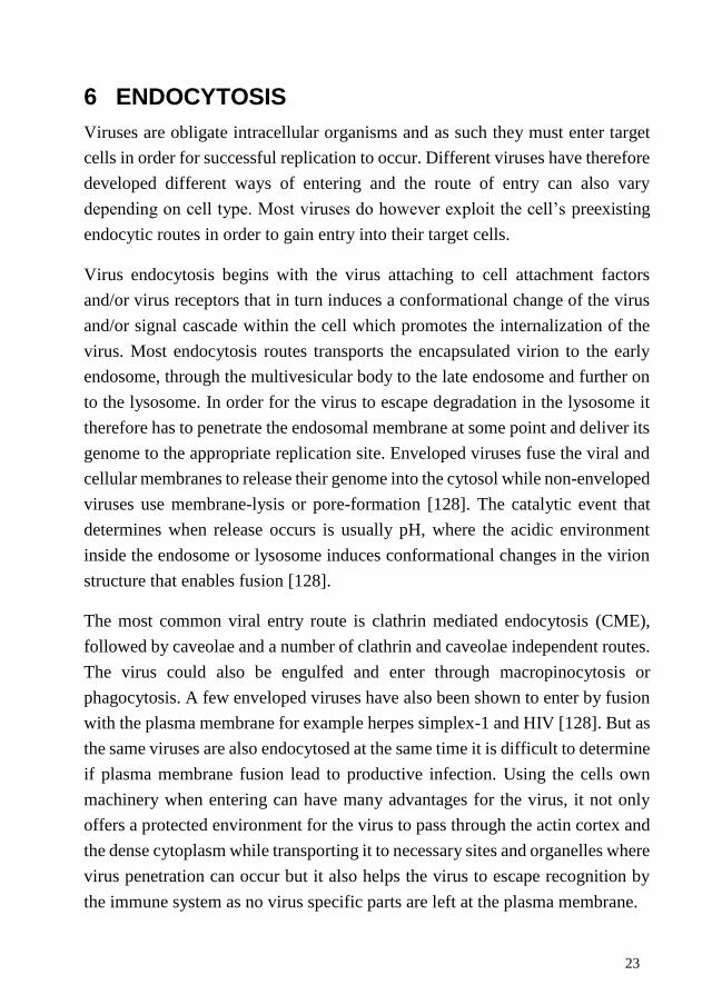
Figure 5: Schematic depiction of different endocytosis routes used by viruses. a)
Clathrin mediated endocytosis, b) Caveolae, c) Lipid raft, d) Clathrin- and
Caveolae-independent endocytosis and e) Macropinocytosis
6.1 CLATHRIN MEDIATED ENDOCYTOSIS
CME is a constitutive event that is found in all eukaryotic cells and normally
used to take up cargo and nutrients, control many plasma membrane activities
and is fundamental in neurotransmission. Clathrin is a self-polymerizing protein
that is composed of three heavy and three light chains that together form a
triskelion shape consisting of three bent limbs radiating from a centre [129]. It
can polymerize into either flat lattices or cages and is not only involved in
endocytosis from the plasma membrane but is also involved in the endosomal
25
sorting complex required for transport (ESCRT)- dependent cargo sorting at the
endosomes, protein secretion from the trans-Golgi network and mitosis [129].
Clathrin can be found in stable islands in the plasma membrane or become
recruited through receptor mediated signaling. Some viruses like Reovirus,
Influenza A and Vesicular stomatitis virus (VSV) have for example been shown
to induce de novo formation at the site of binding [130]. The clathrin triskelions
form a lattice-like coat on the cytoplasmic surface of the plasma membrane.
Binding of clathrin to the membrane is mediated by adaptor protein 2 (AP2) that
links the membrane cargo to clathrin and accessory proteins like epsin and
AP180 [129]. AP2 is considered to be an absolute requirement for CME [129],
some are however controversially suggesting that for example VSV can enter by
CME without AP2 [130]. Yet this is still very controversial and needs to be
further analyzed.
The process of clathrin coated invagination continues, leading to the formation
of deeply invaginated pits and the formation of a vesicle neck. Clathrin
polymerization helps in the formation and constriction of the vesicle neck,
helping to bring the membranes surrounding the neck into close apposition
[131]. The fission of the vesicle is then made by the membrane scission protein
dynamin, a large GTPase that form a helical polymer around the vesicle neck
[130]. After internalization the clathrin coat is removed by uncoating proteins,
Hsc70 and auxilin to form a naked vesicle [132]. The vesicle and its cargo is
then transported to Rab5 positive early endosomes while the clathrin triskelia
and accessory proteins are recycled to perform more CME. Some virus like VSV
exit in the early endosome while others like Influenza A continues through the
multivesicular body to fuse with the late endosome [130]. It is thought that the
later stages of endocytosis (formation of curved coats and vesicle budding) can
occur independently of what cargo is present in the coated pit. Thus, cargo
recruitment to the clathrin-containing lattice structure is the key sorting step
defining the specificity of the internalization process [132].
26
The first virus that was shown to enter by CME was Semliki Forest virus (SFV)
[133] but today CME is well established as the main entry route for viruses [130].
Yet in some cases like for Influenza A and lymphocytic choriomeningitis virus,
CME is only one of several entry pathways that the virus can use [130].
6.2 CAVEOLAE AND LIPID RAFT ENTRY
Caveolae and lipid rafts are found in cholesterol rich domains of the plasma
membrane and are involved in cellular endocytic processes and signaling. Lipid
rafts is a specialized cholesterol- and sphingolipid-enriched membrane
microdomain that can influence membrane fluidity, receptor clustering and
assembly of signaling molecules [130]. Caveolae are seen in electron microscope
as small regular shaped membrane invaginations [134] and can exist either as
single caveolae or clusters of multiple caveolae [135]. They are important in the
regulation of various signaling processes, such as nitric oxide activity, and in
cholesterol uptake and trafficking [134]. The major structural protein, caveolin-
1 forms a coat-like surface around the vesicle and is necessary for the formation
of invaginated caveolae [135]. Caveolin is present in the plasma membrane as
well as on intracellular structures [135]. It is found in nonmuscle cells with the
exception of neurons and leukocytes [131]. Expression of caveolin-1 alone is
sufficient to cause the formation of caveolae [135]. Under normal conditions
caveolae do not appear to be involved in endocytosis and are kept at the plasma
membrane by actin filaments [134]. They do however perform short range “kiss
and run” movements, but whether this is in fact is endocytosis is not yet clear
[134]. Although caveolae are not ordinarily internalized they can be if stimulated
by for example Simian virus 40 (SV40) that initiate a signal cascade that leads
to the depolymerization of the actin cytoskeleton [135]. The fission protein is as
for CME, dynamin [135].
27
Caveolae has mainly been suggested to be involved in viral endocytosis for
members of the polynoma family i.e. SV40, BK and JC virus [130]. Where the
endocytosed polynoma viruses pass through the early and late endosome and are
released in the ER. The role of caveolar as a primary route of viral entry has
however come in question in recent years and it has now been suggested that
only a fraction of SV40 and probably other polynomaviruses enter via caveolae
and that the fraction may vary with cell type and virus [130]. The rest use a
related caveolin-1 independent mechanism. Both routes appear to be sensitive to
cholesterol depletion, and require Arf1 and dynamic actin [130]. Internalization
through caveolae also appears to be slower, dynamin-2 dependent and more
dependent on actin dynamics than the noncaveolar pathway [130].
6.3 CLATHRIN AND CAVEOLAE INDEPENDENT ENDOCYTOSIS
As already stated, some viruses have been shown to become endocytosed in a
clathrin and caveolae/caveolin-1 independent way. The number of viruses that
can use these routes are expanding but at the moment very little specific
information exists regarding these routes. For Influenza A it has been shown that
1/3 of the virus particles are endocytosed in a clathrin and caveolin-1
independent way and that this infection is just as effective and that trafficking
through early to late endosomes appear to similar to CME [136]. An IL-2
tentative route of entry has been suggested for Rhesus rota virus and possibly
also for SARS virus [130]. In both cases it was shown to be clathrin and caveolin-
1 independent and cholesterol and dynamin dependent. Adeno-associated virus
2 was recently suggested to enter through the GEEC/CLIC pathway [137], which
is another clathrin and cavoelin-1 independent route. Not much is known
regarding this route and so far this is only virus that has been proposed to use
this route.
28
6.4 MACROPINOCYTOSIS AND PHAGOCYTOSIS
Macropinocytosis is a cargo triggered endocytosis route which normally is
activated by growth factor. This triggers the activation of a complex signal
cascade that induces changes in the actin filaments dynamics and triggers plasma
membrane ruffling [138]. These ruffles will eventually collapse back towards
the plasma membrane creating an uncoated irregularly shaped vacuole. It is
mostly a transient process responsible for the internalization of fluid, solutes and
sometimes particles into large vacuoles [139]. It is strictly dependent on cortical
actin but independent of dynamin and does not require binding to a specific
receptor [140]. The vacuole can become acidified and intersect with endocytic
vesicles, making it a possible entry pathway [128]. Macropinocytosis is the main
route of entry for Ebola virus, although a fraction of the virus also uses CME
[141]. Human adenovirus, Influenza A and Vaccinia virus has also been
suggested to use this route for part or all of their entry [138].
Unlike the other entry routes, phagocytosis is only found in specialized cells
such as macrophages and amoeba, where it is used for uptake of large particles
[130]. Mimivirus has been suggested to use phagocytosis [142] while herpes
simplex virus 1 has been suggested to use phagocytosis-like uptake [143].
6.5 BUNYAVIRIDAE ENTRY AND RECEPTORS
Bunyaviridae entry has been shown to be pH dependent with some virus, like
CCHFV, Hantavirus and Oropouche virus, entering by CME [24, 144, 145,
Paper I]. While others, like Uukuniemi virus (Phlebovirus) has been shown to
enter cells mainly through clathrin-independent endocytosis [146] while Rift
valley fever virus (Orthobunyavirus) was shown to be using caveolae [147].
Viruses express glycoproteins on their surface, by which they can attach to
cellular receptors or attachment factors. Although not all viruses require specific
receptors for attachment and internalization most virus attach to some cellular
receptor or attachment factor. There are numerous viral receptor expressed on
29
cell surfaces and not all receptors are expressed on all cells. Some viruses have
therefore evolved to use different receptors depending on the cell their infecting
[124]. The availability and position of a specific receptor can determine whether
a virus is able to infect that particular cell [123]. For Bunyaviridae viruses, DC-
SIGN and β3 integrin have been demonstrated as receptors [148, 149]. A recent
report suggested nucleolin to be essential for CCHFV entry [23], but whether it
acts as a receptor or an attachment factor aiding in the entry of the virus remains
to be investigated.
30
7 IN SITU DETECTION OF NUCLEIC ACID
In situ detection has the advantage of giving the precise and spatial localization
of specific nucleic acid sequences in their natural setting. This gives the
possibility to correlate the single cell results to that of the surrounding cells or
tissue. There are a number of ways to detect nucleic acids in situ.
7.1 ISH AND FISH
In situ hybridization (ISH) is a technique where labelled DNA probes are
hybridized to specific target sequence in fixed samples giving a localized
detection of nucleic acids. It was first performed using radio-labelled probes
[150] but had limitations in resolution and probe instability, therefore the
technique was further developed. With the addition of fluorescence (FISH,
fluorescent in situ hybridization), the method became more applicable and more
than one target could be detected simultaneously since different fluorophores
could be used for different targets [151, 152]. The detection can be done either
directly, so that the fluorescence could be analyzed immediately after
hybridization, or indirectly, where probes are labeled, for example with hapten,
and then detected with fluorescent-labeled antibodies against hapten. The
technique has primarily been used for detection of chromosome abnormalities.
During the last few years several modifications of FISH have been presented.
Among them adding locked nucleic acids (LNAs) to improve resolution and
sensitivity [153, 154]. LNAs are a class of RNA analogs with exceptionally high
affinity towards complementary DNA and RNA [153].
Both ISH and FISH have been used for the detection of mRNA in situ, both
individually and as multiplexed analysis [150, 155-157]. Hybridization-based
methods are however usually semiquantitative and do not allow precise digital
quantification of the signals. They can also not discriminate between highly
similar sequences, making them unsuitable for studying cell-specific allelic
expression or expression of splice variants [158].
31
7.2 PADLOCK PROBES
In oligonucleotide ligation assay (OLA), two oligonucleotides bind next to each
other and are ligated by a DNA ligase if they form a perfect match [159]. This
allows for the distinction of single nucleotide variants at the ligation junction.
Padlock probe is a further development of OLA. Instead of having two different
oligonucleotides, a padlock probe is one long linear oligonucleotide where the
3’- and 5’ ends bind juxaposed to each other on the target sequence. The ends of
the padlock probe are joined together by a target-non-complementary DNA
backbone that is later used as detection site. If the two ends are correctly
hybridized to the target sequence a nick is formed between them. The nick can
then be closed by a DNA ligase, creating a closed “locked” circular DNA
molecule [160]. A locked probe remains attached to its target sequence and can
withstand highly stringent wash conditions, further reducing the amount of non-
specific signals [160]. The ligated probe can then be detected by labeled
oligonucleotides towards the target-non-complementary DNA backbone of the
padlock probe.
Padlock probe detection is highly specific as two target complementary
sequences at the correct position are required for ligation and the DNA ligase
has a strong preference for a perfect match, particular at the 3’-end [161].
Mismatched padlock probes will therefore not be circularized. Accordingly,
padlock probes can therefore be used for detection of single nucleotide
differences [162, 163] and can easily be utilized against multiple targets with
limited cross-reactivity between probes [163]. However, to effectively detect the
padlock probes signal in situ, amplification is needed.
7.2.1 Rolling circle amplification
Rolling circle amplification (RCA), creates a linear single stranded DNA
molecule (RCP, rolling circle product) consisting of tandem repeats of the
complementary sequence of a single stranded circle [164]. If correctly ligated,
padlock probes can be used as templates for RCA, thereby increasing detection
32
sensitivity. In order for the RCA to proceed more than 1 turn around the
template, a polymerase with strand displacement activity is needed, Phi29
polymerase has this. It also has two other important activities for RCA, 3’-5’
exonuclease activity on single stranded DNA as well as polymerase activity
[165, 166]. This means that it can remove the non-base-paired nucleotides
downstream of the padlock probe and when reaching the probe bound to the
target, it can perform polymerization using the target strand as a primer. RCA of
a ligated padlock probe can also be initiated by an external primer. Alternatively,
the DNA can be cut site-specifically before amplification by introducing an G:A
mismatch, where the padlock probe contains a G which is mismatched with an
A in the target molecule [167]. The padlock probe should then be designed so
that the mismatch is located at the end of the padlock probe binding site on the
target sequence. MutY enzyme will then remove the mismatched A in target
molecule, leaving an abasic site in the target molecule. This site is then
recognized and cleaved by Endo IV, enabling free access for the Phi29
polymerase to start the RCA [167].
RCA is a very specific way to detect padlock probes since only ligated probes
can serve as templates and the produced RCPs will still remain attached to the
target sequence [168]. The long RCP spontaneously collapses into a micron-
sized object that can be visualized by hybridization with fluorescence-labelled
oligonucleotides [169] , which have the same sequence as the backbone of the
padlock probe. As the RCP consists of repeated sequences, this will create a local
enrichments of fluorophores over background, visual as a single dot in the
microscope [169, 170]. The fluorescent dots can then be quantitatively counted
[171]. Due to the specificity of the padlock probe, RCA can be performed against
multiple nucleic acid targets. Padlock probes and RCA was first used to detect
mitochondrial DNA in situ, where the DNA first was made single stranded to
enable hybridization and ligation of the padlock probe [172].
33
Figure 6: Schematic representation of RNA detection using LNA-primer,
padlock probe and target-primed RCA.
7.2.2 RNA detection in situ using padlock probes and RCA
RNA can serve as template for the ligation of padlock probes but it has less
sensitivity and specificity compared to DNA template ligation [165, 173]. A way
to overcome the problems with low sensitivity and specificity of RNA template
ligation in situ, is to first perform a reverse transcription step where a primer
containing LNA modified bases is used to first convert the RNA into
complementary (c) DNA. The LNA primer then ensures that the cDNA remains
attached to the target RNA even after RNaseH digestion as the LNA primer
34
protects bound RNA from being degraded [174]. The cDNA can then be detected
by padlock probes and RCA. This method has been used to distinguish single
nucleotide differences of actin in cultured cells and tissue, and had a detection
efficiency of approximately 30% of available transcripts as determined by qPCR
[174]. With the addition of RCA and LNA primers, RNA viruses could
effectively be detected with padlock probes both in solution [175] and in situ
[Paper II].
35
8 PRELIMININARY RESULTS
CCHFV entry is independent of human αV, α5, β3 or β5 integrins.
We have previously shown that CCHFV enters by clathrin-mediated endocytosis
[Paper I], but there is, however, no clear data on which receptor CCHFV use. A
number of viruses have been shown to use integrins as cellular receptors including
another Bunyaviridae member, Hantavirus, that was shown to use β3 integrin
during entry [148]. Although not the only receptor for Hantavirus, it has been
suggested that the binding of hantavirus to β3 integrins could have an effect on
cellular integrity by relocation of VE-cadherin and thereby be a cause for the
vascular permeability observed in patients [176, 177]. Integrins are
transmembrane glycoproteins that consist of an α and β subunit and mediate cell-
matrix and cell-cell adhesions [178]. The number of varieties of α and β subunits
produce ligand selectivity to extracellular matrix. Integrins transmit signals,
outside-in and inside-out and regulate cell survival and migration [123].
To investigate if CCHFV could be using human αV, α5, β3 or β5 integrins during
their entry, we used cells that were manipulated to express human integrins to see
if this increases internalization as compared to the control cell line. We used
Chinese Hamster ovary (CHO-B2) cells, transfected with cDNA to continuously
express either human αV integrin (CHO-B2-αV) or α5 integrin (CHO-B2-α5) as
well as Chinese hamster melanoma (CS1) cells that had been transfected with
cDNA encoding either human β3 (CS1-β3) or β5 (CS1-β5) integrin protein. The
CHO-B2 cells were grown in Dulbecco’s modified Eagle´s minimal essential
medium (DMEM) with 10% FBS, 2mM L-glutamine, 1% NEAA, 100units/ml
penicillin, 100ug/ml streptomycin, and 10mM hepes with addition of 700µg/ml of
geneticin (all from Gibco, Life technologies) for the CHO-B2-αV and CHO-B2-
α5 cells. The CS1 cells were grown in RPMI medium with 10% FBS, 2mM L-
glutamine, 100units/ml penicillin, 100ug/ml streptomycin, and 10mM hepes and
with the addition of 500µg/ml of geneticin (all from Gibco, Life technologies) for
the CS1-β3 and CS1-β5 cells. Cells were seeded on 24 well plates and grown to
80% confluence. The same number of cells was always used for all compared cell
lines, as CS1 cells grow as suspension cells due to the lack of anchoring integrins.
36
Cells were infected with CCHFV Ibar 10200 (moi 1, as determined by Vero
titration) for 1h at 37°C in a 5% CO2 humidified atmosphere. The adherent cells
were rinsed twice in PBS and the nonadherent cells were centrifuged and rinsed
twice with PBS before replenished in fresh medium, added to the original well
and incubated for 24h at 37°C in a 5% CO2 humidified atmosphere. At which point
the nonadherent cells were centrifuged and cell pellet added TRIzol, the lysed cells
were then combined with lysed attached cells in the original well. All handling of
the virus was carried out at the BSL-4 facility at the Public Health Agency of
Sweden, Solna, Sweden. Total RNA extraction, cDNA synthesis and quantitative
real-time PCR was performed as previously described [25]. Amplifications was
always performed in triplicates and relative amount of CCHFV S segment
transcripts were calculated with the 2-(∆∆Ct) method in relation to GAPDH (and
with the reference Ct set to 1). All assays included noninfected cells and were
always negative for CCHFV RNA.
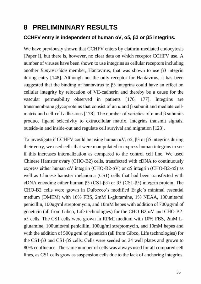
Surprisingly, we found that cells not expressing human αV, α5, β3 or β5 integrins
had the highest level of infection. These data suggest that these integrins are not
essential for CCHFV entry. In paper III in this thesis, we hypothesize that the shift
towards apical entry that occur when infected dendritic cells or their supernatant
is added could be explained by a translocation of an viral receptor from the
basolateral side to the apical side. This has previously been shown for adenovirus
infected macrophages that release soluble factors, including IL-8, causing a
translocation of the viral receptor αvβ3 from the basolateral to the apical side
[127]. We can here show that CCHFV infection is not dependent on either human
αV, α5, β3 or β5 integrin. The reason for the change in entry side in polarized cell
is therefore not due to translocation of these integrins and therefore remains to be
further investigated.
37
Figure: Cells expressing or lacking human integrins were infected with CCHFV
for 24h and then analyzed by RT-PCR for CCHFV RNA levels. Results are
shown as fold change to noninfected cells. A) CS1 cells lacking or expressing
human integrins β3 or β5. CHO cells expressing or lacking (B) human integrin
αV or (C) human integrin α5.
A
B
C
38
9 RESULTS AND DISCUSSION
9.1 CCHFV ENTRY (PAPER I)
In order for virus to replicate, they have to enter a target cell. Viruses have
therefore evolved a of numbers ways to do this. Hantavirus, another
Bunyaviridae member, was known to enter cells though clathrin-mediated
endocytosis in a pH-dependent manner [144] but nothing was known regarding
CCHFV’s entry.
We therefore sought to investigate two common viral entry routes; the clathrin-
mediated and caveolae endocytosis by reducing the expression of the main
proteins of each pathway, clathrin and caveolin-1 respectivly. The siRNA
knockdown of caveolin-1, as confirmed by qPCR and western blot, had no effect
on the level of CCHFV infection, suggesting that caveolae is not necessary for
CCHFV infection. This was further confirmed by the fact that Vero E6 cells, one
of the most commonly used laboratory cell line for CCHFV infection was
discovered to have no or very low levels of caveolin-1, yet it had the highest
level of CCHFV infection of the investigated cell lines in this study.
On the contrary, by treating cells with sucrose or CPZ, we found an indication
that CCHFV could use clathrin-mediated endocytosis. This was confirmed by a
drastic reduction of infection when clathrin protein and mRNA levels were
reduced. We therefore concluded that CCHFV most likely enters through
clathrin-mediated endocytosis. However, we could not completely knock-down
clathrin, and in accordance with this, some CCHFV infection could still be
detected in the clathrin knock-down treated cells. Thus, we could not exclude
that CCHFV could also use a different clathrin-independent route for entry.
As clathrin-mediated endocytosis transports virus to the early endosomes and
many viruses require a drop in pH to induce a conformational change in their
glycoproteins and thereby escape from the endosome, we also wanted to
investigate pH-dependency. By treating cells with chemicals known to disrupt

39
the acidification of the endosome, we could determine that CCHFV entry is pH-
dependent.
Furthermore, in this paper we also investigated whether membrane-bound
cholesterol was needed for CCHFV entry by treating the cells with a cholesterol-
depletion drug. Caveolae and lipid raft endocytosis are located in cholesterol-
rich parts of the membrane and are therefore highly dependent on cholesterol but
CME also requires cholesterol. By treating the cells at various time-points
before, during or after infection we concluded that CCHFV binding was
unaffected by cholesterol depletion but that cholesterol was needed during or
soon after entry and possibly also during later event, such as transcription or
replication.
Since this paper was published another study has confirmed our results showing
that CCHFV enters by clathrin-mediated endocytosis and that CCHFV escapes
from the early and not the late endosome [24].
9.2 IN SITU DETECTION OF CCHFV RNA (PAPER II)
Single cell detection of RNA has several advantages compared to other
molecular methods as it will give the expression level for each cell, which can
vary significantly from the mean expression detected in a cell population [179,
180]. Visualization of single RNA molecules in situ will give the spatial location
as well as reveal potential interactions within the cell, differences that would not
be detected by for example PCR. We therefore wanted to develop an in situ based
method where we could detect individual CCHFV RNA transcripts using a
fluorescent microscope.
CCHFV is a negative single stranded virus, meaning that during its replication
it must produce a positive RNA strand for transcription and replication. We
therefore wanted to target the different strands and be able to differentially detect
them to investigate their cellular location during infection. Reverse transcription
together with specific detection by padlock probes and target-primed RCA had
40
previously been used to detect individual RNA molecules in situ [174]. Yet it
had never been done on negative stranded RNA viruses.
Therefore, we investigated if this technique could be used to detect and
differentiate between CCHFV vRNA and cRNA in fixed cells as well as
combining this with detection of CCHFV proteins. Cell were infected for various
times and fixed. vRNA or cRNA was then reverse-transcribed into cDNA by
using individual LNA-containing primers. The LNA primer protects the target
from being degraded during RNaseH treatment, leaving the cDNA still attached
to the target. Each cDNA is then detected by a padlock probe which if correctly
ligated becomes circularized. The target is then site specifically cleaved to
produce a starting point for the RCA. The resulting RCP is detected by
hybridization of fluorescently labelled oligonucleotides, making them visual as
single dots that then can be digitally counted.
The method worked well, however some false-positive signals could still be
detected in the technical controls, but they were always significantly lower than
signals observed in infected cells. Both vRNA and cRNA could be detected
individually and together. vRNA was found located throughout the cytoplasm
while cRNA was more concentrated to specific parts of the cell. The assay could
also be used to investigate co-localization between viral RNA and viral proteins.
cRNA was found to co-localize with CCHFV NP within the same cellular
structures while vRNA showed no intra-cellular co-localization with CCHFV
NP.
By combining in situ reverse transcriptase by LNA-primer with detection using
padlock probes and RCA, we were able to detect the different strands as
individual dots when looking in a fluorescence microscope. Unfortunately due
the positioning of the primer and probe, we could not discriminate between
cRNA and mRNA. While these techniques has previously been used for the
detection of positive stranded RNA viruses [181], this was the first time it was
used to detect a negative stranded RNA virus in situ and distinguish between
different RNA strands.
41
9.3 IN VITRO PATHOGENESIS MODEL (PAPER III)
To understand the molecular mechanism behind CCHFV’s pathogenesis, there
is a need to investigate virus-host interaction. To date, there are no good in vivo
or in vitro models available for this. A transwell assay has previously been
developed and used to study the direct effect of CCHFV infection on cell
integrity. It was then found that infection was not sufficient to cause increased
permeability and it was suggested that other factors therefore most likely were
involved. However this assay was based on canine cells. Therefore in order to
confirm these data, we have here established an in vitro assay based on human
epithelial cells. Moreover, this assay was used to study cell-cell interaction, to
further assess other reasons behind the increased vascular permeability observed
in CCHFV.
We infected the human polarized cells and could confirm that direct infection
and replication is not sufficient to cause disruption in cellular integrity. Further
indicating that other elements most likely are important in the development of
the vascular leakage observed during CCHF. Elevated levels of cytokines has
been detected in CCHF patients and in the new animal models [80, 83, 107, 108].
In addition, in vitro experiments have shown that infected moDCs release the
same cytokines that has been found in clinical and in vivo studies [96, 100]. DC
has also been suggested to be one of the primary target cells for CCHFV
infection [81, 83] and to play a role in spreading the virus.
Infected moDCs was therefore added to our polarized human epithelial cells to
study if they affected the permeability of these cells. No change in permeability
could be detected regardless of the side the infected moDCs were added to.
Though we could detect CCHFV RNA in the epithelial cells confirming that DCs
could have a role in the systemic spread of the virus. However, quite surprisingly
we could detect a higher level of infection in the epithelial cells when the infected
moDCs were added on the apical side of the epithelial cells. This is in contrast
to the primarily basolateral viral entry observed when only virus was added. To
determine if the moDCs had an active role in the apical entry we added
supernatant from infected moDCs to either side of the polarized epithelial cells.
42
Again, a primarily apical entry for CCHFV was observed, suggesting that
released soluble factors from infected moDCs might affect CCHFV entry.
A recent report on adenovirus, which exclusively enters from the basolateral
side, found entry to occur from the apical side if infected macrophages were
added on that side. This was mainly due to released IL-8 causing a translocation
of the adenovirus receptor human integrin αvβ3 from the basolateral side to the
apical side [127]. Elevated levels of IL-8 has been shown for CCHFV both in
patients and in vitro [96, 109, 114, 182]. It is currently not clear what receptor
CCHFV is using and in the preliminary results section of this thesis we show
that CCHFV entry is independent of human αv or β3 integrin. The observed shift
towards apical entry must therefore be due to something else. It can however not
be ruled out that CCHFV-infected moDCs can cause translocation of another yet
unknown receptor from the basolateral side to the apical, thereby assisting viral
entry from the apical side.
More experiments are however needed in order to determine the reason for the
change towards apical entry for CCHFV. As well as more research on possible
CCHFV receptors and the role of macrophages and dendritic cells in
disseminating and aiding CCHFV entry. Knowledge regarding the exact
mechanism behind CCHFV’s pathogenesis would greatly improve research on
specific treatments.

43

44
10 CONCLUDING REMARKS
Crimean-Congo hemorrhagic fever virus can cause severe human disease and
unfortunately not much is known about this virus. Work in this thesis has
contributed to new knowledge regarding the entry route into cells and established
a new technique to visualization and discriminate between vRNA and cRNA in
a single cell. Within this thesis I also established an in vitro model to study cell-
cell interactions that be used to study molecular mechanisms behind CCHFV’s
pathogenesis.
Viruses have to enter a host cell in order to replicate its genome, in paper I, we
found a strong dependency for clathrin protein for CCHFV’s entry. We could
also demonstrate a dependence for a lower pH during CCHFV entry. We
therefore concluded, and this has since been confirmed by another group, that
CCHFV enters through clathrin-mediated endocytosis in a pH-dependent
manner. However, more research is needed to characterize more specific details
regarding CCHFV’s entry and uncoating process, work that potentially could
contribute to the development of new antivirals.
In paper II, a new technique for the visualization of individual RNA molecules
for CCHFV was established. We used this technique to detect the different
strands of RNA that is produced during CCHFV’s replication as well as to
investigate their potential co-localization with the viral nucleocapsid protein.
vRNA was detected throughout the cytoplasm and did not co-localize with
CCHFV NP while cRNA was found to be more concentrated to particular
regions within the cell. cRNA was also found to co-localize with the viral
nucleocapsid protein within these regions. This new technique enables
visualization of vRNA and cRNA transcripts and their interaction with viral
protein, which could be used to characterize different steps of the viral
replication cycle.
45
In paper III, a new in vitro model system is presented where moDCs were added
to polarized human epithelial cells to study the effect on the epithelial cell
integrity. This was done as a model system of the vascular permeability that is
observed in CCHF patients. While neither direct infection with virus nor the
addition of moDCs had an effect on the integrity of the epithelial cell layer in our
model. Surprisingly, we observed an entry shift from a primarily basolateral entry
when only virus was added, to a primarily apical entry when CCHFV-infected
moDCs were added. The shift in entry for CCHFV was also observed when
supernatant from infected moDCs were added apically. The reason for this
remains unclear but we speculate that it could be due to released soluble factors
from the dendritic cells that possibly could cause a translocation of a yet unknown
viral receptor from the basolateral to the apical side. However this needs to be
further investigated.
Taken together, we established several new in vitro model systems to study
CCHFV’s interaction with host cells. We also show data demonstrating the entry
pathway for CCHFV into mammalian cells. These data and tools will hopefully
facilitate and promote research on virus-host interactions which in turn may result
in the development of new anti-virals.
46
11 POPULÄRVETENSKAPLIG SAMMANFATTNING
Krim-Kongo blödarfeber (CCHF) är en allvarlig sjukdom som drabbar människor.
Den kan i allvarligast fall ge mycket svåra symtom med blödningar och organ
kollaps. Sjukdomen orsakas av viruset Krim-Kongo blödarfebervirus (CCHFV),
som i naturen sprids av fästingar. Människor smittas genom fästingbett, hantering
av smittade eller fästingbärande djur eller vård av andra CCHF sjuka människor.
Det finns idag ingen specifik behandling eller vaccin utan man kan bara behandla
symtomen. Viruset finns idag i delar av Afrika, sydöstra Europa och sydvästra
Asien. Flest fall förekommer för tillfället i Turkiet som sedan 2002 har haft över
6000 fall. CCHFV är ett negativt enkelsträngat RNA virus, där RNA:t eller
arvsmassan som det också kallas är uppdelat på tre olika segment som tillsammans
kodar för virusets 4 strukturella proteiner. Att arvsmassan är av negativ polaritet
gör att viruset först måste tillverka en likadan positiv mall innan det kan utnyttja
cellens maskineri för att tillverka sina egna proteiner och nytt virus RNA. Arbetet
i den här avhandlingen har gjorts för att öka kunskapen om hur CCHFV tar sig in
i celler, dess replikation och hur det eventuellt kan orsaka sjukdom. Kunskap som
förhoppningsvis kan leda till bättre behandlingsmetoder.
Virus kan inte replikera sin arvsmassa själv, utan är beroende av värdceller för att
kunna föröka sig. För att kunna göra det måste viruset ta sig in i cellen. Det kan
virus göra på många olika sätt och i denna avhandling undersöker vi två av de
vanligaste sätten för virus att ta sig in i celler för att se om CCHFV använder dessa.
Det gjorde vi bland annat genom att nedreglera två cellproteiner som är
nödvändiga för de olika vägarna. På så vis kunde vi komma fram till att CCHFV
behöver cellproteinet clatrin, för att kunna ta sig in i cellen. Vi kom också fram till
att viruset behöver en pH-sänkning för att kunna ta sig in i cellen. I andra virus
brukar en pH-sänkning medföra en förändring i ytproteinerna hos viruset, så att
det kan ta sig ur den vesikel som det transporterats in i och börja replikera sin
arvsmassa. Exakt hur pH värdet påverkar CCHFVs ytprotein är dock än så länge
oklart.
47
Vi har också använt en ny teknik för att kunna studera CCHFVs replikation, där
vi visualiserar och kan skilja positivt och negativt strängat virus RNA åt, inne i
fixerade celler. Teknik som denna är viktiga verktyg för att lära oss mer om hur
viruset replikerar i cellen. Med denna metod kan man se varje enskilt RNA som
en lysande punkt inne i cellen när man studerar dem i fluorescence mikroskop. Vi
kunde även färga för virus proteiner för att studera om de befann sig på samma
ställe som virus RNA:t i cellen.
Vi har även studerat hur viruset skulle kunna påverka stabiliteten mellan celler,
vilket skulle kunna orsaka t.ex de inre blödningar som ses i patienter. Det har vi
gjort genom att odla ut epitelceller på ett membran tills de var täta och
polariserade, dvs. har en övre och en undre sida som delvis är olika. Sedan
infekterade vi epitelcellerna med virus för att se om det räckte med bara virus
infektion för att cellerna skulle förlora sin täthet. Vi kom fram till bara virus
infektion inte var tillräckligt utan troligen måste människans immunförsvar också
vara med och påverka. Därför infekterade vi även en speciell typ av immunceller,
dendritiska celler, med CCHFV. Dendritiska celler har en viktig roll i
immunförsvaret och kan både utsöndra cytokiner, vilket kan påverka andra celler
att gå i försvarsställning och även presentera en bit av viruset för en T cell så att
den aktiveras, och tillverkar antikroppar mot just det viruset. När vi tillsatte
CCHFV infekterade dendritiska celler till våra polariserade epitelceller så
påverkade det inte heller stabiliteten mellan cellerna. Vi såg dock att viruset nu
hade mycket lättare att ta sig in från den övre sidan än den undre. Det är motsatsen
till det vi såg när vi bara tillsatte virus och vi spekulerar om den denritiska cellen
utsöndrar något som hjälper viruset att ta sig in från den övre sidan av
epitelcellerna. Exakt vad det skulle kunna vara är i dagsläget oklart.
Antalet fall av Krim-Kongo blödarfeber har de senaste åren ökat och viruset har
även hittats på nya geografiska plaster. Med tanke på de svåra symtom som
sjukdomen kan ge och att det idag inte finns någon specifik behandling är det
därför viktigt att öka kunskapen om detta virus. Något som denna avhandling har
gjort.

48
12 ACKNOWLEDGEMENTS
This work could not have been done without the help of so many people, both at work
and outside. I would like to thank all of you, for the support, encouragement and help
over the years. There are some that I would especially like to thank.
Ali, my main supervisor, for taking me on as a student and inviting me into your
group. Thank you for your guidance and support when it felt like my experiments
were going nowhere. I have learnt tremendously.
Mats, thank you for agreeing to be me cosupervisor, for your guidance and
enthusiasm about padlock probes.
Sara H, for great help with the padlock probes and answering all my questions, and
for all the hours we spent at the microscope.
Melinda, for all the support in the beginning and believing in me. Thank you!
Vithia, for scientific discussions and loong hours in P4. For always caring and looking
out for me.
The “blond team”, Annette and Anne-Marie, for all the help, laughs and good times.
Mehrdad, for all the extra P4 hours.
Gunnel for help around the lab.
Ida, Sara Å and Helen for welcoming me into the group when I first arrived.
Caroline, Andreas, Sebastian and Cristiano, for being part of our group if only for a
short time.
An extra thanks to all the P4 buddies, Melinda, Ida, Anne-Marie, Sara, Helen,
Vithia, Annette, Gunnel and Mehrdad, for staying the extra hours so I could finish
my work.
Our wonderful former coordinator, Monika, for always making sure that us PhDs
weren’t left out and for all the help with various things.
Jonas, for tremendous support and scientific input.

49
Sofie and Malin for being great friends as wells sharing the adventure of combining
motherhood with PhD studies. Sofie for your dark humor and all the laughs. Malin for
your kindness and care.
Anna, good friend, officemate and partner in the padlock experience. Good luck with
the post-doc!
Shawon, for all the fun in the office and outside and for all the long scientific
discussions.
Karin, for always being such a positive force and for all your care. You are going to
be a great post-doc!
Jenny and Tanja, for great fun at the office and at conferences.
Nina, for your ironic view on everything.
The lunch crew: Jonas, Mörner, Hardestam, Nina, Jenny, Sofie and David for all
the wonderful discussions and for teaching me more than I ever wanted to know about
beer brewing.
Jolle, Sofie and Filiz for long lunches.
Tara, because your so “söt and snäll”.
To all my bosses over the years, Johan Struwe, Gunnar Sandström and in particular
Andreas Bråve.
The former biosafety unit, particular Sandor and Tuija for all the help with suits and
for always coming up with new scenarios for the trainings.
To the technical staff, particularly Tage and Kjell for all the help over the years.
To all other past and present coworkers, for all the assistance.
The “Linkan crew”, for being so supportive and for all the fun over the years.
Karin, Maria, Elin, Annika and Kerstin for all dinners and coffees’. I miss our
Wednesday dinner dates.

50
To extended and close family members for all the support and assistance over the
years. This would never have happened if it weren’t for all of you.
Mikael and Karin for taking me in when I first came to Stockholm.
Christer, Ulrika, Liam, Thomas, Tova, Eva, Fredrik, Lage and Svante for
encouragement.
My in-laws, Hasse and Anette, for all support and help with “vabb”.
My parents, Håkan and Kerstin for support and encouragement and for always
believing in me. Thank you so much.
Last but never least
Per, my wonderful husband, I could not have done this without your love and support.
For always keeping my feet on the ground and giving me a pause from work.
Albin for showing me love, and for all the hugs and kisses
51
13 REFERENCES
1. Hoogstraal, H., The epidemiology of tick-borne Crimean-Congo
hemorrhagic fever in Asia, Europe, and Africa. J Med Entomol, 1979.
15(4): p. 307-417.
2. Hoogstraal, H., Changing patterns of tickborne diseases in modern
society. Annu Rev Entomol, 1981. 26: p. 75-99.
3. Casals, J., Antigenic similarity between the virus causing Crimean
hemorrhagic fever and Congo virus. Proc Soc Exp Biol Med, 1969.
131(1): p. 233-6.
4. Elliott, R.M., Bunyaviruses and climate change. Clin Microbiol Infect,
2009. 15(6): p. 510-7.
5. Obijeski, J.F., et al., Structural proteins of La Crosse virus. J Virol, 1976.
19(3): p. 985-97.
6. Bente, D.A., et al., Crimean-Congo hemorrhagic fever: history,
epidemiology, pathogenesis, clinical syndrome and genetic diversity.
Antiviral Res, 2013. 100(1): p. 159-89.
7. Lasecka, L. and M.D. Baron, The molecular biology of nairoviruses, an
emerging group of tick-borne arboviruses. Arch Virol, 2013.
8. Martin, M.L., et al., Distinction between Bunyaviridae genera by surface
structure and comparison with Hantaan virus using negative stain
electron microscopy. Arch Virol, 1985. 86(1-2): p. 17-28.
9. Donets, M.A., et al., Physicochemical characteristics, morphology and
morphogenesis of virions of the causative agent of Crimean hemorrhagic
fever. Intervirology, 1977. 8(5): p. 294-308.
10. Clerx, J.P., J. Casals, and D.H. Bishop, Structural characteristics of
nairoviruses (genus Nairovirus, Bunyaviridae). J Gen Virol, 1981. 55(Pt
1): p. 165-78.
11. Marriott, A.C. and P.A. Nuttall, Comparison of the S RNA segments and
nucleoprotein sequences of Crimean-Congo hemorrhagic fever, Hazara,
and Dugbe viruses. Virology, 1992. 189(2): p. 795-9.
12. Clerex-Van Haaster, C.M., et al., The 3' terminal RNA sequences of
bunyaviruses and nairoviruses (Bunyaviridae): evidence of end sequence
52
generic differences within the virus family. J Gen Virol, 1982. 61 (Pt 2):
p. 289-92.
13. Hewlett, M.J., R.F. Pettersson, and D. Baltimore, Circular forms of
Uukuniemi virion RNA: an electron microscopic study. J Virol, 1977.
21(3): p. 1085-93.
14. Wang, Y., et al., Structure of Crimean-Congo hemorrhagic fever virus
nucleoprotein: superhelical homo-oligomers and the role of caspase-3
cleavage. J Virol, 2012. 86(22): p. 12294-303.
15. Carter, S.D., et al., Structure, function, and evolution of the Crimean-
Congo hemorrhagic fever virus nucleocapsid protein. J Virol, 2012.
86(20): p. 10914-23.
16. Bergeron, E., M.J. Vincent, and S.T. Nichol, Crimean-Congo
hemorrhagic fever virus glycoprotein processing by the endoprotease
SKI-1/S1P is critical for virus infectivity. J Virol, 2007. 81(23): p. 13271-
6.
17. Sanchez, A.J., et al., Crimean-congo hemorrhagic fever virus
glycoprotein precursor is cleaved by Furin-like and SKI-1 proteases to
generate a novel 38-kilodalton glycoprotein. J Virol, 2006. 80(1): p. 514-
25.
18. Haferkamp, S., et al., Intracellular localization of Crimean-Congo
Hemorrhagic Fever (CCHF) virus glycoproteins. Virol J, 2005. 2: p. 42.
19. Bertolotti-Ciarlet, A., et al., Cellular localization and antigenic
characterization of crimean-congo hemorrhagic fever virus
glycoproteins. J Virol, 2005. 79(10): p. 6152-61.
20. Altamura, L.A., et al., Identification of a novel C-terminal cleavage of
Crimean-Congo hemorrhagic fever virus PreGN that leads to generation
of an NSM protein. J Virol, 2007. 81(12): p. 6632-42.
21. Frias-Staheli, N., et al., Ovarian tumor domain-containing viral
proteases evade ubiquitin- and ISG15-dependent innate immune
responses. Cell Host Microbe, 2007. 2(6): p. 404-16.
22. Ahmed, A.A., et al., Presence of broadly reactive and group-specific
neutralizing epitopes on newly described isolates of Crimean-Congo
hemorrhagic fever virus. J Gen Virol, 2005. 86(Pt 12): p. 3327-36.

53
23. Xiao, X., et al., Identification of a putative Crimean-Congo hemorrhagic
fever virus entry factor. Biochem Biophys Res Commun, 2011. 411(2):
p. 253-8.
24. Garrison, A.R., et al., Crimean-Congo hemorrhagic fever virus utilizes a
clathrin- and early endosome-dependent entry pathway. Virology, 2013.
444(1-2): p. 45-54.
25. Simon, M., et al., Microtubule-dependent and microtubule-independent
steps in Crimean-Congo hemorrhagic fever virus replication cycle.
Virology, 2009. 385(2): p. 313-22.
26. Andersson, I., et al., Role of actin filaments in targeting of Crimean
Congo hemorrhagic fever virus nucleocapsid protein to perinuclear
regions of mammalian cells. J Med Virol, 2004. 72(1): p. 83-93.
27. Charrel, R.N., et al., Tick-borne virus diseases of human interest in
Europe. Clin Microbiol Infect, 2004. 10(12): p. 1040-55.
28. Ergonul, O., Crimean-Congo hemorrhagic fever virus: new outbreaks,
new discoveries. Curr Opin Virol, 2012. 2(2): p. 215-20.
29. Ergonul, O., Crimean-Congo haemorrhagic fever. Lancet Infect Dis,
2006. 6(4): p. 203-14.
30. Filipe, A.R., C.H. Calisher, and J. Lazuick, Antibodies to Congo-
Crimean haemorrhagic fever, Dhori, Thogoto and Bhanja viruses in
southern Portugal. Acta Virol, 1985. 29(4): p. 324-8.
31. Estrada-Pena, A., et al., Crimean-Congo hemorrhagic fever virus in ticks,
Southwestern Europe, 2010. Emerg Infect Dis, 2012. 18(1): p. 179-80.
32. Lindeborg, M., et al., Migratory birds, ticks, and crimean-congo
hemorrhagic fever virus. Emerg Infect Dis, 2012. 18(12): p. 2095-7.
33. Jaureguiberry, S., et al., Imported Crimean-Congo hemorrhagic Fever. J
Clin Microbiol, 2005. 43(9): p. 4905-7.
34. Barr, D.A., et al., First confirmed case of Crimean-Congo haemorrhagic
fever in the UK. Lancet, 2013. 382(9902): p. 1458.
35. Yilmaz, G.R., et al., The epidemiology of Crimean-Congo hemorrhagic
fever in Turkey, 2002-2007. Int J Infect Dis, 2009. 13(3): p. 380-6.
54
36. Yagci-Caglayik, D., G. Korukluoglu, and Y. Uyar, Seroprevalence and
risk factors of Crimean-Congo hemorrhagic fever in selected seven
provinces in Turkey. J Med Virol, 2014. 86(2): p. 306-14.
37. Rodriguez, L.L., et al., Molecular investigation of a multisource outbreak
of Crimean-Congo hemorrhagic fever in the United Arab Emirates. Am J
Trop Med Hyg, 1997. 57(5): p. 512-8.
38. Khan, A.S., et al., An outbreak of Crimean-Congo hemorrhagic fever in
the United Arab Emirates, 1994-1995. Am J Trop Med Hyg, 1997.
57(5): p. 519-25.
39. Shepherd, A.J., et al., Experimental studies on the replication and
transmission of Crimean-Congo hemorrhagic fever virus in some African
tick species. Am J Trop Med Hyg, 1989. 40(3): p. 326-31.
40. Gargili, A., S. Thangamani, and D. Bente, Influence of laboratory animal
hosts on the life cycle of Hyalomma marginatum and implications for an
in vivo transmission model for Crimean-Congo hemorrhagic fever virus.
Front Cell Infect Microbiol, 2013. 3: p. 39.
41. Zeller, H.G., J.P. Cornet, and J.L. Camicas, Experimental transmission of
Crimean-Congo hemorrhagic fever virus by west African wild ground-
feeding birds to Hyalomma marginatum rufipes ticks. Am J Trop Med
Hyg, 1994. 50(6): p. 676-81.
42. Gordon, S.W., K.J. Linthicum, and J.R. Moulton, Transmission of
Crimean-Congo hemorrhagic fever virus in two species of Hyalomma
ticks from infected adults to cofeeding immature forms. Am J Trop Med
Hyg, 1993. 48(4): p. 576-80.
43. Nuttall, P.A. and M. Labuda, Tick-host interactions: saliva-activated
transmission. Parasitology, 2004. 129 Suppl: p. S177-89.
44. Shepherd, A.J., P.A. Leman, and R. Swanepoel, Viremia and antibody
response of small African and laboratory animals to Crimean-Congo
hemorrhagic fever virus infection. Am J Trop Med Hyg, 1989. 40(5): p.
541-7.
45. Swanepoel, R., et al., Experimental infection of ostriches with Crimean-
Congo haemorrhagic fever virus. Epidemiol Infect, 1998. 121(2): p. 427-
32.
55
46. Shepherd, A.J., et al., Field and laboratory investigation of Crimean-
Congo haemorrhagic fever virus (Nairovirus, family Bunyaviridae)
infection in birds. Trans R Soc Trop Med Hyg, 1987. 81(6): p. 1004-7.
47. Athar, M.N., et al., Crimean-Congo hemorrhagic fever outbreak in
Rawalpindi, Pakistan, February 2002: contact tracing and risk
assessment. Am J Trop Med Hyg, 2005. 72(4): p. 471-3.
48. Burney, M.I., et al., Nosocomial outbreak of viral hemorrhagic fever
caused by Crimean Hemorrhagic fever-Congo virus in Pakistan, January
1976. Am J Trop Med Hyg, 1980. 29(5): p. 941-7.
49. van Eeden, P.J., et al., A nosocomial outbreak of Crimean-Congo
haemorrhagic fever at Tygerberg Hospital. Part I. Clinical features. S
Afr Med J, 1985. 68(10): p. 711-7.
50. Celikbas, A.K., et al., Crimean-Congo Hemorrhagic Fever among
Health Care Workers, Turkey. Emerg Infect Dis, 2014. 20(3): p. 477-9.
51. Gozel, M.G., et al., Recommended precaution procedures protect
healthcare workers from Crimean-Congo hemorrhagic fever virus. Int J
Infect Dis, 2013. 17(11): p. e1046-50.
52. Goldfarb, L.G., et al., An epidemiological model of Crimean
hemorrhagic fever. Am J Trop Med Hyg, 1980. 29(2): p. 260-4.
53. Bodur, H., et al., Subclinical infections with Crimean-Congo
hemorrhagic fever virus, Turkey. Emerg Infect Dis, 2012. 18(4): p. 640-
2.
54. Swanepoel, R., et al., Epidemiologic and clinical features of Crimean-
Congo hemorrhagic fever in southern Africa. Am J Trop Med Hyg,
1987. 36(1): p. 120-32.
55. Mostafavi, E., B. Pourhossein, and S. Chinikar, Clinical symptoms and
laboratory findings supporting early diagnosis of Crimean-Congo
hemorrhagic fever in Iran. J Med Virol, 2014.
56. Ergonul, O., et al., Analysis of risk-factors among patients with Crimean-
Congo haemorrhagic fever virus infection: severity criteria revisited.
Clin Microbiol Infect, 2006. 12(6): p. 551-4.
57. Joubert, J.R., et al., A nosocomial outbreak of Crimean-Congo
haemorrhagic fever at Tygerberg Hospital. Part III. Clinical pathology
and pathogenesis. S Afr Med J, 1985. 68(10): p. 722-8.
56
58. Duh, D., et al., Viral load as predictor of Crimean-Congo hemorrhagic
fever outcome. Emerg Infect Dis, 2007. 13(11): p. 1769-72.
59. Cevik, M.A., et al., Viral load as a predictor of outcome in Crimean-
Congo hemorrhagic fever. Clin Infect Dis, 2007. 45(7): p. e96-100.
60. Swanepoel, R., et al., The clinical pathology of Crimean-Congo
hemorrhagic fever. Rev Infect Dis, 1989. 11 Suppl 4: p. S794-800.
61. Shepherd, A.J., R. Swanepoel, and P.A. Leman, Antibody response in
Crimean-Congo hemorrhagic fever. Rev Infect Dis, 1989. 11 Suppl 4: p.
S801-6.
62. Dokuzoguz, B., et al., Severity scoring index for Crimean-Congo
hemorrhagic fever and the impact of ribavirin and corticosteroids on
fatality. Clin Infect Dis, 2013. 57(9): p. 1270-4.
63. Cevik, M.A., et al., Clinical and laboratory features of Crimean-Congo
hemorrhagic fever: predictors of fatality. Int J Infect Dis, 2008. 12(4): p.
374-9.
64. Gunes, T., et al., Crimean-Congo hemorrhagic fever virus in high-risk
population, Turkey. Emerg Infect Dis, 2009. 15(3): p. 461-4.
65. Ergonul, O., Treatment of Crimean-Congo hemorrhagic fever. Antiviral
Res, 2008. 78(1): p. 125-31.
66. Vassilenko, S.M., et al., Specific intravenous immunoglobulin for
Crimean-Congo haemorrhagic fever. Lancet, 1990. 335(8692): p. 791-2.
67. Christova, I., et al., Crimean-Congo hemorrhagic fever, southwestern
Bulgaria. Emerg Infect Dis, 2009. 15(6): p. 983-5.
68. Kubar, A., et al., Prompt administration of Crimean-Congo hemorrhagic
fever (CCHF) virus hyperimmunoglobulin in patients diagnosed with
CCHF and viral load monitorization by reverse transcriptase-PCR. Jpn J
Infect Dis, 2011. 64(5): p. 439-43.
69. Keshtkar-Jahromi, M., et al., Crimean-Congo hemorrhagic fever: current
and future prospects of vaccines and therapies. Antiviral Res, 2011.
90(2): p. 85-92.
70. Graci, J.D. and C.E. Cameron, Mechanisms of action of ribavirin against
distinct viruses. Rev Med Virol, 2006. 16(1): p. 37-48.
57
71. Koksal, I., et al., The efficacy of ribavirin in the treatment of Crimean-
Congo hemorrhagic fever in Eastern Black Sea region in Turkey. J Clin
Virol, 2010. 47(1): p. 65-8.
72. Papa, A., E. Papadimitriou, and I. Christova, The Bulgarian vaccine
Crimean-Congo haemorrhagic fever virus strain. Scand J Infect Dis,
2011. 43(3): p. 225-9.
73. Mousavi-Jazi, M., et al., Healthy individuals' immune response to the
Bulgarian Crimean-Congo hemorrhagic fever virus vaccine. Vaccine,
2012. 30(44): p. 6225-9.
74. Spik, K., et al., Immunogenicity of combination DNA vaccines for Rift
Valley fever virus, tick-borne encephalitis virus, Hantaan virus, and
Crimean Congo hemorrhagic fever virus. Vaccine, 2006. 24(21): p.
4657-66.
75. Ghiasi, S.M., et al., Mice orally immunized with a transgenic plant
expressing the glycoprotein of Crimean-Congo hemorrhagic fever virus.
Clin Vaccine Immunol, 2011. 18(12): p. 2031-7.
76. Buttigieg, K.R., et al., A Novel Vaccine against Crimean-Congo
Haemorrhagic Fever Protects 100% of Animals against Lethal
Challenge in a Mouse Model. PLoS One, 2014. 9(3): p. e91516.
77. Tignor, G.H. and C.A. Hanham, Ribavirin efficacy in an in vivo model of
Crimean-Congo hemorrhagic fever virus (CCHF) infection. Antiviral
Res, 1993. 22(4): p. 309-25.
78. Smirnova, S.E., A comparative study of the Crimean hemorrhagic fever-
Congo group of viruses. Arch Virol, 1979. 62(2): p. 137-43.
79. Bereczky, S., et al., Crimean-Congo hemorrhagic fever virus infection is
lethal for adult type I interferon receptor-knockout mice. J Gen Virol,
2010. 91(Pt 6): p. 1473-7.
80. Bente, D.A., et al., Pathogenesis and immune response of Crimean-
Congo hemorrhagic fever virus in a STAT-1 knockout mouse model. J
Virol, 2010. 84(21): p. 11089-100.
81. Burt, F.J., et al., Immunohistochemical and in situ localization of
Crimean-Congo hemorrhagic fever (CCHF) virus in human tissues and
implications for CCHF pathogenesis. Arch Pathol Lab Med, 1997.
121(8): p. 839-46.
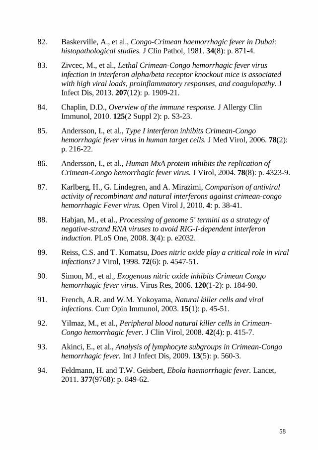
58
82. Baskerville, A., et al., Congo-Crimean haemorrhagic fever in Dubai:
histopathological studies. J Clin Pathol, 1981. 34(8): p. 871-4.
83. Zivcec, M., et al., Lethal Crimean-Congo hemorrhagic fever virus
infection in interferon alpha/beta receptor knockout mice is associated
with high viral loads, proinflammatory responses, and coagulopathy. J
Infect Dis, 2013. 207(12): p. 1909-21.
84. Chaplin, D.D., Overview of the immune response. J Allergy Clin
Immunol, 2010. 125(2 Suppl 2): p. S3-23.
85. Andersson, I., et al., Type I interferon inhibits Crimean-Congo
hemorrhagic fever virus in human target cells. J Med Virol, 2006. 78(2):
p. 216-22.
86. Andersson, I., et al., Human MxA protein inhibits the replication of
Crimean-Congo hemorrhagic fever virus. J Virol, 2004. 78(8): p. 4323-9.
87. Karlberg, H., G. Lindegren, and A. Mirazimi, Comparison of antiviral
activity of recombinant and natural interferons against crimean-congo
hemorrhagic Fever virus. Open Virol J, 2010. 4: p. 38-41.
88. Habjan, M., et al., Processing of genome 5' termini as a strategy of
negative-strand RNA viruses to avoid RIG-I-dependent interferon
induction. PLoS One, 2008. 3(4): p. e2032.
89. Reiss, C.S. and T. Komatsu, Does nitric oxide play a critical role in viral
infections? J Virol, 1998. 72(6): p. 4547-51.
90. Simon, M., et al., Exogenous nitric oxide inhibits Crimean Congo
hemorrhagic fever virus. Virus Res, 2006. 120(1-2): p. 184-90.
91. French, A.R. and W.M. Yokoyama, Natural killer cells and viral
infections. Curr Opin Immunol, 2003. 15(1): p. 45-51.
92. Yilmaz, M., et al., Peripheral blood natural killer cells in Crimean-
Congo hemorrhagic fever. J Clin Virol, 2008. 42(4): p. 415-7.
93. Akinci, E., et al., Analysis of lymphocyte subgroups in Crimean-Congo
hemorrhagic fever. Int J Infect Dis, 2009. 13(5): p. 560-3.
94. Feldmann, H. and T.W. Geisbert, Ebola haemorrhagic fever. Lancet,
2011. 377(9768): p. 849-62.
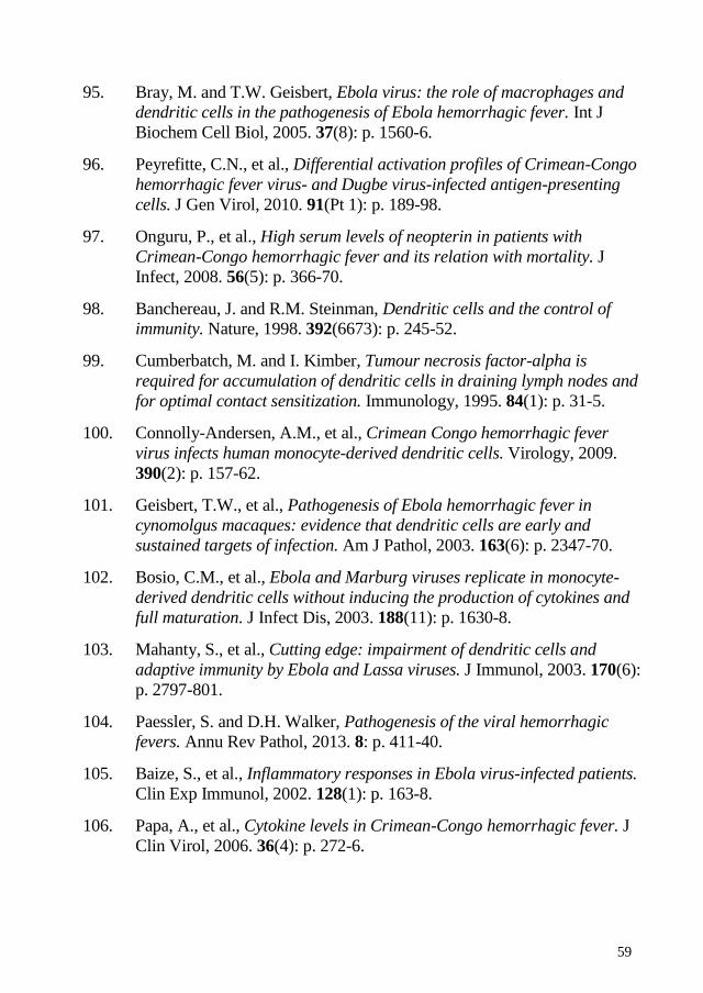
59
95. Bray, M. and T.W. Geisbert, Ebola virus: the role of macrophages and
dendritic cells in the pathogenesis of Ebola hemorrhagic fever. Int J
Biochem Cell Biol, 2005. 37(8): p. 1560-6.
96. Peyrefitte, C.N., et al., Differential activation profiles of Crimean-Congo
hemorrhagic fever virus- and Dugbe virus-infected antigen-presenting
cells. J Gen Virol, 2010. 91(Pt 1): p. 189-98.
97. Onguru, P., et al., High serum levels of neopterin in patients with
Crimean-Congo hemorrhagic fever and its relation with mortality. J
Infect, 2008. 56(5): p. 366-70.
98. Banchereau, J. and R.M. Steinman, Dendritic cells and the control of
immunity. Nature, 1998. 392(6673): p. 245-52.
99. Cumberbatch, M. and I. Kimber, Tumour necrosis factor-alpha is
required for accumulation of dendritic cells in draining lymph nodes and
for optimal contact sensitization. Immunology, 1995. 84(1): p. 31-5.
100. Connolly-Andersen, A.M., et al., Crimean Congo hemorrhagic fever
virus infects human monocyte-derived dendritic cells. Virology, 2009.
390(2): p. 157-62.
101. Geisbert, T.W., et al., Pathogenesis of Ebola hemorrhagic fever in
cynomolgus macaques: evidence that dendritic cells are early and
sustained targets of infection. Am J Pathol, 2003. 163(6): p. 2347-70.
102. Bosio, C.M., et al., Ebola and Marburg viruses replicate in monocyte-
derived dendritic cells without inducing the production of cytokines and
full maturation. J Infect Dis, 2003. 188(11): p. 1630-8.
103. Mahanty, S., et al., Cutting edge: impairment of dendritic cells and
adaptive immunity by Ebola and Lassa viruses. J Immunol, 2003. 170(6):
p. 2797-801.
104. Paessler, S. and D.H. Walker, Pathogenesis of the viral hemorrhagic
fevers. Annu Rev Pathol, 2013. 8: p. 411-40.
105. Baize, S., et al., Inflammatory responses in Ebola virus-infected patients.
Clin Exp Immunol, 2002. 128(1): p. 163-8.
106. Papa, A., et al., Cytokine levels in Crimean-Congo hemorrhagic fever. J
Clin Virol, 2006. 36(4): p. 272-6.
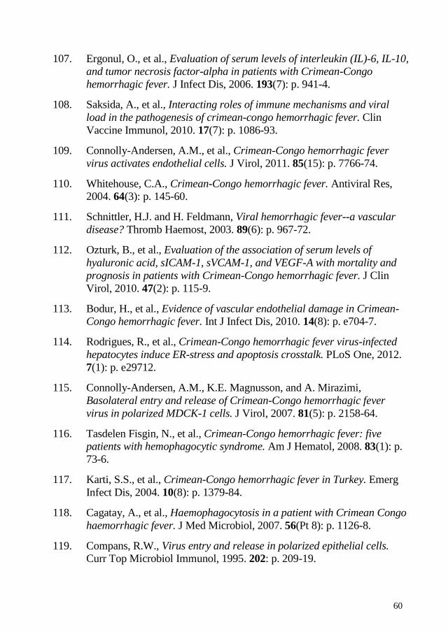
60
107. Ergonul, O., et al., Evaluation of serum levels of interleukin (IL)-6, IL-10,
and tumor necrosis factor-alpha in patients with Crimean-Congo
hemorrhagic fever. J Infect Dis, 2006. 193(7): p. 941-4.
108. Saksida, A., et al., Interacting roles of immune mechanisms and viral
load in the pathogenesis of crimean-congo hemorrhagic fever. Clin
Vaccine Immunol, 2010. 17(7): p. 1086-93.
109. Connolly-Andersen, A.M., et al., Crimean-Congo hemorrhagic fever
virus activates endothelial cells. J Virol, 2011. 85(15): p. 7766-74.
110. Whitehouse, C.A., Crimean-Congo hemorrhagic fever. Antiviral Res,
2004. 64(3): p. 145-60.
111. Schnittler, H.J. and H. Feldmann, Viral hemorrhagic fever--a vascular
disease? Thromb Haemost, 2003. 89(6): p. 967-72.
112. Ozturk, B., et al., Evaluation of the association of serum levels of
hyaluronic acid, sICAM-1, sVCAM-1, and VEGF-A with mortality and
prognosis in patients with Crimean-Congo hemorrhagic fever. J Clin
Virol, 2010. 47(2): p. 115-9.
113. Bodur, H., et al., Evidence of vascular endothelial damage in Crimean-
Congo hemorrhagic fever. Int J Infect Dis, 2010. 14(8): p. e704-7.
114. Rodrigues, R., et al., Crimean-Congo hemorrhagic fever virus-infected
hepatocytes induce ER-stress and apoptosis crosstalk. PLoS One, 2012.
7(1): p. e29712.
115. Connolly-Andersen, A.M., K.E. Magnusson, and A. Mirazimi,
Basolateral entry and release of Crimean-Congo hemorrhagic fever
virus in polarized MDCK-1 cells. J Virol, 2007. 81(5): p. 2158-64.
116. Tasdelen Fisgin, N., et al., Crimean-Congo hemorrhagic fever: five
patients with hemophagocytic syndrome. Am J Hematol, 2008. 83(1): p.
73-6.
117. Karti, S.S., et al., Crimean-Congo hemorrhagic fever in Turkey. Emerg
Infect Dis, 2004. 10(8): p. 1379-84.
118. Cagatay, A., et al., Haemophagocytosis in a patient with Crimean Congo
haemorrhagic fever. J Med Microbiol, 2007. 56(Pt 8): p. 1126-8.
119. Compans, R.W., Virus entry and release in polarized epithelial cells.
Curr Top Microbiol Immunol, 1995. 202: p. 209-19.
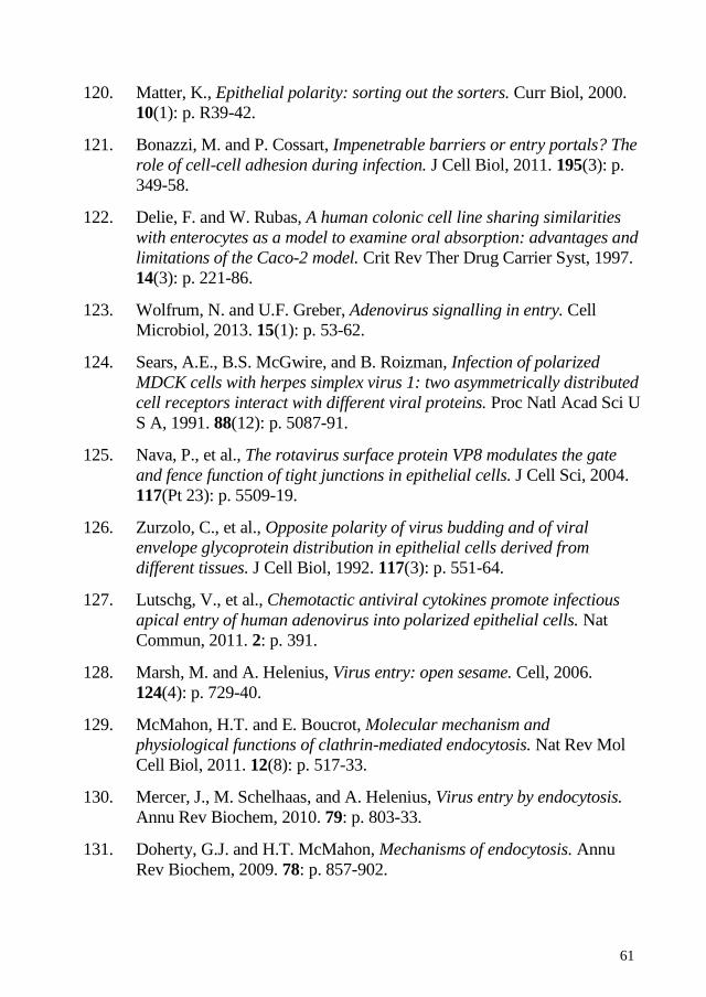
61
120. Matter, K., Epithelial polarity: sorting out the sorters. Curr Biol, 2000.
10(1): p. R39-42.
121. Bonazzi, M. and P. Cossart, Impenetrable barriers or entry portals? The
role of cell-cell adhesion during infection. J Cell Biol, 2011. 195(3): p.
349-58.
122. Delie, F. and W. Rubas, A human colonic cell line sharing similarities
with enterocytes as a model to examine oral absorption: advantages and
limitations of the Caco-2 model. Crit Rev Ther Drug Carrier Syst, 1997.
14(3): p. 221-86.
123. Wolfrum, N. and U.F. Greber, Adenovirus signalling in entry. Cell
Microbiol, 2013. 15(1): p. 53-62.
124. Sears, A.E., B.S. McGwire, and B. Roizman, Infection of polarized
MDCK cells with herpes simplex virus 1: two asymmetrically distributed
cell receptors interact with different viral proteins. Proc Natl Acad Sci U
S A, 1991. 88(12): p. 5087-91.
125. Nava, P., et al., The rotavirus surface protein VP8 modulates the gate
and fence function of tight junctions in epithelial cells. J Cell Sci, 2004.
117(Pt 23): p. 5509-19.
126. Zurzolo, C., et al., Opposite polarity of virus budding and of viral
envelope glycoprotein distribution in epithelial cells derived from
different tissues. J Cell Biol, 1992. 117(3): p. 551-64.
127. Lutschg, V., et al., Chemotactic antiviral cytokines promote infectious
apical entry of human adenovirus into polarized epithelial cells. Nat
Commun, 2011. 2: p. 391.
128. Marsh, M. and A. Helenius, Virus entry: open sesame. Cell, 2006.
124(4): p. 729-40.
129. McMahon, H.T. and E. Boucrot, Molecular mechanism and
physiological functions of clathrin-mediated endocytosis. Nat Rev Mol
Cell Biol, 2011. 12(8): p. 517-33.
130. Mercer, J., M. Schelhaas, and A. Helenius, Virus entry by endocytosis.
Annu Rev Biochem, 2010. 79: p. 803-33.
131. Doherty, G.J. and H.T. McMahon, Mechanisms of endocytosis. Annu
Rev Biochem, 2009. 78: p. 857-902.
62
132. Sorkin, A., Cargo recognition during clathrin-mediated endocytosis: a
team effort. Curr Opin Cell Biol, 2004. 16(4): p. 392-9.
133. Helenius, A., et al., On the entry of Semliki forest virus into BHK-21
cells. J Cell Biol, 1980. 84(2): p. 404-20.
134. Sandvig, K., et al., Clathrin-independent endocytosis: from nonexisting
to an extreme degree of complexity. Histochem Cell Biol, 2008. 129(3):
p. 267-76.
135. van Deurs, B., et al., Caveolae: anchored, multifunctional platforms in
the lipid ocean. Trends Cell Biol, 2003. 13(2): p. 92-100.
136. Lakadamyali, M., M.J. Rust, and X. Zhuang, Endocytosis of influenza
viruses. Microbes Infect, 2004. 6(10): p. 929-36.
137. Nonnenmacher, M. and T. Weber, Adeno-associated virus 2 infection
requires endocytosis through the CLIC/GEEC pathway. Cell Host
Microbe, 2011. 10(6): p. 563-76.
138. Yamauchi, Y. and A. Helenius, Virus entry at a glance. J Cell Sci, 2013.
126(Pt 6): p. 1289-95.
139. Mercer, J. and A. Helenius, Gulping rather than sipping:
macropinocytosis as a way of virus entry. Curr Opin Microbiol, 2012.
15(4): p. 490-9.
140. Amyere, M., et al., Origin, originality, functions, subversions and
molecular signalling of macropinocytosis. Int J Med Microbiol, 2002.
291(6-7): p. 487-94.
141. Aleksandrowicz, P., et al., Ebola virus enters host cells by
macropinocytosis and clathrin-mediated endocytosis. J Infect Dis, 2011.
204 Suppl 3: p. S957-67.
142. Ghigo, E., et al., Ameobal pathogen mimivirus infects macrophages
through phagocytosis. PLoS Pathog, 2008. 4(6): p. e1000087.
143. Clement, C., et al., A novel role for phagocytosis-like uptake in herpes
simplex virus entry. J Cell Biol, 2006. 174(7): p. 1009-21.
144. Jin, M., et al., Hantaan virus enters cells by clathrin-dependent receptor-
mediated endocytosis. Virology, 2002. 294(1): p. 60-9.
63
145. Santos, R.I., et al., Oropouche virus entry into HeLa cells involves
clathrin and requires endosomal acidification. Virus Res, 2008. 138(1-
2): p. 139-43.
146. Lozach, P.Y., et al., Entry of bunyaviruses into mammalian cells. Cell
Host Microbe, 2010. 7(6): p. 488-99.
147. Harmon, B., et al., Rift Valley fever virus strain MP-12 enters
mammalian host cells via caveola-mediated endocytosis. J Virol, 2012.
86(23): p. 12954-70.
148. Gavrilovskaya, I.N., et al., beta3 Integrins mediate the cellular entry of
hantaviruses that cause respiratory failure. Proc Natl Acad Sci U S A,
1998. 95(12): p. 7074-9.
149. Lozach, P.Y., et al., DC-SIGN as a receptor for phleboviruses. Cell Host
Microbe, 2011. 10(1): p. 75-88.
150. Gall, J.G. and M.L. Pardue, Formation and detection of RNA-DNA
hybrid molecules in cytological preparations. Proc Natl Acad Sci U S A,
1969. 63(2): p. 378-83.
151. Bauman, J.G., et al., A new method for fluorescence microscopical
localization of specific DNA sequences by in situ hybridization of
fluorochromelabelled RNA. Exp Cell Res, 1980. 128(2): p. 485-90.
152. Hopman, A.H., et al., Bi-color detection of two target DNAs by non-
radioactive in situ hybridization. Histochemistry, 1986. 85(1): p. 1-4.
153. Silahtaroglu, A.N., N. Tommerup, and H. Vissing, FISHing with locked
nucleic acids (LNA): evaluation of different LNA/DNA mixmers. Mol
Cell Probes, 2003. 17(4): p. 165-9.
154. Silahtaroglu, A., et al., LNA-modified oligonucleotides are highly
efficient as FISH probes. Cytogenet Genome Res, 2004. 107(1-2): p. 32-
7.
155. Singer, R.H. and D.C. Ward, Actin gene expression visualized in chicken
muscle tissue culture by using in situ hybridization with a biotinated
nucleotide analog. Proc Natl Acad Sci U S A, 1982. 79(23): p. 7331-5.
156. Dirks, R.W., et al., Simultaneous detection of different mRNA sequences
coding for neuropeptide hormones by double in situ hybridization using
FITC- and biotin-labeled oligonucleotides. J Histochem Cytochem,
1990. 38(4): p. 467-73.
64
157. Femino, A.M., et al., Visualization of single RNA transcripts in situ.
Science, 1998. 280(5363): p. 585-90.
158. Weibrecht, I., et al., In situ detection of individual mRNA molecules and
protein complexes or post-translational modifications using padlock
probes combined with the in situ proximity ligation assay. Nat Protoc,
2013. 8(2): p. 355-72.
159. Landegren, U., et al., A ligase-mediated gene detection technique.
Science, 1988. 241(4869): p. 1077-80.
160. Nilsson, M., et al., Padlock probes: circularizing oligonucleotides for
localized DNA detection. Science, 1994. 265(5181): p. 2085-8.
161. Luo, J., D.E. Bergstrom, and F. Barany, Improving the fidelity of
Thermus thermophilus DNA ligase. Nucleic Acids Res, 1996. 24(15): p.
3071-8.
162. Nilsson, M., et al., Padlock probes reveal single-nucleotide differences,
parent of origin and in situ distribution of centromeric sequences in
human chromosomes 13 and 21. Nat Genet, 1997. 16(3): p. 252-5.
163. Baner, J., et al., Parallel gene analysis with allele-specific padlock
probes and tag microarrays. Nucleic Acids Res, 2003. 31(17): p. e103.
164. Fire, A. and S.Q. Xu, Rolling replication of short DNA circles. Proc Natl
Acad Sci U S A, 1995. 92(10): p. 4641-5.
165. Lagunavicius, A., et al., Novel application of Phi29 DNA polymerase:
RNA detection and analysis in vitro and in situ by target RNA-primed
RCA. RNA, 2009. 15(5): p. 765-71.
166. Blanco, L. and M. Salas, Characterization of a 3'----5' exonuclease
activity in the phage phi 29-encoded DNA polymerase. Nucleic Acids
Res, 1985. 13(4): p. 1239-49.
167. Howell, W.M., et al., Glycosylases and AP-cleaving enzymes as a
general tool for probe-directed cleavage of ssDNA targets. Nucleic
Acids Res, 2010. 38(7): p. e99.
168. Lizardi, P.M., et al., Mutation detection and single-molecule counting
using isothermal rolling-circle amplification. Nat Genet, 1998. 19(3): p.
225-32.
169. Jarvius, J., et al., Digital quantification using amplified single-molecule
detection. Nat Methods, 2006. 3(9): p. 725-7.
65
170. Melin, J., et al., Homogeneous amplified single-molecule detection:
Characterization of key parameters. Anal Biochem, 2007. 368(2): p.
230-8.
171. Allalou, A. and C. Wahlby, BlobFinder, a tool for fluorescence
microscopy image cytometry. Comput Methods Programs Biomed, 2009.
94(1): p. 58-65.
172. Larsson, C., et al., In situ genotyping individual DNA molecules by
target-primed rolling-circle amplification of padlock probes. Nat
Methods, 2004. 1(3): p. 227-32.
173. Nilsson, M., et al., RNA-templated DNA ligation for transcript analysis.
Nucleic Acids Res, 2001. 29(2): p. 578-81.
174. Larsson, C., et al., In situ detection and genotyping of individual mRNA
molecules. Nat Methods, 2010. 7(5): p. 395-7.
175. Baner, J., et al., Microarray-based molecular detection of foot-and-
mouth disease, vesicular stomatitis and swine vesicular disease viruses,
using padlock probes. J Virol Methods, 2007. 143(2): p. 200-6.
176. Shrivastava-Ranjan, P., P.E. Rollin, and C.F. Spiropoulou, Andes virus
disrupts the endothelial cell barrier by induction of vascular endothelial
growth factor and downregulation of VE-cadherin. J Virol, 2010. 84(21):
p. 11227-34.
177. Gorbunova, E., I.N. Gavrilovskaya, and E.R. Mackow, Pathogenic
hantaviruses Andes virus and Hantaan virus induce adherens junction
disassembly by directing vascular endothelial cadherin internalization in
human endothelial cells. J Virol, 2010. 84(14): p. 7405-11.
178. Hynes, R.O., Integrins: versatility, modulation, and signaling in cell
adhesion. Cell, 1992. 69(1): p. 11-25.
179. Levsky, J.M. and R.H. Singer, Gene expression and the myth of the
average cell. Trends Cell Biol, 2003. 13(1): p. 4-6.
180. Bengtsson, M., et al., Gene expression profiling in single cells from the
pancreatic islets of Langerhans reveals lognormal distribution of mRNA
levels. Genome Res, 2005. 15(10): p. 1388-92.
181. Wang, B., et al., Rapid and sensitive detection of severe acute respiratory
syndrome coronavirus by rolling circle amplification. J Clin Microbiol,
2005. 43(5): p. 2339-44.
66
182. Papa, A., et al., Emergence of Crimean-Congo haemorrhagic fever in
Greece. Clin Microbiol Infect, 2010. 16(7): p. 843-7.
“It’s a trap” –Admiral Ackbar, Star Wars