REGULATION OF BK VIRUS DNA REPLICATION BY …
198
REGULATION OF BK VIRUS DNA REPLICATION BY TRANSCRIPTION FACTORS AND NONCODING RNAS A Dissertation Presented to The Faculty of the Graduate School At the University of Missouri In Partial Fulfillment Of the Requirements for the Degree Doctor of Philosophy By BO LIANG Dr. William R. Folk, Dissertation Supervisor MAY 2011
Transcript of REGULATION OF BK VIRUS DNA REPLICATION BY …

REGULATION OF BK VIRUS DNA REPLICATION BY
TRANSCRIPTION FACTORS AND NONCODING RNAS
A Dissertation
Presented to
The Faculty of the Graduate School
At the University of Missouri
In Partial Fulfillment
Of the Requirements for the Degree
Doctor of Philosophy
By
BO LIANG
Dr. William R. Folk, Dissertation Supervisor
MAY 2011

The undersigned, appointed by the dean of the Graduate School,
have examined the entitled
REGULATION OF BK VIRUS DNA REPLICATION BY TRANSCRIPTION FACTORS AND NONCODING RNAS
Presented by Bo Liang
A candidate for the degree of
Doctor of Philosophy
And hereby certify that, in their opinion, it is worthy of acceptance.
DR. WILLIAM R. FOLK
DR. MICHAEL J. IMPERIALE
DR. DAVID J. PINTEL
DR. MARK HANNINK
DR. DAVID SETZER


ii
ACKNOWLEDGEMENTS
In the fall of 2004, I came to United States with one goal to achieve – to become a Ph.D. After my
first few weeks’ study in University of Missouri-Columbia, I was a little bit worried, because I
realized that this goal was not an easy accomplish with my awkward English and limited research
experience.
But now, I did it!
I know how many difficulties I had encountered and how much effort I have made to realize this
goal. Fortunately, I did not do this alone.
Dr. William Folk, my Ph.D advisor, is always supportive to me. He influences me with his great
passion in science and encourages me with his positive altitude. I would have not been able to
achieve this without his patient guidance and higher expectation. I appreciate that he has been
training me to become an independent scientist and has provided me a lot of opportunities to
develop my skills in scientific writing, student mentoring and public speaking, which are all
required for a successful scientist.
My Ph.D committee members, Drs. Mark Hannink, David Pintel, Michael Imperiale and David
Setzer gave me invaluable advices and suggestions for my research.
Colleagues in our laboratory helped me a lot during my Ph.D training. In particular, Sarah
Scanlon assisted me with experiments and encouraged me whenever I made progress. I Dr.
Alexander Kenzior always gave me enlightening ideas and helped me solve technical problems in
experiments. Olga Kenzior supervised and taught me hand-by-hand at some basic experiments
while I was new in the lab. I also enjoyed working together with other labmates including Lu Lu,

iii
Jonathan Morrand, Khalid Alam, David Kirby, Sangho Bok, Xingrong Wu, Joseph Whittenberg
and Doris Shoemaker, for they have been creating a pleasant and friendly environment in our
laboratory.
Colleagues from neighboring laboratories also helped me with my experiment by generously
sharing equipments and reagents. Michelle Mooney helped me with hypoxia experiments; Drs.
Marc Johnson, Donald Burke, Shrikesh Sachdev and Toshihiko Ezashi provided me a lot of DNA
constructs for my experiments; Dr. Michael Riley taught me how to do plaque assays.
The collaboration with Dr. Heinz-Peter Nasheuer and Dr. Michael Imperiale has been very
successful, which generated several publications and enriched the content of my dissertation.
Without the help from Irina Tikhanovich and Dr. Nasheuer, the story of my dissertation would not
be complete.
In addition, I would also like to give special thanks to my lovely friends both in China and U.S,
including Wei Gui, Shishan Shi, Dr. Guoshi Li, Dr. Rod Becher and Dr. Kim Becher. During my
time of disappointment, depression and stress, they expressed their strong support and
encouragement, which recharged me and gave me the strength and confidence to face the
challenges.
This work is dedicated to my family, including my parents, wife and son, who are always proud of
me and confident in me. I am greatly indebted to my parents, who spent most of their effort to
educate me and promoted me to pursue higher accomplishment, which laid a solid foundation for
my Ph.D training. My wife is always very supportive to my work; particularly after we had our baby
Bo-yuan, she quitted her job and spent most of her effort to take care of Bo-yuan, so that I could
concentrate on my dissertation.

iv
Finally, I would like to thank everyone who I did not mention here but helped me in my life and
study.
This is not an end, but just a beginning for a new journey.

v
TABLE OF CONTENTS
ACKNOWLEDGEMENTS...................................................................................... ii
LIST OF FIGURES............................................................................................... vi
LIST OF ABBREVIATIONS.................................................................................. ix
ABSTRACT ........................................................................................................... x
Chapter
1. INTRODUCTION ......................................................................................... 1
2. STIMULATION OF BK VIRUS DNA REPLICATION BY NFI-FAMILY TRANSCRIPTION FACTORS............................................................... 42
3. ACETYLATION OF BKV LARGE T ANTIGEN AND MODULATION OF BKV DNA REPLICATION BY HISTONE ACETYLTRANSFERASES (HATS)………………………………………………………………………108
4. RESTRICTION OF BKV DNA REPLICATION IN MURINE CELLS AND INHIBITION OF BKV DNA REPLICATION BY NONCODING RNAs....................................................................................................145
5. PROPOSED MODEL AND FUTURE DIRECTIONS……………………...170
VITA................................................................................................................... 185

vi
LIST OF FIGURES
Figure Page
1.1 Organization of NCCRs of BKV, JCV and mPyV…………………………….7
1.2 Organization of BKV NCCR and transcription factor binding sites in core
origin flanking sequences………………………………………………………….....10
1.3 Structure and functional domains of Tags…………………………………..15
2.1 Stimulation of BKV DNA replication by enhancer…………………………..54
2.2 In vitro Tag pull-down assays…………………………………………………57
2.3 Expression of NFI isotypes in different cells………………………………...63
2.4 Co-Immunoprecipitation (Co-IP) assays to detect Tag interaction with NFI
transcription factors…………………………………………………………………...65
2.5 DNA replication of BKV templates with synthetic NFI sites……………….70
2.6 EMSA assays to detect NFI binding to BKV NCCR………………………. 73
2.7 Competitive DNA replication assays in the presence of a wildtype BKV
template (pBC-wt-BKV) as competitor………………………………………………79
2.8. DNA replication assay by titrating the transient expression of
BKTAg…………………………………………………………………………………..82

vii
2.9 Competitive replication assays with mutant competitors…………………..84
2.10 Monopolymerase assays with purified NFIC/CTF1………………………...87
2.11 Co-IP assays to detect interaction of NFI with primase……………………89
2.12 Proposed model for NFIs stimulating BKV DNA replication………………91
3.1 Acetylation of BKV Tag in vitro and in vivo…………………………………116
3.2 In vitro acetylation of truncated Tag…………………………………………120
3.3 Determining acetylation site of BKV Tag. …………………………………..124
3.4 DNA replication assays with Gal-4 fusion HATs…………………………...130
3.5 CHX chase assays to determine the protein stability of BKV Tag affected by
acetylation…………………………………………………………………………….135
4.1 In vivo DNA replication assays of chimearic templates……………………151
4.2 In vitro DNA replication assays………………………………………………155
4.3 Determination of nature of inhibitory activity………………………………..158
4.4 Effect of srRNAs on BKV DNA replication in vitro and in vivo……………161
5.1 BKV DNA replication in HK-2 cells under hypoxia/reoxygenation
conditions……………………………………………………………………………..173

viii
5.2 Expression of NFI, VEGF and E2F3 in HK-2 cells under normoxia (Green
bars) and hypoxia (Red bars) conditions…………………………………………176
ix
LIST OF ABBREVIATIONS
aa amino acid
bp base pair(s)
DMEM Dulbcco’s modified Eagle’s medium
DMSO dimethyl sulfoxide
DTT dithiothreitol
EDTA ethylenediaminetetraacetic acid
kb kilo base pairs
nt nucleotide
ori origin of DNA replication
core-ori core origin of DNA replication
Tag large T antigen
BKV BK virus
JCV JC virus
mPyV mouse polyomavirus
SV40 Simian virus 40
NFI Nuclear Factor I
HAT Histone acetyltransferase
PMSF phenylmethylsulfonyl fluoride
SDS sodium dodecyl sulfate

x
REGULATION OF BK VIRUS DNA REPLICATION BY
TRANSCRIPION FACTORS AND NONCODING RNAS
Bo Liang
Dr. William R. Folk, Dissertation Supervisor
ABSTRACT
Human polyomavirus BK (BKV) asymptomatically infects 80~90% of people during early
childhood, and establishes a life-long persistent infection without causing overt clinical symptoms.
High level BKV replication occurs predominantly in urogenital tracts of immune-suppressed
patients following renal transplantation and bone marrow transplantation, which cause
polyomavirus associated nephropathy (PVAN) and hemorrhagic cystitis, respectively. PVAN has
become a leading cause for renal transplantation failures since tacrolimus and mycophenolate
mofetil began to be widely used in transplantation patients in 1995. Epidemiological studies
indicate that around 30% of kidney transplantation patients are at risk of developing PVAN and
50% of PVAN patients are at risk of transplantation failure.
This dissertation explores the mechanisms by which the genomic noncoding control region
(NCCR) regulates BKV DNA replication in cell culture and might be related to the establishment
of BKV persistent infection and pathogenesis of PVAN.

xi
These studies indicate that cellular transcription factor NFI interacts with the BKV NCCR
and stimulates BKV DNA replication in vivo and in vitro. Also, the data reveal that isotypes NFIA
and NFIB strongly interact with BKV large T antigen (Tag) and NFIC interacts with DNA
polymerase-α primase (pol-α primase), suggesting NFI-family transcription factors may stimulate
BKV DNA replication through recruitment of Tag and pol-α primase. In contrast, ectopic
expression of PCAF/GCN5 histone acetyltransferases inhibit BKV DNA replication. Tag has a site
for acetylation by PCAF/GCN5, but inhibition of BKV DNA replication by PCAF/GCN5 is not due
to acetylation of Tag, suggesting PCAF/GCN5 target other component(s) of DNA replication
machinery. Possible targets include nucleosomes associated with the NCCR and other
components of the replication machinery. A search for these targets is proposed, and possible
functions of acetylation on BKV Tag are discussed.
BKV DNA does not replicate in murine cells. We and our collaborators have found that this
host-restriction of BKV DNA replication involves not only incompatibility of BKV Tag with mouse
pol-α primase, but also inhibitory small noncoding RNAs in murine cells, termed srRNAs, that act
through BKV NCCR. Specific srRNAs were sequenced and cloned. In vitro transcribed srRNAs
inhibit BKV replication in vitro; and ectopic expression of a specific srRNA strongly inhibits BKV
DNA replication in vivo in human cells. Surprisingly, srRNAs from human cancer cells stimulate
BKV DNA replication in vitro, suggesting cell type specific expression of srRNAs has distinct role
in regulation of BKV DNA replication. We propose that differential expression of srRNAs may
have implication in the viral tropism, establishment of persistent infection and reactivation.

1
CHAPETER 1
Introduction
BKV Pathogenesis and Polyomavirus associated nephropathy (PVAN)
Human polyomavirus BK (BKV) was firstly isolated in 1971 from the urine of a kidney
transplantation patient with ureteral stenosis (84). Epidemiological studies indicate that
BKV asymptomatically infects 80~90% of people during early childhood (37, 105, 123,
124, 193). BKV might be transmitted transplacentally(20, 21)or through the fecal-oral
route and respiratory tract (18, 19, 89, 110, 164, 211, 229). After initial infection, BKV
establishes a life-long persistent infection in kidneys without causing overt clinical
symptoms(37, 68, 100). Reactivation of BKV occurs predominantly in urogenital tracts of
immune-suppressed patients following renal transplantation and bone marrow
transplantation, which cause polyomavirus associated nephropathy (PVAN) and
hemorrhagic cystitis, respectively(102, 105).
PVAN has become a leading cause for renal transplantation failures since 1995, when
tacrolimus and mycophenolate mofetil (MMF) began to be widely used in transplantation
patients (102, 103, 177). Epidemiological studies indicated that around 30% of kidney
transplantation patients are at risk of developing PVAN (103, 160) and 50% of PVAN
patients are at risk of transplantation failure (42). The other risk factors for PVAN are
mismatch of HLA and use of corticosteroids in anti-rejection therapy (12, 103). PVAN is
the consequence of extensive BKV replication in kidney allografts, which causes necrotic
injury of kidneys tubules (159). The progression of PVAN can be divided into three stages
(97, 105, 161). Initially, reactivation and massive replication of BKV in kidney tubular
epithelial cells give rise to “inclusions”, the enlarged nuclei filled with viral particles, which

2
shed from the tubular basement membrane and can be detected as “decoy cells”.In urine
samples, these are cytologically useful as surrogate markers for diagnosis of early stage
PVAN (Stage A). Extensive shedding and death of tubular epithelial cells cause tubular
necrosis and release of pro-inflammatory cytokines and chemokines, which promote the
infiltration of leukocytes into the allograft tissue and inflammation, causing
“tubulointerstitial nephritis”(Stage B); the final stage in the progression of PVAN is kidney
tubular fibrosis and atrophy (Stage C), which is highly associated with allograft loss (64).
The current means to control PVAN is to restore immune function by reducing the
immune-suppressive regimen, but this increases the risk of allograft rejection(178). Other
anti-viral treatments have been attempted, such as cidofovir(8, 76, 125, 220),
leflunomide(72, 76, 113, 227), quinolones(6, 81, 172, 181), and immunoglobulin (192),
which are not specific and have high toxicity, adding complications to patients.
Polyomavirus replication in acute and persistent infections
BKV has a non-enveloped icosahedral capsid of 40~45nm and a closed circular double-
stranded DNA genome(136). It is closely related to two other primate polyomaviruses:
SV40 (Simian Virus 40), and JC virus (JCV), the etiological agent for progressive
multifocal leukoencephalopathy (PML). BKV DNAs or viral particles have been detected
in various organs and tissues(182), including kidney(37, 100), tonsil(89), saliva
gland(110), brain(203, 218), lung(218), heart(20), liver(122), lymphocytes(31, 52, 60, 63),
but kidney is the primary site for its persistent infection (36, 37, 100). Because the initial
acute infection of BKV is usually subclinical, little is known about how it replicates during
the initial phase of infection nor how it spreads throughout the body and reaches the
kidney to establish a persistent infection. BKV persists at low levels, perhaps as an
episome (37, 100), in kidneys at only 0.007+/-0.003 copy/cell, which can increase to
3.4+/-1.8 copies/cell after kidney transplantation and to 7738.9+/-1580.4 copies/cell in
allograft kidneys of PVAN patients(179). BKV “viruria” (shedding of virus in urine) occurs

3
in 0.5~20% immune-competent individuals(102) with titers of ~103 copies/ml, and this can
increase to >107~8 copies/ml in late stage PVAN patients, when “viremia” develops (69,
171). This suggests that during persistent infection BKV replicates at an extremely low
level in vivo in immune-competent individuals and the rate of BKV replication increases
dramatically in allograft kidneys.
The mechanism(s) that maintain BKV low-level replication in kidney during persistent
infection are likely to include inhibition of viral replication by immune factors (42) and/or
other inhibitory cellular factor(s). Studies have shown that interferon-gamma inhibits the
replication of BKV in vitro (3); antibodies against BKV capsid protein VP1 have been
detected in the serum of people with latent BKV infection and PVAN patients (98, 124,
162); and cell-mediated immunity is crucial for the control of BKV (17, 70, 237). However,
the immune system does not completely eradicate BKV in the human host, indicating
BKV can evade immune surveillance. A recent study showed that a miRNA expressed by
BKV and JCV targets the ULBP3, a stress-induced ligand expressed on surface of
infected cells, to down-regulate both the native and adaptive immune response against
viral infection(13). Also, high-throughput assays of cellular gene expression in response
to BKV infection in vitro indicated that several genes responsible for immune control are
regulated upon BKV infection (2, 90). But their consequences for viral replication has not
been determined. Replication of BKV might be restricted to a low-level, to escape
immune surveillance, perhaps through control by cis-elements in the viral noncoding
control region (NCCR) or through modulation of large T antigen (Tag) expression or
activity by posttranslational modifications. This is partially supported by the observations
that replication of archetype BKV is very inefficient (27, 69, 88, 189). More details of how
the NCCR and modification of T-Ag might restrict BKV replication will be discussed
below.

4
Also important is why and how BKV replication is reactivated in PVAN. Evidence
suggests that immune-suppression is necessary but not sufficient for PVAN to occur.
PVAN is only highly associated with kidney transplantation patients and rarely occurs in
autologous kidneys of patients with other immunocompromsed conditions (78, 157). BKV
reactivates in some patients with bone marrow transplantation, but this causes
hemorrhagic cystitis, not PVAN (14, 102, 105). Only sporadic cases of HIV+ individuals
have been reported to have symptoms of PVAN(158, 200) and PVAN rarely occurs in
recipients of other organ transplants (liver, heart, pancreas, etc.) even though they
receive similar and in some cases, even stronger immune-suppression regimens (105).
And PVAN is extremely rare in patients with other immune-compromised conditions, such
as systemic lupus erythematosus (SLE)(210) and leukemia(203). These suggest that in
addition to immune suppression, other factors related to certain conditions of allograft
kidney are particularly important for reactivation of BKV and the progression of PVAN.
This might be related to allograft ischemia/reperfusion injury and regeneration,
inflammatory response, which in turn, lead to activation of DNA damage/repair and/or
mitogenic pathways utilized by polyomaviruses (10, 78, 102, 104).
JCV, another human polyomavirus with 80% of homology to BKV genome sequence,
also persists in kidneys of most people and reactivates predominantly in AIDS patients,
however, and this viral reactivation causes Progressive Multifocal Leukoencephalopathy
(PML). Although JC virus replication has been detected in some renal transplantation
patients, the incidence of JCV infection in kidney transplantation is very low and is
generally not associated with disease progression of PVAN (145). This suggests that
BKV has unique properties related to certain treatment conditions used in renal
transplantation that account for its reactivation specifically in kidney allografts and the
pathogenesis of PVAN.

5
Due to the lack of animal model for PVAN, little is known about the how BKV replication
is regulated during acute and persistent infection in vivo in human population. However,
replication of mouse polyomavirus (mPyV) during acute and persistent infection has been
studied extensively in mice. As with other polyomaviruses, mPyV infection has an acute
phase, a clearance phase and a persistent phase (66). The acute phase is usually within
6 days postinfection (dpi) in the respiratory tracts, when viral replication reaches
maximum levels, and the infection spreads systematically from lungs to different organs
such as kidneys, livers and spleens; the viral load reaches up to >1000 copies/cell in
lungs and ~10 copies/cell in kidneys. During the clearance phase, 6~12 dpi, virus titers
drop significantly throughout all the organs, to undetectable levels except for kidneys and
lungs, where virus can be detected at 1 copy/cell in kidneys and 10 copies/cell in lungs.
Finally, in the persistent phase, low level infection is established from 22 dpi to 84 dpi
where viral DNA is present at ~1 copies/cell in lungs and <1 copies/cell in kidneys (65,
66). During the long-term persistent infection, viral DNA exists as a free supercoiled
molecule in lungs and kidneys, without detectable integration into the host genome(66).
Reactivation of mPyV replication can be induced in kidneys, when kidneys are injured by
chemicals (glycerol, cisplatin) or ischemia insult (clamping of kidney arteries), but not by
immune-suppression (methotrexate)(10, 186).
The mPyV noncoding control region (NCCR) controls viral DNA replication during both
acute and persistent infection(184-186). Mutation of specific cis-elements, particularly
transcription factor binding sites, in the enhancer region changes organ-specific
replication of mPyV during acute infection in vivo (184, 185). The enhancer also regulates
persistent infection (186): mPyV with a mutated enhancer that strongly stimulated viral
replication in acute infection failed to establish a long-term persistent infection(186),
suggesting the NCCR regulates viral replication in different ways during acute and
persistent infection. This is reminiscent of what has been observed with BKV with

6
rearranged enhancers, that replicate efficiently in cell culture but are distinct from the
archetype form that persists naturally in kidneys (27, 88, 163).
These observations suggest that the NCCR of BKV has an important role in the
reactivation of replication and the ensuing pathogenesis of PVAN. The major goal of this
dissertation research is to study the mechanism(s) by which the BKV NCCR modulates
viral DNA replication, and to relate these mechanisms to BKV persistent infection and
reactivation in PVAN .
The viral non-coding control region (NCCR) and its regulatory role in polyomavirus
transcription and DNA replication
The NCCR contains the origin of DNA replication and promoter/enhancer elements for
regulation of viral early/late gene expression and DNA replication. (38, 149, 225). NCCRs
of BKV, JCV, SV40 and mPyV have similar organization and can be divided into three
regions: the core origin, early core origin flanking sequence (EF) and late core origin
flanking sequence (enhancer)(Figure 1.1).
The core origin is the minimal sequence required for polymavirus DNA replication in vitro.
Sequences of SV40(50, 51, 54, 130), BKV(58), JCV(47, 80), mPyV(45, 116, 154, 176)
core origins have been well defined. Alignment of these core origin sequence sequences
(Figure 1.1) indicates that they consist of three essential elements: a large T-antigen
(Tag) binding site II (a palindrome of four “G(A/G)GGC” pentanucleotide repeats), an AT-
rich region and an early imperfect palindrome (EP) region. Detailed biochemical analysis
of the SV40 origin suggested that DNA replication initiates upon Tag binding and
formation of double hexamers at the “Tag binding site II” in the presence of ATP (24, 141,
166, 219), followed by melting of duplex DNA in the “EP region” and bending/untwisting

7
Figure 1.1 Organization of NCCRs of BKV, JCV and mPyV. BKV, JCV and mPyV
NCCRs are divided into three regions: core origin (core ori), early flanking sequence (EF)
and Enhancer. Alignment of core ori sequences from BKV, JCV, SV40 and mPyV is
shown. Positions of Early palindrome region (EP region), Tag Binding site II and AT-rich
region of SV40 core ori are marked above the aligned sequences. The configurations of
archetype BKV and JCV NCCRs are illustrated; numbers in brackets are numbers of
nucleotides in each block.

8

9
of DNA in the” AT-rich region” (25, 51, 101, 165). Tag double hexamers unwind the DNA
duplex in opposite directions with the Tag helicase/ATPase activity in the presence of
ATP/Mg2+ (49, 199, 228). Other components of the DNA replication initiation complex,
including RPA, Topoisomerase I and DNA polyomerase -α primase (pol-α primase) are
recruited to the core origin to initiate viral DNA replication.
While the core origin is essential for BKV DNA replication, the EF and enhancer control
viral gene transcription and regulate DNA replication (149, 225). Many transcription
factors binding sites have been identified in the BKV core origin flanking sequences
(Figure 1.2). In the EF region, binding sites for NFkB(86), C/EBP(86), GM-CSF(149, 180)
have been identified, and the NFkB site was shown to stimulate BKV early gene
expression synergistically with C/EBP(180). Although the function of the GM-CSF site is
not clear, it has been reported to be highly mutated (~90%) in PVAN samples suggesting
its importance for BKV reactivation in PVAN(180). In the BKV enhancer region, binding
sites for NFI(29, 30, 59, 91, 137), Sp1(29, 59, 138), GRE/PRE
(glucocoticoid/progesterone responsive element) and ERE (estrogen responsive
element) (150), NF-AT(112), p53(197), AP1(30, 138, 139), Smad3(1) were identified and
shown to regulate early/late gene expression. Many more putative binding sites of
transcription factors, including Ets-family transcription factors (PEA-3, Ets-1 and Spi-
1/PU.1), CREB, AP-2, NFkB were predicted in BKV enhancer region, however, their roles
in BKV transcription are unclear (149).
Transcription factors not only regulate gene transcription, but also modulate DNA
replication (reviewed in 55, 56, 57, 156). Mechanisms by which transcription factors
stimulate polyomavirus DNA include: 1) recruitment of replication factors to the origin to
facilitate assembly of the replication complex: AP-1 and VP16 stimulated mPyV
replication by recruitment of Tag and RPA (99, 109); 2) remodeling of chromatin
configuration into an active form: NFI stimulates SV40 DNA replication by preventing the

10
Figure 1.2 Organization of BKV NCCR and transcription factor binding sites in core origin
flanking sequences. Tag binding and affinity sites are marked with red arrows. Transcription
factor binding sites are marked with black underlines. Six NFI sites are numbered from NFI-1 to -
6.

11

12
formation of repressive chromatin(35) and relievies repressive chromatin through
interaction with histone H3(153); 3) induction of DNA structure changes in the origin to
promote initiation: Sp1 stabilizes the bent structure of SV40 origin DNA caused by Tag
during the initiation(128, 208, 226); 4) facilitating the intracellular localization to promote
DNA replication: Runx1 recruits mPyV origin to replication factories on the nuclear matrix
by In addition to stimulation(33, 155). Transcription factors may also inhibit viral DNA
replication, for instance: Oct-1 inhibits SV40 DNA replication when bound to AT-rich
region in core origin(120); p53 inhibits SV40 DNA replication in vitro and in vivo at the
initiation stage, perhaps by interfering with interaction of Tag with RPA or origin, or
inhibiting helicase activity of Tag (67, 147, 205, 221).
The requirement for core origin flanking sequences for viral DNA replication is different
for different polyomaviruses. The core origin flanking sequences of mPyV is required for
DNA replication in vitro and in vivo (48, 152, 176); while those of SV40 are not absolutely
required in vitro, they stimulate SV40 replication by 10~100 fold in vivo and in vitro (95,
96). One study showed that BKV core origin flanking sequences do not enhance BKV
DNA replication in CV-1 and HeLa cells transiently expressing BKV Tag (58), but the
BKV enhancer stimulates SV40 core origin replication by SV40 Tag in COS-1 cells (58).
The importance of the core origin flanking sequences for BKV DNA replication in human
kidney tubular epithelial cells is not clear. Another study indicated that in addition to core
origin, a 21bp fragment of P block flanking sequence is required for BKV replication in
COS-1 cells (53). These suggest that the requirement of core origin flanking sequences
for polyomavirus DNA replication might be cell-type dependent and the specific
composition of trans-acting elements in different types of cells determines how core origin
flanking sequences regulate polyomavirus DNA replication.
The NCCR sequences of BKV and JCV isolated from clinical samples and selected for
replication in cell culture display great heterogeneity(151). It is now believed that all

13
variants are originated from an archetype NCCR (at-NCCR) through rearrangement of
enhancer sequence by deletion, duplication and insertion during reactivation or passage
of virus in cell culture. Compared with enhancer, core origin and EF region are relatively
stable.
The archetype BKV and JCV viruses have genomes that persist latently in kidney and are
transmitted among the population. For both BKV and JCV, the archetype enhancer has a
linear array of several blocks of sequences arbitrarily designated as P68-Q23-R63-S63 for
BKV(149), and A36-B23-C55-D66-E18-F69 for JCV(11, 79, 232). The most widely studied
archetype strain of BKV is the WW strain (ww-NCCR) directly cloned from urine without
passage in cell culture(32, 146, 188). Other archetype strains with a few single nucleotide
changes have been reported, such as WWT(209) and Dik(213). Phylogenetic analysis of
these archetype sequences indicates that these single-nucleotide changes are due to
random mutations accumulated during the natural evolution of the virus and are not
related to viral pathogenesis (233).
It has been suggested that BKVs with rearranged NCCRs (rr-NCCRs) are generated
through recombination and gain growth advantage through positive selection during in
vivo replication or upon passage of virus in cell culture, so they replicate more efficiently
than virus with at-NCCR. In support of this, quasispecies of BKV with rr-NCCRs were
detected in kidney transplant patients and most of them replicate faster than the
archetype NCCR (at-NCCR) in cell culture (88, 163). Isolates of several BKV rr-NCCRs
from HIV+ individuals also displayed similar enhanced replication activity (27). And rr-
NCCRs with the same configuration were repeatedly detected in a single patient during
the course of PVAN progression (167), suggesting they kept replicating stably at high-
level in PVAN patients. But how the rearrangement of BKV NCCR is related to the
pathogenesis of PVAN is still not clear. The extensive replication of archetype BKV upon
reactivation gives rise to BKVs with rr-NCCRs, some of which may replicate in cells that

14
are usually non-permissive for archetype virus.In support of this, it was shown that rr-
NCCRs were more prevalent in PVAN patient than patients with only viruria(194); and a
recent longitudinal study of BKV genotype in kidney transplantation patients shows a
discordance of appearance of rr-NCCR in plasma and ww-NCCR in urine at the same
time, suggesting the BKVs with rr-NCCR replicate preferentially in different compartments
with ww-NCCR(88). Similarly, JCV archetype also persists in kidney, but the NCCRs of
viruses isolated from brain and CSF of PML patients are rearranged (87). And rearranged
JCV replicates efficiently in cultured glial cells and kidney cells, which are not permissive
for archetype JCV(47, 87). It is hypothesized that the at-NCCR with lower replication
efficiency is particularly adapted for persistent infection because its low level of
replication protects the latent virus from immune surveillance (88).
Large T-antigen (Tag)
Replication of polyomaviruses requires the coordination of Tag with series of cellular
proteins involved in DNA replication, transcription, cell cycle control, cell transformation
and DNA damage response. SV40 Tag’s structural and functional domains have been
well characterized (reviewed in references 4, 5, 23, 34, 73-75). Tags of BKV, JCV and
SV40 have 75% of homology; and mouse polyomavirus (mPyV) shares 45~55%
homology with SV40 Tag (169). So by alignment of their protein sequences, we may infer
the structural and functional domains of BKV and JCV Tags from what’s known about
SV40 Tag (Figure 1.3A). In spite of great similarities, Tags of these closely related
polyomaviruses have subtle but important differences in structure and function, which are
reviewed below.
SV40 Tag is a 90~100kD polypeptide of 708 amino acids and consists of four major
independent structural domains determined from their proteolysis pattern and distinct

15
Figure 1.3 Structure and functional domains of Tags. (A) Alignment of amino acid
sequences of SV40, BKV and JCV Tags. “LxCxE” motifs and NLSs (nuclear localization
signals) are marked with red line box. Phosphorylation sites of SV40 Tag are marked with
blue arrows. Acetylation site of SV40 Tag is marked with red arrow.

16
17
functions (reviewed in references 73, 74, 169) (Figure 1.3B). The N-terminal J domain (1-
82aa) is a homolog of cellular DnaJ chaperon that interacts with Hsc70 to promote
cellular transformation by disrupting Rb-E2F complex (204, 206, 207, 235); in addition, J
domain also facilitates DNA replication in vivo, perhaps by promoting the DNA binding
and hexamer assembly (28, 222). The origin-binding domain (Ori-binding) (131-259aa) is
responsible for binding to the origin of replication and contains the contact interface
between two Tag hexamers(9, 107, 143). The Helicase/ATPase domain (251-627aa) has
intrinsic helicase and ATPase activity, with which Tag double-hexamers unwind duplex
DNA by hydrolysis of ATP(83, 129). The Zn Finger motif (302-320aa) within the
Helicase/ATPase domain is required for Tag hexamer assembly (133) and sequence-
specific origin binding(9, 107). The structures of J domain, Ori-binding domain and
helicase/ATPase domain have been resolved (121, 129, 134). But the structure and
function of the C-terminal host range domain (HR) (628-708aa) is not clear. Replication of
mutant SV40 with deletions of the HR domain is defective in CV-1 cells (170, 216, 217)
and temperature sensitive in BSC cells (40, 170), which can be rescued by providing the
HR domain in trans (173, 217). It also has a similar helper function (hf) to allow
adenovirus to grow in monkey cells when provided in trans (39, 92, 117). HR domain is
not absolutely required for DNA replication, but might function in regulating viral late gene
expression or capsid assembly(201, 202, 238). The linker region between J domain and
DNA binding domain is a relatively unstructured region, which contains the docking sites
for many cellular proteins, including the pRb-related proteins (the “LXCXE” motif)(71,
117), Cul7(7), Bub1(43), Fbw7(224), IRS1(77) as well as a NLS (nuclear localization
signal)(114, 115, 126, 127).
Tags of BKV (695aa) and JCV (688aa) are similar in size compared with SV40 Tag and
have very similar organization of their functional domains. But the amino acid sequence
of mPyV Tag (785aa) is longer than the three Tags of other primate polyomaviruses (85).
The J domain, Ori-binding domain, Helicase/ATPase domain of Tags from SV40, BKV

18
Figure 1.3 (B) Organization of functional domains of SV40 Tag. SV40 Tag (708aa) has
four major functional domains: J domain (aa1-82); origin binding domain (Ori-Binding)
(aa131-259); Helicase/ATPase domain (aa251-627); host range (HR) domain (aa682-
708). Zn-finger and ATPase domain are located in aa302-320 and aa418-627 within
Helicase/ATPase domain. Nuclear localization signal (NLS) is located at aa126-132.
There is a linker sequence between Helicase/ATPase and HR domain, which is less
conserved in SV40, BKV and JCV Tags. Domains within SV40 Tag shown to interact with
cellular proteins including HSC70, pRb family proteins, p53, Cul7, Bub1, Fbw7, IRS1, pol-
α primase, RPA, Topoisomerase I, Nbs1, ATM/ATR, are illustrated below. (C)
Organization of functional domains of mPyV Tag. mPyV Tag (785aa) has three major
function domains: J domain (aa1-79); Ori-Binding (aa282-398); Helicase/ATPase domain
(aa398-785). Two NLSs are located at aa189-195 and aa280-286. Zn-finger and ATPase
domains within Helicase/ATPase domain are located at aa452-472 and aa565-785.

19

20
and JCV are highly conserved, while their C-terminal end regions with a variable linker
and the HR domain are quite different, particularly between SV40 and two human
polyomaviruses, BKV and JCV (Figure 1.3A). Tag of mPyV also has a J domain(15), an
Ori-binding domain(212) and a Helicase/ATPase domain(26), but lacks the C-terminal
region of SV40, BKV and JCV Tags (Figure 1.3C). Remarkably, the unstructured linker
region of mPyV Tag between J domain and Ori-binding domain is ~150aa longer than the
similar linker region in SV40 Tag (Figure 1.3B); and it has two NLSs, the first one (NLS1)
is unique to mPyV, and the other one (NLS2) is conserved with the SV40 NLS (108, 183).
Why mPyV has two NLSs is not clear. The “LxCxE” motif is also present in mPyV Tag’s
linker region(168), but whether other proteins docking to the linker region of SV40 Tag
also bind to mPyV remains unknown.
Tag carries out its functions through interaction with many cellular proteins (Figure 1.3B),
including: 1) proteins of DNA replication machinery, including pol-α primase (41, 61, 62),
RPA(144, 223), Topoisomerase I (82, 118, 187, 198) to facilitate initiation of DNA
replication; 2) proteins responsible for cell cycle control, including pRb, p53 (119, 131,
132, 190), Cul7(7), Bub1(43), Fbw7(224), IRS1(77) to promote S-phase entry and
transformation; 3) components of DNA damage response, including ATM/ATR(44, 196,
236), Nbs1(230) to bypass and co-opt this machinery; 4)transcription factors including
p53 (147, 205, 221), c-Jun(16, 94, 109), Sp1(111), AP2(148) and components of
RNApol-II(RNA polymerase II) transcriptional initiation complex, including TBP(TATA
box-binding protein)(46, 93, 111, 140),TAFs(TBP asoosciated factors)(46) and RNA pol-
II(111), to regulate viral/cellular gene transcription and DNA replication; 5) histone
acetyltransferases (HATs), including CBP/p300(22, 174) and PCAF/GCN5(231), to
acetylate Tag for unknown function. These proteins may also interact with BKV Tag for
similar functions. In support of this, Rb and p53 have been shown to interact with BKV
Tag. Whether BKV Tag interacts with other proteins has not been shown.

21
Another way for Tag to regulate its activity is through post-translational modifications.
Several kinds of post-transplantation modifications of SV40 Tag have been reported (74),
including phosphorylation, amino-terminal and specific lysine acetylations, O-
glycosylation, acylation, adenylylation, poly(ADP-ribosly)-ation. Phosphorylation of SV40
Tag has been studied extensively(reivewed in references 73, 74, 175).
SV40 Tag is phosphorylated at serines and threonines clustered in two regions, one in
the N-terminus linker region between J domain and Ori-binding domain (Ser106, Ser111,
Ser112, Ser120, Ser123, Thr124), and the other in the C-terminus region (Ser639, Ser
665, Ser667, Ser677, Ser679, Thr701)(Figure1.3A). Mutations of Ser120, Ser 123,
Thr124 and Ser679 have significant but distinct effects on viral DNA replication.
Phosphorylation of Ser 120 and Ser 123 inhibits viral DNA replication in vitro, but
mutation of Ser120 and Ser123 to Ala greatly reduce viral replication in vivo(191),
suggesting the inhibition of viral DNA replication by phosphorylation on these sites might
be essential for viral infection cycle in vivo. It was found recently that Ser120 is
phosphorylated by ATM kinase upon SV40 infection, suggesting this or other
phosphorylation sites might be important for the virus to co-opt the DNA damage
response triggered by viral infection (195). In contrast, phosphorylation of Thr124 by cdc3
kinase greatly enhanced Tag’s replication activity in vitro(106, 142); and mutations of
Thr124 to Ala completely abolished SV40 Tag’s origin binding and DNA replication
activity (191). Importantly, the Ser120, Ser123 and Thr124 sites are conserved in BKV
and JCV Tags, suggesting they are essential for viral replication. Phosphorylation of
Ser679 down-regulated DNA replication in vitro and SV40 with mutation at Ser679
replicated better than wildtype virus in vivo(191). However, Ser679 is not conserved in
BKV and JCV Tag (Figure 1.3A), suggesting that BKV and JCV Tag might have a higher
replication activity than SV40. It has recently been shown that phosphorylated Thr701
interacts with tumor suppressor Fwb7 to prevent degradation of cyclinE, perhaps, to

22
promote cell cycle progression and cell transformation(224). The Thr701 is conserved in
BKV and JCV Tag (Figure 1.3A). The functions of other phosphorylation sites remain
unknown.
SV40 Tag can be acetylated by histone acetyltransferases (HATs) CBP/p300 at the K697
residue in the C-terminus Host Range domain through formation of a ternary complex
mediated by p53(22, 174). However, mutation of K697 does not affect the host range
activity of Tag’s C-terminal region (173), suggesting the acetylation has another
modulatory function. Another group of HATs, PCAF/GCN5, but not CBP/p300, acetylates
mouse polyomavirus Tag and stimulates mouse polyomavirus DNA replication, when
tethered to the origin (231), suggesting acetylation of Tag might stimulate polyomavirus
DNA replication. Interestingly, the acetylation site and its surrounding residues of SV40
Tag are highly conserved among SV40, BKV and JCV (Figure 1.3A), suggesting the
acetylation might play an important role in viral replication and/or pathogenesis. Whether
CBP/p300 or PCAF/GCN5 acetylates BKV Tag has not been reported.
Objectives
The primary goal of this thesis research is to determine how cellular factors binding to
BKV NCCR regulate BKV DNA replication and to relate this to the BKV reactivation in
PVAN. This research started from a collaborative effort with Dr. Heinz-Peter Nasheuer
and Dr. Michael Imperiale’s laboratories to study the mechanism(s) responsible for
restriction of BKV DNA replication in murine cells. Several discoveries have been made,
three of which have already been published (135, 214, 215), and others will be submitted
for publication after completion of this dissertation.
Previous studies in the Folk lab have established that transcription factors stimulate
mPyV DNA replication through interaction with viral NCCR(94, 234). We explored how

23
similar mechanism regulates BKV DNA replication; and most importantly, how it
contributes to BKV reactivation in PVAN.
Chapter 2 describes the stimulation of BKV DNA replication by NCCR and identification
of the NFI-family transcription factors that stimulate BKV DNA replication through binding
to NCCR, when Tag and pol-α primase are limiting. Interactions of NFIs with Tag and pol-
α primase have been detected, suggesting NFIs stimulate BKV DNA replication through
recruitment of Tag and pol-α primase to replication origin.
It has been shown that HATs PCAF/GCN5 stimulate mPyV DNA replication, perhaphs
through modification of Tag(231). Whether HATs modulate BKV DNA replication has
been investigated in this dissertation research. As shown in Chapter 3, PCAF/GCN5
HATs acetylate BKV Tag at a K687 residue in the C-terminal HR domain; but
PCAF/GCN5 modulate BKV and mPyV DNA replication in distinct manners. Point
mutations of BKV and mPyV Tags indicate that acetylation of Tags appears to be not
directly involved in DNA replication of either mPyV or BKV (231 and Olga Kenzior
unpublished data). The possible functions of polyomavirus Tags acetylation are
discussed.
The reason why BKV fails to productively infect murine cells remains unclear. Through
collaboration with Drs. Heinz-Peter Nasheuer and Michael Imperiale, combining the use
of in vitro and in vivo DNA replication systems and ex vivo infection model, the restriction
of BKV DNA replication in murine system has been shown to involve incompatibity of
BKV Tag with murine pol-α primase and interaction of inhibitory cellular noncoding RNAs
with BKV NCCR (135, 214, 215), summarized in Chapter 4. This also indicates that in
addition to transcription factors/co-factors, cellular noncoding RNAs also regulate BKV
DNA replication through NCCR.

24
To relate all the above observations to BKV reactivation in PVAN, we developed the
hypothesis that ischemia/reperfusion injury reactivates BKV DNA replication in kidney
allografts(78, 102). Chapter 5 demonstrates our observation that hypoxia can stimulate
BKV DNA replication in cultured human kidney tubular epithelial cells. Accordingly, a
model for BKV reactivation in PVAN is proposed to tie up all observations related with
BKV NCCR: ischemia/reperfusion injury of kidney allografts changes the
activity/expression of transcription factors (NFIs, HATs, etc.) and/or small noncoding
RNAs in kidney tubular epithelial cells, which stimulates BKV replication and promotes
BKV reactivation in PVAN. Other possibilities and the limitation of current studies are also
discussed; and future directions are proposed.

25
Reference
1. Abend, J. R., and M. J. Imperiale. 2008. Transforming growth factor-beta-mediated regulation of BK virus gene expression. Virology 378:6-12.
2. Abend, J. R., J. A. Low, and M. J. Imperiale. 2010. Global effects of BKV infection on
gene expression in human primary kidney epithelial cells. Virology 397:73-79. 3. Abend, J. R., J. A. Low, and M. J. Imperiale. 2007. Inhibitory Effect of Gamma
Interferon on BK Virus Gene Expression and Replication. J. Virol. 81:272-279. 4. Ahuja, D., M. T. Sáenz-Robles, and J. M. Pipas. 2005. SV40 large T antigen targets
multiple cellular pathways to elicit cellular transformation. Oncogene 24:7729-7745. 5. Ali, S. 2001. Cellular transformation by SV40 large T antigen: interaction with host
proteins. Semin. Cancer Biol. 11:15-23. 6. Ali, S. H., A. Chandraker, and J. A. DeCaprio. 2007. Inhibition of Simian virus 40 large
T antigen helicase activity by fluoroquinolones. Antivir Ther 12:1-6. 7. Ali, S. H., J. S. Kasper, T. Arai, and J. A. DeCaprio. 2004. Cul7/p185/p193 binding to
simian virus 40 large T antigen has a role in cellular transformation. J. Virol. 78:2749-2757.
8. Araya, C. E., J. F. Lew, R. S. Iii Fennell, R. E. Neiberger, and V. R. Dharnidharka.
2006. Intermediate-dose cidofovir without probenecid in the treatment of BK virus allograft nephropathy. Pediatr. Transplant. 10:32-37.
9. Arthur, A. K., A. Hoss, and E. Fanning. 1988. Expression of simian virus 40 T antigen
in Escherichia coli: localization of T-antigen origin DNA-binding domain to within 129 amino acids. J. Virol. 62:1999-2006.
10. Atencio, I. A., F. F. Shadan, X. J. Zhou, N. D. Vaziri, and L. P. Villarreal. 1993. Adult
mouse kidneys become permissive to acute polyomavirus infection and reactivate persistent infections in response to cellular damage and regeneration. J. Virol. 67:1424-1432.
11. Ault, G. S., and G. L. Stoner. 1993. Human polyomavirus JC promoter/enhancer
rearrangement patterns from progressive multifocal leukoencephalopathy brain are unique derivatives of a single archetypal structure. J. Gen. Virol. 74:1499-1507.
12. Awadalla, Y., P. Randhawa, K. Ruppert, A. Zeevi, and R. J. Duquesnoy. 2004. HLA
Mismatching Increases the Risk of BK Virus Nephropathy in Renal Transplant Recipients. American Journal of Transplantation 4:1691-1696.
13. Bauman, Y., D. Nachmani, A. Vitenshtein, P. Tsukerman, N. Drayman, N. Stern-
Ginossar, D. Lankry, R. Gruda, and O. Mandelboim. 2011. An Identical miRNA of the Human JC and BK Polyoma Viruses Targets the Stress-Induced Ligand ULBP3 to Escape Immune Elimination. Cell host & microbe 9:93-102.
14. Bedi, A., C. B. Miller, J. L. Hanson, S. Goodman, R. F. Ambinder, P. Charache, R. R.
Arthur, and R. J. Jones. 1995. Association of BK virus with failure of prophylaxis against hemorrhagic cystitis following bone marrow transplantation. Journal of clinical oncology : official journal of the American Society of Clinical Oncology 13:1103-1109.

26
15. Berjanskii, M. V., M. I. Riley, A. Xie, V. Semenchenko, W. R. Folk, and S. R. Van
Doren. 2000. NMR structure of the N-terminal J domain of murine polyomavirus T antigens. Implications for DnaJ-like domains and for mutations of T antigens. The Journal of biological chemistry 275:36094-36103.
16. Bharucha, V. A., K. W. Peden, and G. I. Tennekoon. 1994. SV40 large T antigen with
c-Jun down-regulates myelin P0 gene expression: a mechanism for papovaviral T antigen-mediated demyelination. Neuron 12:627-637.
17. Binggeli, S., A. Egli, S. Schaub, I. Binet, M. Mayr, J. Steiger, and H. H. Hirsch. 2007.
Polyomavirus BK-Specific Cellular Immune Response to VP1 and Large T-Antigen in Kidney Transplant Recipients. American Journal of Transplantation 7:1131-1139.
18. Bofill-Mas, S., M. Formiga-Cruz, P. Clemente-Casares, F. Calafell, and R. Girones.
2001. Potential transmission of human polyomaviruses through the gastrointestinal tract after exposure to virions or viral DNA. J. Virol. 75:10290-10299.
19. Bofill-Mas, S., J. Rodriguez-Manzano, B. Calgua, A. Carratala, and R. Girones. 2010.
Newly described human polyomaviruses Merkel cell, KI and WU are present in urban sewage and may represent potential environmental contaminants. Virology journal 7:141.
20. Boldorini, R., S. Allegrini, U. Miglio, I. Nestasio, A. Paganotti, C. Veggiani, G.
Monga, and V. Pietropaolo. 2010. BK virus sequences in specimens from aborted fetuses. J. Med. Virol. 82:2127-2132.
21. Boldorini, R., C. Veggiani, E. Amoruso, S. Allegrini, U. Miglio, A. Paganotti, R.
Ribaldone, and G. Monga. 2008. Latent human polyomavirus infection in pregnancy: investigation of possible transplacental transmission. Pathology (Phila). 40:72-77.
22. Borger, D. R., and J. A. DeCaprio. 2006. Targeting of p300/CREB Binding Protein
Coactivators by Simian Virus 40 Is Mediated through p53. J. Virol. 80:4292-4303. 23. Borowiec, J. A., F. B. Dean, P. A. Bullock, and J. Hurwitz. 1990. Binding and
unwinding--How T antigen engages the SV40 origin of DNA replication. Cell 60:181-184. 24. Borowiec, J. A., and J. Hurwitz. 1988. ATP stimulates the binding of simian virus 40
(SV40) large tumor antigen to the SV40 origin of replication. Proc. Natl. Acad. Sci. U. S. A. 85:64-68.
25. Borowiec, J. A., and J. Hurwitz. 1988. Localized melting and structural changes in the
SV40 origin of replication induced by T-antigen. The EMBO journal 7:3149-3158. 26. Bradley, M. K., T. F. Smith, R. H. Lathrop, D. M. Livingston, and T. A. Webster. 1987.
Consensus topography in the ATP binding site of the simian virus 40 and polyomavirus large tumor antigens. Proc. Natl. Acad. Sci. U. S. A. 84:4026-4030.
27. Broekema, N. M., J. R. Abend, S. M. Bennett, J. S. Butel, J. A. Vanchiere, and M. J.
Imperiale. 2010. A system for the analysis of BKV non-coding control regions: application to clinical isolates from an HIV/AIDS patient. Virology 407:368-373.
28. Campbell, K. S., K. P. Mullane, I. A. Aksoy, H. Stubdal, J. Zalvide, J. M. Pipas, P. A.
Silver, T. M. Roberts, B. S. Schaffhausen, and J. A. DeCaprio. 1997. DnaJ/hsp40 chaperone domain of SV40 large T antigen promotes efficient viral DNA replication. Genes Dev. 11:1098-1110.

27
29. Cassill, J. A., K. L. Deyerle, and S. Subramani. 1989. Unidirectional deletion and linker scan analysis of the late promoter of the human papovavirus BK. Virology 169:172-181.
30. Chakraborty, T., and G. C. Das. 1989. Identification of HeLa cell nuclear factors that
bind to and activate the early promoter of human polyomavirus BK in vitro. Mol. Cell. Biol. 9:3821-3828.
31. Chatterjee, M., T. B. Weyandt, and R. J. Frisque. 2000. Identification of archetype and
rearranged forms of BK virus in leukocytes from healthy individuals. J. Med. Virol. 60:353-362.
32. Chauhan, S., G. Lecatsas, and E. H. Harley. 1984. Genome analysis of BK (WW) viral
DNA cloned directly from human urine. Intervirology 22:170-176. 33. Chen, L. F., K. Ito, Y. Murakami, and Y. Ito. 1998. The capacity of polyomavirus
enhancer binding protein 2alphaB (AML1/Cbfa2) to stimulate polyomavirus DNA replication is related to its affinity for the nuclear matrix. Mol. Cell. Biol. 18:4165-4176.
34. Cheng, J., J. A. DeCaprio, M. M. Fluck, and B. S. Schaffhausen. 2009. Cellular
transformation by Simian Virus 40 and Murine Polyoma Virus T antigens. Semin. Cancer Biol. 19:218-228.
35. Cheng, L., and T. J. Kelly. 1989. Transcriptional activator nuclear factor I stimulates the
replication of SV40 minichromosomes in vivo and in vitro. Cell 59:541-551. 36. Chesters, P. M., J. Heritage, and D. J. McCance. 1983. Persistence of DNA sequences
of BK virus and JC virus in normal human tissues and in diseased tissues. The Journal of infectious diseases 147:676-684.
37. Chesters, P. M., J. Heritage, and D. J. McCance. 1983. Persistence of DNA sequences
of BK virus and JC virus in normal human tissues and in diseased tissues. J. Infect. Dis. 147:676-684.
38. Cole, C. N., and S. D. Conzen. 2003. Polyomaviridae: the viruses and their replication. .
In Knipe DM, Howley PM, Griffin DE, Lamb RL, Martin MA, and S. SE (ed.), Fields’ Virology. , 4th. ed. Lippincott Williams and Wilkins, New York.
39. Cole, C. N., L. V. Crawford, and P. Berg. 1979. Simian virus 40 mutants with deletions
at the 3' end of the early region are defective in adenovirus helper function. J. Virol. 30:683-691.
40. Cole, C. N., and T. P. Stacy. 1987. Biological properties of simian virus 40 host range
mutants lacking the COOH-terminus of large T antigen. Virology 161:170-180. 41. Collins, K. L., A. A. Russo, B. Y. Tseng, and T. J. Kelly. 1993. The role of the 70 kDa
subunit of human DNA polymerase alpha in DNA replication. The EMBO journal 12:4555-4566.
42. Comoli, P., S. Binggeli, F. Ginevri, and H. H. Hirsch. 2006. Polyomavirus-associated nephropathy: update on BK virus-specific immunity. Transplant Infectious Disease 8:86-94.
43. Cotsiki, M., R. L. Lock, Y. Cheng, G. L. Williams, J. Zhao, D. Perera, R. Freire, A.
Entwistle, E. A. Golemis, T. M. Roberts, P. S. Jat, and O. V. Gjoerup. 2004. Simian virus 40 large T antigen targets the spindle assembly checkpoint protein Bub1. Proc. Natl. Acad. Sci. U. S. A. 101:947-952.

28
44. Dahl, J., J. You, and T. L. Benjamin. 2005. Induction and Utilization of an ATM Signaling Pathway by Polyomavirus. J. Virol. 79:13007-13017.
45. Dailey, L., and C. Basilico. 1985. Sequences in the polyomavirus DNA regulatory region
involved in viral DNA replication and early gene expression. J. Virol. 54:739-749. 46. Damania, B., and J. C. Alwine. 1996. TAF-like function of SV40 large T antigen. Genes
Dev. 10:1369-1381. 47. Daniel, A. M., J. J. Swenson, R. P. Reddy Mayreddy, K. Khalili, and R. J. Frisque.
1996. Sequences within the Early and Late Promoters of Archetype JC Virus Restrict Viral DNA Replication and Infectivity. Virology 216:90-101.
48. de Villiers, J., W. Schaffner, C. Tyndall, S. Lupton, and R. Kamen. 1984. Polyoma
virus DNA replication requires an enhancer. Nature 312:242-246. 49. Dean, F. B., P. Bullock, Y. Murakami, C. R. Wobbe, L. Weissbach, and J. Hurwitz.
1987. Simian virus 40 (SV40) DNA replication: SV40 large T antigen unwinds DNA containing the SV40 origin of replication. Proc. Natl. Acad. Sci. U. S. A. 84:16-20.
50. Deb, S., A. L. DeLucia, C. P. Baur, A. Koff, and P. Tegtmeyer. 1986. Domain structure
of the simian virus 40 core origin of replication. Mol. Cell. Biol. 6:1663-1670. 51. Deb, S., A. L. DeLucia, A. Koff, S. Tsui, and P. Tegtmeyer. 1986. The adenine-thymine
domain of the simian virus 40 core origin directs DNA bending and coordinately regulates DNA replication. Mol. Cell. Biol. 6:4578-4584.
52. Degener, A. M., V. Pietropaolo, C. D. Taranto, L. Jin, F. Ameglio, P. Cordiali-Fei, E.
Trento, L. Sinibaldi, and N. Orsi. 1999. Identification of a new control region in the genome of the DDP strain of BK virus isolated from PBMC. J. Med. Virol. 58:413-419.
53. Del Vecchio, A. M., R. A. Steinman, and R. P. Ricciardi. 1989. An element of the BK
virus enhancer required for DNA replication. J. Virol. 63:1514-1524. 54. DeLucia, A. L., S. Deb, K. Partin, and P. Tegtmeyer. 1986. Functional interactions of
the simian virus 40 core origin of replication with flanking regulatory sequences. J. Virol. 57:138-144.
55. DePamphilis, M. L. 1993. Eukaryotic DNA Replication: Anatomy of An Origin. Annu.
Rev. Biochem. 62:29-63. 56. DePamphilis, M. L. 1993. How transcription factors regulate origins of DNA replication in
eukaryotic cells. Trends Cell Biol. 3:161-167. 57. DePamphilis, M. L. 1988. Transcriptional elements as components of eukaryotic origins
of DNA replication. Cell 52:635-638. 58. Deyerle, K. L., F. G. Sajjadi, and S. Subramani. 1989. Analysis of origin of DNA
replication of human papovavirus BK. J. Virol. 63:356-365. 59. Deyerle, K. L., and S. Subramani. 1988. Linker scan analysis of the early regulatory
region of human papovavirus BK. J. Virol. 62:3378-3387. 60. Dolei, A., V. Pietropaolo, E. Gomes, C. Di Taranto, M. Ziccheddu, M. A. Spanu, C.
Lavorino, M. Manca, and A. M. Degener. 2000. Polyomavirus persistence in

29
lymphocytes: prevalence in lymphocytes from blood donors and healthy personnel of a blood transfusion centre. J. Gen. Virol. 81:1967-1973.
61. Dornreiter, I., W. C. Copeland, and T. S. Wang. 1993. Initiation of simian virus 40 DNA
replication requires the interaction of a specific domain of human DNA polymerase alpha with large T antigen. Mol. Cell. Biol. 13:809-820.
62. Dornreiter, I., A. Hoss, A. K. Arthur, and E. Fanning. 1990. SV40 T antigen binds
directly to the large subunit of purified DNA polymerase alpha. The EMBO journal 9:3329-3336.
63. Dörries, K., E. Vogel, S. Günther, and S. Czub. 1994. Infection of Human
Polyomaviruses JC and BK in Peripheral Blood Leukocytes from Immunocompetent Individuals. Virology 198:59-70.
64. Drachenberg, C. B., J. C. Papadimitriou, H. H. Hirsch, R. Wali, C. Crowder, J.
Nogueira, C. B. Cangro, S. Mendley, A. Mian, and E. Ramos. 2004. Histological Patterns of Polyomavirus Nephropathy: Correlation with Graft Outcome and Viral Load. American Journal of Transplantation 4:2082-2092.
65. Dubensky, T. W., F. A. Murphy, and L. P. Villarreal. 1984. Detection of DNA and RNA
virus genomes in organ systems of whole mice: patterns of mouse organ infection by polyomavirus. J. Virol. 50:779-783.
66. Dubensky, T. W., and L. P. Villarreal. 1984. The primary site of replication alters the
eventual site of persistent infection by polyomavirus in mice. J. Virol. 50:541-546. 67. Dutta, A., J. M. Ruppert, J. C. Aster, and E. Winchester. 1993. Inhibition of DNA
replication factor RPA by p53. Nature 365:79-82. 68. Egli, A., L. Infanti, A. Dumoulin, A. Buser, J. Samaridis, C. Stebler, R. Gosert, and H.
H. Hirsch. 2009. Prevalence of polyomavirus BK and JC infection and replication in 400 healthy blood donors. J. Infect. Dis. 199:837-846.
69. Egli, A., L. Infanti, A. Dumoulin, A. Buser, J. Samaridis, C. Stebler, R. Gosert, and H.
H. Hirsch. 2009. Prevalence of Polyomavirus BK and JC Infection and Replication in 400 Healthy Blood Donors. Journal of Infectious Diseases 199:837-846.
70. Egli, A., S. Köhli, M. Dickenmann, and H. H. Hirsch. 2009. Inhibition of Polyomavirus
BK-Specific T-Cell Responses by Immunosuppressive Drugs. Transplantation 88:1161-1168 1110.1097/TP.1160b1013e3181bca1422.
71. Ewen, M. E., J. W. Ludlow, E. Marsilio, J. A. DeCaprio, R. C. Millikan, S. H. Cheng, E.
Paucha, and D. M. Livingston. 1989. An N-terminal transformation-governing sequence of SV40 large T antigen contributes to the binding of both p110Rb and a second cellular protein, p120. Cell 58:257-267.
72. Faguer, S., H. H. Hirsch, N. Kamar, C. Guilbeau-Frugier, D. Ribes, J. Guitard, L.
Esposito, O. Cointault, A. Modesto, M. Lavit, C. Mengelle, and L. Rostaing. 2007. Leflunomide treatment for polyomavirus BK-associated nephropathy after kidney transplantation. Transplant international : official journal of the European Society for Organ Transplantation 20:962-969.
73. Fanning, E. 1992. Simian virus 40 large T antigen: the puzzle, the pieces, and the
emerging picture. J. Virol. 66:1289-1293.
30
74. Fanning, E., and R. Knippers. 1992. Structure and Function of Simian Virus 40 Large Tumor Antigen. Annu. Rev. Biochem. 61:55-85.
75. Fanning, E., and K. Zhao. 2009. SV40 DNA replication: From the A gene to a
nanomachine. Virology 384:352-359. 76. Farasati, N. A., R. Shapiro, A. Vats, and P. Randhawa. 2005. Effect of leflunomide and
cidofovir on replication of BK virus in an in vitro culture system. Transplantation 79:116-118.
77. Fei, Z. L., C. D'Ambrosio, S. Li, E. Surmacz, and R. Baserga. 1995. Association of
insulin receptor substrate 1 with simian virus 40 large T antigen. Mol. Cell. Biol. 15:4232-4239.
78. Fishman, J. A. 2002. BK Virus Nephropathy - Polyomavirus Adding Insult to Injury. N.
Engl. J. Med. 347:527-530. 79. Flaegstad, T., A. Sundsfjord, R. R. Arthur, M. Pedersen, T. Traavik, and S.
Subramani. 1991. Amplification and sequencing of the control regions of BK and JC virus from human urine by polymerase chain reaction. Virology 180:553-560.
80. Frisque, R. J., G. L. Bream, and M. T. Cannella. 1984. Human polyomavirus JC virus
genome. J. Virol. 51:458-469. 81. Gabardi, S., S. S. Waikar, S. Martin, K. Roberts, J. Chen, L. Borgi, H. Sheashaa, C.
Dyer, S. K. Malek, S. G. Tullius, N. Vadivel, M. Grafals, R. Abdi, N. Najafian, E. Milford, and A. Chandraker. 2010. Evaluation of Fluoroquinolones for the Prevention of BK Viremia after Renal Transplantation. Clinical Journal of the American Society of Nephrology 5:1298-1304.
82. Gai, D., R. Roy, C. Wu, and D. T. Simmons. 2000. Topoisomerase I Associates
Specifically with Simian Virus 40 Large-T-Antigen Double Hexamer-Origin Complexes. J. Virol. 74:5224-5232.
83. Gai, D., R. Zhao, D. Li, C. V. Finkielstein, and X. S. Chen. 2004. Mechanisms of
Conformational Change for a Replicative Hexameric Helicase of SV40 Large Tumor Antigen. Cell 119:47-60.
84. Gardner, S. D., A. M. Field, D. V. Coleman, and B. Hulme. 1971. New human
papovavirus (B.K.) isolated from urine after renal transplantation. Lancet 1:1253-1257. 85. Gjørup, O. V., P. E. Rose, P. S. Holman, B. J. Bockus, and B. S. Schaffhausen. 1994.
Protein domains connect cell cycle stimulation directly to initiation of DNA replication. Proceedings of the National Academy of Sciences 91:12125-12129.
86. Gorrill, T. S., and K. Khalili. 2005. Cooperative interaction of p65 and C/EBP[beta]
modulates transcription of BKV early promoter. Virology 335:1-9. 87. Gosert, R., P. Kardas, E. O. Major, and H. H. Hirsch. 2010. Rearranged JC Virus
Noncoding Control Regions Found in Progressive Multifocal Leukoencephalopathy Patient Samples Increase Virus Early Gene Expression and Replication Rate. J. Virol. 84:10448-10456.
88. Gosert, R., C. H. Rinaldo, G. A. Funk, A. Egli, E. Ramos, C. B. Drachenberg, and H.
H. Hirsch. 2008. Polyomavirus BK with rearranged noncoding control region emerge in

31
vivo in renal transplant patients and increase viral replication and cytopathology. The Journal of experimental medicine 205:841-852.
89. Goudsmit, J., P. Wertheim-van Dillen, A. van Strien, and J. van der Noordaa. 1982.
The role of BK virus in acute respiratory tract disease and the presence of BKV DNA in tonsils. J. Med. Virol. 10:91-99.
90. Grinde, B., M. Gayorfar, and C. H. Rinaldo. 2007. Impact of a polyomavirus (BKV)
infection on mRNA expression in human endothelial cells. Virus Res. 123:86-94. 91. Grinnell, B. W., D. T. Berg, and J. D. Walls. 1988. Negative regulation of the human
polyomavirus BK enhancer involves cell-specific interaction with a nuclear repressor. Mol. Cell. Biol. 8:3448-3457.
92. Grodzicker, T., C. Anderson, P. A. Sharp, and J. Sambrook. 1974. Conditional lethal
mutants of adenovirus 2-simian virus 40 hybrids. I. Host range mutants of Ad2+ND1. J. Virol. 13:1237-1244.
93. Gruda, M. C., J. M. Zabolotny, J. H. Xiao, I. Davidson, and J. C. Alwine. 1993.
Transcriptional activation by simian virus 40 large T antigen: interactions with multiple components of the transcription complex. Mol. Cell. Biol. 13:961-969.
94. Guo, W., W. Tang, X. Bu, V. Bermudez, M. Martin, and W. Folk. 1996. AP1 enhances
polyomavirus DNA replication by promoting T-antigen- mediated unwinding of DNA. J. Virol. 70:4914-4918.
95. Guo, Z. S., C. Gutierrez, U. Heine, J. M. Sogo, and M. L. Depamphilis. 1989. Origin
auxiliary sequences can facilitate initiation of simian virus 40 DNA replication in vitro as they do in vivo. Mol. Cell. Biol. 9:3593-3602.
96. Gutierrez, C., Z. S. Guo, J. Roberts, and M. L. DePamphilis. 1990. Simian virus 40
origin auxiliary sequences weakly facilitate T-antigen binding but strongly facilitate DNA unwinding. Mol. Cell. Biol. 10:1719-1728.
97. Hariharan, S. 2006. BK virus nephritis after renal transplantation. Kidney Int. 69:655-662. 98. Hariharan, S., E. P. Cohen, B. Vasudev, R. Orentas, R. P. Viscidi, J. Kakela, and B.
DuChateau. 2005. BK Virus-Specific Antibodies and BKV DNA in Renal Transplant Recipients with BKV Nephritis. American Journal of Transplantation 5:2719-2724.
99. He, Z., B. T. Brinton, J. Greenblatt, J. A. Hassell, and C. J. Ingles. 1993. The
transactivator proteins VP16 and GAL4 bind replication factor A. Cell 73:1223-1232. 100. Heritage, J., P. M. Chesters, and D. J. McCance. 1981. The persistence of papovavirus
BK DNA sequences in normal human renal tissue. J. Med. Virol. 8:143-150. 101. Hertz, G. Z., M. R. Young, and J. E. Mertz. 1987. The A+T-rich sequence of the simian
virus 40 origin is essential for replication and is involved in bending of the viral DNA. J. Virol. 61:2322-2325.
102. Hirsch, H. H. 2002. Polyomavirus BK nephropathy: a (re-)emerging complication in renal
transplantation. Am J Transplant 2:25-30. 103. Hirsch, H. H., W. Knowles, M. Dickenmann, J. Passweg, T. Klimkait, M. J. Mihatsch,
and J. r. Steiger. 2002. Prospective Study of Polyomavirus Type BK Replication and Nephropathy in Renal-Transplant Recipients. N. Engl. J. Med. 347:488-496.

32
104. Hirsch, H. H., and D. R. Snydman. 2005. BK Virus: Opportunity Makes a Pathogen.
Clin. Infect. Dis. 41:354-360. 105. Hirsch, H. H., and J. r. Steiger. 2003. Polyomavirus BK. The Lancet Infectious Diseases
3:611-623. 106. Hoss, A., I. Moarefi, K. H. Scheidtmann, L. J. Cisek, J. L. Corden, I. Dornreiter, A. K.
Arthur, and E. Fanning. 1990. Altered phosphorylation pattern of simian virus 40 T antigen expressed in insect cells by using a baculovirus vector. J. Virol. 64:4799-4807.
107. Hoss, A., I. F. Moarefi, E. Fanning, and A. K. Arthur. 1990. The finger domain of
simian virus 40 large T antigen controls DNA-binding specificity. J. Virol. 64:6291-6296. 108. Howes, S., B. Bockus, and B. Schaffhausen. 1996. Genetic analysis of polyomavirus
large T nuclear localization: nuclear localization is required for productive association with pRb family members. J. Virol. 70:3581-3588.
109. Ito, K., M. Asano, P. Hughes, H. Kohzaki, C. Masutani, F. Hanaoka, T. Kerppola, T.
Curran, Y. Murakami, and Y. Ito. 1996. c-Jun stimulates origin-dependent DNA unwinding by polyomavirus large Tantigen. The EMBO journal 15:5636-5646.
110. Jeffers, L. K., V. Madden, and J. Webster-Cyriaque. 2009. BK virus has tropism for
human salivary gland cells in vitro: Implications for transmission. Virology 394:183-193. 111. Johnston, S. D., X. M. Yu, and J. E. Mertz. 1996. The major transcriptional
transactivation domain of simian virus 40 large T antigen associates nonconcurrently with multiple components of the transcriptional preinitiation complex. J. Virol. 70:1191-1202.
112. Jordan, J. A., K. Manley, A. S. Dugan, B. A. O'Hara, and W. J. Atwood. 2010.
Transcriptional Regulation of BK Virus by Nuclear Factor of Activated T Cells. J. Virol. 84:1722-1730.
113. Josephson, M. A., D. Gillen, B. Javaid, P. Kadambi, S. Meehan, P. Foster, R.
Harland, R. J. Thistlethwaite, M. Garfinkel, W. Atwood, J. Jordan, M. Sadhu, M. J. Millis, and J. Williams. 2006. Treatment of renal allograft polyoma BK virus infection with leflunomide. Transplantation 81:704-710.
114. Kalderon, D., W. D. Richardson, A. F. Markham, and A. E. Smith. 1984. Sequence
requirements for nuclear location of simian virus 40 large-T antigen. Nature 311:33-38. 115. Kalderon, D., B. L. Roberts, W. D. Richardson, and A. E. Smith. 1984. A short amino
acid sequence able to specify nuclear location. Cell 39:499-509. 116. Katinka, M., and M. Yaniv. 1983. DNA replication origin of polyoma virus: early proximal
boundary. J. Virol. 47:244-248. 117. Kelly, T. J., Jr., and A. M. Lewis, Jr. 1973. Use of nondefective adenovirus-simian virus
40 hybrids for mapping the simian virus 40 genome. J. Virol. 12:643-652. 118. Khopde, S., and D. T. Simmons. 2008. Simian Virus 40 DNA Replication Is Dependent
on an Interaction between Topoisomerase I and the C-Terminal End of T Antigen. J. Virol. 82:1136-1145.

33
119. Kierstead, T. D., and M. J. Tevethia. 1993. Association of p53 binding and immortalization of primary C57BL/6 mouse embryo fibroblasts by using simian virus 40 T-antigen mutants bearing internal overlapping deletion mutations. J. Virol. 67:1817-1829.
120. Kilwinski, J., M. Baack, S. Heiland, and R. Knippers. 1995. Transcription factor Oct1
binds to the AT-rich segment of the simian virus 40 replication origin. J. Virol. 69:575-578.
121. Kim, H. Y., B. Y. Ahn, and Y. Cho. 2001. Structural basis for the inactivation of
retinoblastoma tumor suppressor by SV40 large T antigen. The EMBO journal 20:295-304.
122. Knepper, J. E., and G. diMayorca. 1987. Cloning and characterization of BK virus-
related DNA sequences from normal and neoplastic human tissues. J. Med. Virol. 21:289-299.
123. Knowles, W. A. 2006. Discovery and Epidemiology of the Human Polyomaviruses BK
Virus (BKV) and JC Virus (JCV), p. 19-45. In N. Ahsan (ed.), Polyomaviruses and Human Diseases, vol. 577. Springer New York.
124. Knowles, W. A., P. Pipkin, N. Andrews, A. Vyse, P. Minor, D. W. Brown, and E.
Miller. 2003. Population-based study of antibody to the human polyomaviruses BKV and JCV and the simian polyomavirus SV40. J. Med. Virol. 71:115-123.
125. Kuypers, D. R., A. K. Vandooren, E. Lerut, P. Evenepoel, K. Claes, R. Snoeck, L.
Naesens, and Y. Vanrenterghem. 2005. Adjuvant low-dose cidofovir therapy for BK polyomavirus interstitial nephritis in renal transplant recipients. Am J Transplant 5:1997-2004.
126. Lanford, R. E., and J. S. Butel. 1984. Construction and characterization of an SV40
mutant defective in nuclear transport of T antigen. Cell 37:801-813. 127. Lanford, R. E., P. Kanda, and R. C. Kennedy. 1986. Induction of nuclear transport with
a synthetic peptide homologous to the SV40 T antigen transport signal. Cell 46:575-582. 128. Lednicky, J., and W. R. Folk. 1992. Two synthetic Sp1-binding sites functionally
substitute for the 21-base-pair repeat region to activate simian virus 40 growth in CV-1 cells. J. Virol. 66:6379-6390.
129. Li, D., R. Zhao, W. Lilyestrom, D. Gai, R. Zhang, J. A. DeCaprio, E. Fanning, A.
Jochimiak, G. Szakonyi, and X. S. Chen. 2003. Structure of the replicative helicase of the oncoprotein SV40 large tumour antigen. Nature 423:512-518.
130. Li, J. J., K. W. Peden, R. A. Dixon, and T. Kelly. 1986. Functional organization of the
simian virus 40 origin of DNA replication. Mol. Cell. Biol. 6:1117-1128. 131. Lilyestrom, W., M. G. Klein, R. Zhang, A. Joachimiak, and X. S. Chen. 2006. Crystal
structure of SV40 large T-antigen bound to p53: interplay between a viral oncoprotein and a cellular tumor suppressor. Genes Dev. 20:2373-2382.
132. Lin, J. Y., and D. T. Simmons. 1991. Stable T-p53 complexes are not required for
replication of simian virus 40 in culture or for enhanced phosphorylation of T antigen and p53. J. Virol. 65:2066-2072.

34
133. Loeber, G., J. E. Stenger, S. Ray, R. E. Parsons, M. E. Anderson, and P. Tegtmeyer. 1991. The zinc finger region of simian virus 40 large T antigen is needed for hexamer assembly and origin melting. J. Virol. 65:3167-3174.
134. Luo, X., D. G. Sanford, P. A. Bullock, and W. W. Bachovchin. 1996. Solution structure
of the origin DNA-binding domain of SV40 T-antigen. Nat. Struct. Biol. 3:1034-1039. 135. Mahon, C., B. Liang, I. Tikhanovich, J. R. Abend, M. J. Imperiale, H. P. Nasheuer,
and W. R. Folk. 2009. Restriction of human polyomavirus BK virus DNA replication in murine cells and extracts. J. Virol. 83:5708-5717.
136. Major, E. 2003. Human polyomaviruses. . In H. P. Knipe DM, Griffin DE, Lamb RL,
Martin MA, Straus SE, (ed.), Fields’ Virology, 4th. ed. Lippincott Williams and Wilkins, New York.
137. Markowitz, R. B., and W. S. Dynan. 1988. Binding of cellular proteins to the regulatory
region of BK virus DNA. J Virol 62:3388-3398. 138. Markowitz, R. B., and W. S. Dynan. 1988. Binding of cellular proteins to the regulatory
region of BK virus DNA. J. Virol. 62:3388-3398. 139. Markowitz, R. B., S. Tolbert, and W. S. Dynan. 1990. Promoter evolution in BK virus:
functional elements are created at sequence junctions. J. Virol. 64:2411-2415. 140. Martin, D. W., M. A. Subler, R. M. Munoz, D. R. Brown, S. P. Deb, and S. Deb. 1993.
p53 and SV40 T antigen bind to the same region overlapping the conserved domain of the TATA-binding protein. Biochem. Biophys. Res. Commun. 195:428-434.
141. Mastrangelo, I. A., P. V. Hough, J. S. Wall, M. Dodson, F. B. Dean, and J. Hurwitz.
1989. ATP-dependent assembly of double hexamers of SV40 T antigen at the viral origin of DNA replication. Nature 338:658-662.
142. McVey, D., L. Brizuela, I. Mohr, D. R. Marshak, Y. Gluzman, and D. Beach. 1989.
Phosphorylation of large tumour antigen by cdc2 stimulates SV40 DNA replication. Nature 341:503-507.
143. McVey, D., M. Strauss, and Y. Gluzman. 1989. Properties of the DNA-binding domain
of the simian virus 40 large T antigen. Mol. Cell. Biol. 9:5525-5536. 144. Melendy, T., and B. Stillman. 1993. An interaction between replication protein A and
SV40 T antigen appears essential for primosome assembly during SV40 DNA replication. J. Biol. Chem. 268:3389-3395.
145. Mengelle, C., N. Kamar, J. M. Mansuy, K. Sandres-Saune, F. Legrand-Abravanel, M.
Miedouge, L. Rostaing, and J. Izopet. 2011. JC virus DNA in the peripheral blood of renal transplant patients: a 1-year prospective follow-up in France. J. Med. Virol. 83:132-136.
146. Mew, R. T., G. Lecatsas, O. W. Prozesky, and E. H. Harley. 1981. Characteristics of
BK papovavirus DNA prepared directly from human urine. Intervirology 16:14-19. 147. Miller, S. D., G. Farmer, and C. Prives. 1995. p53 inhibits DNA replication in vitro in a
DNA-binding-dependent manner. Mol. Cell. Biol. 15:6554-6560.

35
148. Mitchell, P. J., C. Wang, and R. Tjian. 1987. Positive and negative regulation of transcription in vitro: enhancer-binding protein AP-2 is inhibited by SV40 T antigen. Cell 50:847-861.
149. Moens, U., T. Johansen, J. I. Johnsen, O. M. Seternes, and T. Traavik. 1995.
Noncoding control region of naturally occurring BK virus variants: sequence comparison and functional analysis. Virus Genes 10:261-275.
150. Moens, U., N. Subramaniam, B. Johansen, T. Johansen, and T. Traavik. 1994. A
steroid hormone response unit in the late leader of the noncoding control region of the human polyomavirus BK confers enhanced host cell permissivity. J. Virol. 68:2398-2408.
151. Moens, U., and M. Van Ghelue. 2005. Polymorphism in the genome of non-passaged
human polyomavirus BK: implications for cell tropism and the pathological role of the virus. Virology 331:209-231.
152. Mueller, C. R., W. J. Muller, and J. A. Hassell. 1988. The polyomavirus enhancer
comprises multiple functional elements. J. Virol. 62:1667-1678. 153. Muller, K., and N. Mermod. 2000. The histone-interacting domain of nuclear factor I
activates simian virus 40 DNA replication in vivo. The Journal of biological chemistry 275:1645-1650.
154. Muller, W. J., C. R. Mueller, A. M. Mes, and J. A. Hassell. 1983. Polyomavirus origin
for DNA replication comprises multiple genetic elements. J. Virol. 47:586-599. 155. Murakami, Y., L.-F. Chen, N. Sanechika, H. Kohzaki, and Y. Ito. 2007. Transcription
factor Runx1 recruits the polyomavirus replication origin to replication factories. J. Cell. Biochem. 100:1313-1323.
156. Murakami, Y., and Y. Ito. 1999. Transcription factors in DNA replication. Frontiers in
bioscience : a journal and virtual library 4:D824-833. 157. Mylonakis, E., N. Goes, R. H. Rubin, A. B. Cosimi, R. B. Colvin, and J. A. Fishman.
2001. BK virus in solid organ transplant recipients: an emerging syndrome. Transplantation 72:1587-1592.
158. Nebuloni, M., A. Tosoni, R. Boldorini, G. Monga, L. Carsana, S. Bonetto, C. Abeli, R.
Caldarelli, L. Vago, and G. Costanzi. 1999. BK virus renal infection in a patient with the acquired immunodeficiency syndrome. Arch. Pathol. Lab. Med. 123:807-811.
159. Nickeleit, V., H. H. Hirsch, I. F. Binet, F. Gudat, O. Prince, P. Dalquen, G. Thiel, and
M. J. Mihatsch. 1999. Polyomavirus infection of renal allograft recipients: from latent infection to manifest disease. Journal of the American Society of Nephrology : JASN 10:1080-1089.
160. Nickeleit, V., T. Klimkait, I. Binet, P. Dalquen, V. Del Zenero, G. Thiel, Mihatsch MJ,
and H. HH. 2000. Testing for Polyomavirus Type BK DNA in Plasma to Identify Renal-Allograft Recipients with Viral Nephropathy. N. Engl. J. Med. 342:1309-1315.
161. Nickeleit, V., and M. J. Mihatsch. 2006. Polyomavirus nephropathy in native kidneys
and renal allografts: an update on an escalating threat. Transplant international : official journal of the European Society for Organ Transplantation 19:960-973.
162. Nilsen, I., T. Flaegstad, and T. Traavik. 1991. Detection of specific IgA antibodies
against BK virus by ELISA. J. Med. Virol. 33:89-94.

36
163. Olsen, G. H., H. H. Hirsch, and C. H. Rinaldo. 2009. Functional analysis of
polyomavirus BK non-coding control region quasispecies from kidney transplant recipients. J. Med. Virol. 81:1959-1967.
164. Pal, A., L. Sirota, T. Maudru, K. Peden, and A. M. Lewis, Jr. 2006. Real-time,
quantitative PCR assays for the detection of virus-specific DNA in samples with mixed populations of polyomaviruses. J. Virol. Methods 135:32-42.
165. Parsons, R., M. E. Anderson, and P. Tegtmeyer. 1990. Three domains in the simian
virus 40 core origin orchestrate the binding, melting, and DNA helicase activities of T antigen. J. Virol. 64:509-518.
166. Parsons, R. E., J. E. Stenger, S. Ray, R. Welker, M. E. Anderson, and P. Tegtmeyer.
1991. Cooperative assembly of simian virus 40 T-antigen hexamers on functional halves of the replication origin. J. Virol. 65:2798-2806.
167. Perets, T. T., I. Silberstein, J. Rubinov, R. Sarid, E. Mendelson, and L. M. Shulman.
2009. High frequency and diversity of rearrangements in polyomavirus bk noncoding regulatory regions cloned from urine and plasma of Israeli renal transplant patients and evidence for a new genetic subtype. J. Clin. Microbiol. 47:1402-1411.
168. Pilon, A., P. Desjardins, J. Hassell, and A. Mes-Masson. 1996. Functional implications
of mutations within polyomavirus large T antigen Rb-binding domain: effects on pRb and p107 binding in vitro and immortalization activity in vivo. J. Virol. 70:4457-4465.
169. Pipas, J. M. 1992. Common and unique features of T antigens encoded by the
polyomavirus group. J. Virol. 66:3979-3985. 170. Pipas, J. M. 1985. Mutations near the carboxyl terminus of the simian virus 40 large
tumor antigen alter viral host range. J. Virol. 54:569-575. 171. Polo, C., J. L. Pérez, A. Mielnichuck, C. G. Fedele, J. Niubò, and A. Tenorio. 2004.
Prevalence and patterns of polyomavirus urinary excretion in immunocompetent adults and children. Clinical Microbiology and Infection 10:640-644.
172. Portolani, M., P. Pietrosemoli, C. Cermelli, A. Mannini-Palenzona, M. P. Grossi, L.
Paolini, and G. Barbanti-Brodano. 1988. Suppression of BK virus replication and cytopathic effect by inhibitors of prokaryotic DNA gyrase. Antiviral Res. 9:205-218.
173. Poulin, D. L., and J. A. DeCaprio. 2006. The carboxyl-terminal domain of large T
antigen rescues SV40 host range activity in trans independent of acetylation. Virology 349:212-221.
174. Poulin, D. L., A. L. Kung, and J. A. DeCaprio. 2004. p53 Targets Simian Virus 40 Large
T Antigen for Acetylation by CBP. J. Virol. 78:8245-8253. 175. Prives, C. 1990. The replication functions of SV40 T antigen are regulated by
phosphorylation. Cell 61:735-738. 176. Prives, C., Y. Murakami, F. G. Kern, W. Folk, C. Basilico, and J. Hurwitz. 1987. DNA
sequence requirements for replication of polyomavirus DNA in vivo and in vitro. Mol. Cell. Biol. 7:3694-3704.

37
177. Purighalla, R., R. Shapiro, J. McCauley, and P. Randhawa. 1995. BK virus infection in a kidney allograft diagnosed by needle biopsy. American journal of kidney diseases : the official journal of the National Kidney Foundation 26:671-673.
178. Ramos, E., C. B. Drachenberg, R. Wali, and H. H. Hirsch. 2009. The decade of
polyomavirus BK-associated nephropathy: state of affairs. Transplantation 87:621-630. 179. Randhawa, P., R. Shapiro, and A. Vats. 2005. Quantitation of DNA of Polyomaviruses
BK and JC in Human Kidneys. J. Infect. Dis. 192:504-509. 180. Randhawa, P., D. Zygmunt, R. Shapiro, A. Vats, K. Weck, P. Swalsky, and S.
Finkelstein. 2003. Viral regulatory region sequence variations in kidney tissue obtained from patients with BK virus nephropathy. Kidney Int. 64:743-747.
181. Randhawa, P. S. 2005. Anti-BK virus activity of ciprofloxacin and related antibiotics.
Clinical infectious diseases : an official publication of the Infectious Diseases Society of America 41:1366-1367; author reply 1367.
182. Reploeg, M. D., G. A. Storch, and D. B. Clifford. 2001. Bk virus: a clinical review.
Clinical infectious diseases : an official publication of the Infectious Diseases Society of America 33:191-202.
183. Richardson, W. D., B. L. Roberts, and A. E. Smith. 1986. Nuclear location signals in
polyoma virus large-T. Cell 44:77-85. 184. Rochford, R., B. A. Campbell, and L. P. Villarreal. 1990. Genetic analysis of the
enhancer requirements for polyomavirus DNA replication in mice. J. Virol. 64:476-485. 185. Rochford, R., B. A. Campbell, and L. P. Villarreal. 1987. A pancreas specificity results
from the combination of polyomavirus and Moloney murine leukemia virus enhancer. Proc. Natl. Acad. Sci. U. S. A. 84:449-453.
186. Rochford, R., J. P. Moreno, M. L. Peake, and L. P. Villarreal. 1992. Enhancer
dependence of polyomavirus persistence in mouse kidneys. J. Virol. 66:3287-3297. 187. Roy, R., P. Trowbridge, Z. Yang, J. J. Champoux, and D. T. Simmons. 2003. The Cap
Region of Topoisomerase I Binds to Sites near Both Ends of Simian Virus 40 T Antigen. J. Virol. 77:9809-9816.
188. Rubinstein, R., and E. H. Harley. 1989. BK virus DNA cloned directly from human urine
confirms an archetypal structure for the transcriptional control region. Virus Genes 2:157-165.
189. Rubinstein, R., B. C. Schoonakker, and E. H. Harley. 1991. Recurring theme of
changes in the transcriptional control region of BK virus during adaptation to cell culture. J. Virol. 65:1600-1604.
190. Schmieg, F. I., and D. T. Simmons. 1988. Characterization of the in vitro interaction
between SV40 T antigen and p53: mapping the p53 binding site. Virology 164:132-140. 191. Schneider, J., and E. Fanning. 1988. Mutations in the phosphorylation sites of simian
virus 40 (SV40) T antigen alter its origin DNA-binding specificity for sites I or II and affect SV40 DNA replication activity. J. Virol. 62:1598-1605.
192. Sener, A., A. A. House, A. M. Jevnikar, N. Boudville, V. C. McAlister, N. Muirhead, F.
Rehman, and P. P. Luke. 2006. Intravenous immunoglobulin as a treatment for BK virus

38
associated nephropathy: one-year follow-up of renal allograft recipients. Transplantation 81:117-120.
193. Shah, K. V., R. W. Daniel, and R. M. Warszawski. 1973. High prevalence of antibodies
to BK virus, an SV40-related papovavirus, in residents of Maryland. J. Infect. Dis. 128:784-787.
194. Sharma, P. M., G. Gupta, A. Vats, R. Shapiro, and P. S. Randhawa. 2007.
Polyomavirus BK non-coding control region rearrangements in health and disease. J. Med. Virol. 79:1199-1207.
195. Shi, Y., G. E. Dodson, S. Shaikh, K. Rundell, and R. S. Tibbetts. 2005. Ataxia-
telangiectasia-mutated (ATM) is a T-antigen kinase that controls SV40 viral replication in vivo. The Journal of biological chemistry 280:40195-40200.
196. Shi, Y., G. E. Dodson, S. Shaikh, K. Rundell, and R. S. Tibbetts. 2005. Ataxia-
telangiectasia-mutated (ATM) Is a T-antigen Kinase That Controls SV40 Viral Replication in Vivo. J. Biol. Chem. 280:40195-40200.
197. Shivakumar, C. V., and G. C. Das. 1996. Interaction of human polyomavirus BK with the
tumor-suppressor protein p53. Oncogene 13:323-332. 198. Simmons, D. T., T. Melendy, D. Usher, and B. Stillman. 1996. Simian Virus 40 Large T
Antigen Binds to Topoisomerase I. Virology 222:365-374. 199. Smelkova, N. V., and J. A. Borowiec. 1998. Synthetic DNA replication bubbles bound
and unwound with twofold symmetry by a simian virus 40 T-antigen double hexamer. J. Virol. 72:8676-8681.
200. Smith, R. D., J. H. Galla, K. Skahan, P. Anderson, C. C. Linnemann, Jr., G. S. Ault,
C. F. Ryschkewitsch, and G. L. Stoner. 1998. Tubulointerstitial nephritis due to a mutant polyomavirus BK virus strain, BKV(Cin), causing end-stage renal disease. J. Clin. Microbiol. 36:1660-1665.
201. Spence, S. L., and J. M. Pipas. 1994. Simian virus 40 large T antigen host range
domain functions in virion assembly. J. Virol. 68:4227-4240. 202. Stacy, T., M. Chamberlain, and C. N. Cole. 1989. Simian virus 40 host range/helper
function mutations cause multiple defects in viral late gene expression. J. Virol. 63:5208-5215.
203. Stoner, G. L., R. Alappan, D. V. Jobes, C. F. Ryschkewitsch, and M. L. Landry. 2002.
BK virus regulatory region rearrangements in brain and cerebrospinal fluid from a leukemia patient with tubulointerstitial nephritis and meningoencephalitis. Am. J. Kidney Dis. 39:1102-1112.
204. Stubdal, H., J. Zalvide, K. S. Campbell, C. Schweitzer, T. M. Roberts, and J. A.
DeCaprio. 1997. Inactivation of pRB-related proteins p130 and p107 mediated by the J domain of simian virus 40 large T antigen. Mol. Cell. Biol. 17:4979-4990.
205. Sturzbecher, H. W., R. Brain, T. Maimets, C. Addison, K. Rudge, and J. R. Jenkins.
1988. Mouse p53 blocks SV40 DNA replication in vitro and downregulates T antigen DNA helicase activity. Oncogene 3:405-413.
206. Sullivan, C. S., S. P. Gilbert, and J. M. Pipas. 2001. ATP-dependent simian virus 40 T-
antigen-Hsc70 complex formation. J. Virol. 75:1601-1610.

39
207. Sullivan, C. S., and J. M. Pipas. 2002. T Antigens of Simian Virus 40: Molecular
Chaperones for Viral Replication and Tumorigenesis. Microbiol. Mol. Biol. Rev. 66:179-202.
208. Sun, D., and L. H. Hurley. 1994. Cooperative bending of the 21-base-pair repeats of the
SV40 viral early promoter by human Sp1. Biochemistry (Mosc). 33:9578-9587. 209. Sundsfjord, A., T. Johansen, T. Flaegstad, U. Moens, P. Villand, S. Subramani, and
T. Traavik. 1990. At least two types of control regions can be found among naturally occurring BK virus strains. J. Virol. 64:3864-3871.
210. Sundsfjord, A., A. Osei, H. Rosenqvist, M. Van Ghelue, Y. Silsand, H.-J. Haga, O. P.
Rekvig, and U. Moens. 1999. BK and JC Viruses in Patients with Systemic Lupus Erythematosus: Prevalent and Persistent BK Viruria, Sequence Stability of the Viral Regulatory Regions, and Nondetectable Viremia. J. Infect. Dis. 180:1-9.
211. Sundsfjord, A., A. R. Spein, E. Lucht, T. Flaegstad, O. M. Seternes, and T. Traavik.
1994. Detection of BK virus DNA in nasopharyngeal aspirates from children with respiratory infections but not in saliva from immunodeficient and immunocompetent adult patients. J. Clin. Microbiol. 32:1390-1394.
212. Sunstrom, N. A., N. H. Acheson, and J. A. Hassell. 1991. Determination of the origin-
specific DNA-binding domain of polyomavirus large T antigen. J. Virol. 65:6998-7003. 213. ter Schegget, J., C. J. Sol, E. W. Baan, J. van der Noordaa, and H. van Ormondt.
1985. Naturally occurring BK virus variants (JL and Dik) with deletions in the putative early enhancer-promoter sequences. J. Virol. 53:302-305.
214. Tikhanovich, I., B. Liang, C. Seoighe, W. R. Folk, and H. P. Nasheuer. 2011. Inhibition
of human BK polyomavirus replication by small non-coding RNAs. J. Virol.:JVI.00547-00511.
215. Tikhanovich, I., and H. P. Nasheuer. 2010. Host-specific replication of BK virus DNA in
mouse cell extracts is independently controlled by DNA polymerase alpha-primase and inhibitory activities. J. Virol. 84:6636-6644.
216. Tornow, J., and C. N. Cole. 1983. Nonviable mutants of simian virus 40 with deletions
near the 3' end of gene A define a function for large T antigen required after onset of viral DNA replication. J. Virol. 47:487-494.
217. Tornow, J., M. Polvino-Bodnar, G. Santangelo, and C. N. Cole. 1985. Two separable
functional domains of simian virus 40 large T antigen: carboxyl-terminal region of simian virus 40 large T antigen is required for efficient capsid protein synthesis. J. Virol. 53:415-424.
218. Vallbracht, A., J. Lohler, J. Gossmann, T. Gluck, D. Petersen, H. J. Gerth, M.
Gencic, and K. Dorries. 1993. Disseminated BK type polyomavirus infection in an AIDS patient associated with central nervous system disease. The American journal of pathology 143:29-39.
219. Valle, M., C. Gruss, L. Halmer, J. M. Carazo, and L. E. Donate. 2000. Large T-antigen
double hexamers imaged at the simian virus 40 origin of replication. Mol. Cell. Biol. 20:34-41.

40
220. Vats, A., R. Shapiro, P. Singh Randhawa, V. Scantlebury, A. Tuzuner, M. Saxena, M. L. Moritz, T. J. Beattie, T. Gonwa, M. D. Green, and D. Ellis. 2003. Quantitative viral load monitoring and cidofovir therapy for the management of BK virus-associated nephropathy in children and adults. Transplantation 75:105-112.
221. Wang, E. H., P. N. Friedman, and C. Prives. 1989. The murine p53 protein blocks
replication of SV40 DNA in vitro by inhibiting the initiation functions of SV40 large T antigen. Cell 57:379-392.
222. Weisshart, K., M. K. Bradley, B. M. Weiner, C. Schneider, I. Moarefi, E. Fanning, and
A. K. Arthur. 1996. An N-terminal deletion mutant of simian virus 40 (SV40) large T antigen oligomerizes incorrectly on SV40 DNA but retains the ability to bind to DNA polymerase alpha and replicate SV40 DNA in vitro. J. Virol. 70:3509-3516.
223. Weisshart, K., P. Taneja, and E. Fanning. 1998. The Replication Protein A Binding Site
in Simian Virus 40†(SV40) T Antigen and Its Role in the Initial Steps of SV40 DNA Replication. J. Virol. 72:9771-9781.
224. Welcker, M., and B. E. Clurman. 2005. The SV40 large T antigen contains a decoy
phosphodegron that mediates its interactions with Fbw7/hCdc4. The Journal of biological chemistry 280:7654-7658.
225. White, M. K., M. Safak, and K. Khalili. 2009. Regulation of gene expression in primate
polyomaviruses. J. Virol. 83:10846-10856. 226. Wilderman, P. J., B. Hu, and M. E. Woodworth. 1999. Conformational changes in
simian virus 40 rearranged regulatory regions: effects of the 21-base-pair promoters and their location. J. Virol. 73:10254-10263.
227. Williams, J. W., B. Javaid, P. V. Kadambi, D. Gillen, R. Harland, J. R. Thistlewaite,
M. Garfinkel, P. Foster, W. Atwood, J. M. Millis, S. M. Meehan, and M. A. Josephson. 2005. Leflunomide for polyomavirus type BK nephropathy. The New England journal of medicine 352:1157-1158.
228. Wold, M. S., J. J. Li, and T. J. Kelly. 1987. Initiation of simian virus 40 DNA replication
in vitro: large-tumor-antigen- and origin-dependent unwinding of the template. Proc. Natl. Acad. Sci. U. S. A. 84:3643-3647.
229. Wong, A. S. Y., V. C. C. Cheng, K. Y. Yuen, Y. L. Kwong, and A. Y. H. Leung. 2008.
High frequency of polyoma BK virus shedding in the gastrointestinal tract after hematopoietic stem cell transplantation: a prospective and quantitative analysis. Bone Marrow Transplant. 43:43-47.
230. Wu, X., D. Avni, T. Chiba, F. Yan, Q. Zhao, Y. Lin, H. Heng, and D. Livingston. 2004.
SV40 T antigen interacts with Nbs1 to disrupt DNA replication control. Genes Dev. 18:1305-1316.
231. Xie, A.-Y., V. P. Bermudez, and W. R. Folk. 2002. Stimulation of DNA Replication from
the Polyomavirus Origin by PCAF and GCN5 Acetyltransferases: Acetylation of Large T Antigen. Mol. Cell. Biol. 22:7907-7918.
232. Yogo, Y., T. Kitamura, C. Sugimoto, T. Ueki, Y. Aso, K. Hara, and F. Taguchi. 1990.
Isolation of a possible archetypal JC virus DNA sequence from nonimmunocompromised individuals. J. Virol. 64:3139-3143.

41
233. Yogo, Y., S. Zhong, Y. Xu, M. Zhu, Y. Chao, C. Sugimoto, H. Ikegaya, A. Shibuya, and T. Kitamura. 2008. Conserved archetypal configuration of the transcriptional control region during the course of BK polyomavirus evolution. The Journal of general virology 89:1849-1856.
234. Yoo, W., M. E. Martin, and W. R. Folk. 1991. PEA1 and PEA3 enhancer elements are
primary components of the polyomavirus late transcription initiator element. J. Virol. 65:5391-5400.
235. Zalvide, J., H. Stubdal, and J. A. DeCaprio. 1998. The J domain of simian virus 40
large T antigen is required to functionally inactivate RB family proteins. Mol. Cell. Biol. 18:1408-1415.
236. Zhao, X., R. J. Madden-Fuentes, B. X. Lou, J. M. Pipas, J. Gerhardt, C. J. Rigell, and
E. Fanning. 2008. Ataxia Telangiectasia-Mutated Damage-Signaling Kinase- and Proteasome-Dependent Destruction of Mre11-Rad50-Nbs1 Subunits in Simian Virus 40-Infected Primate Cells. J. Virol. 82:5316-5328.
237. Zhou, W., M. Sharma, J. Martinez, T. Srivastava, D. J. Diamond, W. Knowles, and S.
F. Lacey. 2007. Functional characterization of BK virus-specific CD4+ T cells with cytotoxic potential in seropositive adults. Viral Immunol. 20:379-388.
238. Zhu, J. Y., P. W. Rice, M. Chamberlain, and C. N. Cole. 1991. Mapping the
transcriptional transactivation function of simian virus 40 large T antigen. J. Virol. 65:2778-2790.

42
CHAPTER 2
Stimulation of BKV DNA replication by NFI-family transcription
factors
INTRODUCTION
Human polyomavirus BK (BKV) persistently infects 80~90% people during early childhood
without causing overt disease(41, 73, 74, 128). BKV DNA has been detected in multiple organs
including liver, lung, brain, tonsil, etc., but the kidney is the primary site for its persistent infection
(31, 62). BKV reactivation in kidney allografts of renal transplantation patients causes
polyomavirus associated nephropathy (PVAN), a major source of allograft loss. During the
asymptomatic persistent infection, BKV is maintained in normal kidneys, perhaps as
episomes(31, 62), at < 0.01 copy/cell, and increases to >1000 copies/cell in allograft kidneys of
PVAN patients (113). How BKV establishes its persistent infection and reactivates in allograft
kidney remains undefined.
As with SV40 and mouse polyomavirus (mPyV), BKV DNA replication and gene transcription are
controlled by the viral noncoding control region (NCCR). Within the NCCR, the core origin is the
minimal sequence for DNA replication, flanked by an early origin flanking sequence (EF) and a
late core origin flanking sequence (“enhancer”), to which transcription factors bind (90, 92)
(Figure 2.1A). Remarkably, in late stage PVAN patients (53) or during passage of virus in cell
culture(59, 123, 133), the BKV enhancer rearranges by duplication, deletion and insertion of four

43
sequence blocks, termed P68, Q39, R63, S63 from archetype BKV. The archetype BKV (e.g. WW,
Dik) with single copy linear configuration of P68-Q39-R63- S63 is the form that persists among most
people (Figure 2.1C). The relation of enhancer rearrangements to PVAN pathogenesis is unclear.
However, it has been shown that rearranged strains replicate more efficiently than archetype BKV
in cell culture, probably because the change of cis-elements in the enhancer provides an
advantage for viral replication (19, 53, 108). This is also observed with JC virus (JCV), another
closely related human polyomavirus that causes progressive multifocal encephalopathy (PML) in
immunosuppressed and AIDS patients(34, 52), suggesting the cis-elements in the enhancer are
important for viral reactivation and replication.
The interaction of cellular trans- acting elements with viral cis-acting elements responsible for
replication is important for BKV persistent infection and reactivation. Studies of mPyV indicated
that specific cis-elements in the mPyV enhancer regulate acute and persistent infection in vivo
(117-119); and the enhancer is required for mPyV replication in vivo and stimulates SV40 DNA
replication in vivo and in vitro(37, 57, 58). We have previously shown that substitution of the BKV
enhancer with the mPyV enhancer significantly changes the efficiency of BKV DNA replication in
cultured human kidney tubular epithelial cells (82); and the cis-elements in the BKV NCCR is
partially responsible for the restriction of BKV DNA replication in murine cells (135). These
observations support the suggestion that the enhancer is important for replication in vivo, but
more detailed study is needed to understand the function of individual elements in the BKV
NCCR and how they interact with host cellular factors to facilitate establishment of persistent
infections and reactivation.
Binding sites for cellular transcription factors NFI(22-24, 39, 83), Sp1(22, 39, 83), NFAT(70),
AP1(23, 39, 83), Smad3(1), ERE and GRE/PRE(91), p53(130), NFkB(51) and C/EBP(51) have
been identified in the BKV enhancers and evidence provided for their importance. Putative
binding sites for Ets-1, PEA3, AP-2, CREB, GM-CSF also have been predicted (90, 114), but
need for their function is not clear. Notably, multiple NFI binding sites have been identified in the

44
BKV archetype (and rearranged) enhancers (22, 39, 83). Each block of P,Q,R,S contains at least
one NFI site, thus all strains of BKV have multiple NFI sites. Particularly, the two NFI sites in P
block (P 24-37) and P-Q junction (P68-Q13) (Figure 2.1C) are present in almost all strains (19, 53,
92, 108, 109, 114), suggesting these NFI sites might be essential for BKV replication. NFI sites
have been shown to up-regulate or down-regulate BKV early/late promoter activity depending on
the enhancer configuration and cell types (22-24, 45, 54, 76), however, a function of NFI sites in
archetype BKV NCCR for DNA replication has not been defined, in part because the archetype
BKV does not grow in cell culture(122).
NFI is a family of transcription factors highly conserved in all vertebrates that bind to “TGGN5~7
GCCAA” sequences in many cellular and viral promoters (35, 55, 56). NFI has four isotypes,
termed NFIA, NFIB, NFIC and NFIX (or NFID), encoded by four different genes (35, 55). These
NFI isotypes have a highly homologous N-terminal DNA binding/dimerization domain while their
C-terminal transactivation domains are completely different(55). The NFI isotypes can form
homo- and hetero-dimers and carry out distinct functions(27, 55). Expression of NFI is ubiquitous,
but expression pattern of NFI isotypes is distinct in different types of cells and changes during cell
differentiation and embryonic development (26, 77, 111, 120). The function of NFI on gene
transcription can be either stimulatory or inhibitory, and the mechanisms involved are complex,
including: to activate transcription by direct interaction with basal transcription factors (TFIIB,
TBP)(72, 138), by displacing repressive histones (3, 47, 116)and interacting with other
transcription factors(32, 36) or HAT (histone acetyltransferase)(28, 79); and repressing
transcription by competition with other transcription factors(12, 105, 106, 139) or other unknown
mechanisms.
NFI was originally identified in HeLa nuclear extracts as a cellular factor that stimulates
adenovirus DNA replication by recruitment of adenovirus DNA polymerase to the origin of
replication(18, 29, 35, 99, 104). Recent studies indicate that it also cooperates with Oct-1
transcription factors to facilitate adenovirus initiation of replication by bending origin DNA (102).

45
Multiple NFI sites were identified on JCV enhancers and expression of different NFI isotypes
determines the tropism of JCV(94, 115, 129). In addition, NFI sites were also identified in
transcription control regions of many other viruses, including MMTV(11, 136, 137), HPV(7, 8,
107), HSV-1(78), CMV(6, 25, 48, 61) and the LTRs of MLV (43, 44, 50, 124), as being important
for regulation of viral gene expression.
Here, we provide evidence for the function of NFI sites in BKV replication: NF1 sites in cis-
stimulate BKV DNA replication, and mutation of NFI sites in the BKV enhancer diminish BKV
DNA replication in competitive DNA replication assays. NFI interacts with Tag and the p58
subunit of DNA polymerase-α primase (pol- α primase), as detected by co-immunoprecipitation
(Co-IP) assays. NFIC/CTF-1 stimulates BKV DNA replication in vitro when pol-a primase is
limiting. We propose that NFI stimulates BKV DNA replication by recruitment of Tag and/or pol-α
primase to replication origin, and may stimulate their activities.
MATERIALS AND METHODS
Plasmids. Test templates pUC-wt-BKV and pUC-Δen-BKV were generated from insertion of PCR
fragments of intact NCCR (positions 5031 to 282) and NCCR without enhancer (positions 5031
to 32) of archetype BKV (Dik strain kindly provided by J. Lednicky; GenBank Accession
#AB211369) into the XmaI/PstI sites of pUC18. pUC-6mtNFIs-BKV was generated by ligation of
synthesized mutant BKV NCCR(GenScript) into XmaI/PstI of pUC18. pUC-5mtNFIsW1-BKV,
pUC-5mtNFIsW2-BKV, pUC-5mtNFIsW3-BKV and pUC-5mtNFIsW6-BKV were mutated from
pUC-6mtNFIs-BKV with relevant oligos using QuickChange Site-Directed mutagenesis kit
(Stratagene). To construct test templates pUC-BKV-1fNFI, pUC-BKV-1rNFI, pUC-BKV-2rNFI,
pUC-BKV-4rNFI, synthetic oligos “CACATGGAATGTAGCCAAAACTGCA” and
“GTTTTGGCTACATTCCATGTGTGCA” were annealed and self-ligated before inserted into PstI
site of pUC-Δen-BKV; and clones were screened by sequencing. Competition templates pBC-wt-
BKV, pBC-BKV-en and pBC-BKV-ori were generated by insertion of PCR fragments of whole

46
NCCR (positions 5031 to 282), enhancer (position 33-282) and core origin (positions 5103 to 32)
of BKV (Dik) into XmaI/PstI, NotI/PstI and NotI/XhoI sites of pBC-Sk(+) respectively. pBC-BKV-
A89G, pBC-BKV-A143G, pBC-BKV-A141T were mutated from pBC-wt-BKV with relevant oligos
using QuickChange Site-Directed mutagenesis kit (Stratagene).
The mammalian expression vector for BKV Tag is pCMV-BKT-Flag containing no SV40 origin as
described (82). pCH-mNFIA1.1, pCH-mNFIB2, pCH-mNFIC2, pCH-mNFIX2 and pCH-empty
were kindly provided by Richard Gronostajski (27). pCH-hNFIC/CTF1 was generated by insertion
of the human NFIC/CTF-1 cDNA amplified by PCR from pCMV-CTF-1∆UTR provided by Nicolas
Mermod (87) using primers “5-AGCTGGGCCCATGGATGAGTTCCAC-3” and “5-
TTGCGCTAGCCTATCCCAGATACCAGGAC-3” into NheI/ApaI sites of pCH-empty vector. pUbi-
HA-Gal4dbd was provided by Olga Kenzior.
Bacteria expression vectors for truncated BKV Tag are pGEX3X-BKTHD, pGEX3X-BKTHDHR,
pGEX3X-BKTHR. They were generated by insertion of PCR amplified fragments into
EcoRI/BamHI sites of pGEX3X vector. Primers used for PCR are: BKTHD
“ATAGGATCCCAGGCTTAAAGGAGCATGATTTTAAC” and
“CGGCCAATTCTTAATCAAGAATACATTTCCCCATG”; BKTHDHR
““ATAGGATCCCAGGCTTAAAGGAGCATGATTTTAAC” and “
ACGCGAATTCTTATTTTGGGGGTGGTGTTTTAG”; BKTHR
“ATATGGATCCCAATTACAAGAGAAGAGGATTCAG” and “
ACGCGAATTCTTATTTTGGGGGTGGTGTTTTAG”.
Cell cultures and antibodies
HK-2 cells (human proximal tubular epithelial cells) were cultured in RPMI medium 1640
(Invitrogen) supplemented with 10% fetal bovine serum (Hyclone), 4 mM L-glutamine and
100U/ml penicillin and 100g/ml streptomycin (Lonza). HEK293 cells (human embryonic kidney

47
epithelial cells) and HeLa cells were cultured Dulbecco’s modified Eagle’s medium (DMEM; low
glucose) (Invitrogen) supplemented with 10% fetal bovine serum (Hyclone), 4 mM L-glutamine
and 100U/ml penicillin and 100g/ml streptomycin (Lonza). RPTECs (renal primary tubular
epithelial cells) were culture in Renal Epithelial Growth Medium with supplements provided
(Lonza). All cells were grown at 37°C with 5% CO2 in a humidified incubator.
In vivo DNA replication assays
HK-2 cells were seeded in 12-wells plates (1X10⁵ cells/well), and incubated overnight at 37°C.
Cells were transiently tranfected with 1.0ug total DNA and 3ul FugeneHD transfection reagent
(Roche) in 1000ul RPMI medium 1640 supplemented with 10% fetal bovine serum (Hyclone), 4
mM L-glutamine without antibiotics. Similarly, human HEK 293 cells were seeded in 12-well
plates (4X 10⁵ cells/well), and incubated overnight at 37°C. Cells were transfected with 0.7ug total
DNA with 2ul LipofectAMINE and 5ul PLUS reagent (Invitrogen). After incubating cells with a
DNA: LipofectAMINE and PLUS mixture for 4 to 5 h in 500ul of serum-free DMEM, the
transfection solution was replaced with 1 ml of DMEM containing 10% FBS. The exact amount of
templates and Tag expression vectors are indicated in each experiment’s description; and total of
amount of DNA were kept constant (1.0ug for HK-2 cells and 0.7ug for HEK293 cells) by adding
pBC-SK(+) or pUC18 empty vectors. Cells were harvested at 48 h P.T (post-transfection), and
low-molecular-weight (LMW) DNAs were extracted following the Hirt protocol(66) and purified
with Promega Miniprep columns. The purified LMW DNAs were digested with EcoRI to linearize
the plasmid, and digested with DpnI to distinguish input from replicated DNA in Tango Buffer
(Fermentas). The DpnI- resistant DNA was resolved from digested DNA by agarose gel
electrophoresis (1%). After capillary transfer of the DNA to a nylon membrane, DpnI-resistant
DNA was detected by Southern blotting with a biotinylated probe of the BKV core origin (~80
nucleotides) and visualized by chemiluminescent nucleic acid detection (Pierce).
In vitro monopolymerase assays

48
The in vitro monopolymerase assays were performed by Dr. Irina Tikhanovich as described (82,
135). Each reaction was supplemented with 0.5 mg of pUC-wt-BKV template DNA, 50 ng
topoisomerase I, 1 mg RPA, 100 ng or ?ng pol-α primase and in 30 mM HEPES pH 7.8, 7 mM
MgAc, 0.1 mM EGTA, 0.5 mM DTT, 200 uM each UTP, GTP, and CTP, 4 mM ATP, 100 uM each
dATP, dGTP, and dTTP, and 10 uM dCTP, 40 mM creatine phosphate, 1 ug creatine kinase, 0.1
mg/ml heat-treated BSA, and 5 uCi [α32P]-dCTP (3000 Ci/mmol, Perkin-Elmer) in 40 ml. The
NFIC/CTF-1(Abcam) was added to the assay as indicated in the figure legends. Purified BKV Tag
(0.2 mg) was added to start the reaction as indicated, and after incubation for 60 min at 37°C, the
reaction products were precipitated with cold 10% (w/v) TCA containing 2.5% (w/v) sodium
pyrophosphate and spotted on glass fiber filters (GF/C; Whatman), washed with 1 M HCl and
analyzed by scintillation counting.
Competitive EMSA assays
Competitive EMSA assays were performed following published procedures (20) with a few
modifications: 4pmol of competitor oligos, 2ug of HeLa nuclear extracts (sc-2120, Santa Cruz) or
0.25ug of purified hNFIC/CTF-1 (Abcam), 50ng of poly(dI.dC) were incubated in 1X HEPES
Binding Buffer (25mM HEPES, pH7.5, 6mM MgCl2, 0.5mM EDTA, 0.5 mM DTT, 50mM NaCl,
0.5mM phenylmethylsulfonyl fluoride (PMSF), 5% Glycerol) for 10 minutes at room-temperature
(R.T); then, 20fmol of Biotin-labeled probe of NFI site was added into the reaction and incubated
for 20 minutes at R.T. For antibody super-shift assays, 1ul of NFI antibody (sc-5567, Santa Cruz)
was added after the binding reaction and incubated for another 10 minutes at R.T. The binding
reaction product was fractionated in 5% non-denaturing polyacrylamide gel (pre-run in 0.5XTBE
at 100V for 30minute) in 0.5XTBE at 100V until the bromophenol blue in the loading buffer
reached bottom of gel. The fractionated probe in the gel was transferred to nylon member in
0.5XTBE at 65V for 30min and UV cross-linked for 15 minute and visualized by chemiluminescent
nucleic acid detection (Pierce).
49
Preparation of nuclear extracts
To prepare large amount of HEK293 cell nuclear extracts for pull-down assays, 10 liters of
HEK293 growing in Joklik’s modified MEM medium supplemented with 5% new born calf serum
to the density of 0.88X10⁶ cells/ml were purchased from The National Cell Culture Center(NCCC)
and the nuclear extracts were prepared with the following steps: the cells pellet was washed in 5
packed cell volumn (pcv) of ice-cold hypotonic buffer (10mM HEPES, pH7.9, 1.5mM MgCl2,
10mMKCl, 0.2mM PMSF, 0.5mM dithiothreitol (DTT) ) and centrifuged with Beckman GH-3.7
rotor at 3000rpm for 5minutes in 4°C; pelleted cells were re-suspended in 3 pcv of ice-cold
hypotonic buffer and allowed to swell on ice for 10 minutes ; swollen cells were transferred to a
pre-chilled glass Dounce tissue grinder (Wheaton Scientific) and homogenized with 10-15 strokes
using type B pestle and centrifuged with Beckman GH-3.7 rotor at 3500rpm for 15 minutes in
4°C; supernatant were removed and the pellets were re-suspended in 1 packed nuclear volume
(pnv) of low salt buffer (20mM HEPES, pH7.9, 25% glycerol, 1.5mM MgCl2, 0.2mM EDTA, 0.2mM
PMSF, 0.5mM DTT); 0.183 pnv of 5M NaCl was added dropwise into the re-suspended nuclei
while mixing gently by swirling; the nuclear extraction mixture was mixed gently on a rotating
platform for 30 minutes in 4°C and centrifuged at 16000rpm(37,000g) in Sorvall SA-600 rotor for
1 hour in 4°C. The supernatants were collected and dialyzed in dialysis buffer (20mM HEPES,
pH7.9, 20% glycerol, 5mM NaCl, 0.2mM EDTA, 0.2mM PMSF, 0.5mM DTT). The final
concentration of extracts was 1.4ug/ul determined by Bradford protein assay.
Small-scale preparations of HK-2, HeLa, RPTEC and HEK293 cells nuclear extracts for Western
Blotting were made using Nuclear Extract Kits (Active Motif) following the procedures
recommended in the instruction manual.
In vitro pull-down assays

50
Full-length Tag pull-down assays: The full-length BKV Tag with Flag epitope tag was expressed
using Bac-to-Bac Baculovirus System (Invitrogen). 1.5X10⁷ of Hi-Five insect cells were seeded in
two 150mm flasks and cultured overnight in 27°C. One flask of Hi-Five cells were infected at
MOI=10 with baculovirus encoding Tag-Flag grown in Sf9 cells; the other was not infected. Both
infected cells and non-infected Hi-Five cells were harvested 48 hours post-infection (P.I) and
lysed in 1ml of 0.5% NP-40 Lysis Buffer (50mM Tris-Cl, pH7.5, 150mM NaCl, 5mM KCl, 1.0mM
MgCl2, 0.5% NP-40, 10% glycerol, 1X PhosSTOP phosphatase inhibitors (Roche), 1X Complete
protease inhibitor cocktail (Roche)) by incubation on a rotating platform in 4°C for 30 minutes;
then the lysate was transferred to a glass Dounce tissue grinder (Wheaton Scientific) and
homogenized with 20 strokes using the type A pestle; the lysate was cleared by centrifugation at
12000rpm(20,000g) in Sorvall SA-600 rotor for 30 minutes in 4°C; the supernatant was
incubated with 60ul of Anti-Flag M2 Affinity Gel (Sigma) by rotating at 4°C for 2 hours; after
incubation, the Anti-Flag M2 Affinity Gel was washed three times with 1ml of ice-cold PBS. Then,
each of the gels incubated with extracts of infected and uninfected Hi-Five cells was incubated
with 1ml of HEK293 cells nuclear extracts (1.4ug/ul) plus 1X Complete protease inhibitor cocktail
(Roche) by rotating in 4°C for 12 hours. The gel was washed three times with 1ml of ice-cold PBS
and boiled 5 minutes with 50ul of 1X SDS sample buffer (63mM Tris-Cl, pH6.8, 10% Glycerol, 2%
SDS, 50 mM DTT, 0.0025% Bromophenol Blue).
Truncated Tags pull-down assays: Various truncated GST-BKTag were expressed in RossettaTM
2 E.coli cells (Novagen), a BL21 strain carrying tRNAs for rare codons to enhance expression of
eukaryotic genes in bacteria. Transformed RossettaTM 2 cells were cultured in LB medium
supplemented with 34ug/ml chloramphenicol and 100ug/ml ampicillin in a shaking platform at
225rpm in 25°C. Expression of the fusion proteins was induced with 1mM isopropyl-1-thio-D-
galactopyranoside (IPTG) when the density of E.coli cells reached 0.4-0.5 (OD600). After
induction, E.coli cells were cultured in 25°C with 225rpm shaking for 20 hours. To purify the
proteins, E. coli cells were lysed by sonication (twice, 300 seconds each, 20% duty cycle,
maximum power) in Buffer L1 (50 mM Tris-Cl, pH 8.0, 250 mM NaCl, and 1 mM dithiothreitol

51
(DTT), 10% glycerol, 1mM PMSF, Complete protease inhibitor cocktail (Roche)). 0.1% NP-40
was added to the lysates after sonication and incubated on ice for 10 minutes. After separating
the soluble supernatant of the cell lysates from the pellet by centrifugation at 12000rpm(20,000g)
in Sorvall SA-600 rotor for 30 minutes in 4°C, the fusion proteins in the supernatant fraction were
incubated with glutathione affinity beads (Amersham Biosciences) in 4°C for 2 hours followed by
washing the beads twice with 20 bed volume of buffer L1. A small portion (1/20) of the beads for
each fusion protein were boiled with SDS sample buffer to check the quantity of bind fusion
proteins by SDS-PAGE followed by Commassie Staining. Beads with approximately equal
amount of each fusion protein (~50ug) were incubated with 800ul of HEK293 nuclear extract
(1.4ug/ul) supplemented with Complete protease inhibitor cocktail (Roche) by rotating in 4°C for
12 hours. The beads were washed 5 times with 1ml of buffer L1, and then boiled 5 minutes with
100ul 1X SDS sample buffer.
All samples were fractionated by SDS-PAGE and analyzed by Commassie Blue Staining and
Western Blotting, followed by Western Bloting with antibodies obtained from Santa Cruz or Cell
Signalling.
Co-immunoprecipitation assays
HEK293 cells (approximately 6X10⁶) were transfected with expression vectors (as indicated in
particular experiments) by using LipofectAMINE and PLUS reagent (Invitrogen) in a 100-mm-
diameter plate following procedures similar to those described in “ in vivo DNA replication
assays”. 48 hours P.T, the cells were lysed with 750ul of 1% Triton Lysis Buffer (50 mM Tris-Cl,
pH 8.0, 150 mM NaCl, 1% Triton X-100, 30uM ethidium bromide, 1 mM phenylmethylsulfonyl
fluoride (PMSF), Complete protease inhibitor cocktail (Roche), 1X PhosSTOP phosphatase
inhibitors (Roche)) by rotating for 1 h at 4°C, and lysates were cleared by centrifugation at
12000rpm(20,000g) in a Sorvall SA-600 rotor for 30 minutes at 4°C. A sample (10ul) was taken
for protein input control, and the remaining supernatants were incubated with 50ul of Anti-Flag M2

52
Affinity Gel (Sigma) or 50ul of Anti-HA Affinity Matrix (Roche) by rotating at 4°C for 2 hours,
followed by four washes with 1ml of 1% Triton Lysis Buffer. Following the last wash, pelleted
beads were suspended in 40ul of 1X SDS sample buffer, boiled for 5 minutes. Samples of 20ul of
the supernatant were loaded for SDS-PAGE and Western Blotting analysis using specific
antibodies.
Protein stability assays
HKE293 cells were seeded in 24-wells plate (2X10⁵ cells/well) and incubated overnight at 37°C.
Cells were transfected with 0.15ug of Tag expression vectors (pCMV-BKT-Flag) and NFI
expression vectors (pCH- empty, NFIA1.1, NFIB2, NFIC2 and NFIX2) by using LipofectAMINE
and PLUS reagent (Invitrogen) following similar procedures described in “in vivo DNA replication
assays”.24 hours post transfection (P.T.), equal volume of fresh complete DMEM containing
200ug/ml cycloheximide (CHX) was added into the culture. Cells were harvested at 0 hour, 12
hours, 24 hours, 36 hours after CHX-treatment and frozen in liquid N2 and stored in -80°C. All
cells were lysed with 100ul of 1% Triton Lysis Buffer (50 mM Tris-Cl, pH 8.0, 150 mM NaCl, 1%
Triton X-100, 1 mM phenylmethylsulfonyl fluoride (PMSF), Complete protease inhibitor cocktail
(Roche), 1X PhosSTOP phosphatase inhibitors (Roche)) for 1 h at 4°C, and lysates were cleared
by centrifugation at 12000rpm(20,000g) in Sorvall SA-600 rotor for 30 minutes in 4°C.
Supernatants were boiled with equal volume of 2X SDS sample buffer for 5 minutes. 20ul of the
boiled samples were loaded for SDS-PAGE and Western Blotting analysis using anti-Flag, anti-
HA and anti-β actin antibodies. Density of each blot was quantified using Quantity One® 1-D
Analysis Software (Bio-Rad).

53
RESULTS
Enhancer in cis- stimulates BKV DNA replication when template and Tag are limiting.
The viral enhancer is required for mPyV DNA replication in vivo(110) and stimulates SV40 DNA
replication in vivo and in vitro (37, 57, 58) and BKV viruses with rearranged enhancers have
higher replication efficiency than those with the archetype enhancer (19, 53, 108). To test if the
enhancer also stimulates BKV DNA replication, replication of BKV templates with the enhancer
(archetype Dik strain) (134) (pUC-wt-BKV) and templates without enhancer (pUC-Δen-BKV) was
compared in transiently transfected HK-2 cells (Figure 2.1A). BKV Tag was expressed with
expression vector pCMV-BKT-Flag. The enhancer in cis- strongly stimulated BKV template
replication when low amounts (10ng) of Tag expression vector and template (10ng) were used
(Figure 2.1B). The stimulatory effect of the enhancer was diminished with higher amounts of input
template (>100ng) and Tag expression vector (>50ng) (data not shown). Similar observations
were also obtained from HEK293 cells and PRTEC cells (data not shown). These indicate that
the enhancer in cis- stimulates BKV DNA replication when the amounts of template and Tag are
low. Transcription factors binding to enhancer might be responsible for the stimulatory effect of
enhancer (Figure 2.1C). We seek to determine which transcription factor(s) play such a role.

54
Figure 2.1 Stimulation of BKV DNA replication by enhancer. (A). Construction of replication
templates. pUC-wt-BKV is template with whole BKV NCCR (Dik strain); pUC-Δen-BKV is
template without enhancer. EF is early flanking sequence; Core-Ori is core origin of replication.
(B). Southern Blot results of replication of BKV test templates with and without enhancer. Arrow
indicates the position of replicated templates; solid line indicates the DpnI digested input
template. (C). Sequence of BKV archetype enhancer and transcription factor binding sites.

55

56
NFI and BKV Tag form complex in vitro.
Transcription factor c-Jun has been shown to stimulate mPyV DNA replication through
recruitment of Tag to origin of replication(68). Transcription factors in HEK293 nuclear extracts
able to interact with BKV Tag were pulled-down by full-length Flag-tagged BKV Tag (BKT-Flag)
and identified by Western Blotting with specific antibodies: NFI, p53, Sp1, c-Jun, but not Ets-1,
CREB, NFkB p65, Smad3, CBP/p300 and PCAF/GCN5 formed stable complexes with BKV Tag
(Figure 2.2A). Which domains of BKV Tag interact with these transcription factors was tested by
GST pull-down assays with GST-tagged truncated BKV Tags (Figure 2.2B). The Tag
Helicase/ATPase domain was able to strongly pull-down NFI, p53, c-jun, but not CREB, NFkB or
PCAF, and very weakly, if at all, Sp1 (Figure 2.2C lane 4 and 5). These observations are
consistent with previous reports that SV40 Tag interacts with p53(71, 81, 125), Sp1(69) and c-
Jun(13); and also consistent with reports that BKV Tag, NFI and Sp1 complex with p53(130) and
p53 interacts with the helicase domain of Tag (71, 81, 125) and Sp1 interacts with origin binding
domain (OBD) of Tag(69). None of the transcription factors tested here interact with the Tag C-
terminal region (Figure 2.2C lane 3). Truncated Tags did not pull down any of the factors that
were not in complex with full-length Tag (Figure 2.2A, C; data not shown).
NFI was pulled-down by both full-length and the helicase domain of BKV Tag. Specific antibodies
for NFIA, NFIB, NFIC were used to distinguish the isotype- specific interaction of NFI with BKV
Tag. Only NFIA was detected in the input of HEK293 nuclear extracts and the pulled-down
fractions with Tag helicase domain (Figure 2.2B panel NFIA). NFIB and NFIC were not detectable
in HEK293 nuclear extracts; and NFIX was not tested due to poor quality of antibody (data not
shown). Thus, the isotype NFIA forms a stable complex with BKV Tag in vitro through Tag’s
helicase/ATPase domain; whether other NFI isotypes stably interact with Tag needs to be tested
with additional assays.
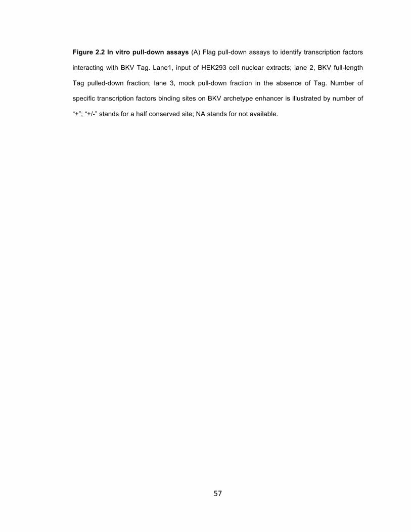
57
Figure 2.2 In vitro pull-down assays (A) Flag pull-down assays to identify transcription factors
interacting with BKV Tag. Lane1, input of HEK293 cell nuclear extracts; lane 2, BKV full-length
Tag pulled-down fraction; lane 3, mock pull-down fraction in the absence of Tag. Number of
specific transcription factors binding sites on BKV archetype enhancer is illustrated by number of
“+”; “+/-” stands for a half conserved site; NA stands for not available.

58

59
Figure 2.2 (B). Illustration of truncation mutants of BKV Tag using in GST pull-down assays. (C).
GST pull-down assays to determine interaction of specific Tag truncation mutants with
transcription factors. Lane 1, HEK293 nuclear extract input; lane 2, GST pull-down fraction
control; lane 3, GST tagged BKT C-terminal region (Cter) pull-down fraction; lane 4, GST tagged
BKT helicase domain (HD) pull-down fraction; lane 5, GST tagged BKV Tag helicase and host
range domain (HDHR) pull-down fraction.

60
61
NFI interacts with BKV Tag in vivo.
To further confirm that NFI interacts with BKV Tag, we attempted to detect their interaction by Co-
Immunoprecipitation(Co-IP) assays. Since NFI expression is ubiquitous, we first compared the
expression of NFIA, NFIB and NFIC in four types of cells including HEK293, HK-2 (human
proximal tubular epithelial cells), HeLa and PRTEC (primary renal tubular epithelial cells), which
are used in our laboratory to study BKV replication. We found that expression patterns of NFI
isotypes differ between these cells (Figure 2.3A-D). A summary of the expression in the four
types of cells is provided in Table 1. All three isotypes are highly expressed in HeLa cells (lane 4
of Figure 2.3B-D), but are poorly expressed in PRTEC cells (lane1 of Figure 2.3B-D). In HEK293
cells, only NFIA is well expressed, and the expression of NFIB and NFIC is barely detectable
(lane 3 of Figure 2.3B-D). In HK-2 cells, NFIA and NFIB are well expressed, but NFIC expression
is low (lane2 of Figure 2.3B-D). Since the expression pattern of NFI isotypes seems to be the
simplest in HEK293 cells and their transfection efficiency is high, we used HEK293 cells to study
in vivo interaction between BKV Tag and NFI by Co-IP assays following ectopic expression of
HA-tagged NFI isotypes and Flag-tagged BKV Tag in HEK293 cells.
When NFI isotypes were precipitated by anti-HA (Figure 2.4A), BKT-Flag was co-precipitated
effectively with HA-NFIB (Figure 2.4A lane 3), while the co-precipitation of BKT-Flag with other
HA-NFI isotypes was much weaker (Figure 2.4A lane 2, 4, 5). In the reciprocal strategy to
precipitate Tag by anti-Flag, more NFIB was co-precipitated by Tag than other NFI isotypes
(Figure 2.4B, lane 2-5). No co-precipitation was detected when BKT-Flag was co-expressed with
an empty NFI expression vector (pCH-empty) (Figure 2.4A lane1; Figure 2.4B lane1) or another
vector expressing an unrelated HA-tagged Gal4 DNA binding domain (pUbi-HA-Gal4dbd), even
though the expression of Tag was much higher (Figure 2.4A lane6; Figure 2.4B lane6).
Endogenous NFkB p65 was included as an internal control to check the quality of in vivo assays
and to see if the in vivo results are in agreement with in vitro results: NFkB p65 co-precipitated
very weakly, if at all, with NFIC and NFIX, and not with NFIA and NFIB (Figure 2.4A, panel “WB:

62
anti-NFkB p65”), which is a totally different pattern compared with BKT-Flag (Figure 2.4A, panel
“WB: anti-Flag”), indicating they are in different protein complexes. But NFkB p65 did not co-
precipitate with BKT-Flag (Figure 2.4B, panel “WB: anti-NFkB p65”), which is consistent with the
in vitro results. Similar results have been obtained from at least two independent repeats.
It is notable that the expression of BKT-Flag from the CMV promoter (pCMV-BKT-Flag) was
differentially affected by different NFI isotypes (Figure 2.4A, “Input” panel for “WB: Anti-Flag”, lane
1-5), which was confirmed with CMV-luciferase reporter assay (Figure 2.4C). However, the
expression of BKT-Flag does not seems to be regulated by NFI only at the transcription level,
because the CMV promoter activity determined in reporter assays (Figure 2.4C) cannot explain
the huge difference at the protein level in Western Blot (Figure 2.4A, “Input” panel for “WB: Anti-
Flag”, lane 1-5). The interaction of Tag with different NFI isotypes might influence the stability of
BKT-Flag. Cyclohexmide (CHX) chase assays indicated that the stability of Tag is greatly
reduced by co-expression of NFIA and NFIX, while co-expression of NFIB and NFIC have little
effect on Tag’s protein stability (Figure 2.4D).

63
Figure 2.3 Expression of NFI isotypes in different cells. Lane 1, 2, 3 and 4 are nuclear
extracts of PRTEC, HK-‐2, HEK293 and HeLa cells respectively. (A). commassie staining of
nuclear extracts from four types of cells; (B). expression of NFIA; (C). expression NFIB;
(D). expression of NFIC.
Table 1. Summery of NFI isotype expression in four types of cells.

64

65
Figure 2.4 Co-Immunoprecipitation (Co-IP) assays to detect Tag interaction with NFI
transcription factors. BKV Tag expression vector is co-transfected with expression vectors for
NFI isotypes (pCH-A, B, C,X or empty) or control vector expressing Gal4 DNA binding domain
(pUbi-HA-Gal4dbd). (A). Immunopreciptiation of HA-tagged NFIs and detection of co-
precipitation of Flag-tagged Tag; (B). Immunoprecipitation of Flag-tagged Tag and detection of
co-precipitation of HA-tagged NFI.

66

67
Figure 2.4 (C). CMV-luciferase assays: HKE293 cells were transfected with pCH-NFIs and
pCMV-Ren at same ratio as pCH-NFIs and pCMV-BKT-Flag in Co-IP assay. Expression of
luciferase was measured 24 hours P.T; (D). Cyclohexmide (CHX) chase assays. Transfected
cells were treated with 200ug/ml CHX 24 hour P.T. Expression of Flag-tagged Tag, HA-tagged
NFIs and β-actin were detected with Western Blotting; (E). Quantification of CHX chase assay
results. Bend density of Flag-Tag was normalized with β-actin; the normalized density at 0 hour
CHX treatment was arbitrarily set as “1” and the normalized densities of later time points were
calculated relatively to the normalized density of 0 hour.

68

69
Synthetic NFI sites in cis- stimulate BKV DNA replication
To determine if NFI stimulates BKV Tag dependent DNA replication in the absence of
interference from other cis-elements in BKV enhancer, the enhancer was substituted by synthetic
NFI binding sites in different orientations and copy numbers (Figure 2.5A). The first NFI site in the
P block of archetype BKV enhancer (NFI-1) was used as the synthetic NFI binding site. This NFI
site was confirmed with EMSA (electrophoretic mobility shift assay) assays as being able to bind
NF1 (data not shown). The synthetic NFI sites stimulate BKV DNA replication (Figure 2.5B lane
2-5); and stimulation of BKV template replication by NFI sites correlates with copy number (Figure
2.5B lane 6-8). Orientation of NFI sites seems not to be important (Figure 2.5B lane 3, 4).
Although NFI sites stimulate BKV DNA replication, the replication of templates with multiple NFI
sites was weaker than replication of BKV templates with the wildtype enhancer, suggesting other
factors in addition to NFI contribute to the stimulatory effect.

70
Figure 2.5 DNA replication of BKV templates with synthetic NFI sites. (A). Illustration of
design of BKV templates with synthetic NFI sites. (B). Southern Blot of DNA replication assays
with BKV templates with synthetic NFI sites.

71
BKV enhancer with mutant NFI sites is defective in NFI binding
Six NFI binding sites have been identified in the BKV archetype enhancer (Figure 2.1C). To
evaluate NFI binding to these sites, a competitive EMSA assay was developed. The Biotin-
labeled oligo conaining the NFI site of the adenovirus ITR (inverted terminal repeat) with high
binding affinity (80) was incubated with HeLa nuclear extracts. A shifted band of oligo caused by
NFI binding was observed in the absence of competitor (Figure 2.6B, lane 1, 6). Adding excess
unlabeled oligo with the same adenovirus ITR NFI site fully competed the binding by NFI in HeLa
extracts (Figure 2.6B, lane 2, 7). However, the competitor with a point mutation in adenovirus ITR
failed to compete for NFI binding (Figure 2.6B, lane3). Adding anti-NFI antibody to the binding
reaction caused “super-shifting” (dash-arrow in Figure 2.6B, lane4), indicating the band shifting
was caused by NFI binding, but not by nonspecific protein binding. An excess of BKV enhancer
PCR fragment also competed for NFI binding (Figure 2.6B, lane5), confirming that NFI can bind
to BKV enhancer (Dik). Oligos of six NFI binding sites in BKV enhancer (Figure 2.6A) were all
able to compete for NFI binding, but with different efficiencies (Figure 2.6B, lane 8-13),
suggesting these NFI sites have different affinities for NFI. Sites closer to the core origin seem to
have higher affinities than distal sites.
Point mutations were introduced into both arms of the NFI consensus binding sequence
“TGGN5~7 GCCAA” of each of the six sites to ensure complete disruption of NFI binding to the
BKV enhancer (Dik) (Figure 2.6A) (86, 121, 126). The design of the mutations was confirmed with
MatInspector (112) to ensure that no significant new transcription factor binding sites were
created by introducing mutations in the BKV enhancer. NFI binding to all mutant NFI sites are
defective in competitive EMSA assays (Figure 2.6C). The competitive binding of mutant BKV
enhancer competitor with mutations on all six NFI sites (6mtNFIs BK enhancer) was greatly
diminished compared with the wild type enhancer competitor (Figure 2.6D, lane 2,3), but some
weak competitive effect was still observed with excessive molar amount of mutant BKV enhancer,

72
which became more obvious when the amount of mutant BKV enhancer competitor was
increased (Figure 2.6D, lane 3-5). However, the mutant enhancer competitor was almost
completely defective in binding when purified hNFIC/CTF1 was used instead of HeLa nuclear
extracts (Figure 2.6E). These results suggest that mutation of six NFI sites greatly reduces NFI
binding to BKV enhancer, however, some cryptic NFI binding site(s) may still be present in the
mutant BKV enhancer, to which NFI binds cooperatively with other transcription factors in the
nuclear extracts.

73
Figure 2.6 EMSA assays to detect NFI binding to BKV NCCR. (A). Sequences of oligos for
NFI sites used in EMSA assays. NFI binding motifs were underlined. Point mutations were
indicated above the mutated positions; (B). Competitive EMSA assays using the NFI site in
Adenovirus inverted terminal repeat (Ad ITR) as probe. Lane 1, in the absence of competitor; lane
2, oligo of NFI site Ad ITR as competitor; lane 3, oligo for mutant NFI site Ad ITR as competitor;
lane 4, in the absence of a competitor, but incubated with anti-NFI antibody for 10min after
binding reaction; lane 5, PCR fragments of BKV enhancer as competitor; lane 6, same as lane 1;
lane 7, same as lane 2; lane 8-13, oligos of NFI sites in BKV enhancer as competitors. Positions
of free probe and shifted probe are indicated with arrows.

74

75
Figure 2.6 (C). Competitive EMSA assays with wildtype (wt) NFI sites and mutant (mt) NFI sites
in BKV enhancer. (D). Competitive EMSA assays with mutant BKV enhancer containing six
mutated NFI sites (6mtNFIs). Lane 1, in the absence of competitor; lane 2, with wildtype BKV
enhancer PCR fragment as competitor; lane 3-5, with increasing amount of 6mtNFIs mutant BKV
enhancer PCR fragment as competitor. (E). Competitive EMSA assays using purified NFIC/CTF1
instead of HeLa nuclear extracts. Lane 1, in the absence of competitor; lane 2, wildtype BKV
enhancer PCR fragment as competitor; lane 3, 6mtNFIs mutant BKV enhancer PCR fragment as
competitor.

76

77
NFI sites on BKV enhancer stimulate BKV DNA replication in vivo
Initially, replication of the mutant BKV template with mutations in all six NFI sites (pUC-6mtNFIs-
BKV) was compared with replication of wildtype template (pUC-wt-BKV) in HK-2 cells. However,
the mutant template replicated with similar efficiency as the wildtype template (data not shown).
We speculate this might be due to the NFI binding to cryptic binding sites, or to redundant
stimulatory effects from other transcription factors.
An alternative competitive DNA replication assay was used to test the effect of mutations in the
NFI sites. A replication competitor (pBC-wt-BKV) with wildtype BKV NCCR in a pBC-backbone
was introduced and transfected together with the test template (in a pUC-backbone) and a Tag
expression vector (pCMV-BKT-Flag). In the presence of the competitor (pBC-wt-BKV), replication
of mutant template (pUC-6mtNFIs-BKV) was greatly reduced compared to replication of the
wildtype template (Figure 2.7A, lane 1, 3), and similar to the replication of template without an
enhancer (Figure 2.7A, lane 2). However, another template with mutations in putative Ets-1
binding sites (pUC-mtEts1s-BKV) replicated at same efficiency as wildtype template (Figure 2.7A,
lane 1, 5), indicating Ets1 is not important for BKV DNA replication.
To determine which sites are mainly responsible for the stimulatory effect of NFI sites, several
mutated NFI sites were changed to wildtype individually. Replication of mutant templates with
only one wildtype NFI site suggests that NFI sites closer to core origin are more important for
BKV DNA replication in the competitive assays (Figure 2.7B, lane 3-7).
The requirement for a competitor in the BKV DNA replication assay to observe the stimulatory
effect of NFI sites suggests that NFI may help to recruit some limiting factors to the replication
complex. Furthermore, that NFI sites closer to core origin are more important than distal sites
suggests that NFI targets components of the initiation complex, including Tag, DNA polymerase-

78
α primase (pol-α primase), RPA and Topoisomerase I. We examine this possibility by the
experiments described below.

79
Figure 2.7 Competitive DNA replication assays in the presence of a wildtype BKV template
(pBC-wt-BKV) as competitor. Solid arrow indicates replicated test template; dashed arrow
indicate replicated competitor template. (A). lane 1, wildtype BKV test template (pUC-wt-BKV);
lane 2, enhancer deletion BKV test template (pUC-Δen-BKV); lane 3, mutant BKV template with
six mutant NFI sites (pUC-6mtNFIs-BKV); lane 4, mutant BKV template with five mutant NFI sites
(5mtNFIs) and the 6th NFI site is wildtype(W6) (pUC-5mtNFIsW6-BKV); lane 5, Ets1 sites mutant
BKV template (pUC-mtEts1s-BKV). (B). lane 1, wildtype BKV test template (pUC-wt-BKV); lane 2,
enhancer deletion BKV test template (pUC-Δen-BKV); lane 3, 6mtNFIs mutant BKV template
(pUC-6mtNFIs-BKV); lane 4, 5mtNFIs mutant BKV template with the 1st NFI being wildtype(W1)
(pUC-5mtNFIsW1-BKV); lane 5, 5mtNFIs mutant BKV template with the 2nd NFI being
wildtype(W2) (pUC-5mtNFIsW2-BKV); lane 6, 5mtNFIs mutant BKV template with the 3rd NFI
being wildtype(W3) (pUC-5mtNFIsW3-BKV); lane 7, 5mtNFIs mutant BKV template with the 6th
NFI being wildtype(W6) (pUC-5mtNFIsW6-BKV).

80

81
Tag is not the sole target for NFI stimulatory activity
That BKV Tag interacts with NFI suggests Tag might be the basis for the effect upon replication
of mutating NF1 sites. To determine if BKV Tag is the limiting factor responsible for the observed
stimulation of BKV DNA replication by NFI in competitive DNA replication assays (Figure 2.7A),
BKV Tag was over-expressed in HK-2 cells and DNA replication of wildtype and NFI sites mutant
templates was compared in the competitive replication assay. Surprisingly, the templates with
mutant NFI sites (pUC-6mtNFIs-BKV) replicated much less efficiently than the wildtype template
(pUC-wt-BKV) even when Tag was over-expressed (Figure 2.8, lane 7,9 and lane 10,12). The
template with a deleted enhancer (pUC-Δen-BKV) also did not replicate when Tag was over-
expressed (Figure 2.8, lane 8, 11), in contrast to its replication in the absence of a competitor
(Figure 2.1). These observations suggest that Tag is not the sole limiting factor for NFI in
competitive replication assays.
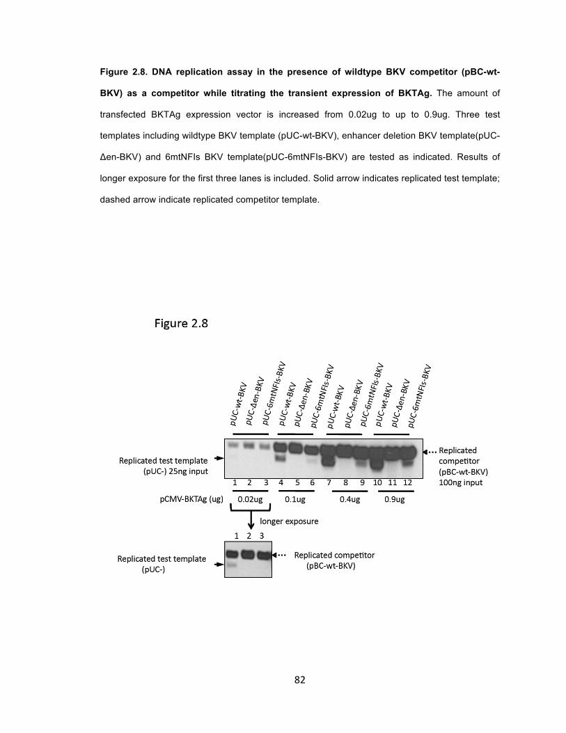
82
Figure 2.8. DNA replication assay in the presence of wildtype BKV competitor (pBC-wt-
BKV) as a competitor while titrating the transient expression of BKTAg. The amount of
transfected BKTAg expression vector is increased from 0.02ug to up to 0.9ug. Three test
templates including wildtype BKV template (pUC-wt-BKV), enhancer deletion BKV template(pUC-
Δen-BKV) and 6mtNFIs BKV template(pUC-6mtNFIs-BKV) are tested as indicated. Results of
longer exposure for the first three lanes is included. Solid arrow indicates replicated test template;
dashed arrow indicate replicated competitor template.

83
NFI stimulates BKV DNA replication by targeting component(s) of the host DNA replication
machinery
To determine factor(s) targeted by NFI, several mutant competitors were constructed and
assessed in replication assays: competitor containing only enhancer (pBC-BKV-en) did not
replicate, due to the lack of core origin; competitor containing only core origin (pBC-BKV-ori)
replicated very inefficiently (data not shown); three competitors containing the complete BKV
NCCR, but each with a single point mutation in the AT-rich or EP region of core origin (pBC-BKV-
A89G, pBC-BKV-A141T, pBC-BKV-A143G) replicated very inefficiently (Figure 2.9A). These
mutant competitors were used in the competition DNA replication assay to examine how NFI sites
in these mutant competitors affect templates replication. BKV Tag was over-expressed to exclude
the possibility that Tag is limiting in the cells. Surprisingly, the enhancer (pBC-BKV-en), core
origin (pBC-BKV-ori) and replication-defective competitors (pBK-BKV-A89G, pBK-BKV-A143G)
are not effective competitors for the unknown cellular factor(s) that stimulates BKV DNA
replication through NFI sites in competitive replication assays in the presence of the wildtype
competitor (pBC-wt-BKV) (Figure 2.9B, lane 3-10). The difference in replication of wildtype and
mutant templates was only observed in the presence of replication competent competitors (pBK-
wt-BKV and pBK-BKV-A141T) (Figure 2.9B, lane 1-2 and 11-12). These observations suggest
that the competitor in the replication assay must be able to replicate efficiently, suggesting the
limiting factor might be a component(s) of the DNA replication machinery that acts beyond
binding of Tag.

84
Figure 2.9 Competitive replication assays with mutant competitors. (A). Replication of
mutant competitors with point mutations(A89G, A141T, A143G) in the core origin. (B).
Competitive DNA replication assays to compare the replication of wildtype (wt) and NFI sites
mutant template (6mtNFIs) in the presence of different competitors: lane 1-2, wildtype competitor
(pBC-wt-BKV); lane 3-4, competitor containing only enhancer (pBC-BKV-en); lane 5-6, competitor
containing only core origin (pBC-BKV-ori); lane 7-8, competitors with point mutation of A89G in
core origin (pBC-BKV-A89G); lane 9-10, competitors with point mutation of A143G in core origin
(pBC-BKV-A143G); lane 11-12, competitors with point mutation of A141T in core origin (pBC-
BKV-A141T). Solid arrow indicates replicated test template; dashed arrow indicate replicated
competitor template.

85

86
NFIC/CTF1 stimulates BKV DNA replication in vitro when DNA polymerase-α primase is
limiting
NFI stimulate initiation of adenovirus DNA replication by recruiting adenovirus DNA polymerase to
origin of replication (18, 29). The proline-rich transactivation domain of NFI (isotype NFIC/CTF1)
stimulates SV40 DNA replication when tethered to the origin (100). This prompted us to assess
whether NFIC/CTF1 can stimulate initiation of BKV DNA replication in the monopolymerase
system that contains Pol-α primase, RPA and Topoisomerase I (82, 135) (Dr. Irina Tikhanovich
did this experiment).
In support of NFI targeting a component of the monopolymerase assay, when pol-α primase was
limiting, the initiation on wildtype BKV template was stimulated strongly by NFIC/CTF1 in a dose-
dependent manner (Figure 2.10, lane 4-6). In contrast, with high levels of pol-α primase, adding
NFIC/CTF1 had no effect in initiation on wildtype BKV template (Figure 2.10, lane 1-3).
Furthermore, no stimulation was observed with the NFI binding mutant BKV template regardless
of pol-α primase level (Figure 2.10, lane 7-12). This indicates that NFIC/CTF1 stimulates initiation
of BKV DNA replication only when pol-α primase is limiting. And pol-α primase might be the
limiting factor targeted by NFI to stimulate BKV DNA replication in the competitive replication
assays.

87
Figure 2.10. Monopolymerase assays with purified NFIC/CTF1. Initiation of wildtype BKV
template (pUC-wt-BKV) and NFI sites mutant BKV template (pUC-6mtNFIs-BKV) were tested
with high (empty columns) and low (solid columns) amount of purified pol-α primase with
increasing amount of purified NFIC/CTF1.

88
NFIC/CTF1 interacts with the p58 subunit of cellular DNA polymerase-α primase
DNA polymerases-α primase consists of four subunits: two smaller subunits, p48 and p58
constitute the primase; and two larger subunits, p68 and p180 constitute the DNA polymerase-α
(88, 89). The p48 is the catalytic subunit of primase and p58 helps with nuclear translocation of
p48 (89). HA tagged human NFIC/CTF was over-expressed with p58 alone or with p48 and p58
in HEK293 cells; the interaction of NFIC/CTF1 with p48 or p58 was examined with Co-IP assays.
HA tagged human NFIC/CTF1 was immunoprecipitated with anti-HA. The p58 was co-
precipitated with NFIC/CTF1 (Figure 2.11 lane 1, panel WB: anti-p58), but p58 co-precipitation
was not detected in three control reactions, in which p58 (Figure 2.11 lane 2, panel WB: anti-p58)
or NFIC/CTF1 (Figure 2.11, lane 3, panel WB: anti-p58) or neither (Figure 2.11, lane 4, panel
WB: anti-p58) was expressed. Because p48 localizes in cytoplasm and its expression is very low
without co-expression of p58 (data not shown), p48 and p58 were co-overexpressed in HEK293
cells with NFIC/CTF-1. However, no co-precipitation of p48 subunit could be detected (Figure 211
lane 5-6, panel WB: anti-p48), although a weak co-precipitation of p58 was observed (Figure 211
lane 5-6, panel WB: anti-p58), probably because most p58 subunit was in complex with over-
expressed p48. This suggests that NFIC/CTF-1 and p48 might interact with the same domain of
p58. No significant interaction between NFIC/CTF-1 and p180 or p68 was observed with Co-IP
assays (data not shown).
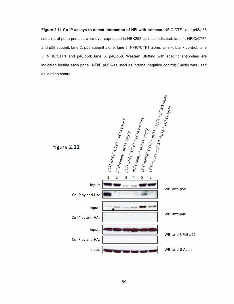
89
Figure 2.11 Co-IP assays to detect interaction of NFI with primase. NFIC/CTF1 and p48/p58
subunits of pol-α primase were over-expressed in HEK293 cells as indicated: lane 1, NFIC/CTF1
and p58 subunit; lane 2, p58 subunit alone; lane 3, NFIC/CTF1 alone; lane 4, blank control; lane
5, NFIC/CTF1 and p48/p58; lane 6, p48/p58. Western Blotting with specific antibodies are
indicated beside each panel. NFkB p65 was used as internal negative control; β-actin was used
as loading control.

90
DISCUSSION
After almost two decades since the first report of PVAN, the mechanism for BKV reactivation still
remains elusive. Immune suppression is partially responsible for reactivation of BKV (2, 15, 42,
60, 140). The involvement of cellular factors induced by the stress related injury, repair,
regeneration and differentiation has been proposed (46, 63, 65). The viral NCCR is the docking
site for cellular transcription factors. Understanding how cis-acting elements on NCCR regulates
BKV replication might provide new insight to the mechanism of BKV reactivation and strategy to
mitigate PVAN.
Here, we found transcription factor NFI binding sites in BKV NCCR in cis- stimulate BKV DNA
replication in vivo and in vitro (Figure 2.5B, Figure 2.7A,B, Figure 2.10); NFI is in complex with
BKV Tag (Figure 2.2A, B, Figure 2.4A, B) and pol-α primase (Figure 2.11 A,B). It is well
established that NFI stimulates adenovirus (Ad2/5) DNA replication(80, 104) through recruitment
of Ad pol-pTP complex (adenovirus DNA polymerase-preterminal protein) to replication origin (18,
29, 99) and/or stabilization of pre-initiation complex(98). According to these observations, we
propose that NFI might also stimulate BKV DNA replication through recruitment of BKV Tag and
pol-α primase to replication origin (Figure 2.12). In support of this, we found that NFI stimulated
initiation of BKV DNA replication in vitro only at low concentration of pol-α primase, whereas at
high concentration of pol-α primase no stimulation was observed (Figure 2.10, lane 1-6). This is
reminiscent of the similar stimulation of adenovirus DNA replication in vitro by NFI, which was
also dependent on concentration of pol-pTP (99). And this is also consistent with the results of in
vivo replication showing that the stimulatory effect of NFI sites is observed only when a
competitor is present (Figure 2.7A,B and data not shown). It seems that the competitor is
competing for the factors that is/are component(s) of DNA replication machinery in addition to
Tag (Figure 2.9B).

91
Figure 2.12 Proposed model for NFIs stimulating BKV DNA replication. When Tag and pol-α
primase are limiting in the cells, NFIs help recruiting them to origin of replication through
interaction and facilitate the assembly of initiation complex to promote DNA replication.

92
Six NFI sites in the BKV archetype NCCR were verified by EMSA assays (Figure 2.6A lane 8-13).
We found these NFI sites not to be equally important for BKV DNA replication: sites closer to core
origin have higher affinity to NFI (Figure 2.6A lane 8-13) and also have stronger stimulatory effect
on BKV DNA replication than distal sites (Figure 2.7B, lane 4-7). These also support the
hypothesis that NFI helps to recruit the components of DNA initiation complex to core origin. That
NFI sites closer to core origin in P 24-37 (NFI-1) and P68-Q13 junction (NFI-2) have stronger
stimulatory activity and almost all rearranged viruses contain the P block and P-Q junction region
(53, 92, 108, 109, 114) suggest that these two NFI sites might be essential for efficient viral DNA
replication in vivo. However, we do not exclude the possibility that these sites might have other
functions in addition to stimulating viral DNA replication.
Other NFI sites in BKV enhancer (Figure 2.1C) might be cis-acting regulators for early/late gene
transcription as reported previously (23, 24, 54, 76). It was determined that the 3rd NFI sites on
archetype BKV enhancer (NFI-3) overlaps the initiation site for late transcription and represses
the late promoter, providing a mechanism for early-late switch of viral gene expression (76). The
NFI-4 site in R block overlaps a Smad3 site that activates the BKV early promoter in responds to
TGF-β(1). Since NFI activity is also be regulated by TGF-β(4), NFI binding to NF-4 might regulate
BKV replication in response to TGF- β induced in kidney allografts. The last two NFI sites (NFI-5
and NFI-6) at the late side of enhancer overlap an ERE (estrogen response element) and
GRE/ERE (glucocorticoid/progesterone response element), both of which independently stimulate
viral replication in transient transfection assays (91). Corticosteroids are used in kidney
transplantation patients as anti-rejection treatment, which is a risk factor for development of
PVAN in renal transplantation patients in addition to HLA-mismatch(64). And BKV reactivation
has been reported extensively among pregnant women(14, 33, 127). So although NFI-5 and NFI-
6 sites have no significant effect in our DNA replication assays, they might function as competitive
inhibitors for hormone-activated nuclear receptors to prevent high-level replication during
persistent infection.

93
The JC virus (JCV) archetype enhancer contains five NFI sites, all of which are highly conserved
with NFI sites in archetype BKV, except that BKV has an additional NFI site (NFI-4) overlapping
the Smad3 site(84). This suggests that NFI might be essential for replication and persistence of
both BKV and JCV. The NFI site close to the core origin of JCV was shown to stimulate JCV DNA
replication in vivo (132), perhaps through similar mechanism as proposed here for BKV. In
support of this, analysis of JCV rearranged enhancers in PML patients also revealed a similar
pattern as with rearranged BKV enhancers in PVAN patients that blocks close to core origin (A to
C for JCV; P and P-Q junction for BKV), which contain the first two NFI sites, are usually
preserved and duplicated (52, 53). NFIX expression is required for JCV propagation in permissive
cells, while NFIA expression is inhibitory for JCV replication in non-permissive cell (93, 115, 129).
Whether it is DNA replication or gene transcription of JCV that is regulated differently by distinct
NFI isotypes is unknown. We have not been able to distinguish the function of different isotypes
in the in vivo replication assays because endogenous NFI isotypes complicate the result
interpretation and no BKV strains have been shown to display a specific tropism in cell culture.
However, using the in vitro monopolymerase assay, we have defined the stimulatory activity of
isotype NFIC/CTF-1 for initiation of BKV DNA replication. The role of other isotypes in DNA
replication will be tested in similar systems in the future.
Although the NFI sites are not absolutely required for BKV DNA replication in DNA replication
assays in the absence of a competitor (data not shown), they stimulate BKV DNA replication
when Tag or pol-α primase is limiting (Figure 2.5B, Figure 2.7A). This stimulatory activity might be
essential for reactivation, because latent BKV persists episomally in kidney tubular epithelial cells
at less than one copy/cell and thus expresses limiting amount of Tag(31, 62, 113). And the
tubular epithelial cells in normal kidney are terminally differentiated quiescent cells dividing at low
rate (16, 103), most of which probably express low amounts of pol-α primase. Stress related
signaling induced by kidney ischemeia/reperfusion injury or inflammatory response during kidney
transplantation might change the NFI isotypes expression or their activity through post-
translational modifications or interaction with other cellular factors, which may promote the NFI

94
recruitment of Tag and/or pol-α primase to replication origin and facilitate the assembly of pre-
initiation complex (Figure 2.12). NFI activity can be modulated by TGF-β(4, 5), TNF-α(5),
oxidative stress(9, 95-97), which are all changed in allograft kidneys (21, 40, 67).
The regulation of BKV replication by NFI-family transcription factors is complicated due to multiple
NFI isotypes/splicing variants and multiple binding sites present on enhancer. In addition to the
proposed mechanism, NFI might also regulate viral DNA replication through chromatin
remodeling (30, 100) or indirectly through modulation of early gene expression(23). How BKV
DNA replication and gene transcription is coordinated by NFI-family transcription factors during
BKV reactivation in PVAN needs to be investigated in detail.
Another possible implication of our proposed model is that NFI might participate in cellular DNA
replication by recruitment of pol-α primase to the cellular DNA replication origin. Around
30,000~50,000 origins are active during replication of mammalian DNA, however, unlike the
prokaryotes DNA replication, the origins of mammalian genome do not have a consensus
sequence and not all the origins are active at the same time(85). And the selection of origins is
related to differentiation and development(85). Like well-established viral systems(101),
transcription factor binding sites and promoters coincide with some eukaryotic cellular replication
origins (75). And several evidences have shown that transcription factors do involve in the cellular
replication (10, 17, 49, 131), perhaps through interaction with components of DNA replication
machinery and/or chromatin remodeling in response to intracellular/extracellular signals during
the development, differentiation and stress response (38, 75). The expression and activity of NFI-
family transcription factors is greatly affected by development, differentiation and stress (9, 26,
77, 96, 97, 111, 120). So it is possible that NFI plays a role in the selection of cellular DNA
replication origins during development/differentiation and in response to stress.

95
Reference
1. Abend, J. R., and M. J. Imperiale. 2008. Transforming growth factor-beta-mediated regulation of BK virus gene expression. Virology 378:6-12.
2. Abend, J. R., J. A. Low, and M. J. Imperiale. 2007. Inhibitory Effect of Gamma Interferon on BK Virus Gene Expression and Replication. J. Virol. 81:272-279.
3. Alevizopoulos, A., Y. Dusserre, M. Tsai-Pflugfelder, T. von der Weid, W. Wahli, and N. Mermod. 1995. A proline-rich TGF-beta-responsive transcriptional activator interacts with histone H3. Genes Dev. 9:3051-3066.
4. Alevizopoulos, A., Y. Dusserre, M. Tsai-Pflugfelder, T. von der Weid, W. Wahli, and N. Mermod. 1995. A proline-rich TGF-beta-responsive transcriptional activator interacts with histone H3. Genes Dev. 9:3051-3066.
5. Alevizopoulos, A., and N. Mermod. 1996. Antagonistic Regulation of a Proline-rich Transcription Factor by Transforming Growth Factor-beta and Tumor Necrosis Factor-alpha. J. Biol. Chem. 271:29672-29681.
6. Angulo, A., D. Kerry, H. Huang, E.-M. Borst, A. Razinsky, J. Wu, U. Hobom, M. Messerle, and P. Ghazal. 2000. Identification of a Boundary Domain Adjacent to the Potent Human Cytomegalovirus Enhancer That Represses Transcription of the Divergent UL127 Promoter. J. Virol. 74:2826-2839.
7. Baldwin, A., M. K. Hypes, L. Pirisi, and K. E. Creek. 2007. NFI is an essential positive transcription factor for human papillomavirus type 16 early gene expression. Open Virol J 1:33-38.
8. Baldwin, A., L. Pirisi, and K. E. Creek. 2004. NFI-Ski Interactions Mediate Transforming Growth Factor {beta} Modulation of Human Papillomavirus Type 16 Early Gene Expression. J. Virol. 78:3953-3964.
9. Bandyopadhyay, S., D. W. Starke, J. J. Mieyal, and R. M. Gronostajski. 1998. Thioltransferase (Glutaredoxin) Reactivates the DNA-binding Activity of Oxidation-inactivated Nuclear Factor I. J. Biol. Chem. 273:392-397.
10. Beall, E. L., J. R. Manak, S. Zhou, M. Bell, J. S. Lipsick, and M. R. Botchan. 2002. Role for a Drosophila Myb-containing protein complex in site-specific DNA replication. Nature 420:833-837.

96
11. Belikov, S., P.-H. Holmqvist, C. √Östrand, and ñ. Wrange. 2004. Nuclear Factor 1 and Octamer Transcription Factor 1 Binding Preset the Chromatin Structure of the Mouse Mammary Tumor Virus Promoter for Hormone Induction. J. Biol. Chem. 279:49857-49867.
12. Bernier, D., H. Thomassin, D. Allard, M. Guertin, D. Hamel, M. Blaquiere, M. Beauchemin, H. LaRue, M. Estable-Puig, and L. Belanger. 1993. Functional analysis of developmentally regulated chromatin-hypersensitive domains carrying the α1-fetoprotein gene promoter and the albumin/α1-fetoprotein intergenic enhancer. Mol. Cell. Biol. 13:1619-1633.
13. Bharucha, V. A., K. W. Peden, and G. I. Tennekoon. 1994. SV40 large T antigen with c-Jun down-regulates myelin P0 gene expression: a mechanism for papovaviral T antigen-mediated demyelination. Neuron 12:627-637.
14. Bhattacharjee, S., and T. Chakraborty. 2004. High reactivation of BK virus variants in Asian Indians with renal disorders and during pregnancy. Virus Genes 28:157-168.
15. Binggeli, S., A. Egli, S. Schaub, I. Binet, M. Mayr, J. Steiger, and H. H. Hirsch. 2007. Polyomavirus BK-Specific Cellular Immune Response to VP1 and Large T-Antigen in Kidney Transplant Recipients. American Journal of Transplantation 7:1131-1139.
16. Bonventre, J. V. 2003. Dedifferentiation and proliferation of surviving epithelial cells in acute renal failure. Journal of the American Society of Nephrology : JASN 14 Suppl 1:S55-61.
17. Bosco, G., W. Du, and T. L. Orr-Weaver. 2001. DNA replication control through interaction of E2F-RB and the origin recognition complex. Nat Cell Biol 3:289-295.
18. Bosher, J., E. C. Robinson, and R. T. Hay. 1990. Interactions between the adenovirus type 2 DNA polymerase and the DNA binding domain of nuclear factor I. The New biologist 2:1083-1090.
19. Broekema, N. M., J. R. Abend, S. M. Bennett, J. S. Butel, J. A. Vanchiere, and M. J. Imperiale. 2010. A system for the analysis of BKV non-coding control regions: application to clinical isolates from an HIV/AIDS patient. Virology 407:368-373.
20. Buratowski, S., and L. A. Chodosh. 2001. Mobility Shift DNA-Binding Assay Using Gel Electrophoresis. John Wiley & Sons, Inc.
21. Campistol, J. M., P. I√±igo, S. Larios, M. n. Bescos, and F. Oppenheimer. 2001. Role of transforming growth factor β-1 in the progression of chronic allograft nephropathy. Nephrology Dialysis Transplantation 16:114-116.

97
22. Cassill, J. A., K. L. Deyerle, and S. Subramani. 1989. Unidirectional deletion and linker scan analysis of the late promoter of the human papovavirus BK. Virology 169:172-181.
23. Chakraborty, T., and G. C. Das. 1989. Identification of HeLa cell nuclear factors that bind to and activate the early promoter of human polyomavirus BK in vitro. Mol. Cell. Biol. 9:3821-3828.
24. Chakraborty, T., and G. C. Das. 1991. Proteins of the nuclear factor-1 family act as an activator of the late promoter in human polyomavirus BK in vitro. The Journal of general virology 72 ( Pt 8):1935-1942.
25. Chang, Y. N., K. T. Jeang, C. J. Chiou, Y. J. Chan, M. Pizzorno, and G. S. Hayward. 1993. Identification of a large bent DNA domain and binding sites for serum response factor adjacent to the NFI repeat cluster and enhancer region in the major IE94 promoter from simian cytomegalovirus. J. Virol. 67:516-529.
26. Chaudhry, A. Z., G. E. Lyons, and R. M. Gronostajski. 1997. Expression patterns of the four nuclear factor I genes during mouse embryogenesis indicate a potential role in development. Dev. Dyn. 208:313-325.
27. Chaudhry, A. Z., A. D. Vitullo, and R. M. Gronostajski. 1998. Nuclear Factor I (NFI) Isoforms Differentially Activate Simple versus Complex NFI-responsive Promoters. J. Biol. Chem. 273:18538-18546.
28. Chaudhry, A. Z., A. D. Vitullo, and R. M. Gronostajski. 1999. Nuclear Factor I-mediated Repression of the Mouse Mammary Tumor Virus Promoter Is Abrogated by the Coactivators p300/CBP and SRC-1. J. Biol. Chem. 274:7072-7081.
29. Chen, M., N. Mermod, and M. S. Horwitz. 1990. Protein-protein interactions between adenovirus DNA polymerase and nuclear factor I mediate formation of the DNA replication preinitiation complex. The Journal of biological chemistry 265:18634-18642.
30. Cheng, L., and T. J. Kelly. 1989. Transcriptional activator nuclear factor I stimulates the replication of SV40 minichromosomes in vivo and in vitro. Cell 59:541-551.
31. Chesters, P. M., J. Heritage, and D. J. McCance. 1983. Persistence of DNA sequences of BK virus and JC virus in normal human tissues and in diseased tissues. J. Infect. Dis. 147:676-684.
32. Chiang, C. M., and R. G. Roeder. 1995. Cloning of an intrinsic human TFIID subunit that interacts with multiple transcriptional activators. Science 267:531-536.

98
33. Coleman, D. V., M. R. Wolfendale, R. A. Daniel, N. K. Dhanjal, S. D. Gardner, P. E. Gibson, and A. M. Field. 1980. A prospective study of human polyomavirus infection in pregnancy. The Journal of infectious diseases 142:1-8.
34. Daniel, A. M., J. J. Swenson, R. P. Reddy Mayreddy, K. Khalili, and R. J. Frisque. 1996. Sequences within the Early and Late Promoters of Archetype JC Virus Restrict Viral DNA Replication and Infectivity. Virology 216:90-101.
35. de Jong, R. N., and P. C. van der Vliet. 1999. Mechanism of DNA replication in eukaryotic cells: cellular host factors stimulating adenovirus DNA replication. Gene 236:1-12.
36. Dechend, R., F. Hirano, K. Lehmann, V. Heissmeyer, S. Ansicau, F. G. Wulczyn, C. Scheidereit, and A. Leutz. 1999. The Bcl-3 oncoprotein acts as a bridging factor between NF-κB/Rel and nuclear co-regulators. Oncogene 18:3316-3323.
37. DeLucia, A. L., S. Deb, K. Partin, and P. Tegtmeyer. 1986. Functional interactions of the simian virus 40 core origin of replication with flanking regulatory sequences. J. Virol. 57:138-144.
38. DePamphilis, M. L. 2003. Eukaryotic DNA Replication Origins: Reconciling Disparate Data. Cell 114:274-275.
39. Deyerle, K. L., and S. Subramani. 1988. Linker scan analysis of the early regulatory region of human papovavirus BK. J. Virol. 62:3378-3387.
40. Djamali, A. 2007. Oxidative stress as a common pathway to chronic tubulointerstitial injury in kidney allografts. American Journal of Physiology - Renal Physiology 293:F445-F455.
41. Egli, A., L. Infanti, A. Dumoulin, A. Buser, J. Samaridis, C. Stebler, R. Gosert, and H. H. Hirsch. 2009. Prevalence of polyomavirus BK and JC infection and replication in 400 healthy blood donors. J. Infect. Dis. 199:837-846.
42. Egli, A., S. Köhli, M. Dickenmann, and H. H. Hirsch. 2009. Inhibition of Polyomavirus BK-Specific T-Cell Responses by Immunosuppressive Drugs. Transplantation 88:1161-1168 1110.1097/TP.1160b1013e3181bca1422.
43. Ethelberg, S., B. Hallberg, J. Lovmand, J. Schmidt, A. Luz, T. Grundstrom, and F. Pedersen. 1997. Second-site proviral enhancer alterations in lymphomas induced by enhancer mutants of SL3-3 murine leukemia virus: negative effect of nuclear factor 1 binding site. J. Virol. 71:1196-1206.

99
44. Ethelberg, S., B. D. Tzschaschel, A. Luz, S. J. Diaz-Cano, F. S. Pedersen, and J. Schmidt. 1999. Increased induction of osteopetrosis, but unaltered lymphomagenicity, by murine leukemia virus SL3-3 after mutation of a nuclear factor 1 site in the enhancer. J. Virol. 73:10406-10415.
45. Ferguson, A. T., and S. Subramani. 1994. Complex functional interactions at the early enhancer of the PQ strain of BK virus. J. Virol. 68:4274-4286.
46. Fishman, J. A. 2002. BK Virus Nephropathy - Polyomavirus Adding Insult to Injury. N. Engl. J. Med. 347:527-530.
47. Gao, B., H. Jaffe, and G. Kunos. 1998. Histone H1 isoforms purified from rat liver bind nonspecifically to the nuclear factor 1 recognition sequence and serve as generalized transcriptional repressors. Mol. Cell. Biochem. 178:187-196.
48. Ghazal, P., H. Lubon, C. Reynolds-Kohler, L. Hennighausen, and J. A. Nelson. 1990. Interactions between cellular regulatory proteins and a unique sequence region in the human cytomegalovirus major immediate-early promoter. Virology 174:18-25.
49. Ghosh, M., G. Liu, G. Randall, J. Bevington, and M. Leffak. 2004. Transcription factor binding and induced transcription alter chromosomal c-myc replicator activity. Mol. Cell. Biol. 24:10193-10207.
50. Golemis, E. A., N. A. Speck, and N. Hopkins. 1990. Alignment of U3 region sequences of mammalian type C viruses: identification of highly conserved motifs and implications for enhancer design. J. Virol. 64:534-542.
51. Gorrill, T. S., and K. Khalili. 2005. Cooperative interaction of p65 and C/EBP[beta] modulates transcription of BKV early promoter. Virology 335:1-9.
52. Gosert, R., P. Kardas, E. O. Major, and H. H. Hirsch. 2010. Rearranged JC Virus Noncoding Control Regions Found in Progressive Multifocal Leukoencephalopathy Patient Samples Increase Virus Early Gene Expression and Replication Rate. J. Virol. 84:10448-10456.
53. Gosert, R., C. H. Rinaldo, G. A. Funk, A. Egli, E. Ramos, C. B. Drachenberg, and H. H. Hirsch. 2008. Polyomavirus BK with rearranged noncoding control region emerge in vivo in renal transplant patients and increase viral replication and cytopathology. The Journal of experimental medicine 205:841-852.
54. Grinnell, B. W., D. T. Berg, and J. D. Walls. 1988. Negative regulation of the human polyomavirus BK enhancer involves cell-specific interaction with a nuclear repressor. Mol. Cell. Biol. 8:3448-3457.

100
55. Gronostajski, R. M. 2000. Roles of the NFI/CTF gene family in transcription and development. Gene 249:31-45.
56. Gronostajski, R. M., S. Adhya, and K. Nagata. 1985. Site-specific DNA binding of nuclear factor I: Analyses of cellular binding sites. Mol. Cell. Biol. 5:964-971.
57. Guo, Z. S., C. Gutierrez, U. Heine, J. M. Sogo, and M. L. Depamphilis. 1989. Origin auxiliary sequences can facilitate initiation of simian virus 40 DNA replication in vitro as they do in vivo. Mol. Cell. Biol. 9:3593-3602.
58. Gutierrez, C., Z. S. Guo, J. Roberts, and M. L. DePamphilis. 1990. Simian virus 40 origin auxiliary sequences weakly facilitate T-antigen binding but strongly facilitate DNA unwinding. Mol. Cell. Biol. 10:1719-1728.
59. Hanssen Rinaldo, C., H. Hansen, and T. Traavik. 2005. Human endothelial cells allow passage of an archetypal BK virus (BKV) strain – a tool for cultivation and functional studies of natural BKV strains. Arch. Virol. 150:1449-1458.
60. Hariharan, S., E. P. Cohen, B. Vasudev, R. Orentas, R. P. Viscidi, J. Kakela, and B. DuChateau. 2005. BK Virus-Specific Antibodies and BKV DNA in Renal Transplant Recipients with BKV Nephritis. American Journal of Transplantation 5:2719-2724.
61. Hennighausen, L., and B. Fleckenstein. 1986. Nuclear factor 1 interacts with five DNA elements in the promoter region of the human cytomegalovirus major immediate early gene. The EMBO journal 5:1367-1371.
62. Heritage, J., P. M. Chesters, and D. J. McCance. 1981. The persistence of papovavirus BK DNA sequences in normal human renal tissue. J. Med. Virol. 8:143-150.
63. Hirsch, H. H. 2002. Polyomavirus BK nephropathy: a (re-)emerging complication in renal transplantation. Am J Transplant 2:25-30.
64. Hirsch, H. H., W. Knowles, M. Dickenmann, J. Passweg, T. Klimkait, M. J. Mihatsch, and J. r. Steiger. 2002. Prospective Study of Polyomavirus Type BK Replication and Nephropathy in Renal-Transplant Recipients. N. Engl. J. Med. 347:488-496.
65. Hirsch, H. H., and J. r. Steiger. 2003. Polyomavirus BK. The Lancet Infectious Diseases 3:611-623.
66. Hirt, B. 1967. Selective extraction of polyoma DNA from infected mouse cell cultures. J. Mol. Biol. 26:365-369.

101
67. Hribova, P., K. Kotsch, I. Brabcova, S. Vitko, H. D. Volk, and J. Lacha. 2005. Cytokines and Chemokine Gene Expression in Human Kidney Transplantation. Transplant. Proc. 37:760-763.
68. Ito, K., M. Asano, P. Hughes, H. Kohzaki, C. Masutani, F. Hanaoka, T. Kerppola, T. Curran, Y. Murakami, and Y. Ito. 1996. c-Jun stimulates origin-dependent DNA unwinding by polyomavirus large Tantigen. The EMBO journal 15:5636-5646.
69. Johnston, S. D., X. M. Yu, and J. E. Mertz. 1996. The major transcriptional transactivation domain of simian virus 40 large T antigen associates nonconcurrently with multiple components of the transcriptional preinitiation complex. J. Virol. 70:1191-1202.
70. Jordan, J. A., K. Manley, A. S. Dugan, B. A. O'Hara, and W. J. Atwood. 2010. Transcriptional Regulation of BK Virus by Nuclear Factor of Activated T Cells. J. Virol. 84:1722-1730.
71. Kierstead, T. D., and M. J. Tevethia. 1993. Association of p53 binding and immortalization of primary C57BL/6 mouse embryo fibroblasts by using simian virus 40 T-antigen mutants bearing internal overlapping deletion mutations. J. Virol. 67:1817-1829.
72. Kim, T. K., and R. G. Roeder. 1994. Proline-rich activator CTF1 targets the TFIIB assembly step during transcriptional activation. Proceedings of the National Academy of Sciences 91:4170-4174.
73. Knowles, W. A. 2006. Discovery and Epidemiology of the Human Polyomaviruses BK Virus (BKV) and JC Virus (JCV), p. 19-45. In N. Ahsan (ed.), Polyomaviruses and Human Diseases, vol. 577. Springer New York.
74. Knowles, W. A., P. Pipkin, N. Andrews, A. Vyse, P. Minor, D. W. Brown, and E. Miller. 2003. Population-based study of antibody to the human polyomaviruses BKV and JCV and the simian polyomavirus SV40. J. Med. Virol. 71:115-123.
75. Kohzaki, H., and Y. Murakami. 2005. Transcription factors and DNA replication origin selection. Bioessays 27:1107-1116.
76. Kraus, R. J., L. Shadley, and J. E. Mertz. 2001. Nuclear factor 1 family members mediate repression of the BK virus late promoter. Virology 287:89-104.
77. Kulkarni, S., and R. M. Gronostajski. 1996. Altered expression of the developmentally regulated NFI gene family during phorbol ester-induced differentiation of human leukemic cells. Cell Growth Differ. 7:501-510.

102
78. Kwun, H. J., S. W. Yim, D. H. Lee, and K. L. Jang. 1999. Activation of the thymidine kinase promoter by herpes simplex virus type 1 immediate early proteins. Mol. Cells 9:277-280.
79. Leahy, P., D. R. Crawford, G. Grossman, R. M. Gronostajski, and R. W. Hanson. 1999. CREB binding protein coordinates the function of multiple transcription factors including nuclear factor I to regulate phosphoenolpyruvate carboxykinase (GTP) gene transcription. J. Biol. Chem. 274:8813-8822.
80. Leegwater, P. A., W. van Driel, and P. C. van der Vliet. 1985. Recognition site of nuclear factor I, a sequence-specific DNA-binding protein from HeLa cells that stimulates adenovirus DNA replication. The EMBO journal 4:1515-1521.
81. Lilyestrom, W., M. G. Klein, R. Zhang, A. Joachimiak, and X. S. Chen. 2006. Crystal structure of SV40 large T-antigen bound to p53: interplay between a viral oncoprotein and a cellular tumor suppressor. Genes Dev. 20:2373-2382.
82. Mahon, C., B. Liang, I. Tikhanovich, J. R. Abend, M. J. Imperiale, H. P. Nasheuer, and W. R. Folk. 2009. Restriction of human polyomavirus BK virus DNA replication in murine cells and extracts. J. Virol. 83:5708-5717.
83. Markowitz, R. B., and W. S. Dynan. 1988. Binding of cellular proteins to the regulatory region of BK virus DNA. J. Virol. 62:3388-3398.
84. Markowitz, R. B., B. A. Eaton, M. F. Kubik, D. Latorra, J. A. McGregor, and W. S. Dynan. 1991. BK virus and JC virus shed during pregnancy have predominantly archetypal regulatory regions. J. Virol. 65:4515-4519.
85. Méchali, M. 2010. Eukaryotic DNA replication origins: many choices for appropriate answers. Nat Rev Mol Cell Biol 11:728-738.
86. Meisterernst, M., I. Gander, L. Rogge, and E. L. Winnacker. 1988. A quantitative analysis of nuclear factor I/DNA interactions. Nucleic Acids Res. 16:4419-4435.
87. Mermod, N., E. A. O'Neill, T. J. Kelly, and R. Tjian. 1989. The proline-rich transcriptional activator of CTF/NF-I is distinct from the replication and DNA binding domain. Cell 58:741-753.
88. Mizuno, T., N. Ito, M. Yokoi, A. Kobayashi, K. Tamai, H. Miyazawa, and F. Hanaoka. 1998. The second-largest subunit of the mouse DNA polymerase alpha-primase complex facilitates both production and nuclear translocation of the catalytic subunit of DNA polymerase alpha. Mol. Cell. Biol. 18:3552-3562.

103
89. Mizuno, T., T. Okamoto, M. Yokoi, M. Izumi, A. Kobayashi, T. Hachiya, K. Tamai, T. Inoue, and F. Hanaoka. 1996. Identification of the nuclear localization signal of mouse DNA primase: nuclear transport of p46 subunit is facilitated by interaction with p54 subunit. J. Cell Sci. 109 ( Pt 11):2627-2636.
90. Moens, U., T. Johansen, J. I. Johnsen, O. M. Seternes, and T. Traavik. 1995. Noncoding control region of naturally occurring BK virus variants: sequence comparison and functional analysis. Virus Genes 10:261-275.
91. Moens, U., N. Subramaniam, B. Johansen, T. Johansen, and T. Traavik. 1994. A steroid hormone response unit in the late leader of the noncoding control region of the human polyomavirus BK confers enhanced host cell permissivity. J. Virol. 68:2398-2408.
92. Moens, U., and M. Van Ghelue. 2005. Polymorphism in the genome of non-passaged human polyomavirus BK: implications for cell tropism and the pathological role of the virus. Virology 331:209-231.
93. Monaco, M. C., B. F. Sabath, L. C. Durham, and E. O. Major. 2001. JC virus multiplication in human hematopoietic progenitor cells requires the NF-1 class D transcription factor. J. Virol. 75:9687-9695.
94. Monaco, M. C. G., B. F. Sabath, L. C. Durham, and E. O. Major. 2001. JC Virus Multiplication in Human Hematopoietic Progenitor Cells Requires the NF-1 Class D Transcription Factor. J. Virol. 75:9687-9695.
95. Morel, Y., and R. Barouki. 1998. Down-regulation of Cytochrome P450 1A1 Gene Promoter by Oxidative Stress. J. Biol. Chem. 273:26969-26976.
96. Morel, Y., and R. Barouki. 2000. The repression of nuclear factor I/CCAAT transcription factor (NFI/CTF) transactivating domain by oxidative stress is mediated by a critical cysteine (Cys-427). Biochem. J. 348:235-240.
97. Morel, Y., N. Mermod, and R. Barouki. 1999. An Autoregulatory Loop Controlling CYP1A1 Gene Expression: Role of H2O2 and NFI. Mol. Cell. Biol. 19:6825-6832.
98. Mul, Y. M., and P. C. Van der Vliet. 1992. Nuclear factor I enhances adenovirus DNA replication by increasing the stability of a preinitiation complex. The EMBO journal 11:751-760.
99. Mul, Y. M., C. P. Verrijzer, and P. C. van der Vliet. 1990. Transcription factors NFI and NFIII/oct-1 function independently, employing different mechanisms to enhance adenovirus DNA replication. J. Virol. 64:5510-5518.

104
100. Muller, K., and N. Mermod. 2000. The histone-interacting domain of nuclear factor I activates simian virus 40 DNA replication in vivo. The Journal of biological chemistry 275:1645-1650.
101. Murakami, Y., and Y. Ito. 1999. Transcription factors in DNA replication. Frontiers in bioscience : a journal and virtual library 4:D824-833.
102. Mysiak, M. E., C. Wyman, P. E. Holthuizen, and P. C. van der Vliet. 2004. NFI and Oct-1 bend the Ad5 origin in the same direction leading to optimal DNA replication. Nucleic Acids Res. 32:6218-6225.
103. Nadasdy, T., Z. Laszik, K. E. Blick, L. D. Johnson, and F. G. Silva. 1994. Proliferative activity of intrinsic cell populations in the normal human kidney. Journal of the American Society of Nephrology : JASN 4:2032-2039.
104. Nagata, K., R. A. Guggenheimer, T. Enomoto, J. H. Lichy, and J. Hurwitz. 1982. Adenovirus DNA replication in vitro: identification of a host factor that stimulates synthesis of the preterminal protein-dCMP complex. Proceedings of the National Academy of Sciences 79:6438-6442.
105. Nehls, M. C., M. L. Grapilon, and D. A. Brenner. 1992. NF-I/Sp1 switch elements regulate collagen α1(I) gene expression. DNA Cell Biol. 11:443-452.
106. Nehls, M. C., R. A. Rippe, L. Veloz, and D. A. Brenner. 1991. Transcription factors nuclear factor I and Sp1 interact with the murine collagen α1(I) promoter. Mol. Cell. Biol. 11:4065-4073.
107. O'Connor, M., and H.-U. Bernard. 1995. Oct-1 Activates the Epithelial-Specific Enhancer of Human Papillomavirus Type 16 via a Synergistic Interaction with NFI at a Conserved Composite Regulatory Element. Virology 207:77-88.
108. Olsen, G. H., H. H. Hirsch, and C. H. Rinaldo. 2009. Functional analysis of polyomavirus BK non-coding control region quasispecies from kidney transplant recipients. J. Med. Virol. 81:1959-1967.
109. Perets, T. T., I. Silberstein, J. Rubinov, R. Sarid, E. Mendelson, and L. M. Shulman. 2009. High frequency and diversity of rearrangements in polyomavirus bk noncoding regulatory regions cloned from urine and plasma of Israeli renal transplant patients and evidence for a new genetic subtype. J. Clin. Microbiol. 47:1402-1411.
110. Prives, C., Y. Murakami, F. G. Kern, W. Folk, C. Basilico, and J. Hurwitz. 1987. DNA sequence requirements for replication of polyomavirus DNA in vivo and in vitro. Mol. Cell. Biol. 7:3694-3704.

105
111. Puzianowska-Kuznicka, M., and Y. B. Shi. 1996. Nuclear factor I as a potential regulator during postembryonic organ development. J. Biol. Chem. 271:6273-6282.
112. Quandt, K., K. Frech, H. Karas, E. Wingender, and T. Werner. 1995. MatInd and MatInspector: new fast and versatile tools for detection of consensus matches in nucleotide sequence data. Nucleic Acids Res. 23:4878-4884.
113. Randhawa, P., R. Shapiro, and A. Vats. 2005. Quantitation of DNA of Polyomaviruses BK and JC in Human Kidneys. J. Infect. Dis. 192:504-509.
114. Randhawa, P., D. Zygmunt, R. Shapiro, A. Vats, K. Weck, P. Swalsky, and S. Finkelstein. 2003. Viral regulatory region sequence variations in kidney tissue obtained from patients with BK virus nephropathy. Kidney Int. 64:743-747.
115. Ravichandran, V., and E. O. Major. 2008. DNA-binding transcription factor NF-1A negatively regulates JC virus multiplication. J. Gen. Virol. 89:1396-1401.
116. Ristiniemi, J., and J. Oikarinen. 1989. Histone H1 binds to the putative nuclear factor I recognition sequence in the mouse α2(I) collagen promoter. J. Biol. Chem. 264:2164-2174.
117. Rochford, R., B. A. Campbell, and L. P. Villarreal. 1990. Genetic analysis of the enhancer requirements for polyomavirus DNA replication in mice. J. Virol. 64:476-485.
118. Rochford, R., B. A. Campbell, and L. P. Villarreal. 1987. A pancreas specificity results from the combination of polyomavirus and Moloney murine leukemia virus enhancer. Proc. Natl. Acad. Sci. U. S. A. 84:449-453.
119. Rochford, R., J. P. Moreno, M. L. Peake, and L. P. Villarreal. 1992. Enhancer dependence of polyomavirus persistence in mouse kidneys. J. Virol. 66:3287-3297.
120. Roulet, E., M. T. Armentero, G. Krey, B. Corthesy, C. Dreyer, N. Mermod, and W. Wahli. 1995. Regulation of the DNA-binding and transcriptional activities of Xenopus laevis NFI-X by a novel C-terminal domain. Mol. Cell. Biol. 15:5552-5562.
121. Roulet, E., P. Bucher, R. Schneider, E. Wingender, Y. Dusserre, T. Werner, and N. Mermod. 2000. Experimental analysis and computer prediction of CTF/NFI transcription factor DNA binding sites. J. Mol. Biol. 297:833-848.
122. Rubinstein, R., and E. H. Harley. 1989. BK virus DNA cloned directly from human urine confirms an archetypal structure for the transcriptional control region. Virus Genes 2:157-165.

106
123. Rubinstein, R., B. C. Schoonakker, and E. H. Harley. 1991. Recurring theme of changes in the transcriptional control region of BK virus during adaptation to cell culture. J. Virol. 65:1600-1604.
124. S¯rensen, K. D., A. B. S¯rensen, L. Quintanilla-Martinez, S. Kunder, J. Schmidt, and F. S. Pedersen. 2005. Distinct roles of enhancer nuclear factor 1 (NF1) sites in plasmacytoma and osteopetrosis induction by Akv1-99 murine leukemia virus. Virology 334:234-244.
125. Schmieg, F. I., and D. T. Simmons. 1988. Characterization of the in vitro interaction between SV40 T antigen and p53: mapping the p53 binding site. Virology 164:132-140.
126. Schneider, R., I. Gander, U. Muller, R. Mertz, and E. L. Winnacker. 1986. A sensitive and rapid gel retention assay for nuclear factor I and other DNA-binding proteins in crude nuclear extracts. Nucleic Acids Res. 14:1303-1317.
127. Shah, K., R. Daniel, D. Madden, and S. Stagno. 1980. Serological investigation of BK papovavirus infection in pregnant women and their offspring. Infect. Immun. 30:29-35.
128. Shah, K. V., R. W. Daniel, and R. M. Warszawski. 1973. High prevalence of antibodies to BK virus, an SV40-related papovavirus, in residents of Maryland. J. Infect. Dis. 128:784-787.
129. Shinohara, T., K. Nagashima, and E. O. Major. 1997. Propagation of the Human Polyomavirus, JCV, in Human Neuroblastoma Cell Lines. Virology 228:269-277.
130. Shivakumar, C. V., and G. C. Das. 1996. Interaction of human polyomavirus BK with the tumor-suppressor protein p53. Oncogene 13:323-332.
131. Simon, I., T. Tenzen, R. Mostoslavsky, E. Fibach, L. Lande, E. Milot, J. Gribnau, F. Grosveld, P. Fraser, and H. Cedar. 2001. Developmental regulation of DNA replication timing at the human beta globin locus. The EMBO journal 20:6150-6157.
132. Sock, E., M. Wegner, and F. Grummt. 1991. DNA replication of human polyomavirus JC is stimulated by NF-I in vivo. Virology 182:298-308.
133. Sundsfjord, A., T. Johansen, T. Flaegstad, U. Moens, P. Villand, S. Subramani, and T. Traavik. 1990. At least two types of control regions can be found among naturally occurring BK virus strains. J. Virol. 64:3864-3871.
134. ter Schegget, J., C. J. Sol, E. W. Baan, J. van der Noordaa, and H. van Ormondt. 1985. Naturally occurring BK virus variants (JL and Dik) with deletions in the putative early enhancer-promoter sequences. J. Virol. 53:302-305.

107
135. Tikhanovich, I., and H. P. Nasheuer. 2010. Host-specific replication of BK virus DNA in mouse cell extracts is independently controlled by DNA polymerase alpha-primase and inhibitory activities. J. Virol. 84:6636-6644.
136. Truss, M., J. Bartsch, A. Schelbert, R. J. Hache, and M. Beato. 1995. Hormone induces binding of receptors and transcription factors to a rearranged nucleosome on the MMTV promoter in vivo. The EMBO journal 14:1737-1751.
137. Vicent, G. P., R. Zaurin, A. S. Nacht, J. Font-Mateu, F. Le Dily, and M. Beato. 2010. Nuclear Factor 1 Synergizes with Progesterone Receptor on the Mouse Mammary Tumor Virus Promoter Wrapped around a Histone H3/H4 Tetramer by Facilitating Access to the Central Hormone-responsive Elements. J. Biol. Chem. 285:2622-2631.
138. Xiao, H., J. T. Lis, J. Greenblatt, and J. D. Friesen. 1994. The upstream activator CTF/NF1 and RNA polymerase II share a common element involved in transcriptional activation. Nucleic Acids Res. 22:1966-1973.
139. Yamada, K., T. Tanaka, and T. Noguchi. 1997. Members of the nuclear factor 1 family and hepatocyte nuclear factor 4 bind to overlapping sequences of the L-II element on the rat pyruvate kinase L gene promoter and regulate its expression. Biochem. J. 324:917-925.
140. Zhou, W., M. Sharma, J. Martinez, T. Srivastava, D. J. Diamond, W. Knowles, and S. F. Lacey. 2007. Functional characterization of BK virus-specific CD4+ T cells with cytotoxic potential in seropositive adults. Viral Immunol. 20:379-388.
108
CHAPTER 3
Acetylation of BKV large T antigen and modulation of BKV DNA
replication by histone acetyltransferases (HATs)
INTRODUCTION
As with SV40 and mouse polyomavirus (mPyV), BK virus (BKV) core origin flanking sequences
regulate BKV DNA replication (28, 46). Various mechanisms by which auxiliary cis-acting
elements regulate viral DNA replication have been suggested (13, 32) including: recruitment of
components of the DNA replication machinery; modulating chromatin structure; activating
replication factors; preventing the binding of inhibitory factors; and facilitating the localization of
template to replication factors in the nuclei. Chromatin remodeling through histone
acetylation/deacetylation by HATs (histone acetyltransferases)/HDACs (histone deacetylases) is
one of the likely mechanisms for how transcription factors can regulate viral DNA replication, as
for transcription (23, 44). In support of this, SV40 minichromosomes replicate more efficiently in
vitro when histones are hyperacetylated or when the acetylated histone N-termini are removed(1,
36).
In addition to histones, HATs and HDACs also target viral or other cellular proteins, to modulate
their activities and functions. Thousands of acetylated cellular proteins have been identified using
high-resolution mass spectrometry; and over fifty acetylated proteins are directly involved in DNA
replication processes (9), many of which, including RPA, RFC, PCNA, topoisomerase I and DNA

109
polymerase δ, etc. are essential components for polyomavirus DNA replication (19). In addition to
these replication factors, large T antigens (Tags) from SV40 and mPyV, are also modified by
acetylation (35, 51). But the functional consequence of the acetylation of polyomavirus Tags
remains unknown. Tag forms a double-hexamer in the presence of ATP and binds the replication
origin to initiate DNA replication by melting DNA duplex in the origin, followed by unwinding of
dsDNA for DNA elongation with its helicase/ATPase activity in the presence of ATP/Mg2+ (4, 17-
19). Steps in these processes might be controlled by acetylation, just as DNA binding activity of
Tag is regulated by phosphorylation (31, 39, 40).
We have previously shown that histone acetyltransfererases (HATs) PCAF/GCN5 acetylate
mPyV Tag and stimulate mouse polyomavirus (mPyV) DNA replication when tethered to the core
origin(51). The function of mPyV Tag acetylation has not been established. Others have observed
that SV40 Tag is acetylated by CBP/p300 at K697 in the C-terminal host range (HR) domain
mediated through interaction with p53(3, 35, 41), and this acetylated lysine is conserved among
Tags of SV40, BKV and JCV(35). But none of the newly identified human polyomaviruses (KI,
WU, Merkel) have this conserved lysine(2, 20, 21), due to the lack of HR domain. Although
acetylation of SV40 Tag K697 has no effect on the host range phenotype of SV40 (34),
acetylation was suggested to regulate protein turnover of Tag (41).
Here we demonstrate that PCAF/GCN5 acetylate both BKV and SV40 Tags; the acetylation site
of BKV Tag is K687, which is conserved with the CBP/P300 acetylation site (K697) in SV40 Tag.
Furthermore, acetylation of BKV Tag appears to somewhat regulate its stability, as previously
reported for SV40 Tag. However, in contrast to the stimulation of mPyV DNA replication observed
with ectopic expression of PCAF/GCN5, BKV and SV40 replication are strongly inhibited by
ectopic expression of PCAF/GCN5; furthermore, mutation of K687 in BKV Tag has no effect upon
the inhibition of replication by ectopic expression of PCAF/GCN5, indicating inhibition of BKV
DNA replication by PCAF/GCN5 is not caused by acetylation of Tag. Instead, PCAF/GCN5 must

110
target other component(s) of the DNA replication machinery. A search for these targets is
proposed, and possible functions of BKV and SV40 Tags’ acetylation are discussed.
MATERIALS AND METHODS
Plasmids. Test templates are pBKGal4, pSV40Gal4. All mammalian expression vectors are in a
same pCMV- backbone, which derives from pcDNA3.1 (Invitrogen) with deletion of the SV40
origin; mammalian expression vectors for Tags are pCMV-BKT-Flag, pCMV-SV40T; for HATs
expression vectors are pCMV-GalDBD, pCMV-PCAF, pCMV-PCAF(Δ65-112), pCMV-
PCAF(Δ573-608), pCMV-GCN5, pCMV-GCN5(DEY), pCMV-GCN5(FTE), pCMV-p300, which are
cloned into the pCMV- backbone from their original expression vectors described in (51). The
cDNAs of truncated fragments of BKV Tag and SV40 Tag were cloned into bacteria expression
vectors pCDFDuet(Novagen) with PCR cloning.
Expression and purification of Tags
The full-length BKV Tag with Flag epitope tag was expressed using Bac-to-Bac Baculovirus
Expression System (Invitrogen). 1.5X10⁷ of Hi-Five insect cells were seeded in 150mm flasks and
cultured overnight at 27°C. Hi-Five cells were infected at MOI=10 with baculovirus encoding Tag-
Flag produced in Sf9 cells. Infected Hi-Five cells were harvested 48 hours post-infection (P.I)
and lysed in 1ml of 0.5% NP-40 Lysis Buffer (50mM Tris-Cl, pH7.5, 150mM NaCl, 5mM KCl,
1.0mM MgCl2, 0.5% NP-40, 0.5mM DTT, 10% glycerol, 1X PhosSTOP phosphatase inhibitors
(Roche), 1X Complete protease inhibitor cocktail (Roche)) by incubation on a rotating platform at
4°C for 30 minutes; then the lysate was transferred to a glass Dounce tissue grinder (Wheaton
Scientific) and homogenized with 20 strokes using a type A pestle; the lysate was cleared by
centrifugation at 12000rpm(20,000g) in a Sorvall SA-600 rotor for 30 minutes in 4°C; the
supernatant was incubated with 60ul of Anti-Flag M2 Affinity Gel (Sigma) by rotating at 4°C for 2
hours; after incubation, the Anti-Flag M2 Affinity Gel was washed once with 0.6ml of ice-cold

111
Wash Buffer 1 (50mM Tris-Cl, pH7.5, 300mM NaCl), twice with 0.6ml of ice-cold Wash Buffer 2
(50mM Tris-Cl, pH7.5, 50mM NaCl) by rotating 5-10 minutes in 4°C. Tag was eluted twice with
0.1ml of ice-cold Elution Buffer A (50mM Tris-Cl, pH7.5, 150mM NaCl, 5mM KCl, 1mM MaCl2,
0.01% NP-40) supplemented with 250mg/ml Flag peptide by rotating at 4°C for 20 minutes. The
two elutions were pooled and dialyzed in dialysis buffer (20mM HEPES, pH8.0, 150mM NaCl,
1mM DTT, 10% glycerol).
Various truncated His- tagged BKV Tags and C-terminal domain of SV40 Tag were expressed in
RossettaTM 2 E.coli cells (Novagen), a BL21 strain carrying tRNAs for rare codons to enhance
expression of eukaryotic genes in bacteria. Transformed RossettaTM 2 cells were cultured in
250ml LB medium supplemented with 34ug/ml chloramphenicol and 40ug/ml spectinomycin in a
shaking platform at 225rpm in 25°C. Expression of the fusion proteins was induced with 1mM
isopropyl-1-thio-D-galactopyranoside (IPTG) when the density of E.coli cells reached 0.4-0.5
(OD600). After induction, E.coli cells were cultured in 25°C with 225rpm shaking for 20 hours. To
purify the proteins, E. coli cells were lysed by sonication (twice, 300 seconds each, 20% duty
cycle, maximum power) in 30ml Buffer L2 (50 mM Tris-Cl, pH 8.0, 250 mM NaCl, and 1 mM β-
Mercaptoethanol, 10% glycerol, 1mM PMSF, Complete protease inhibitor cocktail (Roche)). 0.1%
NP-40 was added into the lysates after sonication and incubated on ice for 10 minutes. After
separating the soluble supernatant of the cell lysates from the pellet by centrifugation at
12,000rpm(20,000g) in Sorvall SA-600 rotor for 30 minutes at 4°C, the fusion proteins in the
supernatant fraction were incubated with 0.4ml TALON Metal Affinity Resin (Clontech) at 4°C for
2 hours followed by washing the beads three times with 5ml of Buffer L2 supplemented with 1mM
PMSF and 5mM imidazole by rotating at 4°C for 5 minutes. Truncated Tag was eluted with 1ml of
Buffer L2 supplemented with 150mM imidazole, followed by a second elution with 1ml of Buffer
L2 supplement with 250mM imidazole with rotating at 4°C for 5 minutes. Concentrations of
purified Tags were determined with the Bradford Protein Assay.
In vitro acetylation assays

112
Acetylation reactions for Tags were performed with a mixture of 5ug of baculovirus-expressed
full-length Tag or 5ug of bacteria-expressed truncated Tags with 1-2ug PCAF(Upstate) or
GCN5(BIOMOL) or CBP (BIOMOL) or p300 (BIOMOL) and 4uM(200pmol) acetyl-coenzyme A
(Sigma) in 50ul of HAT assay buffer (50 mM Tris-Cl pH 8.0, 0.1 mM EDTA, 1.0 mM dithiothreitol
(DTT), 10% glycerol) at 30°C for 1 h. Reaction mixtures were stopped by addition of 2X SDS
sampling buffer. Acetylation was detected by Western blotting by using rabbit anti-acetyl lysine
polyclonal antibody (Cell Signaling).
Acetylation reactions for synthetic peptide (Biomatik) were performed with a mixture of 0.5ug of
peptide with 2ug PCAF(Upstate) and 4uM(200pmol) acetyl-coenzyme A (Sigma) in 50ul of HAT
assay buffer (50 mM Tris-Cl pH 8.0, 10% glycerol) at 30°C for 1 h. The acetylated products were
analyzed with MALDI (Matrix-assisted laser desorption/ionization)/TOF-TOF mass-spectrometry
(MU Proteomic Core).
In vivo acetylation assays
2.2X10⁶ HEK293 cells in 60mm plate were transfected with expression vectors for Tag (1.9ug)
and HATs (1.9ug). 18 hours post-transfection, cells were treated 0.1ug/ml Trichostatin A (TSA)
for 4 hours; then cells were harvested and lysed with 200ul of 1% Triton Lysis Buffer (50 mM Tris-
Cl, pH 8.0, 150 mM NaCl, 1% Triton X-100, 1 mM phenylmethylsulfonyl fluoride (PMSF),
Complete protease inhibitor cocktail (Roche), 1X PhosSTOP phosphatase inhibitors (Roche)) by
rotating for 1 h at 4°C; and lysates were cleared by centrifugation at 12000rpm(20,000g) in
Sorvall SA-600 rotor for 30 minutes at 4°C. The supernatants were incubated with 50ul of Anti-
Flag M2 Affinity Gel (Sigma) or 50ul of Anti-HA Affinity Matrix (Roche) by rotating at 4°C for 2
hours, followed by two washes with 0.5ml of the 1% Triton Lysis Buffer. The pelleted beads were
suspended in 40ul of 1X SDS sample buffer, boiled for 5 minutes. Samples of 20ul of the

113
supernatant were loaded for SDS-PAGE and Western Blotting analysis using rabbit anti-acetyl
lysine polyclonal antibody (Cell Signaling).
In vivo DNA replication assays
HK-2 cells were seeded in 12-wells plates (1X10⁵ cells/well), and incubated overnight at 37°C.
Cells were transiently tranfected with 1.0ug total DNA (0.2ug template + 0.04ug pCMV-BKT-Flag
+ 0.4ug pCMV-HATs (unless indicated) + 0.36ug pUC-empty) and 3ul FugeneHD transfection
reagent (Roche) in 1000ul RPMI medium 1640 supplemented with 10% fetal bovine serum
(Hyclone), 4 mM L-glutamine without antibiotics. Similarly, human HEK 293 cells and CV-1 were
seeded in 12-well plates (4X 10⁵ cells/well and 2X10⁵ cells/well, respectively), and incubated
overnight at 37°C. Both HEK293 and CV-1 cells were transfected with 0.7ug total DNA (0.15ug
template + 0.05ug pCMV-BKT-Flag + 0.5ug pCMV-HAT) with 2ul LipofectAMINE and 5ul PLUS
reagent (Invitrogen). After incubating cells with a DNA: LipofectAMINE and PLUS mixture for 4 to
5 h in 500ul of serum-free DMEM, the transfection solution was replaced with 1 ml of DMEM
containing 10% FBS. Cells were harvested at 48 h P.T (post-transfection). And low-molecular-
weight (LMW) DNAs were extracted following the Hirt protocol(25) and purified with Promega
Miniprep columns. The purified LMW DNAs were digested with EcoRI to linearize the plasmid,
and digested with DpnI to distinguish input from replicated DNA in Tango Buffer (Fermentas). The
DpnI- resistant DNA was resolved from digested DNA by agarose gel electrophoresis (1%). After
capillary transfer of the DNA to a nylon membrane, DpnI-resistant DNA was detected by Southern
blotting with a biotinylated probe of the BKV core origin (~80 nucleotides) and visualized by
chemiluminescent nucleic acid detection (Pierce).
RESULTS
PCAF/GCN5 acetylate BKV Tag in vivo and in vitro.

114
To determine if PCAF acetylates BKV Tag, Flag tagged full-length BKV Tag was expressed in the
baculovirus expression system, purified using Flag affinity chromatography (see Materials and
Methods) and incubated in an in vitro acetylation reaction system containing acetyl-CoA and
purified recombinant PCAF. Acetylated Tag was detected by Western Blotting using anti-acetyl-
lysine (anti-AcK) antibody: a band at size of BKV Tag (90kD) was detected when Tag was
incubated with PCAF (Figure 3.1A, lane 2), and no acetylation of Tag was observed in the
absence of PCAF (Figure 3.1A, lane 2). Although PCAF strongly autoacetylates, there was no
acetylation signal in lanes loaded with PCAF alone at the position corresponding to the size of
BKV Tag (Figure 3.1A, lane3). This experiment indicates that PCAF acetylates BKV Tag in vitro.
In vivo Acetylation of BKV Tag in by PCAF and GCN5 also was detected. Expression vector for
Flag-tagged BKV Tag was transiently co-transfected with HATs (Figure 3.1B) into HEK293 cells.
Transfected cells were treated with TSA for 4 hours at 20h post-transfection (P.T). After
immunoprecipitation from cell extracts using Flag antibody, acetylated Tag was detected with
Western Blotting using anti-AcK antibody. Analogous to what was observed in the in vitro
acetylation assays, PCAF strongly acetylated BKV Tag in vivo (Figure 3.1C, lane 1), while PCAF
mutant (Δ573-608) with a deletion in the HAT domain did not acetylate Tag (Figure 3.1C, lane 3)
and a mutant PCAF (Δ65-112) with deletion in CBP/p300 interaction domain acetylated BKV Tag
at a reduced level (Figure 3.1C, lane 2), suggesting this domain is not required for acetylation, but
may play a regulatory role. GCN5 also acetylates BKV Tag in vivo (Figure 3.1C, lane 5); two
GCN5 mutants with point mutations in the HAT domain behave differently: the FTE mutant is
completely defective in acetylation of BKV Tag (Figure 3.1C, lane 7); but the DEY mutant
acetylates BKV Tag stronger than wildtype GCN5, despite that Tag expression is expressed at
reduced level (Figure 3.1C, lane 6). This indicate that DEY mutation in the HAT domain does not
disrupt the HAT activity of GCN5, but might have increased its acetylation activity for BKV Tag;
acetylation of Tag may reduce the Tag expression by decreasing its protein stability.
Surprisingly, CBP/p300, which acetylate SV40 Tag in vivo (35), do not, if at all, acetylate BKV
Tag in HEK293 cells (Figure 3.1, lane 8-9; very weak bands of corresponding to proteins the size

115
of BKV Tag were detected after extended exposure of film (data not shown)). So PCAF/GCN5,
but not CBP/p300, acetylate BKV Tag in vivo.

116
Figure 3.1 Acetylation of BKV Tag in vitro and in vivo. (A). In vitro acetylation assays. Purified
full-length BKV Tag was acetylated in vitro by PCAF. Lane 1, Tag without PCAF; lane 2, Tag with
PCAF; lane 3, PCAF without Tag. Acetylated signal was detected with anti-aceytlK antibody. The
signal from acetylated Tag and PCAF is indicated with arrow and solid line respectively. Input of
Tag was detected with coomassie staining.

117
Figure 3.1 (B). Structures of histone acetyltransferases (HATs). HAT domains are indicated by
light bars. Two HAT substitution mutants of small form (SF) human GCN5 (FTE and DEY), one
HAT deletion mutant of PCAF (574-608), and one deletion mutant of PCAF in the p300/CBP
interacting domain (65-112) also are indicated. (C). In vivo acetylation assays. Tag was co-
transfected with different HATs into HEK293 cells. Acetylation of Tag in vivo was detected by
Western Blotting using anti-AcK antibody. Input of Tag was detected using anti-Flag antibody.

118

119
BKV Tag C-terminal HR domain is acetylated
Several truncated His tagged BKV Tag proteins (Figure 3.2A) were expressed in E.coli and
purified over TALON resin™. Acetylation of these truncated Tags was tested in the in vitro
acetylation assays. Two truncated BKV Tags containing HR domain were acetylated by PCAF
(Figure 3.2B, lane 2, 3), while the mutant lacking the HR domain was not (Figure 3.2B, lane 1).
This indicates that HR domain of BKV Tag is acetylated, similar with SV40 Tag (35, 41).
To compare the acetylation of BKV and SV40 Tag by different HATs, the same truncated C-
terminal HR domain (Cter) from both Tags were expressed in E.coli and purified. The acetylation
by GCN5 and CBP/ p300 was tested in the in vitro acetylation assays. GCN5, but not CBP/p300,
strongly acetylates BKV Tag (Figure 3.2C, lane 2-4). This is consistent with the observation in
vivo that PCAF/GCN5 acetylates BKV Tag much better than CBP/p300 (Figure 3.1C). SV40 Tag
is acetylated by GCN5 and CBP/p300 (Figure 3.2C, lane 6-8), consistent with previous reports
that SV40 Tag is acetylated by CBP/p300 (35, 41).
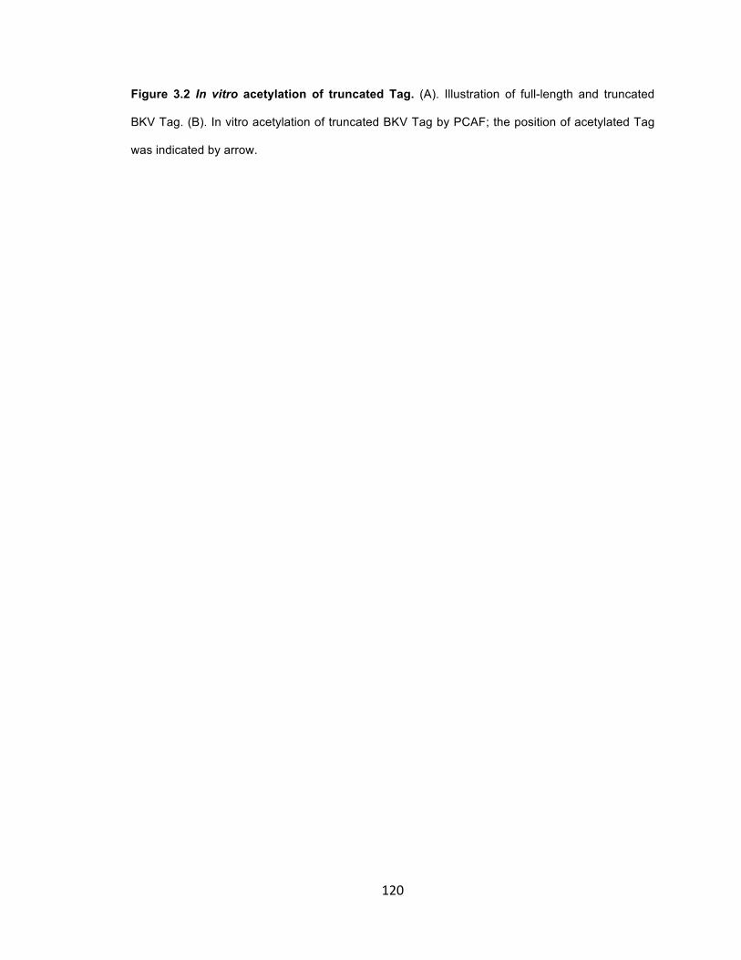
120
Figure 3.2 In vitro acetylation of truncated Tag. (A). Illustration of full-length and truncated
BKV Tag. (B). In vitro acetylation of truncated BKV Tag by PCAF; the position of acetylated Tag
was indicated by arrow.

121

122
Figure 3.2 (C) In vitro acetylation of BKV and SV40 Tags C-terminal truncated fragments by
various HATs. Lane 1-4, Cter of BKV Tag; lane 5-8, Cter of SV40 Tag. The HAT used in the
acetylation assay was indicated above each lane; w/o stands for without HAT. Acetylated
truncated Tag was detected by anti-AcK.

123
K687 is the only acetylation site in BKV Tag
There are four lysines in the HR domain of BKV Tag, which are all located in the most C-terminal
end (681~695aa). To determine which lysine(s) is/are acetylated by PCAF/GCN5, a synthetic
peptide with amino acid sequence from BKV Tag 687-695aa was acetylated by PCAF in vitro,
then analyzed by MALDI-TOF/TOF mass-spectrometry. The MALDI-MS analysis of the peptide
after acetylation shows two strong peaks with a mass difference of 42Da (Figure 3.3A), the
characteristic mass shift for a acetyl group (15). No further +42Da was observed, indicating that
only one lysine is acetylated. De novo sequencing of the acetylated peptide by tandem MS/MS
analysis indicates that K687 is acetylated (Figure 3.3B). The mass of analyzed peptide was 2Da
lower than the theoretical value, due to the disulfide formed between cysteine residues 680 and
685 (Figure 3.3B). Inclusion of DTT in the in vitro acetylation assay confirmed this (data not
shown).
To determine whether K687 is the only acetylation site in BKV Tag, the K687 residue of BKV Tag
was mutated to Arginine (R). Truncated C-terminal BKV Tag (588-695aa) with the HR domain
was tested in the in vitro acetylation assays (Figure 3.3C). While wildtype BKV Tag (588-695)
was acetylated by GCN5 efficiently (Figure 3.3C, lane1), no acetylation of the Tag K687R mutant
was detected with either GCN5 or CBP (Figure 3.3C, lane 2-3). And acetylation of BKV Tag in
vivo by PCAF was also completely disrupted by K687R mutation (Figure 3.3D, lane 1-3). These
data indicate that K687 is the only acetylation site in BKV Tag.

124
Figure 3.3 Determining acetylation site of BKV Tag. (A). Analysis of acetylated BKV Tag C-
terminal peptide (681-695aa) by MALDI-TOF mass spectrometry. The peaks from non-acetylated
and acetylated peptide are indicated by arrow. The acetylation of peptide causes 42Dalton shift
from non-acetylated peptide.

125
Figure 3.3 (B). De novo sequencing of the acetylated peptide by tandem MS/MS analysis.
Identification of ions fragmented at different orientations is indicated by different colors: Red is
from C- to N-terminus; Blue is from N- to C-terminus. Locations of acetylated lysine and the
disulfide bond between two cysteine residues are also indicated.

126
Figure 3.3 (C). In vitro acetylation of wildtype and mutant Cter of BKV Tag (588-695). Lane 1,
acetylation of wildtype BKV Tag by GCN5; lane 2, acetylation of K687R mutant BKV Tag by
GCN5; lane 3, acetylation of K687R mutant BKV Tag by CBP. (D). In vivo acetylation of full-
length wildtype and mutant BKV Tag by PCAF. Lane 1, acetylation of wildtype BKV Tag by
PCAF; lane 2, acetylation of K687R mutant BKV Tag by PCAF; lane 3, acetylation reaction in the
absence of Tag.

127

128
Expression of Gal4-PCAF/GCN5, but not Gal4-CBP/p300 inhibit BKV and SV40 DNA
replication when tethered to the core origin.
To determine whether acetylation of Tag regulates BKV/SV40 DNA replication, expression
vectors for Gal4 fusion HATs (Figure 3.1B) and BKV/SV40 templates with 5XGal4 binding sites
(Figure 3.1A) were co-transfected with cognate Tag expression vectors into HEK293/HK-2 cells
(for BKV) or CV-1 cells (for SV40). The 5XGal4-binding sites replacing the enhancer region of
SV40 and BKV resemble the recruitment of HATs to their replication origins by transcription
factors/co-factors.
Surprisingly, in contrast to mPyV (51), replication of templates with either BKV or SV40 origins
was strongly inhibited by Gal4-PCAF/GCN5, compared with Gal4 without fused HAT (Figure
3.4B, lane 1-2, 6; Figure 3.4C, lane 1-2, 5; Figure 3.4D, lane 1-3). The inhibition by Gal4-
PCAF/GCN5 is dose-dependent on input level of Gal4-GCN5 (Figure 3.4B, lane 2-5; Figure 3.4C,
lane 1-4). However, no inhibition of BKV replication was observed with expression of Gal4-
CBP/p300 (Figure 3.4B, lane 7; Figure 3.4C, lane 6); and strong stimulation of SV40 replication
was observed with expression of Gal4-p300 (Figure 3.4D, lane 4).
To determine if the inhibition of BKV DNA replication by PCAF/GCN5 is dependent on
acetylation, replication of BKV DNA by wildtype and K687R mutant BKV Tags was compared in
HK-2 cells. PCAF/GCN5 co-expression with the K687R mutant Tag inhibits DNA replication
(Figure 3.4E, lane 6-9) as it does with wildtype Tag (Figure 3.4E, lane 1-4), suggesting that
inhibition of BKV DNA replication by PCAF/GCN5 coexpression is not determined by acetylation
of Tag. The acetylation must affect some other function of Tag, such as its stability (41) or
regulation of gene transcription or cellular transformation.
To test whether over-expression of PCAF/GCN5 change the expression of Tag, which could
indirectly affect the BKV DNA replication, expression of BKV Tag in HK-2 cells was tested upon
129
co-expression of PCAF/GCN5. Increased expression of Tag was observed upon co-expression of
PCAF/GCN5(Figure 3.4F). So PCAF/GCN5 must target other cellular proteins to inhibit the BKV
DNA replication.

130
Figure 3.4 DNA replication assays with Gal-4 fusion HATs (A). Structure of Gal4 test
templates for BKV and SV40. The enhancer region of BKV and SV40 NCCR is replaced by
5XGal4 binding sites. (B). Replication of pBKVGal4 template in HK-2 cells transiently expressing
Gal4-fusion HATs. Arrow indicates the replicated template; star indicates DpnI digested input.
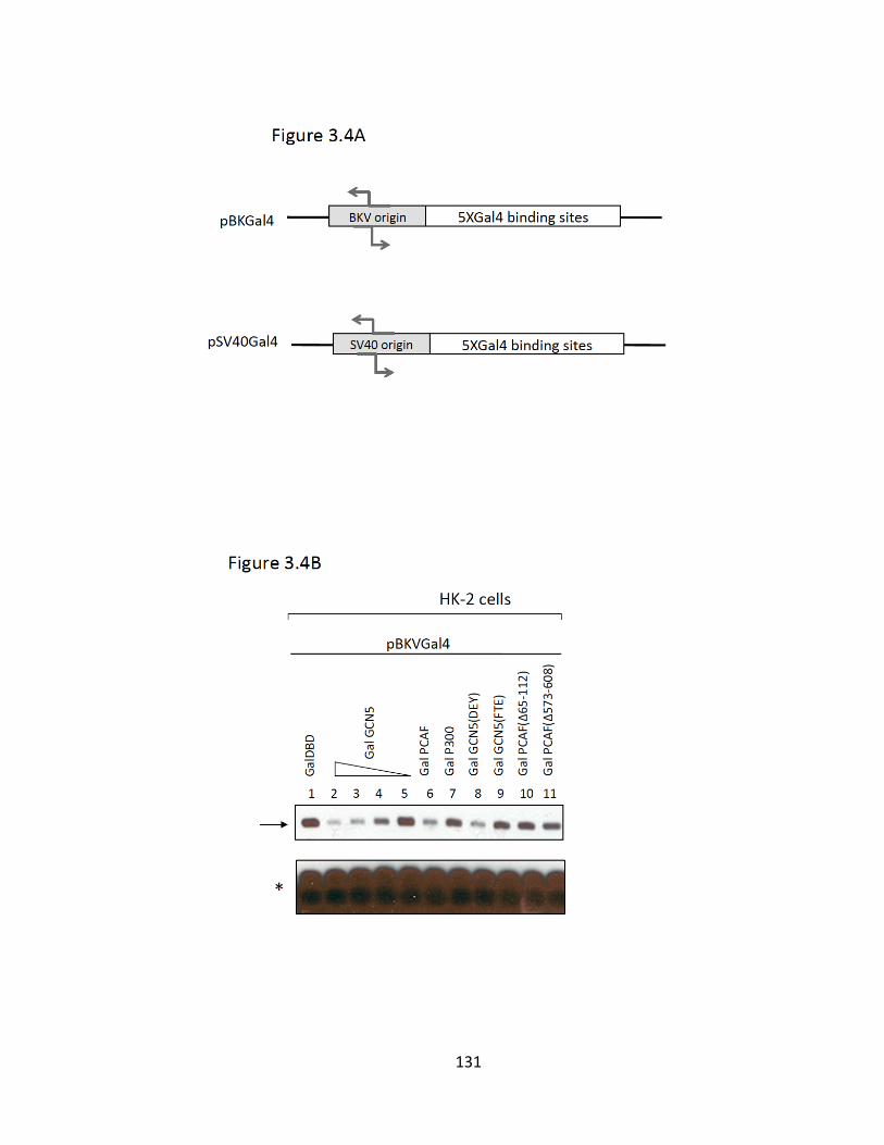
131

132
Figure 3.4 (C) Replication of pBKVGal4 template in HEK293 cells transiently expressing Gal4-
fusion HATs. (D). Replication of pSV40Gal4 template in CV-1 cells transiently expressing Gal4-
fusion HATs. (E). Replication of pBKVGal4 template by either wildtype or K687R mutant BKV Tag
in HK-2 cells. (F). Expression of Tag with co-expression of HATs.

133

134
Acetylation of BKV Tag regulates its protein stability
Although the acetylation of K687 in BKV Tag is not responsible for the inhibition of BKV DNA
replication by PCAF/GCN5, the mutation of K687R reduced BKV DNA replication compared with
wildtype Tag(Figure 3.4E). To examine whether enzymatic activities important for replication are
altered by acetylation, the helicase activity of mutant and wildtype BKV Tag were compared with
in vitro helicase assays, but no significant difference was observed (data not shown). Acetylation
of SV40 Tag has been shown to modulate its protein stability(41). It is possible that K687R
mutant BKV Tag is less stable than with wildtype Tag. The stability of wildtype and mutant BKV
Tag was compared in HEK293 cells with CHX chase assays (Figure 3.5). The transient
acetylation of Tag (Figure 3.5, lane 5-8) and K687R mutation (Figure 3.5, lane 9-12) reduced the
stability of T antigen, compared with wildtype Tag without transient acetylation by PCAF (Figure
3.5, lane 1-4). So both acetylation and the K687R mutation decrease the protein stability of BKV
Tag. This is different from the report that acetylation of SV40 Tag decrease the protein stability,
while K697R mutation stabilize the protein(41). However, our result is consistent with the
replication results showing that BKV DNA replication is less efficient with K687R mutant Tag than
wildtype Tag. The mechanism of how this acetylation regulates stability of Tag is unclear; but it
seems not to be regulated through proteasome-degradation pathway(41).
In conclusion, inhibition of BKV DNA replication by PCAF/GCN5 is not caused by the acetylation
of Tag by PCAF/GCN5; although acetylation of Tag may influence its protein stability, it is not
responsible for the inhibition of BKV DNA replication by PCAF/GCN5. PCAF/GCN5 must act
through other cellular replication factors to modulate BKV DNA replication.
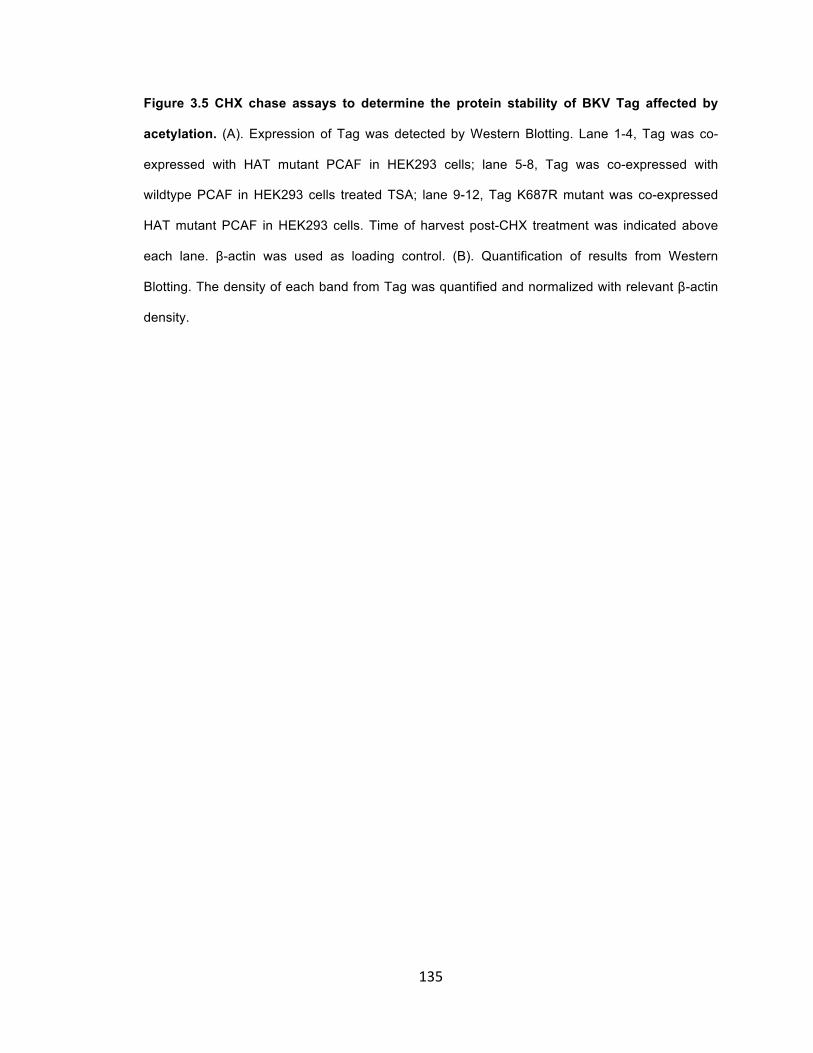
135
Figure 3.5 CHX chase assays to determine the protein stability of BKV Tag affected by
acetylation. (A). Expression of Tag was detected by Western Blotting. Lane 1-4, Tag was co-
expressed with HAT mutant PCAF in HEK293 cells; lane 5-8, Tag was co-expressed with
wildtype PCAF in HEK293 cells treated TSA; lane 9-12, Tag K687R mutant was co-expressed
HAT mutant PCAF in HEK293 cells. Time of harvest post-CHX treatment was indicated above
each lane. β-actin was used as loading control. (B). Quantification of results from Western
Blotting. The density of each band from Tag was quantified and normalized with relevant β-actin
density.

136

137
DISCUSSION
Replication of polyomavirus genome is initiated from the core origin and regulated by core origin
flanking sequences, to which transcription factors/co-factors bind (12, 14, 32). The chromatin
configuration of polyomavirus minichromosomes around the noncoding control region (NCCR)
determines the activity of viral transcription and replication (1, 7, 8, 11, 22, 50). Acetylation of
histones by HATs is an important mechanism of chromatin remodeling and regulation of gene
transcription and DNA replication (1, 23, 36, 37, 44). Besides histones, HATs also acetylate other
cellular or viral proteins to modulate their activities, some of which are known to affect
polyomavirus DNA replication directly, these including SV40/mPyV Tags(35, 51), p53(24, 27, 38),
Sp1(45), c-Jun(49), pRb(6), E2F(29, 30).
Here, we found that BKV Tag is acetylated in vitro and in vivo by PCAF/GCN5 at K687, a
conserved site of Tags from three primate polyomavirues, BKV, JCV and SV40, which however is
not present in newly identified KI, WU and Merkel Cell human polyomaviruses(2, 20, 21).
CBP/p300 acetylates SV40 Tag (K697) in vivo through interactions with p53 (3, 35). And the
interaction with p53 is indispensible for acetylation of SV40 Tag in vivo (35). However, we found
CBP/p300 can acetylate SV40 Tag efficiently in the absence of p53 in vitro (Figure 3.2C),
suggesting that p53 is not absolutely required for acetylation. Instead, it promotes the acetylation
of SV40 Tag by facilitating the interaction between CBP/p300 and Tag in vivo(3). However, no
stable complex between BKV Tag and CBP/p300 or PCAF/GCN5 has been detected in HEK293
cells, while stable interaction between BKV Tag and p53 can be detected (data not shown). This
might be because the p53 in HEK293 cells is not phosphorylated at S15, which enhances the
interaction of p53 with CBP/p300 (3, 16, 26).
Despite the homology in their HR domains, BKV and SV40 Tags are acetylated with different
efficiencies by PCAF/GCN5 and CBP/p300. PCAF/GCN5 acetylates BKV Tag much more
efficiently than CBP/p300 in vivo (Figure 3.1C) and in vitro (Figure 3.2C, lane 1-4). SV40 Tag can

138
be acetylated by either PCAF/GCN5 or CBP/p300 in vitro (Figure 3.2, lane 5-8). This is consistent
with the observation that CBP/p300 acetylates SV40 Tag in vivo (3, 35, 41). Whether
PCAF/GCN5 acetylate SV40 Tag in vivo is unknown. Although down-regulating endogenous
PCAF by siRNA did not affect SV40 Tag acetylation in vivo (35), this might be due to low
expression of endogenous PCAF or to the high expression of endogenous GCN5 and CBP/p300
as redundant sources for acetylation. So in conclusion, PCAF/GCN5 can efficiently acetylate
SV40 and BKV Tags, while CBP/p300 only selectively acetylate SV40 Tag, but not BKV Tag. The
slight difference of amino acid sequence in the HR domains of SV40 and BKV Tag might
determine their different susceptibility for acetylation by different HATs.
PCAF/GCN5, but not CBP/p300, inhibited BKV DNA replication in HK-2 and HEK293 cells when
tethered to the origin (Figure 3.4B lane 1-11; Figure 3.4C). A similar inhibitory effect of
PCAF/GCN5 was also observed with SV40 DNA replication in CV-1 cells (Figure 3.4D, lane 1-9).
This is in contrast to the stimulation of mPyV replication in NIH3T3 and FOP cells by PCAF/GCN5
(51). K687R mutation did not prevent the inhibition by PCAF/GCN5 (Figure 3.4E, lane 6-9)
suggests that PCAF/GCN5 targeting other cellular protein(s) to inhibit BKV DNA replication. The
acetylation site of mPyV Tag has recently been mapped to the second nuclear localization signal
(NLS-2) in the variable domain of mPyV Tag (Olga Kenzior and Folk unpublished data).
Interestingly, mutation of this lysine also did not abrogate the stimulation of mPyV DNA replication
by ectopic expression of PCAF/GCN5 (Olga Kenzior and Folk unpublished data). The distinct
effects of PCAF/GCN5 on polyomavirus DNA replication in primate cells and murine cells
appears not to be determined by acetylation of Tags. The basic DNA replication machineries of
murine and primate polyomavirueses are highly conserved. Then, what could be the reason for
this difference? One most likely possibility is that components of DNA replication machinery or
other related factors in primate and murine cells might be differently modified by PCAF/GCN5 and
have different consequences on DNA replication. Another more complicated possibility is that
PCAF/GCN5 inhibits the same replication factor(s) both in murine cells and primate cells; but
because PCAF/GCN5 stably interact with mPyV Tag and stimulate mPyV DNA replication by
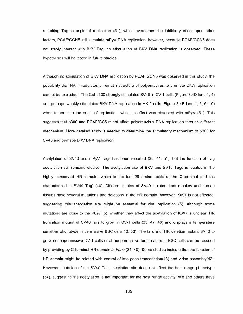
139
recruiting Tag to origin of replication (51), which overcomes the inhibitory effect upon other
factors, PCAF/GCN5 still stimulate mPyV DNA replication; however, because PCAF/GCN5 does
not stably interact with BKV Tag, no stimulation of BKV DNA replication is observed. These
hypotheses will be tested in future studies.
Although no stimulation of BKV DNA replication by PCAF/GCN5 was observed in this study, the
possibility that HAT modulates chromatin structure of polyomavirus to promote DNA replication
cannot be excluded. The Gal-p300 strongly stimulates SV40 in CV-1 cells (Figure 3.4D lane 1, 4)
and perhaps weakly stimulates BKV DNA replication in HK-2 cells (Figure 3.4E lane 1, 5, 6, 10)
when tethered to the origin of replication, while no effect was observed with mPyV (51). This
suggests that p300 and PCAF/GC5 might affect polyomavirus DNA replication through different
mechanism. More detailed study is needed to determine the stimulatory mechanism of p300 for
SV40 and perhaps BKV DNA replication.
Acetylation of SV40 and mPyV Tags has been reported (35, 41, 51), but the function of Tag
acetylation still remains elusive. The acetylation site of BKV and SV40 Tags is located in the
highly conserved HR domain, which is the last 26 amino acids at the C-terminal end (as
characterized in SV40 Tag) (48). Different strains of SV40 isolated from monkey and human
tissues have several mutations and deletions in the HR domain; however, K697 is not affected,
suggesting this acetylation site might be essential for viral replication (5). Although some
mutations are close to the K697 (5), whether they affect the acetylation of K697 is unclear. HR
truncation mutant of SV40 fails to grow in CV-1 cells (33, 47, 48) and displays a temperature
sensitive phonotype in permissive BSC cells(10, 33). The failure of HR deletion mutant SV40 to
grow in nonpermissive CV-1 cells or at nonpermissive temperature in BSC cells can be rescued
by providing by C-terminal HR domain in trans (34, 48). Some studies indicate that the function of
HR domain might be related with control of late gene transcription(43) and virion assembly(42).
However, mutation of the SV40 Tag acetylation site does not affect the host range phenotype
(34), suggesting the acetylation is not important for the host range activity. We and others have

140
found that acetylation of SV40 and BKV Tags might regulate their protein stability(41) (Figure
3.5A, B), but the mechanism is unclear and need to be tested with more sensitive assays, such
as GPS (Global Protein Stability assay)(52).
In conclusion, BKV and SV40 Tags are acetylated specifically at a conserved lysine residue in the
HR domain by CBP/p300 or PCAF/GCN5. The acetylation appears to be not essential for viral
proliferation in cell culture, probably because the HAT’s expression and activity are stable and
constant in the homogeneous cultured cells. The effect of acetylation might be more important in
vivo for the initial acute infection, spreading of virus through the body, establishment of persistent
infection and reactivation, which needs to be tested in animal models.
Reference
1. Alexiadis, V., L. Halmer, and C. Gruss. 1997. Influence of core histone acetylation on
SV40 minichromosome replication in vitro. Chromosoma 105:324-331.
2. Allander, T., K. Andreasson, S. Gupta, A. Bjerkner, G. Bogdanovic, M. A. A. Persson, T. Dalianis, T. Ramqvist, and B. Andersson. 2007. Identification of a Third Human Polyomavirus. J. Virol. 81:4130-4136.
3. Borger, D. R., and J. A. DeCaprio. 2006. Targeting of p300/CREB Binding Protein Coactivators by Simian Virus 40 Is Mediated through p53. J. Virol. 80:4292-4303.
4. Borowiec, J. A., F. B. Dean, P. A. Bullock, and J. Hurwitz. 1990. Binding and unwinding--How T antigen engages the SV40 origin of DNA replication. Cell 60:181-184.
5. Butel, J. S., and J. A. Lednicky. 1999. Cell and Molecular Biology of Simian Virus 40: Implications for Human Infections and Disease. J. Natl. Cancer Inst. 91:119-134.
6. Chan, H. M., M. Krstic-Demonacos, L. Smith, C. Demonacos, and N. B. La Thangue. 2001. Acetylation control of the retinoblastoma tumour-suppressor protein. Nature cell biology 3:667-674.
7. Cheng, L., and T. J. Kelly. 1989. Transcriptional activator nuclear factor I stimulates the replication of SV40 minichromosomes in vivo and in vitro. Cell 59:541-551.

141
8. Cheng, L. Z., J. L. Workman, R. E. Kingston, and T. J. Kelly. 1992. Regulation of DNA replication in vitro by the transcriptional activation domain of GAL4-VP16. Proc. Natl. Acad. Sci. U. S. A. 89:589-593.
9. Choudhary, C., C. Kumar, F. Gnad, M. L. Nielsen, M. Rehman, T. C. Walther, J. V. Olsen, and M. Mann. 2009. Lysine Acetylation Targets Protein Complexes and Co-Regulates Major Cellular Functions. Science 325:834-840.
10. Cole, C. N., and T. P. Stacy. 1987. Biological properties of simian virus 40 host range mutants lacking the COOH-terminus of large T antigen. Virology 161:170-180.
11. Cremisi, C. 1981. The appearance of DNase I hypersensitive sites at the 5' end of the late SV40 genes is correlated with the transcriptional switch. Nucleic Acids Res. 9:5949-5964.
12. DePamphilis, M. L. 1993. Eukaryotic DNA Replication: Anatomy of An Origin. Annu. Rev. Biochem. 62:29-63.
13. DePamphilis, M. L. 1993. How transcription factors regulate origins of DNA replication in eukaryotic cells. Trends Cell Biol. 3:161-167.
14. DePamphilis, M. L. 1988. Transcriptional elements as components of eukaryotic origins of DNA replication. Cell 52:635-638.
15. Dormeyer, W., M. Ott, and M. Schnolzer. 2005. Probing lysine acetylation in proteins: strategies, limitations, and pitfalls of in vitro acetyltransferase assays. Mol Cell Proteomics 4:1226-1239.
16. Dumaz, N., and D. W. Meek. 1999. Serine15 phosphorylation stimulates p53 transactivation but does not directly influence interaction with HDM2. The EMBO journal 18:7002-7010.
17. Fanning, E. 1992. Simian virus 40 large T antigen: the puzzle, the pieces, and the emerging picture. J. Virol. 66:1289-1293.
18. Fanning, E., and R. Knippers. 1992. Structure and Function of Simian Virus 40 Large Tumor Antigen. Annu. Rev. Biochem. 61:55-85.
19. Fanning, E., and K. Zhao. 2009. SV40 DNA replication: From the A gene to a nanomachine. Virology 384:352-359.
20. Feng, H., M. Shuda, Y. Chang, and P. S. Moore. 2008. Clonal integration of a polyomavirus in human Merkel cell carcinoma. Science 319:1096-1100.

142
21. Gaynor, A. M., M. D. Nissen, D. M. Whiley, I. M. Mackay, S. B. Lambert, G. Wu, D. C. Brennan, G. A. Storch, T. P. Sloots, and D. Wang. 2007. Identification of a novel polyomavirus from patients with acute respiratory tract infections. PLoS Pathog 3:e64.
22. Gross, D. S., and W. T. Garrard. 1988. Nuclease hypersensitive sites in chromatin. Annu. Rev. Biochem. 57:159-197.
23. Grunstein, M. 1997. Histone acetylation in chromatin structure and transcription. Nature 389:349-352.
24. Gu, W., and R. G. Roeder. 1997. Activation of p53 sequence-specific DNA binding by acetylation of the p53 C-terminal domain. Cell 90:595-606.
25. Hirt, B. 1967. Selective extraction of polyoma DNA from infected mouse cell cultures. J. Mol. Biol. 26:365-369.
26. Lambert, P. F., F. Kashanchi, M. F. Radonovich, R. Shiekhattar, and J. N. Brady. 1998. Phosphorylation of p53 serine 15 increases interaction with CBP. The Journal of biological chemistry 273:33048-33053.
27. Liu, L., D. M. Scolnick, R. C. Trievel, H. B. Zhang, R. Marmorstein, T. D. Halazonetis, and S. L. Berger. 1999. p53 sites acetylated in vitro by PCAF and p300 are acetylated in vivo in response to DNA damage. Mol. Cell. Biol. 19:1202-1209.
28. Mahon, C., B. Liang, I. Tikhanovich, J. R. Abend, M. J. Imperiale, H. P. Nasheuer, and W. R. Folk. 2009. Restriction of human polyomavirus BK virus DNA replication in murine cells and extracts. J. Virol. 83:5708-5717.
29. Martinez-Balbas, M. A., U. M. Bauer, S. J. Nielsen, A. Brehm, and T. Kouzarides. 2000. Regulation of E2F1 activity by acetylation. The EMBO journal 19:662-671.
30. Marzio, G., C. Wagener, M. I. Gutierrez, P. Cartwright, K. Helin, and M. Giacca. 2000. E2F family members are differentially regulated by reversible acetylation. The Journal of biological chemistry 275:10887-10892.
31. McVey, D., L. Brizuela, I. Mohr, D. R. Marshak, Y. Gluzman, and D. Beach. 1989. Phosphorylation of large tumour antigen by cdc2 stimulates SV40 DNA replication. Nature 341:503-507.
32. Murakami, Y., and Y. Ito. 1999. Transcription factors in DNA replication. Frontiers in bioscience : a journal and virtual library 4:D824-833.
143
33. Pipas, J. M. 1985. Mutations near the carboxyl terminus of the simian virus 40 large tumor antigen alter viral host range. J. Virol. 54:569-575.
34. Poulin, D. L., and J. A. DeCaprio. 2006. The carboxyl-terminal domain of large T antigen rescues SV40 host range activity in trans independent of acetylation. Virology 349:212-221.
35. Poulin, D. L., A. L. Kung, and J. A. DeCaprio. 2004. p53 Targets Simian Virus 40 Large T Antigen for Acetylation by CBP. J. Virol. 78:8245-8253.
36. Quintini, G., K. Treuner, C. Gruss, and R. Knippers. 1996. Role of amino-terminal histone domains in chromatin replication. Mol. Cell. Biol. 16:2888-2897.
37. Rocha, W., and A. Verreault. 2008. Clothing up DNA for all seasons: Histone chaperones and nucleosome assembly pathways. FEBS Lett. 582:1938-1949.
38. Sakaguchi, K., J. E. Herrera, S. Saito, T. Miki, M. Bustin, A. Vassilev, C. W. Anderson, and E. Appella. 1998. DNA damage activates p53 through a phosphorylation-acetylation cascade. Genes Dev. 12:2831-2841.
39. Schneider, J., and E. Fanning. 1988. Mutations in the phosphorylation sites of simian virus 40 (SV40) T antigen alter its origin DNA-binding specificity for sites I or II and affect SV40 DNA replication activity. J. Virol. 62:1598-1605.
40. Shi, Y., G. E. Dodson, S. Shaikh, K. Rundell, and R. S. Tibbetts. 2005. Ataxia-telangiectasia-mutated (ATM) is a T-antigen kinase that controls SV40 viral replication in vivo. The Journal of biological chemistry 280:40195-40200.
41. Shimazu, T., Y. Komatsu, K. I. Nakayama, H. Fukazawa, S. Horinouchi, and M. Yoshida. 2006. Regulation of SV40 large T-antigen stability by reversible acetylation. Oncogene 25:7391-7400.
42. Spence, S. L., and J. M. Pipas. 1994. Simian virus 40 large T antigen host range domain functions in virion assembly. J. Virol. 68:4227-4240.
43. Stacy, T., M. Chamberlain, and C. N. Cole. 1989. Simian virus 40 host range/helper function mutations cause multiple defects in viral late gene expression. J. Virol. 63:5208-5215.
44. Struhl, K. 1998. Histone acetylation and transcriptional regulatory mechanisms. Genes Dev. 12:599-606.

144
45. Suzuki, T., A. Kimura, R. Nagai, and M. Horikoshi. 2000. Regulation of interaction of the acetyltransferase region of p300 and the DNA-binding domain of Sp1 on and through DNA binding. Genes to cells : devoted to molecular & cellular mechanisms 5:29-41.
46. Tikhanovich, I., and H. P. Nasheuer. 2010. Host-specific replication of BK virus DNA in mouse cell extracts is independently controlled by DNA polymerase alpha-primase and inhibitory activities. J. Virol. 84:6636-6644.
47. Tornow, J., and C. N. Cole. 1983. Nonviable mutants of simian virus 40 with deletions near the 3' end of gene A define a function for large T antigen required after onset of viral DNA replication. J. Virol. 47:487-494.
48. Tornow, J., M. Polvino-Bodnar, G. Santangelo, and C. N. Cole. 1985. Two separable functional domains of simian virus 40 large T antigen: carboxyl-terminal region of simian virus 40 large T antigen is required for efficient capsid protein synthesis. J. Virol. 53:415-424.
49. Vries, R. G., M. Prudenziati, C. Zwartjes, M. Verlaan, E. Kalkhoven, and A. Zantema. 2001. A specific lysine in c-Jun is required for transcriptional repression by E1A and is acetylated by p300. The EMBO journal 20:6095-6103.
50. Weiss, E., C. Ruhlmann, and P. Oudet. 1986. Transcriptionally active SV40 minichromosomes are restriction enzyme sensitive and contain a nucleosome-free origin region. Nucleic Acids Res. 14:2045-2058.
51. Xie, A.-Y., V. P. Bermudez, and W. R. Folk. 2002. Stimulation of DNA Replication from the Polyomavirus Origin by PCAF and GCN5 Acetyltransferases: Acetylation of Large T Antigen. Mol. Cell. Biol. 22:7907-7918.
52. Yen, H.-C. S., Q. Xu, D. M. Chou, Z. Zhao, and S. J. Elledge. 2008. Global Protein Stability Profiling in Mammalian Cells. Science 322:918-923.

145
CHAPTER 4
Restriction of BKV DNA replication in murine cells and inhibition
of BKV DNA replication by noncoding RNAs
INTRODUCTION
BKV and JCV polyomaviruses are endemic in humans but in certain individuals can cause
nephropathy and leukoencephalopathy, respectively, and also may be associated with some
cancers and other pathologies (2, 5, 16-18, 27, 29, 49). Recently, additional human
polyomaviruses have been discovered (KI, WU and the Merkel cell polyomavirus (MKV)(1, 13,
15), and preliminary evidence indicates the first two are associated with the oral cavity and
respiratory system and the third is associated with and perhaps the cause of Merkel cell
carcinomas (reviewed by (8, 21, 22)). Remarkably, despite the considerable understanding of the
biology of the Polyomaviridae, there is little understanding of how these viruses establish and
maintain persistent infections, nor are there good means to abrogate or mitigate the infections
and pathologies caused by these viruses.
Replication of polyomavirus DNA requires the large T antigen (Tag), with the host supplying the
other replication factors (12, 19, 28, 46). Polyomavirus Non-Coding Control Regions (NCCRs)
comprise the origins of DNA replication and cis-acting regulatory elements both 5’ and 3’ of the
core origin (12, 19, 28) that bind Tag and cell factors regulating viral transcription and DNA
replication (9, 31). The development of in vitro DNA replication systems has allowed the

146
identification and characterization of the proteins required for polyomavirus replication (26, 28, 30,
34): the Tag helicase activity unwinds DNA in an ATP- and phosphorylation-dependent process
(11, 12, 28, 33, 35, 38, 48), and single-stranded DNA (ssDNA) is bound by replication protein A
(RPA), and topoisomerase I relieves torsional stress ahead of the replication fork (3, 40).
Subsequently the primase activity of DNA polymerase α-primase (Pol-α primase) synthesizes
short RNA primers that are elongated by DNA polymerase α (10). Leading strand synthesis is
completed by DNA polymerase δ, RPA, proliferating nuclear antigen (PCNA) and replication
factor C (RFC). Replication of the lagging strand is mediated by RPA, Pol-α primase, DNA
polymerase δ and auxiliary proteins (35, 38, 39, 48). Heretofore, there has been no hint that small
cellular RNAs might also regulate polyomavirus DNA replication through the viral NCCRs,
although recently it has been determined that small ncRNAs may directly regulate eukaryotic
DNA replication (24), and that RNAs help to recruit the cellular origin recognition complex (ORC)
to the Epstein Barr Virus (EBV) origin of replication and stimulate EBV DNA replication (37).
SV40 (simian virus 40) does not replicate in mouse cells nor do mouse cell extracts support their
in vitro replication, and in turn, mouse polyomavirus (mPyV) does not replicate in human cells or
human cell extracts (6, 23, 42-44). Restriction of SV40 replication occurs at the level of initiation
by Pol-α primase (6, 44, 46). But why BKV replication is restricted in murine cells has not been
investigated.
Our analysis of the restriction of BKV replication in murine cells revealed a more complex
regulation: while the human Pol-α primase complex is required for BKV DNA replication, other
factors within mouse cells and extracts inhibit replication (28, 46). Inhibitory activities were
purified, some of which were determined to be small cellular RNAs, termed small replication
regulatory RNAs (srRNAs), that act through the BKV NCCR. These suggest that cellular small
noncoding RNAs regulate viral DNA replication, which may have great implication in viral tropism,
establishment of persistent infection and reactivation.

147
MATERIALS AND METHODS
Plasmids. pOriBKV (termed B-B-B in Figure 4.1A, Figure 4.2A ) was generated by inserting PCR
fragments of whole NCCR (positions 5031 to 282) of archetype BKV Dik strain (kindly provided by
J. Lednicky; GenBank Accession #AB211369) into the polylinker region(XmaI/PstI) of pUC18
plasmid. Other similar pUC18-based plasmid DNAs with complete viral origins included pOriJCV
(Mad-1 strain), pOriSV40 (SV-S strain), and pOrimPyV (P-P-P; A3 strain). Chimearic templates
were generated by ligation of PCR fragments into XmaI/PstI sites of pUC18 plasmid. The pUC18
plasmid without an insert served as negative control (pOri-) for cell-free DNA replication as well
as a vector for cloning all viral origins.DNAs for replication assays were verified by sequencing
and purified with Qiagen Midiprep kits. pU6 was constructed by insertion of a U6 promoter DNA
fragment cut out with Bgl II and EcoR I from a modified pSirenRetreQ vector into the BamH I and
EcoR I digested pUC18 vector. pU6-B-5-1 and pU6-mY1 were constructed by insertion of
annealed synthetic oligonucleotides of DNA encoding B-5-1 and mY1 RNA sequences into pU6
vectors using BamH I and EcoR I sites. Sequences of oligonucleotides
for B-5-1 are
5’GATCACCGGTCTCACGACGACATTCCAGACGTGGCCTCGTGGGTGCTTCCACGTTGCGAA
CACCCCGATTTCCCGGTCCCTTTTTTGGAA 3’ and
5’AATTTTCCAAAAAAGGGACCGGGAAATCGGGGTGTTCGCAACGTGGAAGCACCCACGAGG
CCACGTCTGGAATGTCGTCGTGAGACCGGT3’;
for mY1 are
5’GATCGGCTGGTCCGAAGGTAGTGAGTTATCTCAATTGATTGTTCACAGTCAGTTACAGATT
GAACTCCTGTTCTACACTTTCCCCCCTTCTCACTACTGCACTTGACTAGTCTTTTTTGGAA 3’
and
5’AATTTTCCAAAAAAGACTAGTCAAGTGCAGTAGTGAGAAGGGGGGAAAGTGTAGAACAGG
AGTTCAATCTGTAACTGACTGTGAACAATCAATTGAGATAACTCACTACCTTCGGACCAGCC
3’.

148
In vivo DNA replication assays. Murine TCMK-1 cells were grown in DMEM
with 10% FBS, seeded in eight-well plates (1 X 10⁵ cells/well), and incubated overnight at 37°C.
Cells were transfected with Lipofectamine and Plus reagent (Invitrogen, Carlsbad, CA) with
expression vectors for TAg (0.6 ug of DNA) and template plasmid (0.4 ug). After incubation of
cells with DNA-Lipofectamine and PLUS reagent mixture for 4 h in 500 ul serum-free DMEM, the
transfection solution was replaced with 2 ml of DMEM containing 20% FBS. Similarly, human
HEK 293 cells were grown in DMEM with 10% FBS, seeded in 12-well plates (4 X10⁵ cells/well),
and incubated overnight at 37°C. Cells were transfected with expression vector for TAg (5 ng),
template plasmid (50 ng), and pUC18 empty vector (0.65 ug) as carrier DNA with Lipofectamine
and Plus reagent. DNA Lipofectamine and Plus reagent mixtures were incubated with HEK 293
cells as described above. Human kidney proximal tubular epithelial cells (HK-2 cells) were grown
in RPMI-1640 with 10% FBS, seeded in 12-well plates (1 x 10⁵ cells/well), and incubated
overnight at 37°C. Cells were transfected with expression vector for TAg (10 ng), template
plasmid pOriBKV (10 ng), srRNA expression vectors pU6-mY1/pU6-B-5-1 or empty control vector
pUC (200 ng), and pBC plasmid vector (500 ng) as carrier DNA using Fugene HD (Roche)
transfection reagent. Cells were harvested at 48 h after transfection, and low-molecular-weight
DNAs were isolated by the Hirt protocol with Promega Miniprep columns, digested with EcoRI to
linearize the plasmid, and digested with DpnI to distinguish input from replicated DNA. The DpnI
resistant DNA was resolved from digested DNA by agarose gel electrophoresis (1%). After
transfer of the DNA to a nylon membrane, DpnI-resistant DNA was detected by Southern blotting
with a biotinylated probe of the lacZ gene (~400 nucleotides) of the pUC18 vector and visualized
by chemiluminescent nucleic acid detection (Pierce).
In vitro DNA replication assays. Replication of DNAs in vitro was assayed as described by
Stadlbauer and coworkers with slight modifications (94). Briefly, the reaction mixtures (30 ul)
contained 20 mM HEPES (pH 7.8); 7 mM magnesium acetate; 1 mM DTT; 4 mM ATP; 200 uM
each CTP, UTP, and GTP; 50 uM dCTP; 100 uM each dATP, dTTP, and dGTP; 40 mM creatine

149
phosphate di-Tris (pH 7.8); 40 ug/ml creatine kinase plus 5 %Ci of [α32P]dCTP (3,000 Ci/mmol);
0.25 ug test plasmid DNA; FM3A or HeLa cell extract (25 to 75 ug of protein); and purified TAgs
at the indicated concentrations. After incubation for 60 min at 37°C, reaction products were
precipitated with cold 10% (wt/vol) trichloroacetic acid containing 2.5% (wt/vol) sodium
pyrophosphate, spotted on glass fiber filters (GF/C; Whatman), washed with 1 M HCl, and
analyzed by scintillation counting.
Monopolymerase DNA replication assay. The BKV and SV40 monopolymerase replication
assay contained 0.25 µg of pOriBKV DNA or 0.5 µg of pOriSV40, respectively. The assay was
supplemented with 50 ng topoisomerase I, 100 ng Pol-primase and 1 µg RPA, in 30 mM HEPES-
KOH, pH 7.8, 7 mM MgAc, 0.1 mM EGTA, 0.5 mM DTT, 200 µM each UTP, GTP, and CTP, 4
mM ATP, 100 µM each dATP, dGTP, and dTTP, and 10µM dCTP, 40 mM creatine phosphate, 1
µg creatine kinase, 0.1 mg/ml heat-treated BSA, and 5 µCi [α32P]-dCTP (3000 Ci/mmol, Perkin-
Elmer) in 40 µl. The small RNAs were added to the assay as indicated in the figure legends.
Purified BKV or SV40 TAg (0.2 µg) was added to start the reaction as indicated, and after
incubation for 60 min at 37°C, the reaction products were precipitated with cold 10% (w/v) TCA
containing 2.5% (w/v) sodium pyrophosphate and spotted on glass fiber filters (GF/C; Whatman),
washed with 1 M HCl and analyzed by scintillation counting.
RESULTS
Restriction of BKV DNA replication in murine cells.
BKV infection of murine cells suggested that BKV DNA replication is restricted in murine cells
(data not shown)(28). To determine whether BKV NCCR is responsible for the restriction,
replication of chimearic BKV templates with BKV core origin and flanking sequences from either
BKV or mPyV were analyzed in human and murine cells (Figure 4.1A). Substitution of BKV core

150
origin flanking sequences with those of mPyV reduced BKV replication efficiency in human cells
(Figure 4.1A lane1-4). These data indicate core origin flanking sequences regulate BKV DNA
replication. However, chimearic templates with the BKV core origin do not replicate in murine
cells (Figure 4.1A, lane 5-8), while templates with the mPyV core origin replicate well (Figure
4.1A, lane 9) (data not shown). BKV and mPyV Tags were equally expressed in murine cells
(Figure 4.1B). These data indicate that while core origin flanking sequences modulate BKV DNA
replication, the core origin is primarily responsible for the restriction of BKV replication in murine
cells.

151
Figure 4.1 In vivo DNA replication assays of chimearic templates (done by Bo Liang) (A) In
vivo DNA replication of BKV and mPyV in human and mouse cells. Vectors expressing BKV TAg
(lanes 1 to 8) or mPyV TAg (lane 9) were cotransfected into human HEK293 (lanes 1 to 4) or
mouse TCMK-1 (lanes 5 to 9) cells together with plasmids containing the complete BKV origin
(lanes 1 and 5, B-B-B), the complete mPyV origin (lane 9, P-P-P), and BKV-mPyV chimeric
origins (lanes 2 to 4 and 6 to 8). At 48 h after transfection, DNA was isolated and analyzed by
Southern blotting. DNA replication products are marked by arrows. (B). Expression of BKV and
mPyV Tags in transfected TCMK-1 cells, detected with anti-Flag antibody in Western Blot. Equal
amount of crude cell lysates were loaded for each sample.

152

153
Inhibition of BKV DNA replication by murine nuclear extracts.
In vitro DNA replication system was established by Mahon et al., with which restriction of BKV
DNA replication in murine cells is reconstituted and studied in vitro (28). In murine FM3A nuclear
extracts, neither BKV nor SV40 (Figure 4.2A, bars 2,3) replicate with cognate Tags; while mPyV
replicates (Figure 4.2A, bars 1,4) with mPyV Tag. Adding human pol α-primase (polymerase α
primase) into FM3A nuclear extracts allowed replication of SV40, but not BKV (Figure 4.2A, bars
5-7). This indicates that mechanisms for the restriction of BKV and SV40 DNA replication in
murine cells are different. Adding either human RPA (hRPA) or Topoisomerase I (hTopo I) did not
stimulate BKV DNA replication in FM3A nuclear extracts (Figure 4.2A, bars 9, 11), although
hRPA and hTopoI are functional and can stimulate mPyV DNA replication in murine extracts
(Figure 4.2A, bars 8, 10). This suggests that restriction of BKV DNA replication is not due to
incompatibility between Tag and RPA or Topoisomerase I.
To determine whether functional interaction occurs between murine pol-α primase and BKV Tag,
monopolymerase system containing purified components of initiation complex (pol-α primase,
RPA, Topoisomerase I, Tag) was established. Using monopolymerase assays, Tikhanovich et al.
found that murine pol-α primase does not support initiation of BKV DNA replication by BKV Tag
(46). However, adding human pol-α primase into murine nuclear extracts does not support BKV
DNA replication (Figure 4.2A, bars 5-7), suggesting in addition to pol-α primase, there is/are other
inhibitory factors in murine nuclear extracts(46).
Mahon et al. also found that replication in the BKV monopolymerase system (BKV template and
Tag, hRPA, hPol-α primase, hRPA) was not affected by human cell extracts added immediately
prior to BKV TAg (Figure 4.2B, compare bars 1 and 2), whereas addition of murine extracts
reduced the incorporation of dNMPs by almost 50% (compare bar 3 with bars 1 and 2)(28). In
comparison, adding murine extracts reduced the incorporation of dNMPs in the SV40
monopolymerase system by only 10% (Figure 4.2C, compare bar 3 with bar 1), and the addition

154
of human extracts to the SV40 system did not influence replication (compare bars 1 and 2)(28).
This indicates that some cellular factors in murine nuclear extracts inhibit the initiation of BKV
DNA replication.

155
Figure 4.2 In vitro DNA replication assays (done by Cathal Mahon) (A). Modulation of
polyomavirus DNA replication in murine cell extracts by human replication factors. In vitro DNA
replication in the presence of equal amounts of purified recombinant BKV, mPyV, and SV40 TAg
(shaded bars) using mouse FM3A cell extracts is shown. Bars 1 to 4 show DNA synthesis in the
presence of mouse cell extracts and either mPyV TAg/P-P-P, BKV TAg/B-B-B, or SV40
Tag/pOriSV40. Incorporation of dNMPs into P-P-P in the absence of Tag served as a negative
control. DNA synthesis with mouse cell extract and additional human DNA Pol-α primase (2 units
of hPol-α primase) is depicted in bars 5 to 7 (mPyV TAg/P-P-P, BKV TAg/B-B-B, and SV40
TAg/pOriSV40, respectively). The DNA synthesis of mousecell extracts with an additional 0.5 %g
of hRPA using mPyV TAg/P-P-P and BKV TAg/B-B-B is presented in bars 8 and 9, respectively,
whereas the influence of human topoisomerase I (hTopo I, 120 ng) on the DNA synthesis in the
presence of mouse cell extracts with mPyV TAg/P-P-P and BKV TAg/B-B-B is shown in bars 10
and 11, respectively. All assays were carried out in triplicate, and the results presented are the
averages from two independent experiments. (B). The effect of human and mouse nuclear
extracts on DNA synthesis by hPol-α primase was determined with a BKV origin of replication.
The incorporation of radioactive dNMPs using the BKV origin of replication as a template was
measured in the presence of buffer but no additional proteins or with 15 ug human or mouse cell
extracts (bars 1, 2, and 3, respectively). DNA synthesis in the presence of DNA lacking an origin
of replication served as a negative control (bar 4). (C) The effect of human and mouse proteins
on the DNA synthesis by human DNA Pol-α primase was determined with an SV40 origin of
replication. The incorporation of radioactive dNMPs was determined in the presence of buffer but
no additional proteins or with 15 ug human or mouse cell extracts (bars 1, 2, and 3, respectively).
DNA synthesis in the presence of DNA lacking an origin of replication served as a negative
control (bar 4). Incorporation of dNMPs into DNA was measured by scintillation counting. DNA
synthesis was determined in duplicate and repeated three times. The averages from these
experiments and the standard deviations are presented.

156
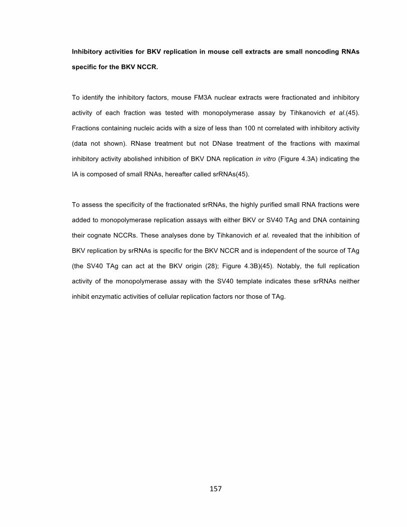
157
Inhibitory activities for BKV replication in mouse cell extracts are small noncoding RNAs
specific for the BKV NCCR.
To identify the inhibitory factors, mouse FM3A nuclear extracts were fractionated and inhibitory
activity of each fraction was tested with monopolymerase assay by Tihkanovich et al.(45).
Fractions containing nucleic acids with a size of less than 100 nt correlated with inhibitory activity
(data not shown). RNase treatment but not DNase treatment of the fractions with maximal
inhibitory activity abolished inhibition of BKV DNA replication in vitro (Figure 4.3A) indicating the
IA is composed of small RNAs, hereafter called srRNAs(45).
To assess the specificity of the fractionated srRNAs, the highly purified small RNA fractions were
added to monopolymerase replication assays with either BKV or SV40 TAg and DNA containing
their cognate NCCRs. These analyses done by Tihkanovich et al. revealed that the inhibition of
BKV replication by srRNAs is specific for the BKV NCCR and is independent of the source of TAg
(the SV40 TAg can act at the BKV origin (28); Figure 4.3B)(45). Notably, the full replication
activity of the monopolymerase assay with the SV40 template indicates these srRNAs neither
inhibit enzymatic activities of cellular replication factors nor those of TAg.

158
Figure 4.3 Determination of nature of inhibitory activity (done by Irina Tikhanovich) (A). Mono
Q chromatography-purified IA from mouse FM3A cells was treated with RNase or DNase, and
then tested in BKV monopolymerase replication assays. (B). srRNA fraction purified from FM3A
extracts were added to the monopolymerase replication assay using DNA containing the BKV
origin of replication with SV40 TAg, or DNA containing the SV40 origin of replication with BKV
TAg (75 ng of RNA with an estimated length of 50 nt (length estimation of RNA is derived from
the cloning results without SELEX) yield a 50% inhibition with an 36-fold molar excess of RNA
over origin DNA). Reactions with BKV TAg and DNA containing the BKV origin of replication
served as controls. The incorporation of dNMPs into the BKV origin DNA was detected by acid
precipitation and scintillation counting. DNA synthesis was determined in duplicates and repeated
three times. The average of incorporation of dNMPs and the standard deviations are presented.

159

160
Specific srRNAs inhibit BKV DNA replication.
Previous observations that templates containing BKV core origins with heterologous murine
polyomavirus (mPyV) origin flanking sequences can be replicated by FM3A mouse cell extracts
supplemented with human Pol-primase complex (46) and that inhibitory srRNAs are specific for
BKV NCCR suggest that the inhibitory activity of srRNAs act through the viral NCCR. Specific
srRNAs that can hybridize with denatured BKV NCCR were enriched and cloned with modified
SELEX (Systematic Evolution of Ligands by EXponential enrichment). cDNAs derived from
purified srRNAs were cloned and transcribed in vitro. Their inhibitory activities were tested in the
in vitro DNA replication systems. The SELEX-selected B-5-1 and B-5-8 srRNAs inhibited in vitro
BKV DNA replication to a greater extent than other RNAs (Figure 4.4A). These works are done by
Irina Tikhanovich(45).
To ascertain if srRNAs function in vivo, the sequence of B-5-1 srRNA was placed under the U6
promoter (pU6-B-5-1). The pU6-B-5-1 was transiently transfected with BKV template and Tag
expression vector into human kidney tubular epithelial cells (HK-2 cells). In multiple independent
assays, BKV template replication in cells transiently expressing B-5-1 srRNA was consistently
inhibited by up to 70% compared to transfections with the vector DNA (pUC) or the vector
expressing an unspecific mouse Y-RNA (pU6-mY1) (Figure 4.4B), indicating srRNAs that inhibit
the BKV replication in vitro also inhibit BKV DNA replication in vivo.
Bioinformatic analysis of B-5-1 and B-5-8 sequences indicates that they are complimentary to
both strands of BKV NCCR; and mutations in the complimentary region disrupting the
complimentarity also concomitantly abolish the inhibitory effect of srRNAs (data not shown) (45).
De novo design of artificially srRNAs hybridizing with both strands of BKV NCCR also confirm
that small noncoding RNAs complimentary to both strands of BKV NCCR can inhibit BKV DNA
replication (45).
161
Figure 4.4 Effect of srRNAs on BKV DNA replication in vitro and in vivo (A) (done by Irina
Tikhanovich) Replication of polyomavirus DNA in the presence of srRNAs. srRNAs were
transcribed in vitro using oligonucleotides with an SP6 promoter; the sequence coding for the
indicated RNAs as templates were assayed for inhibition of BKV DNA replication either in HeLa
extract or in the monopolymerase replication assay. DNA synthesis was determined in duplicates
and repeated three times. The average of incorporation of dNMPs and the standard deviations
are presented. (B) (done by Bo Liang) In vivo BKV DNA replication in the presence of srRNA.
BKV DNA replication in vivo was measured by transfecting HK-2 cells with BKV-origin containing
plasmids and BKV TAg expression vector together with an empty vector (pUC, lane 1), or a
control vector expressing mouse Y1-RNA (pU6-mY1, lane 2; mouse Y1-RNA also does not inhibit
BKV DNA replication in vitro (Tikhanovich and Nasheuer, unpublished data)) or B-5-1 expression
vector (pU6-B-5-1, lane 3). Cells were harvested 48 hours post transfection and replicated BKV
DNA was analyzed by EcoR I and Dpn I digestion, agarose gel electrophoresis and Southern
Blotting. The average quantification of band density of three in vivo DNA replication experiments
and the standard deviation are presented. The amount of Dpn I-resistant replication products in
the transfection of pUC empty vector were arbitrarily set to 100%.

162

163
DISCUSSION
BKV persistently infects most people, but how it establishes latency and reactivates in kidney
allograft and causes PVAN in renal transplantation patients remains poorly defined. Immune-
suppression is required but not sufficient for development of PVAN(14, 18, 32), suggesting other
factors, such as ischemia/reperfusion injury, inflammatory response and tissue regeneration also
contribute to the reactivation of BKV and pathogenesis of PVAN. Replication of polyomavirus is
species-specific: like SV40 and JCV, BKV does not replicate in murine cells. Understanding the
mechanism for the restriction of BKV replication in murine cells may shed light on how BKV
maintains persistent infection and reactivates in kidney allograft. This may have implication for
development of new anti-viral treatment for PVAN and other diseases caused by polyomavirus
infection.
Infection of murine 3T3 cells with BKV suggests that viral DNA replication is restricted in murine
cells and the restriction is not due to low expression of Tag (28). Over-expression of BKV Tag in
murine cells dose not permit replication of BKV template in murine cells, further confirming that
BKV DNA replication is blocked in murine cells (Figure 4.1A, B). Although the fundamental DNA
replication machineries of different members of polyomaviruses are highly similar, the
mechanisms for the host-specificity of their DNA replication seem to be slightly different: the host-
specificity of mPyV and SV40 DNA replication is due to incompatibility of Tag with host pol-α
primase (6, 23, 42-44); restriction of JCV DNA replication in murine cells appears to involve
different mechanism(s), becausue murine pol-α primase supports in vitro JCV DNA replication in
murine cell nuclear extracts (41); BKV Tag is incompatible with murine pol-α primase (46),
however, adding human pol-α primase dose not support BKV DNA replication in murine extracts
(Figure 4.2A), indicating restriction of BKV DNA replication in murine cells is not only due to
incompatibility of Tag with murine pol-α primase, but also involves other inhibitory factor(s)
present in murine cell nuclear extracts (Figure 4.2B,C).

164
Tikhanovich et.al. have fractionated the murine cell nuclear extracts and found that the fraction of
small RNAs(~100nt) is mainly responsible for the inhibition of BKV DNA replication in an in vitro
assay (Figure 4.3A)(45). Further characterization of these small RNAs (srRNA) indicates that
some of them act through BKV NCCR and their inhibitory activity requires the hybridization of
srRNAs with both strands of BKV NCCR(45). It has been proposed that the inhibitory srRNAs act
through hybridization with both strands of BKV NCCR when the replication origin is opened up
during the initiation of DNA replication. Which step(s) during the initiation is/are inhibited is not
clear. RNA aptamers have been shown to inhibit SARS coronavirus helicase activity(20); but
helicase activity of BKV Tag is marginally affected by srRNAs (data not shown)(45), which cannot
account for their strong inhibitory effect on DNA replication. Enzymatic activity of pol-α primase is
unlikely to be significantly affected, because srRNAs hardly affect SV40 DNA replication, which
uses the same pol-α primase complex (data not shown)(45); however, we could not exclude the
possibility that the de novo primer synthesis is blocked by srRNAs. More detailed biochemical
analysis of origin structure during the initiation in the presence and absence of srRNAs is needed
to resolve the mechanism of srRNAs’ action upon BKV DNA replication, and perhaps other
polyomaviruses.
Noncoding RNAs have been shown to be required for chromosomal DNA replication of
mammalian cells through unknown mechanism (7, 25) and also help recruit Origin Recognition
Complex (ORC) to replication origin of Epstein-Barr virus to facilitate initiation of DNA replication
(36). Here we uncovered an inhibitory function of cellular encoded small noncoding RNAs against
BKV DNA replication. Interestingly, in contrast to srRNAs in murine cells (FM3A), the similar
srRNAs fraction from human tumor cells (HeLa) stimulates BKV DNA replication in vitro (data not
shown), suggesting cell type specific expressions of srRNAs may have distinct effect in regulation
of viral DNA replication. The sequences of some srRNAs are similar with some ncRNAs that are
highly over-expressed in cancer cells and are induced by stress or during differentiation (4, 47). It
is tempting to speculate that differential expression of srRNAs in host cells has implication in the

165
viral tropism and viral pathogenesis through regulation of viral DNA replication. Deep sequencing
of small RNAs in infected cells is needed to test this hypothesis.
Reference
1. Allander, T., K. Andreasson, S. Gupta, A. Bjerkner, G. Bogdanovic, M. A. A.
Persson, T. Dalianis, T. Ramqvist, and B. Andersson. 2007. Identification of a Third Human Polyomavirus. J. Virol. 81:4130-4136.
2. Bedi, A., C. B. Miller, J. L. Hanson, S. Goodman, R. F. Ambinder, P. Charache, R. R. Arthur, and R. J. Jones. 1995. Association of BK virus with failure of prophylaxis against hemorrhagic cystitis following bone marrow transplantation. Journal of clinical oncology : official journal of the American Society of Clinical Oncology 13:1103-1109.
3. Borowiec, J. A., F. B. Dean, P. A. Bullock, and J. Hurwitz. 1990. Binding and unwinding--How T antigen engages the SV40 origin of DNA replication. Cell 60:181-184.
4. Bouzinba-Segard, H., A. Guais, and C. Francastel. 2006. Accumulation of small murine minor satellite transcripts leads to impaired centromeric architecture and function. Proceedings of the National Academy of Sciences 103:8709-8714.
5. Brew, B. J., N. W. S. Davies, P. Cinque, D. B. Clifford, and A. Nath. 2010. Progressive multifocal leukoencephalopathy and other forms of JC virus disease. Nat Rev Neurol 6:667-679.
6. Brückner, A., F. Stadlbauer, L. A. Guarino, A. Brunahl, C. Schneider, C. Rehfuess, C. Prives, E. Fanning, and H. P. Nasheuer. 1995. The mouse DNA polymerase a-primase subunit p48 mediates species- specific replication of polyomavirus DNA in vitro. Mol. Cell. Biol. 15:1716-1724.
7. Christov, C. P., E. Trivier, and T. Krude. 2008. Noncoding human Y RNAs are overexpressed in tumours and required for cell proliferation. Br. J. Cancer 98:981-988.
8. Dalianis, T., T. Ramqvist, K. Andreasson, J. M. Kean, and R. L. Garcea. 2009. KI, WU and Merkel cell polyomaviruses: a new era for human polyomavirus research. Semin. Cancer Biol. 19:270-275.

166
9. DePamphilis, M. L. 1993. Eukaryotic DNA replication: anatomy of an origin. Annu. Rev. Biochem. 62:29-63.
10. Eki, T., T. Matsumoto, Y. Murakami, and J. Hurwitz. 1992. The replication of DNA containing the simian virus 40 origin by the monopolymerase and dipolymerase systems. J. Biol. Chem. 267:7284-7294.
11. Fanning, E. 1992. Simian virus 40 large T antigen: the puzzle, the pieces, and the emerging picture. J. Virol. 66:1289-1293.
12. Fanning, E., and K. Zhao. 2009. SV40 DNA replication: From the A gene to a nanomachine. Virology 384:352-359.
13. Feng, H., M. Shuda, Y. Chang, and P. S. Moore. 2008. Clonal integration of a polyomavirus in human Merkel cell carcinoma. Science 319:1096-1100.
14. Fishman, J. A. 2002. BK Virus Nephropathy - Polyomavirus Adding Insult to Injury. N. Engl. J. Med. 347:527-530.
15. Gaynor, A. M., M. D. Nissen, D. M. Whiley, I. M. Mackay, S. B. Lambert, G. Wu, D. C. Brennan, G. A. Storch, T. P. Sloots, and D. Wang. 2007. Identification of a novel polyomavirus from patients with acute respiratory tract infections. PLoS Pathog 3:e64.
16. Hirsch, H. H. 2005. BK virus: opportunity makes a pathogen. Clinical infectious diseases : an official publication of the Infectious Diseases Society of America 41:354-360.
17. Hirsch, H. H. 2002. Polyomavirus BK nephropathy: a (re-)emerging complication in renal transplantation. Am J Transplant 2:25-30.
18. Hirsch, H. H., and J. r. Steiger. 2003. Polyomavirus BK. The Lancet Infectious Diseases 3:611-623.
19. Howley, P. M., and D. M. Livingston. 2009. Small DNA tumor viruses: large contributors to biomedical sciences. Virology 384:256-259.
20. Jang, K. J., N. R. Lee, W. S. Yeo, Y. J. Jeong, and D. E. Kim. 2008. Isolation of inhibitory RNA aptamers against severe acute respiratory syndrome (SARS) coronavirus NTPase/Helicase. Biochem. Biophys. Res. Commun. 366:738-744.
21. Jiang, M., J. R. Abend, S. F. Johnson, and M. J. Imperiale. 2009. The role of polyomaviruses in human disease. Virology 384:266-273.

167
22. Johnson, E. M. 2010. Structural evaluation of new human polyomaviruses provides clues to pathobiology. Trends Microbiol 18:215-223.
23. Kautz, A., A. Schneider, K. Weisshart, C. Geiger, and H. P. Nasheuer. 2001. Different Regions of Primase Subunit p48 Control Mouse Polyomavirus and Simian Virus 40 DNA Replication In Vitro. J. Virol. 75:1751-1760.
24. Krude, T. 2010. Non-coding RNAs: New Players in the Field of Eukaryotic DNA Replication. Subcell. Biochem. 50:105-118.
25. Krude, T., C. P. Christov, O. Hyrien, and K. Marheineke. 2009. Y RNA functions at the initiation step of mammalian chromosomal DNA replication. J. Cell Sci. 122:2836-2845.
26. Li, J. J., and T. J. Kelly. 1984. Simian virus 40 DNA replication in vitro. Proc. Natl. Acad. Sci. U. S. A. 81:6973-6977.
27. Maginnis, M. S., and W. J. Atwood. 2009. JC Virus: An oncogenic virus in animals and humans? Semin. Cancer Biol. 19:261-269.
28. Mahon, C., B. Liang, I. Tikhanovich, J. R. Abend, M. J. Imperiale, H. P. Nasheuer, and W. R. Folk. 2009. Restriction of human polyomavirus BK virus DNA replication in murine cells and extracts. J. Virol. 83:5708-5717.
29. Major, E. O. 2010. Progressive Multifocal Leukoencephalopathy in Patients on Immunomodulatory Therapies*. Annu. Rev. Med. 61:35-47.
30. Murakami, Y., T. Eki, M. Yamada, C. Prives, and J. Hurwitz. 1986. Species-specific in vitro synthesis of DNA containing the polyoma virus origin of replication. Proc. Natl. Acad. Sci. U. S. A. 83:6347-6351.
31. Murakami, Y., and Y. Ito. 1999. Transcription factors in DNA replication. Frontiers in bioscience : a journal and virtual library 4:D824-833.
32. Mylonakis, E., N. Goes, R. H. Rubin, A. B. Cosimi, R. B. Colvin, and J. A. Fishman. 2001. BK virus in solid organ transplant recipients: an emerging syndrome. Transplantation 72:1587-1592.
33. Nasheuer, H. P., H. Pospiech, and J. Syväoja. 2007. Progress towards the anatomy of the eukaryotic DNA replication fork, p. 27 - 68. In D. H. Lankenau (ed.), Genome Integrity: Facets and Perspectives,. Springer, [DOI: 10.1007/7050_012] Genome Dynamics & Stability, Vol. 1, Berlin-Heidelberg-NewYork.

168
34. Nesper, J., R. W. Smith, A. R. Kautz, E. Sock, M. Wegner, F. Grummt, and H. P. Nasheuer. 1997. A cell-free replication system for human polyomavirus JC DNA. J. Virol. 71:7421-7428.
35. Nethanel, T., and G. Kaufmann. 1990. Two DNA polymerases may be required for synthesis of the lagging DNA strand of simian virus 40. J. Virol. 64:5912-5918.
36. Norseen, J., A. Thomae, V. Sridharan, A. Aiyar, A. Schepers, and P. M. Lieberman. 2008. RNA-dependent recruitment of the origin recognition complex. The EMBO journal 27:3024-3035.
37. Norseen, J., A. Thomae, V. Sridharan, A. Aiyar, A. Schepers, and P. M. Lieberman. 2008. RNA-dependent recruitment of the origin recognition complex. EMBO J. 27:3024-3035.
38. Oehlmann, M., C. Mahon, and H. P. Nasheuer. 2007. Comparison of DNA replication in Xenopus laevis and Simian Virus 40. Adv. Exp. Med. Biol. 604:3-16.
39. Rossi, M. L., and R. A. Bambara. 2006. Reconstituted Okazaki fragment processing indicates two pathways of primer removal. J. Biol. Chem. 281:26051-26061.
40. Simmons, D. T., R. Roy, L. Chen, D. Gai, and P. W. Trowbridge. 1998. The activity of topoisomerase I is modulated by large T antigen during unwinding of the SV40 origin. J. Biol. Chem. 273:20390-20396.
41. Smith, R. W., and H. P. Nasheuer. 2003. Initiation of JC virus DNA replication in vitro by human and mouse DNA polymerase alpha-primase. Eur. J. Biochem. 270:2030-2037.
42. Smith, R. W. P., and H. P. Nasheuer. 2000. Control of Papovaviral DNA Replication, p. 67-92. In S. G. Pandalai (ed.), Recent Res. Devel. Virol. Vol. 2, vol. 2. Transworld Research Network, Trivandrum, India.
43. Stadlbauer, F., A. Brueckner, C. Rehfuess, C. Eckerskorn, F. Lottspeich, V. Förster, B. Y. Tseng, and H. P. Nasheuer. 1994. DNA replication in vitro by recombinant DNA-polymerase-α-primase. Eur. J. Biochem. 222:781-793.
44. Stadlbauer, F., C. Voitenleitner, A. Brückner, E. Fanning, and H. P. Nasheuer. 1996. Species-specific replication of simian virus 40 DNA in vitro requires the p180 subunit of human DNA polymerase a-primase. Mol. Cell. Biol. 16:94-104.
45. Tikhanovich, I., B. Liang, C. Seoighe, W. R. Folk, and H. P. Nasheuer. 2011. Inhibition of human BK polyomavirus replication by small non-coding RNAs. J. Virol.:JVI.00547-00511.

169
46. Tikhanovich, I., and H. P. Nasheuer. 2010. Host-specific replication of BK virus DNA in mouse cell extracts is independently controlled by DNA polymerase alpha-primase and inhibitory activities. J. Virol. 84:6636-6644.
47. Ting, D. T., D. Lipson, S. Paul, B. W. Brannigan, S. Akhavanfard, E. J. Coffman, G. Contino, V. Deshpande, A. J. Iafrate, S. Letovsky, M. N. Rivera, N. Bardeesy, S. Maheswaran, and D. A. Haber. 2011. Aberrant Overexpression of Satellite Repeats in Pancreatic and Other Epithelial Cancers. Science 331:593-596.
48. Waga, S., and B. Stillman. 1994. Anatomy of a DNA replication fork revealed by reconstitution of SV40 DNA replication in vitro. Nature 369:207-212.
49. White, M. K., and K. Khalili. 2011. Pathogenesis of Progressive Multifocal Leukoencephalopathy—Revisited. J. Infect. Dis. 203:578-586.

170
CHAPTER 5
Proposed Model and Future Directions
A proposed model: ischemia/reperfusion injury stimulates BKV DNA replication in kidney
tubular epithelial cells.
After initial infection, BKV establishes a life-long persistent infection in kidney tubular epithelial
cells without causing clinical symptoms (13, 19, 23, 42). The immune system suppresses BKV
replication(6, 15), however, it does not completely eradicate BKV from infected kidneys.
Pathogenic reactivation of BKV occurs predominantly in patients following kidney transplantation,
causing polyomavirus associated nephropathy (PVAN)(41), suggesting the relation of BKV
reactivation with particular physiological condition in kidney allografts.
It has been suggested that ischemia/reperfusion injury may be related with BKV reactivation in
kidney allograft (3, 20, 24, 25). This is strongly supported by the evidence that chemical and
mechanical injuries of kidneys can stimulate mPyV reactivation in persistently infected mice(3)
and that SV40 infection triggers host cellular DNA-damage response and SV40 co-opts the DNA
damage/repair pathway to facilitate its own replication(16, 44, 49, 50). This prompted us to
investigate whether BKV DNA replication is stimulated by stresses during the kidney
transplantation. Ischemia/reperfusion is a major cause for kidney allograft injury during renal
transplantation, thus we hypothesized that ischemia/reperfusion injury stimulates BKV DNA
replication in kidney tubular epithelial cells.

171
Because hypoxia treated HK-2 cells closely resembles the epithelial pathophysiology of kidney
from ischemia injury(29), we tested how hypoxia and hypoxia/re-oxygenation affect BKV DNA
replication in HK-2 cells. HK-2 cells were transiently transfected with low amounts of BKV
wildtype template and expression vector for Tag; the replication of BKV template in four
conditions were compared: 1) normoxia (20%O2) for 48 hours; 2) normoxia for 24hours followed
by hypoxia (1% O2) for 24hours; 3) hypoxia for 24hours followed by normoxia for 24hours; 4)
hypoxia 48hours. The effectiveness of hypoxia treatment was confirmed by detection of increased
expression of Hypoxia-Inducible Factor (HIF1α) (data not shown) and Vascular Epithelial Growth
Factor (VEGF) (Figure 5.2) (29). The results of the DNA replication assays indicate that hypoxia
inhibits BKV DNA replication (Figure 5.1A, panel 1, 2, 4), but after re-oxygenation BKV DNA
replication is stimulated ~2 fold compared with replication in normoxia for 48hours (Figure 5.1A,
panel 1, 3). Considering the shorter period of time for active replication after re-oxygenation (24
hours) compared with 48 hours of replication in normoxia condition, the rate of BKV DNA
replication after re-oxygenation should be more than 2 fold higher than the replication rate in
normoxia condition. Although expression of Tag is inhibited by hypoxia, Tag expression level after
re-oxygenation is close to that in normoxia condition (Figure 5.1 B), suggesting the stimulation of
BKV DNA replication is due to modulation of cellular factor(s) or change of Tag’s activity, perhaps
through post-translational modification.
Although the stimulation of BKV DNA replication by hypoxia/re-oxygenation observed from the
preliminary results is not large (Figure 5.1A), it is significant and repeatable. Two major technical
challenges may greatly reduce the effect of ischemia/reperfusion injury. One challenge is that
archetype BKV is hard to grow in cell culture, making it difficult to study the PVAN in cell culture
model. To get around this, we focus on the DNA replication of archetype BKV in cell culture;
however, this strategy neglects the regulation of BKV replication at transcriptional level, which
might also be regulated by ischemia/reperfusion injury. Another technical challenge is the
limitation of the cell culture model that we use. Although the renal proximal tubular epithelial cell
line (HK-2) and primary cells (RPTEC) have some value for study of BKV replication, these cells

172
are still different or cultured under completely different conditions from those in vivo. They have
different differentiation status and potential, lack normal kidney functions, and have altered
metabolism; and their immortalization could change their susceptibility to stress (34). Also, the

173
Figure 5.1 BKV DNA replication in HK-2 cells under hypoxia/reoxygenation conditions. (A)
BKV DNA replication assays under normal and hypoxia conditions. Each panel has three repeats.
Panel 1, normal oxygen condition (normoxia; N) for 48h; panel 2, normoxia for 24h followed by
hypoxia (H) for 24h; panel 3, hypoxia for 24h followed by normoxia 24h; panel 4, hypoxia 48h. (B)
Expression of Tag under normal and hypoxia conditions. Two repeats of each condition were
examined in 7.5% SDS-PAGE gel followed by Western Blotting using anti-Flag; one sample of
each repeat was separated in 6% SDS-PAGE gel for better resolution.

174

175
absence of an immune system and surrounding supportive cells (parenchymal cells) may alter
the stress response from ischemia/reperfusion injury. An animal model is required to
appropriately test the role of hypoxia-reperfusion on BKV reactivation.
Why does hypoxia inhibit BKV DNA replication, while re-oxygenation stimulates replication? First
of all, the inhibition of BKV DNA replication by hypoxia is consistent with the reports that hypoxia
decreases cell proliferation(29) and reduces the expression of cellular factors involved in DNA
replication, such as PCNA, Cyclin D, E2F3, E2F6 etc.(14). Secondly, hypoxia dramatically
changes overall gene expression of kidney tubular epithelial cells, through HIF1α-dependent and
HIF1α-independent pathways(14, 17, 29). Pre-exposure of kidney cells to ischemia makes the
cells less susceptible to subsequent ischemic insult, a process termed “ischemia pre-
conditioning” (7). Studies of animal models indicate that the “ischemia pre-conditioning” is not
only established through the change of differentiation states of kidney cells, but also through the
long-term alteration of protein expression/activity in the affected cells (7, 35, 36), which may last
for weeks(35). So, it possible that in spite of the inhibition of BKV DNA replication, hypoxia
sustainably changes the expression or activity of some cellular factors which stimulates BKV DNA
replication after re-oxygenation. Another possible but not mutually exclusive mechanism is that
re-oxygenation following hypoxia activates the stress-response involving DNA damage/repair
pathways, differentiation/regeneration of injured kidney tubules, which stimulates BKV DNA
replication.
The following sections summarize and speculate how transcription factor NFI, histone
acetyltransferases (HATs) and noncoding RNAs might modulate BKV DNA replication during
ischemia/reperfusion in kidney allograft; future directions related to the hypothesis are proposed.

176
Figure 5.2 Expression of NFI, VEGF and E2F3 in HK-2 cells under normoxia (Green bars)
and hypoxia (Red bars) conditions. The total RNAs in HK-2 cells treated under two conditions
were extracted. The relative amount of mRNAs for each gene is determined by quantitative real-
time RT-PCR (qRT-PCR); the gene expression under normoxia condition was arbitrarily set as
1.0; the expressions of all genes are normalized with expression of β-actin and 18S rRNA. Error
bars are obtained from the standard deviation of three independent repeats.

177
NFI may regulate BKV DNA replication during ischemia/reperfusion.
In chapter 2, we have shown that NFI stimulates BKV DNA replication in HK-2 cells in competitive
DNA replication assays; a specific NFI isotype, NFIC/CTF1 has been shown to stimulate BKV
DNA replication in vitro through recruiting pol-α primase to origin of replication. NFI expression is
ubiquitous and the expression of different isotypes is tissue specific and changes during
development (11, 21, 28, 40). NFI can be either stimulatory or inhibitory depending on specific
isotypes (12, 43), cell types and interaction with other factors. We have observed that isotype
NFIC stimulates BKV DNA replication through recruitment of pol-α primase, but the effect of other
NFI isotypes is unknown. Since NFIA interacts with BKV Tag, it is possible that NFI can also
regulate BKV DNA replication through this interaction. We have shown that NFIA interacts with
helicase domain of Tag; as p53 strongly inhibits SV40 DNA replication through interaction with
Tag helicase domain and interference of helicase activity (30, 46, 48), it is possible that NFIA
inhibits BKV DNA replication by a similar mechanism as p53. We have observed that hypoxia
down-regulates transcription of NFIA and NFIB mRNA in HK-2 cells (Figure 4.2). Hypoxia may
down-regulate the inhibitory NFIA or NFIB to stimulate BKV replication. This can be tested with in
vitro BKV DNA replication assays by adding purified NFIA/NFIB and see if introduction of
NFIA/NFIB inhibits the replication.
Chromatin remodeling activity of NFIC is modulated by TGF-β and TNF-α(1, 2, 27, 37); and DNA
binding activity of NFI is controlled by oxidative stress (5, 31-33). TGF-β, TNF-α and oxidative
stress are induced in kidney by ischemia/reperfusion injury (10, 18, 26). But how these are
related to the stimulation of BKV DNA by hypoxia/re-oxygenation should be studied more
carefully in the future.
HATs may regulate DNA replication during ischemia/reperfusion.

178
In chapter 3, we have shown that BKV/SV40 Tag can be acetylated and different HATs modulate
BKV and SV40 DNA replication in vivo in different ways. Most remarkably, PCAF/GCN5 strongly
inhibit BKV/SV40 DNA replication, in contrast to their stimulatory role on mPyV DNA replication. It
has been recognized recently that HATs are extensively involved in the regulation of gene
expression induced by inflammatory response to various stress related injuries, including
ischemia/reperfusion insult; and many HDAC inhibitors are in clinical trails to examine their role in
anti-inflammation (22). But how the activity and expression of HATs and histone deacetylases
(HDACs) are modulated in ischemia/reperfusion has not been studied in detail. One report
indicated that PCAF is down-regulated in brain ischemia/reperfusion injury(4); another study
observed that HDAC1 is up-regulated in mice kidneys after hypoxia/reperfusion injury(17); and
HDAC1 can deacetylate SV40 Tag, counteracting with PCAF (45). We postulate that
ischemia/reperfusion may stimulate BKV DNA replication by down-regulating the inhibitory PCAF
and up-regulating stimulatory HDAC1.
The function of BKV/SV40 Tags acetylation remains unknown (8, 38, 39, 45) (Chapter 2).
Although the acetylation site is located within the host range (HR) domain, its does not seem to
be related with the host range function of Tag (38). Up till now, all previous studies of Tag
acetylation were carried out in cell culture model, which has limitations for study viral
pathogenesis in vivo. We speculate that acetylation of Tag might be involved in BKV/SV40
reactivation in vivo during ischemia/reperfusion.
Noncoding RNAs may regulate BKV DNA replication during ischemia/reperfusion.
Some cellular noncoding RNAs were found to regulate BKV DNA replication (Chapter 4). Most
interestingly, srRNAs act through BKV NCCR; and small RNAs in human cells stimulate BKV
DNA replication. Dr. Irina Tikhanovich’s preliminary data shows that srRNAs are naturally
expressed in human and murine cells; and the sequences of these srRNA are similar to some
small noncoding RNAs(snRNAs) expressed from heterochromatic and pericentromeric regions of

179
chromosome, which are greatly over-expressed in cancer cells and cells under stress or
differentiation (9, 47). It is quite possible that ischemia/reperfusion injury may also induce the
over-expression of these noncoding RNAs in kidney epithelial cells and stimulate BKV DNA
replication. This hypothesis can be tested by deep sequencing of small RNAs induced in kidney
cells by hypoxia.
Conclusion
Our preliminary results of hypoxia/re-oxygenation stimulating BKV DNA replication (Figure 5.1)
suggests a model that ischemia/reperfusion injury can stimulate BKV DNA replication in kidney
tubular epithelial cells, which may relate the pathogenesis of PVAN to three distinct regulatory
mechanisms for BKV DNA replication as described in this dissertation (Chapter 2-4) and several
published works (Mahon and Liang, et al., 2009; Tikhanovich et al., 2010; Tikhanovich and Liang,
et al., 2011).
Future study should focus on how the hypoxia/re-oxygenation regulates BKV DNA replication
through modulation of cellular transcription factors and/or noncoding RNAs.
Reference
1. Alevizopoulos, A., Y. Dusserre, M. Tsai-Pflugfelder, T. von der Weid, W. Wahli, and
N. Mermod. 1995. A proline-rich TGF-beta-responsive transcriptional activator interacts with histone H3. Genes Dev. 9:3051-3066.
2. Alevizopoulos, A., and N. Mermod. 1996. Antagonistic Regulation of a Proline-rich Transcription Factor by Transforming Growth Factor-beta and Tumor Necrosis Factor-alpha. J. Biol. Chem. 271:29672-29681.
3. Atencio, I. A., F. F. Shadan, X. J. Zhou, N. D. Vaziri, and L. P. Villarreal. 1993. Adult mouse kidneys become permissive to acute polyomavirus infection and reactivate persistent infections in response to cellular damage and regeneration. J. Virol. 67:1424-1432.

180
4. Ayuso, M., L. García-Bonilla, M. Martín, and M. Salinas. 2010. Assessment of Protein Expression Levels After Transient Global Cerebral Ischemia Using an Antibody Microarray Analysis. Neurochem. Res. 35:1239-1247.
5. Bandyopadhyay, S., D. W. Starke, J. J. Mieyal, and R. M. Gronostajski. 1998. Thioltransferase (Glutaredoxin) Reactivates the DNA-binding Activity of Oxidation-inactivated Nuclear Factor I. J. Biol. Chem. 273:392-397.
6. Binggeli, S., A. Egli, S. Schaub, I. Binet, M. Mayr, J. Steiger, and H. H. Hirsch. 2007. Polyomavirus BK-Specific Cellular Immune Response to VP1 and Large T-Antigen in Kidney Transplant Recipients. American Journal of Transplantation 7:1131-1139.
7. Bonventre, J. V. 2002. Kidney ischemic preconditioning. Curr. Opin. Nephrol. Hypertens. 11:43-48.
8. Borger, D. R., and J. A. DeCaprio. 2006. Targeting of p300/CREB Binding Protein Coactivators by Simian Virus 40 Is Mediated through p53. J. Virol. 80:4292-4303.
9. Bouzinba-Segard, H., A. Guais, and C. Francastel. 2006. Accumulation of small murine minor satellite transcripts leads to impaired centromeric architecture and function. Proceedings of the National Academy of Sciences 103:8709-8714.
10. Campistol, J. M., P. I√±igo, S. Larios, M. n. Bescos, and F. Oppenheimer. 2001. Role of transforming growth factor β-1 in the progression of chronic allograft nephropathy. Nephrology Dialysis Transplantation 16:114-116.
11. Chaudhry, A. Z., G. E. Lyons, and R. M. Gronostajski. 1997. Expression patterns of the four nuclear factor I genes during mouse embryogenesis indicate a potential role in development. Dev. Dyn. 208:313-325.
12. Chaudhry, A. Z., A. D. Vitullo, and R. M. Gronostajski. 1998. Nuclear Factor I (NFI) Isoforms Differentially Activate Simple versus Complex NFI-responsive Promoters. J. Biol. Chem. 273:18538-18546.
13. Chesters, P. M., J. Heritage, and D. J. McCance. 1983. Persistence of DNA sequences of BK virus and JC virus in normal human tissues and in diseased tissues. J. Infect. Dis. 147:676-684.
14. Chi, J. T., Z. Wang, D. S. Nuyten, E. H. Rodriguez, M. E. Schaner, A. Salim, Y. Wang, G. B. Kristensen, A. Helland, A. L. Borresen-Dale, A. Giaccia, M. T. Longaker, T. Hastie, G. P. Yang, M. J. van de Vijver, and P. O. Brown. 2006. Gene expression programs in response to hypoxia: cell type specificity and prognostic significance in human cancers. PLoS Med 3:e47.

181
15. Comoli, P., S. Binggeli, F. Ginevri, and H. H. Hirsch. 2006. Polyomavirus-associated nephropathy: update on BK virus-specific immunity. Transplant Infectious Disease 8:86-94.
16. Dahl, J., J. You, and T. L. Benjamin. 2005. Induction and Utilization of an ATM Signaling Pathway by Polyomavirus. J. Virol. 79:13007-13017.
17. Devarajan, P., J. Mishra, S. Supavekin, L. T. Patterson, and S. Steven Potter. 2003. Gene expression in early ischemic renal injury: clues towards pathogenesis, biomarker discovery, and novel therapeutics. Mol. Genet. Metab. 80:365-376.
18. Djamali, A. 2007. Oxidative stress as a common pathway to chronic tubulointerstitial injury in kidney allografts. American Journal of Physiology - Renal Physiology 293:F445-F455.
19. Egli, A., L. Infanti, A. Dumoulin, A. Buser, J. Samaridis, C. Stebler, R. Gosert, and H. H. Hirsch. 2009. Prevalence of polyomavirus BK and JC infection and replication in 400 healthy blood donors. J. Infect. Dis. 199:837-846.
20. Fishman, J. A. 2002. BK Virus Nephropathy - Polyomavirus Adding Insult to Injury. N. Engl. J. Med. 347:527-530.
21. Gronostajski, R. M. 2000. Roles of the NFI/CTF gene family in transcription and development. Gene 249:31-45.
22. Halili, M. A., M. R. Andrews, M. J. Sweet, and D. P. Fairlie. 2009. Histone deacetylase inhibitors in inflammatory disease. Curr Top Med Chem 9:309-319.
23. Heritage, J., P. M. Chesters, and D. J. McCance. 1981. The persistence of papovavirus BK DNA sequences in normal human renal tissue. J. Med. Virol. 8:143-150.
24. Hirsch, H. H. 2002. Polyomavirus BK nephropathy: a (re-)emerging complication in renal transplantation. Am J Transplant 2:25-30.
25. Hirsch, H. H., and D. R. Snydman. 2005. BK Virus: Opportunity Makes a Pathogen. Clin. Infect. Dis. 41:354-360.
26. Hribova, P., K. Kotsch, I. Brabcova, S. Vitko, H. D. Volk, and J. Lacha. 2005. Cytokines and Chemokine Gene Expression in Human Kidney Transplantation. Transplant. Proc. 37:760-763.
182
27. Iozzo, R. V., J. Pillarisetti, B. Sharma, A. D. Murdoch, K. G. Danielson, J. Uitto, and A. Mauviel. 1997. Structural and Functional Characterization of the Human Perlecan Gene Promoter. J. Biol. Chem. 272:5219-5228.
28. Kulkarni, S., and R. M. Gronostajski. 1996. Altered expression of the developmentally regulated NFI gene family during phorbol ester-induced differentiation of human leukemic cells. Cell Growth Differ. 7:501-510.
29. Leonard, M. O., D. C. Cottell, C. Godson, H. R. Brady, and C. T. Taylor. 2003. The role of HIF-1 alpha in transcriptional regulation of the proximal tubular epithelial cell response to hypoxia. The Journal of biological chemistry 278:40296-40304.
30. Miller, S. D., G. Farmer, and C. Prives. 1995. p53 inhibits DNA replication in vitro in a DNA-binding-dependent manner. Mol. Cell. Biol. 15:6554-6560.
31. Morel, Y., and R. Barouki. 1998. Down-regulation of Cytochrome P450 1A1 Gene Promoter by Oxidative Stress. J. Biol. Chem. 273:26969-26976.
32. Morel, Y., and R. Barouki. 2000. The repression of nuclear factor I/CCAAT transcription factor (NFI/CTF) transactivating domain by oxidative stress is mediated by a critical cysteine (Cys-427). Biochem. J. 348:235-240.
33. Morel, Y., N. Mermod, and R. Barouki. 1999. An Autoregulatory Loop Controlling CYP1A1 Gene Expression: Role of H2O2 and NFI. Mol. Cell. Biol. 19:6825-6832.
34. Nony, P. A., and R. G. Schnellmann. 2003. Mechanisms of renal cell repair and regeneration after acute renal failure. The Journal of pharmacology and experimental therapeutics 304:905-912.
35. Park, K. M., A. Chen, and J. V. Bonventre. 2001. Prevention of kidney ischemia/reperfusion-induced functional injury and JNK, p38, and MAPK kinase activation by remote ischemic pretreatment. The Journal of biological chemistry 276:11870-11876.
36. Park, K. M., C. Kramers, M. Vayssier-Taussat, A. Chen, and J. V. Bonventre. 2002. Prevention of kidney ischemia/reperfusion-induced functional injury, MAPK and MAPK kinase activation, and inflammation by remote transient ureteral obstruction. The Journal of biological chemistry 277:2040-2049.
37. Plasari, G., S. Edelmann, F. Hogger, Y. Dusserre, N. Mermod, and A. Calabrese. 2010. Nuclear Factor I-C regulates TGF-{beta}-dependent hair follicle cycling. J. Biol. Chem.

183
38. Poulin, D. L., and J. A. DeCaprio. 2006. The carboxyl-terminal domain of large T antigen rescues SV40 host range activity in trans independent of acetylation. Virology 349:212-221.
39. Poulin, D. L., A. L. Kung, and J. A. DeCaprio. 2004. p53 Targets Simian Virus 40 Large T Antigen for Acetylation by CBP. J. Virol. 78:8245-8253.
40. Puzianowska-Kuznicka, M., and Y. B. Shi. 1996. Nuclear factor I as a potential regulator during postembryonic organ development. J. Biol. Chem. 271:6273-6282.
41. Ramos, E., C. B. Drachenberg, R. Wali, and H. H. Hirsch. 2009. The decade of polyomavirus BK-associated nephropathy: state of affairs. Transplantation 87:621-630.
42. Randhawa, P., R. Shapiro, and A. Vats. 2005. Quantitation of DNA of Polyomaviruses BK and JC in Human Kidneys. J. Infect. Dis. 192:504-509.
43. Roulet, E., M. T. Armentero, G. Krey, B. Corthesy, C. Dreyer, N. Mermod, and W. Wahli. 1995. Regulation of the DNA-binding and transcriptional activities of Xenopus laevis NFI-X by a novel C-terminal domain. Mol. Cell. Biol. 15:5552-5562.
44. Shi, Y., G. E. Dodson, S. Shaikh, K. Rundell, and R. S. Tibbetts. 2005. Ataxia-telangiectasia-mutated (ATM) Is a T-antigen Kinase That Controls SV40 Viral Replication in Vivo. J. Biol. Chem. 280:40195-40200.
45. Shimazu, T., Y. Komatsu, K. I. Nakayama, H. Fukazawa, S. Horinouchi, and M. Yoshida. 2006. Regulation of SV40 large T-antigen stability by reversible acetylation. Oncogene 25:7391-7400.
46. Sturzbecher, H. W., R. Brain, T. Maimets, C. Addison, K. Rudge, and J. R. Jenkins. 1988. Mouse p53 blocks SV40 DNA replication in vitro and downregulates T antigen DNA helicase activity. Oncogene 3:405-413.
47. Ting, D. T., D. Lipson, S. Paul, B. W. Brannigan, S. Akhavanfard, E. J. Coffman, G. Contino, V. Deshpande, A. J. Iafrate, S. Letovsky, M. N. Rivera, N. Bardeesy, S. Maheswaran, and D. A. Haber. 2011. Aberrant Overexpression of Satellite Repeats in Pancreatic and Other Epithelial Cancers. Science 331:593-596.
48. Wang, E. H., P. N. Friedman, and C. Prives. 1989. The murine p53 protein blocks replication of SV40 DNA in vitro by inhibiting the initiation functions of SV40 large T antigen. Cell 57:379-392.
49. Wu, X., D. Avni, T. Chiba, F. Yan, Q. Zhao, Y. Lin, H. Heng, and D. Livingston. 2004. SV40 T antigen interacts with Nbs1 to disrupt DNA replication control. Genes Dev. 18:1305-1316.
184
50. Zhao, X., R. J. Madden-Fuentes, B. X. Lou, J. M. Pipas, J. Gerhardt, C. J. Rigell, and E. Fanning. 2008. Ataxia Telangiectasia-Mutated Damage-Signaling Kinase- and Proteasome-Dependent Destruction of Mre11-Rad50-Nbs1 Subunits in Simian Virus 40-Infected Primate Cells. J. Virol. 82:5316-5328.

185
VITA
Bo Liang was born to Ms. Hongyu Guo and Mr. Shouji Liang on Feburary 21, 1980, in
Wuhan, the Province of Hubei, People’s Republic of China. He had his undergraduate
study in Wuhan University, Wuhan from 1999 to 2003; and received his bachelor’s
degree (B.S) in Biotechnology in June 2003. He worked for a pharmaceutical company in
China after graduation from college in 2004 and worked as research assistant in Xianen
Zhang’s laboratory at Wuhan Institute of Virology, Chinese Academy of Science for a few
months from 2003 to 2004, before he came to United States in August 2004. He
received his Ph.D degree in Genetics Area Program from University of Missouri-‐
Columbia in August 2011. He is presently a postdoctoral fellow in the laboratory of Dr.
Peter Collins at National Institute of Health (NIH), Bethesda, MD.