POLYMERASE-MEMBRANE INTERACTIONS IN VIRAL RNA REPLICATION ...
217
POLYMERASE-MEMBRANE INTERACTIONS IN VIRAL RNA REPLICATION COMPLEX ASSEMBLY by Kenneth Arthur Stapleford A dissertation submitted in partial fulfillment of the requirements for the degree of Doctor of Philosophy (Cellular and Molecular Biology) in The University of Michigan 2009 Doctoral Committee: Assistant Professor David Miller, Chair Associate Professor Amy Chang Associate Professor Kathleen L. Collins Associate Professor Alice Telesnitsky
Transcript of POLYMERASE-MEMBRANE INTERACTIONS IN VIRAL RNA REPLICATION ...
POLYMERASE-MEMBRANE INTERACTIONS IN VIRAL RNA REPLICATION COMPLEX ASSEMBLY
by
Kenneth Arthur Stapleford
A dissertation submitted in partial fulfillment of the requirements for the degree of
Doctor of Philosophy (Cellular and Molecular Biology)
in The University of Michigan 2009
Doctoral Committee: Assistant Professor David Miller, Chair Associate Professor Amy Chang Associate Professor Kathleen L. Collins Associate Professor Alice Telesnitsky

© Kenneth A. Stapleford
2009

ii
ACKNOWLEDGEMENTS
I’d like to thank everyone who made this part of my life possible. I’d like to
give special thanks to David Miller for his encouragement and mentoring
throughout this long process. His help has without a doubt pushed me to
become the scientist that I am today. In addition, I’d like to thank my thesis
committee for helpful discussions and suggestions throughout the years.
Finally, I have to thank my friends and family for shaping me into the person I
am today. I have met countless amazing individuals who have all been influential
in this process and without you this would not be possible. In particular, I want to
give thanks to my family for their continued support and always being there for
me. I would not be where I am in my life without you all.

iii
TABLE OF CONTENTS
ACKNOWLEDGEMENTS ii
LIST OF FIGURES iv
LIST OF TABLES vii
ABSTRACT viii
CHAPTER I INTRODUCTION 1 CHAPTER II MITOCHONDRIAL-ENRICHED ANIONIC PHOSPHOLIPIDS 53 FACILITATE FLOCK HOUSE VIRUS RNA POLYMERASE MEMBRANE ASSOCIATION CHAPTER III THE ROLE OF MITOCHONDRIAL OUTER MEMBRANE 92 PROTEINS IN FLOCK HOUSE VIRUS RNA REPLICATION: UNCOUPLING POLYMERASE TRANSLATION AND RNA SYNTHESIS IN MIM1 DEFICIENT YEAST CHAPTER IV BIOCHEMICAL STUDIES ON THE STRUCTURE AND 145 COMPOSITION OF FHV RNA REPLICATION COMPLEXES CHAPTER V DISCUSSION 179

iv
LIST OF FIGURES
Figure 1.1 Positive-strand RNA virus lifecycle 5 Figure 1.2 Electron micrographs of viral induced membrane 10 changes Figure 1.3 Schematic of FHV Genome and Protein A Organization 12 Figure 1.4 Ultrastructural membrane changes induced by 19 Flock House virus infection Figure 1.5 Mitochondrial targeting signals of amino-terminal 22 anchored mitochondrial outer membrane proteins Figure 1.6 Schematic of yeast mitochondrial outer membrane 25 protein import machinery Figure 1.7 Yeast mitochondrial fusion and fission machinery 30 Figure 1.8 Schematic of the major cellular glycerophospholipids 32 Figure 2.1 FHV protein A specifically associates with mitochondrial 65 membranes in vitro Figure 2.2 Protease-sensitive outer membrane components are not 68 required for protein A mitochondrial membrane association and insertion in vitro Figure 2.3 Mitochondrial import machinery is not required for 70 protein A membrane association and insertion in vitro Figure 2.4 Protein A membrane association is temperature- 72 dependent Figure 2.5 Protein A is a lipid-binding protein with affinity for specific 74 anionic phospholipids

v
Figure 2.6 Protein A binding to liposomes is correlated with 76 CL content Figure 2.7 CL containing liposomes disrupt protein A binding to 78 wildtype mitochondria Figure 3.1 Schematic of the FHV Replicon, pF1 99 Figure 3.2 Complementation of FHV RNA replication in Mim1 105 deficient yeast Figure 3.3 Protein A is membrane-associated in Mim1 deficient 107 yeast Figure 3.4 FHV in vitro replicase activity and membrane-associated 110 RNA levels are reduced in ∆mim1 yeast. Figure 3.5 ∆mim1 yeast lack large protein A complex containing 113 nucleotides and replicase activity Figure 3.6 Assembly of protein A complexes is dependent on 116 viral RNA synthesis Figure 3.7 Complementation of FHV RNA replication with Mim1 133 overexpression Figure 3.8 ER targeted FHV RNA replication is inhibited in Mim1 135 deficient yeast Figure 3.9 Total mitochondrial phospholipid composition of 137 ∆mim1 yeast Figure 4.1 Detergent solubilization of FHV replication complexes 155 Figure 4.2 Co-immunprecipitation of individually solubilized 158 Replication complexes Figure 4.3 Affinity chromatography of protein A complexes 160 Figure 4.4 Blue-native analysis of FHV replication complexes 163 Figure 4.5 Phospholipid analysis of insect cells and yeast 166 mitochondria during viral RNA replication Figure 5.1 Schematic of FHV RNA replication complex states 183

vi
Figure 5.2 Proposed Model for FHV RNA replication complex 186 assembly Figure 5.3 Schematic of the yeast phospholipid biosynthetic 195 pathways Figure 5.4 Conserved amino acid sequences in FHV protein A 200

vii
LIST OF TABLES
Table 1.1 Cellular membranes used by positive-strand RNA viruses 7 Table 1.2 Yeast Mitochondrial Outer Membrane Protein Import 24 Machinery Table 2.1 S. cerevisiae Strains Used in this Study 58 Table 3.1 S. cerevisiae Strains Used in this Study not in the ATCC 97 Deletion Library Table 3.2 Flock House virus RNA Replication in Yeast Strains with 103 Mitochondrial Outer Membrane Protein Deletions or Mutations

viii
ABSTRACT
POLYMERASE-MEMBRANE INTERACTIONS IN VIRAL RNA REPLICATION COMPLEX ASSEMBLY
All characterized positive-strand RNA viruses associate with host intracellular
membranes to facilitate viral genome RNA replication, yet the molecular
mechanisms required for these essential interactions are poorly understood. To
study positive-strand RNA virus replication complex assembly and function I
used the established model alphanodavirus, Flock House virus (FHV). FHV
establishes robust viral RNA replication in the budding yeast Saccharomyces
cerevisiae where it assembles functional viral RNA replication complexes on the
mitochondrial outer membrane. Using this powerful host-pathogen system I took
complementary in vitro, genetic, and biochemical approaches to understand the
polymerase-membrane interactions necessary for replication complex assembly
and function.
To investigate the initial steps in replication complex assembly, I established
an in vitro FHV replicase membrane association assay with which we were able
to recapitulate many of the in vivo characteristics of FHV biology in vitro. We
found the FHV replicase to be a lipid-binding protein that associated with the
mitochondrial outer membrane in a TOM complex independent manner via
specificity for mitochondrial-enriched anionic phospholipids. In addition, we

ix
preformed a targeted genomic analysis to address the role of yeast mitochondrial
outer membrane components in FHV RNA replication. We identified a deletion of
the mitochondrial outer membrane protein Mim1 that led to a significant reduction
in FHV RNA accumulation, and subsequent biochemical studies revealed a role
for Mim1 in FHV replication complex assembly. Finally, we have begun to
explore the composition and structure of FHV RNA replication complexes. We
have developed a blue-native agarose gel electrophoresis system to identify the
presence of two functionally distinct FHV RNA replication complexes in wildtype
yeast. Furthermore, we have employed a lipidomic approach to investigate
dynamic cellular and membrane-specific phospholipid changes associated with
FHV infection and RNA replication. Thus far, we have identified significant FHV
induced changes in global and membrane-specific phospholipid levels further
implicating an important role for phospholipids in viral RNA replication. Taken
together, this thesis provides significant and novel advances in the field of
positive-strand RNA virus biology and identifies new avenues of focus for the
development of anti-viral therapies targeted towards these viral pathogens.

1
CHAPTER I
INTRODUCTION
Positive-strand RNA viruses have the ability to inflict devastating damage to
their host, which in many cases includes humans. However, the ability to
develop effective vaccines and anti-viral therapies targeted towards these viral
pathogens is hindered by our poor understanding of the molecular mechanisms
involved in viral replication and pathogenesis. Viruses, being obligate
intracellular pathogens, maintain an intimate relationship with their host and thus
exploit various cellular components and processes to their advantage. One
common feature of all positive-strand RNA viruses is the use of host intracellular
membranes for the assembly of viral RNA replication complexes, an essential yet
poorly understood process in the viral life cycle. Fortunately, the development of
a powerful host-pathogen system making use of the budding yeast
Saccharomyces cerevisiae and the alphanodavirus Flock House virus provides
the proper molecular tools required to study these specific virus-membrane
interactions. This chapter will provide a detailed background on the
biochemistry, cellular, and molecular biology of this host-pathogen system which
will be sufficient to begin to understand the
2
polymerase-membrane interactions involved in RNA virus replication complex
assembly.
Clinical Relevance of Positive-Strand RNA Viruses
The world of viral pathogens is large, complex, and ever-expanding. In
general, viruses can be divided based on their genetic material which has come
to include double-strand and single-strand, DNA and RNA viruses. Among the
RNA viruses, one group of increasing clinical and economic importance are
those which contain a positive-sense RNA genome. This group of viruses
consists of a long list of clinically relevant viral pathogens including; poliovirus,
hepatitis C virus (HCV), severe acute respiratory syndrome (SARS)-coronavirus,
West Nile virus, dengue virus, and the encephalitic alphaviruses; all of which are
capable of causing significant disease in humans.
More importantly, many of these viruses are still prevalent throughout the
world today. The Center of Disease Control and Prevention (CDC) estimates
that 3.2 million people in the United States are chronically infected with hepatitis
C virus with roughly 170 million people infected worldwide (160). In addition,
SARS and West Nile virus, capable of causing severe respiratory disease and
encephalitis respectively, have emerged in the last decade leading to devastating
effects in Asia and the US. Dengue virus has been endemic in tropical and
subtropical regions of the world for hundreds of years, and the World Health
Organization (WHO) estimates 2.5 billion people live in endemic areas leading to
roughly 50 million dengue infections each year (11, 14). The alphavirus,
Chikungunya virus, has re-emerged in Southeast Asia over the last decade

3
leading to a large epidemic in India in 2006 that subsequently spread to Europe
and northern Italy in 2007. Finally, although poliovirus was eradicated from the
US in 1994 it is still endemic to countries in Africa and the Indian subcontinent
where it can cause problems in unvaccinated individuals.
As one can see, these viruses are significant threats and the necessity for
proper anti-viral therapies is required for their control. Unfortunately, to date
vaccines and anti-viral treatments targeted towards a majority of positive-strand
RNA viruses are not available or are inefficient in the complete clearance of the
virus allowing for persistant infections to continue. Taken together, it is clear that
positive-strand RNA viruses present a severe health risk to many people around
the globe and future work is needed to develop effective therapies to control and
remove these viral threats.
General Biology of Positive-Strand RNA Viruses
Positive-strand RNA viruses are made up of a single-stranded RNA molecule
oriented in the coding sense with a 5’ to 3’ polarity, similar to cellular messenger
RNAs (mRNA). However, unlike cellular mRNA molecules which contain a 5’ 7-
methylguanosine cap and polyadenylated 3’ tail for stablity and efficient
translation, viral RNAs do not always require such modifications. Individual
viruses have been found to use these cellular modifications or combinations of
them as well as specific viral 5’ RNA structural motifs called internal ribosome
entry sites (IRES) for proper translation of viral proteins (39, 60).
Upon infection of the host cell, the viral genome is released from the
infectious virion into the host cytosol where it can quickly make use of the cellular

4
translational machinery and begin translating viral proteins (Figure 1.1). Positive-
strand RNA viruses encode two general types of viral proteins; non-structural
(required for genomic replication) and structural (required for virion assembly).
The non-structural or replicase proteins encode the viral RNA-dependent RNA
polymerase, the viral enzyme required for efficient viral RNA synthesis, as well
as any accessory proteins the virus may need for replication. Once translated,
the non-structural proteins along with the viral genome are trafficked via what are
thought to be host chaperones and cellular co-factors to a host intracellular
membrane where they are assembled into a viral RNA replication complex.
Once established, the viral replication complex synthesizes a negative-strand
RNA template complementary to the positive-strand RNA genome, producing a
double-strand RNA intermediate from which genomic replication can proceed.
During active viral replication, viruses induce changes in the membrane
ultrastructure where they are then able to efficiently replicate multiple copies of
the positive-strand genome that can be encapsided by the viral structural
proteins to produce more infectious virions.
The association of positive-strand RNA viruses with host intracellular
membranes is an essential step for a successful viral life cycle, yet little is known
of how or why this process occurs. Host intracellular membranes play a variety
of roles within the cell which may include: (i) spatial separation of cellular
processes, (ii) concentration of cellular components for dynamic functions, (iii)
structural roles to maintain cellular shape and form, (iv) cellular signalling, and (v)

5
Figure 1.1: Positive-strand RNA virus lifecycle. (1) Viral entry and encoating release the viral RNA into the cytosol. (2) The viral RNA is translated into nonstructural (replicase) and structural (capsid) proteins. (3) Once translated, viral replicase proteins along with the viral genome are trafficked via host components to an intracellular membrane to assemble a viral RNA replication complex. (4) The assembly of viral replication complexes leads to large scale membrane rearrangements to produce "viral replication factories" where a minus- strand template can be synthesized. (5) Upon the synthesis of a minus-strand template, robust genome and sub-genome replication can occur. (6) Finally, viral plus-strand RNA genomes can be packaged and encapsided by the viral structural proteins to produce new infectious virions.

6
function as co-factors for a variety of biological functions. Thus it is not
unreasonable to think that positive-strand RNA viruses have exploited these
same cellular features to facilitate viral RNA replication within the host cell.
Although all positive-strand RNA viruses use host intracellular membranes for
viral replication, the membranes used by specific viruses are diverse (Table 1.1).
Individual viruses have been found associated with the endoplasmic reticulum
(26, 56, 122), Golgi apparatus (63, 123) lysosomes (121), peroxisomes (56),
vacuoles (144), chloroplasts (114), and the mitochondria (52, 90), and these
membranes are necessary for the establishment of viral RNA replication
complexes and subsequent viral genomic replication. This essential link between
cellular membranes and viral RNA replication came from initial studies conducted
with the picornavirus, poliovirus (46, 47). These experiments found that viral
RNA replication could be inhibited by treating poliovirus infected cells with the
pharmacological inhibitor of cellular fatty acid synthetase, cerulenin, or an
inhibitor of ER to Golgi transport, brefeldin A, thus directly implicating the role of
host intracellular membranes in poliovirus replication. Since then, substantial
work has been done pharmacologically and genetically to define the link between
viral RNA replication and cellular phospholipids. In particular, cerulenin has been
used efficiently to inhibit viral replication of semliki forest virus (106), Flock House
virus (58), and coxsackievirus B3 (117) whereas pharmacological inhibitors of
lipid and cholesterol synthesis have been shown to inhibit hepatitis C virus RNA
replication (7, 59, 119). In addition, the genetic deletion of the fatty acid
desaturase, Ole1, has been shown to inhibit brome mosaic virus replication in

7
TABLE 1.1: Cellular Membranes used by Positive-strand RNA viruses Family Virus Membranes Host
Flaviviridae Picornaviridae Coronaviridae Tombusviridae Nodaviridae Bromoviridae Tombavirus Togaviridae Tymoviridae
Hepatitis C virus West Nile virus Dengue virus Yellow Fever virus
Poliovirus
SARS
Tomato Bushy Stunt Virus** Carnation Italian Ringspot virus** Cucumber Necrosis virus**
Flock House virus**
Bromo mosaic virus** Alfalfa mosaic virus
Tobacco mosaic virus
Rubella virus Semliki Forest virus Turnip Yellow Mosaic virus
ER derived ER-golgi ER ER derived ER derived ER-golgi derived Peroxisome, ER* Mitochondria ER Mitochondria ER Vacuole ER Endosomes Lysosomes Chloroplast
Human Human, insect Human, insect Human, insect Human Human Plant Plant Plant Insect Plant Plant Plant Human Human, rodent, insect Plant
* TBSV is able to replicate on the endoplasmic reticulum in the absense of peroxisomes. ** Designants viruses that are able to replicate in yeast.

8
yeast (73, 74) whereas a deletion of acid sphingomyelinase leads to defects in
Sindbis virus replication (100). Taken together, these studies provide valuable
evidence into the necessity of cellular lipid metabolism and host intracellular
membranes for viral RNA replication.
Positive-strand RNA viruses have been shown to use intracellular
membranes for a variety of functions during the complex and dynamic process of
viral RNA replication. Initially, viral replication complexes must be targeted to
specific host intracellular membranes implying the presence of viral and host
compatable targeting signals. Individual viral non-structural proteins encode
membrane-specific targeting signals which have been well characterized (34, 89,
93, 161) and only recently have host membrane components been identified for
proper membrane targeting (105). In most cases, deletion or mutation of these
targeting signals can severely impact viral RNA replication (26, 89), however
there is evidence that functional viral replication complexes can be retargeted to
alternative membranes by changing the viral membrane-specific targeting signal
(21, 91) or by removing the proper host membrane-specific receptor (56). These
studies bring up a few interesting observations; 1) viral targeting signals act as
intracellular addresses and may not play functional roles in viral RNA replication,
and more importantly 2) this suggests the presence of common host components
shared between cellular membranes that are capable of supporting viral RNA
replication. One common component of all intracellular membranes are the
glycerophospholipids which have been shown to be required co-factors for the
function of the Semliki Forest virus nsp1 replicase protein (1), Sindbis virus

9
subgenomic promoter gene expression (120), and Flock House virus replicase
activity (168).
In concert with establishing viral replication complexes, positive-strand RNA
viruses induce dramatic changes in intracellular membrane structure (Figure 1.2)
(67, 90-92, 130, 167). Hepatitis C virus for example (Figure 1.2A), which
associates with the endoplasmic reticulum, causes the induction of what are
termed “membraneous webs” where the ER is turned into small spherical
membrane compartments (93, 167). These membrane rearrangments are
thought to play important structural and protective effects against cellular
nucleases (146) and the host cell innate immune pathways (67, 115) which can
be triggered by double-strand RNA intermediates produced during viral RNA
replication. However, these changes in membrane structure are not unique to
HCV and are induced by a variety of positive-strand RNA viruses (27, 45, 70, 90,
110) yet the mechanisms involved are poorly understood.
In addition, host intracellular membranes play vital roles in positive-strand
RNA virus genome packaging and virion production (153). It is thought that
intracellular membranes provide a fixed platform at which viral RNA replication
and protein production can occur in close proximity (8, 9) as to facilitate a
“replication-coupled packaging” mechanism and ensure efficient genome
packaging (9, 152). Finally, enveloped positive-strand RNA viruses require the
use of host cellular membranes for virion production and budding from the host
cell (77, 100, 140).

10
Figure 1.2: Electron micrographs of viral induced membrane changes. (A) Hepatitis C virus (HCV) replicon in Huh-7 cells (93). (B) Poliovirus infected COS-1 cells (143). (C) Semliki Forest virus (SFV) infected BHK cell (41). (D) SARS-coronavirus infected Vero E6 cells (63). (E) Carnation Italian Ringspot virus infected Nicotiana benthamiana leaves (29) . (F) Yeast expressing brome mosaic virus components (130).

11
Given this, there are a variety of unanswered questions regarding the use of
host intracellular membranes for viral RNA replication. To begin, the viral
components required for viral replication have been studied in detail, yet what
role do cellular components, both protein or lipid play in replication complex
assembly and function? In addition, RNA viruses must assemble complex
macromolecular structures and induce dramatic changes in membrane
architecture, forcing the question; what are the molecular mechanisms and
dynamic processes involved in the assembly of viral RNA replication complexes
within the host cell? Answering these questions will give valuable insight into not
only positive-strand RNA virus biology but also host cellular biology and
membrane function. We will address these questions in the coming chapters, but
first an introduction into the host-pathogen system we use to study viral RNA
replication complex assembly.
Flock House virus
Flock House virus (FHV) is an alphanodavirus and a member of the
Nodaviridae family of positive-strand RNA viruses. FHV is an insect pathogen
and was originally isolated from the grass grub Costelytra zealandica in New
Zealand (131, 153). Due to its simple genome and ability to replicate in a variety
of host cell types it has become a powerful tool to study many aspects of
positive-strand RNA virus biology including; viral RNA replication (54, 55, 58,
111-113, 162), virion structure and assembly (129, 151, 152, 158), and innate
immunity (42, 79, 80, 139).

12
Figure 1.3: Schematic of FHV Genome and Protein A Organization. (A) Flock House virus virions contain a co-packaged bipartite genome consisting of RNA1 (3.1 kb) and RNA 2 (1.4 kb). During active FHV RNA replication a subgenomic RNA3 is produced from the 3' end of RNA1. All FHV RNAs contain a 5'-methylguanosine cap and are non-polyadenylated. RNA1 encodes the nonstructural viral RNA-dependent RNA polymerase, protain A whereas RNA2 encodes the structural capsid precursor protein a which is later cleaved to the mature capsid subunits proteins b and g. RNA3 encodes the RNAi inhibitor protein B2. (B) Protein A contains an amino-terminal mitochondrial targeting signal located between amino acids 1-46. The targeting signal is comprised of a central transmembrane domain flanked by positively charged amino acids. Additional predicted hydrophobic domains are located between amino acids 520-550 and 720-750. Protein A also contains a highly conserved RNA-dependent RNA polymerase domain (RdRp Domain) which contains the catalytic resides glycine-aspartic acid-aspartic acid (GDD) at amino acids 690-692.

13
FHV virions are nonenveloped, icosahedral, and contain a co-packaged
bipartite genome consisting of equimolar amounts of RNA1 (3.1 kb) and RNA2
(1.4 kb) (Figure 1.3A). Both RNAs contain a 5’-methlyguanosine cap and are
non-polyadenylated. 5’ and 3’ of the viral open reading frame contain
untranslated cis-elements which are required for the proper replication of the
genome (5, 13, 76), proper recruitment of RNA1 to the site of viral replication
(148), and genome packing (177). During active RNA replication, FHV also
encodes a subgenomic RNA3 (0.4 kb) from the 3’ end of RNA1 which is not
packaged into mature virions.
FHV RNA1 encodes the non-structural viral RNA-dependent RNA polymerase
(RdRp), protein A (89, 90). Protein A is a large multi-domain protein of 112 kDa
that contains an amino-terminal transmembrane domain and mitochondrial
targeting signal as well as a conserved RdRp catalytic domain containing the
amino acids glycine-aspartic acid-aspartic acid (GDD) located towards the
carboxy-terminus of the protein (108) (Figure 1.3B). In addition, the
transmembrane domain of protein A as well other regions have been shown to
mediate protein A-protein A interactions in vivo (31) as well as viral RNA
recognition (149) and membrane targeting (89). Deletion or mutation of the
mitochondrial targeting signal or catalytic GDD residues abolish protein A
mitochondrial localization (89, 91) and viral RNA replication, respectively (112,
149). Interestingly, the deletion of the mitochondrial targeting signal does not
inhibit FHV RNA replication suggesting it does not play a functional role in FHV
RNA synthesis (91). One possible explanation for this is that although a deletion

14
of the targeting signal disrupts mitochondrial localization, protein A still remains
partially membrane-associated in vivo (89), and these protein-membrane
interactions may be sufficient for RNA replication. Additionally, protein A
contains a variety of predicted hydrophobic regions as well as amphipathic alpha
helices located upstream of the mitochondrial targeting signal which may play
roles in membrane targeting yet have not been characterized in detail (89).
Further speculation on the involvement of other protein A structural motifs in viral
RNA replication will be discussed in Chapter V.
FHV RNA2 encodes the 43 kDa structural capsid precursor protein, protein
alpha, which is arranged into sixty triangular asymmetric subunits to give rise to
the roughly 35 nm capsid (153, 158) . Immature provirions are composed of 180
protein alpha subunits which then undergo a maturation step cleaving protein
alpha into two capsid subunits, protein beta and gamma, generating the mature
and infectious viral particle (129, 153). In addition, the capsid proteins play key
roles in not only the production of virion structure but also in specific packaging of
RNA1 and RNA2 into infectious virions. The carboxy-terminus of protein alpha
has been shown to specifically recognize FHV RNA1 and RNA2 (128) and the
amino-terminus of protein alpha contains regions for RNA2 packaging (82).
Deletions of these portions of the protein lead to the packaging of nonspecific
cellular RNAs into virions. In addition to studies on virion assembly, significant
work with RNA2 has been done to address the dynamic interactions between
FHV RNA replication and RNA packaging. These studies demonstrated that only
capsid proteins that were translated from replicating RNA2 molecules were able

15
to package FHV RNAs into virions (150, 152), suggesting that viral RNA
packaging and RNA replication are functionally linked. Finally, FHV RNA2 plays
key regulatory roles in RNA replication by inhibiting subgenomic RNA3 synthesis
during FHV infection (32, 33).
FHV subgenomic RNA3 encodes protein B2, a potent inhibitor of the RNA
interference innate immune pathway. Protein B2 is a small protein of
approximately 10 kDa that is expressed at high levels early in infection and is
necessary for viral RNA replication and virion production in organisms that
contain the RNA silencing machinery such as insects, plants, and nematodes yet
it is dispensable for replication yeast. Protein B2 has been shown to interact with
dsRNAs in vitro and is thought to function by binding to viral dsRNA
intermediates and short dsRNA products protecting them from degradation and
processing by the cellular RNAi machinery (79). Lastly, similar to RNA2, RNA3
plays regulatory roles in RNA replication by transactivating RNA2 synthesis
during infection (33).
FHV has been used extensively to study the molecular mechanisms involved
in positive-strand RNA virus replication in part due to the relative simplicity of the
virus and development of in vitro and in vivo FHV RNA replication systems. The
study of FHV biology using infectious virions is limited by the cellular tropism of
the virus. Thus in an alternative approach, initial studies identified the ability to
establish robust FHV RNA replication and virion production by introducing virion
free, FHV RNA1 and RNA2 into insect, plant, mammalian, and yeast cells (12,
113, 132). The ability of FHV to replicate in this wide array of host cells brought
16
forth the hypothesis that the host requirements for FHV viral RNA replication
must be conserved between organisms yet the complexity of these requirements
is currently unclear. It was later revealed that the expression of RNA1 alone was
sufficient for the establishment of robust FHV RNA1 replication in insect and
mammalians cells, and the discovery of inducible plasmid-based viral replicons
(self-replicating viral RNAs) quickly advanced the FHV replication field and
allowed for the availability of a variety of host systems in which to study FHV
host-pathogen interactions including Saccharomyces cerevisiae (58, 162).
Finally, the development of whole animal transgenic Drosophila (42) and C.
elegans (80) FHV replication systems within the last few years have proved
important in understanding the role of cellular innate immunity and antiviral
pathways, and will be helpful in understanding other aspects of positive-strand
RNA virus biology in the future.
Host intracellular membranes are essential for FHV RNA replication (58).
The first reports of FHV RNA replication in association with intracellular
membranes began nearly two decades ago with the observation that subcellular
membrane fractions isolated from FHV infected insect cells were able to
synthesize viral dsRNA and ssRNAs in vitro (169). This initial report made two
key findings: 1) FHV replication complexes retained the ability to synthesize
dsRNA products after treatment with the detergent dodecyl β-D-maltoside; and 2)
after nuclease removal of endogenous templates replicase activity can be
restored with the addition of an exogenous template. These two points indicated
that viral dsRNA synthesis is in part independent of membrane structure and that

17
viral RNAs may function in trans for viral RNA synthesis. The results that
replicase function was retained after detergent treatment led to a report in which
specific neutral glycerophospholipids were able to restore ssRNA synthesis when
added back to detergent solubilized replication complexes (168). Taken
together, these two initial reports concluded that FHV RNA replicase activity is
membrane-associated and depends in part on specific glycerophospholipids.
Years later it was discovered that FHV assembles and establishes viral
replication complexes on the mitochondrial outer membrane in insect and yeast
cells where protein A is the only viral protein necessary for RNA replication in
yeast (31, 89, 90, 149). FHV protein A is targeted to the mitochondrial outer
membrane by an amino-terminal membrane targeting signal where it assembles
as an integral membrane protein with the majority of its carboxy-terminus
extended into the cytosol (89). Protein A self-associates in vivo and specifically
interacts with viral RNA1 to target the FHV genome to the mitochondria during
replication complex assembly (31, 149). Interestingly, it has been established
that functional FHV RNA replication can be retargeted to alternative cellular
membranes by exchanging the mitochondrial targeting signal, and in some cases
such as the ER, FHV replication is more efficient than when targeted to the
mitochondria (91). These studies bring forth the possibility the FHV replication is
not membrane specific but the requirements for RNA synthesis may be common
cellular membrane components.
In conjunction with viral RNA replication FHV has been seen, by both global
protein mass spectroscopy (43) and electron microscopy (67, 90), to induce

18
dramatic cellular and mitochondrial changes leading to production of
invaginations in the mitochondrial outer membrane and the creation of what are
now termed “viral replication factories” (67, 90) (Figure 1.4). It is thought that
within these membrane-bound vesicles is the site of active FHV RNA synthesis.
Three-demensional reconstructions of these membrane vesicles by cyro-electron
microscopy have allowed for the detailed visualization of replication complex
containing membrane compartments (67), yet the mechanisms underlying the
formation of these membrane changes or the assembly of the viral RNA
replication complexes are unclear. Thus we took advantage of using the well-
studied virus FHV in combination with the genetically tractable host
Saccharomyces cerevisiae to study the host-pathogen interactions involved in
FHV RNA replication complex assembly and function.
Yeast as a model system for Positive-strand RNA virus replication
The budding yeast has been used as a model eukaryotic system to study
many aspects of cellular and molecular biology, and with the discovery of
plasmid based replicons and viral expression plasmids it has now become a host
model system for the study of positive-strand RNA virus biology as well (95, 96,
104). The yeast research community (www.yeastgenome.org) and the facile
genetics of the organism has provided the availability of an array of genetic
deletion libraries along with well worked out biochemical and cellular biology
techniques. Using these tools, large scale genomic and proteomic screens have
been performed to identify host factors required for positive-strand RNA virus

19
Figure 1.4: Ultrastructural membrane changes induced by Flock House virus infection. Electron micrograph of purified mitochondria isolated from uninfected and FHV infected insect cells. Arrow indicates FHV induced spherules along the mitochondrial outer membrane. Images are courtesy of Dr. David Miller.

20
RNA replication (71, 74, 75, 105, 133, 134, 159, 162). However, although there
has been significant work done using global cellular analyses to identify host
factors necessary for viral replication, there has yet to be a targeted, membrane-
specific approach to understand the role of host components and processes in
positive-strand RNA virus replication complex assembly and function.
Yeast Mitochondrial Biology
FHV assembles functional RNA replication complexes on the mitochondrial
outer membrane in yeast and thus we were interested in understanding the role
of yeast mitochondrial biology in FHV RNA replication. The mitochondria play
essential roles in cellular function which include; energy production, amino acid
metabolism, phospholipid biosynthesis, and apoptosis (30). In order to faciliate
these processes, the mitochondria have become efficient compartmentalized
double-membrane organelles consisting of the inner matrix surrounded by the
mitochondrial inner and outer membranes. The mitochondrial matrix is the major
enzymatic center required for the metabolism of cellular lipids, carbohydrates,
and amino acids; and works in conjunction with the protein-rich mitochondrial
inner membrane which contains cellular proteins necessary for metabolism and
ATP production such as the electron transport chain and ATP synthase. The
mitochondrial outer membrane creates the outer physical barrier of the
mitochondria and is required for protein import, mitochondrial morphology, and
metabolite exchange (30). The intermembrane space is the compartment

21
between the outer and inner membrane and is required for protein folding as well
as metabolism (72, 166).
Yeast Mitochondrial Protein Import
It is estimated that there are roughly 1,000 proteins present in the
mitochondria of the budding yeast (18, 86, 137). Many of these proteins are
encoded by nuclear genes and must be properly targeted to their appropriate
submitochondrial environment. To facilitate this process, mitochondrial proteins
contain a variety of mitochondrial targeting signals (Figure 1.5) (16, 18, 116).
Proteins destined for the mitochondrial matrix contain an amino-terminal
amphipathic α-helix which is cleaved by the mitochondrial peptidase during
import through the mitochondrial outer and inner membranes whereas those
proteins taking residence in the mitochondrial inner membrane contain internal
targeting signals located throughout the protein (145). However, the proteins of
the mitochondrial outer membrane, such as FHV protein A, contain a variety of
mitochondrial targeting signals depending on their structure and orientation.
Mitochondrial outer membrane proteins that contain a more complex structure
and topology, such as β-barrel proteins, contain mitochondrial targeting signals
located within the last transmembrane segment of the protein (72). On the other
hand, there are two types of simple single-transmembrane proteins embedded in
the mitochondrial outer membrane (4, 99, 116, 156). Those with their carboxy-
terminus embedded in the bilayer, or tail-anchored proteins (48, 51, 61), and

22
Figure 1.5: Mitochondrial targeting signals of amino-terminal anchored mitochondrial outer membrane proteins. (A) Schematic of mitochondrial outer membrane protein topology. Signal-anchored and tail-anchored proteins contain mitochondrial targeting signals encoded in their amino- and carbox-terminus respectively. These targeting signals contain a single transmembrane domain flanked by positively charged amino acids. β-barrel proteins contain a more complex topolgy and are targeted via internal targeting signals. (B) Amino acid sequences of mitochondrial targeting signals of yeast mitochondrial outer membrane proteins Tom20, Tom70, OM45, and Flock House virus protein A. Transmembrane domains are in gray and positively charged amino acids are bold.

23
those with the amino-terminus embedded in the bilayer, or signal-anchored
proteins (Figure 1.5A) (15, 156). The mitochondrial targeting signals for these
two types of proteins do not share any primary sequence similarity as the
targeting information is contained by structural determinants within the protein
(156). The targeting signal contains a transmembrane domain of roughly 18-20
amino acids flanked on both sides by an array of positively charged amino acids.
FHV protein A is a signal-anchored protein and contains a mitochondrial
targeting signal similar to those encoded by resident yeast mitochondrial outer
membrane proteins (Figure 1.5B) (89).
Proteins encoded from nuclear genes must first come into contact with the
mitochondrial outer membrane to be imported into the mitochondria or
mitochondrial membranes. To facilitate the import of mitochondrial proteins, the
mitochondrial outer membrane contains a variety of macromolecular protein
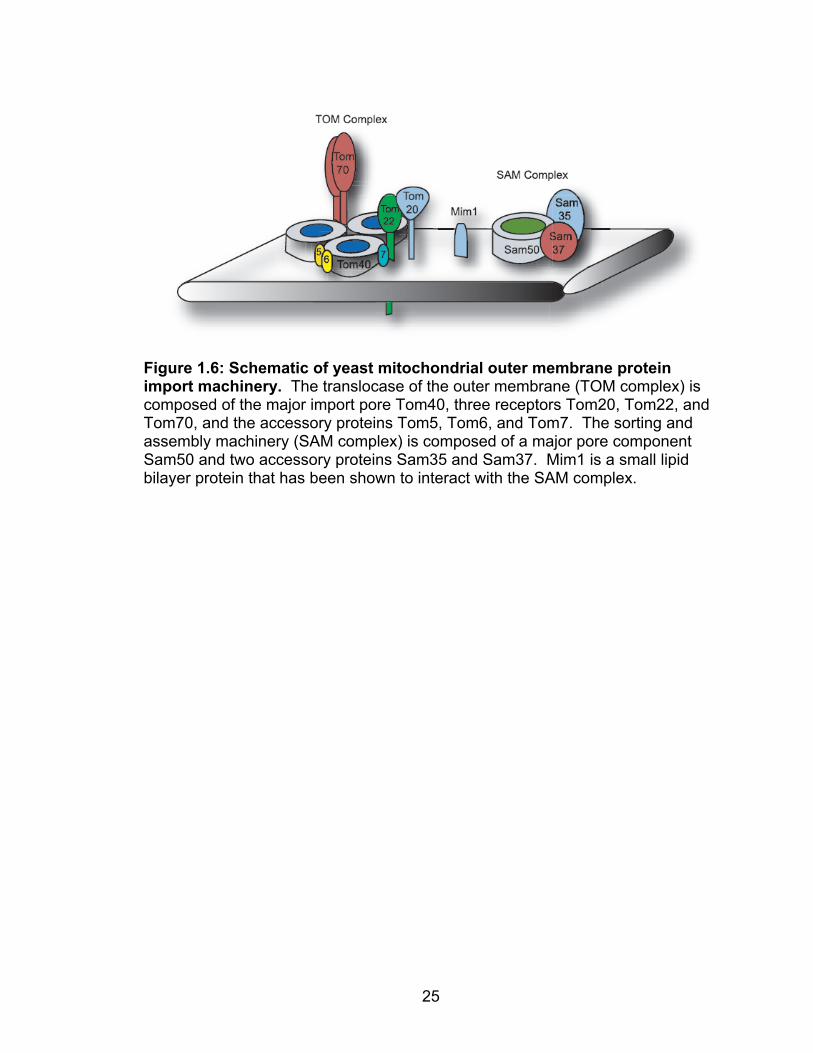
import complexes (16, 18, 99) (Table 1.2, Figure 1.6). The translocase of the
mitochondrial outer membrane (TOM complex) is a large multicomponent protein
structure that functions as the major import pore of the mitochondria (3, 107,
165). The TOM complex consists of three main receptors Tom20, Tom22, and
Tom70, three accessory proteins Tom5, Tom6, and Tom7, and the pore structure
Tom40. The TOM complex is engineered to import the many varieties of
mitochondrial proteins via the individual mitochondrial receptors. Tom20 and
Tom70 are the two main import receptors and are both signal-anchored proteins
similar to FHV protein A (19, 35, 156). Tom20 is the major import receptor and
sits on the periphery of the TOM complex with its amino-terminus embedded in

24
Table 1.2: Yeast Mitochondrial Outer Membrane Protein Import Machinery Component Orientation Mitochondrial Function Translocase of the Mitochondrial Outer Membrane (TOM Complex)
Tom70 Tom20 Tom22 Tom40 Tom5 Tom6 Tom7
Signal-anchored Signal-anchored Tail-anchored β-barrel Tail-anchored Tail-anchored Tail-anchored
Receptor for import of carrier proteins Major mitochondrial protein import receptor Co-receptor for protein import Pore forming subunit TOM complex stability/assembly TOM complex stability/assembly TOM complex stability/assembly
Sorting and Assembly Machinery (SAM Complex)
Sam50 Sam37 Sam35 Mim1
Integral, β-barrel Peripheral Peripheral single transmembrane
Pore forming subunit SAM complex subunit SAM complex subunit TOM complex and signal- anchored protein biogenesis
25
Figure 1.6: Schematic of yeast mitochondrial outer membrane protein import machinery. The translocase of the outer membrane (TOM complex) is composed of the major import pore Tom40, three receptors Tom20, Tom22, and Tom70, and the accessory proteins Tom5, Tom6, and Tom7. The sorting and assembly machinery (SAM complex) is composed of a major pore component Sam50 and two accessory proteins Sam35 and Sam37. Mim1 is a small lipid bilayer protein that has been shown to interact with the SAM complex.
26
the outer membrane (49). The cytosolic tail of Tom20 is highly negatively
charged (172) and provides the physical interactions for the import of complex
mitochondrial outer membrane protein such as the β-barrel proteins porin and
Tom40 (20, 69, 157). On the other hand, Tom70 functions as a dimer for the
import of the carrier domain proteins that contain internal targeting signals such
as the ATP/ADP carrier and phosphate carrier proteins via physical interactions
with tricopeptide repeats present in the cytosolic arm of Tom70 (17, 66, 125,
174). In addition to Tom70, yeast encode a similar protein, Tom71, which has
been shown to play redundant roles with Tom70 as a minor component of the
TOM complex (65, 124) and regulate mitochondrial morphology (66). Tom22 is a
member of the core general import pore and is thought to function along with
Tom20 (171) as a multifunctional organizer of the TOM complex. Tom22, is a
tail-anchored protein with cytosolic and intermembrane space acidic domains
that are thought to function with chaperone-like activity to escort proteins from
the main receptor Tom20, through the import pore, and into the intermembrane
space (97, 171). However, although the import of β-barrel and carrier proteins
require the TOM complex and import receptors this is not the case for single
transmembrane proteins of the mitochondrial outer membrane. It has recently
been shown that both tail-anchored and signal-anchored proteins can be
imported via a TOM complex independent mechanism which has yet to be
identified (61, 84). Tom40 is a β-barrel protein embedded in the mitochondrial
outer membrane and forms the major component and pore of the TOM complex
(2). Tom5 (28, 126), Tom6 (22), and Tom7 (50) are three accessory proteins
27
that are important for TOM complex structural stability as well as maintenance of
mitochondrial morphology (87). The import pore Tom40 is the only component of
the TOM complex that is essential for cellular function, suggesting the TOM
complex receptors and accessory proteins play redundant roles in mitochondrial
function .
In addition to the TOM complex, the mitochondrial outer membrane also
contains the sorting and assembly machinery (SAM complex) (Figure 1.6, Table
1.2) (16, 72, 141, 165). This structure was only recently identified and has been
shown to be required for the proper import and assembly of complex proteins
and protein structures such as the β-barrel protein porin and the TOM complex
(72). Similar to the TOM complex, the SAM complex is a multicomponent protein
complex that consists of a conserved core component Sam50 (Tob55) (23, 68)
and two subunits Sam35 (Tob38) (23) and Sam37 (44, 165) Sam50 is an
integral β-barrel protein whereas Sam35 and Sam37 are peripheral membrane
proteins which are though to function in SAM complex stability. Sam37 was
originally identified in a screen for mutant yeast strains that were defective in
phospholipid biosynthesis and has been found to play many roles within the cell
(44). It was originally thought to be a protein import receptor based on genetic
and physical interactions with Tom70 (44, 125), yet it was later shown to be
necessary for β-barrel protein import and recently it has been implicated in the
import of TOM complex proteins. It has also been implicated in a screen for
genes required for peroxisome function suggesting it may have roles in other
cellular functions (78). Furthermore, the SAM complex has been shown to

28
cooperate with other mitochondrial outer membrane proteins such as Mdm10,
Mdm12, and Mmm1 for the assembly of β-barrel proteins, all of which have been
shown to influence mitochondrial morphology (6, 85). Finally, the small
mitochondrial outer membrane protein Mim1 was recently shown to physically
interact with the SAM complex (15) and has been shown to be required for the
specific import of the signal-anchored receptors Tom20 and Tom70 as well as
the assembly of the TOM complex (155). Of the two major protein import
complexes, the SAM complex seems to play a much more essential role in
mitochondrial biogenesis as only a deletion of the peripheral membrane protein
Sam37 is viable (6, 68). One possible explanation for this is the similarity
between the SAM complex and its bacterial equivalent Omp85, suggesting that
this complex is evolutionarily conserved for cellular function (107). The deletion
of many of the components of the SAM and TOM complexes leads to severe
defects in mitochondrial protein import as well as mitochondrial morphology
suggesting a link between protein import and the maintenance of mitochondrial
shape and structure (142). The deletion of Mdm10 in particular, leads to the
formation of giant-mitochondria which is similar to what is seen in the presence of
active FHV RNA replication (85), yet unfortunately work has not been done to
solidify the link between import components and mitochondrial morphology.
Yeast Mitochondrial Fusion and Fission
Yeast mitochondrial morphology is maintained by the two evolutionarily
conserved opposing forces of mitochondrial fusion and fission (88, 101, 170).

29
The budding yeast has been a valuable host system in which to study these two
processes. The development of yeast deletion strains along with the availability
of molecular biology techniques have led to the identification of the mitochondrial
fusion and fission machinery which is located on the mitochondrial outer
membrane (Figure 1.7). The machinery responsible for mitochondrial fusion
consists of the mitochondrial outer membrane proteins Fzo1 and Ugo1 along with
the mitochondrial inner membrane protein Mgm1 (175). Fzo1 and Mgm1 are
integral membrane GTPase proteins which perform the GTP dependent fusion of
the mitochondrial membranes whereas Ugo1 is an adaptor protein that physically
connects Dnm1 and Mgm1 through the intermembrane space (135, 136). The
mitochondrial fission machinery is composed of the integral mitochondrial outer
membrane protein Fis1 and the peripheral membrane proteins Dnm1 and Mdv1
(109, 163). Dnm1 is a dynamin-related GTPase and the major component of the
mitochondrial fission machinery. Fis1 is a tail-anchored protein that is required
for interactions between Mdv1 and Dnm1 and anchoring of the fission machinery
to the mitochondrial outer membrane (98, 163). In addition to normal
mitochondrial fission, this pathway plays key roles in programmed cell death (36,
62) and peroxisome fission (64, 94). Similar to the mitochondrial protein import
machinery, a deletion of these components leads to dramatic defects in
mitochondrial morphology.

30
Figure 1.7: Yeast mitochondrial fusion and fission machinery. (A) The mitochondrial fusion machinery is composed of the mitochondrial outer membrane (MOM) proteins Fzo1 and Ugo1 and and the mitochondrial inner membrane (MIM) protein Mgm1 with span the intermembrane space (IMS). (B) The mitochondrial fission machinery is composed of the intergral membrane protein Fis1 and peripheral membrane proteins Mdv1 and Dnm1.

31
Yeast Mitochondrial Phospholipid Biology
Although the mitochondria contain roughly 1,000 proteins, the mitochondrial
outer membrane contains only a small fraction of this total. A proteomic analysis
has identified a handful of resident proteins present in the mitochondrial outer
membrane leaving the remaining space to be filled by phospholipids (30, 127,
173). The mitochondrial outer membrane is made primarily of the
glycerophospholipids (Figure 1.8) phosphatidlycholine (PC) (~45%) and
phosphatidlyethanolamine (PE) (~35%), the minor phospholipid components
phosphatidylserine (PS) (~2%) and phosphatidylinositol (PI) (~10%), and the
mitochondrial specific phospholipids phosphatidlyglycerol (PG) and cardiolipin
(CL) (~4%) (25, 147). In particular, the anionic phospholipids PS, PG, PA, and
CL have been shown to be important cellular phospholipids to mediate protein
import and protein-lipid interactions within the cell (37, 83, 103, 176)
Cellular and membrane phospholipid biosynthesis has been studied in great
detail in yeast due to the availability of yeast deletion strains and an easily
manipulatable cellular biology system. The endoplasmic reticulum is the prime
location for the biosynthesis of the major glycerophospholipids. However the
mitochondria contains the enzymatic machinery for the synthesis of PE as well
as the mitochondrial specific phospholipids PG and CL (53, 147) suggesting a
key role for this organelle in phospholipid biosynthesis (147, 154). The
mitochondrial specific phospholipid, cardiolipin, is a diphosphatidylglycerol
molecule that is synthesized from PG by the enzyme cardiolipin synthase (Crd1)
(57). Cardiolipin is located primarily in the mitochondrial inner membranes where

32
Figure 1.8: Schematic of the major cellular glycerophospholipids. Phosphatidylcholine (PC), Phosphatidylethanolamine (PE), Phospha- tidylserine (PS), Phosphatidylinositol (PI), Phosphatidic Acid (PA), Phosphatidylglycerol (PG), and Cardiolipin (CL).

33
it makes up roughly 15% of the total phospholipid composition, and has been
implicated in a variety of mitochondrial functions including mitochondrial protein
import, maintainence of protein-protein interactions, apoptosis, and mitochondrial
morphology (24, 57, 81, 102). Interestingly, although CL makes up roughly 5%
of the mitochondrial outer membrane phospholipids, it can reach concentrations
of up to 20% at “contacts sites” where the mitochondrial outer and inner
membranes come into physical contact (10, 118, 138). Mitochondrial contact
sites are thought to be localized areas where proteins and phospholipids
destined for trafficking into the mitochondrial matrix can easily pass through the
double membrane structure (118, 138). In addition, contacts sites contain a
variety of proteins that are required for protein import and mitochondrial
morphology (40, 164), suggesting that these sites play essential roles in yeast
mitochondrial biogenesis (118).
The mitochondrion is a complex organelle that is vital to cellular function.
Mutations or deletions of mitochondrial proteins can give rise to a variety of
mitochondrial disorders such as Barth Syndrome and dominant optic atrophy
which are caused by defects in cardiolipin synthesis and mitochondrial fusion
respectively (38). Finally, it should be noted that although the study of the
individual processes of protein import, mitochondrial morphology, and
phospholipid biosynthesis can be separated in the laboratory, one has to keep in
mind that all of these processes are intimately linked during mitochondrial
biogenesis and defects in one process can have multiple effects on others.

34
Summary
Positive-strand RNA viruses comprise a long list of viral pathogens capable of
causing both health and economic problems around the world, yet to date there
is little available to fight these pathogens. To aid in the development of anti-viral
therapies, the understanding of essential molecular processes required for
positive-strand RNA virus replication is necessary, and the association of all
positive-strand RNA viruses with host intracellular membranes provides this
crucial step in the viral life cycle on which to focus our studies.
In my thesis we made use of the established model positive-strand RNA
virus, Flock House virus (FHV), and the budding yeast Saccharomyces
cerevisiae to utilize a powerful host-pathogen system with which to study
positive-strand RNA virus replication complex assembly and function. FHV
assembles functional viral RNA replication complexes on the mitochondrial outer
membrane in yeast where protein A is the only viral protein necessary for this
process. Protein A contains a mitochondrial targeting signal similar to those of
host mitochondrial outer membrane proteins and we hypothesize that similar to
yeast mitochondrial outer membranes, FHV uses the yeast mitochondrial import
machinery for the assembly of mitochondrial associated replication complexes.
In order to address this hypothesis we proposed the following three specific aims:

35
Aim1: Establish an FHV in vitro membrane association system (Chapter
II).
Aim2: Investigate the role of individual mitochondrial outer membrane
proteins in FHV RNA replication (Chapter III).
Aim 3. Investigate the composition and structure of FHV RNA replication
Complexes (Chapter IV)

36
REFERENCES
1. Ahola, T., A. Lampio, P. Auvinen, and L. Kaariainen. 1999. Semliki Forest virus mRNA capping enzyme requires association with anionic membrane phospholipids for activity. EMBO J 18:3164-72.
2. Ahting, U., M. Thieffry, H. Engelhardt, R. Hegerl, W. Neupert, and S.
Nussberger. 2001. Tom40, the pore-forming component of the protein-conducting TOM channel in the outer membrane of mitochondria. J Cell Biol 153:1151-60.
3. Ahting, U., C. Thun, R. Hegerl, D. Typke, F. E. Nargang, W. Neupert,
and S. Nussberger. 1999. The TOM core complex: the general protein import pore of the outer membrane of mitochondria. J Cell Biol 147:959-68.
4. Ahting, U., T. Waizenegger, W. Neupert, and D. Rapaport. 2005.
Signal-anchored proteins follow a unique insertion pathway into the outer membrane of mitochondria. J Biol Chem 280:48-53.
5. Albarino, C. G., L. D. Eckerle, and L. A. Ball. 2003. The cis-acting
replication signal at the 3' end of Flock House virus RNA2 is RNA3-dependent. Virology 311:181-91.
6. Altmann, K., and B. Westermann. 2005. Role of essential genes in
mitochondrial morphogenesis in Saccharomyces cerevisiae. Mol Biol Cell 16:5410-7.
7. Amemiya, F., S. Maekawa, Y. Itakura, A. Kanayama, A. Matsui, S.
Takano, T. Yamaguchi, J. Itakura, T. Kitamura, T. Inoue, M. Sakamoto, K. Yamauchi, S. Okada, A. Yamashita, N. Sakamoto, M. Itoh, and N. Enomoto. 2008. Targeting lipid metabolism in the treatment of hepatitis C virus infection. J Infect Dis 197:361-70.
8. Annamalai, P., and A. L. Rao. 2006. Packaging of brome mosaic virus
subgenomic RNA is functionally coupled to replication-dependent transcription and translation of coat protein. J Virol 80:10096-108.
9. Annamalai, P., F. Rofail, D. A. Demason, and A. L. Rao. 2008.
Replication-coupled packaging mechanism in positive-strand RNA viruses: synchronized coexpression of functional multigenome RNA components of an animal and a plant virus in Nicotiana benthamiana cells by agroinfiltration. J Virol 82:1484-95.
37
10. Ardail, D., J. P. Privat, M. Egret-Charlier, C. Levrat, F. Lerme, and P. Louisot. 1990. Mitochondrial contact sites. Lipid composition and dynamics. J Biol Chem 265:18797-802.
11. Asia, W. H. O. R. O. f. S.-E. 2006, posting date. Situation of
Dengue/Dengue Haemorrhagic Fever in the South-East Asia Region. [Online.]
12. Ball, L. A., J. M. Amann, and B. K. Garrett. 1992. Replication of
nodamura virus after transfection of viral RNA into mammalian cells in culture. J Virol 66:2326-34.
13. Ball, L. A., and Y. Li. 1993. cis-acting requirements for the replication of
flock house virus RNA 2. J Virol 67:3544-51. 14. Bartenschlager, R., and S. Miller. 2008. Molecular aspects of Dengue
virus replication. Future Microbiol 3:155-65. 15. Becker, T., S. Pfannschmidt, B. Guiard, D. Stojanovski, D. Milenkovic,
S. Kutik, N. Pfanner, C. Meisinger, and N. Wiedemann. 2008. Biogenesis of the mitochondrial TOM complex: Mim1 promotes insertion and assembly of signal-anchored receptors. J Biol Chem 283:120-7.
16. Becker, T., F. N. Vogtle, D. Stojanovski, and C. Meisinger. 2008.
Sorting and assembly of mitochondrial outer membrane proteins. Biochim Biophys Acta 1777:557-63.
17. Beddoe, T., S. R. Bushell, M. A. Perugini, T. Lithgow, T. D. Mulhern, S.
P. Bottomley, and J. Rossjohn. 2004. A biophysical analysis of the tetratricopeptide repeat-rich mitochondrial import receptor, Tom70, reveals an elongated monomer that is inherently flexible, unstable, and unfolds via a multistate pathway. J Biol Chem 279:46448-54.
18. Bolender, N., A. Sickmann, R. Wagner, C. Meisinger, and N. Pfanner.
2008. Multiple pathways for sorting mitochondrial precursor proteins. EMBO Rep 9:42-9.
19. Brix, J., K. Dietmeier, and N. Pfanner. 1997. Differential recognition of
preproteins by the purified cytosolic domains of the mitochondrial import receptors Tom20, Tom22, and Tom70. J Biol Chem 272:20730-5.
20. Brix, J., S. Rudiger, B. Bukau, J. Schneider-Mergener, and N.
Pfanner. 1999. Distribution of binding sequences for the mitochondrial import receptors Tom20, Tom22, and Tom70 in a presequence-carrying preprotein and a non-cleavable preprotein. J Biol Chem 274:16522-30.

38
21. Burgyan, J., L. Rubino, and M. Russo. 1996. The 5'-terminal region of a tombusvirus genome determines the origin of multivesicular bodies. J Gen Virol 77 ( Pt 8):1967-74.
22. Cao, W., and M. G. Douglas. 1995. Biogenesis of ISP6, a small carboxyl-
terminal anchored protein of the receptor complex of the mitochondrial outer membrane. J Biol Chem 270:5674-9.
23. Chan, N. C., and T. Lithgow. 2008. The peripheral membrane subunits of
the SAM complex function codependently in mitochondrial outer membrane biogenesis. Mol Biol Cell 19:126-36.
24. Chen, S., M. Tarsio, P. M. Kane, and M. L. Greenberg. 2008. Cardiolipin
mediates cross-talk between mitochondria and the vacuole. Mol Biol Cell 19:5047-58.
25. de Kroon, A. I., M. C. Koorengevel, S. S. Goerdayal, P. C. Mulders, M.
J. Janssen, and B. de Kruijff. 1999. Isolation and characterization of highly purified mitochondrial outer membranes of the yeast Saccharomyces cerevisiae (method). Mol Membr Biol 16:205-11.
26. den Boon, J. A., J. Chen, and P. Ahlquist. 2001. Identification of
sequences in Brome mosaic virus replicase protein 1a that mediate association with endoplasmic reticulum membranes. J Virol 75:12370-81.
27. Denison, M. R. 2008. Seeking membranes: positive-strand RNA virus
replication complexes. PLoS Biol 6:e270. 28. Dietmeier, K., A. Honlinger, U. Bomer, P. J. Dekker, C. Eckerskorn, F.
Lottspeich, M. Kubrich, and N. Pfanner. 1997. Tom5 functionally links mitochondrial preprotein receptors to the general import pore. Nature 388:195-200.
29. DiFrano, A. R., M., and G.P. Martelli. 1984. Ultrastructure and Origin of
Cytoplasmic Multivesicular Bodies Induced by Carnation Italian Ringspot Virus. J Gen Virol 65:1233-1237.
30. Dimmer, K. S., and D. Rapaport. 2008. Proteomic view of mitochondrial
function. Genome Biol 9:209. 31. Dye, B. T., D. J. Miller, and P. Ahlquist. 2005. In vivo self-interaction of
nodavirus RNA replicase protein a revealed by fluorescence resonance energy transfer. J Virol 79:8909-19.
39
32. Eckerle, L. D., C. G. Albarino, and L. A. Ball. 2003. Flock House virus subgenomic RNA3 is replicated and its replication correlates with transactivation of RNA2. Virology 317:95-108.
33. Eckerle, L. D., and L. A. Ball. 2002. Replication of the RNA segments of
a bipartite viral genome is coordinated by a transactivating subgenomic RNA. Virology 296:165-76.
34. Elazar, M., K. H. Cheong, P. Liu, H. B. Greenberg, C. M. Rice, and J. S.
Glenn. 2003. Amphipathic helix-dependent localization of NS5A mediates hepatitis C virus RNA replication. J Virol 77:6055-61.
35. Endo, T., and D. Kohda. 2002. Functions of outer membrane receptors in
mitochondrial protein import. Biochim Biophys Acta 1592:3-14. 36. Fannjiang, Y., W. C. Cheng, S. J. Lee, B. Qi, J. Pevsner, J. M.
McCaffery, R. B. Hill, G. Basanez, and J. M. Hardwick. 2004. Mitochondrial fission proteins regulate programmed cell death in yeast. Genes Dev 18:2785-97.
37. Fernandez-Murray, J. P., and C. R. McMaster. 2006. Identification of
novel phospholipid binding proteins in Saccharomyces cerevisiae. FEBS Lett 580:82-6.
38. Foury, F., and M. Kucej. 2002. Yeast mitochondrial biogenesis: a model
system for humans? Curr Opin Chem Biol 6:106-11. 39. Fraser, C. S., J. W. Hershey, and J. A. Doudna. 2009. The pathway of
hepatitis C virus mRNA recruitment to the human ribosome. Nat Struct Mol Biol.
40. Fritz, S., D. Rapaport, E. Klanner, W. Neupert, and B. Westermann.
2001. Connection of the mitochondrial outer and inner membranes by Fzo1 is critical for organellar fusion. J Cell Biol 152:683-92.
41. Froshauer, S., J. Kartenbeck, and A. Helenius. 1988. Alphavirus RNA
replicase is located on the cytoplasmic surface of endosomes and lysosomes. J Cell Biol 107:2075-86.
42. Galiana-Arnoux, D., C. Dostert, A. Schneemann, J. A. Hoffmann, and
J. L. Imler. 2006. Essential function in vivo for Dicer-2 in host defense against RNA viruses in drosophila. Nat Immunol 7:590-7.

40
43. Go, E. P., W. R. Wikoff, Z. Shen, G. O'Maille, H. Morita, T. P. Conrads, A. Nordstrom, S. A. Trauger, W. Uritboonthai, D. A. Lucas, K. C. Chan, T. D. Veenstra, H. Lewicki, M. B. Oldstone, A. Schneemann, and G. Siuzdak. 2006. Mass spectrometry reveals specific and global molecular transformations during viral infection. J Proteome Res 5:2405-16.
44. Gratzer, S., T. Lithgow, R. E. Bauer, E. Lamping, F. Paltauf, S. D.
Kohlwein, V. Haucke, T. Junne, G. Schatz, and M. Horst. 1995. Mas37p, a novel receptor subunit for protein import into mitochondria. J Cell Biol 129:25-34.
45. Grimley, P. M., J. G. Levin, I. K. Berezesky, and R. M. Friedman. 1972.
Specific membranous structures associated with the replication of group A arboviruses. J Virol 10:492-503.
46. Guinea, R., and L. Carrasco. 1991. Effects of fatty acids on lipid
synthesis and viral RNA replication in poliovirus-infected cells. Virology 185:473-6.
47. Guinea, R., and L. Carrasco. 1990. Phospholipid biosynthesis and
poliovirus genome replication, two coupled phenomena. EMBO J 9:2011-6.
48. Habib, S. J., A. Vasiljev, W. Neupert, and D. Rapaport. 2003. Multiple
functions of tail-anchor domains of mitochondrial outer membrane proteins. FEBS Lett 555:511-5.
49. Harkness, T. A., F. E. Nargang, I. van der Klei, W. Neupert, and R. Lill.
1994. A crucial role of the mitochondrial protein import receptor MOM19 for the biogenesis of mitochondria. J Cell Biol 124:637-48.
50. Honlinger, A., U. Bomer, A. Alconada, C. Eckerskorn, F. Lottspeich,
K. Dietmeier, and N. Pfanner. 1996. Tom7 modulates the dynamics of the mitochondrial outer membrane translocase and plays a pathway-related role in protein import. EMBO J 15:2125-37.
51. Horie, C., H. Suzuki, M. Sakaguchi, and K. Mihara. 2003. Targeting and
assembly of mitochondrial tail-anchored protein Tom5 to the TOM complex depend on a signal distinct from that of tail-anchored proteins dispersed in the membrane. J Biol Chem 278:41462-71.
52. Hwang, Y. T., A. W. McCartney, S. K. Gidda, and R. T. Mullen. 2008.
Localization of the Carnation Italian ringspot virus replication protein p36 to the mitochondrial outer membrane is mediated by an internal targeting signal and the TOM complex. BMC Cell Biol 9:54.
41
53. Jiang, F., M. T. Ryan, M. Schlame, M. Zhao, Z. Gu, M. Klingenberg, N. Pfanner, and M. L. Greenberg. 2000. Absence of cardiolipin in the crd1 null mutant results in decreased mitochondrial membrane potential and reduced mitochondrial function. J Biol Chem 275:22387-94.
54. Johnson, K. L., and L. A. Ball. 1999. Induction and maintenance of
autonomous flock house virus RNA1 replication. J Virol 73:7933-42. 55. Johnson, K. L., and L. A. Ball. 1997. Replication of flock house virus
RNAs from primary transcripts made in cells by RNA polymerase II. J Virol 71:3323-7.
56. Jonczyk, M., K. B. Pathak, M. Sharma, and P. D. Nagy. 2007. Exploiting
alternative subcellular location for replication: tombusvirus replication switches to the endoplasmic reticulum in the absence of peroxisomes. Virology 362:320-30.
57. Joshi, A. S., J. Zhou, V. M. Gohil, S. Chen, and M. L. Greenberg. 2008.
Cellular functions of cardiolipin in yeast. Biochim Biophys Acta. 58. Kampmueller, K. M., and D. J. Miller. 2005. The cellular chaperone heat
shock protein 90 facilitates Flock House virus RNA replication in Drosophila cells. J Virol 79:6827-37.
59. Kapadia, S. B., and F. V. Chisari. 2005. Hepatitis C virus RNA replication
is regulated by host geranylgeranylation and fatty acids. Proc Natl Acad Sci U S A 102:2561-6.
60. Kauder, S. E., and V. R. Racaniello. 2004. Poliovirus tropism and
attenuation are determined after internal ribosome entry. J Clin Invest 113:1743-53.
61. Kemper, C., S. J. Habib, G. Engl, P. Heckmeyer, K. S. Dimmer, and D.
Rapaport. 2008. Integration of tail-anchored proteins into the mitochondrial outer membrane does not require any known import components. J Cell Sci 121:1990-8.
62. Kitagaki, H., Y. Araki, K. Funato, and H. Shimoi. 2007. Ethanol-induced
death in yeast exhibits features of apoptosis mediated by mitochondrial fission pathway. FEBS Lett 581:2935-42.
63. Knoops, K., M. Kikkert, S. H. Worm, J. C. Zevenhoven-Dobbe, Y. van
der Meer, A. J. Koster, A. M. Mommaas, and E. J. Snijder. 2008. SARS-coronavirus replication is supported by a reticulovesicular network of modified endoplasmic reticulum. PLoS Biol 6:e226.

42
64. Koch, A., Y. Yoon, N. A. Bonekamp, M. A. McNiven, and M. Schrader. 2005. A role for Fis1 in both mitochondrial and peroxisomal fission in mammalian cells. Mol Biol Cell 16:5077-86.
65. Koh, J. Y., P. Hajek, and D. M. Bedwell. 2001. Overproduction of PDR3
suppresses mitochondrial import defects associated with a TOM70 null mutation by increasing the expression of TOM72 in Saccharomyces cerevisiae. Mol Cell Biol 21:7576-86.
66. Kondo-Okamoto, N., J. M. Shaw, and K. Okamoto. 2008.
Tetratricopeptide repeat proteins Tom70 and Tom71 mediate yeast mitochondrial morphogenesis. EMBO Rep 9:63-9.
67. Kopek, B. G., G. Perkins, D. J. Miller, M. H. Ellisman, and P. Ahlquist.
2007. Three-dimensional analysis of a viral RNA replication complex reveals a virus-induced mini-organelle. PLoS Biol 5:e220.
68. Kozjak, V., N. Wiedemann, D. Milenkovic, C. Lohaus, H. E. Meyer, B.
Guiard, C. Meisinger, and N. Pfanner. 2003. An essential role of Sam50 in the protein sorting and assembly machinery of the mitochondrial outer membrane. J Biol Chem 278:48520-3.
69. Krimmer, T., D. Rapaport, M. T. Ryan, C. Meisinger, C. K.
Kassenbrock, E. Blachly-Dyson, M. Forte, M. G. Douglas, W. Neupert, F. E. Nargang, and N. Pfanner. 2001. Biogenesis of porin of the outer mitochondrial membrane involves an import pathway via receptors and the general import pore of the TOM complex. J Cell Biol 152:289-300.
70. Kujala, P., A. Ikaheimonen, N. Ehsani, H. Vihinen, P. Auvinen, and L.
Kaariainen. 2001. Biogenesis of the Semliki Forest virus RNA replication complex. J Virol 75:3873-84.
71. Kushner, D. B., B. D. Lindenbach, V. Z. Grdzelishvili, A. O. Noueiry, S.
M. Paul, and P. Ahlquist. 2003. Systematic, genome-wide identification of host genes affecting replication of a positive-strand RNA virus. Proc Natl Acad Sci U S A 100:15764-9.
72. Kutik, S., D. Stojanovski, L. Becker, T. Becker, M. Meinecke, V.
Kruger, C. Prinz, C. Meisinger, B. Guiard, R. Wagner, N. Pfanner, and N. Wiedemann. 2008. Dissecting membrane insertion of mitochondrial beta-barrel proteins. Cell 132:1011-24.
73. Lee, W. M., and P. Ahlquist. 2003. Membrane synthesis, specific lipid
requirements, and localized lipid composition changes associated with a positive-strand RNA virus RNA replication protein. J Virol 77:12819-28.
43
74. Lee, W. M., M. Ishikawa, and P. Ahlquist. 2001. Mutation of host delta9 fatty acid desaturase inhibits brome mosaic virus RNA replication between template recognition and RNA synthesis. J Virol 75:2097-106.
75. Li, Z., J. Pogany, T. Panavas, K. Xu, A. M. Esposito, T. G. Kinzy, and
P. D. Nagy. 2009. Translation elongation factor 1A is a component of the tombusvirus replicase complex and affects the stability of the p33 replication co-factor. Virology 385:245-60.
76. Lindenbach, B. D., J. Y. Sgro, and P. Ahlquist. 2002. Long-distance
base pairing in flock house virus RNA1 regulates subgenomic RNA3 synthesis and RNA2 replication. J Virol 76:3905-19.
77. Lindenbach, B. D. a. R. C. M. 2001. Flaviviridae: The Viruses and Their
Replication, p. 991-1041. In K. D. M. a. H. P.M. (ed.), Fields Virology, Fourth ed, vol. 1. Lippincott Williams & Wilkins, Philadelphia, PA.
78. Lockshon, D., L. E. Surface, E. O. Kerr, M. Kaeberlein, and B. K.
Kennedy. 2007. The sensitivity of yeast mutants to oleic acid implicates the peroxisome and other processes in membrane function. Genetics 175:77-91.
79. Lu, R., M. Maduro, F. Li, H. W. Li, G. Broitman-Maduro, W. X. Li, and
S. W. Ding. 2005. Animal virus replication and RNAi-mediated antiviral silencing in Caenorhabditis elegans. Nature 436:1040-3.
80. Lu, R., E. Yigit, W. X. Li, and S. W. Ding. 2009. An RIG-I-Like RNA
helicase mediates antiviral RNAi downstream of viral siRNA biogenesis in Caenorhabditis elegans. PLoS Pathog 5:e1000286.
81. Lutter, M., M. Fang, X. Luo, M. Nishijima, X. Xie, and X. Wang. 2000.
Cardiolipin provides specificity for targeting of tBid to mitochondria. Nat Cell Biol 2:754-61.
82. Marshall, D., and A. Schneemann. 2001. Specific packaging of nodaviral
RNA2 requires the N-terminus of the capsid protein. Virology 285:165-75. 83. Meglei, G., and G. A. McQuibban. 2009. The Dynamin-Related Protein
Mgm1p Assembles into Oligomers and Hydrolyzes GTP To Function in Mitochondrial Membrane Fusion (dagger). Biochemistry 48:1774-84.
84. Meineke, B., G. Engl, C. Kemper, A. Vasiljev-Neumeyer, H.
Paulitschke, and D. Rapaport. 2008. The outer membrane form of the mitochondrial protein Mcr1 follows a TOM-independent membrane insertion pathway. FEBS Lett 582:855-60.

44
85. Meisinger, C., M. Rissler, A. Chacinska, L. K. Szklarz, D. Milenkovic, V. Kozjak, B. Schonfisch, C. Lohaus, H. E. Meyer, M. P. Yaffe, B. Guiard, N. Wiedemann, and N. Pfanner. 2004. The mitochondrial morphology protein Mdm10 functions in assembly of the preprotein translocase of the outer membrane. Dev Cell 7:61-71.
86. Meisinger, C., A. Sickmann, and N. Pfanner. 2008. The mitochondrial
proteome: from inventory to function. Cell 134:22-4. 87. Meisinger, C., N. Wiedemann, M. Rissler, A. Strub, D. Milenkovic, B.
Schonfisch, H. Muller, V. Kozjak, and N. Pfanner. 2006. Mitochondrial protein sorting: differentiation of beta-barrel assembly by Tom7-mediated segregation of Mdm10. J Biol Chem 281:22819-26.
88. Merz, S., M. Hammermeister, K. Altmann, M. Durr, and B.
Westermann. 2007. Molecular machinery of mitochondrial dynamics in yeast. Biol Chem 388:917-26.
89. Miller, D. J., and P. Ahlquist. 2002. Flock house virus RNA polymerase
is a transmembrane protein with amino-terminal sequences sufficient for mitochondrial localization and membrane insertion. J Virol 76:9856-67.
90. Miller, D. J., M. D. Schwartz, and P. Ahlquist. 2001. Flock house virus
RNA replicates on outer mitochondrial membranes in Drosophila cells. J Virol 75:11664-76.
91. Miller, D. J., M. D. Schwartz, B. T. Dye, and P. Ahlquist. 2003.
Engineered retargeting of viral RNA replication complexes to an alternative intracellular membrane. J Virol 77:12193-202.
92. Miller, S., and J. Krijnse-Locker. 2008. Modification of intracellular
membrane structures for virus replication. Nat Rev Microbiol 6:363-74. 93. Moradpour, D., R. Gosert, D. Egger, F. Penin, H. E. Blum, and K.
Bienz. 2003. Membrane association of hepatitis C virus nonstructural proteins and identification of the membrane alteration that harbors the viral replication complex. Antiviral Res 60:103-9.
94. Motley, A. M., G. P. Ward, and E. H. Hettema. 2008. Dnm1p-dependent
peroxisome fission requires Caf4p, Mdv1p and Fis1p. J Cell Sci 121:1633-40.
95. Nagy, P. D. 2008. Yeast as a model host to explore plant virus-host
interactions. Annu Rev Phytopathol 46:217-42.

45
96. Nagy, P. D., and J. Pogany. 2006. Yeast as a model host to dissect functions of viral and host factors in tombusvirus replication. Virology 344:211-20.
97. Nargang, F. E., D. Rapaport, R. G. Ritzel, W. Neupert, and R. Lill.
1998. Role of the negative charges in the cytosolic domain of TOM22 in the import of precursor proteins into mitochondria. Mol Cell Biol 18:3173-81.
98. Naylor, K., E. Ingerman, V. Okreglak, M. Marino, J. E. Hinshaw, and J. Nunnari. 2006. Mdv1 interacts with assembled dnm1 to promote mitochondrial division. J Biol Chem 281:2177-83.
99. Neupert, W., and J. M. Herrmann. 2007. Translocation of proteins into
mitochondria. Annu Rev Biochem 76:723-49. 100. Ng, C. G., I. Coppens, D. Govindarajan, J. Pisciotta, V. Shulaev, and
D. E. Griffin. 2008. Effect of host cell lipid metabolism on alphavirus replication, virion morphogenesis, and infectivity. Proc Natl Acad Sci U S A 105:16326-31.
101. Okamoto, K., and J. M. Shaw. 2005. Mitochondrial morphology and
dynamics in yeast and multicellular eukaryotes. Annu Rev Genet 39:503-36.
102. Osman, C., M. Haag, C. Potting, J. Rodenfels, P. V. Dip, F. T. Wieland,
B. Brugger, B. Westermann, and T. Langer. 2009. The genetic interactome of prohibitins: coordinated control of cardiolipin and phosphatidylethanolamine by conserved regulators in mitochondria. J Cell Biol 184:583-96.
103. Ostrander, D. B., M. Zhang, E. Mileykovskaya, M. Rho, and W.
Dowhan. 2001. Lack of mitochondrial anionic phospholipids causes an inhibition of translation of protein components of the electron transport chain. A yeast genetic model system for the study of anionic phospholipid function in mitochondria. J Biol Chem 276:25262-72.
104. Panavas, T., E. Serviene, J. Pogany, and P. D. Nagy. 2008. Genome-
wide screens for identification of host factors in viral replication. Methods Mol Biol 451:615-24.
105. Pathak, K. B., Z. Sasvari, and P. D. Nagy. 2008. The host Pex19p plays
a role in peroxisomal localization of tombusvirus replication proteins. Virology 379:294-305.
106. Perez, L., R. Guinea, and L. Carrasco. 1991. Synthesis of Semliki Forest
virus RNA requires continuous lipid synthesis. Virology 183:74-82.

46
107. Pfanner, N., N. Wiedemann, C. Meisinger, and T. Lithgow. 2004. Assembling the mitochondrial outer membrane. Nat Struct Mol Biol 11:1044-8.
108. Poch, O., I. Sauvaget, M. Delarue, and N. Tordo. 1989. Identification of
four conserved motifs among the RNA-dependent polymerase encoding elements. EMBO J 8:3867-74.
109. Praefcke, G. J., and H. T. McMahon. 2004. The dynamin superfamily:
universal membrane tubulation and fission molecules? Nat Rev Mol Cell Biol 5:133-47.
110. Prentice, E., W. G. Jerome, T. Yoshimori, N. Mizushima, and M. R.
Denison. 2004. Coronavirus replication complex formation utilizes components of cellular autophagy. J Biol Chem 279:10136-41.
111. Price, B. D., P. Ahlquist, and L. A. Ball. 2002. DNA-directed expression
of an animal virus RNA for replication-dependent colony formation in Saccharomyces cerevisiae. J Virol 76:1610-6.
112. Price, B. D., M. Roeder, and P. Ahlquist. 2000. DNA-Directed
expression of functional flock house virus RNA1 derivatives in Saccharomyces cerevisiae, heterologous gene expression, and selective effects on subgenomic mRNA synthesis. J Virol 74:11724-33.
113. Price, B. D., R. R. Rueckert, and P. Ahlquist. 1996. Complete replication
of an animal virus and maintenance of expression vectors derived from it in Saccharomyces cerevisiae. Proc Natl Acad Sci U S A 93:9465-70.
114. Prod'homme, D., A. Jakubiec, V. Tournier, G. Drugeon, and I. Jupin.
2003. Targeting of the turnip yellow mosaic virus 66K replication protein to the chloroplast envelope is mediated by the 140K protein. J Virol 77:9124-35.
115. Quinkert, D., R. Bartenschlager, and V. Lohmann. 2005. Quantitative
analysis of the hepatitis C virus replication complex. J Virol 79:13594-605. 116. Rapaport, D. 2003. Finding the right organelle. Targeting signals in
mitochondrial outer-membrane proteins. EMBO Rep 4:948-52. 117. Rassmann, A., A. Henke, N. Jarasch, F. Lottspeich, H. P. Saluz, and
T. Munder. 2007. The human fatty acid synthase: a new therapeutic target for coxsackievirus B3-induced diseases? Antiviral Res 76:150-8.

47
118. Reichert, A. S., and W. Neupert. 2002. Contact sites between the outer and inner membrane of mitochondria-role in protein transport. Biochim Biophys Acta 1592:41-9.
119. Sagan, S. M., Y. Rouleau, C. Leggiadro, L. Supekova, P. G. Schultz, A.
I. Su, and J. P. Pezacki. 2006. The influence of cholesterol and lipid metabolism on host cell structure and hepatitis C virus replication. Biochem Cell Biol 84:67-79.
120. Saito, K., M. Nishijima, and O. Kuge. 2006. Phosphatidylserine is
involved in gene expression from Sindbis virus subgenomic promoter. Biochem Biophys Res Commun 345:878-85.
121. Salonen, A., L. Vasiljeva, A. Merits, J. Magden, E. Jokitalo, and L.
Kaariainen. 2003. Properly folded nonstructural polyprotein directs the semliki forest virus replication complex to the endosomal compartment. J Virol 77:1691-702.
122. Schaad, M. C., P. E. Jensen, and J. C. Carrington. 1997. Formation of
plant RNA virus replication complexes on membranes: role of an endoplasmic reticulum-targeted viral protein. EMBO J 16:4049-59.
123. Schlegel, A., T. H. Giddings, Jr., M. S. Ladinsky, and K. Kirkegaard.
1996. Cellular origin and ultrastructure of membranes induced during poliovirus infection. J Virol 70:6576-88.
124. Schlossmann, J., R. Lill, W. Neupert, and D. A. Court. 1996. Tom71, a
novel homologue of the mitochondrial preprotein receptor Tom70. J Biol Chem 271:17890-5.
125. Schlossmann, J., and W. Neupert. 1995. Assembly of the preprotein
receptor MOM72/MAS70 into the protein import complex of the outer membrane of mitochondria. J Biol Chem 270:27116-21.
126. Schmitt, S., U. Ahting, L. Eichacker, B. Granvogl, N. E. Go, F. E.
Nargang, W. Neupert, and S. Nussberger. 2005. Role of Tom5 in maintaining the structural stability of the TOM complex of mitochondria. J Biol Chem 280:14499-506.
127. Schmitt, S., H. Prokisch, T. Schlunck, D. G. Camp, 2nd, U. Ahting, T.
Waizenegger, C. Scharfe, T. Meitinger, A. Imhof, W. Neupert, P. J. Oefner, and D. Rapaport. 2006. Proteome analysis of mitochondrial outer membrane from Neurospora crassa. Proteomics 6:72-80.

48
128. Schneemann, A., and D. Marshall. 1998. Specific encapsidation of nodavirus RNAs is mediated through the C terminus of capsid precursor protein alpha. J Virol 72:8738-46.
129. Schneemann, A., W. Zhong, T. M. Gallagher, and R. R. Rueckert.
1992. Maturation cleavage required for infectivity of a nodavirus. J Virol 66:6728-34.
130. Schwartz, M., J. Chen, W. M. Lee, M. Janda, and P. Ahlquist. 2004.
Alternate, virus-induced membrane rearrangements support positive-strand RNA virus genome replication. Proc Natl Acad Sci U S A 101:11263-8.
131. Scotti, P. D., S. Dearing, and D. W. Mossop. 1983. Flock House virus: a
nodavirus isolated from Costelytra zealandica (White) (Coleoptera: Scarabaeidae). Arch Virol 75:181-9.
132. Selling, B. H., R. F. Allison, and P. Kaesberg. 1990. Genomic RNA of
an insect virus directs synthesis of infectious virions in plants. Proc Natl Acad Sci U S A 87:434-8.
133. Serva, S., and P. D. Nagy. 2006. Proteomics analysis of the tombusvirus
replicase: Hsp70 molecular chaperone is associated with the replicase and enhances viral RNA replication. J Virol 80:2162-9.
134. Serviene, E., Y. Jiang, C. P. Cheng, J. Baker, and P. D. Nagy. 2006.
Screening of the yeast yTHC collection identifies essential host factors affecting tombusvirus RNA recombination. J Virol 80:1231-41.
135. Sesaki, H., and R. E. Jensen. 2004. Ugo1p links the Fzo1p and Mgm1p
GTPases for mitochondrial fusion. J Biol Chem 279:28298-303. 136. Sesaki, H., S. M. Southard, M. P. Yaffe, and R. E. Jensen. 2003.
Mgm1p, a dynamin-related GTPase, is essential for fusion of the mitochondrial outer membrane. Mol Biol Cell 14:2342-56.
137. Sickmann, A., J. Reinders, Y. Wagner, C. Joppich, R. Zahedi, H. E.
Meyer, B. Schonfisch, I. Perschil, A. Chacinska, B. Guiard, P. Rehling, N. Pfanner, and C. Meisinger. 2003. The proteome of Saccharomyces cerevisiae mitochondria. Proc Natl Acad Sci U S A 100:13207-12.
138. Simbeni, R., L. Pon, E. Zinser, F. Paltauf, and G. Daum. 1991.
Mitochondrial membrane contact sites of yeast. Characterization of lipid components and possible involvement in intramitochondrial translocation of phospholipids. J Biol Chem 266:10047-9.

49
139. Singh, G., S. Popli, Y. Hari, P. Malhotra, S. Mukherjee, and R. K. Bhatnagar. 2009. Suppression of RNA silencing by Flock house virus B2 protein is mediated through its interaction with the PAZ domain of Dicer. Faseb J.
140. Stertz, S., M. Reichelt, M. Spiegel, T. Kuri, L. Martinez-Sobrido, A.
Garcia-Sastre, F. Weber, and G. Kochs. 2007. The intracellular sites of early replication and budding of SARS-coronavirus. Virology 361:304-15.
141. Stojanovski, D., B. Guiard, V. Kozjak-Pavlovic, N. Pfanner, and C.
Meisinger. 2007. Alternative function for the mitochondrial SAM complex in biogenesis of alpha-helical TOM proteins. J Cell Biol 179:881-93.
142. Stojanovski, D., M. Rissler, N. Pfanner, and C. Meisinger. 2006.
Mitochondrial morphology and protein import--a tight connection? Biochim Biophys Acta 1763:414-21.
143. Suhy, D. A., T. H. Giddings, Jr., and K. Kirkegaard. 2000. Remodeling
the endoplasmic reticulum by poliovirus infection and by individual viral proteins: an autophagy-like origin for virus-induced vesicles. J Virol 74:8953-65.
144. Van Der Heijden, M. W., J. E. Carette, P. J. Reinhoud, A. Haegi, and J.
F. Bol. 2001. Alfalfa mosaic virus replicase proteins P1 and P2 interact and colocalize at the vacuolar membrane. J Virol 75:1879-87.
145. van der Laan, M., M. Rissler, and P. Rehling. 2006. Mitochondrial
preprotein translocases as dynamic molecular machines. FEMS Yeast Res 6:849-61.
146. van Hemert, M. J., S. H. van den Worm, K. Knoops, A. M. Mommaas,
A. E. Gorbalenya, and E. J. Snijder. 2008. SARS-coronavirus replication/transcription complexes are membrane-protected and need a host factor for activity in vitro. PLoS Pathog 4:e1000054.
147. van Meer, G., D. R. Voelker, and G. W. Feigenson. 2008. Membrane
lipids: where they are and how they behave. Nat Rev Mol Cell Biol 9:112-24.
148. Van Wynsberghe, P. M., and P. Ahlquist. 2009. 5' cis elements direct
nodavirus RNA1 recruitment to mitochondrial sites of replication complex formation. J Virol.
149. Van Wynsberghe, P. M., H. R. Chen, and P. Ahlquist. 2007. Nodavirus
RNA replication protein a induces membrane association of genomic RNA. J Virol 81:4633-44.

50
150. Venter, P. A., N. K. Krishna, and A. Schneemann. 2005. Capsid protein synthesis from replicating RNA directs specific packaging of the genome of a multipartite, positive-strand RNA virus. J Virol 79:6239-48.
151. Venter, P. A., D. Marshall, and A. Schneemann. 2009. Dual roles for an
arginine-rich motif in specific genome recognition and localization of viral coat protein to RNA replication sites in flock house virus-infected cells. J Virol 83:2872-82.
152. Venter, P. A., and A. Schneemann. 2007. Assembly of two independent
populations of flock house virus particles with distinct RNA packaging characteristics in the same cell. J Virol 81:613-9.
153. Venter, P. A., and A. Schneemann. 2008. Recent insights into the
biology and biomedical applications of Flock House virus. Cell Mol Life Sci 65:2675-87.
154. Voelker, D. R. 2003. New perspectives on the regulation of
intermembrane glycerophospholipid traffic. J Lipid Res 44:441-9. 155. Waizenegger, T., S. Schmitt, J. Zivkovic, W. Neupert, and D.
Rapaport. 2005. Mim1, a protein required for the assembly of the TOM complex of mitochondria. EMBO Rep 6:57-62.
156. Waizenegger, T., T. Stan, W. Neupert, and D. Rapaport. 2003. Signal-
anchor domains of proteins of the outer membrane of mitochondria: structural and functional characteristics. J Biol Chem 278:42064-71.
157. Walther, D. M., and D. Rapaport. 2008. Biogenesis of mitochondrial
outer membrane proteins. Biochim Biophys Acta. 158. Walukiewicz, H. E., J. E. Johnson, and A. Schneemann. 2006.
Morphological changes in the T=3 capsid of Flock House virus during cell entry. J Virol 80:615-22.
159. Wang, R. Y., J. Stork, and P. D. Nagy. 2009. A key role for heat shock
protein 70 in the localization and insertion of tombusvirus replication proteins to intracellular membranes. J Virol 83:3276-87.
160. Wasley, A., S. Grytdal, and K. Gallagher. 2008. Surveillance for acute
viral hepatitis--United States, 2006. MMWR Surveill Summ 57:1-24. 161. Weber-Lotfi, F., A. Dietrich, M. Russo, and L. Rubino. 2002.
Mitochondrial targeting and membrane anchoring of a viral replicase in plant and yeast cells. J Virol 76:10485-96.

51
162. Weeks, S. A., and D. J. Miller. 2008. The heat shock protein 70 cochaperone YDJ1 is required for efficient membrane-specific flock house virus RNA replication complex assembly and function in Saccharomyces cerevisiae. J Virol 82:2004-12.
163. Wells, R. C., L. K. Picton, S. C. Williams, F. J. Tan, and R. B. Hill.
2007. Direct binding of the dynamin-like GTPase, Dnm1, to mitochondrial dynamics protein Fis1 is negatively regulated by the Fis1 N-terminal arm. J Biol Chem 282:33769-75.
164. Westermann, B. 2008. Molecular machinery of mitochondrial fusion and
fission. J Biol Chem 283:13501-5. 165. Wiedemann, N., V. Kozjak, A. Chacinska, B. Schonfisch, S. Rospert,
M. T. Ryan, N. Pfanner, and C. Meisinger. 2003. Machinery for protein sorting and assembly in the mitochondrial outer membrane. Nature 424:565-71.
166. Wiedemann, N., N. Pfanner, and A. Chacinska. 2006. Chaperoning
through the mitochondrial intermembrane space. Mol Cell 21:145-8. 167. Wolk, B., B. Buchele, D. Moradpour, and C. M. Rice. 2008. A dynamic
view of hepatitis C virus replication complexes. J Virol 82:10519-31. 168. Wu, S. X., P. Ahlquist, and P. Kaesberg. 1992. Active complete in vitro
replication of nodavirus RNA requires glycerophospholipid. Proc Natl Acad Sci U S A 89:11136-40.
169. Wu, S. X., and P. Kaesberg. 1991. Synthesis of template-sense, single-
strand Flockhouse virus RNA in a cell-free replication system. Virology 183:392-6.
170. Yaffe, M. P. 1999. Dynamic mitochondria. Nat Cell Biol 1:E149-50. 171. Yamano, K., Y. Yatsukawa, M. Esaki, A. E. Hobbs, R. E. Jensen, and
T. Endo. 2008. Tom20 and Tom22 share the common signal recognition pathway in mitochondrial protein import. J Biol Chem 283:3799-807.
172. Yano, M., K. Terada, and M. Mori. 2004. Mitochondrial import receptors
Tom20 and Tom22 have chaperone-like activity. J Biol Chem 279:10808-13.

52
173. Zahedi, R. P., A. Sickmann, A. M. Boehm, C. Winkler, N. Zufall, B. Schonfisch, B. Guiard, N. Pfanner, and C. Meisinger. 2006. Proteomic analysis of the yeast mitochondrial outer membrane reveals accumulation of a subclass of preproteins. Mol Biol Cell 17:1436-50.
174. Zara, V., A. Ferramosca, L. Capobianco, K. M. Baltz, O. Randel, J.
Rassow, F. Palmieri, and P. Papatheodorou. 2007. Biogenesis of yeast dicarboxylate carrier: the carrier signature facilitates translocation across the mitochondrial outer membrane. J Cell Sci 120:4099-106.
175. Zhang, Y., and D. C. Chan. 2007. New insights into mitochondrial fusion.
FEBS Lett 581:2168-73. 176. Zhong, Q., and M. L. Greenberg. 2005. Deficiency in mitochondrial
anionic phospholipid synthesis impairs cell wall biogenesis. Biochem Soc Trans 33:1158-61.
177. Zhong, W., R. Dasgupta, and R. Rueckert. 1992. Evidence that the
packaging signal for nodaviral RNA2 is a bulged stem-loop. Proc Natl Acad Sci U S A 89:11146-50.

53
CHAPTER II
MITOCHONDRIAL-ENRICHED ANIONIC PHOSPHOLIPIDS FACILITATE
FLOCK HOUSE VIRUS RNA POLYMERASE MEMBRANE ASSOCIATION
Positive-strand RNA viruses make up a large and diverse group of microbial
pathogens responsible for significant clinical diseases in humans. The
availability of vaccines and antiviral therapies targeted towards these viruses is
limited, and thus the study of common molecular mechanisms involved in
positive-strand RNA virus pathogenesis is important for the identification of novel
candidate antiviral targets. One universal characteristic of all positive-strand
RNA viruses is the necessity to assemble viral RNA replication complexes on
host intracellular membranes. Viral replication complexes have been found
associated with a variety of intracellular membranes, including the endoplasmic
reticulum (ER), endosomes, peroxisomes, chloroplasts, and the mitochondria
(32). To facilitate the formation of viral RNA replication complexes, individual
viruses encode replicase proteins containing membrane-specific targeting
signals, several of which have been characterized in detail (6, 29, 31, 45, 47).
However, to date the host components present on organelle-specific membranes
that facilitate targeting and assembly are poorly defined, and only a limited

54
number of cellular membrane components have been shown to be important in
viral RNA replication complex assembly (16, 35, 37). The identification of host
factors necessary for this essential step in the positive-strand RNA virus life cycle
will enhance our understanding of the molecular mechanisms of replication
complex assembly, and may highlight possible targets for future antiviral
therapies.
To investigate the role of host factors in viral RNA replication complex
assembly, we use the established and versatile model alphanodavirus Flock
House virus (FHV). FHV is the best studied member of the Nodaviridae family
and has been used extensively to investigate positive-strand RNA virus
replication (7, 14, 15, 30, 31, 40, 41), host innate immunity (9, 24), and virus
assembly (26, 48). FHV virions contain a co-packaged bipartite genome
consisting of RNA1 (3.1 kb) and RNA2 (1.4 kb), which encode protein A, the viral
RNA-dependent RNA polymerase, and the capsid protein precursor,
respectively. Additionally, during active RNA replication a small subgenomic
RNA3 (0.4 kb) co-linear with the 3’ end of RNA1 is produced, which encodes
protein B2, a potent inhibitor of the innate immunity RNA interference pathway (9,
24). FHV RNA replication is supported in cells derived from a wide variety of
genetically tractable hosts, including Drosophila melanogaster (30),
Caenorhabditis elegans (24), and the budding yeast Saccharomyces cerevisiae
(31, 40, 41), which makes FHV a unique model virus to study the molecular
mechanisms of host-pathogen interactions that control RNA replication complex
assembly.

55
FHV protein A is the only viral protein necessary for RNA replication complex
assembly, a process that takes place on the mitochondrial outer membrane (29-
31). Protein A is targeted to the mitochondrial outer membrane via an amino-
terminal targeting signal (29). This signal resembles those present in signal-
anchored proteins that are targeted to the mitochondrial outer membrane (4, 20,
27, 56), suggesting that FHV may use established mitochondrial targeting and
trafficking machinery for assembly. Mitochondrial protein import has been
studied extensively in yeast in part due to the facile genetics of this model
organism and the development of an easily manipulated in vitro import system
(23, 46, 54, 58). The majority of mitochondrial proteins are translated from
nuclear-encoded genes within the cytosol and therefore must be properly
trafficked and targeted to their final mitochondrial compartments. The import of
most proteins into mitochondria is facilitated by the translocase of the
mitochondrial outer membrane (TOM) complex, a large macromolecular structure
consisting of the main import component, Tom40; three receptors, Tom20,
Tom22, and Tom70; and three accessory proteins, Tom5, Tom6, and Tom7 (8,
38, 42). However, it has recently been shown that some mitochondrial outer
membrane proteins containing a signal-anchor amino-terminal domain, including
the NADH-cytochrome b5 reductase isoform encoded by the yeast MCR1 gene,
can use an undefined TOM complex-independent mechanism for membrane
targeting and insertion (27).
In this report, we investigated the role of host mitochondrial outer membrane
components in the binding and insertion of protein A to mitochondria, an initial

56
step of FHV RNA replication complex assembly. To facilitate these studies we
established an in vitro protein A translation and membrane association system
with which we were able to recapitulate many of the in vivo biochemical
characteristics of protein A. We found that protein A rapidly and specifically
associated with yeast, insect, and mammalian mitochondria after in vitro
translation, and furthermore acquired resistance to alkaline extraction, consistent
with its characteristic as an integral membrane protein in vivo (29, 30). Protein A
membrane insertion was independent of protease-sensitive outer membrane
components and the main component of the TOM complex, Tom40. However,
we found that protein A associated efficiently with anionic phospholipids, and in
particular the mitochondrial-specific anionic phospholipid cardiolipin. These
studies provide insight into the early steps of FHV RNA replication complex
assembly, and implicate organelle membrane lipids in the specific intracellular
targeting of positive-strand RNA virus replication.

57
MATERIALS AND METHODS
Yeast strains, Drosophila cells, and culture conditions. Yeast strains
used in this study are listed in Table 1. For isolation of mitochondria, yeast were
grown in YPG (1% [wt/vol] yeast extract, 2% [wt/vol] peptone, 3% glycerol) or
YPD (1% [wt/vol] yeast extract, 2% [wt/vol] peptone, 2% dextrose) media.
Drosophila S2 cells were grown in Schneider’s Drosophila media with 10% heat-
inactivated fetal bovine serum at 25°C as previously described (18). Mammalian
baby hamster kidney (BHK) cells were grown at 37°C with 5% CO2 in Dulbecco’s
modified Eagle’s media (DMEM) supplemented with 5% bovine growth serum, 10
units per ml penicillin, and 10 µg per ml streptomycin.
Plasmids and antibodies. Standard molecular biology techniques were
used for the production of all plasmids. The protein A in vitro expression
plasmid, pIVT-PA, was generated by placing the open reading frame from pS2FA
(18) into the EcoRI/XbaI sites of pCMV-TnT (Promega, Madison, WI). The β-
galactosidase (β-gal) in vitro expression plasmid, pIVT-LacZ, was generated by
placing the SpeI/AgeI fragment from pMT/V5/LacZ (BD Biosciences, San Jose,
CA) into the AvrII/BspEI sites of pIVT-PA. The protein A-green fluorescent
protein (GFP) chimera in vitro expression plasmid, pIVT-PA1-46/GFP, was
generated by placing the PstI/BamHI fragment from pPA1-46/GFP (29) into the
same sites of pGEM-4Z (Promega). The GFP in vitro expression plasmid, pIVT-
GFP, was generated by first placing the PstI/HindIII fragment from pGal-YFP into
the same sites of pGEM-4Z to create pIVT-YFP, followed by placing the

58
TABLE 2.1: S. cerevisiae Strains Used in this Study Strain/Source Genotype BY4743 (WT) (ATCC) MATa/MATα his3∆1/his3∆1 leu2∆0/leu2∆0 lys2∆0/+ met15∆0/+ ura3∆0/ura3∆0 ∆tom70 (ATCC) MATa/MATα his3∆1/his3∆1 leu2∆0/leu2∆0 lys2∆0/+ met15∆0/+ ura3∆0/ura3∆0 tom70∆/tom70∆ YPH499 (WT) (58) MATα ade2-101 his3∆200 leu2∆1 ura3-52 trp1-∆63 lys-801 MM112-C (∆tom20) (58) MATα ade2-101 his3∆200 leu2∆1 ura3-52 trp1-∆63 lys-801 tom20::URA3 (Yep13-TOM22) KKY3.7 (WT -TK8) (58) MATα his3-∆200 leu2-3,112 ade2-101 suc2-∆9 trp1- ∆901 ura3- 52 tom40::HIS3 (pRS314-Tom40) KKY3.3 (TK4) tom40-3 (2) MATα his3-∆200 leu2-3,112 ade2-101 suc2-∆9 trp1- ∆901 ura3-52 tom40::HIS3 (pRS314-Tom40-3) KKY3.6 (TK7) tom40-6 (58) MATα his3-∆200 leu2-3,112 ade2-101 suc2-∆9 trp1- ∆901 ura3-52 tom40::HIS3 (pRS314-Tom40-6)
59
BspEI/HindIII fragment from pPA1-46/GFP (29) into the same sites of pIVT-YFP.
The in vitro expression plasmid encoding the ATP/ADP carrier protein (AAC) was
generously provided by F. Ulrich Hartl (Max-Planck-Institute of Biochemistry,
Germany). In vitro expression plasmids encoding Tom20, porin, or His6-tagged
pSu9-dihydrofolate reductase (DHFR) have been previously described (2, 27).
We expressed and purified pSu9-DHFR from E. coli BL21 cells by nickel affinity
chromatography as previously described (2, 27). Mouse monoclonal antibodies
against yeast porin were purchased from Molecular Probes (Carlsbad, CA).
Rabbit polyclonal antibodies against yeast Tim44 or Tom40 were generously
provided by Donna M. Gordon (University of Pennsylvania) or Nikolaus Pfanner
(University of Freiburg, Germany), respectively. Rabbit polyclonal antibodies
against yeast Tom20 and Tom70 have been previously described (2, 27).
Mitochondrial isolation and in vitro membrane association. Yeast
mitochondria were isolated as previously described (28) and stored in SEM
buffer (250 mM sucrose, 1 mM EDTA, 10 mM 3-morpholinopropanesulphonic
acid [MOPS]-KOH, pH 7.2) at -80°C. Drosophila S2 cell and BHK mitochondria
were isolated by mechanical disruption and differential centrifugation as
previously described (11, 18). mRNAs were generated by in vitro transcription
with SP6 or T7 RNA polymerase (Epicentre, Madison, WI) from pGEM-4Z- or
pCMV-TnT-based vectors, respectively. Initial in vitro translation experiments
with wheat germ, S. cerevisiae, and D. melanogaster cell-free lysates failed to
produce detectable full length protein A (data not shown), and thus we used
commercially available nuclease-treated rabbit reticulocyte lysates (RRL)

60
(Promega) for all subsequent experiments. Proteins for in vitro assays were
translated using RRLs in the presence of 400 µCi/ml [35S] methionine + cysteine
(Amersham) with optimized amounts of in vitro transcribed mRNAs for 1 h at
25°C. Translation mixtures (5 µl per 100 µl reaction) were incubated with
isolated mitochondria (50 µg total protein per 100 µl reaction) in import buffer
(250 mM sucrose, 80 mM KCl, 5 mM MgCl2, 10 mM MOPS-KOH [pH 7.2], 0.25
mg/ml bovine serum albumin, 2 mM ATP, 2 mM NADH) at 25°C. Import
reactions were diluted with 4 volumes of SEM buffer, mitochondria were pelleted
by centrifugation at 12,000 x g for 15 min, resuspended in sodium dodecyl sulfate
polyacrylamide gel electrophoresis (SDS-PAGE) sample buffer (62.5 mM Tris-
HCl; pH 6.8, 2% SDS, 10% [w/v] glycerol, 0.001 mg/ml bromophenol blue, 5% 2-
mercaptoethanol), and heated to 95°C for 5 min. For carbonate extractions,
pelleted membranes were resuspended in 0.1 M sodium carbonate (pH 11.5)
and incubated on ice for 30 min. Extracted membranes were reisolated by
centrifugation at 55,000 x g for 30 min at 4°C, and resuspended in sample buffer.
Porin and AAC protein import were analyzed by incubating mitochondria with 100
µg per ml proteinase K for 5 min on ice followed by the addition 1 mM
phenylmethanesulphonyl fluoride (PMSF). Mitochondria were reisolated by
centrifugation at 12,000 x g for 15 min and resuspended in sample buffer.
Denatured samples were separated by SDS-PAGE, gels were fixed in 25%
isopropanol:10% acetic acid, incubated in 1 M sodium salicylate for 1 h, dried
under vacuum, and exposed to autoradiography film. Digital images were
obtained using an Alpha Innotech Fluorchem 8900, quantified with AlphaEase

61
FC software, and final images were prepared with Adobe Photoshop software.
All contrast adjustments to the final images were done prior to cropping.
Flotation of membranes and liposomes. In vitro membrane association
reactions were brought to 300 µl with flotation buffer (50 mM 4-(2-hydroxyethyl)-
1-piperazineethanesulfonic acid [HEPES], 50 mM KCl, 2 mM MgCl2, pH 7.5) and
mixed with 900 µl 50% Nycodenz solution to yield a final concentration of 37.5%
Nycodenz. Samples were loaded under 3.2 ml 35% and 0.2 ml 5% Nycodenz
solutions, and centrifuged at 134,000 x g for 20 h at 4°C in a Beckman MLS-50
swing bucket rotor. Fractions of equal volume were collected and analyzed by
SDS-PAGE and fluorography as described above. E. coli and porcine brain total
lipid extracts as well as purified 1,2-dioleoyl-sn-glycero-3-phosphoethanolamine
(PE), 1,2-dioleoyl-sn-glycero-3-phosphocholine (PC), 1,2-dioleoyl-sn-glycero-3-
[phospho-L-serine] (PS), L-a-phosphatidylinositol (PI), 1,2-dioleoyl-sn-glycero-3-
phosphate (PA), 1,2-dioleoyl-sn-glycero-3-[phospho-rac-(1-glycerol)] (PG),
1,1'2,2'-tetraoleoyl cardiolipin (CL), and 1-oleoyl-2-[12-[(7-nitro-2-1,3-
benzoxadiazol-4-yl)amino]dodecanoyl]-sn-glycero-3-phosphocholine (NBD-PC)
were purchased from Avanti Polar Lipids (Alabaster, AL). Chloroform solvent
was removed under vacuum centrifugation for 1 h and lipids were resuspended
in HEPES buffer (20 mM HEPES-KOH, pH 7.2) by vortexing at 4°C for 1 h.
Lipids (250 µg per 100 µl reaction) were incubated with in vitro translation
mixtures in protein import buffer for 30 min at 25°C, diluted to 1 ml with 85%
sucrose in HEPES buffer to a final sucrose concentration of 71.25%, overlaid
with 2.5 ml 65% sucrose and 1 ml 10% sucrose in HEPES buffer, and

62
centrifuged at 134,000 x g for 20 h at 4°C in a Beckman MLS-50 swing bucket
rotor. Gradients were fractionated and analyzed as described above.
Statistics. We used a two-tailed Student’s t-test assuming equal variances
for comparative statistical analyses and considered a p-value < 0.05 as
significant. Unless otherwise indicated all quantitative data represent results
from at least 3 independent experiments and are presented as the means ±
standard errors of the means.

63
RESULTS
In vitro translated FHV protein A associates specifically with
mitochondria. FHV assembles RNA replication complexes on the mitochondrial
outer membrane (29, 30). To investigate the initial steps of replication complex
assembly and identify host components responsible for protein A membrane
association we developed an in vitro mitochondrial binding assay, similar to the in
vitro system used to study the import and assembly of endogenous mitochondrial
proteins (23, 46, 58). We initially attempted to purify full length FHV RNA
polymerase produced in E. coli for in vitro experiments, but protein A produced
with this heterologous system was highly insoluble (D. Miller, unpublished data).
Thus, we explored an in vitro translation system, where FHV protein A was
synthesized from a synthetic mRNA template in a nuclease-treated RRL. Initial
studies with a carboxy-terminal epitope-tagged construct and subsequent
immunoblotting with epitope tag-specific antibodies demonstrated that full length
protein A was produced (data not shown). For all subsequent experiments 35S-
radiolabeled protein was used to enhance detection sensitivity.
We incubated in vitro translation mixtures with mitochondria isolated from
cells capable of supporting robust FHV RNA replication, including wildtype
YPH499 yeast (31, 40, 41), Drosophila S2 cells (30), or mammalian BHK cells
(15). Mitochondria were subsequently pelleted by centrifugation and membrane
association was analyzed by SDS-PAGE and fluorography of pelleted fractions
(Figure 2.1). We found protein A rapidly sedimented in the presence of

64
mitochondria from yeast, insect, or mammalian cells, whereas β-gal, a similar
sized (~100 kDa) control protein that was translated as efficiently as protein A
showed negligible mitochondrial association and sedimentation (Figure 2.1A). In
the absence of mitochondria neither protein A nor β-gal were found in significant
amounts in the pellet fractions, indicating that the in vitro translated proteins were
not forming aggregates that rapidly sedimented with centrifugation (Fig. 1A,
bottom gel). Maximal binding of protein A was 70-80% of input protein and was
seen after 10 to 20 min. Protein A association with mitochondria was
independent of ATP levels, and extended incubations past 30 min did not
significantly increase the fraction of bound protein A (data not shown).
Furthermore, 60% of protein A translated in vitro remained associated with
mitochondrial membranes even after alkaline extraction with 0.1 M sodium
carbonate, suggesting that protein A behaved as an integral membrane protein
after in vitro translation and mitochondrial association, similar to results obtained
in vivo with both yeast and cultured Drosophila cells (29, 30). Based on the
comparable results with yeast, insect, and mammalian mitochondria, and the
ready availability of yeast strains with deletions or mutations in specific
mitochondrial outer membrane components, we used yeast mitochondria for all
subsequent studies unless otherwise indicated.
To examine the biochemical characteristics of in vitro translated protein A and
further exclude the possibility of aggregation due to the multiple hydrophobic
regions within the protein (29), we used membrane flotation assays (Figure
2.1B). In vitro translation or membrane association reactions were mixed with

65
Figure 2.1: FHV protein A specifically associates with mitochondrial membranes in vitro. (A) Equal amounts of in vitro translated 35S-labelled protein A (PtnA) (lanes 1-4) or β-gal (lanes 5-8) were incubated in the absence of mitochondria or with isolated yeast (S. cerevisiae), insect (D. melanogaster), or mammalian (BHK cells) mitochondria at 25°C for the indicated time period. Mitochondrial source is shown in bold on the left. Mitochondria were pelleted by centrifugation and membrane association was analyzed by SDS-PAGE and fluorography. (B) In vitro translated protein A was incubated in the presence or absence of purified yeast mitochondria and detergent, subjected to equilibrium centrifugation in nycodenz gradients, and equal volume fractions were analyzed as described above. The bottom image represents an immunoblot for porin to identify membrane containing fractions from yeast mitochondria. (C) In vitro translated GFP or a GFP chimera fused to the first 46 amino acids of protein A (PA1-46/GFP) was incubated with isolated wildtype yeast mitochondria for 20 min at 25°C, separated into pellet and supernatant fractions by centrifugation, and membrane association was quantified as the percent of total in vitro translated protein present in the pellet fraction.
66
nycodenz, subjected to equilibrium centrifugation, and fractionated to analyze
protein A distribution in low-density and high-density regions, which correspond
to membrane-associated and soluble or aggregated proteins, respectively (29,
30). Protein A was recovered in high-density fractions in the absence of
mitochondria (Figure 2.1B, first gel), whereas with the addition of mitochondria
protein A became membrane associated and floated to the low-density fractions
(Figure 2.1B, second gel), indicating that protein A was not aggregating after in
vitro translation and mitochondrial association. Similar results were obtained
when mitochondria were extracted with 0.1 M sodium carbonate prior to flotation,
further confirming the protein A was inserted as an integral membrane protein
(data not shown). As a control, the addition of the detergent Triton X-100 shifted
protein A distribution back to the high-density fractions (Figure 2.1B, third gel).
Immunoblotting against the mitochondrial outer membrane protein porin was
used as a control to identify membrane containing fractions (Figure 2.1B, bottom
blot).
To explore membrane specificity of in vitro translated protein A, we initially
used constructs with defined amino-terminal deletions that disrupt mitochondrial
targeting and membrane association in vivo (29). Deletions of amino acids 9-45
or 9-135 reduced protein A membrane association with mitochondria in vitro by
20% or 40%, respectively, and there was a direct correlation between the levels
of membrane association in vitro with purified mitochondria and in vivo with intact
yeast (R = 0.999, p < 0.02). We also conducted gain-of-function studies and
found that the amino-terminal region containing 46 amino acids from protein A,

67
which includes a mitochondrial targeting signal (29), was sufficient to increase
the in vitro mitochondrial association of a GFP fusion construct ten-fold (Figure
2.1C). To further test membrane specificity, we examined the ability of wildtype
protein A to associate with canine microsomes in vitro. As a control for these
experiments, we used a chimeric protein A where the amino-terminal
mitochondrial targeting signal was replaced with a hepatitis C virus NS5B ER
targeting signal (31), and performed the assays cotranslationally to mimic protein
import in the ER. We found that 51.9 ± 6.0% of the protein A-ER chimera
associated with canine microsomes in vitro compared to only 26.8 ± 6.2% of
wildtype protein A (p < 0.004). We concluded from these data that FHV protein A
translated in vitro recapitulated important membrane specificity and biochemical
characteristics of protein produced in vivo.
The in vitro membrane insertion of protein A is independent of Tom20,
Tom70, and the general import pore. The majority of host mitochondrial
proteins utilize the TOM complex for transport to their appropriate mitochondrial
compartments (8, 38, 42). Thus, we examined the role of this macromolecular
complex in FHV protein A mitochondrial membrane association and insertion
using both biochemical and genetic approaches. Several TOM components,
such as the primary receptors Tom20 and Tom70 are exposed to the cytosol (5,
19, 23) and therefore susceptible to protease digestion. We treated mitochondria
isolated from YPH499 yeast with proteinase K to degrade accessible outer
membrane components and analyzed protein A membrane association and
subsequent bilayer insertion in vitro by assessing resistance to alkaline extraction

68
Figure 2.2: Protease-sensitive outer membrane components are not required for protein A mitochondrial membrane association and insertion in vitro. (A) Immunoblot analysis of yeast mitochondrial membrane proteins after proteinase K digestion of purified mitochondria. (B) In vitro translated porin was incubated with untreated (closed squares) or proteinase K (PK)-treated (open squares) yeast mitochondria for the indicated times. Mitochondria were isolated by centrifugation and porin membrane insertion was analyzed by resistance to protease-digestion followed by SDS-PAGE and fluorography. Data are presented as percentage of porin insertion at 10 min in untreated mitochondria. (C) In vitro translated protein A (PtnA) was incubated with untreated (closed circles) or proteinase K (PK)-treated (open circles) mitochondria for the indicated times. Mitochondria were isolated by centrifugation and protein A membrane insertion was analyzed by resistance to alkaline extraction followed by SDS-PAGE and fluorography. Data are presented as percentage of protein A insertion at 10 min in untreated mitochondria.

69
(Figure 2.2). Immunoblot examination of protease-treated mitochondria revealed
the near complete loss of Tom20 and Tom70, whereas a pore-forming protein
wholly embedded in the mitochondrial outer membrane (porin) or an inner
membrane protein (Tim44) were relatively unaffected by protease treatment
(Figure 2.2A). Protease-treated mitochondria showed a marked defect in the
insertion of porin (Figure 2.2B), an endogenous host protein previously shown to
use the Tom20 receptor for proper membrane insertion (23). In contrast, protein
A showed a slight increase in insertion efficiency upon the removal of protease-
sensitive outer membrane components (Figure 2.2C). These results suggested
that protease-sensitive components were not required for efficient protein A
mitochondrial membrane association and insertion.
To further examine the role of specific mitochondrial outer membrane
components in protein A insertion, we conducted in vitro assays with
mitochondria purified from yeast strains with deletions or mutations in individual
TOM components (Table 1 and Figure 2.3). We verified the genotype of deletion
strains by PCR (data not shown) and immunoblotting (Figure 2.3A), and used
purified mitochondria for protein A insertion assays. Protein A insertion was not
significantly reduced using mitochondria from ∆tom20 or ∆tom70 compared to
wildtype yeast, whereas insertion or import of porin or AAC, which have
previously been shown to depend primarily on Tom20 or Tom70, respectively
(23, 61), was reduced in mitochondria from the appropriate deletion strain (Figure
2.3B). We also examined the role of Tom40, the main component of the general

70
Figure 2.3: Mitochondrial import machinery is not required for protein A membrane association and insertion in vitro. (A) Immunoblot analysis of purified mitochondria from ∆tom20, ∆tom70, and corresponding wildtype (WT) yeast strains (see Table 1). (B) In vitro translated protein A (PtnA), porin, or AAC were incubated with mitochondria from ∆tom70 (black bars), and ∆tom20 (grey bars) yeast for 10 min at 25°C. Protein A and porin membrane insertion were analyzed as described in Fig. 2. AAC import was analyzed by resistance to protease-digestion followed by SDS-PAGE and fluorography. Data are presented as percent of mitochondrial insertion or import compared to control wildtype yeast. (C) Protein A insertion into mitochondria from wildtype (squares), tom40-3 (triangles), or tom40-6 (circles) yeast. Mitochondrial membrane insertion was analyzed as described in Fig. 2 and data are presented as percentage of control mitochondrial from wildtype yeast at 10 min. (D) Wildtype yeast mitochondria were incubated with excess recombinant Su9-DHFR for 5 min prior to incubation with in vitro translation mixtures. Protein A and porin insertion were analyzed at 5 and 20 min as described in Fig. 2. Quantitative data are presented as the percentage of protein A or porin inserted at 20 min in untreated samples and are the averages of two independent experiments.

71
import pore, in protein A membrane insertion. Since Tom40 is an essential
protein, we made use of two temperature-sensitive (ts) strains, tom40-3 and
tom40-6. Mitochondria from tom40-3 have a defect in Tom20 import (2),
whereas mitochondria from tom40-6 have not been shown to have any defects in
protein import but do have reduced levels of assembled TOM complex (58).
Wildtype and ts Tom40 strains were grown at 25°C for mitochondrial isolation,
and prior to insertion assays mitochondria were incubated at 37°C for 15 min to
induce the ts phenotype through temperature-dependent destabilization of
Tom40. Mitochondria from tom40-3 or tom40-6 yeast showed no reduction in
protein A insertion but rather showed a trend toward increased insertion
efficiency (Figure 2.3C), similar to results with protease-treated mitochondria
(Figure 2.2C). To further examine the role of the TOM complex in protein A
mitochondrial insertion we conducted competition experiments with purified
pSu9-DHFR, a chimeric protein that competes for protein insertion through the
main import pore (2, 23, 27). Protein A insertion into wildtype mitochondria was
not inhibited by Su9-DHFR, whereas porin insertion was substantially reduced
(Figure 2.3D). Taken together, these data suggested that protein A association
and insertion into the mitochondrial membrane did not require Tom20, Tom70,
and the general import pore.
Protein A is a lipid binding protein with affinity for specific anionic
phospholipids. The observation that protein A membrane association and
insertion was independent of protease-sensitive outer membrane components
and Tom40 (Figures 2.2 and 2.3) suggested that non-protein components, such

72
Figure 2.4: Protein A membrane association is temperature-dependent. (A) In vitro translated Tom20 (upper graph) or protein A (lower graph) was incubated with purified yeast mitochondria at 4°C (open symbols) or 25°C (closed symbols) for the indicated time period. Mitochondria were isolated by centrifugation and protein membrane association was analyzed as described in Fig. 1. Data are presented as the percentage of control membrane association at 20 min and 25°C. (B) In vitro translated protein A was incubated with purified Drosophila mitochondria at 4°C (open symbols) or 25°C (closed symbols) for the indicated time period and membrane association was analyzed as described above.
73
as membrane lipids, may be involved. To initially test this hypothesis we
examined the temperature dependence of protein A-membrane interactions in
vitro (Figure 2.4). Protein-protein interactions are often temperature
independent, and this characteristic is frequently used to study processes such
as initial receptor-ligand binding at low temperatures to avoid receptor
internalization or remodeling after ligand-mediated activation. We found that
Tom20 binding to purified mitochondria occurred equally well at 4°C and 25°C
(Figure 2.4A, upper graph), consistent with its known interaction with the import
pore Tom40 during mitochondrial membrane binding and insertion (2, 4). We
obtained similar temperature-independent results with porin (data not shown). In
contrast, protein A membrane association with purified wildtype yeast (Figure
2.4A, bottom graph) and Drosophila (Figure 2.4B) mitochondria was significantly
reduced at 4°C to less than 20% of the levels seen at 25°C.
To directly examine potential protein A-lipid interactions, we used lipid
extracts from E. coli and brain with in vitro binding assays. We selected these
extracts due in part to their differences in phospholipid composition. For
example, E. coli lipid extracts contain a lipid composition similar to that of
eukaryotic mitochondria with large quantities of the anionic phospholipids PG
(15.1%) and CL (9.8%) (Avanti Polar Lipids product data). In contrast, brain
extracts contain a more diverse group of phospholipids with low concentrations of
anionic phospholipids and a larger quantity of neutral phospholipids. We
incubated in vitro translated protein A or β-gal with liposomes generated from
individual lipid extracts for 30 min, and subsequently examined liposome binding

74
Figure 2.5: Protein A is a lipid-binding protein with affinity for specific anionic phospholipids. (A) In vitro translated protein A or β-gal was incubated with liposomes generated from E. coli or brain lipid extracts, subjected to equilibrium centrifugation in sucrose gradients, and fractions were analyzed by SDS-PAGE and fluorography. (B) In vitro translated protein A was incubated with liposomes generated from equal amounts of the indicated purified lipids and analyzed as described above.

75
by equilibrium gradient centrifugation (Figure 2.5A). The control protein β-gal did
not bind to liposomes generated with either lipid extract (Figure 2.5A, lower
gradient, and data not shown). In contrast, protein A associated with liposomes
generated from E. coli lipid extracts, where approximately 25% partitioned into
the low density fractions (Figure 2.5A, middle gradient). Only minimal protein A
binding was seen with liposomes generated from brain extracts (Figure 2.5A,
upper gradient).
To further explore potential phospholipid selectivity, we analyzed the in vitro
binding capabilities of protein A with liposomes generated from equal amounts of
purified individual membrane phospholipids (Figure 2.5B). We observed
substantial protein A binding to the anionic phospholipids CL and PA, moderate
binding to the anionic phospholipids PG and PS, and negligible binding to the
anionic phospholipids PI and the neutral lipids PC and PE. The absence of
significant protein A binding to PC was not due to decreased flotation, as control
experiments with fluorescent PC demonstrated almost complete partitioning of
this neutral lipid into upper low-density gradient fractions (data not shown). Of
interest with the purified lipid experiments was protein A binding to the
mitochondrial-enriched anionic phospholipids CL, PA, and PG (17, 55), and in
particular the mitochondrial-specific CL, where protein A was recovered almost
exclusively in the low-density fractions (Figure 2.5B, top gradient, fractions 1 and
2).
The diphosphatidyl-based lipid CL is found in small concentrations in the
mitochondrial outer membrane, with the notable exception of the contact sites

76
Figure 2.6: Protein A binding to liposomes is correlated with CL content. (A) In vitro translated protein A was incubated with liposomes containing the indicated CL and PC concentrations (Mol %) and analyzed as described in Fig. 5. (B) Correlation of CL content and protein A-liposome binding. Percent flotation was calculated as the ratio of the amount of protein A in the upper four gradient samples (fractions 1-4) compared to the total signal (fractions 1-10).

77
between mitochondrial outer and inner membranes, where it can increase to
roughly 20% of total membrane phospholipids (3, 51). To further confirm the CL-
binding capabilities of protein A, we generated liposomes containing increasing
concentrations of CL relative to PC and assayed protein A in vitro binding by
gradient flotation assays (Figure 2.6). Protein A showed CL concentration-
dependent flotation, where approximately 20% of protein A partitioned to low
density fractions when the total CL concentration was 20% (Figure 2.6A, bottom
gradient, and 2.6B). The positive correlation between CL concentration and
protein A flotation (Fig. 6B, R = 0.92) was not due to changes in liposome
partitioning within sucrose gradients, as control experiments with fluorescent PC-
CL liposomes demonstrated equivalent flotation characteristics regardless of CL
concentration (data not shown).
To examine the role of CL in protein A association with intact mitochondria,
we initially conducted in vitro membrane association assays with mitochondria
purified from ∆crd1 yeast, which lack the biosynthetic enzyme CL synthase and
hence produce no CL (12). Protein A membrane association in vitro was not
significantly different between mitochondria from ∆crd1 and wildtype yeast (data
not shown). However, the deletion of CL synthase results in the increased
accumulation of PG (12), which along with PA is a precursor in the CL
biosynthetic pathway and an anionic phospholipid to which protein A also
significantly bound in flotation assays (Figure 2.5B). To avoid the potential
confounding effects of altered phospholipid composition in yeast strains with
deletions or mutations of specific phospholipid biosynthesis genes, we

78
Figure 2.7: CL containing liposomes disrupt protein A binding to wildtype mitochondria. (A) Mitochondria from YPH499 yeast (50 µg total protein) were incubated with in vitro translated protein A and liposomes (50 µg) containing 100% PC (lane 1), 80% PC:20% CL (lane 2), or 95% PC:5% CL (lane 3) for 10 min at 25°C. Samples were separated by differential centrifugation at 1,000 x g for 5 min at 4°C, pellets were resuspended in SDS-PAGE buffer, and samples were analyzed by fluorography. (B) Quantitative analysis of liposome competition assays. Data are presented as the percentage of protein A present in the mitochondrial pellet compared to competition with 100% PC liposomes.

79
subsequently conducted differential centrifugation competition experiments with
mitochondria from wildtype yeast and CL-containing liposomes (Figure 2.7). We
initially used fluorescent PC-CL liposomes to optimize differential centrifugation
conditions and minimize liposome fusion or co-sedimentation with intact
mitochondria, and achieved conditions under which approximately 15% of the
input lipid-associated fluorescence signal pelleted with mitochondria regardless
of the CL liposome concentration (data not shown). Under these optimized
conditions, liposomes containing 20% CL reduced protein A association with
wildtype mitochondria by approximately 25% compared to PC-only liposomes,
whereas minimal competition was seen with 5% CL liposomes (Figure 2.7A and
2.B). Similar results were obtained with mitochondria from Drosophila cells (data
not shown). Taken together, these data indicated that FHV protein A is a lipid-
binding protein with affinity for specific anionic phosopholipids, and in particular
the mitochondrial-specific phospholipid CL.

80
DISCUSSION
In this study, we investigated the interactions between FHV protein A and
intracellular membranes, an initial and important step in the assembly of viral
RNA replication complexes. Based on our results we conclude that protein A is a
lipid-binding protein with particular affinity for specific anionic phospholipids.
Furthermore, both biochemical and genetic studies indicated that protease-
accessible membrane proteins, and in particular the TOM complex components
Tom20, Tom70, and Tom40, were not required for protein A interactions with the
mitochondrial membrane. However, we cannot exclude a role for protease-
resistant and possibly membrane-embedded non-TOM complex mitochondrial
outer membrane proteins in FHV protein A-membrane interactions. Indeed,
although we saw almost complete association of in vitro translated protein A with
intact mitochondria, we obtained only partial association with lipid extracts or
liposomes, with the exception of 100% CL liposomes. Furthermore, changes in
membrane-embedded protein conformation may have accounted for the dramatic
temperature dependence we observed in protein A mitochondrial binding.
Nonetheless, the results with protein A in this study are consistent with those
published for some endogenous mitochondrial outer membrane proteins that
contain an amino-terminal signal-anchor transmembrane domain that resemble
the targeting sequence of FHV protein A, suggesting a potentially similar
pathway for membrane insertion (27, 57). The published study with Mcr1 used
mitochondrial import assays similar to the system described in this manuscript,

81
including the use of protease digestion, TOM receptor component deletion
strains, and Su9-DHFR competition experiments, but did not explore lipid binding
(27). Further experiments with both cellular mitochondrial proteins and FHV
protein A will be required to fully define this pathway.
The observation that protein A has lipid-binding capabilities is consistent with
the well described association between positive-strand RNA virus replication
complexes and intracellular membranes (32, 49). Host membranes likely play
multiple roles in viral RNA replication, which may include: (i) serving as a scaffold
for assembly of a macromolecular structure such as an RNA replication complex;
(ii) shielding viral RNA replication intermediates such as double-stranded RNA
from cellular innate antiviral pathways; and (iii) providing co-factors for optimal
enzymatic activity of viral replicase proteins. These functions could be mediated
by either protein or lipid constituents within particular organelle membranes.
Thus far the predominant emphasis in the field has been placed on identifying
either membrane-resident proteins or cytosolic proteins that become membrane
associated upon viral RNA replication complex assembly (13, 34). The results
presented in this report indicate that lipids, and in particular anionic
phospholipids, may also play important roles in viral RNA replication complex
assembly.
The speculative role of lipids in replication complex assembly is consistent
with the observation that the replicase protein nsP1 from Semliki Forest, a
positive-strand RNA virus that assembles its replication complexes on
membranes derived from endosomes and lysosomes (52), also binds anionic

82
phospholipids (1). FHV protein A binding to predominantly anionic phospholipids
suggests that ionic forces may mediate in part the interactions with
phospholipids. Indeed, for nsP1 the anionic phospholipid interaction domain was
mapped to an amphipathic α-helix with a cluster of positively charged residues
(1). Sequence and predicted secondary structural analyses has revealed the
presence of several similar amphipathic α-helices within the FHV protein A
coding region that may be involved in anionic phospholipid interactions (K.
Stapleford and D. Miller, unpublished data). However, the selectivity of protein A
for certain anionic phospholipids suggests that non-ionic forces also contribute to
protein A-lipid interactions.
The ability of protein A to interact with several different anionic phospholipids
suggests a level of “promiscuity” that may help explain the robustness of FHV
RNA replication in cells derived from multiple organisms from several kingdoms
(24, 30, 41) and the relative ease with which FHV RNA replication complexes
can be retargeted to alternative intracellular membranes such as the ER (31).
However, the ubiquitous nature of anionic phospholipids and the substantial
amounts of PI and PS present in many cellular membranes, including the ER
(62), indicates that net membrane charge cannot be the sole determinant of
replication complex targeting. Nonetheless, protein A did not bind all anionic
phospholipids, but rather showed preferential interactions with CL, PA, and PG,
which are enriched in mitochondrial membranes (55), suggesting that specific
anionic phospholipids can influence FHV replication complex targeting.
Furthermore, we cannot exclude a role for local charge clusters within membrane

83
microdomains or the impact of protease-resistant endogenous membrane protein
interactions with anionic phospholipids in the membrane-specific targeting of
FHV RNA replication complexes.
The hypothesis that anionic phospholipids play targeting or structural roles in
FHV RNA replication complex assembly is particularly interesting given the
results with CL, which we identified as a significant interaction partner of protein
A. CL is a cellular phospholipid whose distribution is almost entirely limited to the
mitochondria (17). Although the majority of CL is found in the inner mitochondrial
membrane, it is also present in high localized concentrations in the outer
membrane at contacts sites between inner and outer membranes (3, 44). One
might speculate that FHV RNA replication complexes may initially assemble at or
near these contact sites via interactions between protein A and CL. This
proposed mechanism resembles the targeting of the pro-apoptotic promoter tBID
to CL that can initiate cytochrome c-mediated apoptotic cell death (10, 25). The
potential similarity between protein A-CL and tBID-CL interaction is intriguing
given the recent demonstration that FHV infection induces apoptosis in cultured
Drosophila cells (50), although the delayed onset of FHV-induced apoptosis until
approximately 12 h post-infection suggests that a threshold of protein A-CL
interactions may be required. In addition, it is possible that protein A-mediated
disruption of the inner-outer membrane contact sites, an essential substructure
needed to maintain mitochondrial shape (44), may lead to some of the
morphological alterations that are seen in mitochondria from yeast or Drosophila
cells that contain active FHV RNA replication complexes (30, 31). This is

84
consistent with the observation that components of the endogenous
mitochondrial fission and fusion machinery can localize to these contact sites
(44, 53), and that disruption of the normal fission-fusion processes can lead to
abnormal mitochondrial morphology (36, 43) that also resemble mitochondria in
cells with active FHV RNA replication (29, 31). Furthermore, the dimeric nature
of CL, which consists of four acyl chains attached to diphosphatidylglycerol,
imparts a unique conical structure that favors a hexagonal HII phase that may
play a role in the membrane curvatures necessary to produce the spherules
associated with FHV RNA replication complexes (22, 30).
Although the focus of this study was to identify host components involved in
protein A-membrane interactions, an initial step in FHV replication complex
assembly, we found that protein A translated in RRLs had RNA polymerase
activity when provided with an excess of exogenous virion template RNA
template (data not shown). However, we did not observe the FHV RNA
replication complex activity that has been described with membrane preparations
from FHV-infected Drosophila cells (59, 60) and replicon-expressing yeast (31),
which includes the production of single-stranded products, with in vitro translated
protein A in the presence of whole mitochondria or liposomes. The particularly
difficult feat of de novo assembly of fully functional viral RNA replication
complexes using a cell-free in vitro translation system has only been
accomplished for a select few positive-strand RNA viruses, including poliovirus,
which requires the use of uninfected mammalian cell extracts (33), the plant
pathogens tomato mosaic virus, brome mosaic virus, and turnip crinkle virus,

85
which require the use of evacuolated plant cell extracts (21), and tomato bushy
stunt virus, which uses a related system that employs a yeast cell-free extract
and purified recombinant viral proteins (39). Further studies with FHV will be
required to identify the optimal conditions under which fully functional viral RNA
replication complexes can be formed in vitro, and both the results presented in
this report and others (59, 60) indicate that the inclusion of specific phospholipids
may be a particularly important aspect of these studies.
In summary, the studies presented in this report demonstrate that FHV
protein A mitochondrial association and membrane insertion is mediated by a
TOM-complex independent mechanism, similar to what has been seen
previously for mitochondrial outer membrane signal-anchored proteins, and
provide evidence for the importance of host membrane-specific phospholipids in
positive-strand RNA virus replication complex assembly. Future in vitro and in
vivo studies using this established host-pathogen system will give further insight
into the role of phospholipids in membrane-specific targeting as well as the
biochemical mechanisms involved in positive-strand RNA virus replication
complex assembly and function.

86
REFERENCES
1. Ahola, T., A. Lampio, P. Auvinen, and L. Kaariainen. 1999. Semliki Forest virus mRNA capping enzyme requires association with anionic membrane phospholipids for activity. EMBO J. 18:3164-3172.
2. Ahting, U., T. Waizenegger, W. Neupert, and D. Rapaport. 2005.
Signal-anchored proteins follow a unique insertion pathway into the outer membrane of mitochondria. J. Biol. Chem. 280:48-53.
3. Ardail, D., J. P. Privat, M. Egret-Charlier, C. Levrat, F. Lerme, and P.
Louisot. 1990. Mitochondrial contact sites. Lipid composition and dynamics. J. Biol. Chem. 265:18797-18802.
4. Becker, T., S. Pfannschmidt, B. Guiard, D. Stojanovski, D. Milenkovic,
S. Kutik, N. Pfanner, C. Meisinger, and N. Wiedemann. 2008. Biogenesis of the mitochondrial TOM complex: Mim1 promotes insertion and assembly of signal-anchored receptors. J. Biol. Chem. 283:120-127.
5. Brix, J., K. Dietmeier, and N. Pfanner. 1997. Differential recognition of
preproteins by the purified cytosolic domains of the mitochondrial import receptors Tom20, Tom22, and Tom70. J. Biol. Chem. 272:20730-20735.
6. den Boon, J. A., J. Chen, and P. Ahlquist. 2001. Identification of
sequences in Brome mosaic virus replicase protein 1a that mediate association with endoplasmic reticulum membranes. J. Virol. 75:12370-12381.
7. Eckerle, L. D., C. G. Albarino, and L. A. Ball. 2003. Flock House virus
subgenomic RNA3 is replicated and its replication correlates with transactivation of RNA2. Virology 317:95-108.
8. Endo, T., and D. Kohda. 2002. Functions of outer membrane receptors in
mitochondrial protein import. Biochim. Biophys. Acta 1592:3-14. 9. Galiana-Arnoux, D., C. Dostert, A. Schneemann, J. A. Hoffmann, and
J. L. Imler. 2006. Essential function in vivo for Dicer-2 in host defense against RNA viruses in Drosophila. Nat. Immunol. 7:590-597.
10. Gonzalvez, F., J. J. Bessoule, F. Rocchiccioli, S. Manon, and P. X.
Petit. 2005. Role of cardiolipin on tBid and tBid/Bax synergistic effects on yeast mitochondria. Cell Death Differ. 12:659-667.
11. Graham, J. M. 2001. Isolation of mitochondria from tissues and cells by
differential centrifugation. Curr. Protoc. Cell. Biol. Chapter 3:Unit 3 3.

87
12. Jiang, F., M. T. Ryan, M. Schlame, M. Zhao, Z. Gu, M. Klingenberg, N. Pfanner, and M. L. Greenberg. 2000. Absence of cardiolipin in the crd1 null mutant results in decreased mitochondrial membrane potential and reduced mitochondrial function. J. Biol. Chem. 275:22387-22394.
13. Jiang, Y., E. Serviene, J. Gal, T. Panavas, and P. D. Nagy. 2006.
Identification of essential host factors affecting tombusvirus RNA replication based on the yeast Tet promoters Hughes Collection. J. Virol. 80:7394-7404.
14. Johnson, K. L., and L. A. Ball. 1999. Induction and maintenance of
autonomous Flock House virus RNA1 replication. J. Virol. 73:7933-7942. 15. Johnson, K. L., and L. A. Ball. 1997. Replication of Flock House virus
RNAs from primary transcripts made in cells by RNA polymerase II. J. Virol. 71:3323-3327.
16. Jonczyk, M., K. B. Pathak, M. Sharma, and P. D. Nagy. 2007. Exploiting
alternative subcellular location for replication: tombusvirus replication switches to the endoplasmic reticulum in the absence of peroxisomes. Virology 362:320-330.
17. Joshi, A. S., J. Zhou, V. M. Gohil, S. Chen, and M. L. Greenberg. 2009.
Cellular functions of cardiolipin in yeast. Biochim. Biophys. Acta. 1793:212-218.
18. Kampmueller, K. M., and D. J. Miller. 2005. The cellular chaperone heat
shock protein 90 facilitates Flock House virus RNA replication in Drosophila cells. J. Virol. 79:6827-6837.
19. Kemper, C., S. J. Habib, G. Engl, P. Heckmeyer, K. S. Dimmer, and D.
Rapaport. 2008. Integration of tail-anchored proteins into the mitochondrial outer membrane does not require any known import components. J. Cell Sci. 121:1990-1998.
20. Komiya, T., S. Rospert, G. Schatz, and K. Mihara. 1997. Binding of
mitochondrial precursor proteins to the cytoplasmic domains of the import receptors Tom70 and Tom20 is determined by cytoplasmic chaperones. EMBO J. 16:4267-4275.
21. Komoda, K., S. Naito, and M. Ishikawa. 2004. Replication of plant RNA
virus genomes in a cell-free extract of evacuolated plant protoplasts. Proc. Natl. Acad. Sci. U.S.A. 101:1863-1867.
88
22. Kopek, B. G., G. Perkins, D. J. Miller, M. H. Ellisman, and P. Ahlquist. 2007. Three-dimensional analysis of a viral RNA replication complex reveals a virus-induced mini-organelle. PLoS Biol. 5:e220.
23. Krimmer, T., D. Rapaport, M. T. Ryan, C. Meisinger, C. K.
Kassenbrock, E. Blachly-Dyson, M. Forte, M. G. Douglas, W. Neupert, F. E. Nargang, and N. Pfanner. 2001. Biogenesis of porin of the outer mitochondrial membrane involves an import pathway via receptors and the general import pore of the TOM complex. J. Cell Biol. 152:289-300.
24. Lu, R., M. Maduro, F. Li, H. W. Li, G. Broitman-Maduro, W. X. Li, and
S. W. Ding. 2005. Animal virus replication and RNAi-mediated antiviral silencing in Caenorhabditis elegans. Nature 436:1040-1043.
25. Lutter, M., M. Fang, X. Luo, M. Nishijima, X. Xie, and X. Wang. 2000.
Cardiolipin provides specificity for targeting of tBid to mitochondria. Nat. Cell Biol. 2:754-761.
26. Marshall, D., and A. Schneemann. 2001. Specific packaging of nodaviral
RNA2 requires the N-terminus of the capsid protein. Virology 285:165-175.
27. Meineke, B., G. Engl, C. Kemper, A. Vasiljev-Neumeyer, H.
Paulitschke, and D. Rapaport. 2008. The outer membrane form of the mitochondrial protein Mcr1 follows a TOM-independent membrane insertion pathway. FEBS Lett. 582:855-860.
28. Meisinger, C., T. Sommer, and N. Pfanner. 2000. Purification of
Saccharomcyes cerevisiae mitochondria devoid of microsomal and cytosolic contaminations. Anal. Biochem. 287:339-342.
29. Miller, D. J., and P. Ahlquist. 2002. Flock House virus RNA polymerase
is a transmembrane protein with amino-terminal sequences sufficient for mitochondrial localization and membrane insertion. J. Virol. 76:9856-9867.
30. Miller, D. J., M. D. Schwartz, and P. Ahlquist. 2001. Flock House virus
RNA replicates on outer mitochondrial membranes in Drosophila cells. J. Virol. 75:11664-11676.
31. Miller, D. J., M. D. Schwartz, B. T. Dye, and P. Ahlquist. 2003.
Engineered retargeting of viral RNA replication complexes to an alternative intracellular membrane. J. Virol. 77:12193-12202.
32. Miller, S., and J. Krijnse-Locker. 2008. Modification of intracellular
membrane structures for virus replication. Nat. Rev. Microbiol. 6:363-374.

89
33. Molla, A., A. V. Paul, and E. Wimmer. 1991. Cell-free, de novo synthesis of poliovirus. Science 254:1647-1651.
34. Nagy, P. D. 2008. Yeast as a model host to explore plant virus-host
interactions. Annu. Rev. Phytopathol. 46:217-242. 35. Nishikiori, M., K. Dohi, M. Mori, T. Meshi, S. Naito, and M. Ishikawa.
2006. Membrane-bound tomato mosaic virus replication proteins participate in RNA synthesis and are associated with host proteins in a pattern distinct from those that are not membrane bound. J. Virol. 80:8459-8468.
36. Otsuga, D., B. R. Keegan, E. Brisch, J. W. Thatcher, G. J. Hermann,
W. Bleazard, and J. M. Shaw. 1998. The dynamin-related GTPase, Dnm1p, controls mitochondrial morphology in yeast. J. Cell Biol. 143:333-349.
37. Pathak, K. B., Z. Sasvari, and P. D. Nagy. 2008. The host Pex19p plays
a role in peroxisomal localization of tombusvirus replication proteins. Virology 379:294-305.
38. Pfanner, N., N. Wiedemann, C. Meisinger, and T. Lithgow. 2004.
Assembling the mitochondrial outer membrane. Nat. Struct. Mol. Biol. 11:1044-1048.
39. Pogany, J., J. Stork, Z. Li, and P. D. Nagy. 2008. In vitro assembly of
the tomato bushy stunt virus replicase requires the host heat shock protein 70. Proc. Natl. Acad. Sci. U.S.A. 105:19956-19961.
40. Price, B. D., P. Ahlquist, and L. A. Ball. 2002. DNA-directed expression
of an animal virus RNA for replication-dependent colony formation in Saccharomyces cerevisiae. J. Virol. 76:1610-1616.
41. Price, B. D., M. Roeder, and P. Ahlquist. 2000. DNA-Directed
expression of functional Flock House virus RNA1 derivatives in Saccharomyces cerevisiae, heterologous gene expression, and selective effects on subgenomic mRNA synthesis. J. Virol. 74:11724-11733.
42. Rapaport, D. 2002. Biogenesis of the mitochondrial TOM complex.
Trends Biochem. Sci. 27:191-197. 43. Rapaport, D., M. Brunner, W. Neupert, and B. Westermann. 1998.
Fzo1p is a mitochondrial outer membrane protein essential for the biogenesis of functional mitochondria in Saccharomyces cerevisiae. J. Biol. Chem. 273:20150-20155.

90
44. Reichert, A. S., and W. Neupert. 2002. Contact sites between the outer and inner membrane of mitochondria-role in protein transport. Biochim. Biophys. Acta 1592:41-49.
45. Schaad, M. C., P. E. Jensen, and J. C. Carrington. 1997. Formation of
plant RNA virus replication complexes on membranes: role of an endoplasmic reticulum-targeted viral protein. EMBO J. 16:4049-4059.
46. Schlossmann, J., and W. Neupert. 1995. Assembly of the preprotein
receptor MOM72/MAS70 into the protein import complex of the outer membrane of mitochondria. J. Biol. Chem. 270:27116-27121.
47. Schmidt-Mende, J., E. Bieck, T. Hugle, F. Penin, C. M. Rice, H. E.
Blum, and D. Moradpour. 2001. Determinants for membrane association of the hepatitis C virus RNA-dependent RNA polymerase. J. Biol. Chem. 276:44052-44063.
48. Schneemann, A., W. Zhong, T. M. Gallagher, and R. R. Rueckert.
1992. Maturation cleavage required for infectivity of a nodavirus. J. Virol. 66:6728-6734.
49. Schwartz, M., J. Chen, W. M. Lee, M. Janda, and P. Ahlquist. 2004.
Alternate, virus-induced membrane rearrangements support positive-strand RNA virus genome replication. Proc. Natl. Acad. Sci. U.S.A. 101:11263-11268.
50. Settles, E. W., and P. D. Friesen. 2008. Flock House virus induces
apoptosis by depletion of Drosophila inhibitor-of-apoptosis protein DIAP1. J. Virol. 82:1378-1388.
51. Simbeni, R., L. Pon, E. Zinser, F. Paltauf, and G. Daum. 1991.
Mitochondrial membrane contact sites of yeast. Characterization of lipid components and possible involvement in intramitochondrial translocation of phospholipids. J. Biol. Chem. 266:10047-10049.
52. Spuul, P., A. Salonen, A. Merits, E. Jokitalo, L. Kaariainen, and T.
Ahola. 2007. Role of the amphipathic peptide of Semliki forest virus replicase protein nsP1 in membrane association and virus replication. J. Virol. 81:872-883.
53. Stojanovski, D., M. Rissler, N. Pfanner, and C. Meisinger. 2006.
Mitochondrial morphology and protein import--a tight connection? Biochim. Biophys. Acta 1763:414-421.

91
54. Stuart, R. A., and C. M. Koehler. 2007. In vitro analysis of yeast mitochondrial protein import. Curr. Protoc. Cell Biol. Chapter 11:Unit 11 19.
55. van Meer, G., D. R. Voelker, and G. W. Feigenson. 2008. Membrane
lipids: where they are and how they behave. Nat. Rev. Mol. Cell. Biol. 9:112-124.
56. Waizenegger, T., T. Stan, W. Neupert, and D. Rapaport. 2003. Signal-
anchor domains of proteins of the outer membrane of mitochondria: structural and functional characteristics. J. Biol. Chem. 278:42064-42071.
57. Walther, D. M., and D. Rapaport. 2009. Biogenesis of mitochondrial
outer membrane proteins. Biochim. Biophys. Acta. 1793:42-51. 58. Wiedemann, N., V. Kozjak, A. Chacinska, B. Schonfisch, S. Rospert,
M. T. Ryan, N. Pfanner, and C. Meisinger. 2003. Machinery for protein sorting and assembly in the mitochondrial outer membrane. Nature 424:565-571.
59. Wu, S. X., P. Ahlquist, and P. Kaesberg. 1992. Active complete in vitro
replication of nodavirus RNA requires glycerophospholipid. Proc. Natl. Acad. Sci. U.S.A. 89:11136-11140.
60. Wu, S. X., and P. Kaesberg. 1991. Synthesis of template-sense, single-
strand Flock House virus RNA in a cell-free replication system. Virology 183:392-396.
61. Young, J. C., N. J. Hoogenraad, and F. U. Hartl. 2003. Molecular
chaperones Hsp90 and Hsp70 deliver preproteins to the mitochondrial import receptor Tom70. Cell 112:41-50.
62. Zinser, E., and G. Daum. 1995. Isolation and characterization of
organelles from the yeast, Saccharomyces cerevisiae. Yeast 11:494-536.

92
CHAPTER III
THE ROLE OF MITOCHONDRIAL OUTER MEMBRANE PROTEINS IN FLOCK
HOUSE VIRUS RNA REPLICATION: UNCOUPLING POLYMERASE
TRANSLATION AND RNA SYNTHESIS IN MIM1 DEFICIENT YEAST
All characterized positive-strand RNA viruses employ the use of host
intracellular membranes to facilitate viral replication complex assembly and
subsequent genomic replication (7, 33). Individual viruses have been found
associated with a wide variety of host membranes including the endoplasmic
reticulum (ER) (6, 46), endosomes (45), Golgi appartatus (18, 47), peroxisomes
(16), chloroplasts (43), and the mitochondria (10, 31). This membrane specificity
is facilitated by membrane-specific targeting signals encoded by viral replicase
components (6, 30, 43, 46, 48), which in turn ensure the proper assembly of the
viral replication complex on the appropriate membrane. To date, many of the
viral determinants required for viral RNA replication complex assembly have
been described in detail whereas the study of required membrane-specific host
components has received only minor attention (16, 37, 39).
To study the role of host intracellular membranes and membrane components
in positive-strand RNA virus RNA replication we use the well-studied insect
pathogen, Flock House virus (FHV). The FHV viral life cycle shares many

93
common features seen among positive-strand RNA viruses. These include the
membrane-specific targeting and assembly of functional membrane-associated
replication complexes (19, 30, 31), the exploitation of various cellular processes
and host factors for viral replication (5, 17, 59), and the induction of large-scale
membrane rearrangements (19, 31, 32), thus making it an ideal model system to
study many aspects of positive-strand RNA virus biology. FHV contains a simple
bipartite genome consisting of RNA1 (3.1 kb) and RNA2 (1.4 kb) which encode
the nonstructural RNA-dependent RNA polymerase, protein A (14, 15, 42), and
structural capsid proteins respectively (27, 49). During active FHV genomic
replication a subgenomic RNA3 (0.4 kb) is produced encoding the RNAi inhibitor,
protein B2, a viral component required for FHV RNA replication in cells that
contain an RNA interference innate immune pathway (20, 25, 53). Furthermore,
FHV is able to establish robust genomic replication in a wide variety of well-
studied host cell types including Drosophila melanogaster (17, 31),
Caenorhabditis elegans (25, 26) , mammalian cells (15), plants cells (50), and
Saccharomyces cerevisiae (30, 42, 59), suggesting the presence of common
molecular mechanisms used for FHV RNA replication.
The budding yeast has been used extensively as a model eukaryotic system
to study not only cellular processes but also viral RNA replication. The facile
genetics of the organism along with the vast array of available cellular and
molecular biology techniques make it an ideal model system for the identification
of host factors required for positive-strand RNA virus replication (13, 21, 35, 52).
Recent yeast genomic and proteomic analyses have identified a variety of

94
cytosolic host components required for viral replication including the yeast Hsp70
family of chaperones and co-chaperones (13, 51, 59), transcription and
translation factors (12, 24, 51), and factors influencing membrane dynamics (23,
39). However, with few exceptions (16, 37, 39), little is known regarding the role
of host membrane-specific components in viral RNA replication.
Flock House virus assembles functional RNA replication complexes on the
mitochondrial outer membrane in yeast, and thus we were interested in exploring
the role of yeast mitochondrial outer membrane proteins in FHV RNA replication.
Yeast mitochondrial biogenesis has been studied in great detail, leading to the
identification of the cellular machinery required for various complex processes
involved in mitochondrial function. A proteomic analysis of the yeast
mitochondrial outer membrane (64) has identified roughly 30 resident proteins
which function to facilitate a variety of cellular processes including proper
mitochondrial protein import and sorting (36, 40) and the maintenance of
mitochondrial morphology (38, 60). The translocase of the mitochondrial outer
membrane (TOM complex) as well as the sorting and assembly machinery (SAM
complex) are used to ensure the proper assembly of many of the mitochondrial
outer membrane proteins in yeast (22, 36) and thus may play an important role in
FHV RNA replication complex assembly and function.
In this study we used a targeted genomic approach utilizing yeast strains
harboring deletions of mitochondrial outer membrane proteins to explore the role
of membrane-specific host components in FHV RNA replication. Of the deletion
strains studied, we found that a majority had no significant effect on FHV RNA

95
replication whereas several deletion strains led to significant increases or
decreases in viral replication. In particular, the deletion of the small
mitochondrial outer membrane protein Mim1 led to a significant reduction in FHV
RNA accumulation yet did not influence protein A translation or membrane
association. A biochemical analysis of membrane-associated FHV replication
complexes isolated from Mim1 deficient yeast revealed a significant reduction in
replicase activity as well as mitochondrial-associated FHV specific RNAs.
Further blue-native agarose gel electrophoresis (BN-AGE) analysis indicated that
this defect was in part due to the absence of the functional component of the
FHV RNA replication complex suggesting a possible role for Mim1 in FHV
replication complex assembly and function. Taken together, these studies reveal
the host and RNA synthesis dependent assembly of two functionally distinct FHV
RNA replication complexes.

96
MATERIALS AND METHODS
Yeast strains, transformations, and culture conditions. Yeast strains used
in this study were purchased from the American Type Culture Collection (ATCC)
in the homozygous diploid BY4743 wildtype background (MATa/MATα
his3∆1/his3∆1 leu2∆0/leu2∆0 lys2∆0/+ met15∆0/+ ura3∆0/ura3∆0) unless
otherwise indicated in Table 3.1. BY4743 deletions were generated by the
insertion of a KanMX cassette in place of the indicated ORF. Yeast were
transformed using the E-Z Transformation Kit (Zymo Research, Orange, CA).
Individual clones were grown to early stationary phase in selectable liquid media
containing 2% [wt/vol] dextrose at 30°C. For the induction of galactose-inducible
plasmids, yeast were washed once with distilled water, resuspended in
selectable liquid media containing 2% [wt/vol] galactose at an OD600 of 0.2, and
grown for 24 h at 25°C for all experiments. Wildtype YPH499 and ∆mim1 strains
were grown to early stationary phase in selectable liquid media containing 2%
[wt/vol] raffinose at 25°C followed by dilution to an OD600 of 0.2 in selectable
liquid media containing 2% [wt/vol] raffinose and 2% [wt/vol] galactose for 24 h at
25°C unless otherwise indicated.
Plasmids and antibodies. The FHV RNA1 expression plasmid pF1 has been
previously described (42). pF1 contains a full-length FHV RNA1 cDNA
containing authentic viral 5’ and 3’ ends under the control of the yeast galactose-
inducible GAL1 promoter. Upon induction of the plasmid in the presence of
galactose, FHV RNA1 can be translated and act as a template for protein A-

97
Table 3.1. S. cerevisiae Strains Used in this Study not in the ATCC Deletion Library Strain/Source Genotype ADM548 (WT) (34) MATα ura3-52 his3∆200 trp1-∆63 ADM575 (∆fzo1) (34) MATα ura3-52 his3∆200 leu2∆1 fzo1::HIS3 YPH499 (WT) (41) MATα ade2-101 his3∆200 leu2∆1 ura3-52 trp1-∆63 lys-801 ∆mim1 (41) MATα ade2-101 his3∆200 leu2∆1 ura3-52 trp1∆63 lys2-801 mim1::his3
MM112-C (∆tom20) (61) MATα ade2-101 his3∆200 leu2∆1 ura3-52 trp1-∆63 lys-801tom20::URA3 (Yep13-TOM22) OL223 (WT) (61) MATα his3-∆200 leu2-∆1 ura3-52 trp1∆63 rho(o/-) OL201 (∆tom22) (61) MATα his3-∆200 leu2-∆1 ura3-52 trp1∆63 tom22::HIS3 rho(o/-) KKY3.7 (WT -TK8) (61) MATα his3-∆200 leu2-3,112 ade2-101 suc2-∆9 trp1- ∆901 ura3- 52 tom40::HIS3 (pRS314-Tom40) KKY3.3 (TK4) tom40-3 (1) MATα his3-∆200 leu2-3,112 ade2-101 suc2-∆9 trp1- ∆901 ura3-52 tom40::HIS3 (pRS314-Tom40-3) KKY3.6 (TK7) tom40-6 (61) MATα his3-∆200 leu2-3,112 ade2-101 suc2-∆9 trp1- ∆901 ura3-52 tom40::HIS3 (pRS314-Tom40-6)

98
dependent viral RNA1 and sub-genomic RNA3 replication (Figure 3.1). The
replication incompetent FHV RNA1 expression plasmid, pF1(GNN), was
generated by PCR and confirmed by sequencing. The Mim1 expression plasmid
encoding yeast Mim1 under its endogenous promoter (pMim1) was previously
described (41). Rabbit polyclonal antibodies against FHV protein A and protein
B2 were previously described (30, 31, 59). Mouse monoclonal antibodies
against yeast porin and 3-phosphoglycerate kinase (PGK1) were purchased from
Molecular Probes (Carlsbad, CA) and rabbit polyclonal antibodies against the
hemagglutinin (HA) epitope were purchased from Santa Cruz Biotech (Santa
Cruz, CA). Antibodies against Mim1 and Tom70 were previously described (41,
58).
RNA and protein analysis. For total protein isolation, equal amounts of cells
were isolated 24 hours after plasmid induction and resuspended in protein
extraction buffer (20 mM Tris, pH 8.0, 200 mM NaCl, 10 mM MgCl2, 1 mM EDTA,
5% glycerol, 1:50 dilution of yeast protease inhibitor cocktail (Sigma)). Cell walls
were removed by vortexing in the presence of glass beads for 10 min at 4°C
followed by the addition of 1% SDS and heating at 100°C for 10 min. Samples
were pelleted by centrifugation at 10,000 x g for 5 min and supernatant aliquots
were removed, mixed with 2X SDS-PAGE sample buffer (125 mM Tris-HCl; pH
6.8, 4% SDS, 20% (w/v) glycerol, 0.002 mg/ml bromophenol blue, 10% 2-
mercaptoethanol), and stored at -20°C for further analysis. Total protein
extractions were separated by SDS-PAGE, transferred in polyvinylidene fluoride
(PVDF) membranes, and immunoblotted as previously described (31). For total
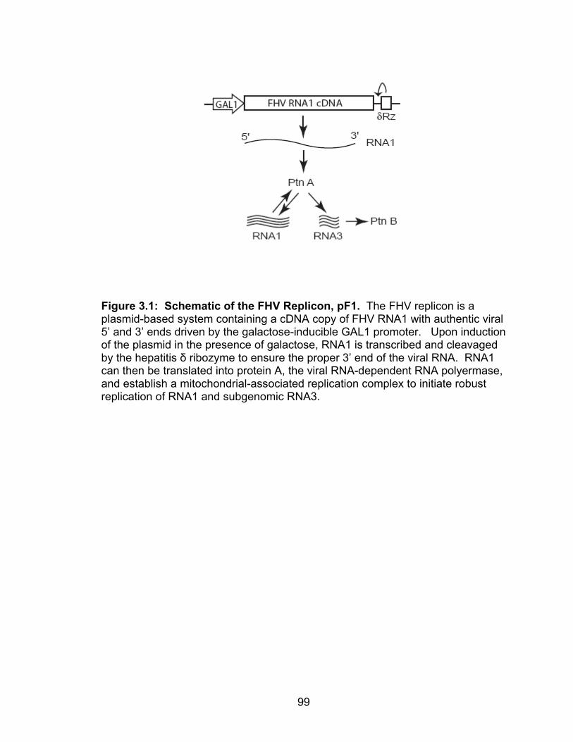
99
Figure 3.1: Schematic of the FHV Replicon, pF1. The FHV replicon is a plasmid-based system containing a cDNA copy of FHV RNA1 with authentic viral 5’ and 3’ ends driven by the galactose-inducible GAL1 promoter. Upon induction of the plasmid in the presence of galactose, RNA1 is transcribed and cleavaged by the hepatitis δ ribozyme to ensure the proper 3’ end of the viral RNA. RNA1 can then be translated into protein A, the viral RNA-dependent RNA polyermase, and establish a mitochondrial-associated replication complex to initiate robust replication of RNA1 and subgenomic RNA3.

100
RNA isolation, equal amounts of cells were subject to hot acidic phenol extraction
as previously described (30, 32, 59) and stored at -80°C until northern blot
analysis. RNA was separated on a 1.2% agarose-formaldehyde gel, blotted onto
nylon membranes, and probed for positive- and negative-strand RNA1 and RNA3
as previously described (31).
Mitochondrial isolation, carbonate extraction and RNA analysis.
Yeast mitochondria were isolated as previously described (29) and stored in
SEM buffer (250 mM sucrose, 1 mM EDTA, 10 mM 3-
morpholinopropanesulphonic acid (MOPS)-KOH, pH 7.2) at -80°C. For
carbonate extraction, mitochondria were pelleted at 10,000 x g for 15 min at 4°C,
resuspended in 0.1 M sodium carbonate (pH 11.5), and incubated on ice for 30
min. Mitochondria were reisolated by centrifugation at 55,000 x g for 1 h in a
TLA-55 rotor (Beckman), resuspended in SDS-PAGE buffer and subject to
immunoblot analysis as described above. For mitochondrial RNA isolated, RNA
was extract using an RNeasy Mini Kit (Qiagen) following manufacturers’
instructions, and 0.5 µg total RNA was analyzed by northern blotting as
described above.
In vitro RdRp activity. Mitochondria (20 µg total protein) were incubated
in RdRp buffer (50 mM Tris, pH 8.0, 50 mM potassium acetate, 15 mM
magnesium acetate, 40 units RNAsin (Promega), 5 µg/ml Actinomycin D, 1 mM
ATP, 1 mM CTP, 1 mM GTP, 25 µM UTP, 10 µCi [32P]UTP (Fisher Scientific) at
25°C for 3 h. Total RNA was extracted with one volume phenol-chloroform (pH
5.2) and unincorporated nucleotides were removed from the aqueous phase by a

101
Micro Bio-Spin Column (BioRad) following the manufacturer’s instructions.
Nucleic acid dye buffer (0.05% bromophenol blue, 0.05% xylene cyanol FF, 5%
glycerol) was added to each sample and RNAs were separated by 1.4%
agarose-TBE gel. Gels were dried under vacuum and subject to
autoradiography.
Blue-Native Agarose Gel Electrophoresis (BN-AGE). Membrane fractions
(100 µg total protein) were resuspended in solublization buffer (0.5% n-dodecyl
β-D-maltoside (DDM) 20 mM Tris-HCl (pH 7.4), 0.1 mM EDTA, 50 mM NaCl,
10% glycerol, 1 mM PMSF), incubated on ice for 30 min, and centrifuged at
100,000 x g for 15 min at 4°C. The supernatant was removed and mixed with
loading dye (0.5% coomassie brilliant blue G-250, 50 mM aminocaproic acid, 10
mM Bis-Tris, pH 7.0). Samples were then loaded on a 1.5% BN-agarose gel
containing 66.7 mM aminocaproic acid, 50 mM Bis-Tris pH 7.0, ran at 100 V for 2
hour in 50 mM Bis-Tris pH 7.0 as a running buffer. For immunoblotting, gels
were incubated in SDS-PAGE running buffer for 20 min followed by transfer to
PVDF membranes and immunoblot analysis as described (31). To analyze
radiolabeled viral RNAs, gels were tried under vacuum, and analyzed by
autoradiography.
Statistics. We used a two-tailed t-test assuming equal variance for
comparative statistical analysis and considered a p-value of < 0.05 as significant.
Unless otherwise indicated all quantitative data represent results from at least 3
independent experiments and are presented as the means ± standard errors of
the mean.

102
RESULTS
Flock House virus RNA replication in yeast strains harboring
mitochondrial outer membrane protein deletions. Flock House virus utilizes
the mitochondrial outer membrane for RNA replication complex assembly and
subsequent genome replication. To investigate the role of host mitochondrial
outer membrane proteins in FHV RNA replication we took advantage of a well-
established plasmid based FHV replicon system (Figure 3.1) in combination with
yeast strains harboring individual deletions or mutations of mitochondrial outer
membrane proteins (Table 3.2). Yeast deletion strains were transformed with the
FHV replicon pF1 and FHV RNA replication was initiated by growing yeast in the
presence of galactose for 24 hours at 25°C. Total viral RNA production was
analyzed by northern blotting with probe specific for positive-strand RNA1 and
sub-genomic RNA3 (Table 3.2). We found that a majority of the deletion strains
had no significant change in FHV RNA production, yet interestingly there were
several strains that led to significant increases or decreases in FHV RNA
accumulation. In particular we found that two temperature-sensitive mutants of
the major import pore of the mitochondrial outer membrane, Tom40, lead to
significant increases in RNA production, whereas deletions of POR1, TOM6,
DNM1, and MIM1 led to significant defects FHV RNA accumulation.
One possible explanation for these changes in FHV RNA accumulation may
be due to differences in yeast deletion and mutant strain growth rates, whereas
slow growing yeast may accumulate more RNA during the 24 hour period as

103
Table 3.2: Flock House virus RNA Replication in Yeast Strains with Mitochondrial Outer Membrane Protein Deletions or Mutations. Cellular Gene Description Growth (+)RNA3a Function Replication Mitochondrial Fission Mitochondrial Fusion Mitochondrial Protein Import and Assembly Other
DNM1 FIS1 MDV1 FZO1* UGO1 TOM5 TOM6 TOM7 TOM70 TOM72 TOM20* TOM22* TOM40-3* TOM40-6* SAM37 MDM12 MIM1* MCR1 UTH1 MMR1 MSP1 OM45 POR1 POR2 MDM34
Dynamin-related GTPase Localization of Dnm1p and Mdv1p WD repeat protein, interacts with Dnm1p and Fis1p Mitofusin, GTPase Links GTPases Fzo1p and Mgm1p during fusion TOM complex, Import and stability TOM complex, Assembly and stability TOM complex, Assembly and stability TOM complex, carrier proteins TOM complex, similar to Tom70 TOM complex, Main receptor TOM complex, Co-receptor TOM complex, Translocation channel TOM complex, Translocation channel SAM complex, β-barrel protein import Β-barrel protein import and assembly Assembly of TOM complex NADH-cytochrome b5 reductase Mitochondrial autophagy Mitochondrial distribution Protein sorting Unknown function Voltage-dependent Anion Channel1 Voltage-dependent Anion Channel2 Mitochondrial morphology and inheritance
1.0 ± 0.0 1.1 ± 0.0 1.0 ± 0.0
1.2 ± 0.0 1.1 ± 0.0
1.2 ± 0.0 1.2 ± 0.0 1.3 ± 0.1 0.9 ± 0.0 0.9 ± 0.0 1.0 ± 0.1 2.4 ± 0.3 1.3 ± 0.0 1.0 ± 0.1 1.1 ± 0.1 1.2 ± 0.0 1.6 ± 0.1
1.0 ± 0.0 1.1 ± 0.0 1.0 ± 0.0 1.1 ± 0.0 1.1 ± 0.0 1.4 ± 0.0 1.2 ± 0.0 1.3 ± 0.0
0.3 ± 0.0 1.1 ± 0.0 0.9 ± 0.2
1.0 ± 0.1 1.6 ± 0.4
0.8 ± 0.2 0.4 ± 0.0 0.8 ± 0.3 0.8 ± 0.2 1.2 ± 0.3 1.6 ± 0.1 1.2 ± 0.3 2.1 ± 0.3 3.8 ± 0.4 1.2 ± 0.2 1.2 ± 0.3 0.2 ± 0.0
0.7 ± 0.3 1.5 ± 0.5 0.5 ± 0.2 0.9 ± 0.2 1.9 ± 0.5 0.4 ± 0.0 0.8 ± 0.2 2.3 ± 1.1
aRNA3 replication is represented as percent of wildtype. *Not in BY4743 background (see Table 3.1). Tom40-3 and Tom40-6 are temperature-sensitive strains. Bold = p < 0.05

104
compared to a dividing wildtype cell. However, we found that there was no
significant differences in yeast growth rates in those strains which lead to
increased accumulation of FHV RNA. In contrast, the Mim1 deletion strain grew
roughly 1.5 times slower then its wildtype strain yet FHV RNA accumulation was
significantly impaired. Given this and the role of Mim1 in mitochondrial protein
import and TOM complex assembly we chose to further explore the role of Mim1
in FHV RNA replication.
FHV RNA replication is impaired in Mim1 deficient yeast. Mim1 is a small
mitochondrial outer membrane protein of approximately 13 kDa that has been
shown to be required for proper mitochondrial protein import and the assembly of
the yeast TOM complex and therefore may play a vital role in FHV RNA
replication complex assembly and function (3, 9, 11, 41, 58). To investigate the
role of Mim1 in FHV RNA replication we first addressed the ability of exogenously
expressed Mim1 to complement the defect in FHV RNA replication. Yeast
strains were transformed with the FHV replicon plasmid and Mim1 deficient yeast
received either a vector control or plasmid expressing Mim1 under its
endogenous promoter. We confirmed that the Mim1 expression plasmid was
functional by its ability to complement growth of Mim1 deficient yeast at 37ºC
(Figure 3.2A). Yeast were grown in the presence of galactose to induce FHV
RNA replication and we analyzed total FHV positive- and negative-strand RNA1
and RNA3 levels by northern blotting. Mim1 deficient yeast showed a 5-fold
reduction in both positive- and negative strand RNA1 and RNA3 levels compared

105
Figure 3.2: Complementation of FHV RNA Replication in Mim1 Deficient Yeast. (A) Mim1 deleted yeast were transformed with pF1 and either an empty plasmid (pRS315) or a plasmid expressing Mim1 under the control of the yeast Mim1 promoter (pMim1), grown to saturation in selectable media with 2% raffinose, diluted to an OD of 0.5, and diluted in 10-fold increments. Yeast were spotted on plates containing selectable media with dextrose and grown at 37°C. (B) Wildtype, Mim1 deleted (315), and Mim1 complemented yeast (Mim1) containing pF1 were induced in 2% galactose for 24 hours at 25°C. Equal numbers of cells were isolated and total RNA and protein were analyzed by northern blotting and immunoblot as described previously.

106
to wildtype yeast (Figure 3.2B, lanes 4 and 5), yet we were unable to rescue this
defect to any degree with the exogenous expression of Mim1 (Figure 3.2B, lanes
6 and 7). This phenotype correlated with a reduction in protein B2 translation
from RNA3 but was not due to defects in the translation of the polymerase as
protein A levels were found to be equal between all samples. One possible
explanation for this could be that Mim1 expression levels were not sufficient to
complement the FHV RNA replication defect as we found total Mim1 levels to be
approximately 10% of wildtype by immunoblot (Figure 3.2B, compare lanes 2 and
3 with lanes 6 and 7). However, when we looked at membrane fractions isolated
from wildtype and Mim1 complemented yeast we did not see this dramatic
difference in membrane-associated Mim1 levels (Figure 3.3A). Additionally,
corresponding with the ability to rescue the temperature-sensitive phenotype, we
found that expression of Mim1 was able to restore Tom70 and porin protein
levels to that of wildtype (Figure 3.3A) indicating that although Mim1 is expressed
at a low levels it is able to rescue a variety of yeast phenotypes. Nonetheless,
given the significant defect in FHV RNA accumulation in Mim1 deficient yeast
and the role of Mim1 in mitochondrial protein import and assembly we chose to
further investigate the role of Mim1 in FHV RNA replication using the
endogenous expression system.
FHV protein A is membrane-associated and behaves as an integral
membrane protein in Mim1 deficient yeast. The observation that protein A is
translated properly in Mim1 deficient yeast suggest that the defect may occur at a
post-translational step in replication complex assembly. After translation, FHV

107
Figure 3.3: Protein A is membrane-associated in Mim1 deficient yeast. (A) Yeast expressing pF1 were fractionated into supernatant (S12) and crude membrane (P12) fractions. 20 µg of total protein was separated by SDS-PAGE and proteins were analyzed by immunblotting. (B) 100 µg of total crude membrane fractions (T) were treated with 0.1 M sodium carbonate, pH 11.5 for 30 min on ice. Samples were then separated by centrifugation at 55,000 x g for 30 min to yield a supernatant (S) and pellet (P) fraction. Proteins were separated by SDS-PAGE and analyzed as previously described.

108
protein A is targeted to the mitochondrial outer membrane in wildtype yeast (30)
and thus it is possible that the RNA replication defect is due to improper
membrane association of protein A. To address if protein A was membrane
associated, we fractionated pF1 expressing wildtype, Mim1 deficient, and Mim1
complemented yeast by differential centrifugation to obtain supernatant (S12)
and crude membrane fractions (P12). In all strains, protein A was found to
fractionate in the membrane fraction similar to the mitochondrial outer membrane
proteins Tom70, porin, and Mim1 (Figure 3.3A). To rule out the possibility that
protein A is aggregating after lysis in Mim1 deficient and Mim1 complemented
yeast strains we performed membrane flotation assays with each strain and
confirmed that protein A was membrane-associated (data not shown).
Additionally, given the role of Mim1 in mitochondrial protein import, we addressed
whether membrane bound protein A behaved as an integral membrane protein
as seen in wildtype yeast (30). We found that in all strains, protein A remained
membrane associated after alkaline treatment similar to the β-barrel protein porin
whereas we were able to remove portions of soluble protein PGK into the
supernatant fraction. These data indicate that the membrane bound replicase is
inserted as an integral membrane protein (Figure 3.3B) in Mim1 deficient yeast.
Taken together, these studies show that protein A is tightly membrane-
associated and inserted as an integral membrane protein as seen in wildtype
yeast.
FHV membrane-associated RNA replication complexes are less active in
Mim1 deficient yeast. Given that protein A is translated and membrane-

109
associated in Mim1 deficient yeast we hypothesized that the defect in FHV RNA
replication is at the level of replicase function. To address the specific activity of
FHV RNA replication complexes we incubated normalized amounts of protein A
containing membrane-associated viral replication complexes in an in vitro RdRp
assay and subsequently analyzed the ability of the replicase to synthesize viral
RNAs in vitro. As a control, membrane fractions not expressing protein A
showed very little background whereas wildtype yeast expressing pF1
synthesized distinct FHV RNA products (Figure 3.4A, lane 2-3). However, with
both the Mim1 deficient and Mim1 complemented membrane fractions there was
a significant reduction in all FHV RNA species (Figure 3.4A, lanes 4-7),
suggesting a defect in FHV replication complex function.
The FHV replication complex is composed of two major viral components, the
viral RNA-dependent RNA polymerase and the viral RNA template. One reason
for reduced activity could be the lack of an RNA template associated with the
membrane bound replicase. It has been shown that during replication complex
assembly, protein A promotes the membrane association of RNA1 and this
interaction is required for efficient RNA replication (55, 56). We addressed
membrane-associated FHV RNA by extracting RNA from equal amounts of
isolated membrane fractions and probing for both positive- and negative-strand
FHV RNAs. Interestingly compared to wildtype, we found a significant reduction
in both membrane-associated FHV positive- and negative-strand RNA1 and
RNA3 in both Mim1 deficient and Mim1 complemented yeast (Figure 3.4B).

110
Figure 3.4: FHV in vitro replicase activity and membrane-associated RNA levels are reduced in ∆mim1 yeast. (A) Membrane fractions (20 µg total protein) were incubated in the presence of RdRp buffer for 3 hours at 25° followed by the addition of an equal volume of phenol:cholorform (pH 5.2). Unincorporated nucleotides were removed from the aqueous phase, RNAs were separated on a 1.2% TBE-agarose gel, and analyzed by autoradiography. (B) Total RNA was isolated from membrane fractions (100 µg total protein) using the RNeasy Kit and 0.5 µg total RNA was analyzed by northern blotting as previously described.

111
Taken together, these data suggest that FHV RNA replication complexes isolated
from Mim1 deficient yeast are defective at the level of replicase activity.
Mim1 deficient yeast lack a large FHV protein A complex capable of RNA
synthesis. Given the role of Mim1 in mitochondrial protein import and protein
complex assembly, it is possible that protein A is translated and membrane-
associated but not assembled properly into a native FHV RNA replication
complex. The assembly of yeast native mitochondrial outer membrane
complexes has been studied in detail in part due to the development of blue-
native polyacrylamide gel electrophoresis (BN-PAGE). This technique has been
used to separate large macromolecular membrane-protein complexes based on
size, however it is unable to efficiently resolve much larger protein-RNA
complexes. In an alternative approach, we used blue-native agarose gel
electrophoresis (BN-AGE) followed by immunoblotting to visualize protein A
containing complexes (Figure 3.5). FHV RNA replication complexes were
solubilized with the mild detergent n-dodecyl β-D-maltoside (DDM) which has
been used previously to study FHV replicase function in vitro (62, 63). When we
analyzed FHV RNA replication complexes from wildtype yeast we found there to
be two distinct protein A containing complexes (Figure 3.5A). Interestingly, we
found that membrane-associated protein A complexes from Mim1 deficient yeast
were missing the larger band but retained equal amounts of the faster migrating
smaller complex.
To further examine these two distinct protein complexes we were interested in
which of the complexes retained FHV RNA replicase activity. We first subjected

112
membrane fractions to an in vitro RdRp assay to label actively synthesized viral
RNAs followed by solublization in DDM and separation of protein-RNA
complexes by BN-AGE. We hypothesized that the fraction of protein A that
retained replicase activity would incorporate radiolabeled nucleotides into the
newly synthesized RNA strands allowing us to visualize these complexes. We
found that in wildtype yeast the slower migrating protein A complex was capable
of RNA synthesis as it was the only complex to incorporate nucleotides into the
synthesized RNAs (Figure 3.5B, lane 2). This complex was not present in Mim1
deficient yeast (Figure 3.5B) suggesting that the FHV RNA replication defect in
Mim1 deficient yeast is due to a defect in the assembly of large protein A
containing complexes capable of RNA synthesis.
The assembly of wildtype FHV replication complexes is dependent on
active viral RNA synthesis. These data with Mim1 deficient yeast suggest the
presence of two functionally distinct FHV protein A containing complexes which
are required for efficient FHV RNA replication. In addition, the FHV replicon
system is designed to establish robust protein A-dependent RNA replication, and
we hypothesized that in using this system protein A levels would be correlated
with (+)RNA1 such that a defect in FHV RNA1 production would yield a similar
defect in protein levels. This effect as been seen previously in yeast deficient in
YDJ1 (59), however although we observed significant differences in positive-
strand RNA1 levels there were no differences in protein A accumulation between
wildtype and Mim1 deficient yeast (Figure 3.2B). One possible explanation for
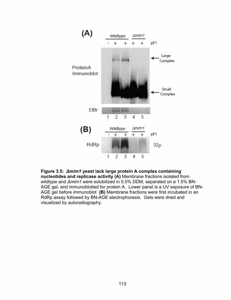
113
Figure 3.5: ∆mim1 yeast lack large protein A complex containing nucleotides and replicase activity (A) Membrane fractions isolated from wildtype and ∆mim1 were solubilized in 0.5% DDM, separated on a 1.5% BN-AGE gel, and immunoblotted for protein A. Lower panel is a UV exposure of BN-AGE gel before immunoblot (B) Membrane fractions were first incubated in an RdRp assay followed by BN-AGE electrophoresis. Gels were dried and visualized by autoradiography.

114
this is that plasmid based transcription of RNA1 from pF1, which is a 2-micron
plasmid, is sufficient to make up for the lack of RNA1 produced from FHV
replication. However, this is unlikely as we have obtained similar results using a
variety of pF1 constructs expressed from centromeric plasmids (data not shown).
This suggests that protein A translation is independent of active protein A-
dependent (+)RNA1 production. We chose to address this directly in wildtype
yeast by separating protein A translation from protein A-dependent RNA
replication with the introduction of an active site mutation into the FHV replicon
(Figure 3.6A). This would allow RNA1 to be transcribed and translated into
protein A as before, but this protein A (containing a GDD to GNN mutation) would
be unable to further amplify more (+)RNA1 to be used for translation. Using this
system, we compared protein A and (+)RNA1 production between the replication
incompetent replicon and the wildtype replicon. We found that similar to what is
seen in Mim1 deficient yeast, equal amounts of protein A were produced
between the wildtype and replication incompetent replicons yet the amount of the
(+)RNA1 was significantly different (Figure 3.6B). This data confirms that RNA1
dependent polymerase translation and genome replication can be separated in
vivo. If this is the case, then this may explain the two protein A containing
complexes we see with BN-AGE. To address this, we investigated the assembly
of protein A containing complexes using the replication incompetent replicon by
BN-AGE. Interestingly, we found that both protein complexes which were
present with the wildtype replicon were missing in yeast expressing the
replication incompetent replicon (Figure 3.6C). These data suggest a possible

115
stepwise assembly mechanism for FHV RNA replication complexes which is
dependent on functional viral RNA synthesis.

116
Figure 3.6: Assembly of protein A complexes is dependent on viral RNA synthesis. (A) Schematic of replication incompetent replicon containing GDD to GNN mutation. (B) Immunoblot and northern blot of FHV RNA replication with wildtype and replication competent replions. (C) BN-AGE analysis of membrane associated FHV replication complexes in yeast expressing the wildtype and replication incompontent replicons.

117
DISCUSSION
In this report we have taken a targeted genomic approach to investigate the
role of yeast mitochondrial outer membrane proteins in Flock House virus RNA
replication. We have identified a variety of mitochondrial outer membrane
deletion strains that lead to increases or decreases in FHV RNA replication which
include members of the protein import and fission machinery. In addition, we
identified Mim1 which led to a significant reduction in positive- and negative-
strand FHV RNA1 and RNA3 accumulation. This defect is independent of FHV
polymerase translation or membrane-association as we found equal amounts of
protein A to behave as an integral membrane protein in Mim1 deficient yeast.
However, we found membrane-associated FHV RNA replication complexes from
Mim1 deficient yeast to have reduced in vitro activity and an analysis of
membrane-associated viral RNAs yielded lower levels of positive- and negative-
strand viral RNAs associated with membrane fractions. Lastly, we found ∆mim1
yeast to lack the functional subunit of the FHV RNA replication complex
suggesting a potential role for Mim1 in FHV replication complex assembly.
These data provide evidence for the role of viral RNA synthesis in the assembly
of functional FHV RNA replication complexes, and suggest an important role for
host components in this process.
Of particular interest is the observation that protein A translation and RNA1
production are uncoupled using the FHV replicon. It is thought that during active
FHV RNA replication, RNA1 that is produced from protein A-dependent RNA
synthesis can then be used as a template for protein A translation and

118
subsequent replication complex assembly, and that defects in FHV RNA
replication yield similar defects in polymerase production, a phenotype that has
been observed with FHV RNA replication in YDJ1 deficient yeast (59). However,
the deletion of Mim1 sheds light on the fact that these two processes may be
mutually exclusive. Positive-strand RNA viruses in particular must begin with
one genomic template which is used for a variety of processes including; the
initial translation of viral proteins, membrane trafficking and replication complex
assembly, genomic replication, and finally packaging of the genome to produce
new infectious virions. It is thus not unreasonable that these processes may be
temporally or spatially linked during the viral life cycle. Studies with FHV RNA2
have shown that RNA replication, translation, and packaging are linked during
FHV replication (57) yet the molecular mechanisms involved in these processes
are unknown. One possible mechanism came from work with Brome Mosaic
virus where viral RNAs localize with P-bodies during replication complex
assembly and these discrete cellular compartments may separate translating
from replicating viral RNAs (4). Future studies of FHV RNA replication with
various yeast deletions such as Mim1 will provide insight into the complex and
dynamic process of viral replication complex assembly and RNA replication.
Furthermore, the presence of two functionally distinct FHV RNA replication
complexes is intriguing. It is currently hypothesized, based on quantitative
analyses of protein and RNA levels, that viral RNA replication complexes are
composed primarily of non-functional subunits whereas the functional subunits
make up a small portion which contain the viral RNAs (19, 44). Our data are in
119
line with this hypothesis indicating that not only are there two protein complexes
but one complex contains RNA and replicase activity, thus providing biochemical
evidence to support this theory. Furthermore, we found that FHV RNA synthesis
and replication complex assembly are intimately linked providing another
example of the coupling of viral processes during RNA replication. It will be
interesting to further explore the assembly of these complexes as well as the
structure and composition that is required for RNA synthesis.
In addition to Mim1 we found a variety of other yeast mitochondrial outer
membrane deletions which led to changes in FHV RNA replication. Of particular
interest were members of the TOM complex, which is important for the import
and assembly of the majority of mitochondrial proteins in yeast, and the
mitochondrial fission machinery. We have previously shown that the TOM
complex is not require for the proper in vitro membrane association of protein A
(Chapter II) yet functional studies presented in this chapter indicate that deletions
or mutations in TOM complex components can significantly influence FHV RNA
replication. Similar to Mim1, deletions of protein import and fission components
lead to changes in not only protein import but also changes in mitochondrial
morphology (2, 28, 54) suggesting possible changes in membrane phospholipid
components as secondary effects in these deletion strains. Mim1 has been
shown to physically interact with members of the SAM complex (3). One
membrane protein in particular Sam37 was originally identified in a screen
looking for genes regulating cellular phospholipids biosynthesis (8). We did not
find any changes in FHV RNA replication in yeast lacking Sam37 using the

120
replicion system however, we did find that when using the trans-replication
system a deletion of this protein leads to roughly an 8-fold increase in FHV RNA
replication (K. Stapleford and D. Miller, unpublished data). The reason for these
differences in FHV RNA replication between replication systems is currently
unclear but future work exploring this may provide valuable information regarding
viral replication. Nonetheless, these data indicate a possible functional role for
host protein and lipid components in FHV RNA replication. Unfortunately, work
with Sam37 and phospholipid biosynthesis was not followed up but a detailed
phospholipid analysis of the mitochondrial outer membrane from many of the
deletion strains used in this studied would yield valuable information not only for
FHV RNA replication but for yeast mitochondrial biology as well.
In summary these studies identify several mitochondrial outer membrane
deletion yeast strains that have dramatic changes in FHV RNA replication. In
particular, a deletion of Mim1 leads to significant reductions in RNA synthesis in
part due a defect in the assembly of functional viral RNA replication complexes, a
process which is dependent on active RNA synthesis and host processes. Mim1
deficient yeast have provided a system with which viral RNA replication can be
dissected in vivo to better understand the molecular mechanisms involved in
these processes. Future studies with Mim1 deficient yeast and other
mitochondrial outer membrane deletion yeast strains will provide valuable insight
into the cellular components and processes which govern positive-strand RNA
virus RNA replication.

121
REFERENCES
1. Ahting, U., T. Waizenegger, W. Neupert, and D. Rapaport. 2005. Signal-anchored proteins follow a unique insertion pathway into the outer membrane of mitochondria. J Biol Chem 280:48-53.
2. Altmann, K., and B. Westermann. 2005. Role of essential genes in
mitochondrial morphogenesis in Saccharomyces cerevisiae. Mol Biol Cell 16:5410-7.
3. Becker, T., S. Pfannschmidt, B. Guiard, D. Stojanovski, D. Milenkovic,
S. Kutik, N. Pfanner, C. Meisinger, and N. Wiedemann. 2008. Biogenesis of the mitochondrial TOM complex: Mim1 promotes insertion and assembly of signal-anchored receptors. J Biol Chem 283:120-7.
4. Beckham, C. J., H. R. Light, T. A. Nissan, P. Ahlquist, R. Parker, and
A. Noueiry. 2007. Interactions between brome mosaic virus RNAs and cytoplasmic processing bodies. J Virol 81:9759-68.
5. Castorena, K. M., S. A. Weeks, K. A. Stapleford, A. M. Cadwallader,
and D. J. Miller. 2007. A functional heat shock protein 90 chaperone is essential for efficient flock house virus RNA polymerase synthesis in Drosophila cells. J Virol 81:8412-20.
6. den Boon, J. A., J. Chen, and P. Ahlquist. 2001. Identification of
sequences in Brome mosaic virus replicase protein 1a that mediate association with endoplasmic reticulum membranes. J Virol 75:12370-81.
7. Denison, M. R. 2008. Seeking membranes: positive-strand RNA virus
replication complexes. PLoS Biol 6:e270. 8. Gratzer, S., T. Lithgow, R. E. Bauer, E. Lamping, F. Paltauf, S. D.
Kohlwein, V. Haucke, T. Junne, G. Schatz, and M. Horst. 1995. Mas37p, a novel receptor subunit for protein import into mitochondria. J Cell Biol 129:25-34.
9. Hulett, J. M., F. Lueder, N. C. Chan, A. J. Perry, P. Wolynec, V. A.
Likic, P. R. Gooley, and T. Lithgow. 2008. The transmembrane segment of Tom20 is recognized by Mim1 for docking to the mitochondrial TOM complex. J Mol Biol 376:694-704.
10. Hwang, Y. T., A. W. McCartney, S. K. Gidda, and R. T. Mullen. 2008.
Localization of the Carnation Italian ringspot virus replication protein p36 to the mitochondrial outer membrane is mediated by an internal targeting signal and the TOM complex. BMC Cell Biol 9:54.
122
11. Ishikawa, D., H. Yamamoto, Y. Tamura, K. Moritoh, and T. Endo. 2004. Two novel proteins in the mitochondrial outer membrane mediate beta-barrel protein assembly. J Cell Biol 166:621-7.
12. Jaag, H. M., J. Stork, and P. D. Nagy. 2007. Host transcription factor
Rpb11p affects tombusvirus replication and recombination via regulating the accumulation of viral replication proteins. Virology 368:388-404.
13. Jiang, Y., E. Serviene, J. Gal, T. Panavas, and P. D. Nagy. 2006.
Identification of essential host factors affecting tombusvirus RNA replication based on the yeast Tet promoters Hughes Collection. J Virol 80:7394-404.
14. Johnson, K. L., and L. A. Ball. 1999. Induction and maintenance of
autonomous flock house virus RNA1 replication. J Virol 73:7933-42. 15. Johnson, K. L., and L. A. Ball. 1997. Replication of flock house virus
RNAs from primary transcripts made in cells by RNA polymerase II. J Virol 71:3323-7.
16. Jonczyk, M., K. B. Pathak, M. Sharma, and P. D. Nagy. 2007. Exploiting
alternative subcellular location for replication: tombusvirus replication switches to the endoplasmic reticulum in the absence of peroxisomes. Virology 362:320-30.
17. Kampmueller, K. M., and D. J. Miller. 2005. The cellular chaperone heat
shock protein 90 facilitates Flock House virus RNA replication in Drosophila cells. J Virol 79:6827-37.
18. Knoops, K., M. Kikkert, S. H. Worm, J. C. Zevenhoven-Dobbe, Y. van
der Meer, A. J. Koster, A. M. Mommaas, and E. J. Snijder. 2008. SARS-coronavirus replication is supported by a reticulovesicular network of modified endoplasmic reticulum. PLoS Biol 6:e226.
19. Kopek, B. G., G. Perkins, D. J. Miller, M. H. Ellisman, and P. Ahlquist.
2007. Three-dimensional analysis of a viral RNA replication complex reveals a virus-induced mini-organelle. PLoS Biol 5:e220.
20. Korber, S., P. Shaik Syed Ali, and J. C. Chen. 2009. Structure of the
RNA-Binding Domain of Nodamura Virus Protein B2, a Suppressor of RNA Interference (dagger) , (double dagger). Biochemistry.
21. Kushner, D. B., B. D. Lindenbach, V. Z. Grdzelishvili, A. O. Noueiry, S.
M. Paul, and P. Ahlquist. 2003. Systematic, genome-wide identification of host genes affecting replication of a positive-strand RNA virus. Proc Natl Acad Sci U S A 100:15764-9.

123
22. Kutik, S., D. Stojanovski, L. Becker, T. Becker, M. Meinecke, V. Kruger, C. Prinz, C. Meisinger, B. Guiard, R. Wagner, N. Pfanner, and N. Wiedemann. 2008. Dissecting membrane insertion of mitochondrial beta-barrel proteins. Cell 132:1011-24.
23. Lee, W. M., and P. Ahlquist. 2003. Membrane synthesis, specific lipid
requirements, and localized lipid composition changes associated with a positive-strand RNA virus RNA replication protein. J Virol 77:12819-28.
24. Li, Z., J. Pogany, T. Panavas, K. Xu, A. M. Esposito, T. G. Kinzy, and
P. D. Nagy. 2009. Translation elongation factor 1A is a component of the tombusvirus replicase complex and affects the stability of the p33 replication co-factor. Virology 385:245-60.
25. Lu, R., M. Maduro, F. Li, H. W. Li, G. Broitman-Maduro, W. X. Li, and
S. W. Ding. 2005. Animal virus replication and RNAi-mediated antiviral silencing in Caenorhabditis elegans. Nature 436:1040-3.
26. Lu, R., E. Yigit, W. X. Li, and S. W. Ding. 2009. An RIG-I-Like RNA
helicase mediates antiviral RNAi downstream of viral siRNA biogenesis in Caenorhabditis elegans. PLoS Pathog 5:e1000286.
27. Marshall, D., and A. Schneemann. 2001. Specific packaging of nodaviral
RNA2 requires the N-terminus of the capsid protein. Virology 285:165-75. 28. Meisinger, C., M. Rissler, A. Chacinska, L. K. Szklarz, D. Milenkovic,
V. Kozjak, B. Schonfisch, C. Lohaus, H. E. Meyer, M. P. Yaffe, B. Guiard, N. Wiedemann, and N. Pfanner. 2004. The mitochondrial morphology protein Mdm10 functions in assembly of the preprotein translocase of the outer membrane. Dev Cell 7:61-71.
29. Meisinger, C., T. Sommer, and N. Pfanner. 2000. Purification of
Saccharomcyes cerevisiae mitochondria devoid of microsomal and cytosolic contaminations. Anal Biochem 287:339-42.
30. Miller, D. J., and P. Ahlquist. 2002. Flock house virus RNA polymerase
is a transmembrane protein with amino-terminal sequences sufficient for mitochondrial localization and membrane insertion. J Virol 76:9856-67.
31. Miller, D. J., M. D. Schwartz, and P. Ahlquist. 2001. Flock house virus
RNA replicates on outer mitochondrial membranes in Drosophila cells. J Virol 75:11664-76.
32. Miller, D. J., M. D. Schwartz, B. T. Dye, and P. Ahlquist. 2003.
Engineered retargeting of viral RNA replication complexes to an alternative intracellular membrane. J Virol 77:12193-202.

124
33. Miller, S., and J. Krijnse-Locker. 2008. Modification of intracellular membrane structures for virus replication. Nat Rev Microbiol 6:363-74.
34. Mozdy, A. D., J. M. McCaffery, and J. M. Shaw. 2000. Dnm1p GTPase-
mediated mitochondrial fission is a multi-step process requiring the novel integral membrane component Fis1p. J Cell Biol 151:367-80.
35. Nagy, P. D. 2008. Yeast as a model host to explore plant virus-host
interactions. Annu Rev Phytopathol 46:217-42. 36. Neupert, W., and J. M. Herrmann. 2007. Translocation of proteins into
mitochondria. Annu Rev Biochem 76:723-49. 37. Nishikiori, M., K. Dohi, M. Mori, T. Meshi, S. Naito, and M. Ishikawa.
2006. Membrane-bound tomato mosaic virus replication proteins participate in RNA synthesis and are associated with host proteins in a pattern distinct from those that are not membrane bound. J Virol 80:8459-68.
38. Okamoto, K., and J. M. Shaw. 2005. Mitochondrial morphology and
dynamics in yeast and multicellular eukaryotes. Annu Rev Genet 39:503-36.
39. Pathak, K. B., Z. Sasvari, and P. D. Nagy. 2008. The host Pex19p plays
a role in peroxisomal localization of tombusvirus replication proteins. Virology 379:294-305.
40. Pfanner, N., N. Wiedemann, C. Meisinger, and T. Lithgow. 2004.
Assembling the mitochondrial outer membrane. Nat Struct Mol Biol 11:1044-8.
41. Popov-Celeketic, J., T. Waizenegger, and D. Rapaport. 2008. Mim1
functions in an oligomeric form to facilitate the integration of Tom20 into the mitochondrial outer membrane. J Mol Biol 376:671-80.
42. Price, B. D., M. Roeder, and P. Ahlquist. 2000. DNA-Directed
expression of functional flock house virus RNA1 derivatives in Saccharomyces cerevisiae, heterologous gene expression, and selective effects on subgenomic mRNA synthesis. J Virol 74:11724-33.
43. Prod'homme, D., A. Jakubiec, V. Tournier, G. Drugeon, and I. Jupin.
2003. Targeting of the turnip yellow mosaic virus 66K replication protein to the chloroplast envelope is mediated by the 140K protein. J Virol 77:9124-35.

125
44. Quinkert, D., R. Bartenschlager, and V. Lohmann. 2005. Quantitative analysis of the hepatitis C virus replication complex. J Virol 79:13594-605.
45. Salonen, A., L. Vasiljeva, A. Merits, J. Magden, E. Jokitalo, and L.
Kaariainen. 2003. Properly folded nonstructural polyprotein directs the semliki forest virus replication complex to the endosomal compartment. J Virol 77:1691-702.
46. Schaad, M. C., P. E. Jensen, and J. C. Carrington. 1997. Formation of
plant RNA virus replication complexes on membranes: role of an endoplasmic reticulum-targeted viral protein. EMBO J 16:4049-59.
47. Schlegel, A., T. H. Giddings, Jr., M. S. Ladinsky, and K. Kirkegaard.
1996. Cellular origin and ultrastructure of membranes induced during poliovirus infection. J Virol 70:6576-88.
48. Schmidt-Mende, J., E. Bieck, T. Hugle, F. Penin, C. M. Rice, H. E.
Blum, and D. Moradpour. 2001. Determinants for membrane association of the hepatitis C virus RNA-dependent RNA polymerase. J Biol Chem 276:44052-63.
49. Schneemann, A., W. Zhong, T. M. Gallagher, and R. R. Rueckert.
1992. Maturation cleavage required for infectivity of a nodavirus. J Virol 66:6728-34.
50. Selling, B. H., R. F. Allison, and P. Kaesberg. 1990. Genomic RNA of
an insect virus directs synthesis of infectious virions in plants. Proc Natl Acad Sci U S A 87:434-8.
51. Serva, S., and P. D. Nagy. 2006. Proteomics analysis of the tombusvirus
replicase: Hsp70 molecular chaperone is associated with the replicase and enhances viral RNA replication. J Virol 80:2162-9.
52. Serviene, E., Y. Jiang, C. P. Cheng, J. Baker, and P. D. Nagy. 2006.
Screening of the yeast yTHC collection identifies essential host factors affecting tombusvirus RNA recombination. J Virol 80:1231-41.
53. Singh, G., S. Popli, Y. Hari, P. Malhotra, S. Mukherjee, and R. K.
Bhatnagar. 2009. Suppression of RNA silencing by Flock house virus B2 protein is mediated through its interaction with the PAZ domain of Dicer. Faseb J.
54. Stojanovski, D., M. Rissler, N. Pfanner, and C. Meisinger. 2006.
Mitochondrial morphology and protein import--a tight connection? Biochim Biophys Acta 1763:414-21.
126
55. Van Wynsberghe, P. M., and P. Ahlquist. 2009. 5' cis elements direct nodavirus RNA1 recruitment to mitochondrial sites of replication complex formation. J Virol.
56. Van Wynsberghe, P. M., H. R. Chen, and P. Ahlquist. 2007. Nodavirus
RNA replication protein a induces membrane association of genomic RNA. J Virol 81:4633-44.
57. Venter, P. A., and A. Schneemann. 2007. Assembly of two independent
populations of flock house virus particles with distinct RNA packaging characteristics in the same cell. J Virol 81:613-9.
58. Waizenegger, T., S. Schmitt, J. Zivkovic, W. Neupert, and D.
Rapaport. 2005. Mim1, a protein required for the assembly of the TOM complex of mitochondria. EMBO Rep 6:57-62.
59. Weeks, S. A., and D. J. Miller. 2008. The heat shock protein 70
cochaperone YDJ1 is required for efficient membrane-specific flock house virus RNA replication complex assembly and function in Saccharomyces cerevisiae. J Virol 82:2004-12.
60. Westermann, B. 2008. Molecular machinery of mitochondrial fusion and
fission. J Biol Chem 283:13501-5. 61. Wiedemann, N., V. Kozjak, A. Chacinska, B. Schonfisch, S. Rospert,
M. T. Ryan, N. Pfanner, and C. Meisinger. 2003. Machinery for protein sorting and assembly in the mitochondrial outer membrane. Nature 424:565-71.
62. Wu, S. X., P. Ahlquist, and P. Kaesberg. 1992. Active complete in vitro
replication of nodavirus RNA requires glycerophospholipid. Proc Natl Acad Sci U S A 89:11136-40.
63. Wu, S. X., and P. Kaesberg. 1991. Synthesis of template-sense, single-
strand Flockhouse virus RNA in a cell-free replication system. Virology 183:392-6.
64. Zahedi, R. P., A. Sickmann, A. M. Boehm, C. Winkler, N. Zufall, B.
Schonfisch, B. Guiard, N. Pfanner, and C. Meisinger. 2006. Proteomic analysis of the yeast mitochondrial outer membrane reveals accumulation of a subclass of preproteins. Mol Biol Cell 17:1436-50.

127
APPENDIX
PRELIMINARY STUDIES ON THE ROLE OF YEAST MIM1 IN FHV RNA
REPLICATION
We have identified a deletion of the mitochondrial outer membrane protein
Mim1 that leads to a significant reduction in FHV RNA accumulation. We found
this defect to be in part due to the absence of a functional FHV replication
complex which leads to a decrease in membrane-associated replicase activity
and membrane-associated viral RNA. We attempted to rescue this defect with
the exogenous expression of Mim1 under the control of its endogenous promoter
but have thus far been unsuccessful. These results were particularly interesting
in that we confirmed that Mim1 was functional by its ability to complement protein
defects and temperature-sensitive phenotypes associated with the deletion of
Mim1. Taken together, these data suggest that FHV RNA replication may
depend on other cellular processes in which the exogenous expression of Mim1
can not complement. To this end, the molecular role of Mim1 in FHV RNA
replication complex assembly and function is currently unclear.
Mim1 is a small mitochondrial outer membrane protein of approximately 13
kDa that was originally identified, along with Sam35, in a screen for yeast
proteins involved in mitochondrial β-barrel protein import and assembly (9).

128
Since then, additional studies have implicated Mim1 in the import of the signal-
anchored receptor proteins, Tom20 and Tom70, as well as the assembly of the
TOM complex (2, 8, 22). Furthermore, Mim1 has been shown to physically
interact with the mitochondrial outer membrane SAM complex although a
functional interaction between these two complexes has yet to be shown (2).
Finally, the deletion of Mim1 has been shown to lead to dramatic changes in
mitochondrial morphology similar to those seen in the deletion of various
mitochondrial protein import machinery components (1). Given this, we were
interested in identifying the specific role of Mim1 in FHV RNA replication complex
assembly and function.

129
MATERIALS AND METHODS
Yeast strains, transformations, and culture conditions. The wildtype
yeast strain YPH499 (MATα ade2-101 his3∆200 leu2∆1 ura3-52 trp1-∆63 lys-
801) and ∆mim1 (MATα ade2-101 his3∆200 leu2∆1 ura3-52 trp1∆63 lys2-801
mim1::HIS3) were obtained from Dr. Doron Rapaport (University of Tubingen,
Germany). Yeast were transformed using the E-Z Transformation Kit (Zymo
Research, Orange, CA). Individual clones were grown to early stationary phase
in selectable liquid media containing 2% [wt/vol] raffinose at 30°C. For the
induction of galactose-inducible plasmids, yeast were washed once with distilled
water, resuspended in selectable liquid media containing 2% raffinose [wt/vol]
and 2% [wt/vol] galactose at an OD600 of 0.2, and grown for 24 h at 25°C for all
experiments. For mitochondrial phospholipid extractions, yeast were grown in
YPDGal (1% yeast extract [wt/vol], 2% peptone [wt/vol], 1% dextrose [wt/vol],
and 1% galactose [wt/vol] at 25°C for 48 hours.
Plasmids and antibodies. The FHV RNA and protein expression plasmids
pF1, pF1fs, pFA-C/HA, and pFA-HCV-HA have been previously described (16,
19, 23). The Mim1 overexpression plasmid, pMIM1-OX, encoding yeast Mim1
under the triosephosphate isomerase (TPI) promoter was previously described
(18). Rabbit polyclonal antibodies raised against FHV protein A and protein B2
have been previously described (11, 15). Rabbit polyclonal antibodies against
the hemagglutinin (HA) epitope and mouse polyclonal antibodies against 3-
phosphoglycerate kinase (PGK1) were purchased from Santa Cruz Biotech

130
(Santa Cruz, CA) and Molecular Probes (Carlsbad, CA) respectively. Rabbit
polyclonal antibodies against Mim1 were previously described (18).
RNA and protein analysis. For total protein isolation, equal amounts of cells
were isolated 24 hours after plasmid induction and resuspended in protein
extraction buffer (20 mM Tris, pH 8.0, 200 mM NaCl, 10 mM MgCl2, 1 mM EDTA,
5% glycerol, 1:50 dilution of yeast protease inhibitor cocktail (Sigma)). Cell walls
were removed by vortexing in the presence of glass beads for 10 min at 4°C
followed by the addition of 1% SDS and heating at 100°C for 10 min. Samples
were pelleted by centrifugation at 10,000 x g for 5 min and supernatant aliquots
were removed, mixed with 2X SDS-PAGE sample buffer (125 mM Tris-HCl; pH
6.8, 4% SDS, 20% (w/v) glycerol, 0.002 mg/ml bromophenol blue, 10% 2-
mercaptoethanol), and stored at -20°C for further analysis. Total protein
extractions were separated by SDS-PAGE, transferred in polyvinylidene fluoride
(PVDF) membranes, and immunoblotted as previously described (15). For total
RNA isolation, equal amounts of cells were subject to hot acidic phenol extraction
as previously described and stored at -80°C until northern blot analysis. RNA
was separated on a 1.2% agarose-formaldehyde gel, blotted onto nylon
membranes, and probed for positive- and negative-strand RNA1 and RNA3 as
previously described (15).
Phospholipid extraction and analysis. Total mitochondrial phospholipids
were extracted as previously described (3). Yeast mitochondria (100 µg total
protein) were resuspended in 100 µl SEM buffer followed by the addition of 375
µl chloroform:methanol (1:2) and vortexed vigorously. Samples were extracted

131
again with 125 µl chloroform and 125 µl H2O with vortexing in between each
step. The aqueous (top) and organic (bottom) layers were separated by
centrifugation at 1,000 rpm for 5 min at room temperature. The organic layer
was removed and phospholipids were dried by removing the solvent under air
stream. Samples were sent to the Kansas Lipidomic Research Center (Kansas
State University) for mass spectroscopy analysis.

132
RESULTS
Complementation of FHV RNA replication with URA3 auxotrophic
marker. We previously attempted to complement the FHV RNA accumulation
defect with the exogenous expression of Mim1 under the control of its
endogenous promoter, yet we were unable to rescue the defect to any degree.
One possible explanation for this could be that although the expression of Mim1
with this system was able to complement the yeast temperature-sensitive
phenotypes, it is insufficient to rescue the FHV phenotype. To address this we
attempted to rescue the FHV RNA replication defect by introducing a Mim1
overexpression plasmid under the control of the strong triosephosphate
isomerase (TPI) promoter. As seen previously, overexpression of Mim1 was
able to rescue the temperature-sensitive phenotype (Figure 3.7A), yet
interestingly we found FHV RNA replication increased over wildtype levels by the
control plasmid alone (Figure 3.7B, lanes 4 and 5) whereas the overexpression
of Mim1 brought RNA levels back down towards wildtype (Figure 3.7B, lanes 6
and 7). Protein B2 translation correlated with RNA3 production whereas there
was no change in protein A expression between the strains indicating that this
defect was not due to increased polymerase accumulation. These results were
unexpected as the only differences between the two control plasmids used in the
Mim1 endogenous and overexpression systems were the auxotrophic markers
(LEU vs URA) and the plasmid copy number (centromeric vs 2-micron).

133
Figure 3.7: Complementation of FHV RNA replication with Mim1 overexpression. (A). Yeast strains were transformed with the control plasmid pRS426 or Mim1 overexpression plasmid pMim1-OX, grown inSpotted selective liquid media, and plated in ten-fold dilutions onto selective plates with 2% dextrose. Strains were grown at 37°C for 4 days. (B). Yeast were induced for 24 hours in selectable liquid media with 2% galactose at 25°C. Equal amounts of cells were isolated, protein and RNA was extracted, and analyzed by northern and western blotting respectively.
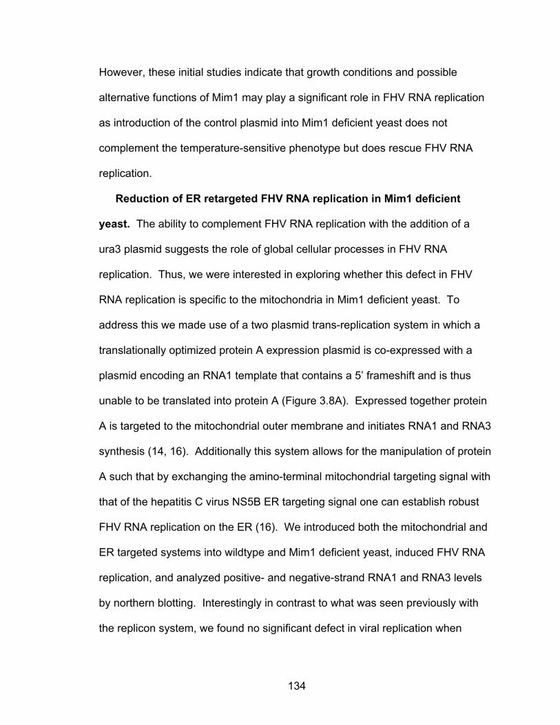
134
However, these initial studies indicate that growth conditions and possible
alternative functions of Mim1 may play a significant role in FHV RNA replication
as introduction of the control plasmid into Mim1 deficient yeast does not
complement the temperature-sensitive phenotype but does rescue FHV RNA
replication.
Reduction of ER retargeted FHV RNA replication in Mim1 deficient
yeast. The ability to complement FHV RNA replication with the addition of a
ura3 plasmid suggests the role of global cellular processes in FHV RNA
replication. Thus, we were interested in exploring whether this defect in FHV
RNA replication is specific to the mitochondria in Mim1 deficient yeast. To
address this we made use of a two plasmid trans-replication system in which a
translationally optimized protein A expression plasmid is co-expressed with a
plasmid encoding an RNA1 template that contains a 5’ frameshift and is thus
unable to be translated into protein A (Figure 3.8A). Expressed together protein
A is targeted to the mitochondrial outer membrane and initiates RNA1 and RNA3
synthesis (14, 16). Additionally this system allows for the manipulation of protein
A such that by exchanging the amino-terminal mitochondrial targeting signal with
that of the hepatitis C virus NS5B ER targeting signal one can establish robust
FHV RNA replication on the ER (16). We introduced both the mitochondrial and
ER targeted systems into wildtype and Mim1 deficient yeast, induced FHV RNA
replication, and analyzed positive- and negative-strand RNA1 and RNA3 levels
by northern blotting. Interestingly in contrast to what was seen previously with
the replicon system, we found no significant defect in viral replication when
135
Figure 3.8: ER targeted FHV RNA replication is inhibited in Mim1 deficient yeast. (A). Schematic of FHV trans replication system. The translationally optimized plasmid (pFA) and replication competent RNA1 plasmid (pF1fs) are co-expressed in yeast and lead to RNA1 and RNA3 synthesis. (B). Wildtype and Mim1 deficient yeast were transformed with mitochondrial or ER targeted protein A molecules, plasmids were induced with galactose, and RNA replication was analyzed by northering and western blotting as previously described.

136
wildtype protein A was used (Figure 3.8B, lanes 2-5). However, when protein A
was targeted to the ER we found a significant reduction in both positive- and
negative-strand RNA1 and RNA3 levels in Mim1 deficient yeast (Figure 3.8B,
lanes 6-9). These data suggest that the deletion of Mim1 leads to a defect in
FHV RNA replication that is not mitochondrial-specific but may play a broader
role which influences multiple membranes.
Mim1 deficient yeast have defects membrane phospholipid
composition. The observation that ER retargeted FHV replication was reduced
in Mim1 deficient yeast suggests the presence of global cellular defects in the
absence of Mim1. Cellular phospholipids play key roles in FHV RNA replication
and thus we hypothesize there may be defects in phospholipid biosynthesis in
Mim1 deficient yeast. To address this directly we isolated mitochondria from the
wildtype and ∆mim1 yeast, extracted the total mitochondrial phospholipids, and
analyzed the phospholipid content by mass spectroscopy. We found there to be
dramatic changes in mitochondrial phospholipid composition in Mim1 deficient
yeast (Figure 3.9A), and identified reductions in total PC and PE levels which
corresponded with increases in mitochondrial PI content (Figure 3.9A). In
particular, we found decreases in unsaturated PC species which have been
implicated in FHV replicase function in vitro (Figure 3.9B) (24). In addition to
these preliminary experiments preformed by our laboratory, similar results were
obtained using thin-layer chromatography (TLC) (personal communication from
Dr. Doron Rapaport). Taken together, these data provide initial evidence that
Mim1 may play a role in maintanence of mitochondrial phospholipid composition.

137
Figure 3.9. Total mitochondrial phospholipid composition of ∆mim1 yeast. (A). Changes in total mitochondrial phospholipid species from wildtype and ∆mim1 yeast. (B). Changes in phosphatidylcholine species in wildtype and ∆mim1 yeast.
138
DISCUSSION
In these preliminary studies we have begun to address the molecular
deficiencies in ∆mim1 yeast responsible for the defect in FHV RNA replication
and replication complex assembly. We attempted to complement the FHV RNA
accumulation defect by the overexpression of Mim1, and found unexpectedly that
FHV RNA accumulation could be rescued by the expression of a control plasmid
expressing the URA3 auxotrophic marker. These data suggest a possible role
for pyrimidine biosynthesis in FHV RNA replication. Furthermore, using a trans
replication system we found that ER targeted FHV RNA replication was also
defective in Mim1 deficient yeast, indicating that Mim1 may play a global role
within the cell influencing multiple membranes. Finally, we addressed the
mitochondrial phospholipid composition of ∆mim1 yeast and found large changes
in total mitochondrial phospholipids content including unsaturated species of PC.
These data provide initial insights into a possible molecular role of Mim1 in FHV
RNA replication and suggests that membrane phospholipid composition may be
a key determinant in FHV RNA replication.
This notion of Mim1 being involved in phospholipid biology is not
unreasonable as Mim1 is a small protein composed mostly of a highly conserved
transmembrane domain thus allowing for close physical contact with the lipid
bilayer. The transmembrane itself is sufficient for Mim1 function in vivo (18), and
a deletion of Mim1 leads to various defects influencing mitochondrial morphology
(1). We found that by using the Mim1 overexpression system, Mim1 was able to
complement the temperature-sensitive phenotype but to our surprise FHV RNA

139
replication was complemented by only expressing a control plasmid encoding a
URA3 auxotrophic marker. It is possible that although the protein defects are not
complemented in order to rescue growth at 37°C, other cellular processes are
complemented leading to increased FHV RNA replication. The URA3 gene plays
an essential role in the production of the nucleotides UTP and CTP, two
molecules which play multiple roles within the cell including nucleic acid and
phospholipid biosynthesis. This idea of a global cellular process is further
confirmed by the defect in FHV replication when replication complexes are
targeted to the ER. In addition, we have confirmed that Mim1 deficient yeast
contain changes in total mitochondrial phospholipid content that correlate with
reductions in viral RNA synthesis. It will be interesting to repeat these analyses
with ER membranes to confirm these observations.
These data suggest that membrane phospholipid content may be an
important determinant in viral RNA replication. Initial studies on the retargeting of
FHV RNA replication complexes made the observation that FHV RNA replication
was more efficient when targeted to the ER as opposed to the mitochondria (16).
In addition, other viruses have been shown to use alternative membranes for
viral replication (10). One possible explanation for this phenotype may involve
the different lipid compositions of cellular membranes which can act to facilitate
viral RNA replication. It is currently unclear as to what specific role Mim1 plays in
these processes as there is a possibility of defects in phospholipid biosynthesis
directly or in lipid trafficking, both processes which could lead to defects in FHV
RNA replication.

140
The synthesis of many of the major phospholipids occurs in the ER and thus
they must be trafficked to other organelles (20, 21). The mitochondria is capable
of synthesizing its own phospholipids such as phosphatidylethanolamine (PE),
and the mitochondrial specific phospholipids cardiolipin and phosphatidyglycerol,
yet it is unable to synthesize phosphatidylcholine (PC). PC, along with PE, make
up the majority of phospholipids found in cellular membranes suggesting that
proper trafficking of these particular phospholipids is important for a variety of
cellular functions (21). It is well known that phospholipids play important roles in
positive-strand RNA virus membrane targeting and replication (5, 17). The
addition of the fatty acid synthetase inhibitor cerulinen has been shown to reduce
viral replication in FHV (4) and poliovirus (6, 7) in vitro and deletion of the fatty
acid saturase Ole1 leads to defects in bromo mosaic virus RNA replication in
yeast (12, 13). Additionally, membrane-associated FHV replication complex
activity can be stimulated in vitro with the addition of exogenous PE and PC (24),
indicating an important role for specific phospholipids in viral RNA replication.
One may speculate that in Mim1 deficient yeast there is a defect in phospholipid
biosynthesis or trafficking which ultimately leads to defects in both mitochondrial
and ER targeted FHV RNA replication.
In summary, we have identified a potential role for Mim1 in cellular
phospholipid biosynthesis and a possible mechanism which regulates FHV RNA
replication and replication complex assembly in yeast. It will be interesting to
further investigate the role of Mim1 in these cellular and FHV related processes.
The observation that URA3 expression can complement FHV RNA replication is

141
intriguing and begs the question as to what specific role pyrimidine biosynthesis
plays in viral RNA replication? Studies are currently in progress to investigate
the role of a variety of plasmid encoded auxotrophic makers on FHV RNA
replication. In addition, we would like to confirm the defects in total mitochondrial
phospholipids levels as well as phospholipid changes unique to the mitochondrial
outer membrane where FHV RNA replication takes place. Future studies will
provide further insight into the role of membrane phospholipids in positive-strand
RNA virus replication.

142
REFERENCES
1. Altmann, K., and B. Westermann. 2005. Role of essential genes in mitochondrial morphogenesis in Saccharomyces cerevisiae. Mol Biol Cell 16:5410-7.
2. Becker, T., S. Pfannschmidt, B. Guiard, D. Stojanovski, D. Milenkovic,
S. Kutik, N. Pfanner, C. Meisinger, and N. Wiedemann. 2008. Biogenesis of the mitochondrial TOM complex: Mim1 promotes insertion and assembly of signal-anchored receptors. J Biol Chem 283:120-7.
3. Bligh, E. G., and W. J. Dyer. 1959. A rapid method of total lipid extraction
and purification. Can J Biochem Physiol 37:911-7. 4. Castorena, K. M., S. A. Weeks, K. A. Stapleford, A. M. Cadwallader,
and D. J. Miller. 2007. A functional heat shock protein 90 chaperone is essential for efficient flock house virus RNA polymerase synthesis in Drosophila cells. J Virol 81:8412-20.
5. Denison, M. R. 2008. Seeking membranes: positive-strand RNA virus
replication complexes. PLoS Biol 6:e270. 6. Guinea, R., and L. Carrasco. 1991. Effects of fatty acids on lipid
synthesis and viral RNA replication in poliovirus-infected cells. Virology 185:473-6.
7. Guinea, R., and L. Carrasco. 1990. Phospholipid biosynthesis and
poliovirus genome replication, two coupled phenomena. EMBO J 9:2011-6.
8. Hulett, J. M., F. Lueder, N. C. Chan, A. J. Perry, P. Wolynec, V. A.
Likic, P. R. Gooley, and T. Lithgow. 2008. The transmembrane segment of Tom20 is recognized by Mim1 for docking to the mitochondrial TOM complex. J Mol Biol 376:694-704.
9. Ishikawa, D., H. Yamamoto, Y. Tamura, K. Moritoh, and T. Endo. 2004.
Two novel proteins in the mitochondrial outer membrane mediate beta-barrel protein assembly. J Cell Biol 166:621-7.
10. Jonczyk, M., K. B. Pathak, M. Sharma, and P. D. Nagy. 2007. Exploiting
alternative subcellular location for replication: tombusvirus replication switches to the endoplasmic reticulum in the absence of peroxisomes. Virology 362:320-30.

143
11. Kampmueller, K. M., and D. J. Miller. 2005. The cellular chaperone heat shock protein 90 facilitates Flock House virus RNA replication in Drosophila cells. J Virol 79:6827-37.
12. Lee, W. M., and P. Ahlquist. 2003. Membrane synthesis, specific lipid
requirements, and localized lipid composition changes associated with a positive-strand RNA virus RNA replication protein. J Virol 77:12819-28.
13. Lee, W. M., M. Ishikawa, and P. Ahlquist. 2001. Mutation of host delta9
fatty acid desaturase inhibits brome mosaic virus RNA replication between template recognition and RNA synthesis. J Virol 75:2097-106.
14. Miller, D. J., and P. Ahlquist. 2002. Flock house virus RNA polymerase
is a transmembrane protein with amino-terminal sequences sufficient for mitochondrial localization and membrane insertion. J Virol 76:9856-67.
15. Miller, D. J., M. D. Schwartz, and P. Ahlquist. 2001. Flock house virus
RNA replicates on outer mitochondrial membranes in Drosophila cells. J Virol 75:11664-76.
16. Miller, D. J., M. D. Schwartz, B. T. Dye, and P. Ahlquist. 2003.
Engineered retargeting of viral RNA replication complexes to an alternative intracellular membrane. J Virol 77:12193-202.
17. Miller, S., and J. Krijnse-Locker. 2008. Modification of intracellular
membrane structures for virus replication. Nat Rev Microbiol 6:363-74. 18. Popov-Celeketic, J., T. Waizenegger, and D. Rapaport. 2008. Mim1
functions in an oligomeric form to facilitate the integration of Tom20 into the mitochondrial outer membrane. J Mol Biol 376:671-80.
19. Price, B. D., M. Roeder, and P. Ahlquist. 2000. DNA-Directed
expression of functional flock house virus RNA1 derivatives in Saccharomyces cerevisiae, heterologous gene expression, and selective effects on subgenomic mRNA synthesis. J Virol 74:11724-33.
20. van Meer, G., D. R. Voelker, and G. W. Feigenson. 2008. Membrane
lipids: where they are and how they behave. Nat Rev Mol Cell Biol 9:112-24.
21. Voelker, D. R. 2003. New perspectives on the regulation of
intermembrane glycerophospholipid traffic. J Lipid Res 44:441-9. 22. Waizenegger, T., S. Schmitt, J. Zivkovic, W. Neupert, and D.
Rapaport. 2005. Mim1, a protein required for the assembly of the TOM complex of mitochondria. EMBO Rep 6:57-62.

144
23. Weeks, S. A., and D. J. Miller. 2008. The heat shock protein 70 cochaperone YDJ1 is required for efficient membrane-specific flock house virus RNA replication complex assembly and function in Saccharomyces cerevisiae. J Virol 82:2004-12.
24. Wu, S. X., P. Ahlquist, and P. Kaesberg. 1992. Active complete in vitro
replication of nodavirus RNA requires glycerophospholipid. Proc Natl Acad Sci U S A 89:11136-40.

145
CHAPTER IV
BIOCHEMICAL STUDIES ON THE STRUCTURE AND COMPOSITION OF FHV
RNA REPLICATION COMPLEXES
Positive-strand RNA viruses have been associated with a variety of diseases
in plants and animals, and as a consequence this has driven the necessity to
further understand the molecular mechanisms involved in viral RNA replication
and pathogenesis. In order to establish robust genomic replication viruses must
assemble functional RNA replication complexes on host intracellular membranes
(8, 30, 35). Individual viruses encode a variety of replicase proteins that make up
these membrane-bound complexes, which can range in complexity from one viral
protein in the Nodaviridae family (16, 36) to as many as 16 nonstructural proteins
in the Coronaviridae family (49, 51). In addition, these complexes are
hypothesized to contain various host components required for RNA synthesis
(33, 44) as well as the viral RNA genome (38, 54, 62) which for some viruses can
include 2 or more segments (34, 57). The identification of viral components has
led to biochemical and proteomic characterizations (6, 11, 27, 38, 44, 51) of
positive-strand RNA virus replication complexes and has identified a variety of
host factors that have been shown to be associated with viral RNA replication
complexes (6, 33, 44) . However, to date only a handful of viral replication

146
complexes have been characterized in detail, and further studies regarding the
composition and structure of these macromolecular protein-RNA complexes is
necessary to understand their function in viral RNA replication.
To study positive-strand RNA virus replication complex structure and
composition we use the well-established model alphanodavirus, Flock House
virus (FHV). FHV has been used extensively to study many aspects of positive-
strand RNA biology including; viral RNA replication (16, 17, 28, 37, 60), virion
assembly (42, 43, 55, 56), innate immunity (22, 46), and host-pathogen
interactions (7, 18, 48). The FHV bipartite RNA genome consists of RNA1 (3.1
kb) and RNA2 (1.4 kb) which are both co-packaged into infectious virions. RNA1
encodes protein A, the viral RNA-dependent RNA polymerase and during active
FHV RNA replication a subgenomic RNA3 (0.4 kb) is produced from the 3’ end of
RNA1 which encodes protein B2, a potent inhibitor of RNA silencing (22). FHV
RNA2 encodes the structural capsid subunit, protein alpha, which is necessary
for the production of mature virions. Furthermore, FHV is capable of robust RNA
replication in a variety of well-studied eukaryotic model systems such as
Drosophila melanogaster (13, 18, 28), Caenorhabditis elegans (22, 23), and
Saccharomyces cerevisiae (29, 36, 54, 60) providing powerful host systems in
which to study viral replication complex assembly and function.
FHV assembles functional viral RNA replication complexes on the
mitochondrial outer membrane where protein A is the only viral protein necessary
for this process in yeast (27, 28, 54). Protein A is an integral, signal-anchored
membrane protein that is directed to the mitochondrial outer membrane via an

147
amino-terminal targeting signal (27). During replication complex assembly,
protein A recruits the viral RNA genome to the mitochondrial outer membrane
(53, 54), and self-associates to form protein A oligomers which are necessary for
RNA synthesis (11). The initial steps in this process use a TOM complex
independent mechanism for mitochondrial import (48), similar to what has been
seen for other yeast mitochondrial outer membrane proteins (3, 24). Given this
we hypothesized that FHV RNA replication complexes possess biochemical
characteristics similar to those of yeast mitochondrial outer membrane protein
complexes.
Yeast mitochondrial biogenesis has been studied in detail in part due to the
availability of yeast strains harboring deletions of mitochondrial proteins as well
as established biochemical and cellular biology techniques. The ability to isolate
purified yeast mitochondria and submitochondrial membranes has led to the
biochemical characterization and purification of mitochondrial outer membrane
protein complexes (2, 4, 31) and complete proteomic analyses of the yeast
mitochondria and mitochondrial compartments (10, 41, 45, 64). In addition, the
development of blue-native polyacrylamide gel electrophoresis (BN-PAGE), a
technique used to separate native protein complexes by size, has aided in
visualizing the structure of large macromolecular protein complexes in vivo and in
vitro (32, 39, 40, 61).
In this report we addressed the biochemical composition and structure of the
FHV RNA replication complex by affinity chromatography, native gel
electrophoresis, and lipidomic analysis. We used a crosslinking technique to

148
isolate protein A complexes by affinity chromatography, however we were unable
to reproducibly identify any complex-associated host components by mass
spectroscopy. In an additional approach to investigate complex structure, we
used blue-native agarose gel electrophoresis to separate functional FHV RNA
replication complexes by size. With this technique we were able to efficiently
separate FHV replication complexes and identified the presence of two
functionally distinct replication complexes in wildtype yeast. Finally, we have
taken a lipidomic approach to understand the cellular and membrane-specific
phospholipid requirements for replication complex assembly and function. These
studies provide insight into the composition and structure of FHV replication
complexes and develop techniques with which to study FHV replication complex
assembly and function in the future.
149
MATERIALS AND METHODS
Yeast Strains, transformations, and growth conditions. The wildtype
diploid yeast strains BY4743 (MATa/MATα his3∆1/his3∆1 leu2∆0/leu2∆0
lys2∆0/+ met15∆0/+ ura3∆0/ura3∆0) and YPH499 (MATα ade2-101 his3∆200
leu2∆1 ura3-52 trp1-∆63 lys-801) were purchase from the American Type
Culture Collection (ATCC) and obtained from Dr. Doron Rapaport respectively.
Yeast were transformed as previously described (27). Individual yeast clones
were grown to early stationary phase in liquid selectable media with 2% dextrose
[wt/vol] for 48 hours at 30°C. Yeast were washed with ddH2O, resuspended at
an OD600 of 0.2 in selectable media with 2% galactose [wt/vol]. For the growth of
YPH499, yeast were grown in selectable media with 2% raffinose [wt/vol] to early
saturation phase, washed with ddH2O, and diluted to an OD600 of 0.2 in
selectable liquid media with 2% raffinose and 2% galactose [wt/vol]. All yeast
strains were grown for 24 hours at 25°C for all experiments.
Plasmid and antibodies. Standard molecular biology techniques were used
for the generation of all plasmids. The yeast FHV expression plasmids pF1,
pF1fs, pFA, and pFA-C/HA have been previously described (27, 36). The yellow
fluorescent protein (YFP)-tagged endoplasmic reticulum targeted protein A, pFA-
HCV-YFP, was generated by moving the NheI/HindIII fragment from pFA-YFP
into the same sites of pFA-HCV (29). Rabbit polyclonal antibodies against FHV
protein A have been previously described (28). Rabbit polyclonal antibodies

150
against the hemagglutinin (HA)-epitope and green fluorescent protein (GFP)
were purchased from Santa Cruz Biotechnology (Santa Cruz, CA).
Yeast mitochondrial isolation. Yeast mitochondria were isolated as
previously described (26), resuspended at 5 mg/ml in SM buffer (250 mM
sucrose, 10 mM MOPS-KOH, pH 7.2), and stored at -80°C. To remove
contaminating membranes, crude mitochondria (5 mg/ml) were subject to 10
strokes of dounce homogenization, loaded on a sucrose step gradient (15%,
23%, 32%, 60% (w/v) sucrose in 10 mM MOPS-KOH, pH 7.2), and centrifuged at
134,000 x g for 1 hour at 4°C. Mitochondria were removed from the 32%/60%
interface, diluted with one volume SM buffer, and reisolated by centrifugation at
12,000 x g for 15 min at 4°C.
Detergent solubilization and co-immunoprecipitation. Mitochondrial
samples (100 µg total protein) were resuspended in 1 ml solublization buffer
(TBS with 1 mM PMSF, 1 mg/ml leupeptin, 1 mg/ml Aprotinin containing one of
the following detergent concentrations [w/v]; TritonX-100 (0.5%),
dodecylmaltoside (DDM) (0.5%), CHAPS (1%), octyl glucopyranoside (1%),
lysophosphatidyl choline (0.5%), taurodeoxycholate (0.5%), Brij 35 (0.5%),
Digitonin (0.5%), Saponin (0.5%), Na-deoxycholate (0.5%), nonidet P-40 (0.5%),
C16TAB (0.5%), Tween 80 (0.5%)). Samples were solubilized on ice for 30
mins, centrifuged at 17,000 x g for 10 min at 4°C, and 900 µl of the supernatant
was removed for immunoprecipitation. Supernatants were precleared with 20 µl
αIgG-agarose beads for 1 hour, beads were removed by centrifugation at 10,000
x g for 1 min, and 20 µl α-HA-agarose beads (Sigma Aldrich) were added

151
overnight at 4°C. Beads were reisolated by centrifugation at 10,000 x g for 1
min, samples were washed 3 times with respective solublization buffer,
resuspended in 2X SDS-PAGE buffer, and heated to 100°C for 10 mins.
Samples were separated by SDS-PAGE and analyzed by immunoblotting.
Chemical crosslinking and affinity chromatography of FHV replication
complexes. Mitochondria isolated from yeast expressing pF1fs and pFA or pFA-
C/HA were resuspended at a concentration of 4 mg/ml in PBS and cross-linked
with 0.25 mM 3,3´-dithiobis[sulfosuccinimidylpropionate] (DTSSP) (Pierce,
Rockford, IL) on ice for 2 hours. To stop crosslinking, 50 mM Tris-HCl (pH 7.4)
was added and samples were incubated for an addition 15 min on ice.
Mitochondria were reisolated by centrifugation at 12,000 x g for 15 min,
resuspended in swelling buffer (10 mM MOPS pH 7.2, 1 mM EDTA), and
incubated on ice for 30 min. Membranes were sonicated on ice 3 times for 5 min
each and centrifuged at 100,000 x g for 15 min. The mitochondrial pellet was
resuspended in solubilization buffer (0.5% TX-100, TBS, 0.5 mM EDTA, 1mM
PMSF), placed on ice for 1 hour, and then centrifuged at 100,000 x g for 15 min
and diluted into 20 ml solublization buffer. Samples were placed onto columns
containing α-HA agarose beads overnight, washed with RIPA buffer (1% TX-100,
1% Na-Deoxycholate, 0.1% SDS, 150 mM NaCl, 50 mM Tris-HCl (pH 7.4), 1 mM
EDTA), and eluted with 150 mM Tris-HCl, 50 mM NaCl, pH 2.5. Samples were
brought to neutral pH with the address of 1 M Tris (pH 8.0). Elution fractions
were analyzed by SDS-PAGE as previously described (28). For mass
spectroscopy protein analysis, elutions were concentrated with an Ambicon Ultra

152
centrifugation filter and TCA precipitated. Mass spectroscopy analysis was
preformed by the Michigan Proteome Consortium (University of Michigan).
Blue-Native Agarose Gel Electrophoresis (BN-AGE). Membrane fractions
(100 µg total protein) were resuspended in solublization buffer (0.5% n-dodecyl
β-D-maltoside (DDM) 20 mM Tris-HCl (pH 7.4), 0.1 mM EDTA, 50 mM NaCl,
10% glycerol, 1 mM PMSF), incubated on ice for 30 min, and centrifuged at
100,000 x g for 15 min at 4°C. The supernatant was removed and mixed with
loading dye (0.5% coomassie brilliant blue G-250, 50 mM aminocaproic acid, 10
mM Bis-Tris, pH 7.0). Samples were then loaded on a 1.5% BN-agarose gel
containing 66.7 mM aminocaproic acid, 50 mM Bis-Tris pH 7.0, ran at 100 V for 2
hour in 50 mM Bis-Tris pH 7.0 as a running buffer. For immunoblotting, gels
were incubated in SDS-PAGE running buffer for 20 min followed by transfer to
PVDF membranes and immunoblot analysis as described (28). To analyze
radiolabeled viral RNAs, gels were tried under vacuum, and analyzed by
autoradiography.
In vitro RdRp Assay. Mitochondria (20 µg total protein) were incubated in
RdRp buffer (50 mM Tris, pH 8.0, 50 mM potassium acetate, 15 mM magnesium
acetate, 40 units RNAsin (Promega), 5 µg/ml Actinomycin D, 1 mM ATP, 1 mM
CTP, 1 mM GTP, 25 µM UTP, 10 µCi [32P]UTP (Amersham)) at 25°C for 3 h.
Total RNA was extracted with one volume phenol-chloroform (pH 5.2) and
unincorporated nucleotides were removed from the aqueous phase by a Micro
Bio-Spin Column (BioRad) following manufacturers’ instructions. Nucleic acid
dye buffer (0.05% bromophenol blue, 0.05% xylene cyanol FF, 5% glycerol) was

153
added to each sample and RNAs were separated by 1.4% agarose-TBE gel.
Gels were dried under vacuum and subject to autoradiography. For BN-AGE
analysis, 2X solublization buffer was added to RdRp reactions, placed on ice for
30 min, and centrifuged as described previously.
Phospholipid extraction and analysis. Total mitochondrial phospholipids
were extracted as previously described (5). Yeast mitochondria (100 µg total
protein) were resuspended in 100 µl SEM buffer followed by the addition of 375
µl chloroform:methanol (1:2) and vortexed vigorously. Samples were extracted
again with 125 µl chloroform and 125 µl H2O with vortexing in between each
step. The aqueous (top) and organic (bottom) layers were separated by
centrifugation at 1,000 rpm for 5 min at room temperature. The organic layer
was removed and the phospholipids were dried by removing the solvent under air
stream. Samples were sent to the Kansas Lipidomic Research Center (Kansas
State University) for mass spectroscopy analysis.

154
RESULTS
Isolation of protein A membrane complexes by affinity chromatography.
Affinity purification of viral RNA replication has been used successfully to identify
complex-associated host components (33, 44), and thus we were interested in
using this approach for the isolation of FHV RNA replication complexes. Flock
House virus assembles functional RNA replication complexes on the
mitochondrial outer membrane in yeast (27). The budding yeast has provided a
powerful tool to study mitochondrial protein complex composition and structure,
and biochemical isolation of native mitochondrial outer membrane protein
complexes have been completed (4, 31). To begin, we first used a detergent
analysis to identify a detergent that was capable of maintaining the structural
integrity and stability of FHV replication complexes. Protein A is the only viral
protein required for replication complex assembly and the ability of protein A to
self-associate is essential for FHV replication complex function (11). Thus we
used protein A self-association as a marker for proper replication complex
stability. To address the relative stability of replication complex structure we
used a well established FHV trans replication system which utilizes a two plasmid
approach to assemble functional membrane-associated replication complexes
(see Chapter III) (29, 54). We co-expressed protein A containing a carboxy-
terminal HA or YFP tag along with a replication competent viral RNA in the same
cell, isolated membrane fractions, solubilized the replication complexes with
various detergents, and analyzed the ability to co-immunoprecipitation the two

155
Figure 4.1: Detergent solubilization of FHV replication complexes. Yeast membrane fractions expressing pFA-C/HA and pFA-YFP were solubilized in the indicated detergent, immunopreciptated with anti-HA agarose beads, boiled, and immuno-blotted for HA or YFP.
156
protein A molecules (Figure 4.1). We found that only the detergents Tx-100,
DDM, digitonin, Brij 35, and NP-40 retained the ability to efficiently co-
immunoprecipitate protein A whereas anionic and zwitterionic detergents
inhibited the ability to immunoprecipitate either protein suggesting dramatic
changes in protein structure. Interestingly, the treatment with the cationic
detergent C16TAB had no effect on the ability to immunoprecipitate protein A but
specifically disrupted protein A-protein A interactions. These data suggest that
certain detergents are able to disrupt protein-protein interactions whereas
treatment with mild detergents has minimal effects on FHV protein A-protein A
self-association.
Given this, we then attempted to isolate membrane-associated protein A
replication complexes by affinity chromatography. We expressed wildtype or HA-
tagged protein A along with a replication competent viral RNA in trans, isolated
crude membrane fractions, and solublized the replication complexes with Tx-100,
a detergent shown not to disruption protein A-protein A interactions. We then
isolated complexes by affinity chromatography with anti-HA agarose beads and
eluted associated proteins by low pH. Interestingly, although we were able to
sufficiently immunoprecipitate HA-tagged protein A as seen by boiling the beads
before elution, initial studies were unsuccessful at eluting protein complexes from
the anti-HA beads. We hypothesized that given the strong interactions between
protein A molecules, this may be due to the association of multi-protein A
complexes after solubilization.

157
To address this directly we expressed HA- and YFP-tagged protein A in
separate yeast cells, isolated membrane fractions, and solubilized them
separately with Tx-100. We then mixed the two solublized membrane fractions
together and co-immunoprecipitated protein A as before (Figure 4.2A). We found
that we were still able to co-immunoprecipitate the two protein complexes after
mixing individually solubilized membrane fractions isolated from different yeast,
suggesting the ability of separate protein complexes to interact after
solubilization. In addition, we also took advantage of the ability to re-target
protein A to the endoplasmic reticulum (ER) by exchanging the amino-terminal
mitochondrial targeting signal with that of the ER targeting signal of the hepatitis
C virus NS5B protein (29). We hypothesized that if replication complexes could
interact in solution then the solublization of membrane fractions isolated from
yeast expressing both ER and mitochondrial targeted FHV replication complexes
in the same cell would also interact (Figure 4.2B). We co-expressed an HA-
tagged mitochondrial targeted protein A along with an YFP-tagged ER targeted
protein A and investigated the ability to co-immunoprecipate these two proteins
after solublization of isolated membrane fractions (Figure 4.2B). Interestingly, we
found that even when FHV RNA replication complexes were targeted to the
different membranes in the same cell we could still co-immunoprecipitate the
protein complexes after solubilization of the membrane fractions. These data
confirm that protein A complexes can associate after solublization and may be
inhibiting the ability to elute the complexes after affinity chromatography. An
additional explanation for these results may be that although detergent

158
Figure 4.2: Co-immunprecipitation of individually solubilized replication complexes. (A). Individual yeast mitochondrial expressing pFA-C/HA or pFA-YFP were isolated, solubilized with 0.5% Tx-100, mixed together and co-immunoprecipitated as described previously. (B) Schmatic of duel membrane targeting protein A in one yeast cell. (C). Co-immunoprecipitation of solubilized membrane fraction containing ER and mitochondrial associated replication complexes.

159
treatment does not disrupt protein A-protein A interactions it does cause other
conformational changes within the complex leading to the association of
replication complexes in solution. To address this we chose to first chemically
crosslink the membrane-associated protein complexes to fix them into their
native confirmation and then assay the ability to co-immunoprecipate the
differentially tagged protein A molecules. As a control for protein crosslinking we
used the detergent C16TAB which we saw previously had no effect on
immunprecipitation but disrupted protein A-protein A interactions. We found that
after chemical crosslinking, C16TAB was unable to disrupt protein A-protein A
interaction confirming that replication complexes were being crosslinked on the
membrane (Figure 4.3A). We then individually crosslinked membrane fractions
containing HA-tagged or YFP-tagged protein A, solublized these complexes with
Tx-100, and looked for the ability of these complexes to interact by co-
immunopreciptation as in Figure 4.2. We found that chemical crosslinking
membrane bound protein complexes inhibited the ability to co-immunoprecipitate
solublized protein complexes (Figure 4.3B) suggesting they were fixed in
conformation different than that caused by detergent treatment alone. These
data indicate that detergent treatment is able to induce changes in protein
structure different from that of the membrane bound complex causing them to
associate in solution. We then applied this crosslinking technique before affinity
chromatography and found that we were now able to efficiently elute protein A
complexes from the HA beads (Figure 4.3C). Silver stain analysis of protein A
containing elution fractions revealed that protein A was the prominent protein

160
Figure 4.3: Affinity chromatography of protein A complexes. (A). Yeast mitochondria expressing HA- and YFP tagged protein A were incubated with increasing amounts of the crosslinker DTSSP, solubilized with C16TAB and co-immunoprecipitated as described previously. (B). individual yeast mitochondrial containing HA- or YFP-tagged protein A were crosslinked with increasing amounts of DTSSP, solubilized with Tx-100, mixed together, and co-immunoprecipitated as before. (C). Yeast mitochondria expressing wildtype or HA-tagged protein A were isolated by affinity chromatography with anti-HA beads, washed with RIPA buffer, and eluted with low pH (2.5). M = membrane, S = start solubilization. (D). Silver stain of combined elution fractions 5-7 separated on 10% SDS-PAGE gel.

161
species and we found a variety of protein bands that were not present in the
control (Figure 4.3D). We concentrated and TCA precipitated the elution
fractions, and analyzed the specific bands by mass spectroscopy.
However, no host proteins were reproducibly identified using this approach.
Analysis of native FHV replication complexes. In an alternative approach
we were interested in analyzing the native structure of FHV RNA replication
complexes. Native yeast mitochondrial outer membrane protein complexes have
been studied in great detail in part due to the development of blue-native
polyacrylamide gel electrophoresis (BN-PAGE) (39, 40). This technique is a
modified version of native electrophoresis which was developed to examine
protein complexes in their native cellular conformation. However, native
electrophoresis separates protein complexes based on a mass to charge ratio
which may vary between complexes. To avoid this potential problem, BN-PAGE
utilizes the negatively charged dye coomassie brilliant blue G250 which coats
protein complexes and allows them to be efficiently separated by size.
In particular, BN-PAGE has been used to investigate the large protein
complexes present in the mitochondria and mitochondrial membranes such as
those necessary for protein import and assembly (25, 50, 58, 59). We thus
chose to use this technique to investigate the relative size and structure of the
FHV RNA replication complex. Membrane fractions isolated from yeast
containing FHV RNA replication complexes were solubilized with digitonin, a mild
detergent used to solublized mitochondrial complexes, and separated on a 4-
12% bis-tris acrylamide gel. We found FHV replication complexes to be large

162
protein complexes in comparison to the mitochondrial outer membrane TOM
complex of approximately 440 kDa (Figure 4.4A). In addition we compared
membrane fractions containing FHV RNA replication complexes isolated from
yeast and insect cells. We found that the replication complexes were of similar
size between species and between infected cells and cells expressing an FHV
replicon, suggesting that functional FHV RNA replication complexes are large
protein structures (Figure 4.4B).
However, although we were able to visualize native protein A complexes by
BN-PAGE, these complexes were large and only entered the upper 4% portion of
the acrylamide gel after running the gel for approximately 24 hours. It is possible
that although BN-PAGE is efficient for complexes which contain only protein
components it is not sufficient for larger protein-RNA complexes such as viral
RNA replication complexes. The FHV RNA replication complex is hypothesized
to contain large RNA molecules in RNA1 and much larger in the case of double-
stranded RNA1 intermediates which are formed during active FHV RNA
replication. We thus developed a modified version of BN-PAGE by using
agarose in place of acrylamide to increase the pore size of the gel and the ability
to separate large protein-RNA complexes (15, 20). In this case, we used the
detergent DDM for solublization as it found previously to not interfere with
replicase function (63). Using blue-native agarose gel electrophoresis (BN-AGE)
we could easy visualize large protein A complexes, and more importantly, we
found that BN-AGE revealed two protein A containing complexes which
contained nucleotides when visualized with ethidium bromide (Figure 4.4C).

163
Figure 4.4: Blue-native analysis of FHV replication complexes. (A). Blue- Native analysis of isolated yeast mitochondria expressing pFA-C/HA and pF1fs. Mitochondria were solubilized in 1% digitonin and separated on a 4-12% Bis-Tris acrylamide gel. Gels were immunoblotted for HA and yeast Tom40. (B). Membrane fractions isolated from insect and yeast cells were solubilize and separated as described previously. (C). Isolated yeast mitochondria were solubilized in 0.5% dodecylmaltoside and separated on a 1.5% blue-native agarose gel. Gels were immunoblotted with antibodies to protein A. (D). Yeast mitochondria harboring FHV replication complexes were incubated in a RdRp assay and separated by BN-AGE. Lane 1 and 2 are phenol:cholorform extraction of RdRp assay.

164
We additionally addressed which of the two protein A species retained replicase
function by first incubating membrane fractions in an RdRp assay to label
complex associated RNAs with radiolabled nucleotides followed by BN-AGE
analysis. We found there only to be one product which retained replicase activity
as seen by autoradiography (Figure 4.4D, lane 3) and we confirmed this to be
mediated in part by a protein component as proteinase K treatment causes a
shift in the radiolabled band (Figure 4.4C, lane 4). These data indicate that BN-
AGE can efficiently separate FHV replication complexes and that the FHV
replication complex consists of two functionally distinct protein A containing
complexes.
Lipidomic analysis of mitochondria harboring FHV RNA replication
complexes. Viral infection and RNA replication are dynamic processes likely
involving many cellular components and processes. A global mass spectroscopy
analysis of FHV infected insect cells has identified dramatic changes in cellular
proteins and metabolites (14), yet an analysis of cellular and membrane specific
phospholipid changes has not been completed. Cellular phospholipids have
been shown to play important structural and functional roles in positive-strand
RNA virus replication (1, 48, 63). Flock House virus induces large scale
mitochondrial membrane changes (Figure 1.5, Chapter I) during replication and
thus we were interested in exploring the role of cellular phospholipids in these
processes (28). Recently, cellular lipidomics has emerged as a new area of
cellular biology to investigate cellular lipid compositions (9, 12, 52), and we used
this approach to analyze the phospholipid composition of total cellular and
165
mitochondrial phospholipids in the presence of FHV infection and RNA
replication. Initially, we extracted lipids from whole FHV infected insect cells and
analyzed phospholipid composition by mass spectroscopy. We found that in
both infected cells and cells expressing an FHV replicon that there was an
increase in total phosphatidylcholine (PC) levels and more specifically PC
molecules containing unsaturated acyl side chains (Figure 4.5A). We performed
similar analyses with purified yeast mitochondria expressing the FHV replicon
and obtained similar results (Figure 4.5B-D). Taken together, these data indicate
that FHV induces dramatic phospholipid changes during infection and RNA
replication.

166
Figure 4.5: Phospholipid analysis of insect cells and yeast mitochondria during viral RNA replication. (A). Total phospholipid composition of whole insect cell extracts. (B). Total PC content from yeast mitochondria. (C). Specific PC species from yeast mitochondria. (D). Specific changes in unsaturated PC species.

167
DISCUSSION
These studies have investigated the structure and composition of Flock
House virus RNA replication complexes. We have shown that treatment with
specific detergents was able to disrupt replication complex protein-protein
interactions whereas other detergents may not disrupt these interactions but do
induce conformational changes in complex structure. These conformational
changes led to the association of solublized replication complexes and the
inability to elute the complexes after affinity chromatography. To avoid this, we
used a chemical crosslinking approach to fix FHV replication complexes on the
mitochondrial outer membrane before solubilization, and found we were now able
to isolate protein A complexes from yeast mitochondria and analyze associated
proteins by mass spectroscopy. However, thus far we have been unable to
confidently identify any host proteins in these analyses. In addition, we have
addressed native FHV replication complexes by blue-native agarose gel
electrophoresis and have identified two high molecular weight and functionally
distinct protein A containing complexes. Lastly, we have taken a lipidomic
approach to analyze cellular and membrane-specific phospholipid changes
during FHV RNA replication and FHV infection. These data provide initial insight
into the biochemical characterization of the Flock House virus RNA replication
complexes and establish a starting point for the future studies of replication
complex structure and composition.
The observation that FHV protein-protein interactions were not disrupted
by specific detergent treatment is consistent with initial studies in which the in

168
vitro FHV replicase activity was not disrupted by treatment with dodecylmaltoside
(DDM) (63), however these studies were not followed up with other detergents.
A biochemical analysis of the Tomato Mosaic virus (ToMV) 180K and 130K
replicase proteins revealed that treatment with various detergents had differential
effects on both in vitro replicase activity and protein structure further supporting
our results with FHV (33). Detergents can have varying effects on both protein
and lipid structure, and one possible explanation for these differences in
replicase function could be due to the ability of detergents to remove
phospholipid co-factors which are required for RNA synthesis. Previous studies
exploring the native structure of the yeast TOM complex revealed that there is
roughly a 40-fold difference in the concentration of TOM complex associated
phospholipids simply between the detergents DDM and digitonin leading to major
effects in complex function (47). Thus one may speculate that detergents can
have many effects on protein associated phospholipid content and protein
structure leading to variety of biochemical and functional consequences.
These data are particularly important when choosing a detergent for the study
of viral RNA replication complex structure and function. We found that the
commonly used detergent Triton X-100 was able to alter replication complex
structure and function suggesting that this may not be the best detergent for
these studies and data obtained using different detergents should be analyzed
and interpreted carefully. Future studies will need to be completed using a
variety of detergents in order to be able to make accurate conclusions on FHV
replication complex biology

169
With this in mind, there are many reasons why we were unable to identify
complex associated host proteins by affinity chromatography. It is possible that
detergent treatment removed loosely associate proteins or altered the protein
structure leading to their release. However, we attempted to get around this by
first crosslinking the membrane bound complexes before solubilization. This
approach was successful at inhibiting the detergent C16TAB to disrupt protein A-
protein A interactions and inhibiting proteins complex to associate in solution
indicating that FHV RNA replication complexes were fixed in one confirmation.
Using this approach, we were able to identify several yeast host proteins in one
analysis whereas a second analysis only identified protein A. One protein of
interest was Isn1, the Inosine 5'-monophosphate (IMP)-specific 5'-nucleotidase
which is necessary for the breakdown of IMP to inosine in the cell. Interestingly,
Isn1 was identified in a proteomic screen for proteins that interact with the tomato
bushy stunt virus p33 replicase protein suggesting that this protein may be
involved in viral RNA replication (21). As this gene is necessary for nucleotide
metabolism it may also be involved in specific metabolism of nucleotides
necessary for viral RNA synthesis or related processes yet additional
experiments will have to be done to confirm these interactions.
In addition, the observation that we were only able to identify protein A in one
analysis brings forth the intriguing hypothesis that protein A is the sole protein
component of the FHV RNA replication complex. This idea is supported by the
fact that FHV can establish RNA replication in many cell types suggesting that
the requirements necessary for FHV RNA replication may be minimal.

170
Furthermore, protein A mitochondrial membrane association has shown to be
mediated by membrane phospholipids and not the core protein import machinery
suggesting a simplified and common system to establish viral replication
complexes (see Chapter II). Finally, although host chaperones have been
implicated in FHV RNA replication (7, 18, 60) physical interactions between
protein A and chaperones have not been seen yet.
An alternative approach to identifying host components is to use BN-AGE
which we have developed as a technique to efficiently separate FHV replication
complexes (15, 20). The observation that FHV replication complexes consist of
two functionally distinct complexes is consistent with proposed theories of viral
replication complex structure (19, 38). The study of viral component
stoichiometry within FHV infected cells has estimated there to be roughly 100
protein A molecules per spherule yet only one negative-strand RNA1 and two
negative-strand RNA2 templates (19). This suggests that within a given spherule
there is only one replication complex containing a negative-strand RNA and thus
capable of RNA synthesis, and would imply that the remainder of protein A
molecules are used for structural or undetermined purposes in RNA synthesis.
Our data indicate the presence of a large complex which contains replicase
activity and a smaller complex that is not functional but contains much more
protein A. It will be interesting to isolate these individual complexes and
investigate their content to identify viral and host factors in native FHV replication
complexes.

171
Finally, although the composition of the FHV replication complex may be
simple, the changes we saw by lipidomic analysis indicate FHV infection and
RNA replication are dynamic and complicated processes. The observation that
FHV infection and RNA replication lead to increases in phosphatidycholine levels
in both insect and yeast cells was particularly interesting. These data are
consistent with previous in vitro studies which identified PC and other specific
phospholipids as required for FHV replicase activity suggesting a functional role
for PC in RNA synthesis (62). In addition, the specific increase in unsaturated
PC species was consistent with in vitro finding indicating that long unsaturated
acyl side chains are important for FHV RNA replication (62). However, although
these analyses looked at global cellular and mitochondrial changes it will be
equally important to investigate mitochondrial outer membrane specific changes
as this is the proposed site of FHV RNA replication.
In summary, this report provides initial studies on FHV RNA replication
complex structure and composition. In addition, it establishes a variety of
biochemical techniques in which to study the biochemical and functional
characteristics of viral RNA replication complexes. These data provide the first
biochemical evidence of two functionally distinct FHV RNA replication complexes
by BN-AGE and implicate important roles for host cellular phospholipids in
replication complex assembly and function. Future studies are in progress to
further characterize the FHV RNA replication complex structure and composition.

172
REFERENCES
1. Ahola, T., A. Lampio, P. Auvinen, and L. Kaariainen. 1999. Semliki Forest virus mRNA capping enzyme requires association with anionic membrane phospholipids for activity. EMBO J 18:3164-72.
2. Ahting, U., C. Thun, R. Hegerl, D. Typke, F. E. Nargang, W. Neupert,
and S. Nussberger. 1999. The TOM core complex: the general protein import pore of the outer membrane of mitochondria. J Cell Biol 147:959-68.
3. Ahting, U., T. Waizenegger, W. Neupert, and D. Rapaport. 2005.
Signal-anchored proteins follow a unique insertion pathway into the outer membrane of mitochondria. J Biol Chem 280:48-53.
4. Becker, T., S. Pfannschmidt, B. Guiard, D. Stojanovski, D. Milenkovic,
S. Kutik, N. Pfanner, C. Meisinger, and N. Wiedemann. 2008. Biogenesis of the mitochondrial TOM complex: Mim1 promotes insertion and assembly of signal-anchored receptors. J Biol Chem 283:120-7.
5. Bligh, E. G., and W. J. Dyer. 1959. A rapid method of total lipid extraction
and purification. Can J Biochem Physiol 37:911-7. 6. Burnham, A. J., L. Gong, and R. W. Hardy. 2007. Heterogeneous
nuclear ribonuclear protein K interacts with Sindbis virus nonstructural proteins and viral subgenomic mRNA. Virology 367:212-21.
7. Castorena, K. M., S. A. Weeks, K. A. Stapleford, A. M. Cadwallader,
and D. J. Miller. 2007. A functional heat shock protein 90 chaperone is essential for efficient flock house virus RNA polymerase synthesis in Drosophila cells. J Virol 81:8412-20.
8. Denison, M. R. 2008. Seeking membranes: positive-strand RNA virus
replication complexes. PLoS Biol 6:e270. 9. Dennis, E. A. 2009. Lipidomics joins the omics evolution. Proc Natl Acad
Sci U S A 106:2089-90. 10. Dimmer, K. S., and D. Rapaport. 2008. Proteomic view of mitochondrial
function. Genome Biol 9:209. 11. Dye, B. T., D. J. Miller, and P. Ahlquist. 2005. In vivo self-interaction of
nodavirus RNA replicase protein a revealed by fluorescence resonance energy transfer. J Virol 79:8909-19.

173
12. Ejsing, C. S., J. L. Sampaio, V. Surendranath, E. Duchoslav, K. Ekroos, R. W. Klemm, K. Simons, and A. Shevchenko. 2009. Global analysis of the yeast lipidome by quantitative shotgun mass spectrometry. Proc Natl Acad Sci U S A 106:2136-41.
13. Galiana-Arnoux, D., C. Dostert, A. Schneemann, J. A. Hoffmann, and
J. L. Imler. 2006. Essential function in vivo for Dicer-2 in host defense against RNA viruses in drosophila. Nat Immunol 7:590-7.
14. Go, E. P., W. R. Wikoff, Z. Shen, G. O'Maille, H. Morita, T. P. Conrads,
A. Nordstrom, S. A. Trauger, W. Uritboonthai, D. A. Lucas, K. C. Chan, T. D. Veenstra, H. Lewicki, M. B. Oldstone, A. Schneemann, and G. Siuzdak. 2006. Mass spectrometry reveals specific and global molecular transformations during viral infection. J Proteome Res 5:2405-16.
15. Henderson, N. S., L. G. Nijtmans, J. G. Lindsay, E. Lamantea, M.
Zeviani, and I. J. Holt. 2000. Separation of intact pyruvate dehydrogenase complex using blue native agarose gel electrophoresis. Electrophoresis 21:2925-31.
16. Johnson, K. L., and L. A. Ball. 1999. Induction and maintenance of
autonomous flock house virus RNA1 replication. J Virol 73:7933-42. 17. Johnson, K. L., and L. A. Ball. 1997. Replication of flock house virus
RNAs from primary transcripts made in cells by RNA polymerase II. J Virol 71:3323-7.
18. Kampmueller, K. M., and D. J. Miller. 2005. The cellular chaperone heat
shock protein 90 facilitates Flock House virus RNA replication in Drosophila cells. J Virol 79:6827-37.
19. Kopek, B. G., G. Perkins, D. J. Miller, M. H. Ellisman, and P. Ahlquist.
2007. Three-dimensional analysis of a viral RNA replication complex reveals a virus-induced mini-organelle. PLoS Biol 5:e220.
20. Lee, C. S., R. Das, and R. Reed. 2003. Agarose gel separation/isolation
of RNA-protein complexes. Curr Protoc Mol Biol Chapter 27:Unit 27 1. 21. Li, Z., D. Barajas, T. Panavas, D. A. Herbst, and P. D. Nagy. 2008.
Cdc34p ubiquitin-conjugating enzyme is a component of the tombusvirus replicase complex and ubiquitinates p33 replication protein. J Virol 82:6911-26.

174
22. Lu, R., M. Maduro, F. Li, H. W. Li, G. Broitman-Maduro, W. X. Li, and S. W. Ding. 2005. Animal virus replication and RNAi-mediated antiviral silencing in Caenorhabditis elegans. Nature 436:1040-3.
23. Lu, R., E. Yigit, W. X. Li, and S. W. Ding. 2009. An RIG-I-Like RNA
helicase mediates antiviral RNAi downstream of viral siRNA biogenesis in Caenorhabditis elegans. PLoS Pathog 5:e1000286.
24. Meineke, B., G. Engl, C. Kemper, A. Vasiljev-Neumeyer, H.
Paulitschke, and D. Rapaport. 2008. The outer membrane form of the mitochondrial protein Mcr1 follows a TOM-independent membrane insertion pathway. FEBS Lett 582:855-60.
25. Meisinger, C., S. Pfannschmidt, M. Rissler, D. Milenkovic, T. Becker,
D. Stojanovski, M. J. Youngman, R. E. Jensen, A. Chacinska, B. Guiard, N. Pfanner, and N. Wiedemann. 2007. The morphology proteins Mdm12/Mmm1 function in the major beta-barrel assembly pathway of mitochondria. EMBO J 26:2229-39.
26. Meisinger, C., T. Sommer, and N. Pfanner. 2000. Purification of
Saccharomcyes cerevisiae mitochondria devoid of microsomal and cytosolic contaminations. Anal Biochem 287:339-42.
27. Miller, D. J., and P. Ahlquist. 2002. Flock house virus RNA polymerase
is a transmembrane protein with amino-terminal sequences sufficient for mitochondrial localization and membrane insertion. J Virol 76:9856-67.
28. Miller, D. J., M. D. Schwartz, and P. Ahlquist. 2001. Flock house virus
RNA replicates on outer mitochondrial membranes in Drosophila cells. J Virol 75:11664-76.
29. Miller, D. J., M. D. Schwartz, B. T. Dye, and P. Ahlquist. 2003.
Engineered retargeting of viral RNA replication complexes to an alternative intracellular membrane. J Virol 77:12193-202.
30. Miller, S., and J. Krijnse-Locker. 2008. Modification of intracellular
membrane structures for virus replication. Nat Rev Microbiol 6:363-74. 31. Model, K., T. Prinz, T. Ruiz, M. Radermacher, T. Krimmer, W.
Kuhlbrandt, N. Pfanner, and C. Meisinger. 2002. Protein translocase of the outer mitochondrial membrane: role of import receptors in the structural organization of the TOM complex. J Mol Biol 316:657-66.

175
32. Nijtmans, L. G., N. S. Henderson, and I. J. Holt. 2002. Blue Native electrophoresis to study mitochondrial and other protein complexes. Methods 26:327-34.
33. Nishikiori, M., K. Dohi, M. Mori, T. Meshi, S. Naito, and M. Ishikawa.
2006. Membrane-bound tomato mosaic virus replication proteins participate in RNA synthesis and are associated with host proteins in a pattern distinct from those that are not membrane bound. J Virol 80:8459-68.
34. Noueiry, A. O., and P. Ahlquist. 2003. Brome mosaic virus RNA
replication: revealing the role of the host in RNA virus replication. Annu Rev Phytopathol 41:77-98.
35. Novoa, R. R., G. Calderita, R. Arranz, J. Fontana, H. Granzow, and C.
Risco. 2005. Virus factories: associations of cell organelles for viral replication and morphogenesis. Biol Cell 97:147-72.
36. Price, B. D., M. Roeder, and P. Ahlquist. 2000. DNA-Directed
expression of functional flock house virus RNA1 derivatives in Saccharomyces cerevisiae, heterologous gene expression, and selective effects on subgenomic mRNA synthesis. J Virol 74:11724-33.
37. Price, B. D., R. R. Rueckert, and P. Ahlquist. 1996. Complete replication
of an animal virus and maintenance of expression vectors derived from it in Saccharomyces cerevisiae. Proc Natl Acad Sci U S A 93:9465-70.
38. Quinkert, D., R. Bartenschlager, and V. Lohmann. 2005. Quantitative
analysis of the hepatitis C virus replication complex. J Virol 79:13594-605. 39. Reisinger, V., and L. A. Eichacker. 2006. Analysis of membrane protein
complexes by blue native PAGE. Proteomics 6 Suppl 2:6-15. 40. Reisinger, V., and L. A. Eichacker. 2008. Solubilization of membrane
protein complexes for blue native PAGE. J Proteomics 71:277-83. 41. Schmitt, S., H. Prokisch, T. Schlunck, D. G. Camp, 2nd, U. Ahting, T.
Waizenegger, C. Scharfe, T. Meitinger, A. Imhof, W. Neupert, P. J. Oefner, and D. Rapaport. 2006. Proteome analysis of mitochondrial outer membrane from Neurospora crassa. Proteomics 6:72-80.
42. Schneemann, A., and D. Marshall. 1998. Specific encapsidation of
nodavirus RNAs is mediated through the C terminus of capsid precursor protein alpha. J Virol 72:8738-46.

176
43. Schneemann, A., W. Zhong, T. M. Gallagher, and R. R. Rueckert. 1992. Maturation cleavage required for infectivity of a nodavirus. J Virol 66:6728-34.
44. Serva, S., and P. D. Nagy. 2006. Proteomics analysis of the tombusvirus
replicase: Hsp70 molecular chaperone is associated with the replicase and enhances viral RNA replication. J Virol 80:2162-9.
45. Sickmann, A., J. Reinders, Y. Wagner, C. Joppich, R. Zahedi, H. E.
Meyer, B. Schonfisch, I. Perschil, A. Chacinska, B. Guiard, P. Rehling, N. Pfanner, and C. Meisinger. 2003. The proteome of Saccharomyces cerevisiae mitochondria. Proc Natl Acad Sci U S A 100:13207-12.
46. Singh, G., S. Popli, Y. Hari, P. Malhotra, S. Mukherjee, and R. K.
Bhatnagar. 2009. Suppression of RNA silencing by Flock house virus B2 protein is mediated through its interaction with the PAZ domain of Dicer. FASEB J.
47. Stan, T., U. Ahting, M. Dembowski, K. P. Kunkele, S. Nussberger, W.
Neupert, and D. Rapaport. 2000. Recognition of preproteins by the isolated TOM complex of mitochondria. EMBO J 19:4895-902.
48. Stapleford, K. A., D. Rapaport, and D. J. Miller. 2009. Mitochondrial-
Enriched Anionic Phospholipids Facilitate Flock House Virus Rna Polymerase Membrane Association. J Virol.
49. Tan, Y. J., S. G. Lim, and W. Hong. 2005. Characterization of viral
proteins encoded by the SARS-coronavirus genome. Antiviral Res 65:69-78.
50. van der Laan, M., M. Meinecke, J. Dudek, D. P. Hutu, M. Lind, I.
Perschil, B. Guiard, R. Wagner, N. Pfanner, and P. Rehling. 2007. Motor-free mitochondrial presequence translocase drives membrane integration of preproteins. Nat Cell Biol 9:1152-9.
51. van Hemert, M. J., S. H. van den Worm, K. Knoops, A. M. Mommaas,
A. E. Gorbalenya, and E. J. Snijder. 2008. SARS-coronavirus replication/transcription complexes are membrane-protected and need a host factor for activity in vitro. PLoS Pathog 4:e1000054.
52. van Meer, G. 2005. Cellular lipidomics. EMBO J 24:3159-65.

177
53. Van Wynsberghe, P. M., and P. Ahlquist. 2009. 5' cis elements direct nodavirus RNA1 recruitment to mitochondrial sites of replication complex formation. J Virol.
54. Van Wynsberghe, P. M., H. R. Chen, and P. Ahlquist. 2007. Nodavirus
RNA replication protein a induces membrane association of genomic RNA. J Virol 81:4633-44.
55. Venter, P. A., N. K. Krishna, and A. Schneemann. 2005. Capsid protein
synthesis from replicating RNA directs specific packaging of the genome of a multipartite, positive-strand RNA virus. J Virol 79:6239-48.
56. Venter, P. A., D. Marshall, and A. Schneemann. 2009. Dual roles for an
arginine-rich motif in specific genome recognition and localization of viral coat protein to RNA replication sites in flock house virus-infected cells. J Virol 83:2872-82.
57. Venter, P. A., and A. Schneemann. 2008. Recent insights into the
biology and biomedical applications of Flock House virus. Cell Mol Life Sci 65:2675-87.
58. Waizenegger, T., and D. Rapaport. 2007. Analyzing import intermediates
of mitochondrial proteins by blue native gel electrophoresis. Methods Mol Biol 372:287-95.
59. Waizenegger, T., S. Schmitt, J. Zivkovic, W. Neupert, and D.
Rapaport. 2005. Mim1, a protein required for the assembly of the TOM complex of mitochondria. EMBO Rep 6:57-62.
60. Weeks, S. A., and D. J. Miller. 2008. The heat shock protein 70
cochaperone YDJ1 is required for efficient membrane-specific flock house virus RNA replication complex assembly and function in Saccharomyces cerevisiae. J Virol 82:2004-12.
61. Wittig, I., and H. Schagger. 2008. Features and applications of blue-
native and clear-native electrophoresis. Proteomics 8:3974-90. 62. Wu, S. X., P. Ahlquist, and P. Kaesberg. 1992. Active complete in vitro
replication of nodavirus RNA requires glycerophospholipid. Proc Natl Acad Sci U S A 89:11136-40.
63. Wu, S. X., and P. Kaesberg. 1991. Synthesis of template-sense, single-
strand Flockhouse virus RNA in a cell-free replication system. Virology 183:392-6.

178
64. Zahedi, R. P., A. Sickmann, A. M. Boehm, C. Winkler, N. Zufall, B. Schonfisch, B. Guiard, N. Pfanner, and C. Meisinger. 2006. Proteomic analysis of the yeast mitochondrial outer membrane reveals accumulation of a subclass of preproteins. Mol Biol Cell 17:1436-50.

179
CHAPTER V
DISCUSSION
Positive-strand RNA viruses comprise a long list of viral pathogens of which
the molecular mechanisms required for viral RNA replication and pathogenesis
are poorly understood. One characteristic of all positive-strand RNA viruses is
the association of viral RNA replication with host intracellular membranes, an
essential yet unclear process in the viral life cycle. To study these necessary
virus-membrane interactions we used the versatile model system Flock House
virus which establishes functional RNA replication complexes on the
mitochondrial outer membrane (18, 19). Given this, we hypothesized that
mitochondrial-specific host components and processes are required to facilitate
the assembly of the FHV RNA replication complex. To address this hypothesis,
we employed complementary in vitro, genetic, and biochemical approaches using
the well studied host organism Saccharomyces cerevisiae. This thesis explores
the dynamic processes and host-pathogen interactions involved in positive-
strand RNA virus replication complex assembly, and provides novel insight into
the molecular mechanisms involved in viral and cellular biology.

180
To begin, we took an in vitro approach to study the initial steps of replication
complex assembly and protein A mitochondrial membrane association and
insertion (Chapter II). We hypothesized, based on similarities of protein A with
other mitochondrial outer membrane proteins, that FHV utilizes the yeast TOM
complex for the insertion of protein A into the mitochondrial outer membrane. To
investigate these initial steps, we established an in vitro protein A mitochondrial
association system similar to those used for the study of yeast mitochondrial
protein import (1, 33). We efficiently translated protein A in a rabbit reticulocyte
lysate and incubated in vitro translation mixtures with mitochondrial fractions
isolated from various organisms capable of supporting FHV RNA replication. We
found protein A to efficiently associate with each mitochondrial fraction as well as
exhibit many of the in vivo biochemical characteristics of protein A biology such
as mitochondrial specificity and a resistance to alkaline extraction. Using this
system, we determined that protein A was inserted into the mitochondrial outer
membrane independent of protease-sensitive outer membrane components and
the yeast TOM complex. Furthermore, we found protein A membrane
association to be temperature-dependent and identified protein A as a lipid-
binding protein with specificity for mitochondrial-enriched anionic phospholipids.
These data are consistent with the import of yeast signal- and tail-anchored
mitochondrial outer membrane proteins which use a TOM complex independent
mechanism for mitochondrial insertion (11, 17). However, these data provide
novel insight into the role of membrane phospholipids in mitochondrial protein
import and positive-strand RNA virus replication complex targeting.

181
Secondly, we took a targeted genomic approach to investigate the role of host
membrane-specific components in FHV RNA replication (Chapter III). We
analyzed FHV RNA replication using an established replicon system in
conjugation with yeast harboring deletions or mutations in individual
mitochondrial outer membrane components. Of the deletions studied, we found
that a majority of outer membrane proteins had no effect on FHV RNA replication
whereas several strains led to significant increases or decreases in FHV RNA
accumulation. A deletion of the mitochondrial outer membrane protein Mim1 led
to the greatest defect in FHV RNA accumulation, and given the role of Mim1 in
protein import and protein complex assembly we chose to further explore the role
of this deletion in FHV RNA replication (3, 10, 37).
To explore the role of Mim1 in FHV RNA accumulation, we attempted to
rescue the FHV phenotype with the exogenous expression of Mim1 but have
thus far been unsuccessful. There are several possible explanations for this
phenotype which include; potential secondary, non-specific effects introduced
during the generation of the ∆mim1 yeast strain, as well as expression level
effects as the exogenous expression of Mim1p yielded levels much lower then
wildtype yeast. We are currently addressing these hypotheses to further
understand the role of Mim1p in FHV RNA replication. However, we did find that
FHV RNA accumulation was independent of polymerase translation and
membrane association suggesting a post-translation defect in ∆mim1 yeast.
Further biochemical analysis of membrane-associated FHV RNA replication
complexes revealed that Mim1 deficient yeast were defective in the assembly of

182
a large functional FHV RNA replication complex which led to a decrease in RNA
synthesis, FHV replicase activity, and membrane-associated viral RNA levels.
The observation that polymerase translation did not correlate with FHV RNA
replication in ∆mim1 yeast was interesting and thus we attempted to recapitulate
this process in wildtype yeast by introducing an active site mutation into the
wildtype FHV replicon. We hypothesized this mutation would disrupt viral RNA
replication and as a consequence decrease polymerase translation. However,
there was no effect on protein A translation yet FHV RNA replication was
severely impaired indicating that polymerase translation is independent of viral
RNA replication. Furthermore, BN-AGE studies revealed that active FHV RNA
synthesis is required for the formation of viral replication complexes. These data
are consistent with reports regarding the coupled processes of positive-strand
RNA virus translation and RNA replication and provide the first biochemical
evidence of two functionally distinct FHV RNA replication complexes (2, 35).
Moreover, these studies with ∆mim1 yeast and the replication incompetent
replicon have now allowed us to begin to understand the mechanisms involved in
FHV replication complex assembly. Taken together, we hypothesize that FHV
replication complexes are assembled in a stepwise fashion that involves both
host cell processes and active viral RNA synthesis, allowing us to create a model
of the at least three different states of FHV replication complex assembly (Figure
5.1).
Finally, we performed biochemical studies to investigate the structure and
composition of FHV RNA replication complexes (Chapter IV). To investigate

183
Figure 5.1: Schematic of FHV RNA replication complex states. We have identified three potential replication complex scenarios in these studies. In the first (Wildtype) protein A is assembled into two functionally distinct replication complexes (FHVI and FHVII) of which FHVII is capable of robust RNA synthesis. In the second (∆mim1) protein A is assembled only into FHVI leading to a defect in RNA synthesis, suggesting a role for Mim1 in this process. In the third (GNN), protein A is translated but neither replication complex is formed indicating a role for RNA synthesis in replication complex assembly.
184
replication complex associated host components, we established a protein A
affinity chromatography system which takes advantage of a crosslinking
approach to fix membrane bound protein A complexes before solubilization. We
were able to efficiently isolate protein A complexes, yet we were unable to
reproducibly identify any host proteins. Furthermore, during these studies we
found that various detergents had differential effects on FHV RNA replication
complex protein interactions and protein structure providing initial insight into the
use of detergents to study FHV biology. In an alternative approach, we
employed the use of blue-native gel electrophoresis to explore the native size
and structure of FHV RNA replication complexes. In initial studies we primarily
used the common technique of blue-native polyacrylamide gel electrophoresis
(BN-PAGE) but found that we were not able to efficiently resolve the FHV
protein-RNA complexes (36, 38). Given this, we developed a blue-native
agarose gel electrophoresis (BN-AGE) system with which were able to efficiently
separate large FHV protein-RNA complexes (9, 15). This technique allowed for
the visualization of two functionally distinct FHV replication complexes and
provides a valuable tool to study positive-strand RNA virus replication complex
assembly in the future. Finally, we took a lipidomic approach to investigate the
cellular and membrane-specific phospholipid changes involved in FHV RNA
replication. We found increases in cellular phosphotidylcholine levels during FHV
infection and RNA replication in insect cells as well as similar changes in
mitochondrial phospholipid content in yeast.

185
In summary, we have taken complementary approaches to investigate the
polymerase-membrane interactions in FHV RNA replication complex assembly
and function. We have established an in vitro membrane association assay
which has identified mitochondrial anionic phospholipids as potential host
determinants for the initial steps in FHV replication complex assembly. In
addition, we have investigated the role of a majority of the yeast mitochondrial
outer membrane proteins in FHV RNA replication, and explored the role of Mim1
in detail. Finally, we have used biochemical approaches to investigate the FHV
RNA replication complex structure and composition as well as to define a variety
of techniques to be used in future experiments. Taken together, these data
provide insight into the molecular mechanisms involved in positive-strand RNA
virus replication complex membrane targeting and association, coupling of RNA
translation and replication, and replication complex structure, allowing us to build
a stepwise model for FHV replication complex assembly (Figure 5.2).
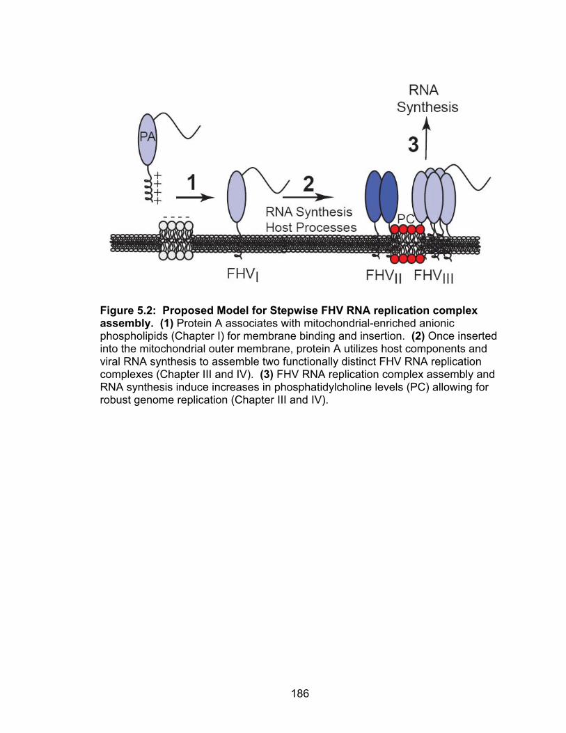
186
Figure 5.2: Proposed Model for Stepwise FHV RNA replication complex assembly. (1) Protein A associates with mitochondrial-enriched anionic phospholipids (Chapter I) for membrane binding and insertion. (2) Once inserted into the mitochondrial outer membrane, protein A utilizes host components and viral RNA synthesis to assemble two functionally distinct FHV RNA replication complexes (Chapter III and IV). (3) FHV RNA replication complex assembly and RNA synthesis induce increases in phosphatidylcholine levels (PC) allowing for robust genome replication (Chapter III and IV).
187
Unanswered Questions and Future Studies
These studies give rise to many unanswered questions including:
1. What are the protein A structural determinants that facilitate membrane
association and insertion?
2. What are the biophysical characteristics of protein A membrane
association?
3. What role does Mim1 and other mitochondrial membrane components
play in FHV replication complex assembly and function?
4. What is the composition and structure of functional FHV replication
complexes?
5. What are the dynamic processes involved in the assembly of FHV RNA
replication complexes?
We will address these questions as well as future directions in the following
pages.

188
What are the protein A structural determinants that facilitate protein A
membrane association and insertion?
This question was originally addressed in yeast by expressing a variety of
protein A deletion constructs and analyzing protein A membrane association
(18). Interestingly, it was found that many of these deletions, including deletions
of the mitochondrial-targeting signal, still remained membrane-associated
possibly due to the presence of multiple membrane binding domains within
protein A. However, these studies looked at protein A-membrane interactions 24
hours after protein A induction, suggesting that a variety of confounding effects
influencing membrane association could take place during this long time period.
In addition, these studies did not address the specific domains responsible for
the initial steps in protein A membrane association directly after translation.
These experiments would be difficult in vivo as the identification of a cytosolic
pool of protein A has not been seen in insect or yeast cells suggesting that
protein A associates rapidly with the mitochondrial membrane in vivo (5).
To investigate these initial steps in replication complex assembly we will use
the in vitro FHV membrane association system described in Chapter II. One
advantage of this system is the ability to easily manipulate the system to address
various aspects of protein A and membrane biology. I have created in vitro
protein A deletion constructs which we will use to directly study the role of protein
A domains on the initial steps of membrane-association. Furthermore, as protein
A is a lipid-binding protein it will be interesting to see what domains are required
for the association with specific mitochondrial-enriched anionic phospholipids.

189
One may speculate that protein A contains multiple domains which are required
for the specific interactions with membrane components either protein or lipid,
and studies using this in vitro system will allow us to identify these domains.
What are the biophysical characteristics of protein A membrane
association?
The observation that protein A membrane-association is temperature-
dependent was unexpected as we hypothesized protein A to behave similarly to
the yeast mitochondrial outer membrane proteins Tom20 and porin (13). One
possible explanation for this phenotype is that protein A membrane-association is
energy dependent and thus inhibited at low temperatures. However, we found
that exogenous ATP was despensible for protein A membrane-association and
preliminary studies enzymatically depleting rabbit reticulocyte lysate ATP levels
led to no changes in protein A membrane association (data not shown). These
data suggest an alternative mechanism for the temperature-dependence of
protein A membrane-association.
However, before we can address these mechanisms we first need to address
the temperature-dependence of protein A binding to purified liposomes. These
experiments are important to determine if the temperature-sensitive phenotype is
specific to mitochondria or all lipid vesicles. One could speculate that the
mitochondria contains a component that undergoes a conformational change at
low temperature leading to low protein A binding, and experiments using
liposomes would address this possibility. Nonetheless, the data we observed
190
with mitochondria brings up two potential hypotheses for this phenotype; 1) the
cold temperature induces structural changes in protein A or membrane proteins
inhibiting the ability to bind mitochondria, or 2) protein A membrane-association
is dependent on membrane fluidity which is decreased at low temperatures.
To address the first hypothesis, we will take advantage of the multiple
cysteine residues located throughout protein A suggesting that disulfide bonds
are required for protein folding. We will incubate in vitro translated protein A with
increasing concentrations of the reducing agent DTT to disrupt intramolecular
disulfide bonds to change protein structure, and address membrane-association
at low temperature. We hypothesize that if protein A structure is the key
determinant for membrane binding that DTT treatment will lead to changes in
membrane association. It is of course possible that the disruption of disulfide
bonds will have no effect on in vitro membrane association, and as a
complementary approach we will repeat this experiments with increasing
amounts of urea, a denaturing agent that has been used for the study of yeast
mitochondrial protein import (37).
To address the second hypothesis we will first address the ability of protein A
to bind mitochondrial fractions at increasing temperatures. If membrane fluidity
plays a role in protein A membrane association we would expect that increasing
the temperature would lead to increased membrane binding of protein A. One
potential downfall of this approach is that increasing temperature may also affect
protein A structure leading to difficult to interrupt results. To address membrane
fluidity directly we will introduce purified ergosterol, a molecule similar to

191
cholesterol, into mitochondria and liposomes to decrease membrane fluidity and
look at protein A membrane-association at 25°C. It has been shown that the
introduction of ergosterol into purified liposomes is able to decrease the
membrane insertion of the yeast mitochondrial outer membrane protein Fis1
suggesting that membrane fluidity may play important roles in protein import (11).
The idea of membrane fluidity in protein A mitochondrial import is intriguing as
we have shown cellular and mitochondrial-specific phospholipids to be involved
in a variety of processes in the FHV RNA replication including membrane binding
and RNA synthesis. However, we have not ruled out the possibility that
protease-resistant mitochondrial protein components are involved in protein A
membrane-association. Taken together, future studies using this in vitro
membrane association system will provide further insight into the host
components and biophysical mechanisms required for protein A membrane-
association.
What role does Mim1 and other mitochondrial membrane components play
in FHV replication complex assembly and function?
The ∆mim1 yeast strain has provided valuable insight into various processes
in FHV RNA replication complex assembly and function but still the specific role
of Mim1 remains unclear. It is possible that Mim1 is directly responsible for the
FHV assembly defects but until we are able to exogenously complement this
system with Mim1 we cannot make this conclusion. We have attempted to
complement this system using two different expression systems but have thus far

192
obtained inconclusive results. Of particular interest are the results obtained
using the Mim1 overexpression system, in which the control 2-micron plasmid
containing a URA3 marker was able to complement FHV RNA replication in
∆mim1 yeast. In an alternative approach we are generating a similar Mim1
overexpression plasmid using a 2-micro plasmid with a LEU2 marker instead of
the URA3 in an attempt to exogenously complement the FHV defect.
However, we cannot completely ignore the ability of URA3 to complement
FHV RNA replication in Mim1 deficient yeast. One possible explanation for these
results is that FHV RNA replication is dependent on the de novo synthesis of
pyrimidines as opposed to salvaging these metabolites from the media. To
address this in wildtype yeast we explored the effect of adding increasing
amounts of exogenous uracil into the media during FHV RNA replication.
Surprisingly, FHV RNA synthesis was enhanced with increasing amounts of
uracil suggesting that FHV RNA synthesis is not dependent on de novo synthesis
of these metabolites. Given the fact that FHV is an RNA virus it is not
unreasonable that pyrimidine biosynthesis is important for genomic synthesis.
One may hypothesize that having the URA3 gene or excess uracil present during
viral replication increases the cellular uracil concentration which leads to an
increase in FHV RNA replication. This is an interesting idea but may be unlikely
as UTP functions as a feedback inhibitor of pyrimidine biosynthesis shutting
down this pathway in the presence of extra UTP (27), however we can not
completely rule out this hypothesis just yet.

193
An additional explanation for the ability to complement viral replication with
URA3 or UTP may involve CTP, the product of UTP amination and end product
of pyrimidine biosynthesis. UTP and more importantly CTP is not only required
for the synthesis of nucleic acids but it also plays major roles in cellular
phospholipid biosynthesis (Figure 5.3)(4). S. cerevisiae has been used
extensively to study phospholipid biosynthesis and in particular the specific role
of CTP in these processes (4, 16, 22). It has been shown that increases in
cellular CTP concentrations correlate with increases in cellular
phosphatidylcholine levels in vivo (16). These data are particularly interesting
given the preliminary data analyzing the mitochondrial phospholipid composition
of wildtype and ∆mim1 yeast (Chapter III).
Taken together we hypothesize that Mim1 plays an important role in
maintaining cellular phospholipid levels and a deletion of this protein leads to
decreases in phosphatidylcholine levels and FHV RNA replication. The presence
of URA3 gene in ∆mim1 yeast is able to rescue the defect in phosphatidylcholine
levels allowing for a complementation of FHV RNA replication. To address this
hypothesis we will need to confirm the role of Mim1 in phospholipid biosynthesis.
We will repeat the mass spectroscopy analysis of ∆mim1 yeast mitochondria as
well as biochemically altering the intracellular phospholipid composition of ∆mim1
yeast to rescue FHV RNA replication. In particular, we will grow yeast in the
absence of inositol, a potent regulator of phospholipid biosynthesis as well as in
the presence of choline which can be used for phosphatidylcholine synthesis (4).
The growth of wildtype yeast in the presence of inositol has been shown to

194
increase phosphatidylinositol levels leading to a corresponding decrease in
phosphatidylcholine levels (Figure 5.3A) (4). In addition, the supplementation of
the media with exogenous choline provides an alternative mechanism to increase
phosphatidylcholine levels through the Kennedy pathway (Figure 5.3B). We
expect that growth of ∆mim1 yeast in these conditions will increase
phosphatidylcholine levels leading to an increase in FHV RNA synthesis.
Furthermore given the FHV replication in wildtype yeast induces changes in
phospholipids, it will be interesting to investigate the overall role of phospholipid
biosynthesis on FHV RNA replication by similar methods as described above.
Although ∆mim1 yeast provide an interesting system to study viral RNA
replication it was not the only membrane-specific deletion strain that lead to
changes in FHV RNA replication. We observed that two temperature sensitive
mutants of Tom40, the major import pore, led to significant increases in RNA
synthesis where as deletions of TOM6, DNM1, and POR1 reduced RNA
replication. Of particular interest are the opposing effects of TOM complex
components on FHV RNA replication. It is possible that deletions in these
proteins lead to protein import defects that positively and negatively influence
FHV RNA synthesis. In addition, a deletion of Dnm1, the major component of the
mitochondrial fission machinery, leads to constitutive mitochondrial fusion and
severe mitochondrial morphology defects possibly influencing FHV replication as
well (20). Lastly, Por1 is the major voltage-dependent anion channel (VDAC)

195
Figure 5.3: Schematic of the yeast phospholipid biosynthetic pathways. (A) CDP-DAG pathway for the de novo synthesis of glycerophospholipids. (B) Kennedy pathway (salvage pathway) for phospholipid biosynthesis.

196
and most abundant protein in the mitochondria. In addition, porin has been
implicated in apoptosis apart from its normal cellular function in channeling
metabolites (39, 41). To facilitate these functions porin has been localized to
mitochondrial contact sites, a hypothesized site for protein A-membrane
interactions (8, 25). One may speculate that given the abundance of porin in the
mitochondria, it is an important determinant in contact site structure and function,
and a deletion of porin disrupts these sites leading to defects in FHV replication
complex assembly.
Nonetheless it should be noted that one reoccuring theme between all of
these deletion strains including Mim1 are their effects on mitochondrial
morphology and membrane structure. This idea is intriguing given that FHV
induces dramatic changes in membrane morphology, and it is possible that
deletions or mutations in certain host proteins governing membrane morphology
will have effects on FHV replication. Furthermore, defects in mitochondrial
morphology may lead to corresponding defects in phospholipid composition yet
little work has been done thus far to link these processes. In the future it will be
necessary to address the role of these mitochondrial components in viral RNA
replication to better understand the role of host components in these processes.
What is the composition and structure of functional FHV replication
complexes?
The identification of two functionally distinct replication complexes begs the
questions as to what are their molecular compositions? We have attempted to
197
explore the composition of FHV replication complexes by affinity chromatography
and mass spectroscopy yet we were unable to identify any host components with
this technique. However, we have now developed an electrophoresis technique
in which to efficiently separate these complexes. BN-AGE has the advantage of
removing smaller contaminating proteins to clean up the protein analysis, and in
the future we will isolate these complexes by cutting the proteins out of the gel
and separating the components by SDS-PAGE or by isolating the native
complexes by preparative electrophoresis. Nonetheless, our “unsuccessful”
attempts at identifying host components by affinity chromatography provide an
interesting suggestion that protein A may be in the only viral component of FHV
replication complexes. One may hypothesis that genetically simple viruses, in
the case of FHV which encodes only one nonstructural protein, would need more
help from the host to facilitate RNA synthesis then a more complex virus such as
HCV, which encodes 6 nonstructural proteins. However in fact, the opposite may
be true in that genetically simple viruses may need very little from the host, and
work with FHV suggests this may be the case. It should also be noted that the
complexes we have identified are only composed of the viral components protein
A and RNA1.
In a natural infection RNA2 and capsid protein would be present within the
cell thus adding to the system. It will be interesting to investigate membrane-
associated replication complexes isolated from infected cells as well as
introducing RNA2 into the yeast replication system. These future studies will

198
give a detailed analysis of the FHV RNA replication complex and identify the host
components present in these complexes if any.
Flock House virus protein A has been biochemically defined as a signal-
anchored, integral membrane protein with its amino-terminus imbedded in the
lipid bilayer and carboxy-terminus exposed to the cytosol (Figure 1.6)(18). The
results that lead to this predicted protein topology were base on
immunofluorescence studies using epitope tagged protein A molecules in
combination with a commonly used differential detergent treatment using saponin
and TX-100. In this thesis we have shown that treatment of membrane fractions
with TX-100 led to protein structural and functional changes and it is possible that
similar events occurred during protein topology studies. One possible
explanation for the results that were obtained previously is that the epitope tag
which was hypothesized to be protected by the mitochondrial outer membrane
was instead protected by the protein complex structure. Treatment of these
structures with TX-100 induced a conformational change in protein structure and
exposed the hidden epitope and immunofluorescent signal. To directly address
the topology of signal- or tail-anchored proteins we can take advantage of the
fact that a small portion of protein A which contains the amino-terminus and
transmembrane domain is embedded in the bilayer and resistant to proteinase
degradation (1). We can then treat membrane fractions with protease and
separate small protein fragments by urea-acrylamide gel electrphoresis.
However, when we used this approach to look at protein A topology we were
unable to find small membrane protected fragments, indicating that the amino-

199
terminus may be exposed to the cytosol and degraded by the protease.
Furthermore, both in vivo and in vitro membrane association studies using
protein A deletions revealed that large deletions of protein A, which included the
amino-terminal transmembrane domain still remained membrane associated.
These data suggest that there are multiple membrane interacting regions of
protein A which have not been well characterized and future detailed biochemical
studies will provide insight into these domains.
In addition to hydrophobic membrane binding domains protein A also
contains a variety of highly conserved amino acid sequences (Figure 5.4). To
begin, protein A contains a common A/GxxxA/G motif in the amino-terminal
transmembrane domain. This structural motif has been shown to mediate
transmembrane domain interactions in such proteins as Mim1 and other yeast
proteins (24, 28, 31). The idea of transmembrane domain interactions is
supported by FRET studies which indicated that deletion of the transmembrane
domain inhibited protein A self-association (7). In addition, it is possible that this
motif mediates interactions with host proteins as well.
Secondly, protein A contains three highly conserved carrier protein domains
with the consenses sequence Px(D/E)xxR/K (14, 26). These domains have been
found in metabolite carrier proteins such as the ATP/ADP carrier protein and
phosphate carrier protein and are essential for their function (21, 40). In addition,
a single carrier domain has been found in the mitochondrial fusion protein Ugo1
which is also required for membrane fusion (6). The presence of

200
Figure 5.4: Conserved amino acid sequences in FHV protein A

201
these three conserved domains is very interesting, and based on the known
topology of carrier proteins it may give insight to the topology of protein A. In
addition, we are curious as to function of these carrier domains in FHV RNA
replication. Given that protein A is the only viral protein necessary for FHV RNA
synthesis it makes sense that the polymerase contains many functional domains
to facilitate this process. It is possible that during RNA replication protein A acts
to move metabolites such as nucleotides into and out of the viral replication
factories aiding in viral replication, and the fact that one of the carrier domains is
located just downstream of the catalytic resides may suggest such a mechanism.
In addition, protein A contains the conserved caspase-8 cleavage site, IETD
(34). This sequence is contained within the conserved replicase domains found
in a variety of RNA-dependent RNA polymerases, and the aspartic acid provides
a constant residue when lined up with the sequence of other viral RdRps (23).
Furthermore, the other caspase cleavage sequences LETD and DEVD have
been found in a variety of positive-strand RNA viruses including members of the
alphavirus family (23). The idea of a caspase cleavage signal is interesting given
the link between FHV, cardiolipin, and apoptosis (32). In particular, cardiolipin
provides a key membrane determinant for the recruitment of apoptotic proteins
including caspase-8, and thus it is possible that cardiolipin provides a platform to
localize FHV protein A to these mitochondrial areas as well (30).
Finally, protein A contains a variety of common protein-protein interaction
motifs including three CX5C motifs, indicating that disulfide bonds may play
important roles in FHV replication complex structure and function (12), and

202
several PxxP motifs which are responsible for the binding of adaptor proteins
(29). Unfortunately, a specific analysis of these Flock House virus motifs has not
been completed. It will be important to specifically mutate key residue in these
sequences and explore their role in viral replicase function. However, it must be
noted that since the replication complex is composed of two functionally distinct
pools of protein A it is not unreasonable to think that there are also two
structurally distinct replication complexes as well. Future analysis of these two
FHV replication complexes will provide valuable insight into the correct structure
and composition of viral replication complexes.
What are the dynamic temporal and spatial processes involved in the
assembly of FHV RNA replication complexes?
In this thesis we have looked at two time points in FHV replication complex
assembly. The first involves the initial steps in replication complex assembly of
protein A-membrane interactions and the second is an end point analysis of
functional FHV RNA replication complexes. However, one has to wonder about
the dynamic processes occurring between these two points. We have identified
dramatic cellular lipid changes induced by FHV RNA replication and evidence
with ∆mim1 yeast suggests these lipid changes are necessary for the assembly
of functional replication complexes. Future studies will be conducted to explore
the temporal and spatial requirements for FHV RNA replication complex
assembly. We will take advantage of BN-AGE to analyze the assembly of
complexes over time as well as submitochondrial fractionation techniques to

203
investigate the mitochondrial localization of these complexes during RNA
replication. In addition we will analyze the temporal pattern of phospholipid
changes in association with replication complex assembly to piece together the
involvement of these cellular processes in replication complex assembly. These
future studies will offer valuable insight in to the assembly of viral RNA replication
complexes and will provide the first complete biochemical analysis of positive-
strand RNA virus replication complex assembly.
Overall Significance
In summary, this thesis has provided novel insight into the molecular
mechanisms and host processes involved in positive-strand RNA virus replication
complex assembly and function. These studies have led to the development of a
variety of in vitro and biochemical techniques which we have used to identify key
roles for cellular phospholipids in positive-strand RNA virus membrane targeting
and replication complex assembly and function. Furthermore, we have provided
the first biochemical evidence of the presence of two functionally distinct viral
replication complexes of which their assembly is host and viral dependent. This
work has opened many avenues of future study in FHV and yeast biology, but
more importantly it has provided a starting point to focus on common molecular
mechanisms shared by all positive-strand RNA viruses with the hope of
understanding the complex processes of viral RNA replication and pathogenesis.

204
REFERENCES
1. Ahting, U., T. Waizenegger, W. Neupert, and D. Rapaport. 2005. Signal-anchored proteins follow a unique insertion pathway into the outer membrane of mitochondria. J Biol Chem 280:48-53.
2. Annamalai, P., and A. L. Rao. 2006. Packaging of brome mosaic virus
subgenomic RNA is functionally coupled to replication-dependent transcription and translation of coat protein. J Virol 80:10096-108.
3. Becker, T., S. Pfannschmidt, B. Guiard, D. Stojanovski, D. Milenkovic,
S. Kutik, N. Pfanner, C. Meisinger, and N. Wiedemann. 2008. Biogenesis of the mitochondrial TOM complex: Mim1 promotes insertion and assembly of signal-anchored receptors. J Biol Chem 283:120-7.
4. Carman, G. M., and G. M. Zeimetz. 1996. Regulation of phospholipid
biosynthesis in the yeast Saccharomyces cerevisiae. J Biol Chem 271:13293-6.
5. Castorena, K. M., S. A. Weeks, K. A. Stapleford, A. M. Cadwallader,
and D. J. Miller. 2007. A functional heat shock protein 90 chaperone is essential for efficient flock house virus RNA polymerase synthesis in Drosophila cells. J Virol 81:8412-20.
6. Coonrod, E. M., M. A. Karren, and J. M. Shaw. 2007. Ugo1p is a
multipass transmembrane protein with a single carrier domain required for mitochondrial fusion. Traffic 8:500-11.
7. Dye, B. T., D. J. Miller, and P. Ahlquist. 2005. In vivo self-interaction of
nodavirus RNA replicase protein a revealed by fluorescence resonance energy transfer. J Virol 79:8909-19.
8. Fritz, S., D. Rapaport, E. Klanner, W. Neupert, and B. Westermann.
2001. Connection of the mitochondrial outer and inner membranes by Fzo1 is critical for organellar fusion. J Cell Biol 152:683-92.
9. Henderson, N. S., L. G. Nijtmans, J. G. Lindsay, E. Lamantea, M.
Zeviani, and I. J. Holt. 2000. Separation of intact pyruvate dehydrogenase complex using blue native agarose gel electrophoresis. Electrophoresis 21:2925-31.
10. Hulett, J. M., F. Lueder, N. C. Chan, A. J. Perry, P. Wolynec, V. A.
Likic, P. R. Gooley, and T. Lithgow. 2008. The transmembrane segment of Tom20 is recognized by Mim1 for docking to the mitochondrial TOM complex. J Mol Biol 376:694-704.

205
11. Kemper, C., S. J. Habib, G. Engl, P. Heckmeyer, K. S. Dimmer, and D. Rapaport. 2008. Integration of tail-anchored proteins into the mitochondrial outer membrane does not require any known import components. J Cell Sci 121:1990-8.
12. Koehler, C. M. 2004. The small Tim proteins and the twin Cx3C motif.
Trends Biochem Sci 29:1-4. 13. Krimmer, T., D. Rapaport, M. T. Ryan, C. Meisinger, C. K.
Kassenbrock, E. Blachly-Dyson, M. Forte, M. G. Douglas, W. Neupert, F. E. Nargang, and N. Pfanner. 2001. Biogenesis of porin of the outer mitochondrial membrane involves an import pathway via receptors and the general import pore of the TOM complex. J Cell Biol 152:289-300.
14. Kunji, E. R., and A. J. Robinson. 2006. The conserved substrate binding
site of mitochondrial carriers. Biochim Biophys Acta 1757:1237-48. 15. Lee, C. S., R. Das, and R. Reed. 2003. Agarose gel separation/isolation
of RNA-protein complexes. Curr Protoc Mol Biol Chapter 27:Unit 27 1. 16. McDonough, V. M., R. J. Buxeda, M. E. Bruno, O. Ozier-
Kalogeropoulos, M. T. Adeline, C. R. McMaster, R. M. Bell, and G. M. Carman. 1995. Regulation of phospholipid biosynthesis in Saccharomyces cerevisiae by CTP. J Biol Chem 270:18774-80.
17. Meineke, B., G. Engl, C. Kemper, A. Vasiljev-Neumeyer, H.
Paulitschke, and D. Rapaport. 2008. The outer membrane form of the mitochondrial protein Mcr1 follows a TOM-independent membrane insertion pathway. FEBS Lett 582:855-60.
18. Miller, D. J., and P. Ahlquist. 2002. Flock house virus RNA polymerase
is a transmembrane protein with amino-terminal sequences sufficient for mitochondrial localization and membrane insertion. J Virol 76:9856-67.
19. Miller, D. J., M. D. Schwartz, and P. Ahlquist. 2001. Flock house virus
RNA replicates on outer mitochondrial membranes in Drosophila cells. J Virol 75:11664-76.
20. Mozdy, A. D., J. M. McCaffery, and J. M. Shaw. 2000. Dnm1p GTPase-
mediated mitochondrial fission is a multi-step process requiring the novel integral membrane component Fis1p. J Cell Biol 151:367-80.
21. Nelson, D. R., C. M. Felix, and J. M. Swanson. 1998. Highly conserved
charge-pair networks in the mitochondrial carrier family. J Mol Biol 277:285-308.
206
22. Ostrander, D. B., D. J. O'Brien, J. A. Gorman, and G. M. Carman. 1998. Effect of CTP synthetase regulation by CTP on phospholipid synthesis in Saccharomyces cerevisiae. J Biol Chem 273:18992-9001.
23. Poch, O., I. Sauvaget, M. Delarue, and N. Tordo. 1989. Identification of
four conserved motifs among the RNA-dependent polymerase encoding elements. EMBO J 8:3867-74.
24. Popov-Celeketic, J., T. Waizenegger, and D. Rapaport. 2008. Mim1
functions in an oligomeric form to facilitate the integration of Tom20 into the mitochondrial outer membrane. J Mol Biol 376:671-80.
25. Reichert, A. S., and W. Neupert. 2002. Contact sites between the outer
and inner membrane of mitochondria-role in protein transport. Biochim Biophys Acta 1592:41-9.
26. Robinson, A. J., C. Overy, and E. R. Kunji. 2008. The mechanism of
transport by mitochondrial carriers based on analysis of symmetry. Proc Natl Acad Sci U S A 105:17766-71.
27. Rodriguez, M., T. A. Good, M. E. Wales, J. P. Hua, and J. R. Wild.
2005. Modeling allosteric regulation of de novo pyrimidine biosynthesis in Escherichia coli. J Theor Biol 234:299-310.
28. Russ, W. P., and D. M. Engelman. 2000. The GxxxG motif: a framework
for transmembrane helix-helix association. J Mol Biol 296:911-9. 29. Saksela, K., G. Cheng, and D. Baltimore. 1995. Proline-rich (PxxP)
motifs in HIV-1 Nef bind to SH3 domains of a subset of Src kinases and are required for the enhanced growth of Nef+ viruses but not for down-regulation of CD4. EMBO J 14:484-91.
30. Scorrano, L. 2008. Caspase-8 goes cardiolipin: a new platform to provide
mitochondria with microdomains of apoptotic signals? J Cell Biol 183:579-81.
31. Senes, A., D. E. Engel, and W. F. DeGrado. 2004. Folding of helical
membrane proteins: the role of polar, GxxxG-like and proline motifs. Curr Opin Struct Biol 14:465-79.
32. Settles, E. W., and P. D. Friesen. 2008. Flock house virus induces
apoptosis by depletion of Drosophila inhibitor-of-apoptosis protein DIAP1. J Virol 82:1378-88.
33. Stuart, R. A., and C. M. Koehler. 2007. In vitro analysis of yeast
mitochondrial protein import. Curr Protoc Cell Biol Chapter 11:Unit 11 19.

207
34. Vachova, L., and Z. Palkova. 2007. Caspases in yeast apoptosis-like death: facts and artefacts. FEMS Yeast Res 7:12-21.
35. Venter, P. A., and A. Schneemann. 2007. Assembly of two independent
populations of flock house virus particles with distinct RNA packaging characteristics in the same cell. J Virol 81:613-9.
36. Waizenegger, T., and D. Rapaport. 2007. Analyzing import intermediates
of mitochondrial proteins by blue native gel electrophoresis. Methods Mol Biol 372:287-95.
37. Waizenegger, T., S. Schmitt, J. Zivkovic, W. Neupert, and D.
Rapaport. 2005. Mim1, a protein required for the assembly of the TOM complex of mitochondria. EMBO Rep 6:57-62.
38. Wittig, I., and H. Schagger. 2008. Features and applications of blue-
native and clear-native electrophoresis. Proteomics 8:3974-90. 39. Zaid, H., S. Abu-Hamad, A. Israelson, I. Nathan, and V. Shoshan-
Barmatz. 2005. The voltage-dependent anion channel-1 modulates apoptotic cell death. Cell Death Differ 12:751-60.
40. Zara, V., A. Ferramosca, L. Capobianco, K. M. Baltz, O. Randel, J.
Rassow, F. Palmieri, and P. Papatheodorou. 2007. Biogenesis of yeast dicarboxylate carrier: the carrier signature facilitates translocation across the mitochondrial outer membrane. J Cell Sci 120:4099-106.
41. Zheng, Y., Y. Shi, C. Tian, C. Jiang, H. Jin, J. Chen, A. Almasan, H.
Tang, and Q. Chen. 2004. Essential role of the voltage-dependent anion channel (VDAC) in mitochondrial permeability transition pore opening and cytochrome c release induced by arsenic trioxide. Oncogene 23:1239-47.