VECTOR-BORNE DISEASES - … · 0 college of medical technologists of puerto rico continuing...
22
COLLEGE OF MEDICAL TECHNOLOGISTS OF PUERTO RICO CONTINUING EDUCATION PROGRAM VECTOR-BORNE DISEASES BY: SANDRA MARTÍNEZ, MS, MT (ASCP)
Transcript of VECTOR-BORNE DISEASES - … · 0 college of medical technologists of puerto rico continuing...

0
COLLEGE OF MEDICAL TECHNOLOGISTS OF PUERTO RICO
CONTINUING EDUCATION PROGRAM
VECTOR-BORNE DISEASES
BY: SANDRA MARTÍNEZ, MS, MT (ASCP)

1
LEARNING OBJECTIVES
As a result of reading this educational module on Vector-Borne Diseases, the medical technologist
will be able to:
1. Review concepts of the epidemiology of some vector-borne diseases.
2. Name the vector, host or reservoir, causative agent, geographic distribution and availability
of a vaccine of the diseases presented.
INTRODUCTION
The World Health Organization (WHO) reports that vector-borne diseases account for over 17%
of all infectious diseases worldwide. Vector-borne diseases are major public health concern. Globally,
every year there are more than 1 billion cases and over 1 million deaths from vector-borne diseases
such as dengue, malaria, Chagas disease, yellow fever, Zika and chikungunya, among others.
Vectors are living organisms that can transmit infectious diseases between humans or from
animals to humans. Many of these vectors are bloodsucking insects, which ingest disease-producing
microorganisms during a blood meal from an infected host (human or animal) and later inject it into a
new host during their subsequent blood meal. Arthropod-borne viruses (arboviruses) comprise the
largest class of vector-borne human pathogens
Mosquitoes are the best known disease vector. Others include ticks, louses, flies, sandflies, fleas,
triatomine (kissing) bugs and some freshwater aquatic snails. (See Table I)
Distribution of these diseases is determined by a complex dynamic of environmental and social
factors. Globalization of travel and trade, unplanned urbanization, increased human traffic through
isolated areas, breakdown in public health measures, poor sanitation conditions and environmental
challenges such as climate change are having a significant impact on disease transmission in recent
years. Some diseases, such as dengue, chikungunya, West Nile virus and Zika are emerging in
countries where they were previously unknown.
Table I includes some of the vector-borne diseases. From this table were chosen six vector-
borne diseases of which some key facts will be discussed.

2
VECTOR-BORNE DISEASES
A. Yellow Fever
Geographic Distribution
Sub-Saharan Africa and tropical South America.
Transmission
Yellow fever is an acute viral hemorrhagic disease transmitted by infected mosquitoes. The
"yellow" in the name refers to the jaundice that affects some patients. Yellow fever virus (YFV)
is an RNA virus that belongs to the genus Flavivirus. It is related to West Nile, St. Louis
encephalitis, and Japanese encephalitis viruses. Yellow fever virus is transmitted to people
primarily through the bite of infected Aedes or Haemagogus species mosquitoes. Mosquitoes
acquire the virus by feeding on infected primates (human or non-human) and then can transmit
the virus to other primates (human or non-human). People infected with yellow fever virus are
infectious to mosquitoes shortly before the onset of fever and up to 5 days after onset.
Symptoms
The majority of persons infected with yellow fever virus have no illness or only mild illness.
In persons who develop symptoms, the incubation period (time from infection until illness) is
typically 3–6 days.
The initial symptoms include sudden onset of fever, chills, severe headache, back pain,
general body aches, nausea, and vomiting, fatigue, and weakness. Most persons improve after
the initial presentation.
After a brief remission of hours to a day, roughly 15% of cases progress to develop a more
severe form of the disease. The severe form is characterized by high fever, jaundice, bleeding,
gastrointestinal hemorrhages (black vomit) and eventually shock and failure of multiple
organs.
Laboratory Diagnosis
In early stages: RT-PCR testing of viral mRNA on blood and other samples.
In later stages: ELISA and Plaque-reduction neutralization tests (PRNT)
Treatment
No specific treatments have been found to benefit patients with yellow fever. Whenever
possible, yellow fever patients should be hospitalized for supportive care and close
observation.
Treatment is directed at symptomatic relief or life-saving interventions. Rest, fluids, and use of
pain relievers and medication to reduce fever may relieve symptoms of aching and fever.
Care should be taken to avoid certain medications, such as aspirin or other nonsteroidal anti-
inflammatory drugs (e.g. ibuprofen, naproxen), which may increase the risk of bleeding.

3
Yellow fever patients should be protected from further mosquitoes exposure (staying indoors
and/or under a mosquitoes net) for up to 5 days after the onset of fever. This way, yellow fever
virus in their bloodstream will be unavailable to uninfected mosquitoes, thus breaking the
transmission cycle and reducing risk to the persons around them.
Other
The majority of infected persons will be asymptomatic or have mild disease with complete
recovery.
In persons who become symptomatic but recover, weakness and fatigue may last several
months.
Among those who develop severe disease, 20–50% may die.
Those who recover from yellow fever generally have lasting immunity against subsequent
infection.
Prevention
Avoid mosquitoes bites
Control of mosquitoes population
Get vaccinated
o Vaccination is recommended for persons aged ≥9 months that are traveling to or
living in areas at risk YFV transmission in South America and Africa. Yellow fever
vaccine may be required for entry in certain countries. The yellow fever vaccine
certification required for entry in certain countries is valid after 10 days of vaccine
administration.
B. West Nile Encephalitis
Geographic Distribution
Africa, Europe, Central Asia and North America.
Transmission
West Nile virus (WNV) is most commonly transmitted to humans by mosquitoes. In nature,
West Nile virus cycles between mosquitoes (especially Culex species) and birds. Some infected
birds, can develop high levels of the virus in their bloodstream and mosquitoes can become
infected by biting these infected birds. After about a week, infected mosquitoes can pass the
virus to more birds when they bite.
Mosquitoes with West Nile virus also bite and infect people, horses and other mammals.
However, humans, horses and other mammals are ‘dead end’ hosts. This means that they do
not develop high levels of virus in their bloodstream, and cannot pass the virus on to other
biting mosquitoes.

4
Additional routes of human infection have also been documented. It is important to note that
these methods of transmission represent a very small proportion of cases:
Blood transfusions
Organ transplants
Exposure in a laboratory setting
From mother to baby during pregnancy, delivery, or breastfeeding
Symptoms
Most people infected with WNV will have no symptoms.
About 1 in 5 people who are infected will develop fever, headache, tiredness, and body
aches, occasionally with a skin rash on the trunk of the body and swollen lymph glands
usually lasting only a few days.
Encephalitis, meningitis, or meningoencephalitis occurs in approximately 1% of infected
individuals.
Individuals older than 50 years and the immunocompromised are at higher risk for serious
disease.
Laboratory Diagnosis
RT-PCR
Treatment
No vaccine or specific antiviral treatments for West Nile virus infection are available.
Over-the-counter pain relievers can be used to reduce fever and relieve some symptoms
In severe cases, patients often need to be hospitalized to receive supportive treatment,
such as intravenous fluids, pain medication, and nursing care.
Prevention
The most effective way to avoid West Nile virus disease is to prevent mosquitoes bites.
C. Lyme Disease
Geographic Distribution
Worldwide distribution
Transmission
Lyme disease is caused by the spirochetes belonging to the Borrelia species complex and is
transmitted to humans through the bite of infected Ixodes ticks. Lyme disease was first
recorded in 1977 when an unusual cluster of children were diagnosed with arthritis in a town
by the name Lyme in Connecticut.
5
Ticks can attach to any part of the human body but are often found in hard-to-see areas such
as the groin, armpits, and scalp. In most cases, the tick must be attached for 36 to 48 hours
or more before the Lyme disease bacterium can be transmitted.
Most humans are infected through the bites of immature ticks called nymphs. Nymphs are tiny
(less than 2 mm) and difficult to see; they feed during the spring and summer months. Adult
ticks can also transmit Lyme disease bacteria, but they are much larger and are more likely to
be discovered and removed before they have had time to transmit the bacteria. Adult Ixodes
ticks are most active during the cooler months of the year.
Symptoms
Untreated Lyme disease can produce a wide range of symptoms, depending on the stage of
infection.
◊Early Signs and Symptoms (3 to 30 days after tick bite)
Fever, chills, headache, fatigue, muscle and joint aches, and swollen lymph
nodes
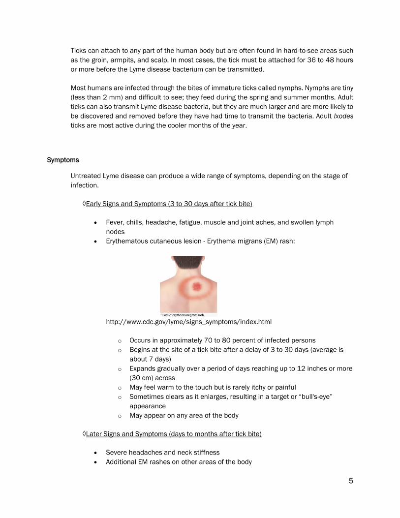
Erythematous cutaneous lesion - Erythema migrans (EM) rash:
http://www.cdc.gov/lyme/signs_symptoms/index.html
o Occurs in approximately 70 to 80 percent of infected persons
o Begins at the site of a tick bite after a delay of 3 to 30 days (average is
about 7 days)
o Expands gradually over a period of days reaching up to 12 inches or more
(30 cm) across
o May feel warm to the touch but is rarely itchy or painful
o Sometimes clears as it enlarges, resulting in a target or “bull's-eye”
appearance
o May appear on any area of the body
◊Later Signs and Symptoms (days to months after tick bite)
Severe headaches and neck stiffness
Additional EM rashes on other areas of the body

6
Arthritis with severe joint pain and swelling, particularly the knees and other large
joints.
Facial or Bell's palsy (loss of muscle tone or droop on one or both sides of the
face)
Intermittent pain in tendons, muscles, joints, and bones
Heart palpitations or an irregular heart beat (Lyme carditis)
Episodes of dizziness or shortness of breath
Inflammation of the brain and spinal cord
Nerve pain
Shooting pains, numbness, or tingling in the hands or feet
Problems with short-term memory
Laboratory Diagnosis
The Center for Disease Control and Prevention (CDC) currently recommends a two-step process
when testing blood for evidence of antibodies against the Lyme disease bacteria. Both steps
can be done using the same blood sample.
The first step uses EIA (enzyme immunoassay) or rarely, an IFA (indirect immunofluorescence
assay). If this first step is negative, no further testing of the specimen is recommended, but if
the first step is positive or indeterminate (sometimes called "equivocal"), the second step
should be performed. The second step uses Western blot test. Results are considered positive
only if the EIA/IFA and the immunoblot are both positive.
The two steps of Lyme disease testing are designed to be done together. CDC does not
recommend skipping the first test and just doing the Western blot. Doing so will increase the
frequency of false positive results and may lead to misdiagnosis and improper treatment.
Treatment
Patients treated with appropriate antibiotics in the early stages of Lyme disease usually
recover rapidly and completely. Antibiotics commonly used for oral treatment include
doxycycline, amoxicillin, or cefuroxime axetil. Patients with certain neurological or cardiac
forms of illness may require intravenous treatment with drugs such as ceftriaxone or penicillin.
Prevention
Using insect repellent
Removing ticks promptly
Applying pesticides
Reducing tick habitat

7
D. Tularemia
Geographic Distribution
Worldwide
In the United States infections have been reported from all states (most common in Oklahoma,
Missouri and Arkansas), except Hawaii.
Transmission
Tularemia is a disease of animals and humans caused by the bacterium Francisella tularensis.
It can enter the human body through the skin, eyes, mouth, or lungs. Rabbits, hares, and
rodents are especially susceptible and often die in large numbers during outbreaks. Humans
can become infected through several routes, including:
Tick and deer fly bites
Skin contact with infected animals
Ingestion of contaminated water
Inhalation of contaminated aerosols or agricultural dusts
Laboratory exposure
Francisella tularensis is highly infectious when grown in culture, and laboratory-acquired
infections have been documented. The isolation of F. tularensis from clinical specimens,
especially if unanticipated, can generate concern among laboratory workers about possible
exposure. Workers who report sniffing a culture plate or conducting procedures that
generate aerosols are probably at greater risk than those who simply worked with the
organism on the bench.
In addition, F. tularensis is considered a biologic weapon.
Symptoms
Tularemia can be difficult to diagnose. It is a rare disease, and the symptoms can be mistaken
for other, more common, illnesses. For this reason, it is important to share with the health care
provider any likely exposures, such as tick and deer fly bites, or contact with sick or dead
animals.
The signs and symptoms of tularemia vary depending on how the bacteria enter the body.
Illness ranges from mild to life-threatening. All forms are accompanied by fever, which can be
as high as 104 °F. Main forms of this disease are listed below:
Ulceroglandular This is the most common form of tularemia and usually occurs
following a tick or deer fly bite or after handling of an infected animal. A skin ulcer
appears at the site where the bacteria entered the body. The ulcer is accompanied
by swelling of regional lymph glands, usually in the armpit or groin.

8
Glandular Similar to ulceroglandular tularemia but without an ulcer. Also generally
acquired through the bite of an infected tick or deer fly or from handling sick or
dead animals.
Oculoglandular This form occurs when the bacteria enter through the eye. This can
occur when a person is butchering an infected animal and touches his or her eyes.
Symptoms include irritation and inflammation of the eye and swelling of lymph
glands in front of the ear.
Oropharyngeal This form results from eating or drinking contaminated food or
water. Patients with orophyangeal tularemia may have sore throat, mouth ulcers,
tonsillitis, and swelling of lymph glands in the neck.
Pneumonic This is the most serious form of tularemia. Symptoms include cough,
chest pain, and difficulty breathing. This form results from breathing dusts or
aerosols containing the organism. It can also occur when other forms of tularemia
(e.g. ulceroglandular) are left untreated and the bacteria spread through the
bloodstream to the lungs.
Typhoidal This form is characterized by any combination of the general symptoms
(without the localizing symptoms of other syndromes)
Laboratory Diagnosis
Microscopy is limited by the fact the organisms are extremely small and frequently
overlooked in clinical specimens.
Culture on cysteine-supplemented media (e.g. chocolate agar, buffered charcoal yeast
extract agar) is sensitive if prolonged incubation is used.
Serology can be used to confirm clinical diagnosis; fourfold increase in titer or single titer ≥
1:160; high titers can persist for months to years.
Treatment
Antibiotics used to treat tularemia include streptomycin, gentamicin, doxycycline, and
ciprofloxacin. Treatment usually lasts 10 to 21 days depending on the stage of illness and the
medication used. Although symptoms may last for several weeks, most patients completely
recover.
Prevention
Use insect repellents
Wear long pants, long sleeves, and long socks to keep ticks and deer flies off your skin.
Remove attached ticks promptly with fine-tipped tweezers.
Don’t drink untreated surface water.
Don’t mow over sick or dead animals.
Use gloves when handling hunted animals, especially rabbits, muskrats, prairie dogs, and
other rodents.
Cook game meat thoroughly before eating.

9
E. Malaria
Geographic Distribution
Africa, Latin America, parts of the Caribbean, Asia (including South Asia, Southeast Asia, and
the Middle East), Eastern Europe, and the South Pacific.
Transmission
Malaria is caused by Plasmodium parasites. The parasites are spread to people through the
bites of infected female Anopheles mosquitoes. There are 5 parasite species (see Table I) that
cause malaria in humans, and 2 of these species – P. falciparum and P. vivax – pose the
greatest threat.
P. falciparum is the most prevalent malaria parasite on the African continent. It is
responsible for most malaria-related deaths globally.
P. vivax is the dominant malaria parasite in most countries outside of sub-Saharan
Africa.
The long lifespan and strong human-biting habit of the African vector species is the main
reason why nearly 90% of the world's malaria cases are in Africa.
Biology
The natural ecology of malaria involves malaria parasites infecting successively two types of
hosts: humans and female Anopheles mosquitoes. In humans, the parasites grow and multiply
first in the liver cells and then in the red cells of the blood. In the blood, successive broods of
parasites grow inside the red cells and destroy them, releasing daughter parasites
("merozoites") that continue the cycle by invading other red cells.
The blood stage parasites are those that cause the symptoms of malaria. When certain forms
of blood stage parasites ("gametocytes") are picked up by a female Anopheles mosquitoes
during a blood meal, they start another, different cycle of growth and multiplication in the
mosquitoes.
After 10-18 days, the parasites are found (as "sporozoites") in the mosquito's salivary glands.
When the Anopheles mosquito takes a blood meal on another human, the sporozoites are
injected with the mosquito's saliva and start another human infection when they parasitize the
liver cells.
Thus the mosquito carries the disease from one human to another (acting as a "vector").
Differently from the human host, the mosquitoes vector does not suffer from the presence of
the parasites.

10
Symptoms
Malaria is an acute febrile illness. In a non-immune individual, symptoms appear 7 days or
more (usually 10–15 days) after the infective mosquitoes bite. The first symptoms – fever,
headache, chills and vomiting – may be mild and difficult to recognize as malaria. If not treated
within 24 hours, P. falciparum malaria can progress to severe illness, often leading to death.
Children with severe malaria frequently develop one or more of the following symptoms: severe
anemia, respiratory distress in relation to metabolic acidosis, or cerebral malaria. In adults,
multi-organ involvement is also frequent. In malaria endemic areas, people may develop
partial immunity, allowing asymptomatic infections to occur.
Laboratory Diagnosis
Smear microscopy remains the gold standard for malaria diagnosis.
Rapid diagnostic tests (RDTs) offer a useful alternative to microscopy in situations where
reliable microscopic diagnosis is not immediately available. Although RDTs can detect
malaria antigens within minutes, they cannot determine the species, are less sensitive for
diagnosis, and cannot quantify parasitemia. In addition, CDC recommends that positive
and negative results always be confirmed by microscopy.
PCR
Treatment
Treatment of malaria depends on many factors including disease severity, the species of
malaria parasite causing the infection and the part of the world in which the infection was
acquired. The latter two characteristics help determine the probability that the organism is
resistant to certain antimalarial drugs. Additional factors such as age, weight, and pregnancy
status may limit the available options for malaria treatment.
Prevention
Malaria elimination: defined as the interruption of local transmission of a specified malaria
parasite in a defined geographical area.

11
F. Chagas Disease (American trypanosomiasis)
T. cruzi trypomastigote in a thin blood smear stained with Giemsa.
http://www.cdc.gov/dpdx/trypanosomiasisAmerican/index.html
Geographic Distribution
Latin American countries
Transmission
In Latin America, T. cruzi parasites are mainly transmitted by contact with feces/urine of
infected blood-sucking triatomine bugs. These bugs typically live in the wall or roof cracks of
poorly-constructed homes in rural or suburban areas. Normally they hide during the day and
become active at night when they feed on human blood. They usually bite an exposed area of
skin such as the face, and the bug defecates close to the bite. The parasites enter the body
when the person instinctively smears the bug feces or urine into the bite, the eyes, the mouth,
or into any skin break.
T. cruzi can also be transmitted by:
consumption of food contaminated with T. cruzi through, for example, contact with
infected triatomine bug feces or urine;
blood transfusion from infected donors;
passage from an infected mother to her newborn during pregnancy or childbirth;
organ transplants using organs from infected donors; and
laboratory accidents
Symptoms
Chagas disease presents itself in two phases. The initial, acute phase lasts for about 2 months
after infection. During the acute phase, a high number of parasites circulate in the blood but
in most cases symptoms are absent or mild. In less than 50% of people bitten by a triatomine
bug, characteristic first visible signs can be a skin lesion or a purplish swelling of the lids of
one eye. Additionally they can present fever, headache, enlarged lymph glands, pallor, muscle
pain, difficulty in breathing, swelling, and abdominal or chest pain.
During the chronic phase, the parasites are hidden mainly in the heart and digestive muscles.
Up to 30% of patients suffer from cardiac disorders and up to 10% suffer from digestive
(typically enlargement of the esophagus or colon), neurological or mixed alterations. In later

12
years the infection can lead to sudden death or heart failure caused by progressive destruction
of the heart muscle and its nervous system.
Laboratory Diagnosis
The diagnosis of Chagas disease can be made by observation of the parasite in a blood smear
by microscopic examination. A thick and thin blood smear are made and stained for
visualization of parasites. However, a blood smear works well only in the acute phase of
infection when parasites are seen circulating in blood.
Diagnosis of chronic Chagas disease is made after consideration of the patient's clinical
findings, as well as by the likelihood of being infected, such as having lived in an endemic
country. Diagnosis is generally made by testing with at least two different serologic tests (most
commonly, ELISA, immunoblot, and immunofluorescent antibody test).
PCR testing may also help detect acute infection, but is not a useful diagnostic test for chronic-
phase infections since parasites are not detectable in the peripheral blood during this phase.
Treatment
Antitrypanosomal drug treatment is always recommended for acute, early congenital, and
reactivated T. cruzi infection and for chronic T. cruzi infection in children aged <18 years old.
In adults, treatment is usually recommended. In the United States, treatment drugs
(benznidazole and nifurtimox) are provided only by CDC under investigational protocols.
Prevention
Vector control is the most effective method of prevention in Latin America. Blood screening is
necessary to prevent infection through transfusion and organ transplantation.
REFERENCES
1. CDC: www.cdc.gov
2. CDC Travelers’ Health: Chapter 3-Infectious Diseases Related to Travel-
http://wwwnc.cdc.gov/travel/yellowbook/2016/table-of-contents
3. WHO: www.who.int
4. Murray, P. R., Rosenthal, K. S., & Pfaller, M. A. (2016). Medical Microbiology (8th ed.).
Philadelphia, PA: Elsevier.

13
Table I: List of Some Vector-Borne Diseases in Humans1
DISEASE VECTOR HOST/ RESERVOIR
CAUSATIVE AGENT
CAUSATIVE AGENT GENUS
GEOGRAPHIC DISTRIBUTION
MAJOR DISEASE MANIFESTATION
VACCINE
VIRUSES
Dengue fever and dengue hemorrhagic fever
Aedes Mosquitoes
Humans, monkeys
Dengue virus Flavivirus Tropical, subtropical regions around the world
Muscle, joint pain, dengue hemorrhagic fever, dengue shock
syndrome
No
Yellow fever Aedes and Haemogogus
spp. mosquitoes
Humans, monkeys
Yellow fever virus
Flavivirus Africa, South America
Hepatitis and hemorrhagic fever
Yes
Chikungunya fever Aedes mosquitoes
Humans, monkeys
Chikungunya virus
Alphavirus Africa, Asia, Europe, the
Oceania and Pacific Islands, Americas
Fever, arthralgia, arthritis
No
Zika fever Aedes mosquitoes
Unknown Zika virus Flavivirus Tropical, subtropical regions around the world
Fever, rash, joint pain, or conjunctivitis
No
Japanese encephalitis
Culex mosquitoes
Pigs, wading birds
Japanese encephalitis
virus
Flavivirus Asia, western Pacific
Encephalitis, seizures in children
Yes
West Nile encephalitis
Culex mosquitoes
Birds West Nile virus Flavivirus Africa, Europe, Central Asia, North
America
Encephalitis No
Crimean-Congo Hemorrhagic fever
Ixodid ticks (genus
Hyalomma)
Numerous wild and domestic animals, such
as cattle, goats, sheep and
hares
Nairovirus Nairovirus Eastern and southern Europe, Mediterranean, northwestern
China, central Asia, Africa, the Middle
East and the Indian subcontinent
Hemorrhagic fever, No

14
DISEASE VECTOR HOST/ RESERVOIR
CAUSATIVE AGENT
CAUSATIVE AGENT GENUS
GEOGRAPHIC DISTRIBUTION
MAJOR DISEASE MANIFESTATION
VACCINE
Tick-borne encephalitis
Ixodes sp. ticks
Small rodents/ Ixodes spp. ticks
Tick-borne encephalitis virus
Flavivirus Europe, the former Soviet Union, Asia
Encephalitis No in the USA Available in other countries
Powassan encepahalitis
Ixodes ticks Small mammals
Powassan virus Flavivirus North America, Russia
Encephalitis No
BACTERIAS
Lyme disease Ixodes ticks Mice, deer, domestic pets, hard ticks
Spirochetes: B. burgdoferi, other Borrelia species
Borrelia Worldwide distribution
Erythema migrans, cardiac, neurologic, or rheumatologic abnormalities
No
Relapsing fever (epidemic)
Epidemic: human body louse Pediculus humanus
Human Spirochete: B. recurrentis
Borrelia Ethiopia, Rwanda, Andean foothills
Fever and septicemia separated by afebrile periods
No
Relapsing fever (endemic)
Endemic: Ornithodoros soft ticks
Rodents, small mammals, soft ticks
Spirochetes: Borrelia spp.
Borrelia Worldwide, western part of the United States
Fever and septicemia separated by afebrile periods
No

15
DISEASE VECTOR HOST/ RESERVOIR
CAUSATIVE AGENT
CAUSATIVE AGENT GENUS
GEOGRAPHIC DISTRIBUTION
MAJOR DISEASE MANIFESTATION
VACCINE
Rocky Mountain spotted fever
Hard ticks (dog and wood tick)
Rodents Rickettsia rickettsii
Rickettsia North, Central and South America
Fever, rash, headache No
Tularemia Ticks (dog and wood) and deer flies (Chrysops spp)
Wild mammals, domestic animals, birds, fish
Francisella tularensis
Francisella Worldwide distribution. Most common in USA in Oklahoma, Missouri, Arkansas
Vary depending on how the bacteria enter the body. Six different forms of clinical presentation
Yes but rarely used for human disease
Plague Fleas Rats Yersinia pestis Yersinia Africa, the former Soviet Union, the Americas and Asia.
Sudden onset of fever, chills, head and body-aches and weakness, vomiting and nausea. Two forms of clinical manifestations: bubonic and pneumonic plague
No

16
DISEASE VECTOR HOST/ RESERVOIR
CAUSATIVE AGENT
CAUSATIVE AGENT GENUS
GEOGRAPHIC DISTRIBUTION
MAJOR DISEASE MANIFESTATION
VACCINE
Anaplasmosis (previously known as human granulocytic ehrlichiosis, recently been called human granulocytic anaplasmosis)
Ticks Ixodes scapularis, Ixodes pacificus
Anaplasma phagocyte-philum
Anaplasma North America (Upper Midwest and Northeast), Europe, Asia
Fever, headache, chills, and muscle aches
No
Ehrlichiosis [Ehrlichia chaffeensis (human monocytic erlichiosis), and Ehrlichia ewingii (human granulocytic ehrlichiosis)]
Lone star tick
Ehrlichia chaffeensis: (Deer, dogs, foxes,coyote,wolves) Ehrlichia ewingii: (Doogs, deer)
Ehrlichia chaffeensis, Ehrlichia ewingii and Ehrlichia spp.
Ehrlichia E. chaffeensis: North and South America, Asia. E. ewingii: North America (uncommon in Missouri)
Fever, headache, fatigue, and muscle aches
No
PARASITES
Malaria Anopheles spp. mosquitoes
Humans, monkeys
P. falciparum, P.vivax, P.ovale, P. malariae, P. knowlesi
Plasmodium Tropical, subtropical and temperate regions
Fever, chills, and flu-like illness
No

17
DISEASE VECTOR HOST/ RESERVOIR
CAUSATIVE AGENT
CAUSATIVE AGENT GENUS
GEOGRAPHIC DISTRIBUTION
MAJOR DISEASE MANIFESTATION
VACCINE
Lymphatic filariasis Anopheles, Culex, Aedes and Mansonia mosquitoes
Human Nematodes: Wuchereria bancrofti, Brugia malayi, and B.timori
Wuchereria Brugia
Tropical, subtropical regions around the world
Fever, lymphangitis, lymphadenitis with chills, recurrent febrile attacks. Filarial elephantiasis
No
Leishmaniasis Sandflies Human Leishmania spp. Leishmania Parts of the tropics, subtropics, southern Europe
Most common form is cutaneous leishmaniasis, which causes skin sores. The other main form is visceral leishmaniasis, which affects several internal organs (usually spleen, liver, and bone marrow) and can be life threatening
No
Ghagas disease (American trypanosomiasis)
Triatomine bugs (kissing bugs)
Human Trypanosoma cruzi
Trypanosoma Latin American countries
Two disease phases: acute (fever and swelling around the site of inoculation) and chronic (20 - 30% of infected people will develop debilitating and sometimes life-threatening medical problems).
No
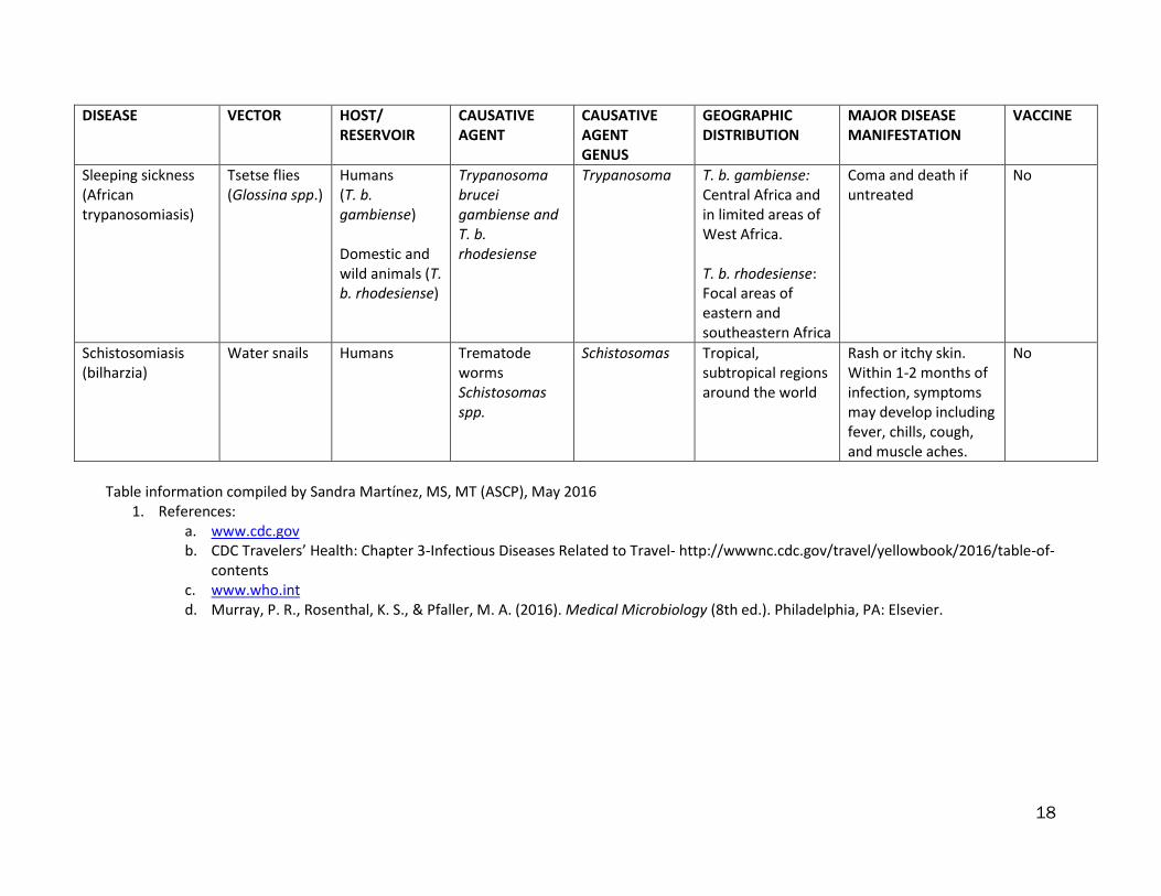
18
DISEASE VECTOR HOST/ RESERVOIR
CAUSATIVE AGENT
CAUSATIVE AGENT GENUS
GEOGRAPHIC DISTRIBUTION
MAJOR DISEASE MANIFESTATION
VACCINE
Sleeping sickness (African trypanosomiasis)
Tsetse flies (Glossina spp.)
Humans (T. b. gambiense) Domestic and wild animals (T. b. rhodesiense)
Trypanosoma brucei gambiense and T. b. rhodesiense
Trypanosoma T. b. gambiense: Central Africa and in limited areas of West Africa. T. b. rhodesiense: Focal areas of eastern and southeastern Africa
Coma and death if untreated
No
Schistosomiasis (bilharzia)
Water snails Humans Trematode worms Schistosomas spp.
Schistosomas Tropical, subtropical regions around the world
Rash or itchy skin. Within 1-2 months of infection, symptoms may develop including fever, chills, cough, and muscle aches.
No
Table information compiled by Sandra Martínez, MS, MT (ASCP), May 2016
1. References: a. www.cdc.gov b. CDC Travelers’ Health: Chapter 3-Infectious Diseases Related to Travel- http://wwwnc.cdc.gov/travel/yellowbook/2016/table-of-
contents c. www.who.int d. Murray, P. R., Rosenthal, K. S., & Pfaller, M. A. (2016). Medical Microbiology (8th ed.). Philadelphia, PA: Elsevier.

VECTOR-BORNE DISEASES TEST
Por: Lcda. Sandra Martínez, MS, MT(ASCP)
Course: 20-170-65 Date: February 1, 2017
Name: __________________________________ License#: ___________________
Envíe la hoja de preguntas debidamente contestada e identificada con su nombre y número de
licencia por correo a la siguiente dirección: Colegio de Tecnólogos Médicos de PR * Programa de
Educación Continua * F-1 Ave. San Patricio * Guaynabo, PR 00968 ó vía fax (787)792-6627. * Para
otorgar 0.2 UEC, usted debe obtener un 80% de contestaciones correctas o más.
19
______
Answers to the questions are found in the module and Table I.
I. Match each question type with one attribute:
_____ 1. Lyme disease a) African trypanosomiasis
_____ 2 Plague b) Black vomit
_____ 3. Sleeping sickness c) Borrelia recurrentis
_____ 4 Chagas disease d) Erythema migrans
_____ 5 Relapsing fever (epidemic) e) Bubonic manifestation
_____ 6. Crimean-Congo hemorrhagic f) Powassan virus
fever
_____ 7. Encephalitis g) Kissing bugs
_____ 8. Yellow fever h) Nairovirus
II. Choose the best answer:
1. In the acute phase of Chagas disease is characteristic a:
a) Erythematous cutaneous lesion
b) Skin ulcer at the site of the bite
c) Purplish swelling of the lids of one eye
d) Skin rash on the trunk of the body
2. Which of the following organisms is considered a bioterrorism agent?
a) Francisella tularensis
b) Plasmodium falciparum
c) Erlichia chaffeensis
d) Trypanosoma cruzi
3. The most common form of tularemia is:
a) Pneumonic c) Typhoida
b) Oropharyngeal d) Ulceroglandular
4. In countries outside the African continent the most prevalent malaria parasite is:
a) Plamodium ovale
b) Plasmodium knowlesi
c) Plasmodium vivax
d) Plasmodium malariae
______
______
______

VECTOR-BORNE DISEASES TEST
Por: Lcda. Sandra Martínez, MS, MT(ASCP)
Course: 20-170-65 Date: February 1, 2017
Name: __________________________________ License#: ___________________
Envíe la hoja de preguntas debidamente contestada e identificada con su nombre y número de
licencia por correo a la siguiente dirección: Colegio de Tecnólogos Médicos de PR * Programa de
Educación Continua * F-1 Ave. San Patricio * Guaynabo, PR 00968 ó vía fax (787)792-6627. * Para
otorgar 0.2 UEC, usted debe obtener un 80% de contestaciones correctas o más.
20
5. The gold standard test for malaria diagnosis is PCR.
a) True b) False
6. A blood sample was analyzed by EIA for evidence of antibodies against Lyme disease.
The result was positive. The next step should be:
a) Confirm by IFA
b) Confirm by Western blot
c) Confirm by PCR
d) No further step is required.
7. The transmission cycle of the West Nile virus by the Culex mosquitoes is human-to-
vector-to human.
a) True b) False
8. A missionary has planned a trip to Liberia. This country requires a proof of yellow fever
(YF) vaccination. The missionary received the vaccine on June 15. He arrived in Liberia
on June 20. The entry of the missionary to Liberia was denied because the YF
vaccination is valid after ___________ days of vaccine administration.
a) 6 days (June 21 is the valid date for entry)
b) 15 days (June 30 is the valid date for entry)
c) 20 days (July 5 is the valid date for entry)
d) 10 days (June 25 is the valid date for entry)
9. The causative agent of Rocky Mountain spotted fever is Rickettsia rickettsii.
a) True b) False
10. Case study retrieved from:
http://www.cdc.gov/malaria/references_resources/interactive_training/ph-2/index.html
A 49-year-old man from Pennsylvania receives 4 units of packed red blood cells (PRBCs)
on January 15 while undergoing hip replacement surgery. He is again hospitalized on
February 1 with fever, hypotension, and renal failure. Peripheral blood smears show
malaria infection. The patient has never traveled outside the United States.
The blood donor was born in West Africa, had lived in Europe, and then returned to West
Africa, where he had lived for approximately 20 years before immigrating to the United
States 2 years ago.
A. Among the modes of malaria infection below, which one is the most likely?
a) Infection during travel overseas.
b) Congenital malaria
c) Infection by local Anopheles mosquito
d) Blood transfusion
______
______
______
______
______
______

0