Effect of Mycobacterium leprae on neurotrophins expression ...

THE IMPACT OF Mycobacterium leprae: A COMPREHENSIVE META-ANALYSIS
OF THE PALEOPATHOLOGICAL LITERATURE
by
Mallory Alexis Schreier
A thesis
submitted in partial fulfillment
of the requirements for the degree of
Master of Arts in Anthropology
Boise State University
May 2016

© 2016
Mallory Alexis Schreier
ALL RIGHTS RESERVED

BOISE STATE UNIVERSITY GRADUATE COLLEGE
DEFENSE COMMITTEE AND FINAL READING APPROVALS
of the thesis submitted by
Mallory Alexis Schreier
Thesis Title: The Impact of Mycobacterium leprae: A Comprehensive Meta-Analysis of the Paleopathological Literature
Date of Final Oral Examination: 10 March 2016
The following individuals read and discussed the thesis submitted by student Mallory Alexis Schreier, and they evaluated her presentation and response to questions during the final oral examination. They found that the student passed the final oral examination.
Kristin Snopkowski, Ph.D. Chair, Supervisory Committee
Samantha Blatt, Ph.D. Member, Supervisory Committee
John Ziker, Ph.D. Member, Supervisory Committee
The final reading approval of the thesis was granted by Kristin Snopkowski, Ph.D., Chair of the Supervisory Committee. The thesis was approved for the Graduate College by John R. Pelton, Ph.D., Dean of the Graduate College.

iv
DEDICATION
To my mom and dad. Thank you for everything.

v
ACKNOWLEDGEMENTS
This thesis could not have been possible without the help of many individuals. I’d
like to thank my advisor and committee members for having the patience of saints
through all the times I needed their help. Thank you Dr. Kristin Snopkowski, Dr. John
Ziker, and Dr. Samantha Blatt. I am especially grateful to the staff and students in the
anthropology department, particularly its powerhouse, Faith Brigham. Additional thanks
goes to Dr. Charlotte Roberts at the University of Dunham, Dr. Helen D. Donoghue at
UCL, Dr. Joel Blondiaux et Centre d’Etudes Paléopathologiques du Nord in Selvigny,
France. Dr. Francisco J. Silva at University of Valencia, and Dr. Clark Larsen and the
Ohio State University’s Badia Pozzeveri field school in Italy.
My love and thanks goes out to my grandmother, who passed away suddenly
during the final stages of this thesis, for always supporting everything I ever did and
inspiring a love of adventure in me. This thesis is dedicated in loving memory of Barbara
Schreier. The final thanks goes out to my parents for all their love and for understanding
why I needed to move two thousand miles away from home to study bioarchaeology.

vi
ABSTRACT
Leprosy, caused by the bacteria Mycobacterium leprae and Mycobacterium
lepromatosis, is a chronic, infectious disease that eventually causes disfiguring skin
lesions, nerve damage, and muscle weakness. Even though leprosy has been nearly
eliminated in many parts of the world today, it remains endemic in India, Myanmar,
Nepal, Brazil, and a few African countries. Unfortunately, this infectious disease is not
limited to just modern populations. In the past, leprosy spread globally and was a
pervasive, degenerating disease. The literature traces leprosy back to 1550 BCE although
there is possible skeletal evidence of leprosy in Rajasthan, India from 2000 BCE,
suggesting it originated there and spread on a larger scale, but leprosy’s dissemination
remains uncertain.
Presently, numerous scientific articles exist on the paleopathology of leprosy, but
no meta-analysis of leprosy has ever been done. In this paper, a meta-analysis was
conducted on 1,645 paleopathological cases of leprosy found in 102 sites ranging from
3125 BCE to 1905 CE. This meta-analysis statistically tested the prevalence of leprosy
based on the paleopathological literature to chart the pathogen’s occurrence. First, a
comprehensive search was conducted on previously published peer-reviewed literature to
identify archaeological sites where leprosy was reported. These were geographically and
temporally grouped together to trace the disease’s effect in the varying populations over
time. Second, the null hypotheses that the frequency and distribution of bone lesions due
to leprosy did not change through time were tested. Results suggest that the frequency
vii
and distribution of bone lesions did change over time, increasing in frequency in the Iron
Ages and the Middle Ages, contrary to the null hypotheses.
Additionally, the average age at death of a leprosy sufferer rose almost 12 years
from the Bronze Age to the Iron Age (26.6 years versus 38.3 years). Age at death
remained relatively constant through the Middle Ages. There were more male skeletons
than female skeletons (N = 312 versus N = 221) although females died at a younger age
than males in all time periods analyzed except the Bronze Age.
This project suggests that Mycobacterium leprae originated in South Asia, slowly
reaching Europe where it spread quickly and prospered for over 400 years, dramatically
declined worldwide, and was eventually introduced to the New World likely through
colonialism and the slave trade. Due to a rise in co-infection with other pathogens and
improved social conditions, Europeans likely developed a natural resistance to leprosy.
Leprosy’s current global situation is also discussed, with 1.15 million infected individuals
as of 2013 (World Health Organization, 2016). This is the first meta-analysis examining
leprosy’s global imprint in the archaeological record and provides evidence for how bone
lesion frequency and distribution changed across time and space.

viii
TABLE OF CONTENTS
DEDICATION ......................................................................................................................... iv
ACKNOWLEDGEMENTS ...................................................................................................... v
ABSTRACT ............................................................................................................................. vi
LIST OF TABLES ................................................................................................................... xi
LIST OF FIGURES ................................................................................................................ xii
LIST OF ABBREVIATIONS ................................................................................................ xiii
CHAPTER ONE: INTRODUCTION ....................................................................................... 1
Brief Introduction.......................................................................................................... 1
Leprosy ............................................................................................................. 1
Historical Background ...................................................................................... 3
Leprosy Today ................................................................................................ 10
Purpose ........................................................................................................................ 11
The Value of a Meta-Analysis ........................................................................ 11
CHAPTER TWO: LITERATURE REVIEW ......................................................................... 14
Review of Past Studies: Geographical Division ......................................................... 14
Leprosy in Northern Europe ........................................................................... 14
Leprosy in Central and Western Europe ......................................................... 15
Leprosy in the Mediterranean ......................................................................... 15
Leprosy in Asia, Oceania, and the New World .............................................. 16
Bioarchaeological Considerations .................................................................. 18

ix
CHAPTER THREE: METHODS AND MATERIALS ......................................................... 21
Data Collection ........................................................................................................... 21
Literature Search ............................................................................................. 21
Criteria for Consideration and Exclusion ....................................................... 21
Geographic and Chronological Groupings: .................................................... 24
Distribution of Lesions ................................................................................... 25
Frequency of Lesions ...................................................................................... 27
Measurement of Variables .......................................................................................... 27
Data Analysis .................................................................................................. 27
CHAPTER FOUR: RESULTS ............................................................................................... 29
Sample Size ................................................................................................................. 29
Sex................................................................................................................... 34
Age .................................................................................................................. 35
Does Frequency of Leprosy Lesions Change through Time? ......................... 39
Lesion Distribution ..................................................................................................... 40
Lesions ............................................................................................................ 40
Does Distribution of Lesions Change over Time? .......................................... 41
How does the Proportion of Lesion Type Change across Time Period? ........ 41
Does Lesion Type Change through Time by Region? .................................... 41
Does the Proportion of Leprosy Skeletons per Site Change? ......................... 46
CHAPTER FIVE: DISCUSSION ........................................................................................... 52
Does Bone Lesion Type Vary by Time Period? ......................................................... 52
How Does the Rate of Leprosy Change over Time? .................................................. 53

x
Demographics ............................................................................................................. 54
Health .......................................................................................................................... 55
Leprosariums............................................................................................................... 58
Transmission and aDNA ............................................................................................. 59
Limitations in a Meta-Analysis ................................................................................... 62
Biocultural Applications ............................................................................................. 64
Conclusion and Further Directions ............................................................................. 65
Review ............................................................................................................ 65
Further Directions ........................................................................................... 65
REFERENCES ....................................................................................................................... 67
APPENDIX A ......................................................................................................................... 83
References Included in Data ....................................................................................... 83
APPENDIX B ......................................................................................................................... 88
All References Searched ............................................................................................. 88
APPENDIX C ......................................................................................................................... 89
References Excluded from Data ................................................................................. 89

xi
LIST OF TABLES
Table 1. Descriptions of temporal groups and the corresponding dates for each geographical region ................................................................................... 25
Table 2. Descriptions of leprosy lesions. ................................................................ 26
Table 3. Details on the literature search from mentioned databases. Completed December 2015 ......................................................................................... 31
Table 4. Average ages of study sample – with estimation of terminology on the left and without estimation on the right .................................................... 37
Table 5. Age composition within the study sample – includes the distribution with and without estimated ages ............................................................... 38
Table 6. Lesion distribution total (N = 301), based on facies leprosa-only (or face-only), post-cranial only, or on both (facies leprosa and other) ......... 41
Table 7A. Contingency table of Chi-square test measuring lesion types – Northern Europe ....................................................................................................... 42
Table 7B. Contingency table of Chi-square test measuring lesion types – Central and Western Europe .................................................................................. 43
Table 7C. Contingency table of Chi-square test measuring lesion types – Mediterranean ........................................................................................... 43
Table 7D. Contingency table of Chi-square test measuring lesion types – Asia ....... 43
Table 8. Frequency distribution of median age at death for each time period in the combined sex sample – with estimated terminology (N = 611) ..... 44
Table 9. Frequency distribution of median age at death for each time period in the combined sex sample – without estimated terminology (N = 424) ................................................................................................... 44
Table 10. Leprosariums used in the study organized by date (N = 13) .................... 47
Table 11. Leprosariums that were not used in the study organized by date (N = 4) ....................................................................................................... 47

xii
LIST OF FIGURES
Figure 1. Frequency of leprosy in 2011 worldwide per 10,000 population (WHO, 2012) ............................................................................................ 11
Figure 2. Histogram overlaid with a normal curve of density (y) to number of Events (x) .............................................................................................. 32
Figure 3. Histogram overlaid with a normal curve measuring density (y) the distribution of the Total (x) number of skeletons at each site ................... 33
Figure 4. Histogram of the proportion of leprosy skeletons at each site ................. 34
Figure 5. Number of males and females (only for those reporting sex, N = 533)... 35
Figure 6. Number of males, females, unknown, and not reported (N = 1,645) ...... 35
Figure 7. Frequency of leprosy lesions at each burial site to time period in BP (N = 95). Includes Bronze Age skeletons ................................................. 39
Figure 8. Frequency of leprosy lesions at each burial site to time period in BP (N = 92). Does not include Bronze Age skeletons ................................... 40
Figure 9. Multi-Way ANOVA of median age at death for each time period and geographical region in the combined sex sample – includes estimated terminology of age (N = 611) ................................................... 45
Figure 10. Multi-Way ANOVA of median age at death for each time period and geographical region in the combined sex sample – does not include estimated terminology of age (N = 412) ................................................... 46
Figure 11. Forest plot of a meta-analysis of single proportions. Excludes Events under 10 but includes leprosariums .......................................................... 49
Figure 12. Forest plot of a meta-analysis of single proportions. Excludes Events under 10 and leprosariums ........................................................................ 50
Figure 13. Forest plot of a meta-analysis of single proportions organized temporally by region. Excludes Events under 10 and leprosariums ......... 51
Figure 14. Dissemination of leprosy in the world [from Monot et al. (2005)] .......... 62
xiii
LIST OF ABBREVIATIONS
APM Alveolar Process of the Maxilla
ANS Anterior Nasal Spine
BA Bronze Ages
EME Early Modern Era
ENIC Endonasal Inflammatory Changes
F Female
HD Hansen’s Disease
IA Iron Ages
M Male
MA Middle Ages
MNI Minimum-Number of Individuals
MDT Multidrug Therapy
M. leprae Mycobacterium leprae
SE Standard Error
TB Tuberculosis
U Unknown

1
CHAPTER ONE: INTRODUCTION
Brief Introduction
Paleopathology is defined as the study of pathological conditions found in human
remains or contexts. Pathological studies of skeletal data have demonstrated that health
and diet were at an all-time low in the medieval period (Porter, 1999:25). Today,
meanwhile, medicines and antibiotics serve to cure some of the worst infectious diseases.
Modern medicine and medical research work toward furthering disease and injury
prevention, cure, and individual wellbeing.
Leprosy
Leprosy, also known as Hansen’s disease, is a chronic, curable infectious disease
mainly causing disfiguring skin lesions, nerve damage, and muscle weakness. It is
transmitted by nasal or oral droplets although only about 10% exposed to the disease
actually get it (Covey, 2001:315). Although it is curable, blindness, paralysis, and severe
disfigurement can manifest if unchecked and untreated.
More than 1.15 million people suffer from leprosy worldwide, with 215,557 new
cases reported in 2013, mainly in South Asia, Africa, and South America (World Health
Organization, 2016). However, except for pockets in Angola, Brazil, the Central African
Republic, India, Madagascar, Nepal, Myanmar, and Tanzania, the disease is classified as
eliminated, defined as below one case per 10,000 population (ibid).
There are two species of bacteria that cause leprosy: Mycobacterium leprae and
Mycobacterium lepramatosis. Mycobacterium lepramatosis was discovered in 2008 in
2
Mexican populations and is a lesser agent of leprosy than Mycobacterium leprae (Han et
al., 2008). M. leprae is an aerobic bacillus bacteria, enclosed in a characteristic waxy
coating, resembling Mycobacterium tuberculosis in its size and shape.
Leprosy can be diagnosed as paucibacillary, a more moderate form of leprosy
containing just a few lesions, or multibacillary, which is more severe and indicates more
than five lesions present. Based on the severity of the symptoms, WHO classifies the
disease into six classes through a Ridley-Jopling system, beginning with early
indeterminant (I) leprosy and continuing with polar tuberculoid (TT) leprosy, borderline
tuberculoid (BT) leprosy, mid-borderline (BB) leprosy, borderline lepromatous (BL)
leprosy, and polar lepromatous (LL) leprosy (Pardillo et al., 2007:1096). Lepromatous
leprosy (BL and LL) is the more contagious form of leprosy since the body is unable to
form a resistance. Usually, BL or LL sufferers develop nodules all over body and face.
Tuberculoid leprosy (TT and BT) is a less contagious form of leprosy. The skin grows
dry, discolored, and loses feeling and most often affects the fingers and toes and is a
common cause of blindness.
Furthermore, this chronic infectious disease is difficult to acquire, but a weakened
immune system increases risk of disease, and signs of it may not show for 6 months up to
a few decades after initial contact. M. leprae is extremely slow growing after contact and
needs to be in an intracellular environment within its host (Donoghue et al., 2015:5141).
Even after a diagnosis of leprosy, it can be years until lesions appear on the body. In the
first stages of the disease, lesions are barely noticeable and do not itch and therefore, are
often ignored by the individual. As the disease progresses, nerve damage and other

3
complications arise, including the characteristic deformities on the face and extremities
(World Health Organization, 2016).
Historical Background
Today, the diagnosis and treatment of leprosy is unproblematic and most
countries have achieved elimination at a national level (Bennett et al., 2008:198-9). In the
past, before a cure was available, there was widespread fear of contracting leprosy which
lead to the social exclusion of individuals who displayed symptoms of the disease. It is
believed that it was first recorded on an ancient Egyptian papyrus document written
around 1550 BCE, and around 600 BCE, Indian writings describe a skin disease that
appears to have been leprosy (Hulse, 1972; Dharmendra, 1947). Numerous hymns of the
four thousand year old Sanskrit text, Atharva Veda, discuss health problems that some
scholars believe to be leprosy. The Sushruta Samhita (600 BCE), an ancient Sanskrit text
describing primeval surgery and Indian medicine, is considered the oldest Indian writing
describing the physical malformations of leprosy (Bloomfield, 2004).
The word tsara’ath in the Leviticus 13:9-46 (the Torah or the Old Testament),
translated as ‘leprosy,’ used to be considered the oldest reference to the disease in history.
Tsara’ath in Hebrew was later translated into Greek as lepra, or “scaly.” However, it is
now thought that ‘leprosy’ was used to categorize a broad variety of skin diseases
(Møller-Christensen, 1967; Aufderheide and Rodriguez-Martin, 1998; Lechat, 2002),
designating a state of moral uncleanness and ritual impurity for all sufferers (Grmek,
1991; Roberts and Manchester, 1995; Zias, 2002; Mariotti et al., 2005:311). The New
Testament also contains references to lepers, but describes malformations that are
unlikely characteristic of clinical leprosy.
4
For many years, leprosy was feared and misunderstood since many people
thought of it as a hereditary disease, a curse, or even a punishment from God or some
higher deity (Bennett et al., 2008:199). European sufferers were outcasts from society
and had to ring bells to warn others of their approach. Victims were also pressured to not
procreate, believing that the disease was hereditary. The public perception that people
with the disease were unclean was derived from biblical scriptures, fearing that leprosy
was a “moral disease” (Covey, 2001:316). Leviticus 13:44-46 states, “Now whosoever
shall be defiled with the leprosy [tsara’ath], and is separated by the judgement of the
priest, shall have his clothes hang lose, his head bare, his mouth covered with a cloth, and
he shall cry out that he is defiled and unclean. All the time that he is infected and unclean,
he shall dwell alone without the camp” (ibid).
This social stigma continued for most of its history, contributing to the barrier in
self-reporting and medical treatment. It is nearly impossible to quantify the patients of
centuries past based on the historical literature, so scholars have turned to other sources
to help portray leprosy’s history, especially demographic information such as church
records and bioarchaeology. Leprosariums, usually sustained by the church, maintained
some of the best medical records during the medieval ages.
Leprosy became a serious health problem in the medieval era and leprosy
asylums, or “leprosariums,” also called “lazar houses,” typically run by monastic orders,
were established in mass quantities in the United Kingdom, Denmark, Portugal, France,
Germany, Sweden, Czech Republic, Italy, and Hungary (Robbins et al., 2009:5669;
Antunes-Ferreira et al, 2013; Boldsen, 2001; Magilton et al., 2008). While episcopal
support and private endowments financed most medieval European leprosariums,

5
leprosariums in Western Europe “had other unique sources of income” (Miller and
Nesbit, 2014:130). In most cases, these leprosariums charged an admission fee by its
patrons and their families or ran a business on the side to earn revenue. The business set-
up of these leprosariums excluded some needy candidates who could not afford its
services. Outside of Europe, leprosariums also became widespread in India during the
medieval period (Granoff 1998:220). After the medieval era, leprosariums grew popular
outside of Europe and were built in countries such as Japan, Syria, Israel, South America,
and a few in the United States (Miller and Nesbitt 2014:73).
There is debate in the paleopathological literature on the oldest known skeleton
with leprosy. Recently, a skeleton dating to 2000 BCE in Rajasthan, India was excavated
bearing signs of leprosy (Robbins et al., 2009). Also in 2009, Köhler and colleagues,
published a site report from Abony-Turjányos dűlő, Hungary on 2 probable leprous
skeletons from 3215 BCE, but as of 2015, this article has not been translated into English
(Köhler et al., 2009). This case from Hungary has not been cited in any subsequent
paleopathological publications on leprosy.
In the 2007, Roberts provided an account of two burial cists dating to 2300 c. –
2000 c. BCE in Dryburn Bridge, East Lothian, Scotland. One of these cists (burial 11)
contained a leprous 6-8 year old child pathological by identified with rhinomaxillary
changes (2007:18-25). In contrast to Köhler et al., this report has been cited in the
subsequent paleopathological literature yet articles published in 2015 still refer to the
burial in Balathal, Rajasthan, India as the “oldest” documented case of leprosy
(Kjellström, 2012; Taylor et al., 2013:17). In light of this confusion, one of the main

6
goals of this systematic review of the literature is to provide evidence to resolve this
debate.
Since leprosy does not act as an immediate killer, it has remained more stable
than most other infectious diseases (Boldsen, 2009:409; Monot et al., 2005). Modern
leprosy is a disease that the sufferers die with, not from (Boldsen, 2005:165). Leprosy is
frequently seen in co-infection with other diseases, observed in burials dating to the
Yersinia pestis and Mycobacterium tuberculosis epidemics a few hundred years ago
(Pálfi and Molnár, 2011; Donoghue et al., 2005).
Additionally, according to Monot et al. (2005), leprosy was much more of an
epidemic in the past than it is today. The skeletal pathology and epidemiology of another
well-known Mycobacterium, Mycobacterium tuberculosis, also an epidemic in the past,
remains a big threat to global health. Both M. tuberculosis and M. leprae grow
remarkably slow for bacteria, taking tuberculosis and leprosy, respectively, months or
sometimes years to develop. Around one-third of the world’s population are infected with
tuberculosis and about 2 million people die each year from the disease while mortalities
from leprosy number around 4,000 annually (Stone et al., 2009).
Both leprosy and tuberculosis were prevalent, or a common threat, in Europe
throughout the first thousand years after the fall of the Roman Empire, but thereafter
leprosy suddenly declined. It is not known why this occurred, but Donoghue et al.
(2005:389), Roberts & Manchester (1995), and Lietman et al. (1993; 1997) suggest that
cross-immunity protected tuberculosis patients from leprosy. They coexisted in antiquity,
but tuberculosis sufferers had increased mortality and therefore, there was a historical
decline in leprosy to where it is almost non-existent today.

7
Leprosy, unlike TB, is an Old World disease. Most historians agree that the
disease originated in Asia as early as 2000 BCE. By 400 CE, the first leprosariums were
built in Cappadocia and in Europe (Aufderheide and Rodriguez-Martin, 1998). Skeletal
evidence of the disease is scarce until 400 CE, when the disease emerges in the UK
(although not for the first time), likely spreading by the Roman army (Donoghue et al.,
2015; Monot et al., 2005). The Crusades resulted in high leprosy infection rates
throughout most of medieval Europe, with hundreds of leprosariums emerging in
Denmark and the UK. All pre-16th century leprous skeletal remains are from Old World
contexts (Table 1; Stone et al., 2009:71). Leprosy only reached the New World (North
and South America) in the 16th and 17th centuries, likely by European explorers and
migration. During the 19th century CE, Europeans spread the disease into Oceania
(Aufderheide and Rodriguez-Martin, 1998; Smith, 2010:12). However, the earliest
bioarchaeological cases of leprosy in Western Micronesia date from the 7th to the 15th
centuries, making them the oldest known leprous individuals in Oceania (Trembly, 1995).
In the past millennium or two, however, leprosy spread like the common cold and
was a chronic, widespread disease. After possibly originating in Asia, the disease was
ubiquitous in Europe in the medieval period, but many people were likely misdiagnosed.
Before standard terminology for different pathologies were invented, leprosy served as
the “de facto” term for anyone suffering from a skin infection that left lesions on the
bone. Therefore, early reports of leprosy could have been discussing another disease
entirely, affecting the accuracy of the numbers. This problem persisted until Gerhard
Armauer Hansen described the bacteria causing the disease in 1873 (Monot et al., 2005;
Taylor et al., 2006). Despite this supposed “misdiagnosis” with syphilis, yaws, bejel,
8
pinta, tuberculosis, and numerous other skin conditions, leprosy was at full force for two
thousand years. To keep up with the “demand,” the United Kingdom instituted hundreds
leprosariums within 12th to 16th centuries alone (Roberts, 2002a).
Leprosy has since nearly died out in Europe and most of the Western World; with
rare cases appearing in the skeletal record within the past 450 years after a sudden decline
in cases in the 16th century. This is remarkable considering the infection did not arrive in
the New World until the 18th century likely through colonization and the slave trade
(Stone et al., 2009).
Multiple hypotheses to explain leprosy’s drastic decline beginning in 1400 CE
have been proposed. One explanation that has been suggested is that late medieval people
eventually developed increased resistance to the disease after the widespread death of
those most vulnerable to the disease (Mendum et al., 2014; Monot et al., 2009).
Additionally, it is possible that the power of leprosy faltered due to the heavy competition
with other diseases such as Mycobacterium tuberculosis and Yersinia pestis, or the
“Black Death” (Monot et al., 2009:1287). The Black Death spread throughout Europe in
1346-1353 CE, killing 30%-60% of Europe’s population and 25% of the world
population. Lastly, the improved living conditions, better sanitation, and strict quarantine
efforts/superior medicine practices, especially after the Black Death, have been offered as
reasons for leprosy’s unexpected decline (Bennett et al., 2008:199).
Currently, leprosy is a curable disease with the use of multi-drug treatment
(MDT) which may last from 6 to 24 months. Treatment should be provided in the early
stages of the disease in order to prevent permanent disability. Before the World Health
Organization introduced MDT in 1981, the possibility of curing leprosy through medical

9
treatment was not possible as the disease itself was little understood, particularly its
transmission and how to care for someone who had it (World Health Organization, 2016).
Not much is known about medical treatments for leprosy before the medieval ages but for
the most part, early attempts at medicine were basic, naturalistic, and medicated by herbal
remedies.
Medieval physicians, especially during the Renaissance era, used a wide variety
of treatments to try to care and cure the disease. Some of these methods included rubbing
hydnocarpus oil on the body, eating white lilies or the soil of ant-hills, spilling blood,
drinking mercury, and amateur dissection and amputation (Covey, 2001:319; Rasmussen
et al., 2008). Spiritual health was also closely linked to physical health as prayer and
religion served as the most prescribed medicine. To answer the spiritual aspects of
leprosy, many churches and monasteries ran and maintained leprosariums and promoted
pilgrimages (Covey, 2001:319).
Although tremendous advances have occurred in the past three decades in the
understanding and treatment of the disease, much remains unknown about the disease’s
transmission and pathogenesis (Bennett et al., 2008:198). It is a considerable debate as to
whether the bacteria Mycobacterium leprae originated in ancient Europe, Eastern Africa,
India, or the Near East centuries ago. One study, using comparative genomics from 21
countries, traced the strain of M. leprae by sequencing rare single-nucleotide
polymorphisms of leprosy back to Eastern Africa or the Near East (SNP-type 1) and
showed that leprosy was likely then spread through human migration and colonialism
(Monot et al., 2005). The disease then grew rampant in Western Africa and Europe until
about 450 years ago. This 2005 study is further explored in Chapter 5.

10
The most accepted hypothesis, similar to that in Monot (2005) is that leprosy
originated in Southern Asia, spread through the Old World, and was introduced to the
New World by Spaniards, through the African slave trade, and colonialism. The
possibility of an African origin has been thoughtfully considered as well but that is likely
no longer an option after the discovery of skeletal evidence for leprosy in Rajasthan,
India, the oldest known case of leprosy at 2000 BCE (Grmek, 1991; Robbins et al., 2009)
Leprosy Today
With the introduction of multidrug therapy (MDT) by the World Health
Organization in 1981, leprosy is curable and treatment in the early stages prevents
permanent disability. Since 1995, the World Health Organization has supplied the triple
antibiotic course free of charge to all leprosy patients. MDT regimens combine rifampin,
clofazamine, and dapsone in the treatment of leprosy (Bennett et al., 2008:201; Setia et
al., 2006:162). Despite WHO providing MDT free of charge, a 2015 study found that
leprosy affects poor and marginalized communities in developing countries, pushing
affected households deeper into poverty (Chandler et al., 2015). Further out of pocket
expenses and lost productivity (ability to earn an income) keeps disabled individuals at a
constant disadvantage.
Even though leprosy is nearly eliminated, it remains endemic in parts of the
world, with India accounting for about 60% of all registered cases (as of 2012; WER
2012 Index). In regions considered European or ‘New World,’ it remains endemic only in
Brazil (1-2 cases per 10,000 people). Although more rare, there have been cases reported
issue in Venezuela, Paraguay, and the Dominican Republic (based on rate detected per
10,000 in 2011; Figure 1) (ibid).

11
Figure 1. Frequency of leprosy in 2011 worldwide per 10,000 population
(WHO, 2012)
Purpose
The Value of a Meta-Analysis
A meta-analysis fills the gaps in knowledge using a large set of archaeological
findings. Thus, a meta-analysis of the prevalence and impact of leprosy throughout
history is useful to trace the pathways of the disease in addition to providing insight into
why M. leprae died out in most of the Western hemisphere over 450 years ago, yet still
remains a problem in other countries. A meta-analysis takes effect sizes from individual
studies that investigate the same question, quantify the observed effect in a standard way,
and then combines these effects to get a more precise idea of the true effect in the
population (Field, 2013:879).

12
The purpose of this research project is to expand our understanding of the impact
of leprosy on past populations through a meta-analysis on the paleopathological literature
by compiling and categorizing published and unpublished independent archaeological
data on leprosy. A meta-analysis serves to provide a more comprehensive overview of
this ancient chronic disease and its effect through time. This project uses statistical
techniques to identify patterns among the study results.
While Roberts (forthcoming) is publishing a compendium of leprosy through
history and numerous scientific articles exist on the archaeology of leprosy, no meta-
analysis of leprosy has ever been done. This research is the first meta-analysis examining
leprosy in the archaeological record, although there are meta-analyses on other
pathologies (tuberculosis, Holloway et al., 2011; malaria, Setzer, 2014; Smith-Guzmàn,
2015, os acromiale, Yammine, 2014; syphilis, Boekhout, 2009). Bratschi et al. (2015)
recently published a systematic literature review in Leprosy Review on the current
knowledge of the transmission of Mycobacterium leprae, concluding that there are no
studies that indisputably demonstrate the mechanisms behind the transmission of leprosy.
A paleopathological-based meta-analysis has the potential for enormous broader
impacts in bioarchaeology and paleopathology. Despite considerable excellent work done
studying leprosy, much about the infectious disease remains unknown, including its
origin, initial transmission routes, and the timing of the spread of the disease (Robbins et
al., 2009). This study is organized two-fold: First, I grouped archaeological findings on
leprosy geographically and traced the disease’s effect, or frequency, in the varying
populations. Second, I tested two null hypotheses; (1) the frequency of bone lesions due
to leprosy did not change through time and (2) the distribution of lesions throughout the

13
skeleton did not change over time. These null hypotheses are based on two research
questions asking how the frequency of leprosy changes through time and how the
distribution of lesions throughout the skeleton changes through time. It is assumed that
the distribution of lesions throughout the skeleton correspond to the type of leprosy the
individual had: lepromatous leprosy or tuberculoid. If the null hypotheses are confirmed,
then leprosy has remained constant throughout time and the type of leprosy has not
changed. If the null hypotheses are falsified, however, then the frequency and type of
leprosy have changed over time. With higher frequencies of leprosy, it is assumed that
there was increased migration, increased population density, and overall decreased
sanitation.

14
CHAPTER TWO: LITERATURE REVIEW
Review of Past Studies: Geographical Division
Leprosy in Northern Europe
Due to the pioneering work of Møller-Christensen and Boldsen, bioarchaeological
information on leprosy in Denmark is exhaustive (Møller-Christensen, 1978, 1967, 1965,
1952; Boldsen, 2013, 2009, 2008, 2006, 2005a, 2005b, 2001). Sixty-nine percent of this
project’s skeletal sample derives from Denmark. Regardless of large numbers of
Mycobacterium leprae in its Scandinavian neighbors, there is no physical evidence of
leprosy in Finland’s skeletal record, although Vuorinen reported a leprosy hospital in
Finland in 1355 CE (Vuorinen, 2002). Explanations for the absence range from
environmental factors to TB co-infections leading to a nonsurvival of identifying leprosy
lesions.
Leprosy is rare in the UK until the Roman period (4th century CE) and then
increases in the early and late Medieval periods; only one case is seen in the post-
Medieval period (Roberts et al., 2007, Roberts, 2002a; Walker, 2009). Eleven percent of
the entire sample used in this data came from the UK. The data also includes Northern
European skeletons from Sweden (Arcini, 1999; Nuorala et al., 2004; Linderholm and
Kjellström, 2011) and Ireland (Murphy and Manchester, 2002). Even though leprosy
disappeared from Middle Europe almost completely by the 18th century, the disease
remained in the Baltic and Scandinavian countries (Nerlich and Zink, 2008:109).

15
Norway, Iceland, and the UK still had issues with leprosy after the medieval ages ended
but by the 1920s, new cases rarely occurred.
Leprosy in Central and Western Europe
Much like Denmark and the UK, Hungary has also received a significant amount
of attention in the paleopathological literature due to leprosy’s large presence before and
during the medieval period (Csóri et al., 2009; Pálfi, 1991; Pálfi et al., 2002; Pálfi and
Molnár, 2009; Marcsik and Molnár, 2007, 2002; Molnár et al., 2015; Mészáros et al.,
2005; Donoghue et al., 2005; Donoghue et al., 2015; Köhler et al., 2009). After the
medieval era, leprosy became rare in Hungary. In the Czech Republic, the discovery of
two Czech individuals by Likovský et al. (2006) suggest that leprosy existed there prior
to the Crusades (2006:1276). Historical evidence cites confirmation of leprosy hospitals
in Czech Republic back to the later medieval period (Strouhal et al., 2002).
Much like the rest of Central and Western Europe, Portugal had a surge in leprosy
cases in the 14th to 16th centuries (Antunes-Ferreira et al., 2013; Ferreiera et al., 2013).
This rise in leprosy is typically associated with increased population density, migration,
and decreased sanitation, leading to a virulent environment for the disease to spread in.
This project includes the data from two leprosariums in Portugal, the Lagos leprosarium
and the Beja leprosarium. Other Central and Western European countries, such as Austria
and Germany, had cases of leprosy in the Iron Age and the medieval era (Gausterer et al.,
2015; Boldsen, 2008; Boldsen et al., 2013; Szilvássy, 1980).
Leprosy in the Mediterranean
Leprosy in Italy has a long history, thanks to human migration and trading. The
earliest case of leprosy in Italy, in Casalecchio di Reno, Bologna, dates to the 4th and 3rd

16
centuries BCE (Mariotti et al., 2005). Paleopathological literature references multiple
cases of leprosy in the Iron Age, but most cases date to the early/middle medieval period
(Rubini and Zias, 2009; Rubini et al., 2012; Rubini et al., 2014; Belcastro et al., 2005).
While only a few skeletons used in this data originate from France (Blondiaux et
al., 2015; Blondiaux et al., 2002), social stigma against leprosy in France has a long place
in the historical literature. In 1321, King Philip V was informed of a plot to poison the
wells and springs of Aquitaine and subsequently, issued a decree that led to riots across
France against lepers. French towns like Pamiers, Toulouse and Rouen, Normandy
erected leprosariums to contain these “vagrants” and soon saw large crowds of people
burn leprosy sufferers alive without trail (Miller and Nesbitt, 2014:96-7). King Charles V
of France followed suit and complained that they “were overtaking Paris” (Covey,
2001:319).
Besides the historical literature and religious texts, Israel and Egypt have
documented bioarchaeological evidence proving that leprosy existed in those two
countries for a long time, providing some of the oldest samples in this project (Matheson
et al., 2009; Molto, 2002; Dzierzykray-Rogalski, 1980). Rafi et al. (1994), Spigelman et
al. (2002), and Møller-Christensen et al. (1966) have also recorded skeletal evidence for
leprosy in Israel and Egypt. Turkey (Rubini et al., 2012), Cyprus (Baker and Bolhofner,
2014), Croatia (Watson and Lockwood, 2009), and Spain (Montiel et al., 2003) also have
skeletal evidence for leprosy.
Leprosy in Asia, Oceania, and the New World
Most of the oldest leprosy cases are found in Asia and the Middle East. The oldest
accepted osteological evidence for leprosy is found in India. Robbins et al. (2009)

17
published findings of the earliest skeletal evidence for leprosy, traced to a Late Indus Age
burial in India from 2000 BCE. Additionally, two individuals with leprous lesions from
Noen U-Loke, Thailand have been dated to 300-200 BCE and 1st to 2nd centuries CE and
six from Armenia date to the 2nd to 1st centuries BCE (Tayles and Buckley, 2004;
Khudaverdyan, 2010). Burial context tells us that leprosy was present, if not prevalent, in
Asia centuries before it was ubiquitous in Europe.
Leprosy traveled throughout Asia during the Iron Age. A single leprous individual
from the Ustyurt Plateau in Uzbekistan dates to the 1st to 4th centuries CE and two have
been found in Georgia dating to the 6th to 10th centuries CE (Blau and Yagodin, 2005;
Neil, 2003). Two dating to the Han Dynasty (206 BCE to 200 CE) were excavated in
Shanxi Province, China (Zhang, 1994). A few centuries later, leprosy was recorded in
Syria (Miller and Nesbit, 2014) and Japan. Historical literature documents leprosy in
Japan as early as the medieval period; however, the earliest skeletal evidence dates to the
18th century CE (Suzuki et al., 2010, 2014).
Leprosy travels to the Pacific through China and Japan during the late Iron Age/
early medieval era and grows virulent after the medieval era ends. In Guam and Saipan,
western Micronesia, six cases of leprosy were found across three Oceanic sites ranging in
date from 7th to 11th centuries CE (Trembly, 1995). People born in Oceania had one of the
highest prevalence of leprosy during the 18th to early 20th centuries outside of India and it
remains an issue in Federated States of Micronesia, the Marshall Islands, Kiribati, Papua
New Guinea, and the Philippines today (WPRO, 2016).
Meanwhile, skeletal evidence for leprosy in Chile and the Netherlands Antilles
date to the Early Modern Era (post-1536 CE) (Gilmore, 2008; Polet, 2011). Since leprosy

18
did not exist in the Americas until post-Columbus exploration and conquest, there are no
leprous skeletal remains dating to the Bronze Age, the Iron Age, or the medieval era.
Leprosy was never endemic in the United States (none of the skeletal samples
originate from there and leprosy was not documented in Hawaii until 1823 CE), but cases
of leprosy in Louisiana were first reported in the 18th century, likely resulting from
migration and the slave trade (Nerlich and Zink, 2008:109). It was endemic in Louisiana
between 1835 and 1970 and relatively high rates were found in Texas, although nowhere
near the rate found in medieval Europe. The first leprosarium in the US existed in
Carville, Louisiana from 1894-1999 and Baton Rouge’s National Hansen’s Disease
Clinical Center remains a leader in research into multidrug therapy (Boeckl, 2011:22).
Bioarchaeological Considerations
All these things considered, there are other factors that affect the placement of
leprous remains in the archaeological record. A frequent problem in correctly assessing
health status in past populations is the conflicting interpretations pathological lesions in
archaeological skeletal populations leave. The “Osteological Paradox,” as Woods et al.
(1994) deems this phenomena, is the problem that arises in bioarchaeological
interpretations since the frequency of lesions in a skeletal sample(s) will always be higher
than in the living population due to heterogeneity of risk and selective mortality (635).
Specifically, those individuals that are most likely to enter the sample (or at the most risk
of dying at any given age from any given factor) with osteological lesions were those old
enough to have lesions form on their skeleton. Those suffering from a pathogen that died
too young or before they endured the disease for too long, lack visible osteological
lesions, appearing as if they never suffered from the disease at all. Donoghue et al. (2005)

19
lamented that, since only about 5% of pre-antibiotic lepromatous leprosy cases involve
bone changes, the number of paleopathology-reported leprosy cases will always be
under-estimate (251). Thus, using skeletal remains to determine leprosy rates may
underestimate the actual numbers of people who experienced the disease. In spite of this
setback, the number of leprosy cases reported from paleopathological literature compared
against the number of the total population examined for lesions provides valuable clues
into the rates of leprosy over space and time. Nevertheless, the Osteological Paradox is
crucial to remember in any bioarchaeological assessment of a past population.
Useful biocultural information is further restricted by present political,
socioeconomic, cultural, and preservation issues. Osteological attempts measuring
leprosy’s distribution are hindered by these factors, which ultimately compromises
accurate estimations of the disease’s proportion and impact throughout time. While
bioarchaeological interpretations suggesting an increase in leprosy in medieval Europe is
supported by the historical literature, the distribution of leprosy in the archaeological
record in many under-represented countries are affected by the aforementioned factors.
Political and socioeconomic instability affect the progression of archaeological
work in a country. The impact leprosy's had in many Middle Eastern, Asian, and African
countries is likely incorrectly assessed since it is difficult for archaeologists to acquire
permits to work in those places. Archaeology has also been impacted by government
propaganda, protests, wars, disinterest, and lack of funding. Bioarchaeology in Denmark
and the UK are very well-funded and supported by academics and locals, leading to a rise
in publications in those countries (Gamble, 2014).
20
Lastly, issues in preservation and diagnosis also impact leprosy’s representation.
There are a few problems found in many osteological investigations (i.e., poor bone
preservation, inter-observer error, variations in recording lesions, and difficulties in
aging) (Judd and Roberts, 1998:44). Leprosy lesions look similar to those caused by
syphilis and tuberculosis, which have caused misidentifications and overestimations in
the past (Suzuki et al., 2010; Holst, 2012). Additionally, diagnosing leprosy by scoring
lesions using a technique that is over 60 years old is not as secure of a diagnosis method
as DNA analysis (Møller-Christensen, 1952). Studies that exclude DNA analysis might
suffer from inaccuracy in their estimations of leprosy’s prevalence.
21
CHAPTER THREE: METHODS AND MATERIALS
Data Collection
Literature Search
A literature search was conducted using a number of online databases including
Academic Search Premier, Google Books, Google Scholar, JSTOR, ProQuest, PubMed,
ScienceDirect, SpingerLink, Web of Science, and Wiley Online Library. Peer-reviewed
articles were also searched for within Leprosy Review, the International Journal of
Leprosy and Other Mycobacterial Diseases, AJPA, the International Journal of
Osteoarchaeology, the International Journal of Paleopathology, and The Past and
Present of Leprosy, edited by Roberts et al. (2002).
Additionally, online publication sharing websites ResearchGate and Academia
were utilized to aid in the search. They were primarily used to find full-text documents.
Through these websites and through email, numerous authors were generous enough to
send copies of their work and answer any further questions. A bioarchaeologist
specializing in tuberculous and leprosy was contacted to inquire about the existence of
any unpublished meta-analyses and thirteen articles were received by the author
themselves.
Criteria for Consideration and Exclusion
The search terms included: “paleopathology,” “leprosy,” “history,” “Hansen’s
[disease],” “skeletal,” “remains,” “lesions,” “Mycobacterium leprae,” “infectious
disease,” “meta-analysis,” and “archaeological remains.” Articles were searched using

22
four different combinations of terms in order to maximize the scope of references used
(Table 3). The four search combinations were: (1) “leprosy” and “paleopathology”; (2)
“leprosy” and “history” or “leprosy” and “skeletal” and “remains”; (3) “leprosy” and
“lesions” or “leprosy” and “paleopathology” and “lesions”; (4) “leprosy” and “meta
analysis” or “leprosy” and “archaeology.” The results of the search terms are explained
and discussed further in Chapter 5.
This review removed duplicate references, book reviews on sources already
considered, publications not related to leprosy, and non-peer reviewed publications (e.g.
letters). All publication dates were considered although some earlier dates were re-
evaluated for accuracy and relevancy. If articles had overlapping skeletal samples, the
more recently published article was used. The earliest article with data used in this
project (supplementary text Appendix B) was published in 1958 and the most recent
article was published in June 2015. The literature search is up to date as of December
2015. All documents returned from searches that were deemed relevant to this study are
included in Appendix A. See Appendix B for articles/book chapters that were not
included. Sources in all languages were considered and publications were found in
English, French, Spanish, Danish, Hungarian, Russian, Ukrainian, German, Swedish,
Czech, Croatian, Hebrew, Italian, Polish, Arabic, Portuguese, Japanese, Afrikaans, and
Turkish.
In a Microsoft excel document (Table 3), the details of the literature search was
carefully organized under each database and sorted by search terms. More details about
the results from the literature search are provided in the Results section (Chapter 4). From
August-September 2015, each of the aforementioned databases were searched for
23
relevant articles using four search term combinations and their results counted.
Additionally, the following topics were not included in the meta-analysis despite being
related to the impact and exposure of M. leprae:
• Articles describing signs and symptoms in living people, not skeletal signs,
• Cases of leprosy that were non-human,
• If cases of leprosy were mentioned but no description was given,
• Only drug treatment for leprosy was described but without bone signs,
• Articles reviewing cases already included from other sources,
• Other diseases than leprosy were the primary focus of the article (leprosy only
mentioned as an example).
• The total number of skeletons at the site, with and without leprosy, was not
provided.
“Not accessible” means that they could not be accessed by public domain or by
the Boise State University library (see supplementary text Appendix B, column C).
Useful articles that were not accessible were acquired either through inter-library loan or
by contacting the author directly.
The meta-analytic approach was developed in 1976 by Gene V. Glass to increase
diagnostic accuracy and address methodological validity in statistical studies (Glass,
1976). It is most often used to assess clinical effectiveness of healthcare or medical
interventions by combining data from two or more randomized control trials (ex: BCG
vaccine in regards to leprosy, Zodpey, 2007; Setia et al., 2006). Its use is not limited to
clinical studies; systematic reviews and meta-analyses are possible and have been done
24
on paleopathological literature (Zweifel et al., 2009; Holloway et al., 2011; Setzer, 2014;
Bratschi et al., 2015).
As Rothstein and Hopewell (2009) noted in the Handbook of Research Synthesis
and Meta-Analysis, “the aim of a high quality research synthesis is to generate as
comprehensive a list as possible of both published and unpublished studies” (105).
Similarly, Johnson and Eagly. (2014) recommend that “every effort should be made to
obtain unpublished studies. Meta-analyses properly have the goal of describing the
universe of studies on a topic or at least an unbiased sample of that universe” (682). This
proved to be difficult for this study, although four relevant unpublished studies were
acquired and used in this project.
Geographic and Chronological Groupings:
Cases were grouped into six regions: Northern Europe, Central and Western
Europe, Mediterranean, Asia, Oceania, and New World. Within each geographical
regions, cases were also organized by cultural time periods. These were termed “Bronze
Age”, “Iron Age”, “Middle Age”, and “Early Modern Era.” The corresponding dates of
these groups are listed in Table 1. Note, that the chronological term MA represents the
Middle Ages, which refers to the same period of time as the medieval era.
The European Middle (or medieval) Ages, listed in Table 1 as 1050 CE-1539 CE,
is a major scheme for analyzing the crux of European history. However, the Post-
classical Era (e.g., any period that immediately follows ancient history), in China began
with the start of the Sui dynasty (600 CE) and ended 100 years before Europe’s medieval
ages (Marks, 2015:24). This seems to be the case for most of the rest of the Asian
countries. Thus, the way these four periods were defined was somewhat restricted, yet out

25
of statistical necessity, some simplifications had to be made (Stone et al., 2009; Roberts,
2002a; Boldsen, 2009).
Table 1. Descriptions of temporal groups and the corresponding dates for each geographical region. Groupings based on information gathered from: Stone et al. (2009); Roberts (2002a), and Boldsen (2009)
Geographical Region and Description (Total = 1,645)
Bronze Age (pre-600 BCE): Total Cases
Iron Age (500 BCE – 1050 CE): Total Cases
Middle Ages (1050 CE-1536 CE): Total Cases
Early Modern Era (1536 CE – 1905 CE): Total Cases
Northern Europe 1 80 1,290 2
Central and Western Europe
2 39 137 0
Asia 1 12 0 3
Mediterranean 0 23 45 0
New World 0 0 0 4
Oceania 0 0 6 0
Total = 4 (0.24%) Total = 154 (9.36%)
Total = 1,478 (89.9%)
Total = 9 (0.55%)
Distribution of Lesions
Osteological lesions were recorded on the skeleton that were characteristic of
leprosy. In this study, lesion distribution was based on facies leprosa-only (or
rhinomaxillary syndrome), post-cranial only, or on both (facies leprosa and other) which
acted as epithets to summarize the lesions reported in publications. These publications
attributed their paleopathological system of lesions to Møller-Christensen (1978). More
recent methodology is illustrated in Boldsen (2005, 2008, 2013), but the sequence
remains practically the same as Møller-Christensen’s (Table 2). The term facies leprosa,
26
introduced by Møller-Christensen in 1952, serves to represent the following skeletal
pathological changes of the skull:
1. “Atrophy of the anterior nasal spine (ANS);
2. Atrophy and recession of the alveolar process of the maxilla (APM)
confined to the incisor region, beginning centrally at the prosthion and
resulting in loosening and eventually loss of relevant teeth;
3. Endonasal inflammatory changes. These endonasal inflammatory
changes (ENIC) constitute the pathological basis of the complex,
which should always be present for the diagnosis of “facies leprosa”,
together with one or both of the other symptoms mentioned” (Møller-
Christensen, 1952, 1978).
Table 2. Descriptions of leprosy lesions (Boldsen, 2008; Møller-Christensen, 1952, 1978).
Number Lesion Description
1 The remodeling of the edge of the nasal aperture
2 Atrophy of the anterior nasal spine
3 Atrophy of the alveolar process on the premaxilla
4 Porosity or perforation of the palate
5 Subperiostal exostoses on fibula
6 General hypertrophy of the fibula
7 Changes to the plantar surface on the fifth metatarsal
The distributions of lesions were compared using chi-square tests. Four
contingency tables were created, each representing a different region, to test whether the

27
proportion of lesion type differed by time period. A Fisher’s exact test was calculated for
each contingency table to deal with the small samples.
Frequency of Lesions
The frequency of lesions where sample size was known, was evaluated by
dividing the total number of skeletons with leprosy lesions by the total number of
skeletons at the site. This was not possible in all cases due to insufficient information
(supplementary text Appendix B). For comparative purposes, a regression analysis was
performed using linear, power, logarithmic, and exponential models (to determine which
model fit the data best) to examine the frequency of leprosy at each site. The results were
charted in histograms.
Measurement of Variables
Data Analysis
The meta-analysis started with an exhaustive systematic review of the literature.
The next research stage was setting boundaries for the sample of studies and selecting the
determination criteria. After that, the third step was locating relevant studies, published
and unpublished. Next, I created the meta-analytic database and decided on how the
variables should be coded. A meta-analysis is the synthesis of compatible effects, giving
greater weight to studies with less variance and more precision (Kovalchik, 2013). This
project analyzed data using the R software (cran.r-project.org).
I used the R package metaprop to examine how proportions of leprosy varied
across different time periods. By examining the forest plots, I inspected whether there
was a trend in proportion of leprosy skeletons by time period. Chi-squared tests were also
examined to determine if a) the proportion of leprosy between the different geographical
28
regions varied; b) the proportion of leprosy varied between the BA, IA, MA, and EME
individuals; and c) if the distribution of lesions between four of the six geographical
regions varied. Since the expected value of cells fell below 5 in at least one cell of each
contingency table, Fisher’s exact test was used to determine if there were differences in
the proportions between the groups.

29
CHAPTER FOUR: RESULTS
Sample Size
A literature search conducted from August to September 2015 from 10 academic
databases using a combination of four search terms (see Table 3) returned a combined
total of 21,641 references. After additional searches (by searching through particular
journals, for instance), the total number of references totaled 21,880. Of these references,
77 met the inclusion criteria, 73 published and 4 unpublished (see Chapter 3).
Every continent, except Antarctica, had data, although the data varied drastically
in quantity. The exact number of leprosy cases reported across 77 papers was 1,645 from
a total number of 102 gravesites. Additionally, 533 of these individuals were sexed and
1,112 were unable to be sexed or the sex was not provided. From these 102 gravesites,
there was a total number of 18,787 skeletons excavated. Thus, the percentage of
individuals showing osteological signs of leprosy expressed in relation to total number of
individuals is 8.76%.
A few search term options were explored to determine the best choice to narrow
down the search and ultimately, the combination “leprosy AND paleopathology” was
deemed most responsive for acquiring relevant references. The other search
combinations, listed in Table 3 and discussed in the next paragraph, were searched as
well, but most relevant articles overlapped with any found in the “leprosy AND
paleopathology” combination, and therefore these search terms were not used. In all,

30
2,104 articles were returned from the search term combination “leprosy AND
paleopathology.” These articles were read and organized by database and relevance.
An additional 107 articles were found via references from papers included in the
study, even though these articles were not found through a database. An additional 58
references were found not through a database, but they were categorized as “not
accessible” (supplementary text Appendix B, column C). In an excel document, all 1,661
articles found through databases were read to determine if they were “useful” or
“somewhat useful” to the project. 208 references were deemed “useful,” 232 references
were deemed “somewhat useful,” and 1,221 references were deemed “not useful.”
“Useful” references contained data that was used in this project, whereas the “somewhat
useful” references were primarily used for supporting information.
In every database listed after the first one (for example, ProQuest; see
supplementary text Appendix B, row 980), there is a column added on whether the article
is a duplicate or “new to the search”. It is a “NO” if it is a duplicate and is a “YES” if it is
not a duplicate. In column D, those that were repeats contained “Repeat” in the row. The
results from Google Books, in all four search combinations, revealed similar results to
those found in Google Scholar. Thus, the references from Google Books are not included
in the Microsoft Excel document (supplementary text Appendix B).
After the literature search was completed, 432 references were removed as
duplicates, 1,221 yielded no relevant information, and 555 were not accessible. “Not
accessible” means that they could not be accessed by public domain or by the Boise State
University library. Most of these “not accessible” articles were either repeats or not
relevant to the study according to its abstract. Useful articles that were not accessible

31
were acquired either through inter-library loan or by contacting the author directly. Refer
to Table 3 for details on the literature search. Additionally, 107 publications were
acquired by following up references from papers included in the study and 13 were
received from Dr. Charlotte Roberts, Dr. Helen D. Donoghue, Dr. Joel Blondiaux, Dr.
Francisco J. Silva, and Dr. Kristin Snopkowski.
Table 3. Details on the literature search from mentioned databases. Completed December 2015

32
Figures 2 and 3 show histograms charting the number of leprous skeletons at each
site (referred to as ‘Events’ for here on) and the total number of skeletons at each site
(referred to as ‘Total’ for the remainder of the thesis). Both figures do not have a normal
distribution and produced very positively skewed data. This indicates that the majority of
sites have small samples (both in the number of leprous skeletons and the total number of
skeletons), while a few sites have large sample sizes. A Shapiro-Wilk test shows that
these distributions are non-normal; Events (W = 0.2248, p < 0.001) and Total (W =
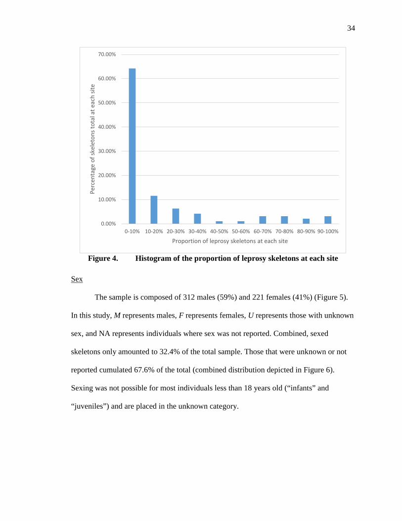
0.7324, p < 0.001). A histogram of proportion of Events at each site (Figure 4) also
shows that the proportion data is not normally distributed, although most sites have less
than 10% leprous skeletons.
Figure 2. Histogram overlaid with a normal curve of density (y) to number of
Events (x) where Events represents number of leprous individuals at a site

33
Figure 3. Histogram overlaid with a normal curve measuring density (y) the
distribution of the Total (x) number of skeletons at each site
34
Figure 4. Histogram of the proportion of leprosy skeletons at each site
Sex
The sample is composed of 312 males (59%) and 221 females (41%) (Figure 5).
In this study, M represents males, F represents females, U represents those with unknown
sex, and NA represents individuals where sex was not reported. Combined, sexed
skeletons only amounted to 32.4% of the total sample. Those that were unknown or not
reported cumulated 67.6% of the total (combined distribution depicted in Figure 6).
Sexing was not possible for most individuals less than 18 years old (“infants” and
“juveniles”) and are placed in the unknown category.
0.00%
10.00%
20.00%
30.00%
40.00%
50.00%
60.00%
70.00%
0-10% 10-20% 20-30% 30-40% 40-50% 50-60% 60-70% 70-80% 80-90% 90-100%
Perc
enta
ge o
f ske
leto
ns to
tal a
t eac
h sit
e
Proportion of leprosy skeletons at each site

35
Figure 5. Number of males and females (only for those reporting sex, N = 533)
Figure 6. Number of males, females, unknown, and not reported (N = 1,645)
Age
Age at death categories follow the organization used by Møller-Christensen
(1978). The age composition of the study sample were sorted into these eight categories:
Males, 312, 59%
Females, 221, 41%
LEPROUS SKELETONS: MALE AND FEMALE
Males Females
19%
13%
3% 65%
DIVISION OF TOTAL SKELETON BY SEX
Males Females Unknown Not Reporting

36
0-6 years old, 7-13 years old, 14-19 years old, 20-29 years old, 30-39 years old, 40-49
years old, 50-59 years old, and 60+ years old. Some references did not report the exact
estimated age of death, using monikers such as adult, young adult, elderly, adolescent,
and middle-aged. In those cases, an age was estimated: adult (40 years old), young adult
(25 years old), elderly (65 years old), adolescent (14 years old), and middle-aged or
mature (50 years old). The age composition with (N = 611) and without (N = 428) the
estimated ages included are combined in Table 5.
The average ages of death of the total sample, male only, female only, and
unknown only were also calculated. In some instances, the average age of death fell
below the other estimates, so averages excluding 0-6 years and 7-14 years values were
also incorporated in Tables 4, calculating an average that did not account for instances of
child mortality.

37
Table 4. Average ages of study sample – with estimation of terminology on the left and without estimation on the right. ‘Adults’ are assumed to be 40 years old, ‘young adults’ are 25 years old, ‘middle-aged adults’ and ‘mature’ are 50 years old, ‘elderly’ adults are 65 years old, and ‘adolescents’ are 14 years old. M stands for male, F stands for female, and U stands for unknown
Average ages of all eight groups
With estimated-aged skeletons included
Without estimated-aged skeletons included
Total 29.91 30.76
M Only 34.02 34.27
F Only 38.03 38.4
U Only 27.33 22.9
Excluding individuals less than 14 years old
Total 37.65 38.77
M Only 37.73 38.02
F Only 38.03 38.4
U Only) 41.12 39.15

38
Table 5. Age composition within the study sample – includes the distribution with and without estimated ages, where ‘adults’ are assumed to be 40 years old, ‘young adults’ are 25 years old, ‘middle-aged adults’ and ‘mature’ are 50 years old, ‘elderly’ adults are 65 years old, and ‘adolescents’ are 14 years old
With estimated-aged skeletons included
Excluding estimated-aged skeletons
Age category Number of skeletons
Percent of total Number of skeletons
Percent of total
0-6 4 0.65% 4 0.93%
7-13 21 3.44% 21 4.90%
14-19 51 8.35% 50 11.68%
20-29 35 5.73% 34 7.94%
30-39 137 22.42% 137 32.00%
40-49 187 30.60% 15 3.50%
50-59 156 25.53% 149 34.81%
60+ 20 3.27% 18 4.20%
Total 611 100% 428 100%
39
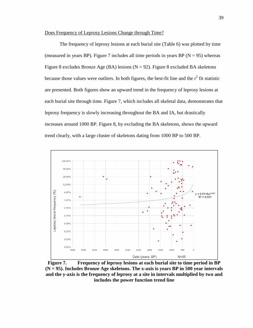
Does Frequency of Leprosy Lesions Change through Time?
The frequency of leprosy lesions at each burial site (Table 6) was plotted by time
(measured in years BP). Figure 7 includes all time periods in years BP (N = 95) whereas
Figure 8 excludes Bronze Age (BA) lesions (N = 92). Figure 8 excluded BA skeletons
because those values were outliers. In both figures, the best-fit line and the r2 fit statistic
are presented. Both figures show an upward trend in the frequency of leprosy lesions at
each burial site through time. Figure 7, which includes all skeletal data, demonstrates that
leprosy frequency is slowly increasing throughout the BA and IA, but drastically
increases around 1000 BP. Figure 8, by excluding the BA skeletons, shows the upward
trend clearly, with a large cluster of skeletons dating from 1000 BP to 500 BP.
Figure 7. Frequency of leprosy lesions at each burial site to time period in BP
(N = 95). Includes Bronze Age skeletons. The x-axis is years BP in 500 year intervals and the y-axis is the frequency of leprosy at a site in intervals multiplied by two and
includes the power function trend line

40
Figure 8. Frequency of leprosy lesions at each burial site to time period in BP (N = 92). Does not include Bronze Age skeletons. Both axes are plotted as natural
logarithms and includes the power function trend line
Lesion Distribution
Lesions
Ninety-five out of 102 sites (93%) of the references that met the inclusion criteria
of the survey reported leprosy lesion type. Osteological changes indicative of leprosy
were recorded in seven locations on the skeleton (Table 2) (Boldsen, 2008; Møller-
Christensen, 1952, 1978). Most references utilized the osteological method of reporting
leprosy lesions developed by Møller-Christensen in 1978. Since not all the articles
utilized a seven-step system, three monikers were used that broadly segregate the
different types: facies leprosa (also known as skull-only), post-cranial-only, and both.
Lesion count of each site based on these three types are recorded in Table 6.
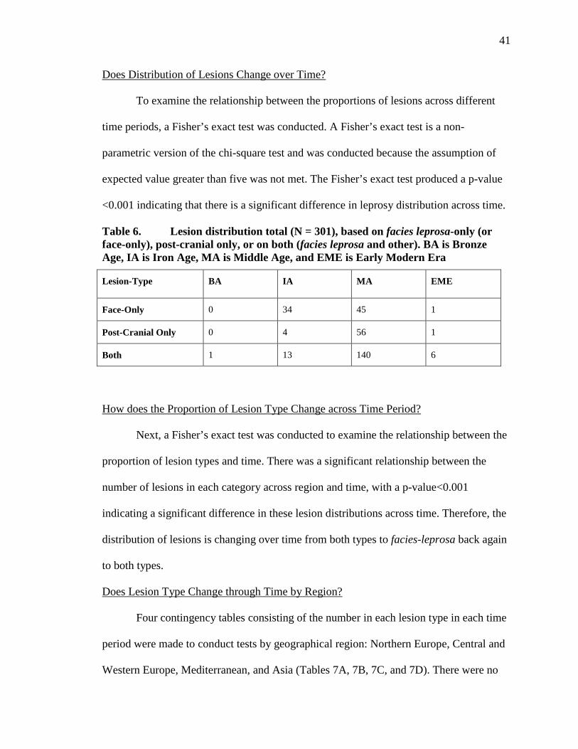
41
Does Distribution of Lesions Change over Time?
To examine the relationship between the proportions of lesions across different
time periods, a Fisher’s exact test was conducted. A Fisher’s exact test is a non-
parametric version of the chi-square test and was conducted because the assumption of
expected value greater than five was not met. The Fisher’s exact test produced a p-value
<0.001 indicating that there is a significant difference in leprosy distribution across time.
Table 6. Lesion distribution total (N = 301), based on facies leprosa-only (or face-only), post-cranial only, or on both (facies leprosa and other). BA is Bronze Age, IA is Iron Age, MA is Middle Age, and EME is Early Modern Era
Lesion-Type BA IA MA EME
Face-Only 0 34 45 1
Post-Cranial Only 0 4 56 1
Both 1 13 140 6
How does the Proportion of Lesion Type Change across Time Period?
Next, a Fisher’s exact test was conducted to examine the relationship between the
proportion of lesion types and time. There was a significant relationship between the
number of lesions in each category across region and time, with a p-value<0.001
indicating a significant difference in these lesion distributions across time. Therefore, the
distribution of lesions is changing over time from both types to facies-leprosa back again
to both types.
Does Lesion Type Change through Time by Region?
Four contingency tables consisting of the number in each lesion type in each time
period were made to conduct tests by geographical region: Northern Europe, Central and
Western Europe, Mediterranean, and Asia (Tables 7A, 7B, 7C, and 7D). There were no

42
specific place of lesions indicated in any Oceania samples and the New World skeletons
were only found in the Early Modern Era (EME) so only four contingency tables were
made. Northern Europe had the largest amount of leprosy lesions (N = 203) in all, with
66.1% falling under MA Northern European skeletons with both lesion-types. There were
only 22 Central and Western European skeletons with lesions, 45% of whom dated to the
IA and only had facies leprosa. There were 59 Mediterranean skeletons with lesion-types,
primarily facies leprosa only in the IA and the MA. In Asia, 69% of the 13 total skeletons
with lesions were from the IA with facies leprosa and 2 EME Asian skeletons had both
lesion-type. Fisher’s exact tests determined that, out of the four geographic areas tested,
only Asia was significant. See Chapter 5 for further discussion on the results from Tables
7A-D.
Table 7A. Contingency table of Chi-square test measuring lesion types – Northern Europe
Lesion-Type IA MA EME
Face-Only 1 5 0
Post-Cranial Only 3 55 1
Both 3 134 1
Fisher’s Exact Test: p-value = 0.1596

43
Table 7B. Contingency table of Chi-square test measuring lesion types – Central and Western Europe
Lesion-Type IA MA
Face-Only 10 4
Post-Cranial Only 1 1
Both 5 1
Fisher’s Exact Test: p-value = 0.7988
Table 7C. Contingency table of Chi-square test measuring lesion types – Mediterranean
Lesion-Type IA MA
Face-Only 14 36
Both 4 5
Fisher’s Exact Test: p-value= 0.434
Table 7D. Contingency table of Chi-square test measuring lesion types – Asia
Lesion-Type BA IA EME
Face-Only 0 9 0
Both 1 1 2
Fisher’s Exact Test: p-value = 0.014
Tables 8 and 9 chart mean age at death by male, female, unknown and total across
the four time periods. Table 8 provides calculated average age with estimated
terminology of age included whereas Table 9 provides averages without the terminology
included.
Figures 9 and 10 consist of the same information as Tables 8 and 9 but categorize
average ages by geographical region as well. All 8 age categories are not represented in

44
some geographical regions and thus, have N/A next to it. Figure 9 includes results with
the estimated terminology of age and Figure 10 excludes those results.
Table 8. Frequency distribution of median age at death for each time period in the combined sex sample – with estimated terminology (N = 611). Numbers in parenthesis represent the number of each calculated for each average
Table 9. Frequency distribution of median age at death for each time period in the combined sex sample – without estimated terminology (N = 424). Numbers in parenthesis represent the number of each calculated for each average
Two multi-way ANOVA’s were conducted to see whether age is significant by
groups: sex (M, F, and U) and time period (BA, IA, MA, and EME). Levene’s test was
used to test for homogeneity of variance. The Levene’s test for Figure 9, which includes
data with estimated terminology, was significant (D.F. =11, F-value=5.37, p-
Calculated Average Age at Death for Each Time Period: With Estimated Terminology:Average Culmulated Age (and number of each)
Average Male Age (and number of each)
Average Female Age (and number of each)
Average Unknown Age (and number of each)
Bronze Age: pre-600 BCE 26.625 (4) 28.5 (2) 42.5 (1) 7 (1)Iron Age: 500 BCE - 1049 CE 38.29 (67) 39.5 (30) 38.66 (18) 36.04 (19)Middle Ages: 1050 CE - 1535 CE 37.25 (530) 40.75 (193) 33.87 (170) 36.47 (167)
Early Modern Era: 1536 CE - 1905 CE 36.44 (8) 35.8 (5) 22.5 (1) 45 (2)
Calculated Average Age at Death for Each Time Period: Without Estimated Terminology:Average Culmulated Age
Average Male Age (and number of
Average Female Age (and number
Average Unknown Age (and number
Bronze Age: pre-600 BCE 26.625 (4) 28.5 (2) 42.5 (1) 7 (1)
Iron Age: 500 BCE - 1049 CE 35.9 (35) 37.07 (22) 38.03 (11) 2.43 (2)
Middle Ages: 1050 CE - 1535 CE 36.87 (378) 40.65 (185) 33.76 (167) 16.98 (26)
Early Modern Era: 1536 CE - 1905 CE 34.5 (7) 32.25 (4) 22.5 (1) 45 (2)

45
value=<0.001), suggesting the assumption of homogeneity of variance is not met. The
ANOVA in Figure 9 states that, with the estimated terminology included, sex by itself is
significant (p<0.001) but time period by itself as well as the combination of sex and time
period together are not significant (p=0.26; p=0.13).
Additionally, the Levene’s test for Figure 10, which does not include data with
estimated terminology, is also significant (DF=11, F-value=1.85, p-value=<0.05). The
ANOVA in Figure 10 states that, without the estimated terminology included, sex by
itself is significant (p<0.001) but time period by itself is not significant (p=0.55).
However, sex and time period together are significant (p<0.01).
These results suggests that the sex (M, F, and U) of the leprous skeletons is a
significant factor to the age at death whether estimated terminology was used in the
ANOVA or not. Time period was a significant factor to the age at death only when
combined with sex and only when estimated terminology is not included (Figure 10).
Since the culminated average ages at death between the four time periods only varied
slightly, it is apparent that sex would need to be significant for the ANOVA’s to be
significant.
Figure 9. Multi-Way ANOVA of median age at death for each time period and geographical region in the combined sex sample – includes estimated terminology of
age (N = 611)

46
Figure 10. Multi-Way ANOVA of median age at death for each time period and
geographical region in the combined sex sample – does not include estimated terminology of age (N = 412)
Does the Proportion of Leprosy Skeletons per Site Change?
Leprosariums likely affect the proportion of leprous individuals in the study.
Table 10 lists the 13 leprosariums used in this study and Table 11 lists the 4 leprosariums
that were not included in the data since they did not include the total number of skeletons.
While leprosariums are crucial in bioarchaeology, the overall representation of leprosy’s
effect was skewed towards those sites with leprosariums. In particular, the data found at
three leprosariums in Denmark made up 69% of this data’s total number of leprous
individuals. Figures 12 and 13 show two forest plots arranged chronologically by date
and time period, respectively, that excludes all leprosariums and samples less than 10
individuals (total) Leprosariums are further discussed in Chapter 5.

47
Table 10. Leprosariums used in the study organized by date (N = 13)
Table 11. Leprosariums that were not used in the study organized by date (N = 4)
References Leprosarium Year (Mid-point)Mendum et al (2014); Magilton et al (2008)
St. Mary Magdalen, Winchester, UK 1043 CE
Wells (1967) Castle Acre, Norfolk, UK 1225 CE
Blondiaux et al (2015)Saint-Thomas d’Aizier, Haute-Normandy, France 1250 CE
Chundun et al (1995) St. Giles, North Yorkshire, UK 1290 CE
Manchester et al (1986) St. Mary and St. Thomas hospital, Ilford, Essex, UK 1300 CE
Farley et al (1989)St. Margaret’s Hospital, Buckinghamshire, UK 1350 CE
Primeau et al (2014) Maribo, Lolland, Denmark 1350 CE
Lee et al (1989); Magilton et al (2008)
St James and St Mary Magdalene, Chichester, UK 1388 CE
Antunes-Ferreira et al (2013) Beja, Portugal 1400 CE
Møller-Christensen (1978) St. Jorgen Hospital, Næstved, Denmark 1400 CE
Boldsen (2001) Kvarteret S:t Jörgen leprosy hospital, Malmö, Skåne, Sweden 1420 CE
Jensen et al (1983) St. Jørgen, Odense, Denmark 1450 CEFerreira et al (2013) Lagos, Algarve, Portugal 1500 CE
References Leprosarium Year (Mid-point)Schultz et al (2002) Lihou Island, Guernsey, UK 1250 CE
Bishop (1983) St. Leonard’s, Nottinghamshire, UK 1350 CE
Matos et al (2013) St. Jørgen leprosarium cemetery, Odense, Denmark 1350 CE
Schmitz-Cliever (1972) Aachen, Germany 1390 CE
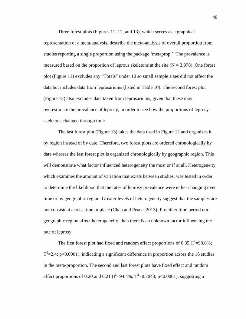
48
Three forest plots (Figures 11, 12, and 13), which serves as a graphical
representation of a meta-analysis, describe the meta-analysis of overall proportion from
studies reporting a single proportion using the package ‘metaprop.’ The prevalence is
measured based on the proportion of leprous skeletons at the site (N = 3,978). One forest
plot (Figure 11) excludes any “Totals” under 10 so small sample sizes did not affect the
data but includes data from leprosariums (listed in Table 10). The second forest plot
(Figure 12) also excludes data taken from leprosariums, given that these may
overestimate the prevalence of leprosy, in order to see how the proportions of leprosy
skeletons changed through time.
The last forest plot (Figure 13) takes the data used in Figure 12 and organizes it
by region instead of by date. Therefore, two forest plots are ordered chronologically by
date whereas the last forest plot is organized chronologically by geographic region. This
will demonstrate what factor influenced heterogeneity the most or if at all. Heterogeneity,
which examines the amount of variation that exists between studies, was tested in order
to determine the likelihood that the rates of leprosy prevalence were either changing over
time or by geographic region. Greater levels of heterogeneity suggest that the samples are
not consistent across time or place (Chen and Peace, 2013). If neither time period nor
geographic region affect heterogeneity, then there is an unknown factor influencing the
rate of leprosy.
The first forest plot had fixed and random effect proportions of 0.35 (I2=98.6%;
T2=2.4; p<0.0001), indicating a significant difference in proportion across the 16 studies
in the meta-proportion. The second and last forest plots have fixed effect and random
effect proportions of 0.20 and 0.21 (I2=94.4%; T2=0.7043; p<0.0001), suggesting a

49
slighter smaller difference in heterogeneity between the 10 studies in this meta-
proportion than the first plot. Therefore, running a meta-proportion that included
leprosariums had more heterogeneity than a meta-proportion that was conducted without
them, as expected. The heterogeneity tells us whether there are significant differences
between those proportions.
The sites with the largest proportion of leprosy were found at leprosariums
(Studies 3, 10, 12, and 16 in Figure 11). By removing those sites, the highest proportion
is in St. Nicholai Kirche in Schleswig Germany at 44% versus the highest proportion of
95% at St. Jørgen, Denmark when leprosariums are included. Additionally, Figures 11
and 12 do not show a clear trend of proportion in those sites dating to the Iron Ages
versus the medieval aged sites, suggesting that proportion is independent of time period.
Figure 11. Forest plot of a meta-analysis of single proportions. Excludes Events
under 10 but includes leprosariums. Organized chronologically by date where Study 1 occurred furthest back in time and Study 16 most recently. Studies: 1 (478 CE;
Lauchheim, Germany), 2 (950 CE; St. John, Norfolk), 3 (1043 CE; St. Mary Magdalen, Winchester), 4 (1050 CE; St. Nicolai Kirche, Schleswig, Germany), 5 (1050 CE; St. Clemens Kirche, Schleswig, Germany), Study 6 (1065 CE; Lund,

50
Sweden), Study 7 (1133 CE; Rathaus Markt, Schleswig, Germany), 8 (1239 CE; Dominikaner Kloster, Schleswig, Germany), 9 (1250 CE; Tirup, Denmark), 10
(1250 CE; Saint-Thomas d’Aizier, France), 11 (1388 CE; St. James and St. Mary Magdalene, Chichester), 12 (1400 CE; St. Jorgen, Denmark), 13 (1420 CE; Malmö,
Denmark), 14 (1410 CE; St. Knud, Odense, Denmark), 15 (1410 CE; St. Albani, Odense, Denmark), 16 (1450 CE; St. Jørgen, Denmark)
Figure 12. Forest plot of a meta-analysis of single proportions. Excludes Events
under 10 and leprosariums. Organized chronologically by date where Study 1 occurred furthest back in time and Study 10 most recently. Studies: 1 (478 CE; Lauchheim, Germany), 2 (950 CE; St. John, Norfolk), 3 (1050 CE; St. Nicolai
Kirche, Schleswig, Germany), 4 (1050 CE; St. Clemens Kirche, Schleswig, Germany), 5 (1065 CE; Lund, Sweden), 6 (1133 CE; Rathaus Markt, Schleswig, Germany), 7 (1239 CE; Dominikaner Kloster, Schleswig, Germany), 8 (1250 CE;
Tirup, Denmark), 9 (1410 CE; St. Knud, Odense, Denmark), 10 (1410 CE; St. Friars, Odense, Denmark)

51
Figure 13. Forest plot of a meta-analysis of single proportions organized
temporally by region. Excludes Events under 10 and leprosariums. Organized by geographic region where Studies 1 through 5 represent Central and Western
Europe and Studies 6 through 10 represent Northern Europe. Studies: 1 (478 CE; Lauchheim, Germany), 2 (1050 CE; St. Nicolai Kirche, Schleswig, Germany), 3
(1050 CE; St. Clemens Kirche, Schleswig, Germany), 4 (1133 CE; Rathaus Markt, Schleswig, Germany), 5 (1239 CE; Dominikaner Kloster, Schleswig, Germany), 6
(950 CE; St. John, Norfolk), 7 (1065 CE; Lund, Sweden), 8 (1250 CE; Tirup, Denmark), 9 (1410 CE; St. Knud, Odense, Denmark), 10 (1410 CE; St. Friars,
Odense, Denmark)
52
CHAPTER FIVE: DISCUSSION
Does Bone Lesion Type Vary by Time Period?
The frequency and distribution of bone lesions was found to vary depending on
the geographical region and time period. All regions except for the New World, saw a
rise in bone lesions after the Bronze Age but significantly decreasing after the Middle
Ages (Tables 7A-D). This finding suggests that leprosy’s large prevalence in most
medieval, non-New World countries correlates with the poor health and high frequency
of disease before the Early Modern Era. The New World only had bone lesions found
post-1536 CE, which corresponds to other research made by comparative genomic
studies that leprosy entered the Western hemisphere after the medieval period. The only
significant result came from Asia, proposing that Asian bone lesions proportions varied
by time period from both to facies-leprosa to both. No significant differences were found
between the proportions of bone lesions across time for the other three geographical
regions.
Additionally, the values of the chi-square test measuring lesion distribution
suggests that there are significant differences in proportion of lesion types by time
periods (BA, IA, MA, and EME) and geographic regions (NE, CWE, M, A, NW, and O).
Skeletons from Northern Europe in the medieval ages are more likely to display both
lesion types than facies-leprosa or post-crania only. Skeletons from Central and Western
Europe in the Iron Age has a greater proportion of facies-leprosa. Skeletons from
medieval Mediterranean countries have a greater proportion of facies leprosa, and Iron
Age Asia has a greater proportion of facies leprosa (Tables 7A-D). This suggests that

53
different parts of the world suffered from either tuberculoid or lepromatous leprosy,
which affects different parts of the body. Additionally, some results are biased towards
one type since there were skeletons that were missing parts of their body, either due to
poor preservation or to burial preference (i.e., ossuary jars in the Czech Republic).
How Does the Rate of Leprosy Change over Time?
Two of the forest plots (Figure 11 and 12) were organized by time to see if there
was a trend of proportions through time while one (Figure 13) was organized
chronologically by region. The studies with the highest proportions of leprosy came from
were from leprosariums (Figure 11), such as the St. Mary Magdalen leprosarium in
Winchester, UK and the Saint-Thomas d’Aizier in France. When the leprosariums were
removed from the data and put in new forest plots, the sites with the highest proportions
were all from Schleswig, Germany.
Organizing by region did not change the proportion values in the forest plot,
predictably, but displayed the pattern the proportions made by region instead of time
period. Notably, Figure 12, which is organized by date, shows a larger frequency of sites
from the 11th and 12th centuries to any other time period. Even without leprosarium data
included, this suggests that leprosy was prevalent in the early to middle medieval ages.
Geographically, Figure 13 has a large proportion of non-leprosarium sites from
Schleswig, Germany and Tirup, Denmark (Studies 2, 3, 4, 5, 8; Figure 13) (Boldsen et
al., 2013; Boldsen, 2005b). Denmark has always been well-represented in leprosy during
the medieval period, but this finding from Germany is unique. Possibly, Schleswig,
Germany did not act as a leprosarium but as a town for leprosy sufferers and non-leprosy

54
sufferers alike could live together and be buried together. More data on this topic is
required, however.
As for the rest of the data, the forest plots show that Denmark had large cases
(10+) of leprosy throughout the 13th and 14th centuries whereas other countries that were
represented early on in the forest plot with large leprosy cases – UK, Germany, and
Sweden – had decreased in frequency. Based on the chronological and temporal
distribution of these sites with 10+ leprosy cases (excluding leprosariums), leprosy was
prevalent throughout Europe but did not impact Denmark until the 13th century, after it
had decreased everywhere else.
Demographics
Leprosy more commonly occurs in males, who are more susceptible to the
disease, than females in most regions of the world, although leprosy can affect all age
groups (Walker, 2009:364). The data set confirms this, as 312 male skeletons had leprosy
whereas only 221 females had it. Individuals under 18 years old and over 60 years old
were poorly represented in the sample, likely because it takes years to develop bone
lesions but most will not survive over the age 60 (Lynnerup and Boldsen, 2012).
Although all age groups were represented, this infectious disease is not spread equally
among the age groups. Based on the information from Table 8, or with the estimated
terminology, the average cumulated ages ranged from 26.6 years to 38.3 years whereas
the average cumulated ranged from 26.6 years to 36.9 years without estimated
terminology factored in (Table 9). Subsequently, providing an estimate of age for those
reported as adult, middle-aged, young adult, adolescent, and elderly did not change the
numbers drastically. Tables 8 and 9 also show that males lived longer in the medieval era

55
than any other time period (40.75 years or 40.65 years) whereas females lived their
longest in the Bronze Age (42.5 years) although this might be due to the small sample
from the BA. While this demographic information is significant and suggests variation in
health and sex-roles through time, the values are not significantly different from one
another so a conclusion about age at death and sex can be made.
When the estimated terminology is included, 40-49 year old adults comprise the
largest portion of the eight age groups (N = 187). This is likely because 173 of those
numbers were “adults” of unknown age, subsequently estimated to be 40 years old. The
appellative “adults” is a common term to call an older skeleton when age is generally
unknown but the range could be anywhere between 20 to 60 years. With an estimation of
40 years for these 173 “adults,” the distribution is predictably skewed towards a large
number of 40 year olds in the sample. Most likely, based on historical information of the
time period, the average age at death for those suffering from leprosy would have ranged
anywhere from 30 to 39 years old (Blondiaux et al., 2015; Boldsen, 2005; Weiss and
Møller-Christensen, 1971), a result that is confirmed by the paleopathological skeletons
used in this study (see Table 5, which includes and excludes data where age was
estimated by this project’s author).
Health
Out of all the 1,645 leprosy cases used in this study, 0.24% were from the BA,
9.4% were from the IA, 89.9% were from the MA, and 0.55% were from the EME. There
was an decrease in prevalence rates from BA (7.02% of cases from that time period) to
IA (1.95% of cases from that time period) then a drastic increase in the MA (14.2% of
cases from that time period) then a drop in cases in the EME (2.1% of cases from that

56
time period). Roberts (2002a) attributes MA’s large increase in leprosy cases to denser
living conditions and decreased sanitation, stimulating the rate of transmission.
There is a steep decline in prevalence rates once the medieval period came to an
end in the 16th century likely due to the aftermath of the Black Death and increased
competition with TB. Skeletal evidence of chronic infectious disease, according to Larsen
(2009), “are primarily associated with agricultural-based populations living in close,
densely crowded communities” (2009:7). It is assumed that, due to a rise in population,
skeletons dating to the Iron Age, Middle Age, and Early Modern Era were from densely
populated, agricultural communities. Agriculture was a relatively new phenomenon to the
Bronze Age skeletons so it is possible that small number of skeletons in the dataset (N =
4) is related to agriculture’s recent introduction. An agriculture-based diet is less healthy
and more labor intensive than a hunter-gatherer diet. It also creates densely-populated
areas, promoting the spread of disease.
A trend observed in the data was that females died at a younger age than males in
all time periods except the Bronze Age (Tables 8 and 9). This finding is significant
because assessing age at death and sex portrays the health conditions and trends of the
population in a given time and place. In contrast to the pattern seen today where females
live longer than males, a significant proportion of women in the Iron Ages and medieval
ages died in association with childbirth. Life expectancy varied by location, but the trend
remained the same. In the mid-late medieval UK, for example, the average life
expectancy at birth for an aristocratic male was around 35 years whereas a female at birth
was expected to live to age 30 (Lancaster, 1990:8). Surprisingly, the average ages for
leprous individuals of both sexes in the data fall within 30 to 40 years, matching

57
estimates of the life expectancy at birth for the time period. This could be because
leprosy, while a disfiguring disease, does not cause death unless it co-infects the
individual with other diseases. In this dataset, the average age at death is higher than life
expectancy at birth in the mid-late medieval UK because not many infants/children are
dying with leprosy, whereas there were likely higher rates of infant/child mortality in the
general population. Those children that did die from leprosy would skew that data
towards a lower average age of death, a trend that is observed for the “unknown”
individuals in Table 4. Assessing the average age of death is affected by the
“Osteological Paradox,” since many children do not have time to develop telling bone
lesions before they die.
In reality, these estimates of life expectancy are at birth but once a person
survives the first few years of life, life expectancy goes up by many years. A 21-year old
individual’s life expectancy in 13th century UK is 64 years. Therefore, it is also
reasonable to conclude that the individuals in the dataset did not outlive non-infected
individuals who, if they lived past the first few years of life, could expect to live well into
their 50s and 60s. The leprosy sufferers usually died at an average age of 35 to 40 years
of age.
However, life expectancy for the general population and those suffering from
leprosy dropped drastically in the 1300-1400s, reflecting the high mortality rates caused
by the Black Plague (see Chapter 1). A 21-year old aristocratic male in the UK was only
expected to survive an additional 25 years in the 14th to 15th centuries (Lancaster, 1990).
Therefore, the average age of death of a non-leprous individual and a leprous individual
during the 1300-1400s were almost the same.

58
Leprosariums
Since successful medical treatment for leprosy did not exist until recently, past
efforts focused on segregating and caring for those individuals afflicted with the disease.
Leprosariums became common during the medieval era, often existing outside the city
limits (Covey, 2001:320). Historical reports relaying accurate numbers of leprosariums at
a given time vary drastically. Roffey (2012) reports “over 300 documented leper
hospitals in the medieval period” (2012:203). Meanwhile, Matthew of Paris, a medieval-
era Benedictine monk, roughly estimated there to be more than 19,000 “lazar houses” in
Europe in the eleventh century (Simpson, 1872:3).
Estimates for the leprosarium count also differ depending on the country. Covey
(2001) reports that by the mid-12th century CE, France had around 2,000 leprosariums,
although there are none from that time period used in the dataset for this thesis, while
England and Scotland had about 220 leprosariums (2001:317). Denmark had 31
leprosariums between 1250 CE to 1550 CE (Bennike et al., 2005). In Portugal, there were
around 70 small leprosariums during the medieval period (Antunes-Ferreira et al.,
2013:149; Carvalho, 1932). Likely, Covey states, there was a misuse of the word
“leprosarium” for hospitals that treated people with leprosy occasionally, exclusively,
sometimes, or never at all (2001:317; Roffey, 2010:217; Rogers et al., 1946).
Due to this misrepresentation of terminology, opinions vary in the literature on
the prevalence of leprosy in medieval Europe. For the most part, authorities of the period
agree that leprosy was a familiar disease during the 11th to 15th centuries among
Europeans, as the disease is consistently reported and referred to in medieval-aged texts.
It is intriguing to see the contrast between the reported number of leprosy hospitals and

59
the cases seen in the bioarchaeological record. This could indicate that leprosy was over-
reported in the historical literature, possibly confusing the disease for another pathogen.
It is interesting, although not surprising given the historical literature, that every
leprosarium, save one, whether its data was used in the study or not, dates to the medieval
era (1050 CE – 1536 CE). The only exception is St. Mary Magdalen in Winchester, UK,
although at 1043 CE only makes this site seven years away from the MA cut-off. While
the bioarchaeological record may not show it, Antunes-Ferreira et al. (2013) states that
leprosy hospitals were around in Europe as far back as 5th century CE (149).
These results also show leprosariums in the paleopathological literature were only
in Europe, primarily in the UK and Denmark. This is unsurprising as the only non-
European paleopathological skeletal sites from the medieval period used in this study
were in Polis Chrysochous, Cyprus, Dakhleh Oasis, Egypt, and western Micronesia for a
combined total of 11 non-European medieval-aged individuals (Baker et al., 2014; Molto,
2002; Trembly, 1995). Out of a total 1,108 burials from these three sites, these 11 leprous
individuals indicate a rate of leprosy in non-European MA to a paltry 0.992% according
to this paleopathological data. In this regard, these results support studies suggesting
Mycobacterium leprae and leprosariums primarily raged through Europe during the
medieval era although this conclusion could be biased based on where bioarchaeological
research occurs (Donoghue et al., 2005, 2015; Matheson et al., 2009; Robbins et al.,
2009).
Transmission and aDNA
Comparative genomic studies have mapped genetic variability between
populations through mapping SNP-types and variable number tandem repeat, or “VNTR”

60
types. “SNP” or single nucleotide polymorphisms, each represent a difference in a single
nucleotide, and have been consistently proposed as participants in susceptibility towards
diseases such as leprosy (Alvarado-Arnez et al., 2015:1; Economou et al., 2013;
Donoghue et al., 2005; Monot et al., 2005). Monot et al. (2005) discovered four different
SNP-types (1, 2, 3, and 4) and in 2009, divided these four types into sixteen SNP-
subtypes.
The four main strains, or types, of Mycobacterium leprae are divided
geographically. Monot et al. (2005) created a map (Figure 2 from Monot et al. 2005;
Figure 14) showing the dissemination of the strains and their geographical pathways.
According to their research, strain 1 occurs in East Africa, Asia, and the Pacific; strain 2
in New Caledonia, Nepal/north India, Ethiopia, and Malawi; strain 3 in North Africa,
Europe, and the Americas; and strain 4 in West Africa, Brazil, and the Caribbean islands
(from Monot et al. Table 3, 2005:1041).
Economou et al. (2013) found evidence of SNP-subtype 2G, a type that is found
mainly in Asia/North Africa, in medieval Swedish remains (465). This finding is unique
and remains one of the few tested samples from Europe that does not belong to SNP-type
3, providing genetic evidence that a transmission of a particular type of the pathogen
from Asia to Scandinavia had occurred during the medieval ages. Mendum et al. (2014)
found evidence for SNP-type 2 strains in two skeletons from the medieval leprosarium of
St Mary Magdalen near Winchester, England (2014). Monot et al. (2005) and Taylor and
Donoghue (2011) have found evidence that SNP-type 3, a predominantly European and
North American type, was present in some parts of Asia/Middle East in the Iron Age and
medieval era. A 1st- 4th century CE adult skeleton from Uzbekistan was of subtype 3L

61
and four young adults from 4th century CE Dakhleh Oasis in Egypt had genotype 3. A
large study by Donoghue et al. (2015), mostly on Hungarian samples, found SNP-
subtypes 3M and 3K, which was also found in a Turkish sample (253). Based on their
results, the authors concluded that Avar (or Caucasus) migration from Central Asia into
Byzantium and Central Europe exposed the disease to local populations.
The exact route of transmission remains unclear, but there are two possible
schemes for the spread of leprosy based on SNP types. The first, which Cole and Singh
(2012) deem more probable, is that M. leprae evolved in East Africa and spread to
Central Asia (SNP-type 2 preceding SNP-type 1, which migrated eastward, and SNP-
types 3 and 4 subsequently follow). The other scheme argues that M. leprae evolved in
Central Asia and spread to East Africa following human migrations (SNP-type 1
preceding SNP-type 2 and SNP-types 3 and 4 subsequently follow). The earliest accepted
evidence (excluding the Bronze Age Scottish and Hungarian samples) of leprosy was
found in a north Indian individual in 2009 and makes a strong argument for scheme two
(Robbins et al., 2009). Thus, this study deems the second scheme the most likely
scenario.
Genotypes 1 and 4 remain mostly unique to their geographic regions – type 1 in
Asia, the Pacific region, and East Africa and type 4 in West Africa and the Caribbean
region, and South America (Monot et al., 2005; Cole and Singh, 2012). SNP-type 1
originated in Asia, spread into East Africa, and then spread into the Pacific region
through possible contact with, or immigration from, China or Japan (Trembly, 1995).
SNP-type 4 was brought over into the Caribbean and South America through the
18th century CE West African slave trade by infected European or North African

62
colonialists, explorers, traders, or West African slaves (Monot et al., 2005:1042). Much
like how SNP-type 3 arrived in North America, infected Europeans with type-4 leprosy
likely brought over the disease into the Caribbean and South America through early
exploration, migration, and the slave trade.
Figure 14. Dissemination of leprosy in the world [from Monot et al. (2005)]. The
colored circles indicate the country of origin sampled and their division into the four SNP types. SNP-type 1 is yellow, SNP-type 2 is orange, SNP-type 3 is purple, and
SNP-type 4 is green. The colored arrows represent migrations (Monot et al., 2005:1042)
Limitations in a Meta-Analysis
Relying on data of human skeletons from bioarchaeological reports means relying
on the authors’ quality of excavation and interpretation of skeletal pathologies.
Additional limiting factors include burial preservation and excavation and post-
excavation treatment which makes the quality of the data highly variable (see Chapter 2;
Roberts et al., 2003:13). Additionally, all articles that were not in English (the author’s

63
native language), Spanish or Hebrew, were translated through an online website. This
could have resulted in possible translation issues or incorrect information.
An apparent issue this project deals with is that several geographic areas and time
periods have small samples sizes, or sometimes, none at all. One of the most obvious
limitations in this study is its reliance on secondary sources. A possible bias lies in
sample studies themselves as they are based on published studies available through the
internet, the Boise State University library or its inter-library loan, or received from the
authors themselves
There is a known bias since 44 sites from 29 different references did not include
total number of individuals excavated and were subsequently thrown out since the
frequency of lesions (or percent of skeletons with leprosy) could not be calculated. In
these excluded sites, 5 were from Sweden, 3 from Denmark, 25 from UK, 1 from Egypt,
2 from Germany, 1 from Indonesia, 3 from Hungary, 2 from Israel, 1 from Poland, and 1
from Ukraine. This data was entered into an excel sheet similarly to the data that was
used in this study (supplementary text Appendix C).
Bioarchaeological literature on leprosariums lead to a densely populated skeletal
samples of people with leprosy in certain geographical areas, which may skew the results
as skeletal samples without any leprosy lesions were not included. This suggests that the
rate of leprosy may be lower than was calculated in this meta-analysis (given that skeletal
samples with 0% leprosy prevalence were not included). Thus, the results of this meta-
analysis are supported by the author with the full understanding that its reliability
depends on the validity of the studies (Walker et al., 2008:438).

64
Biocultural Applications
This thesis’ conclusions are strained by the biocultural considerations discussed in
Chapter 2. The problem of accurate estimation of leprosy’s impact continues to be an
ongoing challenge for bioarchaeology and paleopathology. While this meta-analysis was
successful in portraying leprosy’s frequency and lesion distribution in past populations
based on the available paleopathological literature, the Osteological Paradox, political
instability, and socioeconomic issues creates misrepresentation in accurate
bioarchaeological assessments. Some countries are over-represented and others are
grossly under-represented. It should also be remembered that, due to leprosy’s long
incubation period which affects when bone changes develop, skeletons of children and
those with a low resistant form of the disease are unlikely to show leprous lesions. The
many intrinsic and extrinsic factors that affect whether or not someone shows signs of the
infectious disease further complicates the picture. Clearly, it is unlikely that the frequency
of leprosy was truly 8.76% of the total population. The skeletal material examined here
only represents the individuals reported in the paleopathological literature and any
assumptions on the demographic profile of the leprous individuals will be flawed. The
data presented here tells us something about the impact of leprosy throughout time and
space but the data is biased due to a number of different factors (Roberts, 2002b:216).
More work is needed in order to obtain the real frequency of leprosy. Future careful
excavation, recording, and analysis in additional geographic areas are needed to further
interpretations on the impact and frequency of leprosy.

65
Conclusion and Further Directions
Review
Out of 18,787 total skeletal samples used in this data, the percentage of
individuals that showed osteological signs of leprosy expressed in relation to total
number of individuals was 8.756% (1,645 leprous skeletons). It is unlikely that leprosy
was this prevalent in communities at any time, however. Leprosy was at its peak in
Northern Europe in the high medieval period (1200-1400 CE), slowly declining in the
mid-13th century CE, and then dropping off dramatically by the end of the period (Covey,
2001:320). This peak is correlated to the influx of leprosariums in that geographical
region, especially in the UK and Denmark. Leprosy did not truly take effect in Europe
until the late Iron Age, existing mostly in Asia and the Mediterranean until the sixth
century.
Further Directions
The future of leprosy research is constantly marred by technological and
methodological limitations. To keep up with curing leprosy today, research into leprosy’s
impact in the past will need to continue, especially in non-European and post-medieval
populations. Additionally, continued methodological developments in lesion
identification and prevalence is critical. As new paleopathological research continues to
be published on leprosy, future meta-analyses will need to integrate this information with
the new developing technique of aDNA analyses.
Although there is no vaccine available for the prevention of leprosy, there are
numerous prevention programs ran by the World Health Organization, the Centers for
Disease Control and Prevention, the National Institute of Allergy and Infectious Diseases,

66
the United Nations Children’s Emergency Fund, LEPRA, the International Federation of
Anti-Leprosy Associations, and the Movement of Reintegration of Persons Afflicted by
Hansen’s disease. Ever since the World Health Organization provided MDT for free
worldwide in 1995, leprosy rates keep decreasing every year (World Health
Organization, 2016). However, leprosy is still a problem in dozens of countries with India
accounting for over 60% of the total cases (as of 2012; WER 2012 Index). Despite these
setbacks and continued prevalence of the disease, substantial progress has been made by
providing free treatment to millions of affected individuals and in overcoming social
stigma and exclusion. Hopefully, this progress will be further advanced through
continued ongoing research into the basic understanding of the disease, the history and
cause of its transmission, and improved treatments and vaccines. If it does, leprosy will
one day no longer be one of the leading causes of physical disability and social stigma in
the world as it was in the past (Bennet et al., 2008).

67
REFERENCES
Alvarado-Arnez LE, Amaral EP, Sales-Marques C, Durães SMB, Sarno EN, Pacheco
AG, Lana FCF, Moraes MO. 2015. Association of IL10 polymorphisms and
leprosy: A meta-analysis. PLoS ONE 10:1-13.
Anderson S. 1998. Leprosy in a medieval churchyard in Norwich. In: Anderson S, editor.
Current and recent research in osteoarchaeology. Proceedings of the 3rd meeting
of the Osteoarchaeology research group. Oxford: Oxbow Books. p 31-37.
Antunes-Ferreira N, Matos VMJ, Santos AL. 2013. Leprosy in individuals unearthed near
the Ermida de Santo Andre and leprosarium of Beja, Portugal. Anthropol Sci
121:149-159.
Arcini C. 1999. Health and disease in early Lund - osteopathologic studies of 3305
individuals buried in the first cemetery area of Lund 990-1536. Archaeologica
Lundensia VIII. Lund: Department of Community Health Sciences.
Aufderheide AC, Rodríguez-Martín C. 1998. The Cambridge encyclopedia to human
paleopathology. Cambridge: Cambridge University Press.
Baker B, Bolhofner KL. 2014. Biological and social implications of a medieval burial
from Cyprus for understanding leprosy in the past. Int J Paleopathol 4:17-24.
Barber L, Sibun L. 1998. The medieval hospital of St Nicholas, Lewes, East Sussex:
excavations 1994. Sussex archaeological collections ADS 148:1-42.
Bax L, Yu L-M, Ikeda N, Moons KGM. 2007. A systematic comparison of software
dedicated to meta-analysis of casual studies. BMC Med Res Methodol 7:40.
Belcastro MG, Mariotti V, Facchini F, Dutour O. 2005. Leprosy in a skeleton from the
7th century necropolis of Vicenne-Campochiaro (Molise, Italy). Int J
Osteoarchaeol 15:431-448.

68
Bennett BH, Parker DL, Robson M. 2008. Leprosy: steps along the journey of
eradication. Pub Health Rep 123:198-205.
Bennike P, Lewis ME, Schutkowski H, Valentin F. 2005. Comparison of child morbidity
in two contrasting medieval cemeteries from Denmark. Am J Phys Anthropol
128:734-746.
Blau S, Yagodin V. 2005. Osteoarchaeological evidence for leprosy from western Central
Asia. Am J Phys Anthropol 126:150-158.
Blondiaux J, Naji S, Bocquet-Appel J-P, Colard T, de Broucker A, de Seréville-Niel C.
2015. The Leprosarium of Saint Thomas d'Aizier, the cementochronological proof
of the medieval decline of Hansen disease in Europe? Int J Paleopathol. In press.
Blondiaux J, Durr J, Khouchaf L, Eisenberg LE. 2002. Microscopic study and X-ray
analysis of two 5th century cases of leprosy: palaeoepidemiological inferences.
In: Roberts CA, Lewis ME, Manchester K, editors. The past and present of
leprosy. Archaeological, historical, palaeopathological and clinical approaches.
BAR International series 1054. Oxford: Archaeopress. p 105-110.
Bloomfield M. 2004. Hymns of the Atharva Veda. Whitefish, MT: Kessinger Publishing.
Boekhout TA. 2009. The puzzle of syphilis: a literature review on putting the pieces
together. [thesis].
Boeckl CM. 2011. Images of leprosy: disease, religion, and politics in European art.
Kirksville, MO: Truman State University Press.
Boldsen JL, Rasmussen KL, Riis T, Dittmar M, Weise S. 2013. Schleswig: medieval
leprosy on the boundary between Germany and Denmark. Anthropol Anz 70:273-
287.
Boldsen JL. 2009. Leprosy in medieval Denmark - osteological and epidemiological
analyses. Anthropol Anz 67:407-425.
Boldsen JL. 2008. Leprosy in the early medieval Lauchheim community. Am J Phys
Anthropol 135:301-310.

69
Boldsen JL, Mollerup L. 2006. Outside St. Jørgen: leprosy in the medieval Danish city of
Odense. Am J Phys Anthropol 130:344-351.
Boldsen JL, Freund UH. 2006. Osteological leprosy - epidemiology and diagnosis. Scan J
Foren Sci 12:54-59.
Boldsen JL. 2005a. Testing conditional independence in diagnostic palaeoepidemiology.
Am J Phys Anthropol 128:586-592.
Boldsen JL. 2005b. Leprosy and mortality in the medieval Danish village of Tirup. Am J
Phys Anthropol 126:159-168.
Boldsen JL. 2001. An epidemiological approach to the paleopathological diagnosis of
leprosy. Am J Phys Anthropol 115:380-387.
Boulter S. 1992. Death and disease in medieval Grantham. Unpublished Undergraduate
Dissertation, University of Sheffield.
Brander T, Lynnerup N. 2002. A possible leprosy hospital in Stubbekøbing, Denmark. In:
Roberts CA, Lewis ME, Manchester K, editors. The past and present of leprosy.
Archaeological, historical, palaeopathological and clinical approaches. BAR
International series 1054. Oxford: Archaeopress. p 145-148.
Bratschi MW, Steinmann P, Wickenden A, Gillis TP. 2015. Current knowledge on
Mycobacterium leprae transmission: a systematic literature review. Lepr Rev
86:142-155.
Brothwell DR. 1958. Evidence of leprosy in British archaeological material. Med Hist
2:287-291.
Carvalho AS. 1932. História da lepra em Portugal. Oficinas Gráficas da Sociedade de
Papelaria, Porto.
Chen D-G, Peace KE. 2013. Applied meta-analysis with R. Boca Raton: CRC Press.
Chundun Z, Roberts CA. 1995. Human skeletal remains, In: Cardwell P, editor.
Excavation of the hospital of St Giles by Brompton Bridge, North Yorkshire.
Archaeol J 152:214-220.

70
Cole ST, Singh P. 2012. History and phylogeography of leprosy. In: Nunzi E, Massone
C, editors. Leprosy: a practical guide. London: Springer.
Covey HC. 2001. People with leprosy (Hansen's disease) during the Middle Ages. Soc
Sci J 38:315-321.
Csóri ZS, Donoghue HD, Marcsik A. 2009. Leprosy in the 10th-13th century AD in
Eastern Hungary. Ann Roum Anthropol 46:3-11.
Daniel TM. 2006. The history of tuberculosis. Resp Med 100:1862-1870.
Deeks JJ, Altman DG, Bradburn MJ. 2001. Statistical methods for examining
heterogeneity and combining results from several studies in meta-analysis. In:
Egger M, Smith GD, Altman DG, editors. Systematic reviews in health care:
Meta-analysis in context, 2nd Edition. London: BMJ.
Dewey M. 2015. CRAN task view: meta-analysis. CRAN R-Project. CRAN.
<https://cran.r-project.org/web/views/MetaAnalysis.html>.
Dharmendra R. 1947. Leprosy in ancient Indian medicine. Int J Lepr 15:424–430.
Donoghue HD, Taylor GM, Marcsik A, Molnár E, Pálfi G, Pap I, Teschler-Nicola M,
Pinhasi R, Erdal YS, Veleminsky P, Likovsky J, Belcastro MG, Mariotti V, Riga
A, Rubini M, Zaio P, Besra GS, Lee OY-C, Wu HHT, Minnikin DE, Bull ID,
O’Grady J, Spigelman M. 2015. A migration-driven model for the historical
spread of leprosy in medieval Eastern and Central Europe. Infect Genet Evol
31:250-256.
Donoghue HD, Marcsik A, Matheson C, Vernon K, Nuorala E, Molto JE, Greenblatt CL,
Spigelman M. 2005. Co-infection of Mycobacterium tuberculosis and
Mycobacterium leprae in human archaeological samples: a possible explanation
for the historical decline of leprosy. Proc Biol Sci 272:389-394.
Duhig C. 1998. The human skeletal material In: The Anglo-Saxon cemetery at Edix Hill
(Barrington A), Cambridgeshire. Council for British Archaeology Research
Report.
71
Dzierzykray-Rogalski T. 1980. Paleopathology of the Ptolemaic inhabitants of Dakhleh
Oasis (Egypt). J Hum Evol 9:71-74.
Farley M, Manchester K. 1989. The cemetery of the leper hospital of St Margaret, High
Wycombe, Buckinghamshire. Med Arch 33:82-89.
Ferreira MT, Neves MJ, Wasterlain SN. 2013. Lagos leprosarium (Portugal): evidences
of disease. J Archaeol Sci 40:2298-2307.
Field A. 2013. Discovering statistics using IBM SPSS statistics. London: Sage
Publications. p 1-952.
Field A. 2012. Discovering statistics using R. 12th ed. London: Sage Publications. p 1-
957.
Fornaciari G, Mallegni F, de Leo P. 1999. The leprosy of Henry VII: Incarceration or
isolation? Lancet 353:758.
Gamble JA. 2014. A bioarchaeological approach to stress and health in medieval
Denmark: dental enamel defects and adult health in two medieval Danish
populations. [dissertation].
Gausterer C, Stein C, Teschler-Nicola M. 2015. First genetic evidence of leprosy in early
medieval Austria. Wien Med Wochenschr 165:126-132.
Gilmore JK. 2008. Leprosy at the Lazaretto on St Eustatius, Netherlands Antilles. Int J
Osteoarchaeol 18:72-84.
Gladykowska-Rzeczycka JJ. 1976. A case of leprosy from a medieval burial ground.
Folia Morphol Warsz 35:253–264.
Glass GV. 1976. Primary, secondary, and meta-analysis of research. Educ Res 5:3-8.
Granoff P. 1998. Cures and karma: healing and being healed in Jain religious literature.
In: Baumgarten AI, Assmann J, Stroumsa G, editors. Self, soul, and body in
religious experience. Leiden, Netherlands: Koninklijke Brill.
Grmek MD. 1991. Diseases in the ancient Greek world. Baltimore: John Hopkins
University Press.

72
Han XY, Seo Y-H, Sizer KC, Schoberle Y, May GS, Spencer JS, Li W, Nair RG. 2008.
A new Mycobacterium species causing diffuse lepromatous leprosy. Am J Clin
Path 130:856-864.
Holloway KL, Henneberg RJ, de Barros Lopes M, Henneberg M. 2011. Evolution of
human tuberculosis: a systematic review and meta-analysis of paleopathological
evidence. HOMO J Comp Hum Biol 62:402-458.
Holst M. 2012. Osteological analysis. Dixon Lane and George Street York. York
Osteoarchaeol Rep No 1612.
Hulse EV. 1972. Leprosy and ancient Egypt. Lancet 300:1024-1025.
Jensen KR, Tkocz I. 1983. Skeletmaterialet fra udgravningen af St. Jørgensgården i
Odense. Dansk Med Historisk Arbog 1983.
Johnson BT, Eagly AH. 2014. Meta-analysis of social-personality psychological
research. In: Reis HT, Judd CM, editors. Handbook of research methods in social
and personality psychology (2nd Ed.). Cambridge: Cambridge University Press. p
675-707.
Judd M, Roberts CA. 1998. Fracture patterns at the medieval leper hospital in Chichester.
Am J Phys Anthropol 105:43-55.
Khudaverdyan A. 2010. Pattern of disease in 2nd-1st millennium BC necropolis from
Lchashen, Armenia. Anthropologie 158:239-254.
Kjellström A. 2012. Possible cases of leprosy and tuberculosis in medieval Sigtuna,
Sweden. Int J Osteoarchaeol 22:261-283.
Köhler K, Marcsik A, Donoghue H, Márk L, Hajdu T. 2009. Előzetes eredmények az
Abony 36. lelőhely késő rézkori áldozati gödreiből feltárt embertani leletek
vizsgálata alapján.
Kovalchik S. 2013. Tutorial on meta-analysis in R. R useR! Conference 2013.
Kozak AD, Schultz M. 2006. Prokaza v drevnerusskom Kieve po dannym paleopatologii.
Vestnik Antropologii 14:34–40.

73
Lancaster HO. 1990. Expectations of life: a study in the demography, statistics, and
history of world mortality. New York: Springer-Verlag. p 1-611.
Larsen CS. 2009. Emergence and evolution of agriculture: the impact on human health
and lifestyle. In: Pond WG, Nichols BL, Brown DL, editors. Adequate food for
all: culture, science and technology of food in the 21st century. Boca Raton, FL:
CRC Press Taylor & Francis Group.
Lechat MF. 2002. The palaeoepidemiology of leprosy: an overview. In: Roberts CA,
Lewis ME, Manchester K, editors. The past and present of leprosy.
Archaeological, historical, palaeopathological and clinical approaches. BAR
International series 1054. Oxford: Archaeopress. p 157-162.
Lee F, Magilton J. 1989. The cemetery of the hospital of St. James and St. Mary
Magdalene, Chichester - A case study. World Archaeol 21:273-282.
Lietman T, Porco T, Blower S. 1997. Leprosy and tuberculosis: the epidemiological
consequences of cross- immunity. Am J Publ Health 87:1923-1927.
Likovský J, Urbanová M, Hájek M, Cerny V, Cech P. 2006. Two cases of leprosy from
Žatec (Bohemia), dated to the turn of the 12th century and confirmed by DNA
analysis for Mycobacterium leprae. J Archaeol Sci 33:1276-1283.
Linderholm A, Kjellström A. 2011. Stable isotope analysis of a medieval skeletal sample
indicative of systemic disease from Sigtuna, Sweden. J Archaeol Sci 38:925-933.
Lockwood DJ. 2002. Leprosy elimination – a virtual phenomenon or a reality? BMJ
324:1516-1518.
Lunt DA. 2013. The first evidence for leprosy in early mediaeval Scotland: two
individuals from cemeteries in St. Andrews, Fife, Scotland, with evidence for
normal burial treatment. Int J Osteoarchaeol 23:310-318.
Lynnerup N, Boldsen J. 2012. Leprosy (Hansen’s disease). In: Grauer A, editor. A
companion to paleopathology. Oxford: Wiley-Blackwell. p 458-471.
Magilton J, Lee F, Boylston A. 2008. ‘Lepers outside the gate’. Excavations at the
cemetery of the hospital of St James and St Mary Magdalene, Chichester, 1986-87

74
and 1993: Council for British archaeology research report 158 and Chichester
excavations Volume 10, York.
Manchester K, Roberts CA. 1986. Palaeopathological evidence of leprosy and
tuberculosis in Britain. Unpublished SERC Report (Grant 337.367), University of
Bradford. Unpublished.
Marcsik A, Molnár, Osz B. 2007. Specifikus fertőző megbetegedések csontelváltozásai
történeti népesség körében. JETE Press.
Mariotti V, Dutour O, Belcastro MG, Facchini F, Brasili P. 2005. Probable early presence
of leprosy in Europe in a Celtic skeleton of the 4th-3rd century BC (Casalecchio
di Reno, Bologna, Italy). Int J Osteoarchaeol 15:311-325.
Marks RB. 2015. The origins of the modern world: A global and environmental narrative
from the fifteenth to the twenty-first century. 3rd edition, Lanham: Rowman &
Littlefield.
Matheson CD, Vernon KK, Lahti A, Fratpietro R, Spigelman M, Gibson S, Greenblatt
CL, Donoghue HD. 2009. Molecular exploration of the first-century Tomb of the
Shroud in Akeldama, Jerusalem. PLoS ONE 4:e8319.
Mays S. 1991. The medieval burials from the Blackfriars Friary, School Street, Ipswich,
Suffolk (Excavated 1983-1985). Ancient Monuments Laboratory Report 16/91.
English Heritage: Portsmouth.
Mays S. 1989. The Anglo-Saxon human bone from School Street, Ipswich, Suffolk.
Ancient Monuments Laboratory Report 115/89, English Heritage. Unpublished.
Mendum TA, Schuenemann VJ, Roffey S, Taylor GM, Wu H, Singh P, Tucker K, Hinds
J, Cole ST, Kierzek AM, Nieselt K, Krause J, Stewart GR. 2014. Mycobacterium
leprae genomes from a British medieval leprosy hospital towards understanding
an ancient epidemic. BMC Genomics 15:270.
Mészáros P, Paluch T, Sóskuti K, Sz Wilhelm G. 2006 Felgyő, Kettőshalmi-dűlő. In:
Régészeti Kutatások Magyarországon, 2005- Archaeological investigations in
Hungary, 2005, Budapest, 199-200.
75
Meyer C, Jung C, Kohl T, Poenicke A, Poppe A, Alt KW. 2002. Syphilis 2001: a
palaeopathological reappraisal. HOMO J Comp Hum Biol 53:39-58.
Miller TS, Nesbitt JW. 2014. Walking corpses: leprosy in Byzantium and the medieval
west. Ithaca: Cornell University Press.
Møller-Christensen V. 1978. Leprosy changes of the skull. Odense: Odense University
Press.
Møller-Christensen V. 1967. Evidence of leprosy in earlier peoples. In: Brothwell DR,
Sandison AT, editors. Diseases in antiquity. Springfield, IL: Charles Thomas. p
295– 306.
Møller-Christensen, V. 1965. New knowledge of leprosy through paleopathology. Int J
Lepr Other Mycobact Dis 33:603-610.
Møller-Christensen V, Faber B. 1952. Leprous changes in a material of medieval
skeletons from the St. Georges Court, Næstved. Acta Radiol [Old Series] 37:308–
317.
Molnár E, Donoghue HD, Lee OY-C, Wu HHT, Besra GS, Minnikin DE, Bull ID,
Llewellyn G, Williams CM, Spekker O, Pálfi G. 2015. Morphological and
biomolecular evidence for tuberculosis in 8th century AD skeletons from
Belmegyer-Csomoki Domb, Hungary. Tuberculosis 95:S35-S41.
Molnár E, Marscik A. 2002. Paleopathological evaluation of Hungarian skeletal remains
from the 7th-9th centuries AD. Antropol Portuguesa 19:85-99.
Molto JE. 2002. Leprosy in Roman period skeletons from Kellis 2, Dakhleh, Egypt. In:
Roberts CA, Lewis ME, Manchester K, editors. The past and present of leprosy.
Archaeological, historical, palaeopathological and clinical approaches. BAR
International series 1054. Oxford: Archaeopress. p 179-192.
Monot M, Honoré N, Garnier T, Zidane N, Sheradi S, Paniz-Mondolfi A, Matsuoka M,
Taylor GM, Donoghue HD, Bouwman A, Mays S, Watson C, Lockwood D,
Khamispour A, Dowlati Y, Jianping S, Rea TH, Vera-Cabrera L, Stefani MM,
Banu S, Macdonald M, Sapkota BR, Spencer JS, Thomas J, Harshman K, Singh
P, Busso P, Gattiker A, Rougemont J, Brennan PJ, Cole ST. 2009. Comparative

76
genomic and phylogeographic analysis of Mycobacterium leprae. Nat Gen
41:1282-1289.
Monot M, Honoré N, Garnier T, Araoz R, Coppée J-Y, Lacroix C, Sow S, Spencer JS,
Truman RW, Williams DL, Gelber R, Virmond M, Flageul B, Cho S-N, Ji B,
Paniz-Mondolfi A, Convit J, Young S, Fine PE, Rasolofo V, Brennan PJ, Cole
ST. 2005. On the origin of leprosy. Science 308:1040-1042.
Montiel R, García C, Cañadas MP, Isidro A, Guijo JM, Malgosa A. 2003. DNA
sequences of Mycobacterium leprae recovered from ancient bones. FEMS
Microbiol Lett 226:413-414.
Murphy E, Manchester K. 2002. Evidence for leprosy in medieval Ireland. In: Roberts
CA, Lewis ME, Manchester K, editors. The past and present of leprosy.
Archaeological, historical, palaeopathological and clinical approaches. BAR
International series 1054. Oxford: Archaeopress. p 193-199.
Neil B. 2003. 2003 Anglo-Georgian Nokalakevi expedition: Osteological assessment for
trench A and B. p 1-21.
Nerlich AG, Zink AR. 2008. Past leprae. In: Raoult D, Drancourt M, editors.
Paleomicrobiology: past human infections.
Nuorala E, Donoghue HD, Spigelman M, Götherström A, Hårding B, Grundberg L,
Alexandersen V, Leden I, Lidén K. 2004. Diet and disease in Björned, a Viking-
early medieval site in northern Sweden. Theses and Papers in Scientific
Archaeology. Stockholm: Archaeological Research Laboratory.
Pálfi G, Molnár E. 2009. The paleopathology of specific infectious diseases from
Southeastern Hungary: A brief overview. Acta Biol Szeged 53:111-116.
Pálfi G, Zink A, Haas CJ, Marcsik A, Dutour O, Nerlich AG. 2002. Historical and
palaeopathological evidence of leprosy in Hungary. In: Roberts CA, Lewis ME,
Manchester K, editors. The past and present of leprosy. Archaeological, historical,
palaeopathological and clinical approaches. BAR International series 1054.
Oxford: Archaeopress. p 205-212.
77
Pálfi G. 1997. Maladies dans l'antiquité et au Moyen-Âge. Paléopathologie comparée des
anciens Gallo-Romains et Hongrois. Bulletins et Mémoires de la Société
d’Anthropologie de Paris. Nouvelle série 9:1–206.
Pálfi G. 1991. The first osteoarchaeological evidence of leprosy in Hungary. Int J
Osteoarchaeol 1:99-192.
Pardillo FEF, Fajardo TT, Abalos RM, Scollard D, Gelber RH. 2007. Methods for the
classification of leprosy for treatment purposes. Clin Infect Dis 44:1096-1099.
Pearson K. 1904. Mathematical contributions to the theory of evolution. Philosophical
transactions of the royal society of London. Series A, containing papers of a
mathematical or physical character 195:1-47,405.
Pearson K. 1900. On the criterion that a given system of deviations from the probable in
the case of a correlated system of variables is such that it can be reasonably
supposed to have arisen from random sampling. Philosophical Magazine Series
50:157-175.
Polet C. 2011. Health and diet of ancient Easter Islanders: Contribution of
paleopathology, dental microwear, and stable isotopes. Royal Belgian Institute of
Natural Sciences. p 1-19.
Porter D. 1999. Health, civilization, and the state: a history of public health from ancient
to modern times. London and New York: Routledge.
Powell F. 1996. The human remains. In: Raunds Furnells. The Anglo-Saxon church and
churchyard. English Heritage Archaeological Report.
Primeau C, Linnerup N. 2014. Non-bony lesions and functional losses in leprosy
[abstract]. Am J Phys Anthropol Suppl. p 16.
Rasmussen KL, Boldsen JL, Kristensen HK, Skytte L, Hansen KL, Mølholm L, Grootes
PM, Nadeau M-J, Eriksen KMF. 2008. Mercury levels in Danish Medieval human
bones. J Archaeol Sci 35:2295-2306.
Reader R. 1974. New evidence for the antiquity of leprosy in early Britain. J Archaeol
Sci 1:205-207.

78
Robbins G, Tripthy VM, Misra VN, Mohanty RK, Shinde VS. 2009. Ancient skeletal
evidence for leprosy in India (2000 B.C.). PLoS ONE 4:e5669.
Roberts CA. 2011. Chapter 9: The bioarchaeology of leprosy and tuberculosis. In:
Agarwal SC, Glencross BA, editors. Social bioarchaeology. Oxford: Wiley-
Blackwell. p 252-282.
Roberts CA, Cox M. 2007. The impact of economic intensification and social complexity
on human health in Britain from 4000 BC (Neolithic) and the introduction of
farming to the mid-19th century AD and industrialization (post-Medieval period).
In: Cohen MN, Crane-Kramer GMM, editors. Ancient health: skeletal indicators
of agricultural and economic intensification. Gainesville: University Press of
Florida. p. 149-63.
Roberts CA, Cox M. 2003. Health and disease in Britain. From prehistory to the present
day. Gloucester: Sutton.
Roberts CA, Lewis ME, Manchester K. 2002. The past and present of leprosy.
Archaeological, historical, palaeopathological and clinical approaches. BAR
International series 1054. Oxford: Archaeopress.
Roberts CA. 2002a. Palaeopathology and archaeology: the current state of play. In: The
archaeology of medicine. BAR International series 1046. Oxford: Archaeopress. p
1-20.
Roberts CA. 2002b. The antiquity of leprosy in Britain: the skeletal evidence. In: Roberts
CA, Lewis ME, Manchester K, editors. The past and present of leprosy.
Archaeological, historical, palaeopathological and clinical approaches. BAR
International series 1054. Oxford: Archaeopress. p 213-222.
Roberts CA, Manchester K. 1995. The archaeology of disease. Ithaca, NY: Cornell
University Press.
Roberts J. 2007. Human remains from the cists. In: Dunwell A, editors. Cist burials and
an Iron Age settlement at Dryburn Bridge, Innerwick, East Lothian. Scottish
Archaeological Internet Report 24:18-25.
79
Roffey S. 2012. Medieval leper hospitals in England: An archaeological perspective.
Mediev Archaeol 56:203-233.
Rothstein HR, Hopewell S. 2009. Chapter 6: Grey literature. In: Cooper H, Hedges LV,
Valentine JC, editors. The handbook of research synthesis and meta-analysis.
New York: Russell Sage Foundation. p 103-128.
Rubini M, Zaio P, Roberts CA. 2014. Tuberculosis and leprosy in Italy-new skeletal
evidence. HOMO J Comp Hum Biol 65:13-32.
Rubini M, Dell’Anno V, Giuliani R, Favia P, Zaio P. 2012. The first probable case of
leprosy in Southeast Italy (13th-14th centuries AD, Montecorvino, Puglia). J
Anthropol. p 1-7.
Rubini M, Erdal YS, Spigelman M, Zaio P, Donoghue HD. 2012. Paleopatholoical and
molecular study on two cases of ancient childhood leprosy from the Roman and
Byzantine empires. Int J Osteoarchaeol 24:570-582.
Rubini M, Zaio P. 2009. Lepromatous leprosy in an early mediaeval cemetery in central
Italy (Morrione, Campochiaro, Molise, 6th-8th century AD). J Archaeol Sci
36:2771-2779.
Schmitz-Cliever E. 1972. Das mittelalterliche leprosarium Mela- tan bei Aachen. Clio
Medica 7:13–34.
Schuenemann VJ, Singh P, Mendum TA, Krause-Kyora B, Jäger G, Bos KI, Herbig A,
Economou C, Benjak A, Busso P, Nebel A, Boldsen JL, Kjellström A, Wu H,
Stewart GR, Taylor GM, Bauer P, Lee OY-C, Wu HHT, Minnikin DE, Bersa GS,
Tucker K, Roffey S, Sow SO, Cole ST, Nieselt K, Krause J. 2013. Genome-wide
comparison of medieval and modern Mycobacterium leprae. Science 341:179-
184.
Setia MS, Steinmaus C, Ho CS, Rutherford GW. 2006. The role of BCG in prevention of
leprosy: a meta-analysis. Lancet Infect Dis 6:162-170.
Setzer TJ. 2014. Malaria detection in the field of paleopathology: A meta-analysis of the
state of the art. Acta Trop 140:97-104.

80
Simpson SJY. 1872. On leprosy and leper hospitals in Scotland and England. In: Stuart J,
editor. Archaeological Essays 2:1-184.
Smith SE. 2010. Human and mycobacterial coevolution: The role of genetic
recombination in reconstructing the evolutionary history of these important
human pathogens. [dissertation].
Smith-Guzmán NE. 2015. Cribra orbitalia in the ancient Nile Valley and its connection to
malaria. Int J Paleopathol 10:1-12.
Stone AC, Wilbur AK, Buikstra JE, Roberts CA. 2009. Tuberculosis and leprosy in
perspective. Yearb Phys Anthropol 52:66-94.
Stones JA, editor. 1989. Three Scottish carmelite friaries: excavations at Aberdeen,
Linlithgow and Perth 1980-1986. Society of Antiquaries of Scotland.
Strouhal E, Ladislava H, Likovsky J, Vargova L, Danes J. 2002. Traces of leprosy from
the Czech Kingdom. In: Roberts CA, Lewis ME, Manchester K, editors. The past
and present of leprosy. Archaeological, historical, palaeopathological and clinical
approaches. BAR International series 1054. Oxford: Archaeopress. p 223-232.
Suzuki K, Saso S, Hoshino K, Sakurai J, Tanigawa K, Luo Y, Ishido Y, Mori S, Hirata K,
Ishii N. 2014. Paleopathological evidence and detection of Mycobacterium leprae
DNA from archaeological skeletal remains of Nabe-kaburi (head-covered with
iron pots) burials in Japan. PLoS ONE 9:1-9.
Suzuki K, Takigawa W, Tanigawa K, Nakamura K, Ishido Y, Kawashima A, Wu H,
Akama T, Sue M, Yoshihara A, Mori S, Ishii N. 2010. Detection of
Mycobacterium leprae DNA from archaeological remains in Japan using whole
genome amplification and polymerase chain reaction. PLoS ONE 5:1-8.
Szilvássy J. 1980 Die Skelette aus dem awarischen Gräberfeld von Zwölfaxing in
Niederösterreich. Anthropologische Forschungen 3, Wein, Austria.
Tayles N, Buckley HR. 2004. Leprosy and tuberculosis in Iron Age Southeast Asia? Am
J Phys Anthropol 125:239-256.
81
Taylor GM, Donoghue HD. 2011. Multiple loci variable Number tandem repeat (VNTR)
analysis (MLVA) of Mycobacterium leprae isolates amplified from European
archaeological human remains with Lepromatous leprosy. Microbes Infect
13:923-929.
Taylor GM, Watson CL, Bouwman AS, Lockwood DNJ, Mays SA. 2006. Variable
nucleotide tandem repeat (VNTR) typing of two palaeopathological cases of
lepromatous leprosy from mediaeval England. J Archaeol Sci 33:1569-1579.
Trembly DL. 1995. On the antiquity of leprosy in Western Micronesia. Int J
Osteoarchaeol 7:621-624.
Truman R. 2005. Leprosy in wild armadillos. Lepr Rev 76:198-208.
Vuorinen HS. 2002. History of leprosy in Finland. In: Roberts CA, Lewis ME,
Manchester K, editors. The past and present of leprosy. Archaeological, historical,
palaeopathological and clinical approaches. Oxford: Archaeo Press. p 239–246.
Walker D. 2009. The treatment of leprosy in 19th century London: a case study from St
Marylebone cemetery. Int J Osteoarchaeol 19:364-374.
Walker E, Hernandez AV, Kattan MW. 2008. Meta-analysis: Its strengths and
limitations. Cleve Clin J Med 75:431-439.
Watson CL, Lockwood DNJ. 2009. Single nucleotide polymorphism analysis of
European archaeological M. leprae DNA. PLoS ONE 4:e7547.
Weiss D, Møller-Christensen V. 1971. Leprosy, echinococcosis and amulets: a study of a
medieval Danish inhumation. Med Hist 15:260-267.
Wells C. 1967. A leper cemetery at South Acre, Norfolk. Mediev Archaeol 11:242-248.
Wiggins R, Boylston A, Roberts CA. 1993. Report on human skeletal remains from
Blackfriars, Gloucester (19/91). Unpublished.
Wood JW, Milner GR, Harpending HC, Weis KM. 1992. The osteological paradox. Curr
Anthropol 33:343-370.

82
World Health Organization (WHO). 2016. Leprosy elimination: WHO multidrug therapy
(MDT). Geneva: The World Health Organization.
<http://www.who.int/lep/mdt/en/>.
World Health Organization in the Western Pacific Region (WPRO). 2016. Eliminating
leprosy in the Western Pacific. WHO Western Pacific regional office.
<http://www.wpro.who.int/leprosy/elimination/en/>.
World Health Organization (WHO). 2012. Global leprosy situation. Weekly
epidemiological record (WER) 87:317-328.
Yammine K. 2014. The prevalence of os acromiale: A systematic review and meta-
analysis. Clin Anat 27:610-621.
Zhang Z. 1994. The skeletal evidence of human leprosy and syphilis in ancient China.
Acta Anthropol Sin 13:294–299.
Zias J. 2002. New evidence for the history of leprosy in the Ancient Near East: an
overview. In: Roberts CA, Lewis ME, Manchester K, editors. The past and
present of leprosy. Archaeological, historical, palaeopathological and clinical
approaches. BAR International series 1054. Oxford: Archaeopress. p 259-268.
Zodpey SP. 2007. Protective effect of bacillus Calmette Guerin (BCG) vaccine in the
prevention of leprosy: A meta-analysis. Indian J Dermatol Venereol Leprol 73:86-
93.
Zweifel L, Böni T, Rühli FJ. 2009. Evidence-based palaeopathology: Meta-analysis of
PubMed-listed scientific studies of ancient Egyptian mummies. HOMO J Comp
Hum Biol 60:405-427.

83
APPENDIX A
References Included in Data
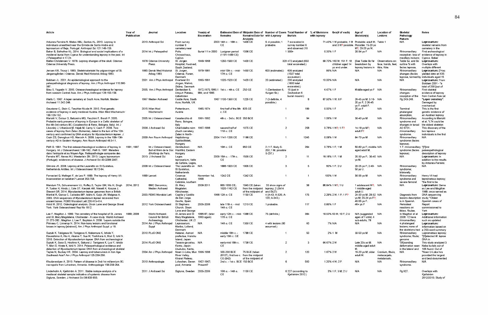
84

85
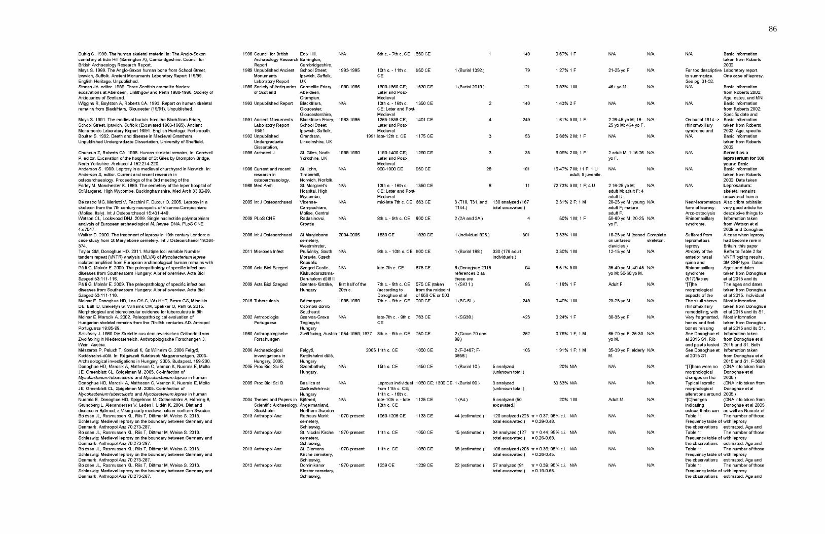
86

87

88
APPENDIX B
All References Searched
[See supplementary content in ScholarWorks.]

89
APPENDIX C
References Excluded from Data
[See supplementary content in ScholarWorks]