The Impact of Endoscopic Ultrasound - University of ... 1 The Impact of Endoscopic Ultrasound...
32
10/30/2015 1 The Impact of Endoscopic Ultrasound Truptesh H. Kothari, MD; MS Assistant Professor, Medicine Interventional Endoscopist, Strong Memorial and Highland Hospital Director, Developmental Endoscopy Lab at UR (DELUR) Division of Gastroenterology & Hepatology University of Rochester Medical Center , Rochester NY November 7, 2015 Objectives Understand and identify the role of Endoscopic Ultrasound (EUS) in diagnosis of gastrointestinal disorders. Types of EUS scopes Where is EUS helpful? Understand where EUS is used for therapy EUS‐directed therapy (FNA, fiducial, celiac plexus block) EUS complementing ERCP As an alternative to surgery and IR Multidisciplinary Cancer Care Algorithm Gardner T B et al. JOP 2010;6:288-292 Staging Mass/Tumor Imaging (CT C/A/P) Tissue/Biopsy (EUS, CT, Surgical) Multidisciplinary Tumor Approach Interventional Endoscopy • Fiducial placement • Celiac plexus neurolysis • ERCP with stenting • Enteral stenting Surgical Oncology Medical and Radiation Oncology Palliative Care Nutrition
Transcript of The Impact of Endoscopic Ultrasound - University of ... 1 The Impact of Endoscopic Ultrasound...

10/30/2015
1
The Impact of Endoscopic Ultrasound
Truptesh H. Kothari, MD; MSAssistant Professor, Medicine
Interventional Endoscopist, Strong Memorial and Highland Hospital
Director, Developmental Endoscopy Lab at UR (DELUR)
Division of Gastroenterology & Hepatology
University of Rochester Medical Center , Rochester NY
November 7, 2015
Objectives Understand and identify the role of Endoscopic
Ultrasound (EUS) in diagnosis of gastrointestinal disorders.
Types of EUS scopes
Where is EUS helpful?
Understand where EUS is used for therapy EUS‐directed therapy (FNA, fiducial, celiac plexus block)
EUS complementing ERCP
As an alternative to surgery and IR
Multidisciplinary Cancer Care Algorithm
Gardner T B et al. JOP 2010;6:288-292
Staging
Mass/Tumor
Imaging (CT C/A/P)
Tissue/Biopsy(EUS, CT, Surgical)
Multidisciplinary Tumor Approach
Interventional Endoscopy• Fiducial placement• Celiac plexus neurolysis• ERCP with stenting• Enteral stenting
Surgical Oncology
Medical and Radiation Oncology
Palliative Care
Nutrition
10/30/2015
2
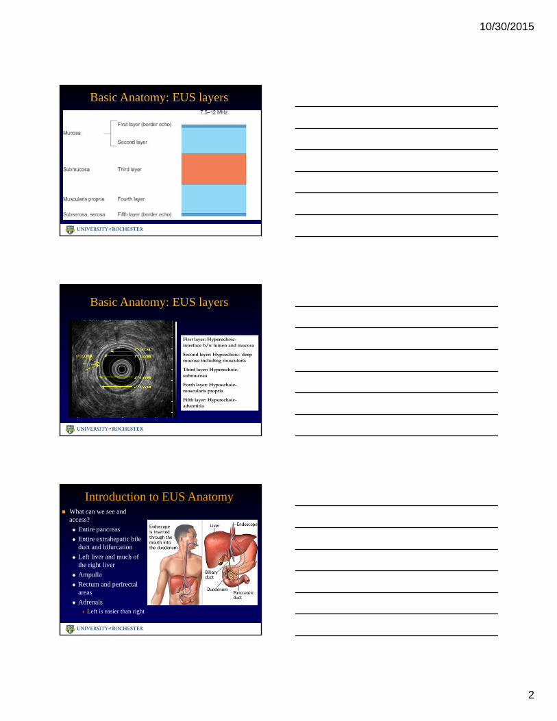
Basic Anatomy: EUS layers
First layer: Hyperechoic-interface b/w lumen and mucosa
Second layer: Hypoechoic- deep mucosa including muscularis
Third layer: Hyperechoic-submucosa
Forth layer: Hypoechoic-muscularis propria
Fifth layer: Hyperechoic-adventitia
Basic Anatomy: EUS layers
Introduction to EUS Anatomy What can we see and
access?
Entire pancreas
Entire extrahepatic bile duct and bifurcation
Left liver and much of the right liver
Ampulla
Rectum and perirectal areas
Adrenals Left is easier than right
10/30/2015
3
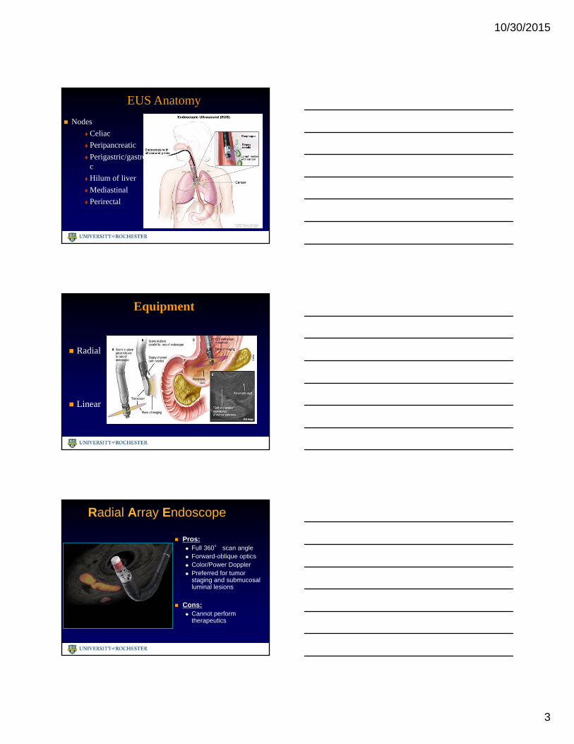
EUS Anatomy
Nodes
Celiac
Peripancreatic
Perigastric/gastrohepatic
Hilum of liver
Mediastinal
Perirectal
Equipment
Radial
Linear
Pros: Full 360° scan angle Forward-oblique optics Color/Power Doppler Preferred for tumor
staging and submucosal luminal lesions
Cons: Cannot perform
therapeutics
Radial Array Endoscope

10/30/2015
4
Doppler Images
Color Doppler ( Color Flow ) Power Doppler
Linear Echoendoscope
Pros: Scanning range: 180o
Elevator like ERCP
Can perform therapeutics/FNA
Color Doppler & Power Doppler for interpreting blood flow conditions
Cons: Not cross‐sectional
Not 360 degrees
Harder to completely visualize mucosa/submucosa
Linear Array EchoendoscopeEUS-FNA

10/30/2015
5
Mini-Probes
Pros: Surface evaluations through the
scope
Through therapeutic endoscope
Used for small or flat lesions b/c it is easier to localize lesions endoscopically
Cons: Depth of penetration and
evaluation is limited
Cannot perform therapeutics
25, 22 and 19 gauge
Disposable
Variable position locking syringe & stopcock.
FNA NEEDLE
25, 22 and 19 gauge
Disposable
Variable position locking syringe & stopcock.
Designed to obtain core tissue
Core biopsy NEEDLE

10/30/2015
6
Technique
PreparationAs for normal upper GI endoscopy
SedationFentanyl and midazolam
Propofol/GA
Antibiotic prophylaxisUsual indications + biopsy / therapeutics
Indications Staging cancers:
Esophageal, gastric, pancreatico-biliary, ampullary, rectal, lung
Confirming EMR potential T1 disease, excluding sub-mucosal involvement
Diagnosis and follow up of benign lesions Submucosal lesions and pancreatic cysts
Investigating RUQ pain and pancreatitis
Indications
Therapeutic applications: FNA Fine needle injection: celiac plexus block,
fiducial placement Pseudocyst drainage EUS guided ERCP
10/30/2015
7
Tumor Staging and Tissue acquisition
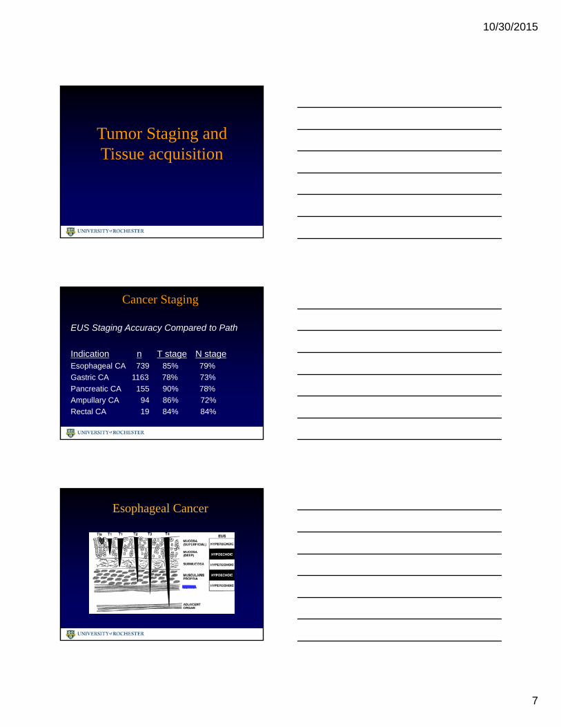
Cancer Staging
EUS Staging Accuracy Compared to Path
Indication n T stage N stageEsophageal CA 739 85% 79%
Gastric CA 1163 78% 73%
Pancreatic CA 155 90% 78%
Ampullary CA 94 86% 72%
Rectal CA 19 84% 84%
Esophageal Cancer
10/30/2015
8
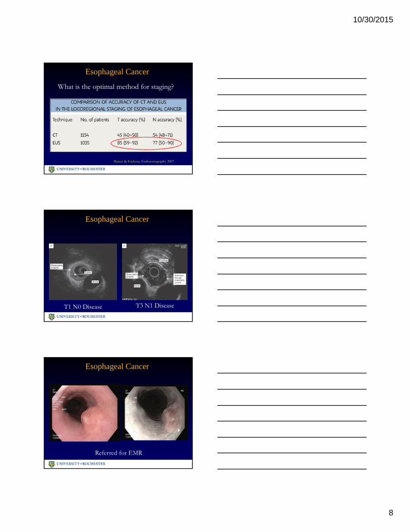
Esophageal Cancer
What is the optimal method for staging?
Hawes & Fockens, Endosonography 2007.
Esophageal Cancer
T1 N0 Disease T3 N1 Disease
Esophageal Cancer
Referred for EMR
10/30/2015
9
Esophageal Cancer
T1 N1 Disease
FNA of 7 mm periesophageal node
Esophageal Cancer
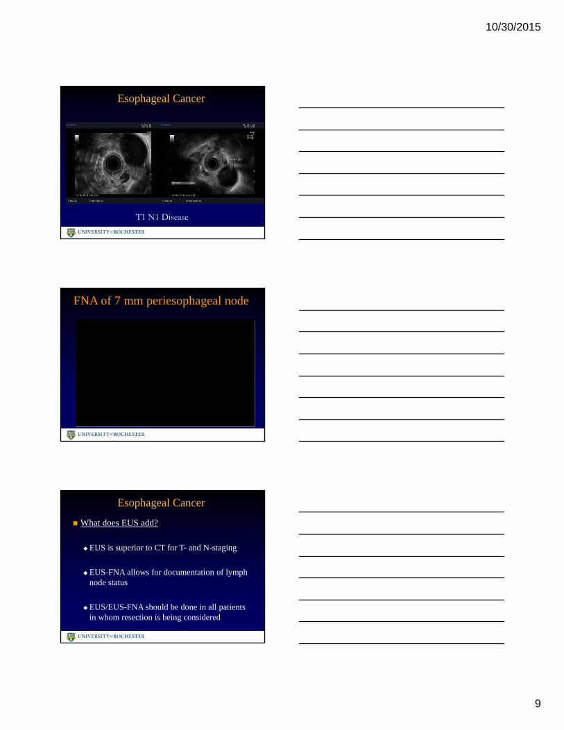
What does EUS add?
EUS is superior to CT for T- and N-staging
EUS-FNA allows for documentation of lymph node status
EUS/EUS-FNA should be done in all patients in whom resection is being considered

10/30/2015
10
Gastric disease- Gastric Cancer
EUS plays a large role in gastric cancer staging
Primary Role: Selecting tumors appropriate for EMR, superior for T staging
Secondary Role: complementing CT for N staging
Gastric disease- Gastric Cancer
T Staging of EUS vs. CT & MRIYear n EUS CT MRI
Kuntz et al., Semin SurgPollowski et al., EndoscopyBhandari et al., GIE
199920042004
828863
73%63%88%
51%44%83%
48%--
75% 59% 48%
Gastric disease- Gastric Cancer
Early Gastric Cancer
60-75% survival
Advanced Gastric Cancer
20-35% Survival
10/30/2015
11
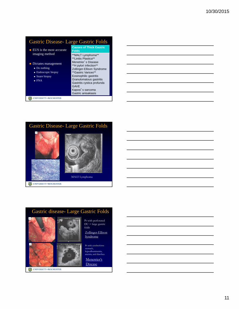
Gastric Disease- Large Gastric FoldsCauses of Thick Gastric Folds
**MALT Lymphoma****Linitis Plastica**Menetrier’s Disease**H pylori infection**Zollinger-Ellison Syndrome**Gastric Varices**Eosinophilic gastritisGranulomatous gastritisGastritis cystica profundaGAVEKaposi’s sarcomaGastric anisakiasis
EUS is the most accurate imaging method
Dictates management Do nothing
Endoscopic biopsy
Snare biopsy
FNA
Gastric Disease- Large Gastric Folds
MALT Lymphoma
Gastric disease- Large Gastric Folds
Pt with perforated DU + large gastric folds
Zollinger-Ellison Syndrome
Pt with cerebreform stomach, hypoalbuminemia, anemia, and diarrhea
Menetrier’s Disease
10/30/2015
12
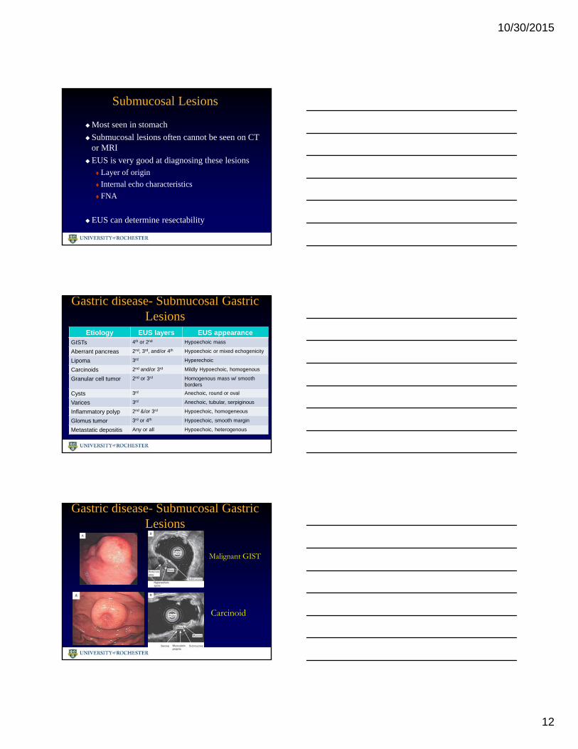
Submucosal Lesions
Most seen in stomach
Submucosal lesions often cannot be seen on CT or MRI
EUS is very good at diagnosing these lesions Layer of origin
Internal echo characteristics
FNA
EUS can determine resectability
Gastric disease- Submucosal Gastric Lesions
Etiology EUS layers EUS appearanceGISTs 4th or 2nd Hypoechoic mass
Aberrant pancreas 2nd, 3rd, and/or 4th Hypoechoic or mixed echogenicity
Lipoma 3rd Hyperechoic
Carcinoids 2nd and/or 3rd Mildly Hypoechoic, homogenous
Granular cell tumor 2nd or 3rd Homogenous mass w/ smooth borders
Cysts 3rd Anechoic, round or oval
Varices 3rd Anechoic, tubular, serpiginous
Inflammatory polyp 2nd &/or 3rd Hypoechoic, homogeneous
Glomus tumor 3rd or 4th Hypoechoic, smooth margin
Metastatic depositis Any or all Hypoechoic, heterogenous
Gastric disease- Submucosal Gastric Lesions
Malignant GIST
Carcinoid

10/30/2015
13
Gastric disease- Submucosal Gastric Lesions
Lipoma
Varices
EUS for Pancreatic Tumors
Sensitivity = 90% Specificity = 100% Accuracy = 94% For lesions as small as sub-cm Yield is enhanced with on-site
cytopathologist FNA primary tumor, LNs, & liver
lesions Evaluate for vascular invasion
Faigel et al. J Clin Onc 1997;15:1439-43Faigle et al. Diagn Cytopath 1998;18:98-109
EUS Advantages over CT-guided Biopsy
Ability to sample lesions (including lymph nodes) too small to be identified by US, CT or MRI
Minimizing the risk of needle track seeding
Ability to obtain accurate local staging

10/30/2015
14
Pancreatic mass FNA
Neuroendocrine lesions: Insulinoma
Hepatobiliary Disease-Pancreatic Cysts
Pancreatic cystic lesions once thought rare are now much more common due to MD-CT/MRI
EUS plays a critical role in differentiating benign vs. malignant lesions

10/30/2015
15
Pancreatic Cysts How can EUS make a diagnosis?
Cyst Morphology- is suggestive but not diagnostic
Solid/cystic mass • Dilated pancreatic duct
Thick wall • Intramural growth
FNA Cytology- high variability
Sensitivity 55% - 89%
Cyst fluid tumor markers-
CEA, CA 19-9, CA 72-4, CA 125 & CA 15-3
Accuracy of CEA (79%) vs. Morphology (51%) vs. Cytology (59%) Brugge et al. Gastro 2004 126:1330-36.
Pancreatic Cysts
Simple CystThin-walled, no solid
component, no debris and normal surrounding pancreas
PseudocystThin-walled anechoic cyst
Pancreatic Cysts
Serous Cystadenoma“honeycomb” appearance Multiple small, microcystsOften may have a central
calcification
Mucinous CystadenomaPapillary projections into the cyst
cavity

10/30/2015
16
Pancreatic cyst FNA
Hepatobiliary Disease-Chronic Pancreatitis
Diagnosis of chronic pancreatitis can be difficult
Abdominal Imaging (CT & MRI) for advanced disease
ERCP has risks & is best in advanced disease
EUS is highly accurate in the diagnosis of chronic pancreatitis and is relatively non-invasive
Chronic Pancreatitis
EUS Criteria for diagnosing Chronic Pancreatitis
Parenchymal Criteria
Hyperechoic FociHyperechoic StrandsHypoechoic LobulesCysts
Ductal Criteria DilatationDilated Side BranchesMain Duct IrregularityHyperechoic Duct MarginsStones

10/30/2015
17
Chronic Pancreatitis
Chronic Pancreatitis
How accurate is EUS in making the diagnosis?
126 patientsUnexplained chronic Abd pain
or suspected chronic pancreatitis
126 underwent ERCP & EUS
PPV > 85%More than 6 criteria
in moderate to severe dz
NPV > 85%Less than 3 criteria
In moderate to severe disease
Sahai et al. GIE 1998; 48:18-25.
Hepatobiliary Disease- Bile Duct Stones
EUS is an excellent non-invasive method to diagnose CBD stones
EUS sensitivity 81% - 100%
MRCP sensitivity 87% - 100%

10/30/2015
18
Bile Duct Stones
Bile Duct Stones
Consider EUS to diagnose CBD stones:
Patients with mid- to low-probability choledocholithiasis
High clinical suspicion but negative MRCP
Hospitals with poor MRCP expertise
Permanent pacemakers
Cerebral aneurysm clips
Claustrophobic patients
Morbidly obese patients
Hepatobiliary Disease- Bile Duct Tumors
Malignant CBD stricturesBrushings alone have
poor yield (desmoplastic)
EUS allows: Evaluation of stricture
Locoregional staging
FNA for diagnosisCBD
cholangiocarcinoma
10/30/2015
19
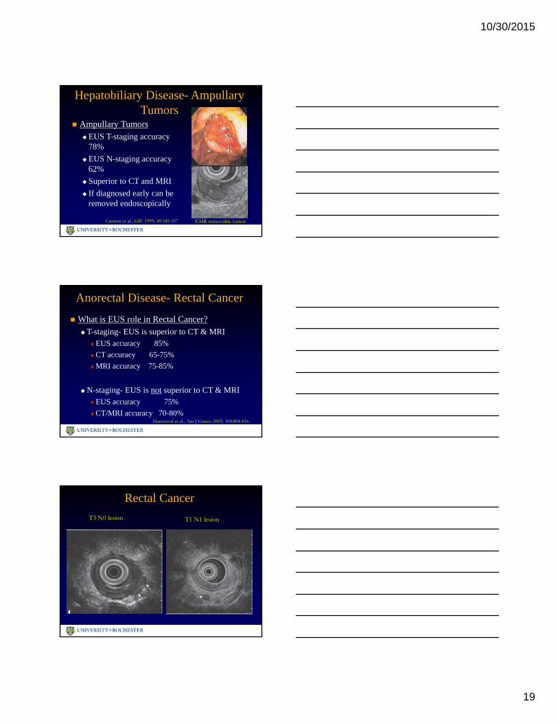
Hepatobiliary Disease- Ampullary Tumors
Ampullary TumorsEUS T-staging accuracy
78%
EUS N-staging accuracy 62%
Superior to CT and MRI
If diagnosed early can be removed endoscopically
EMR removable tumorCannon et al., GIE 1999; 49:349-357
Anorectal Disease- Rectal Cancer
What is EUS role in Rectal Cancer?T-staging- EUS is superior to CT & MRI
EUS accuracy 85%
CT accuracy 65-75%
MRI accuracy 75-85%
N-staging- EUS is not superior to CT & MRI EUS accuracy 75%
CT/MRI accuracy 70-80%Harewood et al., Am J Gastro 2005; 100:808-816.
Rectal Cancer
T3 N0 lesion T1 N1 lesion
10/30/2015
20
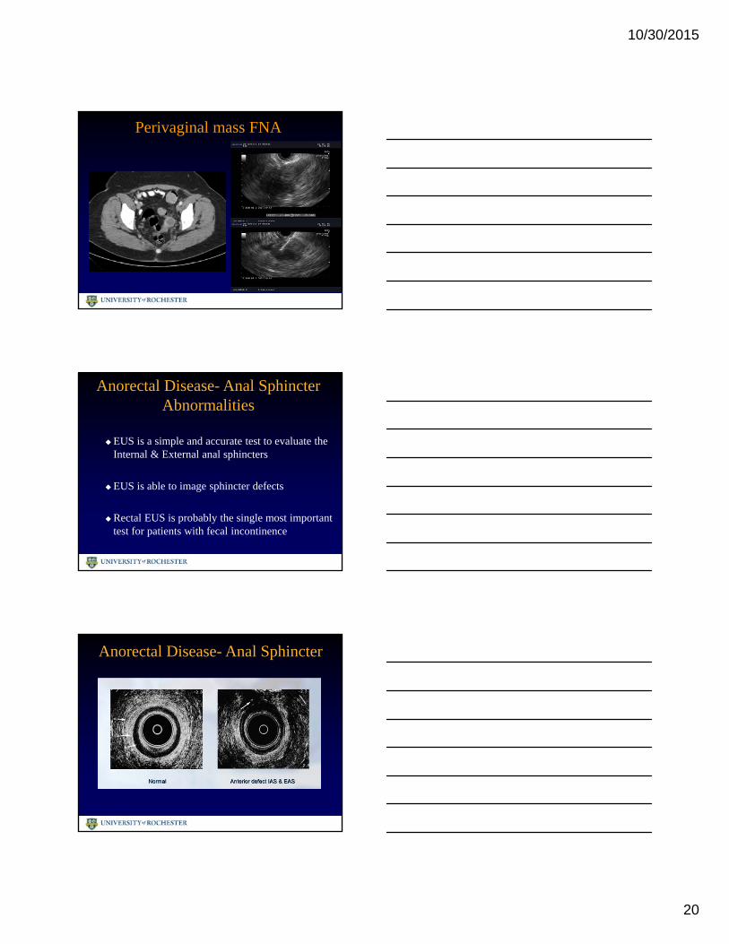
Perivaginal mass FNA
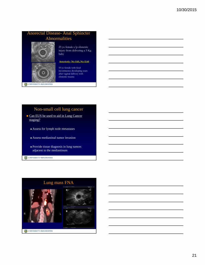
Anorectal Disease- Anal Sphincter Abnormalities
EUS is a simple and accurate test to evaluate the Internal & External anal sphincters
EUS is able to image sphincter defects
Rectal EUS is probably the single most important test for patients with fecal incontinence
Anorectal Disease- Anal Sphincter
10/30/2015
21
Anorectal Disease- Anal Sphincter Abnormalities
25 yo female s/p obstetric injury from delivering a 5 Kg baby
Anteriorly- No IAS, No EAS
55 yo female with fecal incontinence developing years after vaginal delivery with obstetric trauma
Non-small cell lung cancer Can EUS be used to aid in Lung Cancer
staging?
Assess for lymph node metastases
Assess mediastinal tumor invasion
Provide tissue diagnosis in lung tumors adjacent to the mediastinum
Lung mass FNA
10/30/2015
22
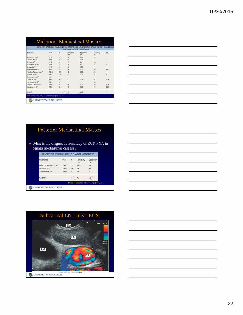
Malignant Mediastinal Masses
Hawes & Fockens, Endosonography 2007.
Posterior Mediastinal Masses
What is the diagnostic accuracy of EUS-FNA in benign mediastinal disease?
Hawes & Fockens, Endosonography 2007.
Subcarinal LN Linear EUS
10/30/2015
23
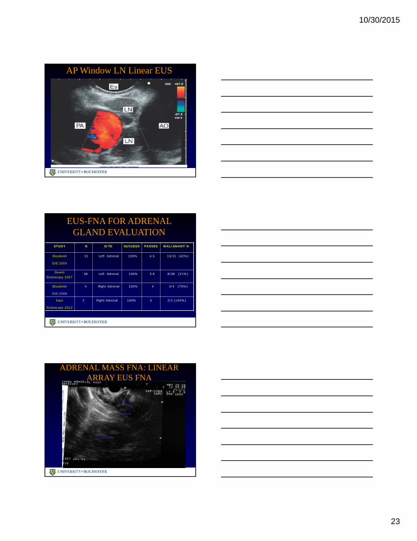
AP Window LN Linear EUS
EUS-FNA FOR ADRENAL GLAND EVALUATION
STUDY N SITE SUCCESS PASSES MALIGNANT %
Eloubeidi
GIE 2004
31 Left Adrenal 100% 4.5 13/31 (42%)
DewittEndoscopy 2007
38 Left Adrenal 100% 3.6 8/38 (21%)
Eloubeidi
GIE 2008
4 Right Adrenal 100% 4 3/4 (75%)
Kaul
Endoscopy 2012
2 Right Adrenal 100% 5 2/2 (100%)
ADRENAL MASS FNA: LINEAR ARRAY EUS FNA
Left Kidney
Adrenal

10/30/2015
24
Clinical Utility of EUS FNA for Diagnosing Liver lesions
Sensitivity of EUS-FNA for the diagnosis of malignancy range from 82 to 94%
DeWitt J et al. Am J Gastroenterol. 2003 Sep;98(9):1976-81
EUS FNA: LIVER METASTASIS
Evaluate for liver metastasis
Tissue Acquisition & Staging: Summary
EUS FNA allows access to anatomically difficult to sample areas
EUS and FNA has a high accuracy and sensitivity and specificity in tumor staging
Expands the horizons for further therapeutic interventions:
Celiac plexus block
Fiducial placement
Biliary drainage

10/30/2015
25
Therapeutic Applications of EUS
Pancreatic Pseudocyst Drainage
Indications for Intervention
Absolute indications
Symptomatic :pain, rapid enlargementGI Luminal ObstructionComplications: infection, bleeding
Traditionally drained by surgery or percutaneouslyby IR
Current standard is EUS-guided approach

10/30/2015
26
Endoscopic Cystgastrostomy
Endoscopic Cystgastrostomy
Necrosectomy

10/30/2015
27
Clash of the Titans:Endoscopy vs Surgery
Pancreatic Pseudocyst DrainageEndoscopic vs Surgical: RCT
Lower post procedural hospital stay 2.65 vs 6.5 days
Direct cost saving of $ 5,738 per case in the EUS group
In complex pseudocysts endoscopy may be employed but surgery can be considered first line in appropriate patients.
Complications: Infection (0% – 8%), bleeding (0% -5%), retroperitoneal perforation (0% - 5%)
Varadarajulu S, Lopes TL, Wilcox CM, et al. EUS versus surgical cyst-gastrostomy for management of pancreatic pseudocysts. Gastrointest Endosc 2008;68:649–55.
Pseudocyst Drainage: Summary
Endoscopic management is considered first-line therapy and is effective
Endoscopic drainage can be accomplished with minimal morbidity and does not complicate surgical approach.
Not all peripancreatic cysts are pseudocysts
Close co-operation between the TITANS……!!!!!

10/30/2015
28
EUS Fine Needle Injection:
Pain management & Fiducial placement
Celiac Plexus Block & Neurolysis
Patients with pancreatic cancer and chronic pancreatitis often have severe debilitating pain
Pain is mediated through neurons in the celiac plexus
Injection of medications into this nerve plexus can provide pain relief
Traditionally has been performed under CT guidance
EUS guided approach is now standard
EUS-Guided Celiac Plexus Block or Neurolysis
Celiac Plexus Block – injection of steroids (triamcinolone)/long acting anesthetic (bupivacaine)
Celiac Neurolyisis – injection of ethanol
EUS allows real time imaging and visualization of celiac ganglion & vascular structures
10/30/2015
29
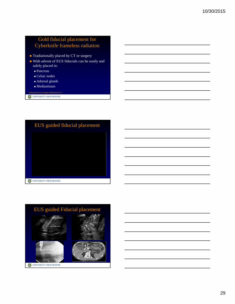
Gold fiducial placement for Cyberknife frameless radiation
Tradiationally placed by CT or surgery
With advent of EUS fiducials can be easily and safely placed in:Pancreas
Celiac nodes
Adrenal glands
Mediastinum
Pishvaian AC,et al. Endosc 2006;64(3):412–7.
EUS guided fiducial placement
EUS guided Fiducial placement

10/30/2015
30
EUS-FNI: Summary
EUS allows for safe and feasible access to celiac plexus and ganglion for neurolysis or block
Gold fiducials can be safely placed in mediastinal and abdominal malignancy with EUS access.
Highly targeted radiotherapy can be delivered
EUS Guided ERCP !
If ERCP fails, is there an alternative to PTC or surgical drainage?
Palliation of Jaundice
EUS Guided Biliary & Pancreatic drainage
ERCP fails in 3-12% of cases:
Difficult/altered anatomy
Tumor at ampulla
EUS guided rendezvous is feasible and has a pooled success rate of 83%. EUS guided PD access
EUS guided CBD accessAbu Dayyeh BK.Gastroenterol Hepatol (N Y) 2012, 8(7):450-456.
10/30/2015
31
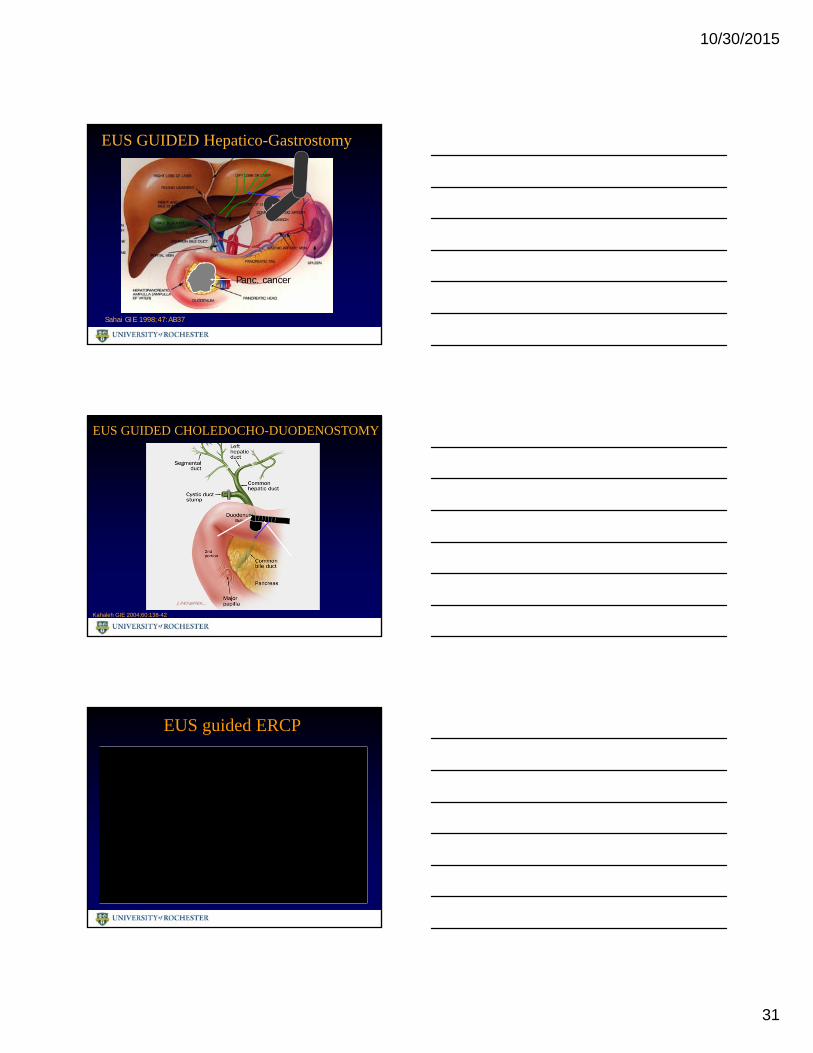
EUS GUIDED Hepatico-Gastrostomy
Sahai GIE 1998;47:AB37
Panc. cancer
EUS GUIDED CHOLEDOCHO-DUODENOSTOMY
Kahaleh GIE 2004;60:138-42
EUS guided ERCP
10/30/2015
32
Summary Interventional EUS has revolutionized medical-
surgical management
Significant shift in management paradigms
Multidisciplinary management is critical
Significant advantage in era of health care reform
Minimally invasive therapeutic EUS options continue to develop
This is just the beginning….!!!
THANK YOU!!