Endoscopic Ultrasound for the Characterization of Subepithelial Lesio
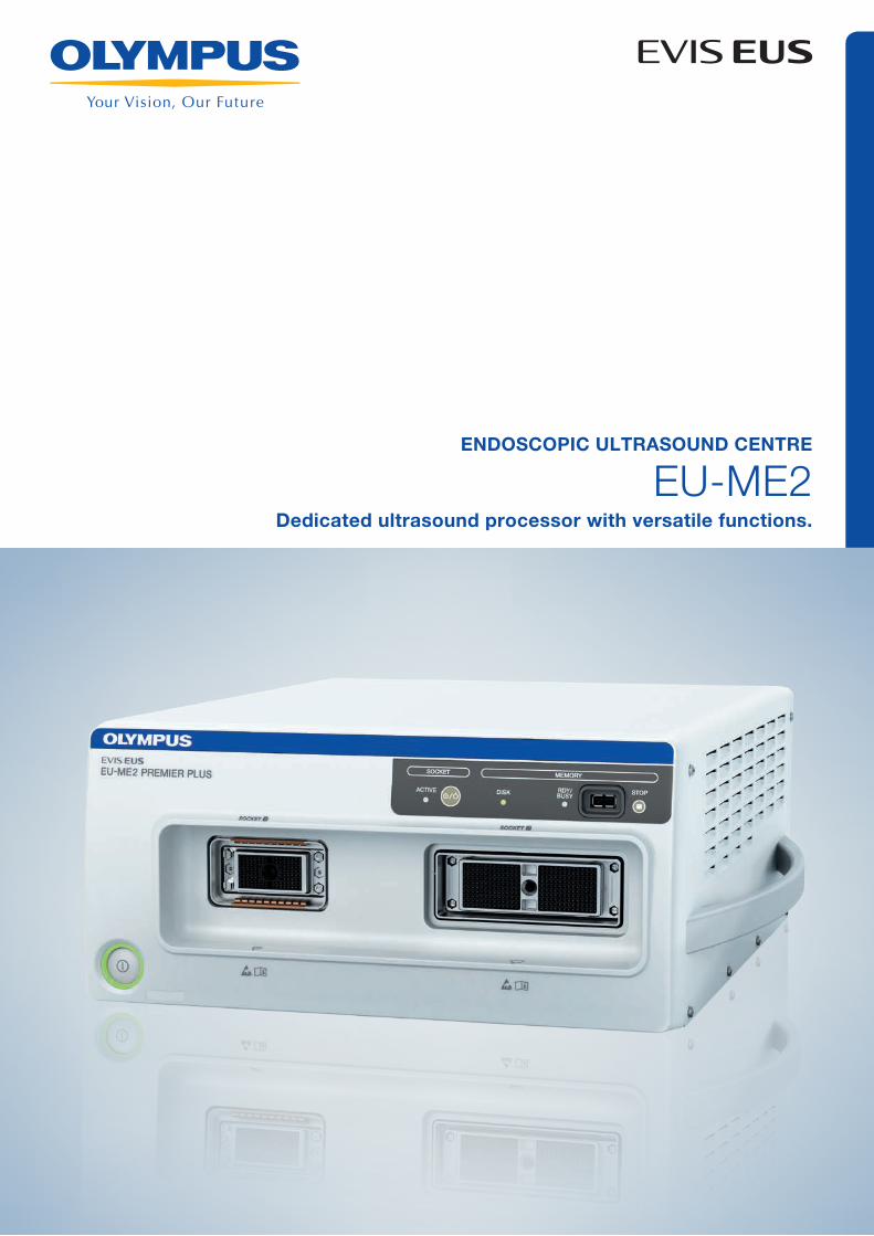
EU-ME2Dedicated ultrasound processor with versatile functions.
ENDOSCOPIC ULTRASOUND CENTRE

ENVISIONING THE FUTURE OF ENDOSONOGRAPHY
The EU-ME2 is a high-quality compact ultrasound processor for use with OLYMPUS endoscopic and
endobronchial ultrasound equipment that has been designed for integration with conventional endoscopy on a
single workstation. With its high resolution and an image display that promotes clear visualisation, the EU-ME2
brings real clarity to your EUS procedures, supporting better detection and characterisation of lesions. A variety
of new features such as harmonic echo and Elastography help to explore the future of endosonography.
Excellent
Improved basic functions ensure
excellent ultrasound imaging.
Unique
New functions offer unique new
possibilities in endosonography.
Specific
Designed specifically to optimise
endosonographic procedures.
Excellent Specific
Unique
EU-ME2

EXCELLENT – IMPROVED BASIC FUNCTIONS ENSURE EXCELLENT ULTRASOUND IMAGING
B-mode
B-mode image quality has been substantially improved, making it possible to support more efficient
localisation of tumours and more accurate identification of tissue properties and boundaries. Clearer image delineation
helps enable more precise orientation for puncturing and aspiration during EUS-FNA and may make it easier to develop
effective therapeutic practices.
Electronic radial scanning
EBUS-TBNA
Electronic curved linear array scanning
UNIQUE – NEW FUNCTIONS OFFER UNIQUE NEW POSSIBILITIES IN ENDOSONOGRAPHY
Tissue Harmonic Echo (THE)
When ultrasound waves are propagated through tissue,
distortion occurs and higher harmonic components are
generated. The THE mode uses these components to build
an image of the targeted area. Potential advantages of
harmonic imaging include improved resolution, an improved
signal-to-noise ratio and fewer artefacts.
Pulse Wave Doppler
Pulse Wave Doppler measures blood
flow velocities at specific locations,
while cross-sectional images are
viewed to spot the target vessel.
H-FLOW
Especially useful for imaging
small vessels around the tip of the
endoscope, the H-FLOW (High
Resolution Flow) mode can help facilitate
more precise manoeuvring during
EUS-FNA or EBUS-TBNA by making it
potentially easier to avoid vessels.
Contrast Harmonic EUS (CH-EUS)
Using technology designed to depict
higher harmonics, the CH-EUS mode
is expected to help realise enhanced
sensitivity to tumours and other
abnormal growths.
Note: Regulations and usage of ultrasound contrast agents vary according to the country where they are used and the type of agents. Please use the ultrasound contrast medium according to the instructions provided with the products.
Elastography
An advanced form of ultrasound, elastography
displays the relative stiffness of tissues by taking
advantage of the deformation caused by the compression
or vibrations generated by the heartbeat or vascular
pulsations.
THE-P (radial)
THE-R (radial)
THE-P (linear)
THE-R (radial)
ELASTOGRAPHY (linear)
ELASTOGRAPHY (radial)

SPECIFIC – DESIGNED SPECIFICALLY TO OPTIMISE ENDOSONOGRAPHIC PROCEDURES
Move the ultrasound probe within the guide sheath, back and forth observing the ultrasound image to assess the lesion.
Fully compatible with a wide range of EUS and EBUS scopes and probes
Integrating both electronic and mechanical scanning technologies, the EU-ME2 is a total endosonography solution
compatible with virtually all available OLYMPUS ultrasound endoscopes and miniature probes, providing access to a
full range of endosonographic applications.
Single monitor and single keyboard
The EU-ME2 features a user-friendly keyboard with a
touch panel and trackball. The picture-in-picture function
is standard, and when available, both endoscopy and
ultrasound images can be displayed on a single monitor.
EVIS-ready and space-saving design
The EU-ME2 is designed to save space in your endoscopy
suite. As an integral part of the OLYMPUS EVIS endoscopy
system, it fits snugly on the standard endoscopy trolley,
leaving plenty of room for all the other equipment you need.
Full support for endobronchial ultrasonography
The EU-ME2 is designed to support a wide range of
EBUS procedures, such as the EBUS GuideSheath
procedure. By placing the GuideSheath with the inserted
miniature probe near the target lesion, the probe can be
withdrawn and forceps or a brush can be conveniently
advanced to the site of the lesion for further sampling.
Advancing the sampling device through the sheath after
the miniature probe has been withdrawn helps to
improve accuracy and shorten the examination time.
Mechanical radial endoscopes
Ultrasound miniature probes
Electronic radial endoscopes
EUS curved linear array endoscopes
EBUS curved linear array endoscopes
CLINICAL CASES
See some of what you can do with the EU-ME2 using various types of ultrasound endoscopes and probes. With the excellent
performance made possible by improved functions, the expanded possibilities offered by new functions, and the efficiency of the
endosonography-specific design, the EU-ME2 will help you envision the future of endosonography.
With a curved linear array ultrasound endoscope
THE-P mode
ELST (elastography) mode
POWER FLOW mode
With an electronic radial ultrasound endoscope
COLOR FLOW mode THE-R mode
ELST (elastography) mode
EBUS
B-mode H-FLOW mode
ELST (elastography) mode
COLOR FLOW mode H-FLOW mode

SpecificationsPower supply Voltage 100–240 V AC (for NTSC), 220–240 V AC (for PAL)
Voltage fluctuation Within ±10%Frequency 50/60 HzFrequency fluctuation Within ±1 HzConsumption (electric power) 370 VA
Size Dimensions Main unit 371 (W) × 175 (H) × 480 (D) mm445 (W) × 184 (H) × 495 (D) mm (max.)
Keyboard 392 (W) × 39 (H) × 207 (D) mmWeight Main unit 22.5 kg
Keyboard 2.5 kg
Classification Type of protection against electric shock
Class I
Degree of protection against electric shock of applied part
TYPE BF applied part Where no classification mark appears, the device is a TYPE BF applied part
Degree of protection against explosion
The ultrasound centre should be kept away from flammable gases
TYPE BF applied part This instrument can safely be applied to any part of the body except the heart
EMC This instrument complies with the standards listed as follows: IEC 60601-1-2: 2001, IEC 60601-2-37: 2007CISPR 11 of emission: Group 1, Class B
Ultrasound scanning format Mechanical scanning, electronic scanning
Mechanical scanning Display mode B-modeScanning Radial scanningCompatible equipment Mechanical radial scanning ultrasound endoscope, miniature probeUsable frequencies C5, C7.5, C12, C20, 7.5, 12, 20 MHz
Display range 2, 3, 4, 6, 9, 12 cm
Image adjustment Gain, contrast, STC, enhance
Display processing Rotation Rotatable (64 steps, clockwise/counterclockwise)
Display area Full circle, bottom sector, top sector, scroll
Direction Normal/inverse
Cine memory Maximum 160 frames, Cine review function
3D 3D display, MPR display
Measurement Distance, area, circumstance
Electronic scanning Display mode B-mode, FLOW mode, PW mode, THE mode, CH-EUS mode, elastography mode
Scanning Radial scanning, curved linear array scanning
Compatible equipment Electronic radial scanning ultrasound endoscope, Electronic curved linear array scanning ultrasound endoscope
Usable frequencies 5, 6, 7.5, 10, 12 MHz
Display range 2, 3, 4, 5, 6, 7, 8, 9, 12 cm
Image adjustment Gain, contrast, STC, enhance, compound
Display processing Display area Radial: Full circle, bottom sector, top sector, scroll Curved linear array: convex
Direction Normal/inverse
Display pattern Single-screen/dual-screen
Cine memory Over 600 frames can be stored depending on the conditions Cine review function
Focus Auto preset Near/far
Focus setting Focus location adjustable, focus number adjustable
FLOW mode COLOR FLOW mode, POWER FLOW mode, H-FLOW mode
PW mode B+PW, Color+PW, Power+PW, H-Flow+PW
Measurement Distance, area, circumstance, PW measurement
THE (Tissue Harmonic Echo) mode *1, *2
THE-P, THE-R
CH-EUS mode *1, *2 Display pattern CH-B, CH-Color
Preset (CH agent type)
2 types, adjustable (middle or low)
Frequency selection 2 types, adjustable (CH-R or CH-P)
TIC analysis Displays the change over time of the average brightness of each ROI
ELST mode (elastography) *2 Pressurisation state guide
Strain graph, pressurisation bar
Strain ratio Displays the amounts of the strain and their ratio in two areas
Recording data Data format Still image BMP, JPEG, 3DV
Movie data *1, *2 AVI
Ancillary equipment Keyboard Keyboard with built-in trackball, LCD touch panel and LED backlit keys
Recording device Video printer (colour/monochrome), DVR
Video system centre Monitor display selection
Endoscopic/ultrasound image
Picture-In-Picture Displays the endoscopic image as PIP sub-display on the ultrasound image
Patient data Shares patient data with the video system centre
*1 Only available on EU-ME2 PREMIER/EU-ME2 PREMIER PLUS *2 Only available on EU-ME2 PREMIER PLUS
EU-ME2
E04
8278
1 · x
.000
· 11
/13
· PR
EU-ME2 PREMIER PLUS
Specifications, design and accessories are subject to change without any notice or obligation on the part of the manufacturer.
Postbox 10 49 08, 20034 Hamburg, GermanyWendenstrasse 14–18, 20097 Hamburg, GermanyPhone: +49 (0)40 237 730, Fax: +49 (0)40 230 761www.olympus-europa.com