Endoscopic ultrasound-guided transmural drainage for ... · Endoscopic ultrasound-guided transmural...
26
Instructions for use Title Endoscopic ultrasound-guided transmural drainage for pancreatic fistula or pancreatic duct dilation after pancreatic surgery Author(s) Onodera, Manabu; Kawakami, Hiroshi; Kuwatani, Masaki; Kudo, Taiki; Haba, Shin; Abe, Yoko; Kawahata, Shuhei; Eto, Kazunori; Nasu, Yuya; Tanaka, Eiichi; Hirano, Satoshi; Asaka, Masahiro Citation Surgical Endoscopy, 26(6), 1710-1717 https://doi.org/10.1007/s00464-011-2097-z Issue Date 2012-06 Doc URL http://hdl.handle.net/2115/52926 Rights The original publication is available at www.springerlink.com Type article (author version) File Information SE26-6_1710-1717.pdf Hokkaido University Collection of Scholarly and Academic Papers : HUSCAP
Transcript of Endoscopic ultrasound-guided transmural drainage for ... · Endoscopic ultrasound-guided transmural...
Instructions for use
Title Endoscopic ultrasound-guided transmural drainage for pancreatic fistula or pancreatic duct dilation after pancreaticsurgery
Author(s) Onodera, Manabu; Kawakami, Hiroshi; Kuwatani, Masaki; Kudo, Taiki; Haba, Shin; Abe, Yoko; Kawahata, Shuhei;Eto, Kazunori; Nasu, Yuya; Tanaka, Eiichi; Hirano, Satoshi; Asaka, Masahiro
Citation Surgical Endoscopy, 26(6), 1710-1717https://doi.org/10.1007/s00464-011-2097-z
Issue Date 2012-06
Doc URL http://hdl.handle.net/2115/52926
Rights The original publication is available at www.springerlink.com
Type article (author version)
File Information SE26-6_1710-1717.pdf
Hokkaido University Collection of Scholarly and Academic Papers : HUSCAP
Endoscopic ultrasound-guided transmural drainage for pancreatic fistula or pancreatic
duct dilatation after pancreatic surgery
Manabu Onodera 1)
, Hiroshi Kawakami 1)
, Masaki Kuwatani 1)
, Taiki Kudo 1)
, Shin Haba 1)
,
Yoko Abe 1)
, Shuhei Kawahata 1)
, Kazunori Eto 1)
, Yuya Nasu 2)
, Eiichi Tanaka 2)
, Satoshi
Hirano 2)
, and Masahiro Asaka 1)
1) Department of Gastroenterology, Hokkaido University Graduate School of Medicine
2) Department of Surgical Oncology, Hokkaido University Graduate School of Medicine
Author contributions: Onodera M and Kawakami H contributed equally to this work
Address correspondence to: Hiroshi Kawakami, MD, PhD.
Department of Gastroenterology, Hokkaido University Graduate School of Medicine, Sapporo,
060-8638, Japan
Tel: +81-11-716-1161
Fax: +81-11-706-7867 (ext.5918)
E-mail: [email protected]
Running title: EUS-TD of pancreatic fistula
Key words: EUS-guided drainage, EUS-guided transmural drainage, pancreatic fistula, stasis
of pancreatic juice, pancreatic surgery

Abstract
Background Endoscopic ultrasound (EUS)-guided drainage is widely used to manage
pancreatic pseudocysts. Several studies have reported the use of EUS-guided drainage for
pancreatic fistula and stasis of pancreatic juice caused by stricture of the pancreatic duct after
pancreatic resection.
Methods In total, 262 patients underwent surgery involving pancreatic resection in our hospital
from April 2005 to March 2010. Ninety patients (34%) developed a grade B or C postoperative
pancreatic fistula (POPF) that required additional treatment. We performed EUS-guided
transmural drainage (EUS-TD) for six patients (2.1%) with a pancreatic fistula or dilatation of
the main pancreatic duct visible by EUS. Eighteen patients (6.8%) received percutaneous
drainage. The success rates of EUS-TD and percutaneous drainage were compared in a
retrospective analysis.
Results EUS-TD was successfully performed without complications in all six cases, with five
of the six patients being successfully treated with only one trial of EUS-TD. The final technical
success rate was 100% for EUS-TD and for percutaneous drainage. Both the short-term and
long-term clinical success rates of EUS-TD were 100%, and those of percutaneous drainage
were 61.1% and 83%, respectively. The differences in these rates were not significant
(short-term success, P = 0.091; long-term success, P = 0.403). However, the period to clinical
success was significantly shorter with EUS-TD (5.8 days) than with percutaneous drainage
(30.4 days; P = 0.0013) in our series.
Conclusions EUS-TD appears to be a safe and technically feasible alternative to percutaneous
drainage and may be considered a first-line therapy for pancreatic fistulas visible by EUS.

Abbreviations:
CT, computed tomography; EUS, endoscopic ultrasound; EUS-TD, EUS-guided transmural
drainage; MPD, main pancreatic duct; POPF, postoperative pancreatic fistula; US, ultrasound;
DPDS, Disconnected pancreatic duct syndrome

Introduction
Postoperative pancreatic fistula (POPF), a serious and potentially lethal complication, is the
main unsolved problem in pancreatic surgery. The principle underlying this treatment is
drainage of the pancreatic fluid that has collected within the abdominal cavity. The surgeon
usually places the drainage tube near the pancreatic stump or the anastomotic site of
pancreatoenterostomy to drain the collected fluid in cases of leakage (pancreatic fistula).
However, additional drainage may be needed when the drainage work during the operation is
inadequate. Despite the expansive growth pattern of pancreatic pseudocysts followed by acute
or chronic pancreatitis, the POPF space extends to the peritoneal cavity between visceral
organs. This space is usually located deep in the peritoneal cavity and it has an irregular and
complex shape. These caused treatment with both surgical and interventional ultrasound (US)
or computed tomography (CT)-guided drainage procedures to be problematic. The incidence
of postoperative pancreatic fistula (POPF) has been reported to be 5–20% after
pancreaticoduodenectomy and is associated with high mortality [1]. The Japanese Society of
Hepato-Biliary-Pancreatic Surgery reported that the rate of POPF after
pancreaticoduodenectomy was 30.2% [1]. Even with recent advances in pancreatic surgery,
POPF remains the major contributor to morbidity after pancreatic resection.
The treatment of choice for POPF is external or internal drainage of pancreatic juice, which
can be accomplished by surgical drainage, image-guided percutaneous drainage, or endoscopic
drainage. A surgical cyst-enteric anastomosis has traditionally been performed, with high
morbidity rates ranging from 7 to 37% [2, 3]. Percutaneous drainage offers satisfying results,
but it requires a long therapy period (several weeks to months) owing to the indwelling catheter
and is occasionally associated with the formation of an external fistula [4, 5]. Furthermore,

cutaneopancreatic fistulas that form after percutaneous drainage are usually intractable.
Endoscopic drainage has been performed increasingly during the last 10 years [6–8].
Two approaches have been used for endoscopic access: through the gastrointestinal wall by
creating a cystogastrostomy/cystoduodenostomy (transmural drainage) or via the papilla
(transpapillary drainage). Both approaches, which usually require the insertion of one or more
plastic stents [9], have success rates of 70–87% and complication rates of 11–34% [10, 11].
Recently, endoscopic ultrasound (EUS)-guided transmural drainage (EUS-TD) has
been widely used to manage pancreatic pseudocysts, and its applications have been extended
gradually with the development of more innovative techniques and devices. Several reports
have indicated the feasibility of EUS-guided drainage of pancreatic fistulae and stasis of
pancreatic juice caused by a pancreatic duct stricture after pancreatic resection [12, 13].
However, no detailed reports are available comparing EUS-TD and percutaneous drainage of a
pancreatic fistula after pancreatic resection. To examine the utility and efficacy of EUS-TD, we
report six consecutive successful pancreatic fistula and pancreatic duct dilatation cases treated
by EUS-TD through the gastrointestinal tract and compare the outcomes with those of cases
treated by conventional percutaneous drainage.
Patients and methods
In total, 262 patients underwent surgery involving pancreatic resection at our hospital from
April, 2005 to March, 2010. A total of 172 patients (65%) did not have POPF and did not
undergo additional treatment. Ninety patients (34%) developed POPF and required additional
interventional treatment because they had symptoms such as fever and abdominal pain.
Sixty-six patients have been cured by conservative therapies, while 24 have not since a convex
array echo endoscope was introduced to our institute in April 2006. When POPFs were visible
by EUS, EUS-TD was the first choice for their treatment. Percutaneous drainage was the
second choice for treatment of refractory POPFs with symptoms or if they were not visible by
EUS. As a result, POPFs in five patients (2.1%) and dilatation of the main pancreatic duct
(MPD) caused by a stricture of the anastomosis in one, were detected by EUS and treated by
EUS-TD, and patients with a POPF not visible by EUS were treated with percutaneous
drainage (18 cases, 6.8%) or with a surgical drain followed by primary surgery (66 cases, 25%)
for the duration above.
In the six patients treated with EUS-TD (Table 1), initial surgical indications were
pancreatic carcinoma in three, bile duct carcinoma in two, and ampullary carcinoma in one.
Pancreatoduodenectomy had been performed in three cases; hepatopancreatoduodenectomy, in
two; and distal pancreatectomy, in one. After surgery, five patients (cases 1–5) developed a
POPF, and one (case 6) developed dilatation of the MPD. Three patients (cases 1, 2, and 6)
experienced fever, abdominal pain, or general fatigue as signs of a pancreatic fistula infection
or stasis of pancreatic juice.
Of the 18 POPF patients treated with percutaneous drainage (Table 1), seven with a
clinical diagnosis of bile duct carcinoma, three with pancreatic carcinoma, two with intraductal
papillary mucinous neoplasm, and one each with ampullary carcinoma, gallbladder cancer, and
mucinous cystic neoplasm had initially undergone pancreaticoduodenectomy or distal
pancreatectomy. Hepatopancreatoduodenectomy had been performed in three patients
diagnosed with bile duct carcinoma (Fig. 1). Data were collected from our endoscopic database
and hospital records. Figure 1 shows a flow diagram of the study participants.

Endoscopic therapy
All EUS-guided procedures were performed with the patient in the lateral or prone
position and under conscious sedation with midazolam and fentanyl. A convex array echo
endoscope (GF-UCT240; Olympus Medical Systems, Tokyo, Japan), an ultrasound system
(ProSound SSD-5000; ALOKA, Tokyo, Japan), and a fluoroscope (CUREVISTA; Hitachi
Medical Corp., Tokyo, Japan) were used for all procedures. All POPF or dilatation of MPD
were visible by EUS, and all punctures were performed through the stomach wall. With regard
to EUS-TD of POPF, first, an internal fistula was created between the POPF and the stomach
by introducing a 19-gauge needle (Echo Tip Ultra; Cook Endoscopy, Osaka, Japan). Successful
POPF access was demonstrated by injection of contrast medium defining a POPF. Next, a
0.035-inch guidewire (Jagwire; Boston Scientific, Tokyo, Japan) was passed through the
needle, introduced into the cavity of the POPF, and coiled within the POPF under fluoroscopic
guidance. The tract was sequentially dilated with an over-the-wire biliary balloon dilator (6
mm, Hurricane RX; Boston Scientific), a Soehendra biliary dilatation catheter (SBDC-6, -7,
-8.5; Cook Endoscopy), or a Soehendra stent retriever (7 Fr; Cook Endoscopy). After dilatation,
a double-pigtail stent (7 Fr, 40 mm; Olympus Medical Systems) was deployed into the
fluid-filled space. Regarding EUS-TD of the dilated MPD, after the pancreatic duct was
identified and regional recurrence was confirmed by color Doppler imaging, the MPD was
punctured with a 19-gauge needle (Echo Tip Ultra; Cook Endoscopy), and an internal fistula
was created between the MPD and the stomach as the initial step. Successful pancreatic duct
access was demonstrated by injection of contrast medium defining a pancreatogram. Next, a
0.035-inch guidewire (Jagwire; Boston Scientific) was passed through the needle and
introduced into the cavity of the MPD under fluoroscopic guidance. The tract was sequentially

dilated with a Soehendra dilatation catheter (SBDC-6; Cook Endoscopy). After dilatation, a
pancreatic stent (5 Fr, 50 mm; Cook Endoscopy) was deployed into the MPD. Representative
images of EUS-TD for POPF associated with the treatment of case 1 are shown in Fig. 2.
Percutaneous drainage of pancreatic fistulas
Percutaneous drainage was performed by an interventional radiologist and a surgeon, who
punctured the pancreatic fistula by inserting a 0.035-inch guidewire into the fistula space and
then placed an indwelling drainage catheter (7.2 Fr strait catheter; O-HIRATATM
, Medico’s
Hirata, Osaka, Japan, or 8.5 Fr pigtail catheter; UltrathaneTM
, Cook Endoscopy) under
fluoroscopy, US, or CT guidance.
Technical and clinical success of EUS-TD and percutaneous drainage
Technical success of EUS-TD was defined as the successful placement of a transmural stent or
percutaneous catheter into the POPF. Short-term clinical success was defined as a 50%
decrease in the POPF or an improvement in the dilated MPD on CT images, and a decrease in
serum amylase level in the drainage fluid 1 week after treatment. Long-term clinical success
was defined as the resolution of the POPF or improvement of the dilated MPD on CT images 1
month after treatment.
Statistical analysis
Non-normally distributed variables are expressed as medians (range), and nonparametric tests
(Mann-Whitney U test) were employed for statistical comparisons. Nominal variables are
expressed as frequencies and percentages. The χ2 test with Yates continuity correction for the

two-way contingency table was used for statistical comparisons. Fisher’s exact test was used in
the case of a small expected frequency. Data were considered significant at a P value <0.05.
Results
The mean period between the onset of POPF and endoscopic therapy was 7.1 months (range,
1–24 months). EUS-TD was successfully performed without complications in all six cases
(rate of technical success, 100%) (Table 2). The size of the fistula decreased remarkably
(average reduction rate, 81.2%; range, 46.8–100%) in the five POPF patients. The MPD
diameter in case 6 improved after MPD stenting. In all six cases, the serum amylase level was
lower after drainage (before drainage: 413 U/L; 1 week after drainage: 65 U/L), reflecting
decreased pancreatic duct pressure. One patient (case 1) developed a growth in the cavity by
occlusion of the indwelling stent 2 months after the first drainage; this case underwent a stent
exchange using a conventional endoscopic approach. No recurrences (mean follow-up, 7.5
months; range, 1–27 months) were observed in the remaining five cases. Table 3 compares the
characteristics of the patient treated by EUS-TD and percutaneous drainage. EUS-TD and
percutaneous drainage each had a final technical success rate of 100%. The short- and
long-term clinical success rates of EUS-TD were both 100%, and those of percutaneous
drainage were 61.1% and 83%, respectively. These rates did not differ significantly between
techniques (short-term clinical success rate, P = 0.091; long-term clinical success rate, P =
0.403). However, the time until clinical success was significantly shorter with EUS-TD (5.8
days) than with percutaneous drainage (30.4 days; P = 0.0013).
Follow-up

After discharge, all patients were followed-up by laboratory investigations and CT. Transmural
stents were not retrieved, even after fluid collection was completed. Endoscopic treatment was
repeated in the event of stent occlusion. No POPF recurrence was observed during the
follow-up time (mean follow-up, 17.7 months; range, 1–47 months).
Discussion
A pancreatic fistula is as an abnormal communication between the pancreatic duct and another
non-epithelized cavity containing pancreas-derived, enzyme-rich fluid [3, 12], which occurs
mainly after pancreatic resection and trauma. Pancreatic juice may spread throughout the
pleura or peritoneum and induce severe inflammation and autodigestion of peripancreatic and
retroperitoneal tissues. A pancreatic pseudocyst, which develops after acute pancreatitis or
accompanying chronic pancreatitis, is a localized collection of fluid rich in amylase and other
pancreatic enzymes and enclosed by a non-epithelized, hard and thick wall [3, 12]. However,
differentiating a POPF from a pancreatic pseudocyst is difficult in some situations. A POPF
represents failure of healing and/or sealing of a pancreatic-enteric anastomosis and a leak of
pancreatic juice into the peritoneal or retroperitoneal cavity.
POPF is treatable by endoscopic, surgical, and/or percutaneous drainage. Endoscopic
transpapillary drainage of the MPD for a pancreatic fistula communicating with the MPD is a
relatively easy, well established, first-step for patients with POPFs [14, 15]. However, the
transpapillary approach can occasionally be difficult owing to distortion of the duodenum
caused by postoperative changes. When distal pancreatectomy is performed, a transpapillary
approach should be considered. However, we did not undertake any endoscopic transpapillary
approaches during the period of this retrospective study. Of the six cases, cases 1–3, 5 and 6

underwent pancreaticoduodenectomy or hepatopancreatoduodenectomy. Although only case 4
underwent a distal pancretectomy, we performed EUS-TD because POPF were readily
visualized by EUS. EUS-TD has been successfully performed for POPFs associated with a
complete MPD rupture caused by trauma, pancreatoenteric anastomotic breakdown, and
occlusion of a pancreatoenteric anastomosis after pancreatic surgery [13, 16, 17]. The present
report demonstrates that drainage of pancreatic fistulas and MPD by EUS-TD through the
gastrointestinal tract was effective and safe in all six cases. EUS-TD allows access to relatively
small and non-bulging pancreatic fistulas and provides visualization of the blood vessels to
reduce the risk of bleeding [18, 19]. EUS-TD also permits internal drainage of pancreatic fluid
into the gut, and direct fistula formation between the POPF space and the gut can facilitate
POPF closure in one step. In contrast, percutaneous drainage may lead to the formation of a
permanent external fistula in 5–10% of cases [4]. Percutaneous drainage also sometimes
requires two or more procedures in a multistep conversion from external to internal drainage
and necessitates a longer hospital stay compared with EUS-TD. Disconnected pancreatic duct
syndrome (DPDS) is an anatomic situation defined by a lack of ductal continuity between
viable secreting pancreatic tissue and the gastrointestinal tract. The visible upstream pancreas
continues secretion without an intact drainage system. The physiologic abnormality of DPDS
results in myriad complications, including pancreatic pseudocysts, pancreatic ascites,
pancreatic fistula, and others [20]. In our study, we believed that differences in the mechanism
of POPF were caused by the methods of surgical anastomosis used. The patients with
pancreatojejunostomy would demonstrate POPFs caused by anastomotic breakdown (case
1–3,5) or complete rupture (case 4), whereas a case with pancratogastrostomy would develop
stasis of the pancreatic juice caused by occlusion of the anastomosis (case 6). EUS-TD to the

dilatation of the MPD was successful in our study. However, Kahaleh et al. [21] reported that
EUS-TD of the MPD dilatation failed in 28.5% of postsurgical patients. Therefore,
EUS-guided pancreatic duct drainage remains difficult, especially in the postoperative
situation. If recurrence develops after EUS-TD of the dilated MPD, surgery should be
considered, despite its higher morbidity and mortality than nonsurgical approaches [22].
Only one patient (case 1) developed a relapse of pancreatic fluid collection due to
stent occlusion and underwent a second EUS-TD for a stent exchange. Although several
reports have indicated the recurrence of pancreatic fluid accumulation following stent removal
after endoscopic transmural drainage, in most patients with a pancreatic fistula who undergo
EUS-TD, stent removal is performed routinely within 2 weeks of fistula resolution on imaging
[6, 23]. The recurrence rate of a pancreatic fistula that requires further endoscopic, surgical, or
percutaneous drainage varies from 10% to 30%, and recurrence often occurs within 1 year after
treatment [6, 7, 10]. A previous randomized controlled trial regarding stent removal after
successful endoscopic transmural drainage of a pancreatic fistula caused by acute pancreatitis
or chronic pancreatitis showed that stent retrieval was associated with a higher recurrence rate
[24]. In this study, during a median follow-up period of 14 months, no patients in the persistent
stent placement group had POPF recurrence, whereas five of the 13 patients (38.4%) in the
stent retrieval group did have POPF recurrence. This result suggests that by maintaining the
stent in place, the cystenterostomy tract may remain patent. This patency could be caused by
the stent, which acts as a wick and maintains communication between the pancreatic fistula and
the stomach. Prolonged transmural stent placement has been adapted as a strategy to prevent
recurrence of POPF [24, 25]. Although Varadarajulu et al. pointed out the risk of stent
migration, they reported placement of a permanent indwelling stent by EUS-TD [26].

Furthermore, the appropriate period for stent placement and optimal stent diameter are
controversial and yet to be determined. Nevertheless, we consider that a stent remaining in its
proper position reduces the recurrence rate.
In our study, the period to clinical success was significantly shorter with EUS-TD than
with percutaneous drainage. In two previous studies [27, 28], a higher failure rate was
associated with percutaneous drainage of pancreatic pseudocysts (58%) compared with
surgical treatment (7%) [27], and surgical and endoscopic interventions for pancreatic
pseudocysts were equally safe and effective compared with percutaneous drainage [28]. Thus,
EUS-TD can be considered a first-line therapy for a pancreatic fistula detectable by EUS when
conservative management is ineffective. When EUS-TD cannot be performed, percutaneous
drainage or surgical drainage can be considered to manage POPF.
No major complications following EUS-TD were noted in our study. Previous studies
have reported complications such as perforation, bleeding, and pancreatitis, although most
were not serious [10, 11]. As major hemorrhage, a rare occurrence during EUS-TD, would
require surgery, EUS-TD should be performed only in tertiary care centers [17].
The limitations of the present study include its retrospective design, the small number
of patients studied, and the evaluation of subjects from a single center. Larger studies are
needed to further understand the current situation of POPF drainage.
In conclusion, EUS-TD is a technically feasible and apparently safe alternative to
percutaneous or surgical drainage for POPF. EUS-TD can be considered as a first-line therapy
for POPFs visible by EUS.

Acknowledgments
The authors declare that there was no grant support and that there are no financial disclosures
or conflicts of interest. We are deeply grateful to Professor Satoshi Kondo (formerly of the
Department of Surgical Oncology, Hokkaido University Graduate School of Medicine), who
provided support during the preparation of this manuscript, but who passed away before its
submission.
References
1. Le Moine O, Matos C, Closset J, Devière J (2004) Endoscopic management of pancreatic
fistula after pancreatic and other abdominal surgery. Best Pract Res Clin Gastroenterol
18:957–975
2. Kawai M, Kondo S, Yamaue H, Wada K, Sano K, Motoi F, et al. (2011) Predictive risk
factors for clinically relevant pancreatic fistula analyzed in 1,239 patients with
pancreaticoduodenectomy: multicenter data collection as a project study of pancreatic surgery
by the Japanese Society of Hepato-Biliary-Pancreatic Surgery. J Hepatobiliary Pancreat Sci
18:601–608
3. Lehman GA (1999) Pseudocysts. Gastrointest Endosc 49:S81–S84
4. Boerma D, van Gulik TM, Obertop H, Gouma DJ (1999) Internal drainage of infected
pancreatic pseudocysts: safe or sorry? Dig Surg 16:501–505
5. Neff R (2001) Pancreatic pseudocysts and fluid collections: percutaneous approaches.
Surg Clin North Am 81:399–403
6. Sohn TA, Yeo CJ, Cameron JL, Geschwind JF, Mitchell SE, Venbrux AC, et al. (2003)
Pancreaticoduodenectomy: role of interventional radiologists in managing patients and
complications. J Gastrointest Surg 7:209–219
7. Baron TH, Harewood GC, Morgan DE, Yates MR (2002) Outcome differences after
endoscopic drainage of pancreatic necrosis, acute pancreatic pseudocysts, and chronic
pancreatic pseudocysts. Gastrointest Endosc 56:7–17
8. Binmoeller KF, Seifert H, Walter A, Soehendra N (1995) Transpapillary and transmural
drainage of pancreatic pseudocysts. Gastrointest Endosc 42:219–224
9. Harewood GC, Wright CA, Baron TH (2003) Impact on patient outcomes of experience in
the performance of endoscopic pancreatic fluid collection drainage. Gastrointest Endosc
58:230–235
10. Delhaye M, Matos C, Devière J (2003) Endoscopic management of chronic pancreatitis.
Gastrointest Endosc Clin N Am 13:717–742
11. Cahen D, Rauws E, Fockens P, Weverling G, Huibregtse K, Bruno M (2005) Endoscopic
drainage of pancreatic pseudocysts: long-term outcome and procedural factors associated with
safe and successful treatment. Endoscopy 37:977–983
12. Hookey LC, Debroux S, Delhaye M, Arvanitakis M, Le Moine O, Devière J (2006)
Endoscopic drainage of pancreatic-fluid collections in 116 patients: a comparison of etiologies,
drainage techniques, and outcomes. Gastrointest Endosc 63:635–643
13. Arvanitakis M, Delhaye M, Bali MA, Matos C, Le Moine O, Devière J (2007) Endoscopic
treatment of external pancreatic fistulas: when draining the main pancreatic duct is not enough.
Am J Gastroenterol 102:516–524
14. Devière J, Bueso H, Baize M, Azar C, Love J, Moreno E, et al. (1995) Complete disruption
of the main pancreatic duct-endoscopic management. Gastrointest Endosc 42:445–451
15. Varadarajulu S, Noone TC, Tutuian R, Hawes RH, Cotton PB (2005) Predictors of
outcome in pancreatic duct disruption managed by endoscopic transpapillary stent placement.
Gastrointest Endosc 61:568–575
16. Katanuma A, Maguchi H, Fukazawa M, Kurita A, Ichiya T, Kin T, et al. (2009)
Endoscopic ultrasonography-guided pancreaticogastrostomy for a case of occlusion of
gastro-pancreatic anastomosis after pancreaticoduodenectomy. Dig Endosc 21:S87–S91
17. Cavallini A, Butturini G, Malleo G, Bertuzzo F, Angelini G, Abu Hilal M, et al. (2011)
Endoscopic transmural drainage of pseudocysts associated with pancreatic resections or

pancreatitis–a comparative study. Surg Endosc 25:1518–1525
18. Giovannini M, Binmoeller K, Seifert H (2003) Endoscopic ultrasound-guided
cystogastrostomy. Endoscopy 35:239–245
19. Krüger M, Schneider AS, Manns MP, Meier PN (2006) Endoscopic management of
pancreatic pseudocysts or abscesses after an EUS-guided 1-step procedure for initial access.
Gastrointest Endosc 63:409–416
20. Lawrence C, Howell DA, Stefan AM, Conklin DE, Lukens FJ, Martin RF, Landes A, Benz
B (2008) Disconnected pancreatic tail syndrome: potential for endoscopic therapy and results
of long-term follow-up. Gastrointest Endosc 67:673–679
21. Kahaleh M, Hernandez AJ, Tokar J, Adams RB, Shami VM, Yeaton P (2007) EUS–guided
pancreaticogastrostomy: analysis of its efficacy to drain inaccessible pancreatic ducts.
Gastrointest Endosc 65:224–230
22. Szentes MJ, Traverso LW, Kozarek RA, Freeny PC (1991) Invasive treatment of
pancreatic fluid collection with surgical and non surgical methods. Am J Surg 161:600-605
23. Cahen D, Rauws E, Fockens P, Weverling G, Huibregtse K, Bruno M (2005) Endoscopic
drainage of pancreatic pseudocysts: long-term outcome and procedural factors associated with
safe and successful treatment. Endoscopy 37:977–983
24. Arvanitakis M, Delhaye M, Bali MA, Matos C, De Maertelaer V, Le Moine O, Devière J
(2007) Pancreatic-fluid collections: a randomized controlled trial regarding stent removal after
endoscopic transmural drainage. Gastrointest Endosc 65:609–619
25. Devière J, Bueso H, Baize M, Azar C, Love J, Moreno E, Cremer M. (1995) Complete
disruption of the main pancreatic duct: endoscopic management. Gastrointest Endosc
42:445-451

26. Varadarajulu S, Wilcox CM. (2011) Endoscopic placement of permanent indwelling
transmural stents in disconnected pancreatic duct syndrome: does benefit outweigh the risks?
Gastrointest Endosc. (in press)
27. Heider R, Meyer AA, Galanko JA, Behrns KE (1999) Percutaneous drainage of pancreatic
pseudocysts is associated with a higher failure rate than surgical treatment in unselected
patients. Ann Surg 229:781–789
28. Johnson MD, Walsh RM, Henderson JM, Brown N, Ponsky J, Dumot J, et al. (2009)
Surgical versus nonsurgical management of pancreatic pseudocysts. J Clin Gastroenterol
43:586–590

Figure legends
Fig. 1. Flow diagram of the study participants. Abbreviations: AC, ampullary carcinoma;
BDC, bile duct carcinoma; DP, distal pancreatectomy; EUS-TD, endoscopic ultrasound-guided
transmural drainage; GBC, gallbladder cancer; HPD, hepatopancreaticoduodenectomy; IPMN,
intraductal papillary mucinous neoplasm; MCN, mucinous cystic neoplasm; NET,
neuroendocrine tumor; PC, pancreatic carcinoma; PD, pancreaticoduodenectomy; POPF,
postoperative pancreatic fistula; SPR, segmental pancreatic resection.
Fig. 2 Endoscopic ultrasound-guided transmural drainage (EUS-TD) technique.
A Computed tomography scan of the abdomen showing a postoperative pancreatic fistula
(arrowhead) that developed 24 months after surgical resection. B EUS image of a pancreatic
fistula (arrowhead) showing a 71-mm hypoechoic area and introduction of a needle (dotted
arrowhead). C, D, E, F Fluoroscopic images of EUS-TD. A fine aspiration needle was used to
puncture the fistula under EUS guidance. A guidewire was introduced through the needle and
was coiled within the fistula cavity under fluoroscopic guidance. Dilatation was performed
using a dilatation catheter. After dilatation, a 7 Fr double-pigtail stent was inserted into the
fistula.

Table 1. Characteristics of patients with a postoperative pancreatic fistula before endoscopic treatment.
AC, ampullary carcinoma; BDC, bile duct carcinoma; DP, distal pancreatectomy; EUS-TD, endoscopic ultrasound-guided transmural drainage; HPD,
hepatopancreaticoduodenectomy; MPD, main pancreatic duct; PC, pancreatic carcinoma; PD, pancreaticoduodenectomy; PG, pancreatogastrostomy; PJ,
pancreatojejunostomy; POPF, postoperative pancreatic fistula.
* Case 6 had a dilatation of the pancreatic duct by occlusion of pancreatogastrostomy. The value indicates the MPD diameter.
Case
Age
(yr)/Gender
Disease Operation Reconstruction Location of POPF
POPF
grade
Size of POPF
(mm)
Duration from
Operation to EUS-TD
(months)
1 50/M PC PD PJ Anastomosis of PJ B 39 6
2 79/M PC PD PJ Anastomosis of PJ B 21 1
3 66/M BDC HPD PJ Anastomosis of PJ B 71 24
4 75/M PC DP (–) Pancreatic body (cut end) B 77 4
5 69/M BDC HPD PJ Anastomosis of PJ B 75 5
6 49/M AC PD PG Dilatation of MPD* B 6 3
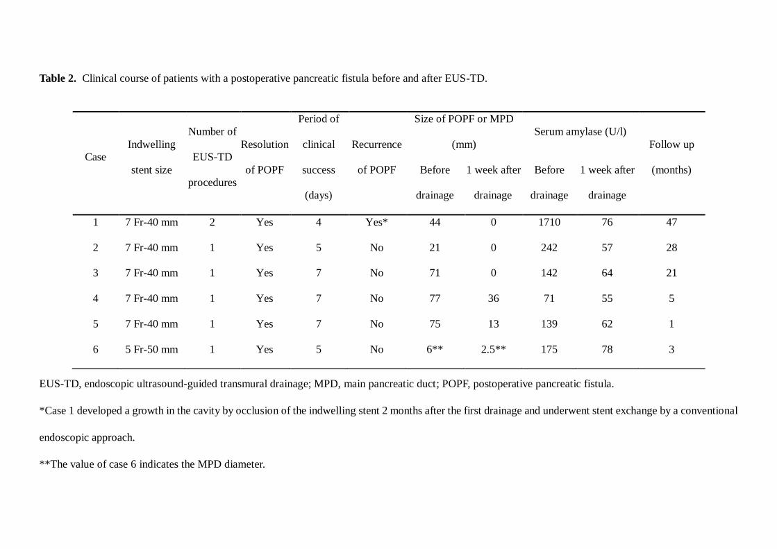
Table 2. Clinical course of patients with a postoperative pancreatic fistula before and after EUS-TD.
EUS-TD, endoscopic ultrasound-guided transmural drainage; MPD, main pancreatic duct; POPF, postoperative pancreatic fistula.
*Case 1 developed a growth in the cavity by occlusion of the indwelling stent 2 months after the first drainage and underwent stent exchange by a conventional
endoscopic approach.
**The value of case 6 indicates the MPD diameter.
Case
Indwelling
stent size
Number of
EUS-TD
procedures
Resolution
of POPF
Period of
clinical
success
(days)
Recurrence
of POPF
Size of POPF or MPD
(mm)
Serum amylase (U/l)
Follow up
(months) Before
drainage
1 week after
drainage
Before
drainage
1 week after
drainage
1 7 Fr-40 mm 2 Yes 4 Yes* 44 0 1710 76 47
2 7 Fr-40 mm 1 Yes 5 No 21 0 242 57 28
3 7 Fr-40 mm 1 Yes 7 No 71 0 142 64 21
4 7 Fr-40 mm 1 Yes 7 No 77 36 71 55 5
5 7 Fr-40 mm 1 Yes 7 No 75 13 139 62 1
6 5 Fr-50 mm 1 Yes 5 No 6** 2.5** 175 78 3
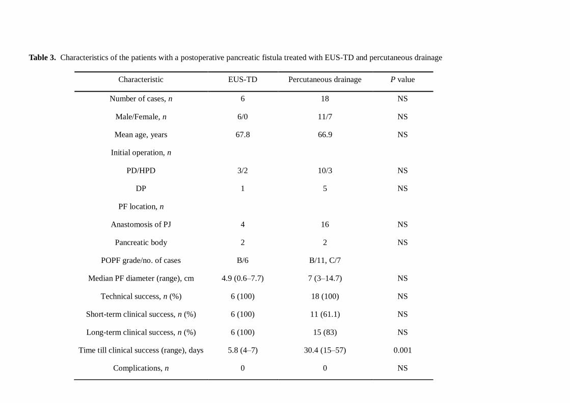
Table 3. Characteristics of the patients with a postoperative pancreatic fistula treated with EUS-TD and percutaneous drainage
Characteristic EUS-TD Percutaneous drainage P value
Number of cases, n 6 18 NS
Male/Female, n 6/0 11/7 NS
Mean age, years 67.8 66.9 NS
Initial operation, n
PD/HPD 3/2 10/3 NS
DP 1 5 NS
PF location, n
Anastomosis of PJ 4 16 NS
Pancreatic body 2 2 NS
POPF grade/no. of cases B/6 B/11, C/7
Median PF diameter (range), cm 4.9 (0.6–7.7) 7 (3–14.7) NS
Technical success, n (%) 6 (100) 18 (100) NS
Short-term clinical success, n (%) 6 (100) 11 (61.1) NS
Long-term clinical success, n (%) 6 (100) 15 (83) NS
Time till clinical success (range), days 5.8 (4–7) 30.4 (15–57) 0.001
Complications, n 0 0 NS

DP, distal pancreatectomy; HPD, hepatopancreaticoduodenectomy; NS, not significant; PD, pancreaticoduodenectomy; PJ,
pancreatojejunostomy; POPF, postoperative pancreatic fistula.

Total n=262
POPF (-) or POPF, gradeA n=172
POPF grade B or C n=90
Conservative treatment
by surgical drain (n=66)
Disease BDC 26 PC 23 NET 5 IPMN 4 AC 2 Others 4 Benign disease 2 Surgery PD 34 HPD 10 DP 19 SPR 3
Percutaneous drainage (n=18)
Disease BDC 10 PC 3 IPMN 2 AC 1 GBC 1 MCN 1 Surgery PD 10 HPD 3 DP 5
EUS-TD (n=6)
Disease PC 3 BDC 2 AC 1 Surgery PD 3 HPD 2 DP 1
Conservative treatment
(n=172)
Disease PC 102 BDC 32 NET 13 GBC 5 IPMN 4 AC 4 Others 6 Benign disease 6 Surgery PD 116 HPD 8 DP 48

A
B
C
E
D
F