Endoscopic ultrasound versus magnetic resonance ...
89
Endoscopic ultrasound versus magnetic resonance cholangiopancreatography for common bile duct stones (Review) Giljaca V, Gurusamy KS, Takwoingi Y, HiggieD, Poropat G, Štimac D, Davidson BR This is a reprint of a Cochrane review, prepared and maintained by The Cochrane Collaboration and published in The Cochrane Library 2015, Issue 2 http://www.thecochranelibrary.com Endoscopic ultrasound versus magnetic resonance cholangiopancreatography for common bile duct stones (Review) Copyright © 2015 The Cochrane Collaboration. Published by John Wiley & Sons, Ltd.
Transcript of Endoscopic ultrasound versus magnetic resonance ...

Endoscopic ultrasound versus magnetic resonance
cholangiopancreatography for common bile duct stones
(Review)
Giljaca V, Gurusamy KS, Takwoingi Y, Higgie D, Poropat G, Štimac D, Davidson BR
This is a reprint of a Cochrane review, prepared and maintained by The Cochrane Collaboration and published in The Cochrane Library2015, Issue 2
http://www.thecochranelibrary.com
Endoscopic ultrasound versus magnetic resonance cholangiopancreatography for common bile duct stones (Review)
Copyright © 2015 The Cochrane Collaboration. Published by John Wiley & Sons, Ltd.

T A B L E O F C O N T E N T S
1HEADER . . . . . . . . . . . . . . . . . . . . . . . . . . . . . . . . . . . . . . .1ABSTRACT . . . . . . . . . . . . . . . . . . . . . . . . . . . . . . . . . . . . . .2PLAIN LANGUAGE SUMMARY . . . . . . . . . . . . . . . . . . . . . . . . . . . . . .3BACKGROUND . . . . . . . . . . . . . . . . . . . . . . . . . . . . . . . . . . . .
Figure 1. . . . . . . . . . . . . . . . . . . . . . . . . . . . . . . . . . . . . . 56OBJECTIVES . . . . . . . . . . . . . . . . . . . . . . . . . . . . . . . . . . . . .7METHODS . . . . . . . . . . . . . . . . . . . . . . . . . . . . . . . . . . . . . .9RESULTS . . . . . . . . . . . . . . . . . . . . . . . . . . . . . . . . . . . . . . .
Figure 2. . . . . . . . . . . . . . . . . . . . . . . . . . . . . . . . . . . . . . 10Figure 3. . . . . . . . . . . . . . . . . . . . . . . . . . . . . . . . . . . . . . 12Figure 4. . . . . . . . . . . . . . . . . . . . . . . . . . . . . . . . . . . . . . 13Figure 5. . . . . . . . . . . . . . . . . . . . . . . . . . . . . . . . . . . . . . 15Figure 6. . . . . . . . . . . . . . . . . . . . . . . . . . . . . . . . . . . . . . 16
20DISCUSSION . . . . . . . . . . . . . . . . . . . . . . . . . . . . . . . . . . . . .21AUTHORS’ CONCLUSIONS . . . . . . . . . . . . . . . . . . . . . . . . . . . . . . .21ACKNOWLEDGEMENTS . . . . . . . . . . . . . . . . . . . . . . . . . . . . . . . .22REFERENCES . . . . . . . . . . . . . . . . . . . . . . . . . . . . . . . . . . . . .36CHARACTERISTICS OF STUDIES . . . . . . . . . . . . . . . . . . . . . . . . . . . . .80DATA . . . . . . . . . . . . . . . . . . . . . . . . . . . . . . . . . . . . . . . .
Test 1. Endoscopic ultrasound. . . . . . . . . . . . . . . . . . . . . . . . . . . . . . . 80Test 2. Magnetic resonance cholangiopancreatography. . . . . . . . . . . . . . . . . . . . . . . 81
81ADDITIONAL TABLES . . . . . . . . . . . . . . . . . . . . . . . . . . . . . . . . . .83APPENDICES . . . . . . . . . . . . . . . . . . . . . . . . . . . . . . . . . . . . .86CONTRIBUTIONS OF AUTHORS . . . . . . . . . . . . . . . . . . . . . . . . . . . . .86DECLARATIONS OF INTEREST . . . . . . . . . . . . . . . . . . . . . . . . . . . . . .87SOURCES OF SUPPORT . . . . . . . . . . . . . . . . . . . . . . . . . . . . . . . . .87DIFFERENCES BETWEEN PROTOCOL AND REVIEW . . . . . . . . . . . . . . . . . . . . .87NOTES . . . . . . . . . . . . . . . . . . . . . . . . . . . . . . . . . . . . . . . .
iEndoscopic ultrasound versus magnetic resonance cholangiopancreatography for common bile duct stones (Review)
Copyright © 2015 The Cochrane Collaboration. Published by John Wiley & Sons, Ltd.

[Diagnostic Test Accuracy Review]
Endoscopic ultrasound versus magnetic resonancecholangiopancreatography for common bile duct stones
Vanja Giljaca1, Kurinchi Selvan Gurusamy2, Yemisi Takwoingi3 , David Higgie4, Goran Poropat1, Davor Štimac1, Brian R Davidson2
1Department of Gastroenterology, Clinical Hospital Centre Rijeka, Rijeka, Croatia. 2Department of Surgery, Royal Free Campus,UCL Medical School, London, UK. 3Public Health, Epidemiology and Biostatistics, University of Birmingham, Birmingham, UK.4North Bristol NHS Trust, Bristol, UK
Contact address: Kurinchi Selvan Gurusamy, Department of Surgery, Royal Free Campus, UCL Medical School, Royal Free Hospital,Rowland Hill Street, London, NW3 2PF, UK. [email protected].
Editorial group: Cochrane Hepato-Biliary Group.Publication status and date: New, published in Issue 2, 2015.Review content assessed as up-to-date: 9 September 2012.
Citation: Giljaca V, Gurusamy KS, Takwoingi Y, Higgie D, Poropat G, Štimac D, Davidson BR. Endoscopic ultrasound versusmagnetic resonance cholangiopancreatography for common bile duct stones. Cochrane Database of Systematic Reviews 2015, Issue 2.Art. No.: CD011549. DOI: 10.1002/14651858.CD011549.
Copyright © 2015 The Cochrane Collaboration. Published by John Wiley & Sons, Ltd.
A B S T R A C T
Background
Endoscopic ultrasound (EUS) and magnetic resonance cholangiopancreatography (MRCP) are tests used in the diagnosis of commonbile duct stones in patients suspected of having common bile duct stones prior to undergoing invasive treatment. There has been nosystematic review of the accuracy of EUS and MRCP in the diagnosis of common bile duct stones using appropriate reference standards.
Objectives
To determine and compare the accuracy of EUS and MRCP for the diagnosis of common bile duct stones.
Search methods
We searched MEDLINE, EMBASE, Science Citation Index Expanded, BIOSIS, and Clinicaltrials.gov until September 2012. Wesearched the references of included studies to identify further studies and of systematic reviews identified from various databases(Database of Abstracts of Reviews of Effects (DARE), Health Technology Assessment (HTA), Medion, and ARIF (Aggressive ResearchIntelligence Facility)). We did not restrict studies based on language or publication status, or whether data were collected prospectivelyor retrospectively.
Selection criteria
We included studies that provided the number of true positives, false positives, false negatives, and true negatives for EUS or MRCP. Weonly accepted studies that confirmed the presence of common bile duct stones by extraction of the stones (irrespective of whether this wasdone by surgical or endoscopic methods) for a positive test, and absence of common bile duct stones by surgical or endoscopic negativeexploration of the common bile duct or symptom free follow-up for at least six months for a negative test, as the reference standard inpeople suspected of having common bile duct stones. We included participants with or without prior diagnosis of cholelithiasis; withor without symptoms and complications of common bile duct stones, with or without prior treatment for common bile duct stones;and before or after cholecystectomy. At least two authors independently screened abstracts and selected studies for inclusion.
1Endoscopic ultrasound versus magnetic resonance cholangiopancreatography for common bile duct stones (Review)
Copyright © 2015 The Cochrane Collaboration. Published by John Wiley & Sons, Ltd.

Data collection and analysis
Two authors independently collected the data from each study. We used the bivariate model to obtain pooled estimates of sensitivityand specificity.
Main results
We included a total of 18 studies involving 2366 participants (976 participants with common bile duct stones and 1390 participantswithout common bile duct stones). Eleven studies evaluated EUS alone, and five studies evaluated MRCP alone. Two studies evaluatedboth tests. Most studies included patients who were suspected of having common bile duct stones based on abnormal liver functiontests; abnormal transabdominal ultrasound; symptoms such as obstructive jaundice, cholangitis, or pancreatitis; or a combination ofthe above. The proportion of participants who had undergone cholecystectomy varied across studies. Not one of the studies was ofhigh methodological quality. For EUS, the sensitivities ranged between 0.75 and 1.00 and the specificities ranged between 0.85 and1.00. The summary sensitivity (95% confidence interval (CI)) and specificity (95% CI) of the 13 studies that evaluated EUS (1537participants; 686 cases and 851 participants without common bile duct stones) were 0.95 (95% CI 0.91 to 0.97) and 0.97 (95% CI0.94 to 0.99). For MRCP, the sensitivities ranged between 0.77 and 1.00 and the specificities ranged between 0.73 and 0.99. Thesummary sensitivity and specificity of the seven studies that evaluated MRCP (996 participants; 361 cases and 635 participants withoutcommon bile duct stones) were 0.93 (95% CI 0.87 to 0.96) and 0.96 (95% CI 0.90 to 0.98). There was no evidence of a differencein sensitivity or specificity between EUS and MRCP (P value = 0.5). From the included studies, at the median pre-test probability ofcommon bile duct stones of 41% the post-test probabilities (with 95% CI) associated with positive and negative EUS test results were0.96 (95% CI 0.92 to 0.98) and 0.03 (95% CI 0.02 to 0.06). At the same pre-test probability, the post-test probabilities associatedwith positive and negative MRCP test results were 0.94 (95% CI 0.87 to 0.97) and 0.05 (95% CI 0.03 to 0.09).
Authors’ conclusions
Both EUS and MRCP have high diagnostic accuracy for detection of common bile duct stones. People with positive EUS or MRCPshould undergo endoscopic or surgical extraction of common bile duct stones and those with negative EUS or MRCP do not needfurther invasive tests. However, if the symptoms persist, further investigations will be indicated. The two tests are similar in terms ofdiagnostic accuracy and the choice of which test to use will be informed by availability and contra-indications to each test. However,it should be noted that the results are based on studies of poor methodological quality and so the results should be interpreted withcaution. Further studies that are of high methodological quality are necessary to determine the diagnostic accuracy of EUS and MRCPfor the diagnosis of common bile duct stones.
P L A I N L A N G U A G E S U M M A R Y
Endoscopic ultrasound versus magnetic resonance cholangiopancreatography for the diagnosis of common bile duct stones
Background
Bile, produced in the liver and stored temporarily in the gallbladder, is released into the small bowel on eating fatty food. The commonbile duct (CBD) is the tube through which bile flows from the gallbladder to the small bowel. Stones in the CBD (CBD stones)are usually formed in the gallbladder before migration into the bile duct. They can obstruct the flow of bile leading to jaundice(yellowish discolouration of skin, whites of the eyes, and dark urine), infection of the bile (cholangitis), and inflammation of thepancreas (pancreatitis), which can be life threatening. Various diagnostic tests can be performed for the diagnosis of CBD stones.Depending upon the availability of resources, these stones are removed endoscopically (usually the case) or may be removed as part ofthe operation performed to remove the gallbladder (it is important to remove the gallbladder since the stones continue to form in thegallbladder and can cause recurrent problems). Prior to removal, invasive tests such as endoscopic retrograde cholangiopancreatography(ERCP) or intraoperative cholangiography (IOC) can be performed to detect CBD stones. However, before performing such invasivetests to diagnose CBD stones, non-invasive tests such as endoscopic ultrasound (EUS) (using ultrasound attached to the endoscope)and magnetic resonance cholangiopancreatography (MRCP) are used to identify people at high risk of having CBD stones so that onlythose at high risk can be subjected to further tests.
Study characteristics
We performed a thorough search for studies that reported the accuracy of EUS or MRCP in the diagnosis of CBD stones. We includeda total of 18 studies involving 2532 participants. Eleven studies evaluated EUS alone, five studies evaluated MRCP alone, and two
2Endoscopic ultrasound versus magnetic resonance cholangiopancreatography for common bile duct stones (Review)
Copyright © 2015 The Cochrane Collaboration. Published by John Wiley & Sons, Ltd.

studies evaluated both tests. A total of 1537 participants were included in the 13 studies that evaluated EUS and 995 participants wereincluded in the seven studies that evaluated MRCP. Most studies included patients who were suspected of having CBD stones basedon abnormal blood tests, abnormal ultrasound, or symptoms such as jaundice or pancreatitis, or a combination of the above. Theproportion of participants who had undergone previous gallbladder removal varied across studies.
Key results
Based on an average sensitivity of 95% for EUS, on average 95 out of 100 people with CBD stones will be detected while the remaining5 people will be missed and will not receive appropriate treatment. The average number of people with CBD stones detected usingEUS may vary between 91 and 97 out of 100 people. The average specificity of 97% for EUS means that on average 97 out of 100people without CBD stones will be identified as not having CBD stones; 3 out of 100 would be false positives and would not receiveappropriate treatment. The average number of false positives could vary between 1 and 6 out of 100 people. For MRCP, an averagesensitivity of 93% means that on average 93 out of 100 people with CBD stones will be detected while the remaining 7 people willbe missed and will not receive appropriate treatment. The average number of people with CBD stones detected using MRCP mayvary between 87 and 96 out of 100 people. With an average specificity of 96% for MRCP, 96 out of 100 people without CBD stoneswill be identified as not having CBD stones; 4 out of 100 would be false positives and would not receive appropriate treatment. Theaverage number of false positives could vary between 2 and 10 out of 100 people. This means that some people with CBD stones can bemissed by EUS and MRCP. Although most people with a negative EUS or MRCP do not need to undergo further invasive tests, in thepresence of persistent symptoms further testing with MRCP if the patient had undergone EUS or EUS if the patient had undergoneMRCP, ERCP, or IOC may be indicated. There is little to choose between EUS and MRCP in terms of diagnostic accuracy.
Quality of evidence
All the studies were of low methodological quality, which may undermine the validity of our findings.
Future research
Further studies of high methodological quality are necessary.
B A C K G R O U N D
Biliary stones are conglomerates of precipitated bile salts that formin the gallbladder or the common bile duct. The common bileduct carries bile from the liver to the duodenum (first part of thesmall intestine). The term ’gallstones’ generally refers to the stonesin the gallbladder while the term ’common bile duct stones’ refersto stones in the common bile duct. Common bile duct stonesmay form inside the common bile duct (primary common bileduct stones), or they may form in the gallbladder and migrateto the common bile duct (secondary common bile duct stones)(Williams 2008). A significant proportion of patients presentingwith common bile duct stones may be asymptomatic (Sarli 2000).In some patients the stones pass silently into the duodenum, andin others the stones cause clinical symptoms like biliary colic, jaun-dice, cholangitis, or pancreatitis (Caddy 2006). The prevalence ofgallstone disease in the general population is about 6% to 15%,with a higher prevalence in females (Barbara 1987; Loria 1994).Only 2% to 4% of people with gallstones become symptomaticwith biliary colic (pain), acute cholecystitis (inflammation), ob-structive jaundice, or gallstone pancreatitis in a year (Attili 1995;
Halldestam 2004), and removal of the gallbladder is recommendedin people with symptomatic gallstones (Gurusamy 2010). Amongpatients who undergo laparoscopic cholecystectomy (removal ofthe gallbladder) for symptomatic gallstones, 3% to 22% of pa-tients also have concomitant common bile duct stones (Arnold1970; Lill 2010; Yousefpour Azary 2011).
Common bile duct stones present in multiple ways. Central andright sided upper abdominal pain is a common presentation(Anciaux 1986; Roston 1997). Jaundice, caused by an impactedstone in the common bile duct leading to obstruction of bile pas-sage into the duodenum, is another presentation. It may subse-quently resolve if the common bile duct stone passes spontaneouslyinto the duodenum. This happens in 54% to 73% of patientswith common bile duct stones in whom cholecystectomy is per-formed for gallstones (Tranter 2003; Lefemine 2011). Another,more dangerous, complication of common bile duct stones is acutecholangitis. Cholangitis is clinically defined by Charcot’s triadwhich includes elevated body temperature, pain under the rightribcage, and jaundice (Raraty 1998; Salek 2009). Acute cholan-
3Endoscopic ultrasound versus magnetic resonance cholangiopancreatography for common bile duct stones (Review)
Copyright © 2015 The Cochrane Collaboration. Published by John Wiley & Sons, Ltd.

gitis is caused by an ascending bacterial infection of the commonbile duct and the biliary tree along with biliary obstruction. Thiscomplication is present in 2% to 9% of patients admitted for gall-stone disease (Saik 1975; Tranter 2003) and a mortality of ap-proximately 24% is recorded (Salek 2009). Common bile ductstones may also cause acute pancreatitis, accounting for 33% to50% of all patients with acute pancreatitis (Corfield 1985; Toh2000). Acute pancreatitis is usually a self-limiting disease and isgenerally sufficiently treated by conservative measures in its mildform (Neoptolemos 1988). However, a more severe pancreatitismay evolve in approximately 27% to 37% of patients with com-mon bile duct stone induced pancreatitis, with mortality around6% to 9% (Mann 1994; Toh 2000).
Suspicion of common bile duct stones can be investigated by lab-oratory liver function tests (Barkun 1994) or imaging tests likeabdominal ultrasound (Ripolles 2009). Further testing may in-clude endoscopic ultrasound (EUS) (Aljebreen 2008), magneticresonance cholangiopancreatography (MRCP) (Stiris 2000), en-doscopic retrograde cholangiopancreatography (ERCP) (Geron1999), and intraoperative cholangiography (IOC) (Fiore 1997).Currently, these are the recommended tests for diagnosis of com-mon bile duct stones. Of these tests, IOC can only be done duringan operation as the test requires surgical cannulation of the com-mon bile duct during cholecystectomy. The other tests may beused before or after cholecystectomy. Usually the first diagnostictests that most patients undergo are liver function tests and ab-dominal ultrasound. Invasive diagnostic tests are usually reservedfor patients with suspected common bile duct stones based onnon-invasive diagnostic tests, or when therapeutic measures arenecessary (Freitas 2006).
Conventional computed tomogram (CT scan), CT cholan-giogram, laparoscopic ultrasound, and ERCP guided intraductalultrasound are of limited use for diagnosing common bile ductstones (Maple 2010).
Target condition being diagnosed
Common bile duct stones. We did not differentiate the targetcondition with respect to common bile duct stone size, degree ofcommon bile duct obstruction, and the presence or absence ofsymptoms.
Index test(s)
MRCP uses a high magnetic field to cause fluctuations of tissuesat a molecular level. These minute fluctuations are then registeredby the receiver as differences in frequencies of fluctuation for thedifferent types of tissues. This information is then combined us-ing computer software to generate high-resolution pictures of the
scanned area. A common bile duct stone is seen as a hypointenseround or oval area of low signal in the hyperintense common bileduct (Stiris 2000; RadiologyInfo 2011).Endoscopic ultrasound combines endoscopy (a flexible tube usedto visualise the food-pipe and stomach) with ultrasound. A for-ward-viewing or side-viewing endoscope with an ultrasound trans-ducer is introduced in the duodenum by visual control, and thenhigh-frequency sound waves are used to inspect the tissues thatare in the proximity. Seeing a hyperechoic round or oval structurewithin the common bile duct is considered a positive test (Fickling2003; Aljebreen 2008).
Clinical pathway
Figure 1 illustrates a diagnostic pathway. Patients that are at riskof having common bile duct stones or are suspected of havingcommon bile duct stones (such as patients with gallbladder stonesor patients that show symptoms and signs of obstructive jaundiceor pancreatitis) will undergo liver function tests and abdominalultrasound as the first step. An abdominal ultrasound is usuallyavailable by the time the person is at risk or is suspected of havingcommon bile duct stones. Usually a combination of both tests isused as triage tests before further testing is done in the second step,but these can be used as the definitive diagnostic tests to carryout a therapeutic option (for example endoscopic or surgical com-mon bile duct exploration) (Williams 2008; ASGE Standards ofPractice Commitee 2010). MRCP or EUS are tests in the secondstep of the diagnostic pathway, which are used as optional triagetests prior to tests used in the third step of the diagnostic pathway;but they can also be used as definitive diagnostic tests to carry outa therapeutic option, that is, some people attempt extraction ofstones irrespective of the ERCP or IOC findings. MRCP and EUSare not usually combined since the positive or negative results ofone or the other is usually accepted for further clinical decisionmaking, without taking into consideration the results of liver func-tion tests or transabdominal ultrasound, as it is generally believedthat MRCP and EUS have better diagnostic accuracy than liverfunction tests or transabdominal ultrasound. ERCP and IOC areused in the third step of the diagnostic pathway. Both tests aredone just before the therapeutic intervention. Therapeutic inter-ventions, such as endoscopic or surgical stone extraction, can thenbe undertaken during the same session. ERCP is done before en-doscopic sphincterotomy and removal of common bile duct stonesusing a Dormia basket or balloon during the same endoscopic ses-sion (Prat 1996; Maple 2010), and IOC is done before surgicalcommon bile duct exploration and removal of common bile ductstones using surgical instruments during an operation for chole-cystectomy (Targarona 2004; Freitas 2006; Chen 2007; Williams2008; ASGE Standards of Practice Commitee 2010; Kelly 2010).
4Endoscopic ultrasound versus magnetic resonance cholangiopancreatography for common bile duct stones (Review)
Copyright © 2015 The Cochrane Collaboration. Published by John Wiley & Sons, Ltd.

Figure 1. The diagnostic pathway for diagnosis of common bile duct stones. Note that ultrasound is
generally performed in all patients at risk or suspected of common bile duct stones.AbbreviationsMRCP:
magnetic resonance cholangiopancreatographyERCP: endoscopic retrograde cholangiopancreatography
MRCP and EUS can be considered as add-on tests in patientswith a positive transabdominal ultrasound or liver function tests.Although most patients can undergo either MRCP or EUS, withthe choice between the tests being determined by the preferenceof the surgeon, EUS is the only add-on test possible in patientswith contra-indications to magnetic resonance imaging such asclaustrophobic patients and patients with cardiac pacemakers (Magnetic Resonance Imaging 2011) while MRCP is the only add-on test possible in patients with Roux-en-Y gastric anastomosissince EUS cannot reach the desired location (Wilson 2010).
Implications of negative tests
In general, patients with negative tests in one step do not undergofurther testing. For example, a person with no suggestion of com-mon bile duct stones on liver function tests and ultrasound willnot undergo further testing for common bile duct stones. Simi-larly, persons having no suggestion of common bile duct stoneson MRCP or EUS will not undergo further testing for commonbile duct stones, and persons with no suggestion of common bile
duct stones on ERCP or IOC will not undergo common bile ductclearance. Individuals with a false negative test result can developcomplications of common bile duct stones such as cholangitis andpancreatitis but the natural history of such patients with negativetests in terms of the frequency with which these complicationsdevelop is not known. However, it is generally recommended thatcommon bile duct stones are removed when they are identifiedbecause of the serious complications associated with their presence(Williams 2008). Although this practice is not evidence-based, thisshows the perception among hepato-pancreato biliary surgeonsand gastroenterologists that it is important not to miss commonbile duct stones.
Prior test(s)
Ultrasound and liver function tests are usually used prior to EUSand MRCP (see Figure 1).
5Endoscopic ultrasound versus magnetic resonance cholangiopancreatography for common bile duct stones (Review)
Copyright © 2015 The Cochrane Collaboration. Published by John Wiley & Sons, Ltd.

Role of index test(s)
EUS and MRCP are employed as add-on tests in the second stepof the diagnostic pathway. If positive, the tests are followed bydiagnostic tests in the third step of the diagnostic pathway. Ifnegative, the diagnosis of common bile duct stones is ruled outand further invasive testing is not performed.
Alternative test(s)
There are no alternative tests to EUS and MRCP that are in rou-tine clinical use at the second step of the diagnostic pathway. CTcholangiography and intravenous cholangiography may be usedin the second step of the diagnostic pathway but are not usedroutinely. A small proportion of surgeons use postoperative en-doscopic sphincterotomy for management of common bile ductstones. In persons in whom postoperative sphincterotomy is usedfor management of common bile duct stones, IOC may be con-sidered as an alternative to EUS and MRCP.
Rationale
There are several other benign and malignant conditions that maypresent in a similar manner to common bile duct stones. Benign(non-cancerous) causes of obstructive jaundice include primarysclerosing cholangitis (Penz-Osterreicher 2011), primary biliarycirrhosis (Hirschfield 2011), chronic pancreatitis (Abdallah 2007),autoimmune pancreatitis (Lin 2008), inflammatory strictures ofthe common bile duct (Krishna 2008), and strictures of the com-mon bile duct caused by prior instrumentation (Lillemoe 2000;Tang 2011). Malignant (cancerous) causes of obstructive jaun-dice include cholangiocarcinoma (Siddiqui 2011), cancer of theampulla of Vater as well as other periampullary cancers (Hamade2005; Choi 2011; Park 2011), and carcinoma of the pancreas(Singh 1990; Kalady 2004). It is important to differentiate be-tween the causes of obstructive jaundice in order to initiate ap-propriate treatment. The correct diagnosis of common bile ductstones is an essential contribution to this differentiation.Common bile duct stones are responsible for a range of compli-cations. Common bile duct stones lead to pancreatitis in about33% to 50% of the patients who have them (Corfield 1985; Toh2000) and cause mortality in about 6% to 9% of these patients(Mann 1994; Toh 2000). Acute cholangitis appears in 2% to 9%of patients admitted for gallstone disease, with mortality around24% (Salek 2009). Therefore, it is important to diagnose com-mon bile duct stones in order to treat patients and prevent suchcomplications.The preferred option for the treatment of common bile ductstones is currently endoscopic sphincterotomy (ES) with balloontrawling followed by laparoscopic cholecystectomy (Ludwig 2001;Spelsberg 2009). Other options include open cholecystectomywith open common bile duct exploration, laparoscopic cholecys-tectomy with laparoscopic common bile duct exploration, and la-paroscopic cholecystectomy with ES (Hong 2006; Dasari 2013). It
has been found that approximately half of patients with jaundice,abnormal liver function tests, and common bile duct dilation onultrasound do not actually have common bile duct stones (Hoyuela1999) and, therefore, these patients undergo invasive proceduresunnecessarily. Accurate diagnosis of common bile duct stonesmay avoid unnecessary procedures and the complications associ-ated with these procedures. Invasive tests can result in complica-tions; for example, endoscopic retrograde cholangiopancreatogra-phy with endoscopic sphincterotomy (ERCP-ES) can have life-threatening complications such as pancreatitis (Gurusamy 2011).Accurate diagnosis of common bile duct stones using non-invasivetests can avoid these complications.Currently, there are no Cochrane reviews of studies assessing theaccuracy of different tests for diagnosing common bile duct stones.This review is one of three reviews evaluating the diagnostic accu-racy of different tests in the diagnosis of common bile duct stonesand will help in the development of an evidence-based algorithmfor diagnosis of common bile duct stones.
O B J E C T I V E S
To determine and compare the accuracy of EUS and MRCP forthe diagnosis of common bile duct stones.
Secondary objectives
To investigate variation in the diagnostic accuracy of EUS andMRCP according to the following potential sources of heterogene-ity.
1. Studies at low risk of bias versus those with unclear or highrisk of bias (as assessed by the QUADAS-2) tool (Table 1).
2. Full text publications versus abstracts (this may indicatepublication bias if there is an association between the results ofthe study and the study reaching full publication) (Eloubeidi2001).
3. Prospective versus retrospective studies.4. Symptomatic versus asymptomatic common bile duct
stones (the presence of symptoms may increase the pre-testprobability). Symptomatic patients are defined as patientsshowing upper right quadrant abdominal pain, jaundice, acutecholangitis or acute pancreatitis (Anciaux 1986; Roston 1997;Raraty 1998; Toh 2000; Tranter 2003).
5. Prevalence of common bile duct stones in each includedstudy. The prevalence of common bile duct stones in thepopulation analysed by each included study may vary and causeheterogeneity. Prevalence may also change with the presence ofpatients with comorbidities that would predispose them tocommon bile duct stones such as primary sclerosing cholangitis,Caroli’s disease, hypercholesterolaemia, sickle cell anaemia, andsphincter of Oddi dysfunction.
6Endoscopic ultrasound versus magnetic resonance cholangiopancreatography for common bile duct stones (Review)
Copyright © 2015 The Cochrane Collaboration. Published by John Wiley & Sons, Ltd.

6. Proportion of patients with previous cholecystectomy.Cholecystectomy may cause dilatation of the common bile duct(Benjaminov 2013) and subsequently change the accuracy of theindex test, particularly imaging modalities.
7. Proportion of patients with common bile duct strictures(only for index tests that use contrast material, as strictures mayprevent contrast material from filling the common bile ductcompletely and, therefore, change the accuracy of the index test).
M E T H O D S
Criteria for considering studies for this review
Types of studies
We included studies providing cross-sectional information com-paring one or more of the index tests against a reference standardin the appropriate patient population (see Participants). We in-cluded studies irrespective of language or publication status, orwhether data were collected prospectively or retrospectively. Weplanned to include comparative studies in which EUS and MRCPwere performed in the same study population, either by givingall patients both index tests or by randomly allocating patients toreceive MRCP or EUS. We planned to exclude diagnostic case-control studies if there were at least four cross-sectional or com-parative studies.
Participants
Patients at risk or suspected of having common bile duct stoneswith or without prior diagnosis of cholelithiasis; with or withoutsymptoms and complications of common bile duct stones or withor without prior treatment for common bile duct stones; and be-fore or after cholecystectomy.
Index tests
Endoscopic ultrasound (EUS) and magnetic resonance retrogradecholangiopancreatography (MRCP).
Target conditions
Common bile duct stones.
Reference standards
We accepted the following reference standards.• For test positives, we accepted confirmation of a common
bile duct stone by extraction of the stone (irrespective of whetherthis was done by surgical or endoscopic methods).
• For test negatives, we acknowledged that there was no wayof being absolutely sure that there were no common bile ductstones. However, we accepted negative results by surgical orendoscopic negative exploration of the common bile duct, orsymptom-free follow-up for at least six months as the referencestandard. Surgical or endoscopic exploration is adequate but it isnot commonly used in patients with negative index tests becauseof its invasive nature. Therefore, we accepted follow-up as a lessadequate reference test. Negative exploration of the common bileduct is likely to be a better reference standard than follow-up forat least six months since most stones already present in thecommon bile duct are likely to be identified and extracted in thisfashion. Six months is an arbitrary choice but we anticipated thatmost common bile duct stones will manifest during this period.
Search methods for identification of studies
Electronic searches
We searched MEDLINE via PubMed (January 1946 to September2012), EMBASE via OvidSP (January 1947 to September 2012),Science Citation Index Expanded via Web of Knowledge (January1898 to September 2012), BIOSIS via Web of Knowledge (Jan-uary 1969 to September 2012), and Clinicaltrials.gov (September2012). The search strategies are shown in Appendix 1. We used acommon search strategy for the three reviews of which this reviewis one. The other two reviews assess the diagnostic accuracy oftransabdominal ultrasound, liver function tests, ERCP, and IOC(Gurusamy 2015a; Gurusamy 2015b). We also identified system-atic reviews from the Database of Abstracts of Reviews of Effects(DARE), Health Technology Assessment (HTA), Medion, andARIF (Aggressive Research Intelligence Facility) databases in orderto search their reference lists (please see searching other resources).
Searching other resources
We searched the references of the included studies and systematicreviews related to the topic to identify further studies. We alsosearched for additional articles related to the included studies byperforming the ’related search’ function in MEDLINE (PubMed)and EMBASE (OvidSP) and a ’citing reference’ search (searchthe articles which cited the included articles) (Sampson 2008) inScience Citation Index Expanded and EMBASE (OvidSP).
7Endoscopic ultrasound versus magnetic resonance cholangiopancreatography for common bile duct stones (Review)
Copyright © 2015 The Cochrane Collaboration. Published by John Wiley & Sons, Ltd.

Data collection and analysis
Selection of studies
Three authors (VG and DH or GP) searched the references inde-pendently for identification of relevant studies. We obtained fulltexts for the references that at least one of the authors consideredrelevant. Two review authors (VG and DH or GP) assessed the fulltext articles independently. Any differences in study selection werearbitrated by KG. We selected the studies that met the inclusioncriteria for data extraction. We included abstracts if sufficient datato create a 2 x 2 table were provided.
Data extraction and management
Two authors (KG and VG) independently extracted the followingdata from each included study.
1. First author of report.2. Year of publication of report.3. Study design (prospective or retrospective; cross-sectional
studies or randomised clinical trials).4. Inclusion and exclusion criteria for individual studies.5. Total number of patients.6. Number of males and females.7. Mean age of the participants.8. Tests carried out prior to index test.9. Index test.
10. Reference standard.11. Number of true positives, false positives, true negatives, andfalse negatives.We sought further information on the diagnostic test accuracy dataand assessment of methodological quality (please see Assessmentof methodological quality) from the authors of the studies, if nec-essary. We resolved any differences between the review authors bydiscussion till a consensus was reached. We extracted the data ex-cluding participants with indeterminate results but recorded thenumber of indeterminates and the reference standard results of thepatients with indeterminate results.
Assessment of methodological quality
We adopted the quality assessment of diagnostic accuracy studiesassessment tool (QUADAS-2) (Whiting 2006; Whiting 2011) forassessment of the methodological quality of included studies asdescribed in Table 1. We considered studies classified at low risk ofbias and low concern regarding applicability to the review questionas studies at low risk of bias. Any differences in the methodolog-ical quality assessments were resolved by discussion between thereview authors until a consensus was reached. We sought furtherinformation from study authors in order to accurately assess themethodological quality of the included studies.
Statistical analysis and data synthesis
To visually explore between study variation in the performance ofeach test, we plotted estimates of sensitivity and specificity fromeach study on forest plots and in receiver operating characteristic(ROC) space. Because our focus of inference was summary points,we used the bivariate model (Reitsma 2005; Chu 2006) to jointlysummarise the sensitivity and specificity of each test. This modelaccounts for between study variability in estimates of sensitivityand specificity through the inclusion of random effects for the logitsensitivity and logit specificity parameters of the bivariate model.Using all available studies (that is, an indirect comparison), wecompared the diagnostic accuracy of EUS and MRCP by includ-ing covariate terms for test type in the bivariate model to estimatedifferences in the sensitivity and specificity of the two tests. Wealso allowed the variances of the random effects and their covari-ance to depend on test type thus allowing the variances to differbetween tests. We used likelihood ratio tests to compare the fit ofdifferent models, and we also compared the estimates of sensitiv-ity and specificity between models to check the robustness of ourassumptions about the variances of the random effects. If stud-ies that evaluated EUS and MRCP in the same study populationwere available, we planned to also perform a direct head-to-headcomparison by limiting the test comparison to such studies. Meta-analyses were performed using the xtmelogit command in Stataversion 13 (Stata-Corp, College Station, Texas, USA).We created a table of pre-test probabilities (using the observedmedian and range of prevalence from the included studies) againstpost-test probabilities. The post-test probabilities were calculatedusing these pre-test probabilities and the summary positive andnegative likelihood ratios were derived by using the Stata ˙diparmcommand and functions of the parameter estimates from the bi-variate model that we fitted to estimate the summary sensitivitiesand specificities.
Investigations of heterogeneity
We visually inspected forest plots of sensitivity and specificity, andsummary ROC plots to investigate the potential sources of hetero-geneity as stated in the Secondary objectives. Where possible giventhe number of included studies, we planned to formally exploreheterogeneity by adding each potential source of heterogeneitylisted above as a covariate in the bivariate model (meta-regressionwith one covariate at a time).
Sensitivity analyses
Exclusion of participants with uninterpretable results can result inan overestimation of diagnostic test accuracy (Schuetz 2012). Inpractice, uninterpretable test results will generally be consideredas test negatives. Therefore, we planned to perform sensitivityanalyses by including uninterpretable test results as test negatives,if sufficient data were available.
8Endoscopic ultrasound versus magnetic resonance cholangiopancreatography for common bile duct stones (Review)
Copyright © 2015 The Cochrane Collaboration. Published by John Wiley & Sons, Ltd.

Assessment of reporting bias
As described in the Investigations of heterogeneity section, weplanned to investigate whether the sensitivity and specificity of thetests differed between studies that were published as full texts andthose that were available only as abstracts.
R E S U L T S
Results of the search
We identified a total of 22,789 references through electronicsearches of MEDLINE (n = 8292), EMBASE (n = 10,029), Sci-
ence Citation Index Expanded and Biosis (n = 4276), and DAREand HTA in the Cochrane Library (n = 192). One additional refer-ence was identified by searching other sources. We excluded 5866duplicates and 16,718 clearly irrelevant references through readingabstracts. We assessed the remaining 206 references for eligibilityby reading the full texts of the publications. We excluded 188 fulltext articles. The main reasons for exclusion were inappropriatereference standards and lack of data to construct the 2 x 2 tablesneeded for meta-analyses. The list of excluded studies and reasonsfor exclusion are listed in the Characteristics of excluded studiestable. We included a total of 18 studies. We were able to obtainadditional information from the authors of two of the studies (Prat1996; Montariol 1998). The flow of studies through the selectionprocess is shown in Figure 2.
9Endoscopic ultrasound versus magnetic resonance cholangiopancreatography for common bile duct stones (Review)
Copyright © 2015 The Cochrane Collaboration. Published by John Wiley & Sons, Ltd.

Figure 2. Flow of studies through the screening process.
10Endoscopic ultrasound versus magnetic resonance cholangiopancreatography for common bile duct stones (Review)
Copyright © 2015 The Cochrane Collaboration. Published by John Wiley & Sons, Ltd.

Characteristics of included studies
The characteristics of the included studies are summarised in theCharacteristics of included studies table. We included a total of 18studies involving 2366 participants in this systematic review. EUSwas evaluated by 13 studies involving 1537 participants (686 par-ticipants with common bile duct stones and 851 participants with-out common bile duct stones), and MRCP was evaluated by sevenstudies involving 996 participants (361 cases and 635 participantswithout common bile duct stones). The median pre-test probabil-ity of common bile duct stones was 0.41, or 41%. The minimumpre-test probability of common bile duct stones in the studies was0.14, and the maximum pre-test probability was 0.68. Fifteen (Prat1996; Norton 1997; Canto 1998; Montariol 1998; De Ledinghen1999; Liu 2001; Boraschi 2002; Jendresen 2002; Kohut 2002;Buscarini 2003; Gautier 2004; Guarise 2005; Ney 2005; Miletic2006; Fernandez-Esparrach 2007) of the 18 included studies werefull text publications. Ten studies (Canto 1998; Montariol 1998;De Ledinghen 1999; Liu 2001; Fazel 2002; Jendresen 2002; Kohut2002; Buscarini 2003; Gautier 2004; Choo 2012) were prospec-tive studies, one study (Ang 2012) was a retrospective study, andit was unclear whether the remaining studies were prospective orretrospective (Prat 1996; Norton 1997; Boraschi 2002; Guarise2005; Ney 2005; Miletic 2006; Fernandez-Esparrach 2007). Tenstudies (Prat 1996; Norton 1997; Canto 1998; De Ledinghen1999; Boraschi 2002; Fazel 2002; Kohut 2002; Buscarini 2003;Fernandez-Esparrach 2007; Ang 2012) included patients whowere suspected of having common bile duct stones based on ab-normal liver function tests; abnormal transabdominal ultrasound;symptoms such as obstructive jaundice, cholangitis, or pancre-atitis; or a combination of the above. One study (Liu 2001) in-cluded only patients with pancreatitis and another study (Ney2005) included patients with abnormal liver function tests or ultra-sound but excluded those with symptoms. One study (Montariol1998) excluded patients with abnormal liver function tests, ab-
normal transabdominal ultrasound, or symptoms; and one study(Choo 2012) included only patients with a positive intraopera-tive cholangiogram. Three studies (Gautier 2004; Guarise 2005;Miletic 2006) reported that they performed the test in patientswith suspected common bile duct stones but the reasons for sus-picion were not stated. The reason for performing the test wasnot stated in the remaining study (Jendresen 2002). Six stud-ies (Norton 1997; Canto 1998; Montariol 1998; Boraschi 2002;Jendresen 2002; Ney 2005) included participants who had notundergone previous cholecystectomy. In one study (Choo 2012)all the participants had undergone cholecystectomy, while in threestudies (Prat 1996; Liu 2001; Buscarini 2003) 8% to 75% of par-ticipants had undergone cholecystectomy. The proportion of par-ticipants who had undergone cholecystectomy was not stated inthe remaining studies. The proportion of patients with commonbile duct strictures was not stated in any of the studies.The criteria for a positive EUS varied between the studiesthat reported their criteria. While the studies used hyperechoicshadowing inside the common bile duct as the main criterion(Norton 1997; Canto 1998; Montariol 1998; De Ledinghen1999; Liu 2001; Kohut 2002; Buscarini 2003; Ney 2005;Fernandez-Esparrach 2007), some studies stipulated that theseshadows should have acoustic shadowing (Canto 1998; Montariol1998; Kohut 2002; Ney 2005) and should be mobile (Ney 2005).The criteria for a positive MRCP were signal defects within thecommon bile duct, defined variably as foci or rounded and ovalin some studies (De Ledinghen 1999; Boraschi 2002; Jendresen2002; Gautier 2004; Guarise 2005; Fernandez-Esparrach 2007).
Methodological quality of included studies
The methodological quality of the included studies is summarisedin Figure 3 and Figure 4. Not one of the included studies wasof high methodological quality. Regarding applicability concerns,none of the studies were of low concern in all three domains.
11Endoscopic ultrasound versus magnetic resonance cholangiopancreatography for common bile duct stones (Review)
Copyright © 2015 The Cochrane Collaboration. Published by John Wiley & Sons, Ltd.

Figure 3. Risk of bias and applicability concerns graph: review authors’ judgements about each domain
presented as percentages across included studies. Each bar shows the number of studies in each category. The
index test domain was evaluated separately for each test. Of the 18 included studies, 7 studies evaluated
MRCP and 13 studies evaluated EUS; the numbers do not add up to 18 because two of the studies evaluated
both tests.
12Endoscopic ultrasound versus magnetic resonance cholangiopancreatography for common bile duct stones (Review)
Copyright © 2015 The Cochrane Collaboration. Published by John Wiley & Sons, Ltd.
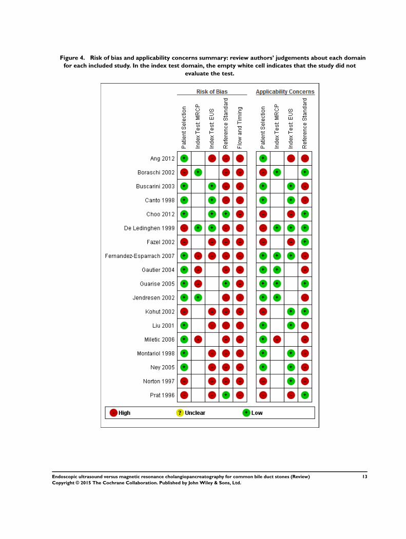
Figure 4. Risk of bias and applicability concerns summary: review authors’ judgements about each domain
for each included study. In the index test domain, the empty white cell indicates that the study did not
evaluate the test.
13Endoscopic ultrasound versus magnetic resonance cholangiopancreatography for common bile duct stones (Review)
Copyright © 2015 The Cochrane Collaboration. Published by John Wiley & Sons, Ltd.

Patient selection domain
In the patient selection domain, 12 studies (Canto 1998;Montariol 1998; Liu 2001; Jendresen 2002; Buscarini 2003;Gautier 2004; Guarise 2005; Ney 2005; Miletic 2006; Fernandez-Esparrach 2007; Ang 2012; Choo 2012) had low risk of bias.Eleven studies (Canto 1998; Montariol 1998; Liu 2001; Jendresen2002; Buscarini 2003; Gautier 2004; Guarise 2005; Ney 2005;Miletic 2006; Fernandez-Esparrach 2007; Ang 2012) had low ap-plicability concerns. The remaining studies were at high risk of biasand were of high concern for applicability because patient recruit-ment was unclear (Norton 1997; De Ledinghen 1999; Boraschi2002; Fazel 2002; Kohut 2002), participants were excluded inap-propriately (Prat 1996), or there were concerns that the partici-pants did not match the types of participants that will undergothese tests in routine clinical practice (Choo 2012).
Index test domain
In the index test domain, seven studies had low risk of bias; fourwere EUS only studies (Prat 1996; Canto 1998; Buscarini 2003;Choo 2012), two (Boraschi 2002; Jendresen 2002) were MRCPonly studies, and one (De Ledinghen 1999) evaluated both EUSand MRCP. The remaining studies were at high risk of bias becauseit was not clear whether the index test results were interpreted with-out knowledge of the reference standard results. Thirteen studieswere of low concern for applicability; seven (Norton 1997; Canto1998; Montariol 1998; Liu 2001; Kohut 2002; Buscarini 2003;Ney 2005) were EUS only studies, four (Boraschi 2002; Jendresen2002; Gautier 2004; Guarise 2005) were MRCP only studies, andtwo (De Ledinghen 1999; Fernandez-Esparrach 2007) were stud-ies of both EUS and MRCP. The remaining studies (Prat 1996;Boraschi 2002; Fazel 2002; Gautier 2004; Guarise 2005; Miletic2006; Ang 2012; Choo 2012) were of high concern for applica-bility because the criteria for a positive test were not stated.
Reference standard domain
In the reference standard domain, three studies (Prat 1996; Guarise2005; Choo 2012) had low risk of bias. The remaining studies wereat high risk of bias because it was either not clear whether the ref-erence standards were interpreted without knowledge of the index
test results (Norton 1997; Canto 1998; De Ledinghen 1999; Liu2001; Boraschi 2002; Fazel 2002; Kohut 2002; Buscarini 2003;Gautier 2004; Ney 2005; Miletic 2006; Fernandez-Esparrach2007; Ang 2012) or it was clear that the reference standards wereinterpreted with the knowledge of the index test results (Montariol1998; Jendresen 2002). Seven studies (Prat 1996; De Ledinghen1999; Boraschi 2002; Fazel 2002; Kohut 2002; Guarise 2005;Choo 2012) gave low concern about applicability. The remain-ing 11 studies (Norton 1997; Canto 1998; Montariol 1998; Liu2001; Jendresen 2002; Buscarini 2003; Gautier 2004; Ney 2005;Miletic 2006; Fernandez-Esparrach 2007; Ang 2012) were of highconcern because endoscopic or surgical clearance of the commonbile duct was achieved in patients with a positive test and clinicalfollow-up was performed in patients with a negative test.
Flow and timing domain
In the flow and timing domain, all 18 studies were at highrisk of bias for the following reasons. Six studies (De Ledinghen1999; Boraschi 2002; Fazel 2002; Guarise 2005; Fernandez-Esparrach 2007; Ang 2012) did not report the time intervalbetween the index test and reference standard, and 11 studies(Norton 1997; Canto 1998; Montariol 1998; Liu 2001; Jendresen2002; Buscarini 2003; Gautier 2004; Ney 2005; Miletic 2006;Fernandez-Esparrach 2007; Ang 2012) did not use the same ref-erence standard since endoscopic or surgical clearance of the com-mon bile duct was achieved in patients with a positive test andclinical follow-up was performed in patients with a negative test.It was not clear whether all the patients were included in the anal-ysis in six studies (Norton 1997; Canto 1998; Fazel 2002; Kohut2002; Ang 2012; Choo 2012), while some patients were excludedfrom the analysis in nine studies (Prat 1996; Montariol 1998; DeLedinghen 1999; Boraschi 2002; Buscarini 2003; Gautier 2004;Guarise 2005; Miletic 2006; Fernandez-Esparrach 2007).
Findings
The results are summarised in Summary of findings, Figure 5, andFigure 6.
14Endoscopic ultrasound versus magnetic resonance cholangiopancreatography for common bile duct stones (Review)
Copyright © 2015 The Cochrane Collaboration. Published by John Wiley & Sons, Ltd.

Figure 5. Forest plot of endoscopic ultrasound and magnetic resonance cholangiopancreatography for
diagnosis of common bile duct stones. The plot shows study specific estimates of sensitivity and specificity
(with 95% confidence intervals). The studies are ordered according to study design (prospective or not),
sensitivity and study identifier; FN = false negative; FP = false positive; TN = true negative; TP = true positive.
15Endoscopic ultrasound versus magnetic resonance cholangiopancreatography for common bile duct stones (Review)
Copyright © 2015 The Cochrane Collaboration. Published by John Wiley & Sons, Ltd.

Figure 6. Summary ROC plot of endoscopic ultrasound and magnetic resonance cholangiopancreatography
for diagnosis of common bile duct stones. For each test, each symbol represents the pair of sensitivity and
specificity from a study and the symbol is scaled according to the sample size of the study. The solid circles
represent the summary sensitivity and specificity for each test. Each summary point is surrounded by a 95%
confidence region.
16Endoscopic ultrasound versus magnetic resonance cholangiopancreatography for common bile duct stones (Review)
Copyright © 2015 The Cochrane Collaboration. Published by John Wiley & Sons, Ltd.

Endoscopic ultrasound (EUS)
The sensitivities of the 13 studies ranged between 0.75 and 1.00,and the specificities ranged between 0.85 and 1.00 (Figure 5). Thesummary sensitivity (95% CI) and summary specificity (95% CI)were 0.95 (95% CI 0.91 to 0.97) and 0.97 (95% CI 0.94 to 0.99).The summary positive and negative likelihood ratios were 34.4(95% CI 15.2 to 78.1) and 0.05 (95% CI 0.03 to 0.09). At themedian pre-test probability of common bile duct stones of 41%,the post-test probabilities (with 95% CI) associated with positiveand negative tests were 0.96 (95% CI 0.92 to 0.98) and 0.03 (95%CI 0.02 to 0.06) respectively. At the minimum pre-test probabilityof 14%, the post-test probabilities associated with positive andnegative tests were 0.85 (95% CI 0.72 to 0.93) and 0.01 (95% CI0.01 to 0.02). At the maximum pre-test probability of 68%, thepost-test probabilities associated with positive and negative testswere 0.99 (95% CI 0.97 to 0.99) and 0.10 (95% CI 0.06 to 0.16).
Magnetic resonance cholangiopancreatography (MRCP)
The sensitivities ranged between 0.77 and 1.00, and the speci-ficities ranged between 0.73 and 0.99 (Figure 5). The summarysensitivity (95% CI) and summary specificity (95% CI) were 0.93(95% CI 0.87 to 0.96) and 0.96 (95% CI 0.89 to 0.98). The sum-mary positive and negative likelihood ratios were 21.7 (95% CI9.3 to 50.7) and 0.07 (95% CI 0.04 to 0.14). At the median pre-test probability of common bile duct stones of 41%, the post-testprobabilities associated with positive and negative tests were 0.94(95% CI 0.87 to 0.97) and 0.05 (95% CI 0.03 to 0.09). At theminimum pre-test probability of 14%, the post-test probabilitiesassociated with positive and negative tests were 0.79 (95% CI 0.61to 0.90) and 0.01 (95% CI 0.01 to 0.02). At the maximum pre-test probability of 68%, the post-test probabilities associated withpositive and negative tests were 0.98 (95% CI 0.95 to 0.99) and0.13 (95% CI 0.08 to 0.23).
Endoscopic ultrasound (EUS) versus magnetic resonance
cholangiopancreatography (MRCP)
Only two studies (De Ledinghen 1999; Fernandez-Esparrach2007) performed EUS and MRCP in the same participants and sowe were unable to perform a direct comparison. We performed anindirect comparison of EUS and MRCP (Figure 6). There was noevidence of a difference in sensitivity or specificity between EUSand MRCP (P value = 0.5).
Investigation of sources of heterogeneity
We were unable to formally explore potential sources of hetero-geneity for MRCP because there were only seven studies. For EUS,we found no evidence of a difference in sensitivity or specificitybetween full text publications (10 studies) and abstracts (3 studies)(P value = 0.5). The prevalence of common bile duct stones inthe studies of EUS ranged between 16% and 63%. There was noevidence of an effect of prevalence on test performance (P value =0.5).We were unable to explore the effect of the following potentialsources of heterogeneity.
1. Studies at low risk of bias versus those at unclear or highrisk of bias: the analysis could not be performed because all thestudies were of low methodological quality.
2. Prospective studies versus retrospective studies: eight studieswere prospective, one was retrospective and four studies did notprovide this information.
3. Symptomatic versus asymptomatic participants: thisinformation was available in five studies only (Norton 1997;Montariol 1998; Buscarini 2003; Ney 2005; Choo 2012). Allparticipants in these studies were symptomatic.
4. Proportion of patients with common bile duct strictures:the information was not available in any of the studies.
5. Proportion of patients with previous cholecystectomy: fourstudies did not include patients with previous cholecystectomyand five studies included between 8% and 100% of such patients.
Sensitivity analyses
Endoscopic ultrasound (EUS)
Two studies (Prat 1996; Buscarini 2003) reported participantswith uninterpretable results together with their reference standardresults. Five studies (Prat 1996; Montariol 1998; De Ledinghen1999; Buscarini 2003; Fernandez-Esparrach 2007) reported unin-terpretable results but did not provide the corresponding referencestandard results. We did not perform sensitivity analyses becausedata were sparse.
Magnetic resonance cholangiopancreatography (MRCP)
None of the studies reported participants with uninterpretable re-sults for whom the reference standard results were available and sowe did not perform sensitivity analyses. Six studies (De Ledinghen1999; Boraschi 2002; Gautier 2004; Guarise 2005; Miletic 2006;Fernandez-Esparrach 2007) reported participants with uninter-pretable results for whom the reference standard results were notavailable.
17Endoscopic ultrasound versus magnetic resonance cholangiopancreatography for common bile duct stones (Review)
Copyright © 2015 The Cochrane Collaboration. Published by John Wiley & Sons, Ltd.
Summary of findings
Population Patients suspected of having common bile duct stones based on symptoms, liver function tests, and ultrasound
Settings Secondary and tertiary care setting in different parts of the world
Index tests Endoscopic ultrasound (EUS) and magnetic resonance cholangiopancreatography (MRCP)
Reference standard Endoscopic or surgical extraction of stones in patients with a positive index test result or clinical follow-up (minimum 6 months) in patients with a negative
index test result
Target condition Common bile duct stones
Number of studies A total of 18 studies were included. Thirteen studies (686 cases, 1537 participants) evaluated EUS and 7 studies (361 cases, 996 participants) evaluated
MRCP. Two of the studies evaluated both tests in the same patients
Methodological quality con-
cerns
All the studies were of poor methodological quality; most studies were at high risk of bias or gave high concern about applicability across all domains of
quality assessment, or both
Pre-test probability1 Test Summary sensitivity (95%
CI)
Summary specificity (95%
CI)
Positive post-test probability
(95% CI)2Negative post-test probabil-
ity (95% CI)3
0.14 EUS 0.95 (0.91 to 0.97) 0.97 (0.94 to 0.99) 0.85 (0.72 to 0.93) 0.01 (0.01 to 0.02)
MRCP 0.93 (0.87 to 0.96) 0.96 (0.89 to 0.98) 0.79 (0.61 to 0.90) 0.01 (0.01 to 0.02)
0.30 EUS 0.95 (0.91 to 0.97) 0.97 (0.94 to 0.99) 0.94 (0.87 to 0.97) 0.02 (0.01, 0.04)
MRCP 0.93 (0.87 to 0.96) 0.96 (0.89 to 0.98) 0.90 (0.80 to 0.96) 0.03 (0.02, 0.06)
0.41 EUS 0.95 (0.91 to 0.97) 0.97 (0.94 to 0.99) 0.96 (0.92 to 0.98) 0.03 (0.02, 0.06)
MRCP 0.93 (0.87 to 0.96) 0.96 (0.89 to 0.98) 0.94 (0.87 to 0.97) 0.05 (0.03 to 0.09)
0.48 EUS 0.95 (0.91 to 0.97) 0.97 (0.94 to 0.99) 0.97 (0.93, 0.99) 0.05 (0.03 to 0.08)
MRCP 0.93 (0.87 to 0.96) 0.96 (0.89 to 0.98) 0.95 (0.90 to 0,98) 0.06 (0.04 to 0.11)18
En
do
sco
pic
ultra
sou
nd
versu
sm
agn
etic
reso
nan
ce
ch
ola
ngio
pan
cre
ato
gra
phy
for
co
mm
on
bile
du
ct
ston
es
(Revie
w)
Co
pyrig
ht
©2015
Th
eC
och
ran
eC
olla
bo
ratio
n.P
ub
lished
by
Joh
nW
iley
&S
on
s,L
td.

0.68 EUS 0.95 (0.91 to 0.97) 0.97 (0.94 to 0.99) 0.99 (0.97 to 0.99) 0.10 (0.06 to 0.16)
MRCP 0.93 (0.87 to 0.96) 0.96 (0.89 to 0.98) 0.98 (0.95 to 0.99) 0.13 (0.08 to 0.23)
Comparison of the diagnostic accuracy of EUS and MRCP: at pre-test probabilities of 14%, 41%, and 68%, out of 100 people with positive EUS, common bile duct stones will be present in
85, 96, and 99 people respectively; while out of 100 people with positive MRCP, common bile duct stones will be present in 79, 94, and 98 people. For the same pre-test probabilities, out of
100 people with negative EUS, common bile duct stones will be present in 1, 3, and 10 people respectively; while out of 100 people with negative MRCP, common bile duct stones will be
present in 1, 5, and 13 people respectively
Conclusions: the performance of EUS and MRCP appears to be comparable for diagnosis of common bile duct stones. The strength of the evidence for the test comparison was weak
because the studies were methodologically flawed, and only two studies made head-to-head comparisons of EUS and MRCP
1 The pre-test probability (proportion with common bile duct stones out of the total number of participants) was computed for each
included study. These numbers represent the minimum, lower quartile, median, upper quartile and the maximum values from the 18
studies.2Post-test probability of common bile duct stones in people with positive index test results.3Post-test probability of common bile duct stones in people with negative index test results.
19
En
do
sco
pic
ultra
sou
nd
versu
sm
agn
etic
reso
nan
ce
ch
ola
ngio
pan
cre
ato
gra
phy
for
co
mm
on
bile
du
ct
ston
es
(Revie
w)
Co
pyrig
ht
©2015
Th
eC
och
ran
eC
olla
bo
ratio
n.P
ub
lished
by
Joh
nW
iley
&S
on
s,L
td.

D I S C U S S I O N
Summary of main results
The results are summarised in Summary of findings. We included13 studies that evaluated the diagnostic accuracy of EUS and sevenstudies that evaluated the diagnostic accuracy of MRCP. The sum-mary sensitivity and specificity of EUS were 0.95 (95% CI 0.91to 0.97) and 0.97 (95% CI 0.94 to 0.99). The summary sensi-tivity and specificity of MRCP were 0.93 (95% CI 0.87 to 0.96)and 0.96 (95% CI 0.89 to 0.98). Sensitivity and specificity didnot differ significantly between the two tests. The median pre-testprobability of common bile duct stones from the included studieswas 41%. This proportion is higher than in the general popula-tion (Barbara 1987; Loria 1994) or in the population of patientsundergoing cholecystectomy for gallbladder stones (Arnold 1979;Lill 2010; Yousefpour Azary 2011). This is probably due to thefact that EUS and MRCP are performed as triage tests in the sec-ond step of the diagnostic pathway, and only preselected patientswith abnormal liver function tests or abnormal abdominal ultra-sound, or both, were included in these studies. The probability ofcommon bile duct stones in such a selected population has beenreported to be about 36% (Rahman 2010), which is similar tothe pre-test probability in this review. For a pre-test probability of41%, the median observed in this review, the post-test probabili-ties associated with positive and negative EUS were 0.96 (95% CI0.92 to 0.98) and 0.03 (95% CI 0.02 to 0.06). At the same pre-test probability, the post-test probabilities associated with positiveand negative MRCP were 0.94 (95% CI 0.87 to 0.97) and 0.05(95% CI 0.03 to 0.09).The choice of whether to use MRCP or EUS will be based onthe availability and expertise to perform these tests, and whetherpatients can tolerate the procedure. For example, MRCP may notbe suitable for people with cardiac pacemakers or claustrophobia.Endoscopic ultrasound may not be suitable for people who haveundergone gastric bypass procedures, including Roux-en-Y anas-tomosis for various indications such as cancer and obesity surgery.The proportion of people with such contra-indications to the testsis likely to be low and it is very unlikely that both tests will beunsuitable in the same person.
Strengths and weaknesses of the review
We conducted a thorough literature search and included full textpublications and abstracts without any language restrictions. Theuse of diagnostic test accuracy filters may lead to the loss of somestudies (Doust 2005) and so we did not use any diagnostic test ac-curacy filters. Two authors independently identified and extracteddata from the studies, potentially decreasing errors related to singledata extraction (Buscemi 2006). To avoid potential bias due to the
use of an inadequate reference standard, we restricted the studiesto those with appropriate reference standards.The major limitation in the review process was our inability to for-mally explore all the potential sources of heterogeneity, as planned,because of limited data. Factors such as the proportion of par-ticipants with previous cholecystectomy may affect test accuracybut this information was not fully available. It was also not pos-sible to perform a direct comparison of the tests because onlytwo studies performed both tests in the same patients. There-fore, the evidence relies on an indirect test comparison which isprone to confounding and may give different results compared toa more reliable direct comparison (Takwoingi 2013). Endoscopicor surgical extraction was used in all participants in only sevenstudies (Prat 1996; De Ledinghen 1999; Boraschi 2002; Fazel2002; Kohut 2002; Guarise 2005; Choo 2012). In the remain-ing 11 studies endoscopic or surgical clearance of the commonbile duct was achieved in patients with a positive index test andclinical follow-up was performed in patients with a negative in-dex test (Norton 1997; Canto 1998; Montariol 1998; Liu 2001;Jendresen 2002; Buscarini 2003; Gautier 2004; Ney 2005; Miletic2006; Fernandez-Esparrach 2007; Ang 2012). This may resultin overestimation of diagnostic accuracy although there was noevidence that this was the case. However, we acknowledge thateven the best reference standard of endoscopic or surgical extrac-tion of common bile duct stones can result in misclassificationand hence an alteration in diagnostic accuracy if one or morestones reach the small bowel without the knowledge of the per-son who performed the common bile duct stone extraction. Theuse of different reference standards may also reflect the belief ofthe study authors about the probability of participants harbouringcommon bile duct stones. It is quite possible that in studies inwhich surgical or endoscopic clearance was performed in all par-ticipants (Prat 1996; De Ledinghen 1999; Boraschi 2002; Fazel2002; Kohut 2002; Guarise 2005; Choo 2012) included partici-pants were at greater risk of having common bile duct stones be-cause of their symptoms (that is, they were more symptomatic)compared to the study in which participants with a positive in-dex test underwent surgical or endoscopic extraction of stones andparticipants with a negative index test were followed up clinically(Norton 1997; Canto 1998; Montariol 1998; Liu 2001; Jendresen2002; Buscarini 2003; Gautier 2004; Ney 2005; Miletic 2006;Fernandez-Esparrach 2007; Ang 2012). This was not evident frompre-test probabilities of common bile duct stones in studies inwhich all participants underwent endoscopic or surgical extrac-tion compared to those in which participants received differentreference standards.The major limitation of the included studies was that none of thestudies were of good methodological quality. There was a highproportion of studies at high risk of bias and with high concernregarding applicability in all the four domains of the QUADAS-2 tool. This makes the validity of the results questionable. Weconsidered endoscopic or surgical extraction of common bile duct
20Endoscopic ultrasound versus magnetic resonance cholangiopancreatography for common bile duct stones (Review)
Copyright © 2015 The Cochrane Collaboration. Published by John Wiley & Sons, Ltd.

stones in all participants as a better reference standard than a com-bination of extraction of common bile duct stones in participantswith a positive index test and clinical follow-up in those with anegative index test. However, we acknowledge that even this idealreference standard can result in misclassification and hence an al-teration in diagnostic test accuracy if one or more stones reach thesmall bowel without the knowledge of the person performing theextraction. Despite these shortcomings, these studies provide thebest available evidence on the topic.There are other published systematic reviews on diagnostic accu-racy of EUS and MRCP for common bile duct stones (Mark 2002;Verma 2006; Ledro-Cano 2007; McMahon 2008). The summarysensitivity of EUS in these systematic reviews ranged from 90%to 93%, and specificity ranged from 96% to 99%. The summarysensitivity of MRCP ranged from 85% to 87% and specificityranged from 93% to 95%. In general, in spite of differences in themethods used, the summary sensitivities and specificities appearbroadly similar between these reviews and the current review.
Applicability of findings to the review question
Most of the participants included in the review had prior abnormaltransabdominal ultrasound or liver function tests or were symp-tomatic, and so the findings of this review are only applicableto such people. The diagnostic accuracy in asymptomatic peoplewith normal ultrasound and liver function tests may be different.The methods of EUS and MRCP that were used in the includedstudies have not changed considerably over time and so the resultsfrom old studies (the earliest publication included in this reviewwas in 1996 for EUS and 1999 for MRCP) are still applicable. Thereference standard that we used in this review is a reliable referencestandard and so the findings are applicable to the review question.However, it should be noted that the tests were performed in sec-ondary or tertiary centres and our findings are therefore applicableonly in this setting. The decision to use these tests as triage testsprior to confirmation with invasive tests in a state-funded healthsystem is dependent upon a formal cost-utility analysis, which isbeyond the scope of this review.In this review, we have assessed the diagnostic test accuracy ofEUS and MRCP in the diagnosis of common bile duct stones.The diagnostic accuracy of these tests for the diagnosis of otherconditions such as benign or malignant biliary stricture and peri-ampullary tumours have not been assessed in this review.
A U T H O R S ’ C O N C L U S I O N S
Implications for practice
Both EUS and MRCP have high diagnostic accuracy for detectionof common bile duct stones. People with positive EUS or MRCP
should undergo endoscopic or surgical extraction of common bileduct stones, and those with negative EUS or MRCP do not needfurther invasive tests. However, further investigations will be in-dicated if symptoms persist. The two tests are similar in terms ofdiagnostic accuracy; the choice of which test to use will be in-formed by availability and contra-indications to each test. How-ever, it should be noted that the results are based on studies thatare of poor methodological quality and so the results should beinterpreted with caution.
Implications for research
Further studies of high methodological quality are necessary. Fu-ture research should be conducted in a prospective manner as closeas possible to the clinical setting in which EUS and MRCP wouldbe used. Such research should use appropriate reference standardsand should not use ERCP or IOC as the reference standards be-cause neither of these tests are 100% accurate (Gurusamy 2015a).We acknowledge that differential verification cannot always beavoided if endoscopic sphincterotomy and extraction of stones areused as the reference standard because of the complications associ-ated with this procedure (Gurusamy 2011). Surgical explorationof the common bile duct is a major surgical procedure and cannotbe undertaken lightly. Based on these considerations, persons witha positive test are likely to undergo endoscopic sphincterotomyand extraction of stones or surgical exploration of the commonbile duct while those with a negative test are likely to be followedup. Such persons should be followed up for at least six months toensure that they do not develop the symptoms of common bileduct stones. Future studies should avoid any inappropriate exclu-sions to ensure that true diagnostic accuracy can be determined.Long-term follow-up of patients with negative tests will help inunderstanding the implications of false negative results and willaid clinical decision making.
Both EUS and MRCP involve additional costs. Whether theseadditional costs are offset by avoiding unnecessary invasive testingin a state-funded healthcare system has to be investigated in formalcost-effectiveness analysis.
A C K N O W L E D G E M E N T S
We thank the Cochrane Hepato-Biliary Group (CHBG) andCochrane Diagnostic Test Accuracy Working Group for their helpin the development of this systematic review. We are grateful toDimitrinka Nikolova and Christian Gluud of the CHBG for theiradvice during preparation of this review. We also thank SarahLouise Klingenberg of the CHBG for her assistance with searchesand obtaining articles, and Bosa Licul of the University of RijekaMedical School Library Services for her help in obtaining some ofthe articles.
21Endoscopic ultrasound versus magnetic resonance cholangiopancreatography for common bile duct stones (Review)
Copyright © 2015 The Cochrane Collaboration. Published by John Wiley & Sons, Ltd.

Contact Editors; Agostino Colli, Italy; Dario Conte, Italy.
This project was funded by the National Institute for Health Re-search.
Disclaimer of the Department of Health: ’The views and opinionsexpressed in the review are those of the authors and do not nec-essarily reflect those of the National Institute for Health Research(NIHR), National Health Services (NHS), or the Department ofHealth’.
R E F E R E N C E S
References to studies included in this review
Ang 2012 {published data only}
Ang TL, Liew SFAP, Ang D, Kwek A, Fock KM, Teo EK.EUS-Guided ERCP in patients with negative cross sectionalimaging but high clinical probability of choledocholithiasis.Gastrointestinal Endoscopy 2012; Vol. 1:AB203.
Boraschi 2002 {published data only}
Boraschi P, Gigoni R, Braccini G, Lamacchia M, Rossi M,Falaschi F. Detection of common bile duct stones beforelaparoscopic cholecystectomy. Acta Radiologica 2002; Vol.43, issue 6:593–8.
Buscarini 2003 {published data only}
Buscarini E, Tansini P, Vallisa D, Zambelli A, BuscariniL. EUS for suspected choledocholithiasis: Do benefitsoutweigh costs? A prospective, controlled study.Gastrointestinal Endoscopy 2003; Vol. 57, issue 4:510–8.
Canto 1998 {published data only}
Canto MI, Chak A, Stellato T, Sivak MV Jr. Endoscopicultrasonography versus cholangiography for the diagnosis ofcholedocholithiasis. Gastrointestinal Endoscopy 1998; Vol.47, issue 6:439–48.
Choo 2012 {published data only}
Choo L, Mishra G, Conway J, Evans JA. Prospective singleblinded study of endoscopic ultrasound prior to endoscopicretrograde cholangio-pancreatography for patients with apositive intra-operative cholangiogram. GastrointestinalEndoscopy 2012; Vol. 1:AB203.
De Ledinghen 1999 {published data only}
De Ledinghen V, Lecesne R, Raymond JM, GenseV, Amouretti M, Drouillard J, et al. Diagnosis ofcholedocholithiasis: EUS or magnetic resonancecholangiography? A prospective controlled study.Gastrointestinal Endoscopy 1999; Vol. 49, issue 1:26–31.
Fazel 2002 {published data only}
Fazel A, Catalano MF, Quadri A, Geenen JE. A comparisonof the diagnostic accuracy of EUS and ERCP in identifyingcommon bile duct stones. Gastrointestinal Endoscopy2002; Vol. 55, issue 5:AB246.
Fernandez-Esparrach 2007 {published data only}
Fernandez-Esparrach G, Gines A, Sanchez M, PagesM, Pellise M, Fernandez-Cruz L, et al. Comparison of
endoscopic ultrasonography and magnetic resonancecholangiopancreatography in the diagnosis ofpancreatobiliary diseases: A prospective study. AmericanJournal of Gastroenterology 2007;102(8):1632–9.
Gautier 2004 {published data only}
Gautier G, Pilleul F, Crombe-Ternamian A, Gruner L,Ponchon T, Barth X, et al. Contribution of magneticresonance cholangiopancreatography to the managementof patients with suspected common bile duct stones.Gastroenterologie Clinique et Biologique 2004; Vol. 28,issue 2:129–34.
Guarise 2005 {published data only}
Guarise A, Baltieri S, Mainardi P, Faccioli N. Diagnosticaccuracy of MRCP in choledocholithiasis. La RadiologiaMedica 2005; Vol. 109, issue 3:239–51.
Jendresen 2002 {published data only}
Jendresen MB, Thorboll JE, Adamsen S, Nielsen H,Gronvall S, Hart-Hansen O. Preoperative routine magneticresonance cholangiopancreatography before laparoscopiccholecystectomy: A prospective study. European Journal ofSurgery 2002; Vol. 168, issue 12:690–4.
Kohut 2002 {published data only}
Kohut M, Nowakowska-Dulawa E, Marek T, Kaczor R,Nowak A. Accuracy of linear endoscopic ultrasonography inthe evaluation of patients with suspected common bile ductstones. Endoscopy 2002; Vol. 34, issue 4:299–303.
Liu 2001 {published data only}
Liu CL, Lo CM, Chan JKF, Poon RTP, Lam CM, Fan ST,et al. Detection of choledocholithiasis by EUS in acutepancreatitis: a prospective evaluation in 100 consecutivepatients. Gastrointestinal Endoscopy 2001; Vol. 54, issue3:325–30.
Miletic 2006 {published data only}
Miletic D, Uravic M, Mazur-Brbac M, Stimac D, PetranovicD, Sestan B. Role of magnetic resonance cholangiographyin the diagnosis of bile duct lithiasis. World Journal ofSurgery 2006; Vol. 30, issue 9:1705–12.
Montariol 1998 {published data only}
Montariol T, Msika S, Charlier A, Rey C, Bataille N, HayJM, et al. Diagnosis of asymptomatic common bile ductstones: Preoperative endoscopic ultrasonography versus
22Endoscopic ultrasound versus magnetic resonance cholangiopancreatography for common bile duct stones (Review)
Copyright © 2015 The Cochrane Collaboration. Published by John Wiley & Sons, Ltd.

intraoperative cholangiography - a multicenter, prospectivecontrolled study. Surgery 1998; Vol. 124, issue 1:6–13.
Ney 2005 {published data only}
Ney MV, Maluf-Filho F, Sakai P, Zilberstein B, Gama-Rodrigues J, Rosa H. Echo-endoscopy versus endoscopicretrograde cholangiography for the diagnosis ofcholedocholithiasis: the influence of the size of the stoneand diameter of the common bile duct. Arquivos deGastroenterologia 2005; Vol. 42, issue 4:239–43.
Norton 1997 {published data only}
Norton SA, Alderson D. Prospective comparison ofendoscopic ultrasonography and endoscopic retrogradecholangiopancreatography in the detection of bile ductstones. British Journal of Surgery 1997; Vol. 84, issue 10:1366–9.
Prat 1996 {published data only}
Prat F, Amouyal G, Amouyal P, Pelletier G, FritschJ, Choury AD, et al. Prospective controlled study ofendoscopic ultrasonography and endoscopic retrogradecholangiography in patients with suspected common-bileduct lithiasis. Lancet 1996; Vol. 347, issue 8994:75–9.
References to studies excluded from this review
Adamek 1998 {published data only}
Adamek HE, Albert J, Weitz M, Breer H, Schilling D,Riemann JF. A prospective evaluation of magnetic resonancecholangiopancreatography in patients with suspected bileduct obstruction. Gut 1998;43(5):680–3.
Agapov 2006 {published data only}
Agapov VK, Romanov VA, Aleksandrov BA, AslibekovMA. The application of endoscopic ultrasonography indiagnostics of choledocholithiasis. [Russian]. Voenno-Meditsinskii Zhurnal 2006; Vol. 327, issue 3:41–3.
Ahn 1998 {published data only}
Ahn T, Matsumoto M, Ueda S, Fukui H.Diagnostic usefulness of magnetic resonancecholangiopancreatography (MRCP) in comparisonwith retrograde cholangiopancreatography (ERCP) forcholelithiasis. [Japanese]. Nippon Rinsho (Japanese Journalof Clinical Medicine) 1998; Vol. 56, issue 11:2923–7.
Ainsworth 2003 {published data only}
Ainsworth AP, Rafaelsen SR, Wamberg PA, DurupJ, Pless TK, Mortensen MB. Is there a differencein diagnostic accuracy and clinical impact betweenendoscopic ultrasonography and magnetic resonancecholangiopancreatography?. Endoscopy 2003; Vol. 35,issue 12:1029–32.
Alcaraz 2000 {published data only}
Alcaraz MJ, De la Morena EJ, Polo A, Ramos A, De laCal MA, Mandly AG. A comparative study of magneticresonance cholangiography and direct cholangiography.Revista Espanola de Enfermedades Digestivas 2000; Vol.92, issue 7:433–8.
Alhayaf 2008 {published data only}
Alhayaf N, Lalor E, Bain V, McKaigney J, SandhaGS. The clinical impact and cost implication of
endoscopic ultrasound on use of endoscopic retrogradecholangiopancreatography in a Canadian universityhospital. Canadian Journal of Gastroenterology 2008; Vol.22, issue 2:138–42.
Aljebreen 2008 {published data only}
Aljebreen A, Azzam N, Eloubeidi MA. Prospective studyof endoscopic ultrasound performance in suspectedcholedocholithiasis. Journal of Gastroenterology andHepatology 2008; Vol. 23, issue 5:741–5.
Al-Jiffry 2010 {published data only}
Al-Jiffry B, El-Fateh A. The use of MRCP incholedocholithiasis to reduce the incidence of negativeERCP. HPB 2010; Vol. 12:415–6.
Amouyal 1994 {published data only}
Amouyal P, Amouyal G, Levy P, Tuzet S, Palazzo L, VilgrainV, et al. Diagnosis of choledocholithiasis by endoscopicultrasonography. Gastroenterology 1994; Vol. 106, issue 4:1062–7.
Anderloni 2012 {published data only}
Anderloni A, Ballare M, Galeazzi M, Pagliarulo M, AndornoS, Del Piano M. The clinical impact of early endoscopicultrasound (EUS) in suspected choledocholithiasis: Aprospective pilot study. Digestive and Liver Disease 2012;Vol. 44:S182.
Anderloni 2012a {published data only}
Anderloni A, Galeazzi M, Ballare M, Pagliarulo M,Andorno S, Del Piano M. Assessment of the utility ofearly endoscopic ultrasound (EUS) in the evaluation ofacute biliary pancreatitis (ABP): A prospective pilot study.Digestive and Liver Disease 2012; Vol. 44:S76.
Ang 2007 {published data only}
Ang TL, Ming FK, Kiong TE. A prospective studyof the impact of clinical parameters and EUS in themanagement of suspected choledocholithiasis. Journal ofGastroenterology and Hepatology 2007; Vol. 22:A220.
Ang 2007a {published data only}
Ang TL, Teo EK, Fock KM. Endosonography- vs.endoscopic retrograde cholangiopancreatography-basedstrategies in the evaluation of suspected common bile ductstones in patients with normal transabdominal imaging.Alimentary Pharmacology & Therapeutics 2007; Vol. 26,issue 8:1163–70.
Aube 2005 {published data only}
Aube C, Delorme B, Yzet T, Burtin P, Lebigot J, PessauxP, et al. MR cholangiopancreatography versus endoscopicsonography in suspected common bile duct lithiasis: Aprospective, comparative study. American Journal ofRoentgenology 2005; Vol. 184, issue 1:55–62.
Aubertin 1996 {published data only}
Aubertin JM, Levoir D, Becheur H, Bouillot JL, BlochF, Petite JP. Prospective comparison of preoperativeendosonography and intraoperative cholangiography duringlaparoscopic cholecystectomy in patients with common bileduct stone risk. Gastroenterology 1996; Vol. 110, issue 4:A446.
23Endoscopic ultrasound versus magnetic resonance cholangiopancreatography for common bile duct stones (Review)
Copyright © 2015 The Cochrane Collaboration. Published by John Wiley & Sons, Ltd.

Aubertin 1996a {published data only}
Aubertin JM, Levoir D, Bouillot JL, Becheur H, Bloch F,Aouad K, et al. Endoscopic ultrasonography immediatelyprior to laparoscopic cholecystectomy: A prospectiveevaluation. Endoscopy 1996; Vol. 28, issue 8:667–73.
Basile 2000 {published data only}
Basile L, Pezzoto C, Roubicek D, Tempra A, LabordaMolteni J. Magnetic resonance cholangiopancreatography:comparative study with direct cholangiography. [Spanish].Acta Gastroenterologica Latinoamericana 2000; Vol. 30,issue 5:487–90.
Becker 1997 {published data only}
Becker CD, Grossholz M, Becker M, Mentha G, dePeyerR, Terrier F. Choledocholithiasis and bile duct stenosis:Diagnostic accuracy of MR cholangiopancreatography.Radiology 1997; Vol. 205, issue 2:523–30.
Berdah 2001 {published data only}
Berdah SV, Orsoni P, Bege T, Barthet M, Grimaud JC,Picaud R. Follow-up of selective endoscopic ultrasonographyand/or endoscopic retrograde cholangiography prior tolaparoscopic cholecystectomy: A prospective study of 300patients. Endoscopy 2001; Vol. 33, issue 3:216–20.
Bhatt 2005 {published data only}
Bhatt C, Shah PS, Prajapati HJ, Modi J. Comparison ofdiagnostic accuracy between USG and MRCP in biliaryand pancreatic pathology. Indian Journal of Radiology andImaging 2005; Vol. 15, issue 2:177–81.
Bilgin 2012 {published data only}
Bilgin M, Toprak H, Burgazli M, Bilgin SS, Chasan R,Erdogan A, et al. Diagnostic value of dynamic contrast-enhanced magnetic resonance imaging in the evaluation ofthe biliary obstruction. Scientific World Journal 2012; Vol.2012:731089.
Boboev 2012 {published data only}
Boboev B. [Usefulness of endoscopic ultrasonography in thediagnosis of choledocholithiasis and inflammatory biliarystrictures]. Vestnik Khirurgii Imeni I. I. Grekova 2012; Vol.171, issue 3:39–41. [0042–4625: (Print)]
Bodula 2011 {published data only}
Bodula A, Pazurek M, Wozniak B, Biernacki R,Antosik-Biernacka A, Winter K, et al. Comparisonof magnetic resonance cholangiopancreatography andendoscopic retrograde cholangiopancreatography in thediagnosis of pancreatobiliary diseases. [Polish]. PrzegladGastroenterologiczny 2011; Vol. 6, issue 3:187–94.
Bokobza 1988 {published data only}
Bokobza B, Leblanc I, Michot F, Bouvier P, Teniere P.Peroperative investigations during surgery for biliarylithiasis. Endoscopy and ultrasonography. [French]. RevueFrancaise de Gastro-Enterologie 1988; Vol. 24, issue 236:729–31.
Boraschi 1999 {published data only}
Boraschi P, Neri E, Braccini G, Gigoni R, Caramella D,Perri G, et al. Choledocolithiasis: Diagnostic accuracy ofMR cholangiopancreatography. Three-year experience.
Magnetic Resonance Imaging 1999; Vol. 17, issue 9:1245–53.
Brisbois 2001 {published data only}
Brisbois D, Plomteux O, Nchimi A, Hock D, DupontP, Delforge M, et al. Value of MRCP for detection ofcholedocholithiasis in symptomatic patients: One-yearexperience with a standardized high resolution breath-holdtechnique. [French]. Journal Belge de Radiologie 2001;Vol. 84, issue 6:258–61.
Calle 2006 {published data only}
Calle G, Hastier P, Chevalier P, Bataille L, Dumas R,Demarquai JF, et al. Cholangiography by magneticresonance for detection of cholelithiasis: comparisonwith endoscopic retrograde cholangiography, operativecholangiography and percutaneous cholangiography.[Spanish]. Revista de Gastroenterologia del Peru 2006; Vol.26, issue 2:115–24.
Calvo 2002 {published data only}
Calvo MM, Bujanda L, Calderon A, Heras I, CabriadaJL, Bernal A, et al. Role of magnetic resonancecholangiopancreatography in patients with suspectedcholedocholithiasis. Mayo Clinic Proceedings 2002; Vol.77, issue 5:422–8.
Canto 1995 {published data only}
Canto M, Chak A, Sivak MV, Blades E, Stellato T.Endoscopic ultrasonography (EUS) versus cholangiographyfor diagnosing extrahepatic biliary stones - aprospective, blinded study in precholecystectomy andpostcholecystectomy patients. Gastrointestinal Endoscopy1995; Vol. 41, issue 4:391.
Catalano 2000 {published data only}
Catalano MF, Sial SH, Geenen JE, Hogan WJ.Endoscopic ultrasonography versus endoscopic retrogradecholangiopancreatography in the evaluation of patients withsuspected choledocholithiasis. Gastrointestinal Endoscopy2000; Vol. 51, issue 4:AB187.
Cervi 2000 {published data only}
Cervi C, Aube C, Tuech JJ, Pessaux P, Regenet N, BurtinP, et al. MR cholangiopancreatography in biliary disease.A prospective study in 60 patients. Annales de Chirurgie2000; Vol. 125, issue 5:428–34.
Chak 1999 {published data only}
Chak A, Hawes RH, Cooper GS, Hoffman B, CatalanoMF, Wong RCK, et al. Prospective assessment of theutility of EUS in the evaluation of gallstone pancreatitis.Gastrointestinal Endoscopy 1999; Vol. 49, issue 5:599–604.
Chan 1996 {published data only}
Chan YL, Chan AC, Lam WW, Lee DW, ChungSS, Sung JJ, et al. Choledocholithiasis: comparisonof MR cholangiography and endoscopic retrogradecholangiography. Radiology 1996; Vol. 200, issue 1:85–9.[0033–8419: (Print)]
Chan 2010 {published data only}
Chan C, Cho SS, Bell CJ, Norton ID. EUS immediatelyprior to planned ERCP is a valuable strategy for the
24Endoscopic ultrasound versus magnetic resonance cholangiopancreatography for common bile duct stones (Review)
Copyright © 2015 The Cochrane Collaboration. Published by John Wiley & Sons, Ltd.

management of intermediate probability choledocholithiasis.Gastrointestinal Endoscopy 2010; Vol. 71, issue 5:AB281.
Chandra 2010 {published data only}
Chandra N, Hodgekiss CH, Gupta PK, Mee AS. Dopatients presenting with obstructive symptoms, abnormalLFTS and gallstones on USS require a MRCP before anERCP?. Gastroenterology 2010; Vol. 1:S212.
Chavez-Valencia 2009 {published data only}
Chavez-Valencia V, Espinosa-Ortega H, Espinoza-PeraltaD, Arce-Salinas C. [Diagnostic performance of biliaryultrasound vs. magnetic resonance cholangiogram inpatients with recurrent biliary obstruction]. Revista deGastroenterología de México 2009; Vol. 74, issue 4:329–33. [0375–0906: (Print)]
Chen 2003 {published data only}
Chen RC, Lin KY, Lii JM, Yang MT, Chen WT, Tu HY, etal. MR cholangiopancreatography: Prospective comparisonof 3-dimensional turbo spin echo and single-shot turbospin echo with ERCP. Journal of the Formosan MedicalAssociation 2003; Vol. 102, issue 3:172–7.
Chen 2012 {published data only}
Chen CC. The efficacy of endoscopic ultrasound for thediagnosis of common bile duct stones as compared toCT, MRCP, and ERCP. Journal of the Chinese MedicalAssociation 2012; Vol. 75, issue 7:301–2.
Chowdhury 1999 {published data only}
Chowdhury A, Bourke MJ, Valiozis I, Williams SJ,Peduto A, Markson G, et al. Magnetic resonancecholangiography (MRC) versus endoscopic retrogradecholangio-pancreaticography (ERCP) in the diagnosis ofbile duct stones & strictures. Gastrointestinal Endoscopy1999; Vol. 49, issue 4:AB154.
Coakley 2002 {published data only}
Coakley FV, Qayyum A. Magnetic resonancecholangiopancreatography. Gastrointestinal Endoscopy2002; Vol. 55, issue 7 Suppl II:S2–S12.
Contractor 2004 {published data only}
Contractor QQ, Karkaria AK, Contractor TQ, DubianMK. Impact of magnetic resonance cholangiographyon endoscopic therapy before and after laparoscopiccholecystectomy. Indian Journal of Gastroenterology 2004;Vol. 23, issue 1:8–11.
Dalton 2005 {published data only}
Dalton SJ, Balupuri S, Guest J. Routine magneticresonance cholangiopancreatography and intra-operativecholangiogram in the evaluation of common bile ductstones. Annals of the Royal College of Surgeons of England2005; Vol. 87, issue 6:469–70.
Danaci 2002 {published data only}
Danaci M, Polat V, Kamali A, Belet U, Incesu L, OzenN, et al. Magnetic resonance cholangiography andultrasonography in the diagnosis of choledocholithiasis.[Turkish]. Ondokuz Mayis Universitesi Tip Dergisi 2002;Vol. 19, issue 3:195–201.
Dancygier 1995 {published data only}
Dancygier H. Endoscopic ultrasonography in extrahepaticobstructive jaundice. Acta Endoscopica 1995; Vol. 25, issue5:511–8.
del Pozo 2011 {published data only}
del Pozo D, Tabernero S, Poves E, Sanz C, Beceiro I,Costero B, et al. Usefulness of endoscopic ultrasonographyin the clinical suspicion of biliary disease. Revista Espanolade Enfermedades Digestivas 2011; Vol. 103, issue 7:345–8.
Demartines 2000 {published data only}
Demartines N, Eisner L, Schnabel K, Fried R, Zuber M,Harder F. Evaluation of magnetic resonance cholangiographyin the management of bile duct stones. Archives of Surgery2000; Vol. 135, issue 2:148–52.
Denis 1993 {published data only}
Denis BJ, Bas V, Goudot C, Frederic M, Bigard MA,Gaucher P. Accuracy of endoscopic ultrasonography(EUS) for diagnosis of common bile-duct stones (CBDS).Gastroenterology 1993; Vol. 104, issue 4:A358.
Derodra 1986 {published data only}
Derodra J. Predictive ability of choledocholithiasisindicators. Annals of Surgery 1986; Vol. 203, issue 3:335.
De Waele 2007 {published data only}
De Waele E, De Beeck BO, De Waele B, Delvaux G.Magnetic resonance cholangiopancreatography in thepreoperative assessment of patients with biliary pancreatitis.Pancreatology 2007; Vol. 7, issue 4:347–51.
Di Angelo 2010 {published data only}
Di Angelo IT, Prochazka V, Holinka M, Konecny M,Zapletalova J. Sensitivity, specificity and predictive value ofcholedocholithiasis by EUS and ERCP. GastrointestinalEndoscopy 2010; Vol. 71, issue 5:AB293.
Di Angelo 2011 {published data only}
Di Angelo IT, Prochazka V, Holinka M, ZapletalovaJ. Endosonography versus endoscopic retrogragecholangiopancreatography in diagnosing extrahepatic biliaryobstruction. Biomedical Papers of the Medical Faculty ofthe University Palacky, Olomouc, Czechoslovakia 2011;Vol. 155, issue 4:339–46.
Dittrick 2005 {published data only}
Dittrick G, Lamont JP, Kuhn JA, Mallat D. Usefulnessof endoscopic ultrasound in patients at high risk ofcholedocholithiasis. Proceedings (Baylor University.Medical Center) 2005; Vol. 18, issue 3:211–3.[0899–8280: (Print)]
Duchmann 1999 {published data only}
Duchmann JC, Benkirane A, Herve E, Barbare JC,Latrive JP, Messerschmitt C. Endoscopic ultrasonographyand endoscopic retrograde cholangiopancreatographyperformed during the same anesthesia session. [French].Gastroenterologie Clinique et Biologique 1999; Vol. 23,issue 10:1028–32.
Dwerryhouse 1998 {published data only}
Dwerryhouse SJ, Brown E, Vipond MN. Prospectiveevaluation of magnetic resonance cholangiography to
25Endoscopic ultrasound versus magnetic resonance cholangiopancreatography for common bile duct stones (Review)
Copyright © 2015 The Cochrane Collaboration. Published by John Wiley & Sons, Ltd.

detect common bile duct stones before laparoscopiccholecystectomy. British Journal of Surgery 1998; Vol. 85,issue 10:1364–6.
Eshghi 2008 {published data only}
Eshghi F. Routine magnetic resonance cholangiographycompared to intra-operative cholangiography in patientswith suspected common bile duct stones. Journal ofMedical Sciences 2008; Vol. 8, issue 1:98–101.
Familiari 2004 {published data only}
Familiari LT. MRCP vs ERCP: A comparative study indiagnosis of common bile duct stones. GastrointestinalEndoscopy 2004; Vol. 59, issue 5:AB198.
Fernandez 2001 {published data only}
Fernandez E, Falco J, Martin J, Brullet E, Campo R,Espinos J, et al. A prospective, comparative study ofmagnetic resonance cholangiopancreatography and directcholangiography in the diagnosis of biliary diseases.[Spanish]. Radiologia 2001; Vol. 43, issue 3:99–104.
Filippone 2003 {published data only}
Filippone A, Ambrosini R, Fuschi M, Marinelli T,Pinto D, Maggialetti A. Clinical impact of MRcholangiopancreatography in patients with biliary disease.[Italian, English]. Radiologia Medica 2003; Vol. 105, issue1–2:27–35.
Galvao 2007 {published data only}
Galvao do Amaral PC, Azaro Filho E, Marques de MenezesEttinger JE, Cunha AG, Cangussu HC, Correa Lima MD,et al. Common bile duct investigation in patients with mildbiliary pancreatitis. When and how? A prospective analysisof 48 patients. Hepato-Gastroenterology 2007; Vol. 54,issue 77:1323–5.
Griffin 2003 {published data only}
Griffin N, Wastle ML, Dunn WK, Ryder SD, BeckinghamIJ. Magnetic resonance cholangiopancreatography versusendoscopic retrograde cholangiopancreatography in thediagnosis of choledocholithiasis. European Journal ofGastroenterology and Hepatology 2003; Vol. 15, issue 7:809–13.
Gul 2010 {published data only}
Gul H, Waheed S, Orakzai R, Nawaz M, SaddiqueU, Roghani IS. Utility of magnetic resonanceCholangiopancreatography in the evaluation of biliary tractobstruction. Journal of Medical Sciences 2010; Vol. 18,issue 2:97–100.
Gupta 2008 {published data only}
Gupta T, Anwar S, Mesenas S. Endoscopic ultrasound(EUS) for choledocholithiasis: accuracy and cost benefitanalysis. Journal of Gastroenterology and Hepatology 2008;Vol. 23:A143.
Hasan 2010 {published data only}
Hasan DI, Almassry HN. Magnetic resonancecholangiopancreatography in conjunction with 3D forassessment of different biliary obstruction causes. EgyptianJournal of Radiology and Nuclear Medicine 2010; Vol. 41,issue 4:483–9.
Hayashi 2002 {published data only}
Hayashi T, Maguchi H, Takahashi K, Katanuma A,Tanaka Y, Yoshida A, et al. Diagnosis of biliary stones byendoscopic ultrasound and intraductal ultrasonography.Gastrointestinal Endoscopy 2002; Vol. 56, issue 4:S128.
Ho 1999 {published data only}
Ho JT, Yap CK. Magnetic resonancecholangiopancreatography: value of using the half-Fourieracquisition single-shot turbo spin-echo (HASTE) sequence.Annals of the Academy of Medicine, Singapore 1999; Vol.28, issue 3:366–70.
Hochwald 1998 {published data only}
Hochwald SN, Dobryansky MB, Rofsky NM, NaikKS, Shamamian P, Coppa G, et al. Magnetic resonancecholangiopancreatography accurately predicts thepresence or absence of choledocholithiasis. Journal ofGastrointestinal Surgery 1998; Vol. 2, issue 6:573–9.[1091–255X: (Print)]
Holzknecht 1998 {published data only}
Holzknecht N, Gauger J, Sackmann M, Thoeni RF,Schurig J, Holl J, et al. Breath-hold MR cholangiographywith snapshot techniques: Prospective comparison withendoscopic retrograde cholangiography. Radiology 1998;Vol. 206, issue 3:657–64.
Hrabar 2009 {published data only}
Hrabar D, Gomercic M, Duvnjak M, Ljubicic N, Pavic T,Nikolic M. Endoscopic ultrasonography in the diagnosis ofcholedocholithiasis. Acta Medica Croatica 2009; Vol. 63Suppl 3:5–9.
Hussein 2002 {published data only}
Hussein FM, Alsumait B, Aman S, Sinan T, Alkandari K,da Hniya MH, et al. Diagnosis of choledocholithiasis andbile duct stenosis by magnetic resonance cholangiogram.Australasian Radiology 2002; Vol. 46, issue 1:41–6.[0004–8461: (Print)]
Isomoto 1998 {published data only}
Isomoto I, Koshiishi T, Fukuda T, Hayashi K. Diagnosisof common bile duct stones by MR imaging, mainly MRcholangiopancreatography. [Japanese]. Nihon IgakuHoshasen Gakkai Zasshi 1998; Vol. 58, issue 11:566–71.
Ito 2001 {published data only}
Ito K, Fujita N, Noda Y, Kobayashi G, Kimura K,Tada T, et al. Clinical efficacy of magnetic resonancecholangiopancreatography in the diagnosis of biliary tractdiseases. [Japanese]. Japanese Journal of Gastroenterology2001; Vol. 98, issue 10:1164–73.
Janssen 2008 {published data only}
Janssen J, Halboos A, Greiner L. EUS accurately predicts theneed for therapeutic ERCP in patients with a low probabilityof biliary obstruction. Gastrointestinal Endoscopy 2008;Vol. 68, issue 3:470–6.
Karakan 2009 {published data only}
Karakan T, Cindoruk M, Alagozlu H, Ergun M, Dumlu S,Unal S. EUS versus endoscopic retrograde cholangiographyfor patients with intermediate probability of bile duct
26Endoscopic ultrasound versus magnetic resonance cholangiopancreatography for common bile duct stones (Review)
Copyright © 2015 The Cochrane Collaboration. Published by John Wiley & Sons, Ltd.
stones: a prospective randomized trial. GastrointestinalEndoscopy 2009; Vol. 69, issue 2:244–52.
Kats 2003 {published data only}
Kats J, Kraai M, Dijkstra AJ, Koster K, ter Borg F, HazenbergHJA, et al. Magnetic resonance cholangiopancreaticographyas a diagnostic tool for common bile duct stones - Acomparison with ERCP and clinical follow-up. DigestiveSurgery 2003; Vol. 20, issue 1:32–7.
Kausar 2005 {published data only}
Kausar A, Kaushik VY, Chang D, Chadwick M. Role ofendoscopic ultrasound following normal ultrasound and/ormagnetic resonance imaging in patients suspected of biliarycalculus disease. Gut 2005; Vol. 54:A72.
Ke 2004 {published data only}
Ke ZW, Zheng CZ, Li JH, Yin K, Chen DL, Hu MG, etal. Evaluation of magnetic resonance cholangiography inpatients with suspected common bile duct stones beforelaparoscopic cholecystectomy. [Chinese]. World ChineseJournal of Digestology 2004; Vol. 12, issue 9:2143–6.
Kejriwal 2004 {published data only}
Kejriwal R, Liang J, Anderson G, Hill A. Magneticresonance imaging of the common bile duct to excludecholedocholithiasis. ANZ Journal of Surgery 2004; Vol. 74,issue 8:619–21.
Kim 2002 {published data only}
Kim JH, Kim MJ, Park SI, Chung JJ, Song SY, Kim KS,et al. MR cholangiography in symptomatic gallstones:Diagnostic accuracy according to clinical risk group.Radiology 2002; Vol. 224, issue 2:410–6.
Kim 2005 {published data only}
Kim YJ, Kim MJ, Kim KW, Chung JB, Lee WJ, Kim JH,et al. Preoperative evaluation of common bile duct stonesin patients with gallstone disease. American Journal ofRoentgenology 2005; Vol. 184, issue 6:1854–9.
Kohut 2003 {published data only}
Kohut M, Nowak A, Nowakowska-Dulawa E, Marek T,Kaczor R. Endosonography with linear array instead ofendoscopic retrograde cholangiography as the diagnostictool in patients with moderate suspicion of common bileduct stones. World Journal of Gastroenterology 2003; Vol.9, issue 3:612–4.
Kondo 2005 {published data only}
Kondo S, Isayama H, Akahane M, Toda N, Sasahira N,Nakai Y, et al. Detection of common bile duct stones:Comparison between endoscopic ultrasonography, magneticresonance cholangiography, and helical-computed-tomographic cholangiography. European Journal ofRadiology 2005; Vol. 54, issue 2:271–5.
Lachter 2000 {published data only}
Lachter J, Rubin A, Shiller M, Lavy A, Yasin K, Suissa A,et al. Linear EUS for bile duct stones. GastrointestinalEndoscopy 2000; Vol. 51, issue 1:51–4.
Laghi 1998 {published data only}
Laghi A, Pavone P, Catalano C, Broglia L, Messina A,Panebianco V, et al. MR Cholangiopancreatography
(MRCP) in the evaluation of bilio-pancreatic diseases.[Italian]. Chirurgia 1998; Vol. 11, issue 5:305–10.
Laokpessi 2001 {published data only}
Laokpessi A, Bouillet P, Sautereau D, Cessot F, DesportJC, Le Sidaner A, et al. Value of magnetic resonancecholangiography in the preoperative diagnosis of commonbile duct stones. American Journal of Gastroenterology2001; Vol. 96, issue 8:2354–9. [0002–9270: (Print)]
Lee 1996 {published data only}
Lee DWH, Chan YL, Chan ACW, Lam WWM, ChungSCS, Sung JY, et al. Magnetic resonance cholangiographycan replace endoscopic retrograde cholangiography for thediagnosis of common bile duct stones. GastrointestinalEndoscopy 1996; Vol. 43, issue 4:381.
Lee 2010 {published data only}
Lee JH, Lee SR, Lee SY, Kim HH, Park JH, Ryu SH, etal. [The usefulness of endoscopic ultrasonography in thediagnosis of choledocholithiasis without common bile ductdilatation]. Korean Journal of Gastroenterology 2010; Vol.56, issue 2:97–102. [1598–9992: (Print)]
Le Rhun 1999 {published data only}
Le Rhun M. Magnetic resonance cholangiography anddiagnosis of choledocolithiasis. [French]. Hepato-Gastroenterology 1999; Vol. 6, issue 3:231–2.
Leytens 2001 {published data only}
Leytens JWA, Van Proosdij MP, Koster K, EeftinckSchattenkerk M, Ziedses des Plantes BG, Hazenberg HJA,et al. Magnetic resonance cholangiopancreatography: Asensitive and specific method of examination in presumptionof choledocholithiasis. [Dutch]. Nederlands Tijdschriftvoor Geneeskunde 2001; Vol. 145, issue 10:478–82.
Liessi 1996 {published data only}
Liessi G, Cesari S, Dell’Antonio C, Avventi P, Spaliviero B,Butini R, et al. Cholangiopancreatography with magneticresonance. Clinical use of a new “inversion-recovery”sequence. [Italian]. La Radiologia Medica 1996; Vol. 92,issue 3:252–6.
Lim 2003 {published data only}
Lim KP, Khan AL, Kirk HJ, Deans H, Koruth M.A prospective comparison of endoscopic retrogradecholangiography with magnetic resonance cholangiographyin choledocholithiasis. British Journal of Surgery 2003; Vol.90:94.
Liu 1999 {published data only}
Liu TH, Consorti ET, Kawashima A, Ernst RD, BlackCT, Greger Jr PH, et al. The efficacy of magneticresonance cholangiography for the evaluation of patientswith suspected choledocholithiasis before laparoscopiccholecystectomy. American Journal of Surgery 1999; Vol.178, issue 6:480–4.
Liu 2005 {published data only}
Liu CL, Fan ST, Lo CM, Tso WK, Wong Y, Poon RTP,et al. Comparison of early endoscopcic ultrasonographyand endoscopic retrograde cholangiopancreatographyin the management of acute biliary pancreatitis: A
27Endoscopic ultrasound versus magnetic resonance cholangiopancreatography for common bile duct stones (Review)
Copyright © 2015 The Cochrane Collaboration. Published by John Wiley & Sons, Ltd.

prospective randomized study. Clinical Gastroenterologyand Hepatology 2005; Vol. 3, issue 12:1238–44.
Lomanto 1997 {published data only}
Lomanto D, Pavone P, Laghi A, Panebianco V,Mazzocchi P, Fiocca F, et al. Magnetic resonance,cholangiopancreatography in the diagnosis of biliopancreaticdiseases. American Journal of Surgery 1997; Vol. 174, issue1:33–8.
Lomas 1999 {published data only}
Lomas DJ, Bearcroft PW, Gimson AE. MRcholangiopancreatography: prospective comparison of abreath-hold 2D projection technique with diagnostic ERCP.European Radiology 1999; Vol. 9, issue 7:1411–7.
Lundorf 2000 {published data only}
Lundorf E, Sorensen JS, Wara P, Kruse A. MR-cholangiography in patients suspected with choledochuscalculi. [Danish]. Ugeskrift for Laeger 2000; Vol. 162,issue 17:2436–8.
Magnuson 1997 {published data only}
Magnuson TH, Regan F, Ahrendt SA, Schaefer DC, BenderJS. Magnetic resonance cholangiography compared toultrasound and other clinical criteria in the preoperativeprediction of choledocholithiasis. Gastroenterology 1997;Vol. 112, issue 4:A1456.
Magnuson 1999 {published data only}
Magnuson TH, Bender JS, Duncan MD, Ahrendt SA,Harmon JW, Regan F. Utility of magnetic resonancecholangiography in the evaluation of biliary obstruction.Journal of the American College of Surgeons 1999; Vol.189, issue 1:63–72.
Makary 2005 {published data only}
Makary MA, Duncan MD, Harmon JW, Freeswick PD,Bender JS, Bohlman M, et al. The role of magneticresonance cholangiography in the management of patientswith gallstone pancreatitis. Annals of Surgery 2005; Vol.241, issue 1:119–24.
Maurea 2009 {published data only}
Maurea S, Caleo O, Mollica C, Imbriaco M, Mainenti PP,Palumbo C, et al. Comparative diagnostic evaluation withMR cholangiopancreatography, ultrasonography and CT inpatients with pancreatobiliary disease. Radiologia Medica2009; Vol. 114, issue 3:390–402. [0033–8362: (Print)]
Meduri 1998 {published data only}
Meduri B, Aubert A, Chiche R, Fritsch J. Laparoscopiccholecystectomy and common bile duct stones: value ofpreoperative endoscopic ultrasonography and endoscopicretrograde cholangiography. Gastroenterologie Clinique etBiologique 1998; Vol. 22, issue 10:759–65.
Mendler 1998 {published data only}
Mendler MH, Bouillet P, Sautereau D, Chaumerliac P,Cessot F, Le Sidaner A, et al. Value of MR cholangiographyin the diagnosis of obstructive diseases of the biliary tree:A study of 58 cases. American Journal of Gastroenterology1998; Vol. 93, issue 12:2482–90.
Meroni 2004 {published data only}
Meroni E, Bisagni P, Bona S, Fumagalli U, Zago M,Rosati R, et al. Pre-operative endoscopic ultrasonographycan optimise the management of patients undergoinglaparoscopic cholecystectomy with abnormal liver functiontests as the sole risk factor for choledocholithiasis: aprospective study. Digestive and Liver Disease 2004; Vol.36, issue 1:73–7.
Miao 2008 {published data only}
Miao L, Fan ZN, Ji GZ, Wen W, Wang X, Xiong GY,et al. Comparative study of ultrasonography, magneticresonance cholangiopancreatography and endoscopicretrograde cholangiopancreatography in common ductstones. [Chinese]. Chinese Journal of Surgery 2008; Vol.46, issue 19:1465–7.
Mirbagheri 2005 {published data only}
Mirbagheri SA, Mohamadnejad M, Nasiri J, Vahid AA,Ghadimi R, Malekzadeh R. Prospective evaluation ofendoscopic ultrasonography in the diagnosis of biliarymicrolithiasis in patients with normal transabdominalultrasonography. Journal of Gastrointestinal Surgery 2005;Vol. 9, issue 7:961–4.
Mofidi 2008 {published data only}
Mofidi R, Lee AC, Madhavan KK, Garden OJ,Parks RW. The selective use of magnetic resonancecholangiopancreatography in the imaging of the axialbiliary tree in patients with acute gallstone pancreatitis.Pancreatology 2008; Vol. 8, issue 1:55–60.
Moon 2005 {published data only}
Moon JH, Cho YD, Cha SW, Cheon YK, Ahn HC, KimYS, et al. The detection of bile duct stones in suspectedbiliary pancreatitis: comparison of MRCP, ERCP, andintraductal US. American Journal of Gastroenterology2005; Vol. 100, issue 5:1051–7.
Morera 2006 {published data only}
Morera FJ, Ripoll F, Garcia-Granero M, Martin J, GarciaMingo J, Millan J, et al. Utility of magnetic resonancecholangiography prior to cholecystectomy in acute biliarypancreatitis. [Spanish]. Cirugia Espanola 2006; Vol. 80,issue 1:27–31.
Morris-Stiff 2009 {published data only}
Morris-Stiff G, Al-Allak A, Frost B, Lewis WG, Puntis MC,Roberts A. Does endoscopic ultrasound have anything tooffer in the diagnosis of idiopathic acute pancreatitis?. JOP:Journal of the Pancreas 2009; Vol. 10, issue 2:143–6.
Munir 2004 {published data only}
Munir K, Bari V, Yaqoob J, Khan DB, Usman MU. The roleof magnetic resonance cholangiopancreatography (MRCP)in obstructive jaundice. Journal of the Pakistan MedicalAssociation 2004; Vol. 54, issue 3:128–32. [0030–9982:(Print)]
Musella 1998 {published data only}
Musella M, Barbalace G, Capparelli G, Carrano A, CastaldoP, Tamburrini O, et al. Magnetic resonance imaging inevaluation of the common bile duct. British Journal ofSurgery 1998; Vol. 85, issue 1:16–9.
28Endoscopic ultrasound versus magnetic resonance cholangiopancreatography for common bile duct stones (Review)
Copyright © 2015 The Cochrane Collaboration. Published by John Wiley & Sons, Ltd.

Nandalur 2008 {published data only}
Nandalur KR, Hussain HK, Weadock WJ, WamstekerEJ, Johnson TD, Khan AS, et al. Possible biliary disease:diagnostic performance of high-spatial-resolution isotropic3D T2-weighted MRCP. Radiology 2008; Vol. 249, issue3:883–90.
Nau 2011 {published data only}
Nau PN, Cowgill S, Ellison E, Melvin W, MuscarellaP. Diagnostic accuracy of magnetic resonancecholangiopancreatography in patients with suspectedcholedocholithiasis. HPB 2011; Vol. 13:48.
Nebiker 2009 {published data only}
Nebiker CA, Baierlein SA, Beck S, Von Flue M, AckermannC, Peterli R. Is routine MR cholangiopancreatography(MRCP) justified prior to cholecystectomy?. Langenbeck’sArchives of Surgery 2009; Vol. 394, issue 6:1005–10.
Neri 2000 {published data only}
Neri V, Ambrosi A, Di Lauro G, Melino R, ValentinoTP. Role of ERCP, MRCP and laparoscopic explorationof common biliary duct. E.A.E.S: Proceedings of the 8thInternational Congress of the European Association forEndoscopic Surgery. E.A.E.S., 2000:167–71.
Norero 2008 {published data only}
Norero E, Norero B, Huete A, Pimentel F, CruzF, Ibanez L, et al. Accuracy of magnetic resonancecholangiopancreatography for the diagnosis of common bileduct stones. [Spanish]. Revista Medica de Chile 2008; Vol.136, issue 5:600–5.
Okaniwa 2002 {published data only}
Okaniwa S, Nakamura Y, Matsuo K, Horigome N, KanekoG, Miyakawa M. Usefulness of endoscopic ultrasonographyin detecting bile duct stones. Gastrointestinal Endoscopy2002; Vol. 56, issue 4:S127.
Palazzo 1995 {published data only}
Palazzo L, Girollet PP, Salmeron M, Silvain C, Roseau G,Canard JM, et al. Value of endoscopic ultrasonography inthe diagnosis of common bile duct stones: Comparison withsurgical exploration and ERCP. Gastrointestinal Endoscopy1995; Vol. 42, issue 3:225–31.
Palazzo 1998 {published data only}
Palazzo L. [Lithiasis of the common bile duct:endoscopic ultrasonography. Results and indications].Gastroentérologie Clinique et Biologique 1998; Vol. 22,issue 5 Suppl:B7–B16. [0399–8320: (Print)]
Palmucci 2010 {published data only}
Palmucci S, Mauro LA, La Scola S, Incarbone S,Bonanno G, Milone P, et al. Magnetic resonancecholangiopancreatography and contrast-enhanced magneticresonance cholangiopancreatography versus endoscopicultrasonography in the diagnosis of extrahepatic biliarypathology. La Radiologia Medica 2010; Vol. 115, issue 5:732–46.
Pamos 1998 {published data only}
Pamos S, Rivera P, Canelles P, Quiles F, OrtiE, Cuquerella J, et al. [Magnetic resonancecholangiopancreatography (MRCP) versus endoscopic
retrograde cholangiopancreatography (ERCP): diagnosticusefulness]. Gastroenterology & Hepatology 1998; Vol. 21,issue 4:174–80. [0210–5705: (Print)]
Pamos 2003 {published data only}
Pamos S, Benages A, Medina E, Martinez SanjuanV. Prospective evaluation of magnetic resonancecholangiopancreatography in patients with biliary disease:comparative study with conventional ultrasonography andendoscopic retrograde cholangiopancreatography diagnosticalgorithm. Digestive and Liver Disease 2003; Vol. 35, issue3:186–92.
Pavone 1996 {published data only}
Pavone P, Laghi A, Catalano C, Broglia L, Fiocca F,Passariello R. Non-invasive evaluation of the biliary treewith magnetic resonance cholangiopancreatography: Initialclinical experience. Italian Journal of Gastroenterology1996; Vol. 28, issue 2:63–9.
Pavone 1996a {published data only}
Pavone P, Laghi A, Catalano C, Broglia L, Panebianco V,Messina A, et al. [Lithiasis of the common bile duct: the roleof cholangiography and magnetic resonance]. RadiologiaMedica 1996; Vol. 91, issue 4:420–3. [0033–8362: (Print)]
Pavone 1996b {published data only}
Pavone P, Laghi A, Catalano C, Panebianco V, Messina A,Pirillo S, et al. [Cholangiography with magnetic resonancein the diagnosis of main common bile duct calculi incandidates to laparoscopic cholecystectomy]. RadiologiaMedica 1996; Vol. 92, issue 6:748–51. [0033–8362:(Print)]
Pavone 1997 {published data only}
Pavone P, Laghi A, Lomanto D, Fiocca F, PanebiancoV, Catalano C, et al. MR cholangiography (MRC)in the evaluation of CBD stones before laparoscopiccholecystectomy. Surgical Endoscopy-Ultrasound andInterventional Techniques 1997; Vol. 11, issue 10:982–5.
Pavone 1997a {published data only}
Pavone P, Laghi A, Panebianco V, Catalano C, Passariello R.MR cholangiopancreatography: technique, indications andclinical results. [Italian]. Radiologia Medica 1997; Vol. 94,issue 6:632–41.
Polkowski 2001 {published data only}
Polkowski M, Regula J, Tilszer A, Rupinski M, WronskaE, Butruk E. Endosonography instead of endoscopicretrograde cholangiography in patients with low-to-moderate probability of bile duct stones - A randomised,prospective comparison of two management strategies.[Polish]. Gastroenterologia Polska 2001; Vol. 8, issue 3:269–76.
Pomakov 2007 {published data only}
Pomakov OA, Mehboob S. Incidental bile duct stonesdiagnosed by EUS. American Journal of Gastroenterology2007; Vol. 102:S177–8.
Pozo 2010 {published data only}
Pozo DD, Tabernero S, Poves E. Usefulness of endoscopicultrasonography in patients with low clinical suspicion of
29Endoscopic ultrasound versus magnetic resonance cholangiopancreatography for common bile duct stones (Review)
Copyright © 2015 The Cochrane Collaboration. Published by John Wiley & Sons, Ltd.

biliary disease. Gastrointestinal Endoscopy 2010; Vol. 71,issue 5:AB287.
Pulpeiro 2000 {published data only}
Pulpeiro JR, Armesto V, Lopez-Roses L, Lancho A,Gonzalez A. Comparison between magnetic resonancecholangiopancreatography (MRCP) and endoscopicretrograde cholangiopancreatography (ERCP). [Spanish].Radiologia 2000; Vol. 42, issue 3:175–81.
Puri 2012 {published data only}
Puri R, Sud R, Thandassery RB. EUS versus ERC inpatients with moderate risk of common bile duct stones.Gastrointestinal Endoscopy 2012; Vol. 1:AB197–8.
Rahman 2010 {published data only}
Rahman R, Ju J, Shamma’s J, Goebel S, Sundaram U.Correlation between MRCP and ERCP findings at a tertiarycare hospital. The West Virginia Medical Journal 2010;Vol. 106, issue 5:14–9.
Regan 1996 {published data only}
Regan F, Fradin J, Khazan R, Bohlman M, Magnuson T.Choledocholithiasis: Evaluation with MR cholangiography.American Journal of Roentgenology 1996; Vol. 167, issue6:1441–5.
Regan 1996a {published data only}
Regan F, Smith D, Khazan R, Bohlman M, SchultzeHaakhH, Campion J, et al. MR cholangiography in biliaryobstruction using half-Fourier acquisition. Journal ofComputer Assisted Tomography 1996; Vol. 20, issue 4:627–32.
Regan 1998 {published data only}
Regan F, Schaefer DC, Smith DP, Petronis JD, BohlmanME, Magnuson TH. The diagnostic utility of HASTEMRI in the evaluation of acute cholecystitis. Journal ofComputer Assisted Tomography 1998; Vol. 22, issue 4:638–42.
Reinhold 1998 {published data only}
Reinhold C, Taourel P, Bret PM, Cortas GA, Mehta SN,Barkun AN, et al. Choledocholithiasis: Evaluation of MRcholangiography for diagnosis. Radiology 1998; Vol. 209,issue 2:435–42.
Roig 1995 {published data only}
Roig MVP, Espinosa RG, Rodero DR. Risk of commonbile-duct stones - selective criteria for preoperativeercp or intraoperative cholangiography in laparoscopiccholecystectomy. British Journal of Surgery 1995; Vol. 82:15.
Roig 1995a {published data only}
Roig MVP, Espinosa RG, Rodero DR. Risk of commonbile-duct stones - selective criteria for preoperativeERCP or intraoperative cholangiography in laparoscopiccholecystectomy. British Journal of Surgery 1995; Vol. 82:15.
Rudowicz-Pietruszewska 2002 {published data only}
Rudowicz-Pietruszewska B, Sasiadek M, Jamrozik-Kruk Z.MRCP with mid-field unit versus ERCP - Comparison of88 cases. [Polish]. Gastroenterologia Polska 2002; Vol. 9,issue 1:51–8.
Sabbagh 2000 {published data only}
Sabbagh L, Velasco B, Hani A, Gutierrez O, Jaramillo C,Sanmiguel C, et al. A prospective evaluation of endoscopicultrasonography reliability for choledocholithiasis andpancreatic involvement diagnosis in acute pancreatic.Gastrointestinal Endoscopy 2000; Vol. 51, issue 4:AB246.
Sajewicz 2006 {published data only}
Sajewicz Z, Wozniak-Stolarska B, Salomon A, PoniewierkaE, Kempinski R. The use of endoscopic ultrasonography inthe diagnosis of the causes of common bile duct dilation.Advances in Clinical and Experimental Medicine 2006; Vol.15, issue 2:293–5.
Sakai 2007 {published data only}
Sakai Y, Tsuyuguchi T, Tsuchiya S, Sugiyama H, MiyakawaK, Ebara M, et al. Diagnostic value of MRCP andindications for ERCP. Hepato-Gastroenterology 2007; Vol.54, issue 80:2212–5.
Salmeron 1994 {published data only}
Salmeron M, Simon JF, Houdart R, Lemann M, Johanet H.Endoscopic ultrasonography (EUS) versus invasive methodsfor the diagnosis of common bile-duct stones (CBDS).Gastroenterology 1994; Vol. 106, issue 4:A357.
Saruc 2001 {published data only}
Saruc M, Tarhan S, Aydede H, Can M, Erhan Y, Ersoz G,et al. Magnetic resonance cholangiopancreatography versusendoscopic retrograde cholangiopancreatography in thediagnosis of common bile duct stones. Turkish Journal ofGastroenterology 2001; Vol. 12, issue 4:263–6.
Scaffidi 2009 {published data only}
Scaffidi MG, Luigiano C, Consolo P, Pellicano R, GiacobbeG, Gaeta M, et al. Magnetic resonance cholangio-pancreatography versus endoscopic retrograde cholangio-pancreatography in the diagnosis of common bile ductstones: A prospective comparative study. Minerva Medica2009; Vol. 100, issue 5:341–7.
Scheiman 2001 {published data only}
Scheiman JM, Carlos RC, Barnett JL, Elta GH, NostrantTT, Chey WD, et al. Can endoscopic ultrasound ormagnetic resonance cholangiopancreatography replaceERCP in patients with suspected biliary disease? Aprospective trial and cost analysis. American Journal ofGastroenterology 2001; Vol. 96, issue 10:2900–4.
Schmidt 2012 {published data only}
Schmidt R, Tannheimer M, Danz B, Benesch S, GeueR. [Clinical relevance of a routinely performed magneticresonance cholangiopancreatography (MRCP) prior tocholecystectomy]. Zentralblatt für Chirurgie 2012; Vol.137, issue 6:541–8. [1438–9592: (Electronic)]
Seifert 2004 {published data only}
Seifert H, Wehrmann T, Hilgers R, Gouder S, Braden B,Dietrich CF. Catheter probe extraductal EUS reliably detectsdistal common bile duct abnormalities. GastrointestinalEndoscopy 2004; Vol. 60, issue 1:61–7.
30Endoscopic ultrasound versus magnetic resonance cholangiopancreatography for common bile duct stones (Review)
Copyright © 2015 The Cochrane Collaboration. Published by John Wiley & Sons, Ltd.

Shafiq 2003 {published data only}
Shafiq K, Thomas E, Brett B, Jamieson C. Evaluating MRCagainst ERCP in the assessment of bile duct stones. Gut2003; Vol. 52:A25.
Shamiyeh 2005 {published data only}
Shamiyeh A, Lindner E, Danis J, SchwarzenlanderK, Wayand W. Short- versus long-sequence MRIcholangiography for the preoperative imaging of thecommon bile duct in patients with cholecystolithiasis.Surgical Endoscopy 2005; Vol. 19, issue 8:1130–4.
Shanmugam 2005 {published data only}
Shanmugam V, Beattie GC, Yule SR, Reid W, LoudonMA. Is magnetic resonance cholangiopancreatography thenew gold standard in biliary imaging?. British Journal ofRadiology 2005; Vol. 78, issue 934:888–93.
Shim 1995 {published data only}
Shim CS, Joo JH, Park CW, Kim YS, Lee JS, Lee MS, et al.Effectiveness of endoscopic ultrasonography in the diagnosisof choledocholithiasis prior to laparoscopic cholecystectomy.Endoscopy 1995; Vol. 27, issue 6:428–32.
Simeone 1997 {published data only}
Simeone A, Carriero A, Armillotta M, Marano R, De MaioG, Scarabino T, et al. Choledocholithiasis: semeiotic anddiagnostic accuracy of cholangiography with magneticresonance. [Italian]. La Radiologia Medica 1997; Vol. 93,issue 5:561–6.
Skorka 1982 {published data only}
Skorka B, Preuss HJ, Baldauf K. Informative value andvalidity of pre-, per- and postoperative radiological bile-duct diagnostics. [German]. Zeitschrift fur ArztlicheFortbildung 1982; Vol. 76, issue 1–2:24–9.
Soto 1996 {published data only}
Soto JA, Barish MA, Yucel EK, Siegenberg D,Ferrucci JT, Chuttani R. Magnetic resonancecholangiography: Comparison with endoscopic retrogradecholangiopancreatography. Gastroenterology 1996; Vol.110, issue 2:589–97.
Soto 2000 {published data only}
Soto JA, Alvarez O, Munera F, Velez SM, Valencia J,Ramirez N. Diagnosing bile duct stones: Comparisonof unenhanced helical CT, oral contrast-enhanced CTcholangiography, and MR cholangiography. AmericanJournal of Roentgenology 2000; Vol. 175, issue 4:1127–34.
Soto 2000a {published data only}
Soto JA, Barish MA, Alvarez O, Medina S. Detection ofcholedocholithiasis with MR cholangiography: Comparisonof three-dimensional fast spin-echo and single- andmultisection half-Fourier rapid acquisition with relaxationenhancement sequences. Radiology 2000; Vol. 215, issue3:737–45.
Sotoudehmanesh 2007 {published data only}
Sotoudehmanesh R, Kolahdoozan S, Asgari AA,Dooghaei-Moghaddam M, Ainechi S. Role of endoscopicultrasonography in prevention of unnecessary endoscopicretrograde cholangiopancreatography - A prospective study
of 150 patients. Journal of Ultrasound in Medicine 2007;Vol. 26, issue 4:455–60.
Sperlongano 2005 {published data only}
Sperlongano P, Pisaniello D, Del Viscovo L, De FalcoM, Parmeggiani D, Piatto A, et al. Efficacy of magneticresonance cholangiopancreatography in detecting commonbile duct lithiasis: our experience. Chirurgia Italiana 2005;Vol. 57, issue 5:635–40.
Srinivasa 2010 {published data only}
Srinivasa S, Sammour T, McEntee B, Davis N,Hill AG. Selective use of magnetic resonancecholangiopancreatography in clinical practice may misscholedocholithiasis in gallstone pancreatitis. CanadianJournal of Surgery 2010; Vol. 53, issue 6:403–7.
Stevens 1996 {published data only}
Stevens PD, Lightdale CJ, Chabot JA, Green PHR, SteinJA, Siegel LM, et al. Endoscopic ultrasound in patientswith suspected common bile duct stones before laparoscopiccholecystectomy. Gastrointestinal Endoscopy 1996; Vol.43, issue 2:13.
Stiris 2000 {published data only}
Stiris MG, Tennoe B, Aadland E, Lunde OC. MRcholangiopancreaticography and endoscopic retrogradecholangiopancreaticography in patients with suspectedcommon bile duct stones. Acta Radiologica 2000; Vol. 41,issue 3:269–72.
Sugiyama 1997 {published data only}
Sugiyama M, Atomi Y. Endoscopic ultrasonography fordiagnosing choledocholithiasis: A prospective comparativestudy with ultrasonography and computed tomography.Gastrointestinal Endoscopy 1997; Vol. 45, issue 2:143–6.
Sugiyama 1998 {published data only}
Sugiyama M, Atomi Y, Hachiya J. Magnetic resonancecholangiography using half-fourier acquisition fordiagnosing choledocholithiasis. American Journal ofGastroenterology 1998; Vol. 93, issue 10:1886–90.
Sverrisson 2012 {published data only}
Sverrisson IP, Bjornsson ES, Ivarsson B, Jonsson HM,Hannesson P, Blondal S. The value of magnetic resonancecholangio-pancreatography (MRCP) in patients suspectedto have choledocholithiasis. Scandinavian Journal ofGastroenterology 2012; Vol. 47:S68.
Taylor 2002 {published data only}
Taylor AC, Little AF, Hennessy OF, Banting SW, Smith PJ,Desmond PV. Prospective assessment of magnetic resonancecholangiopancreatography for noninvasive imaging of thebiliary tree. Gastrointestinal Endoscopy 2002; Vol. 55,issue 1:17–22.
Tennoe 1999 {published data only}
Tennoe B, Stiris MG, Dullerud R, Lunde OC, AadlandE. Magnetic resonance tomography of biliary andpancreatic ducts. [Norwegian]. Tidsskrift for den NorskeLaegeforening 1999; Vol. 119, issue 22:3252–6.
Topal 2003 {published data only}
Topal B, Van de Moortel M, Fieuws S, Vanbeckevoort D,Van Steenbergen W, Aerts R, et al. The value of magnetic
31Endoscopic ultrasound versus magnetic resonance cholangiopancreatography for common bile duct stones (Review)
Copyright © 2015 The Cochrane Collaboration. Published by John Wiley & Sons, Ltd.

resonance cholangiopancreatography in predicting commonbile duct stones in patients with gallstone disease. BritishJournal of Surgery 2003; Vol. 90, issue 1:42–7.
Tripathi 2002 {published data only}
Tripathi RP, Batra A, Kaushik S. Magnetic resonancecholangiopancreatography: evaluation in 150 patients.Indian Journal of Gastroenterology 2002; Vol. 21, issue 3:105–9.
Uehara 1998 {published data only}
Uehara H, Caro L, Cerisoli C, Ghigliani M. Usefulnessof endoscopic ultrasound (EUS) in the diagnosis ofcholedocholithiasis. Gastrointestinal Endoscopy 1998; Vol.47, issue 4:AB155.
Urban 2002 {published data only}
Urban M, Holzer B, Sebesta C, Schmid L, Schiessel R,Hruby W, et al. Efficacy of diagnosis of mechanicalcholestasis by magnetic resonance cholangiography. WorldJournal of Surgery 2002; Vol. 26, issue 3:353–8.
Vaishali 2004 {published data only}
Vaishali MD, Agarwal AK, Upadhyaya DN, ChauhanVS, Sharma OP, Shukla VK. Magnetic resonancecholangiopancreatography in obstructive jaundice. Journalof Clinical Gastroenterology 2004; Vol. 38, issue 10:887–90. [0192–0790: (Print)]
Valji 1996 {published data only}
Valji A, Rawat B, Loewy J, Pfeifer J, Keith R. MRIcholangiography is highly predictive for choledocholithiasis.Gastroenterology 1996; Vol. 110, issue 4:A1424.
Varghese 1999 {published data only}
Varghese JC, Farrell MA, Courtney G, Osborne H, MurrayFE, Lee MJ. A prospective comparison of magneticresonance cholangiopancreatography with endoscopicretrograde cholangiopancreatography in the evaluationof patients with suspected biliary tract disease. ClinicalRadiology 1999; Vol. 54, issue 8:513–20.
Varghese 2000 {published data only}
Varghese JC, Liddell RP, Farrell MA, Murray FE,Osborne DH, Lee MJ. Diagnostic accuracy of magneticresonance cholangiopancreatography and ultrasoundcompared with direct cholangiography in the detection ofcholedocholithiasis. Clinical Radiology 2000; Vol. 55, issue1:25–35.
Vazquez-Sequeiros 2005 {published data only}
Vazquez-Sequeiros E, Boixeda-Miquel D, Moreira-VicenteV, Garcia-Plaza A. Patients with suspicion for bile ductstones and negative MRI cholangiography: EUS to therescue?. Gastrointestinal Endoscopy 2005; Vol. 61, issue 5:AB304.
Vazquez-Sequeiros 2011 {published data only}
Vazquez-Sequeiros E, Tamargo FGP, Boixeda-Miquel D,Milicua JM. Diagnostic accuracy and therapeutic impact ofendoscopic ultrasonography in patients with intermediatesuspicion of choledocholithiasis and absence of findings inmagnetic resonance cholangiography. Revista Espanola deEnfermedades Digestivas 2011; Vol. 103, issue 9:464–71.
Verma 2006 {published data only}
Verma D, Kapadia A, Eisen GM, Adler DG. EUS vs MRCPfor detection of choledocholithiasis. GastrointestinalEndoscopy 2006; Vol. 64, issue 2:248–54.
Watanabe 2003 {published data only}
Watanabe M, Sugano S, Yamamoto K, Kobayashi M,Sumino Y. Diagnosis of choledocholithiasis using magneticresonance cholangiopancreatography (MRCP) 0.5T single-slice 2D-fast advanced spin echo (FASE). Journal of theMedical Society of Toho University 2003; Vol. 50, issue 3:233–41.
Wehrmann 2009 {published data only}
Wehrmann T, Martchenko K, Riphaus A. Catheterprobe extraductal ultrasonography vs. conventionalendoscopic ultrasonography for detection of bile ductstones. Endoscopy 2009; Vol. 41, issue 2:133–7.
Wierzbicka-Paczos 1999 {published data only}
Wierzbicka-Paczos E, Butkiewicz J. Endoscopicultrasonography in the diagnosis of unexplained extrahepaticcholestasis. [Polish]. Polski Merkuriusz Lekarski 1999; Vol.7, issue 40:175–9.
Wong 2012 {published data only}
Wong HP, Chiu YL, Shiu BH, Ho LC. Preoperative MRCPto detect choledocholithiasis in acute calculous cholecystitis.Journal of Hepato-Biliary-Pancreatic Sciences 2012; Vol.19, issue 4:458–64. [1868–6982: (Electronic)]
Zaheer 2011 {published data only}
Zaheer A, Anwar MM, Donohoe CL, O’Keeffe S, MushtaqH, Barry Kelleher T, et al. EUS significantly reduces theburden of ERCP and accurately predicts CBD stones inpatients with suspected biliary obstruction: Consecutiveanalysis of 418 patients. Gastrointestinal Endoscopy 2011;Vol. 1:AB328–9.
Zaydan 2009 {published data only}
Zaydan RA, Muslim OT. Is endoscopic ultrasonographymandatory prior to endoscopic retrogradecholangiopancreatography in patients at high risk ofcholedocholithiasis?. Gastrointestinal Endoscopy 2009;Vol. 69, issue 2:S264.
Zhang 2012 {published data only}
Zhang ZY, Wang D, Ni JM, Yu XR, Zhang L, Wu WJ,et al. Comparison of three-dimensional negative-contrastCT cholangiopancreatography with three-dimensional MRcholangiopancreatography for the diagnosis of obstructivebiliary diseases. European Journal of Radiology 2012; Vol.81, issue 5:830–7.
Zhi 2002 {published data only}
Zhi FC, Yan ZQ, Li XL, Zhu JX, Chen CL, Zhang XL, et al.Prospective study of diagnostic value of magnetic resonancecholangiopancreatography versus endoscopic retrogradecholangiopancreatography in cholangiopancreatic diseases.Chinese Journal of Digestive Diseases 2002; Vol. 3, issue 3:124–6.
Zidi 1997 {published data only}
Zidi S, Prat F, LeGuen O, Frouge C, Pelletier G. Magneticresonance cholangiography (MRCP) in biliary stone disease.
32Endoscopic ultrasound versus magnetic resonance cholangiopancreatography for common bile duct stones (Review)
Copyright © 2015 The Cochrane Collaboration. Published by John Wiley & Sons, Ltd.

A prospective controlled study. Gastroenterology 1997; Vol.112, issue 4:A529.
Zidi 1999 {published data only}
Zidi SH, Prat F, Le Guen O, Rondeau Y, Rocher L, FritschJ, et al. Use of magnetic resonance cholangiography in thediagnosis of choledocholithiasis: Prospective comparisonwith a reference imaging method. Gut 1999; Vol. 44, issue1:118–22.
Additional references
Abdallah 2007Abdallah AA, Krige JEJ, Bornman PC. Biliary tractobstruction in chronic pancreatitis. HPB 2007;9(6):421–8.
Anciaux 1986Anciaux ML, Pelletier G, Attali P, Meduri B, Liguory C,Etienne JP. Prospective study of clinical and biochemicalfeatures of symptomatic choledocholithiasis. DigestiveDiseases and Sciences 1986;31(5):449–53.
Arnold 1970Arnold DJ, Zollinger RW, Bartlett RM, Telesz W, King RG,Strait J, et al. 28621 cholecystectomies in Ohio. Resultsof a survey in Ohio hospitals by the gallbladder surveycommittee, Ohio Chapter, American College of Surgeons.American Journal of Surgery 1979;119(6):714–7.
Arnold 1979Arnold DJ, Zollinger RW, Bartlett RM, Telesz W, King RG,Strait J, et al. 28621 cholecystectomies in Ohio. Resultsof a survey in Ohio hospitals by the gallbladder surveycommittee, Ohio Chapter, American College of Surgeons.American Journal of Surgery 1979;119(6):714–7.
ASGE Standards of Practice Commitee 2010Maple JT, Ben-Menachem T, Anderson MA, AppalaneniV, Banerjee S, ASGE Standards of Practice Commitee2010. The role of endoscopy in the evaluation of suspectedcholedocholithiasis. Gastrointestinal Endoscopy 2010;71(1):1–9.
Attili 1995Attili AF, De Santis A, Capri R, Repice AM, Maselli S. Thenatural history of gallstones: The GREPCO experience.The GREPCO group. Hepatology 1995;21(3):655–60.
Barbara 1987Barbara L, Sama C, Morselli Labate AM, Taroni F,Rusticali AG, Festi D, et al. A population based study onthe prevalence of gallstone disease: the Sirmione study.Hepatology 1987;7(5):913–7.
Barkun 1994Barkun AN, Barkun JS, Fried GM, Ghitulescu G, SteinmetzO, Pham C, et al. Useful predictors of bile duct stones inpatients undergoing laparoscopic cholecystectomy. Annals
of Surgery 1994;220(1):32–9.
Benjaminov 2013Benjaminov F, Leichtman G, Naftali T, Half EE, KonikoffFM. Effects of age and cholecystectomy on common bileduct diameter as measured by endoscopic ultrasonography.Surgical Endoscopy 2013;27(1):303–7.
Buscemi 2006Buscemi N, Hartling L, Vandermeer B, Tjosvold L, KlassenTP. Single data extraction generated more errors than doubledata extraction in systematic reviews. Journal of ClinicalEpidemiology 2006;59(7):697–703.
Caddy 2006Caddy GR, Tham DC. Gallstone disease: symptoms,diagnosis and endoscopic management of common bile ductstones. Best Practice & Research. Clinical Gastroenterology2006;20(6):1085–101.
Chen 2007Chen YK, Pleskow DK. SpyGlass single-operator peroralcholangiopacreatoscopy system for the diagnosis andtherapy of bile duct disorders: a clinical feasibility study(with video). Gastrointestinal Endoscopy 2007;65(6):832–41.
Choi 2011Choi SB, Kim W, Song TJ, Suh SO, Kim YC, Choi SY.Surgical outcomes and prognostic factors for ampulla ofVater cancer. Scandinavian Journal of Surgery 2011;100(2):92–8.
Chu 2006Chu H, Cole SR. Bivariate meta-analysis of sensitivity andspecificity with sparse data: a generalized linear mixedmodel approach. Journal of Clinical Epidemiology 2006;59(12):1331–2.
Corfield 1985Corfield AP, Cooper MJ, Williamson RCN. Acutepancreatitis: a lethal disease of increasing incidence. Gut1985;26(7):724–9.
Dasari 2013Dasari BV, Tan CJ, Gurusamy KS, Martin DJ, Kirk G,McKie L, et al. Surgical versus endoscopic treatment of bileduct stones. Cochrane Database of Systematic Reviews 2013,Issue 9. [DOI: 10.1002/14651858.CD003327.pub3]
Doust 2005Doust JA, Pietrzak E, Sanders S, Glasziou PP. Identifyingstudies for systematic reviews of diagnostic tests was difficultdue to the poor sensitivity and precision of methodologicfilters and the lack of information in the abstract. Journal ofClinical Epidemiology 2005;58(5):444–9.
Eloubeidi 2001Eloubeidi MA, Wade SB, Provenzale D. Factorsassociated with acceptance and full publication of GIendoscopic research originally published in abstract form.Gastrointestinal Endoscopy 2001;53(3):275–82.
Fickling 2003Fickling WE, Wallace MB. Endoscopic ultrasound andupper gastrointestinal disorders. Journal of Clinical
Gastroenterology 2003;36(2):103–10.
Fiore 1997Fiore NF, Ledniczky G, Wiebke EA, Broadie TA, PruittAL, Goulet RJ, et al. An analysis of perioperativecholangiography in one thousand laparoscopiccholecystectomies. Surgery 1997;122(4):817–23.
33Endoscopic ultrasound versus magnetic resonance cholangiopancreatography for common bile duct stones (Review)
Copyright © 2015 The Cochrane Collaboration. Published by John Wiley & Sons, Ltd.

Freitas 2006Freitas ML, Bell RL, Duffy AJ. Choledocholithiasis:evolving standard for diagnosis and management. World
Journal of Gastroenterology 2006;12(20):3162–7.
Geron 1999Geron N, Reshef R, Shiller M, Kniaz D, Eitan A. Therole of endoscopic retrograde cholangiopancreatography inthe laparoscopic era. Surgical Endoscopy, Ultrasound and
Interventional Techniques 1999;13(5):452–6.
Gurusamy 2010Gurusamy KS, Davidson BR. Surgical treatment ofgallstones. Gastroenterology Clinics of North America 2010;39(2):229-44, viii.
Gurusamy 2011Gurusamy K, Sahay SJ, Burroughs AK, Davidson BR.Systematic review and meta-analysis of intraoperative versuspreoperative endoscopic sphincterotomy in patients withgallbladder and suspected common bile duct stones. British
Journal of Surgery 2011;98(7):908–16.
Gurusamy 2015aGurusamy KS, Giljaca V, Higgie D, Poropat G,Stimac D, Davidson BR. Endoscopic retrogradecholangiopancreatography versus intraoperativecholangiography for diagnosis of common bile duct stones.Cochrane Database of Systematic Reviews 2015, Issue 2.[DOI: 10.1002/14651858.CD010339]
Gurusamy 2015bGurusamy KS, Giljaca V, Higgie D, Poropat G, Stimac D,Davidson BR. Ultrasound versus liver function tests fordiagnosis of common bile duct stones. Cochrane Database
of Systematic Reviews 2015, Issue 2. [DOI: 10.1002/14651858.CD011548]
Halldestam 2004Halldestam I, Enell EL, Kullman E, Borch K. Developmentof symptoms and complications in individuals withasymptomatic gallstones. British Journal of Surgery 2004;91(6):734–8.
Hamade 2005Hamade AM, Al-Bahrani AZ, Owera AMA, Hamoodi AA,Abid GH, Bani Hani OI, et al. Therapeutic, prophylactic,and preresection applications of laparoscopic gastric andbiliary bypass for patients with periampullary malignancy.Surgical Endoscopy 2005;19(10):1333–40.
Hirschfield 2011Hirschfield GM, Gershwin ME. Primary biliary cirrhosis:one disease with many faces. Israel Medical Association
Journal 2011;13(1):55–9.
Hong 2006Hong DF, Xin Y, Chen DW. Comparison of laparoscopiccholecystectomy combined with intraoperative endoscopicsphincterotomy and laparoscopic exploration of thecommon bile duct for cholecystocholedocholithiasis.Surgical Endoscopy and Other Interventional Techniques 2006;20(3):424–7.
Hoyuela 1999Hoyuela C, Cougat E, Pretcha P, Collera P, Espinos J, MarcoC. Must ERCP be routinely performed if choledocholithiasisis suspected?. Digestive Surgery 1999;16(5):411–4.
Kalady 2004Kalady MF, Peterson B, Baillie J, Onaitis MW, Abdul-Wahab OI, Howden JK, et al. Pancreatic duct strictures:identifying risk of malignancy. Annals of Surgical Oncology2004;11(6):581–8.
Kelly 2010Kelly MD. Results of laparoscopic bile duct explorationvia choledochotomy. ANZ Journal of Surgery 2010;80(10):694–8.
Krishna 2008Krishna RP, Kumar A, Singh RK, Sikora S, Saxena R,Kapoor VK. Xanthogranulomatous inflammatory stricturesof extrahepatic biliary tract: presentation and surgicalmanagement. Journal of Gastrointestinal Surgery 2008;12(5):836–41.
Ledro-Cano 2007Ledro-Cano D. Suspected choledocholithiasis:endoscopic ultrasound or magnetic resonance cholangio-pancreatography? A systematic review. European Journal of
Gastroenterology & Hepatology 2007;19:1007–11.
Lefemine 2011Lefemine V, Morgan RJ. Spontaneous passage of commonbile duct stones in jaundiced patients. Hepatobiliary and
Pancreatic Diseases International 2011;10(2):209–13.
Lill 2010Lill S, Rantala A, Pekkala E, Sarparanta H, Huhtinen H,Rautava P, et al. Elective laparoscopic cholecystectomywithout routine intraoperative cholangiography: aretrospective analysis of 1101 consecutive cases.Scandinavian Journal of Surgery 2010;99(4):197–200.
Lillemoe 2000Lillemoe KD, Melton GB, Cameron JL, Pitt HA, CampbellKA, Talamini MA, et al. Postoperative bile duct strictures:management and outcome in the 1990s. Annals of Surgery2000;232(3):430–41.
Lin 2008Lin LF, Huang PT, Ho KS, Tung JN. Autoimmune chronicpancreatitis. Journal of the Chinese Medical Association 2008;71(1):14–22.
Loria 1994Loria P, Dilengite MA, Bozzzoli M, Carubbi F, Messora R,Sassatelli R, et al. Prevalence rates of gallstone disease inItaly. The Chianciano population study. European Journal
of Epidemiology 1994;10(2):143–50.
Ludwig 2001Ludwig K, Kockerling F, Hohenberger W, Lorenz D.Surgical therapy for cholecysto-/choledocholithiasis. Resultsof a Germany-wide questionnaire sent to 859 clinicsincluding 123,090 cholecystectomies [Die chirurgischeTherapie der cholecysto–/choledocholithiasis. Ergenbisseeiner deutschlandweiten Umfrage an 859 Kliniken mit
34Endoscopic ultrasound versus magnetic resonance cholangiopancreatography for common bile duct stones (Review)
Copyright © 2015 The Cochrane Collaboration. Published by John Wiley & Sons, Ltd.

123.090 cholecystektomien]. Der Chirurg 2001;72(10):1170–8.
Magnetic Resonance Imaging 2011What is an MRI scan and what can it do?. Drug andTherapeutics Bulletin 2011;49(12):141–4.
Mann 1994Mann DV, Hershman MJ, Hittinger R, Glazer G.Multicentre audit of death from acute pancreatitis. British
Journal of Surgery 1994;81(6):890–3.
Maple 2010Maple JT, Ben-Menachem T, Anderson MA, AppalaneniV, Banerjee S, ASGE Standards of Practice Commitee2010. The role of endoscopy in the evaluation of suspectedcholedocholithiasis. Gastrointestinal Endoscopy 2010;71(1):1–9.
Mark 2002Mark DH, Flamm CR, Aronson N. Evidence-basedassessment of diagnostic modalities for common bile ductstones. Gastrointestinal Endoscopy 2002;56(6 Suppl):S190–4.
McMahon 2008McMahon CJ. The relative roles of magnetic resonancecholangiopancreatography (MRCP) and endoscopicultrasound in diagnosis of common bile duct calculi: acritically appraised topic. Abdominal Imaging 2008;33:6–9.
Neoptolemos 1988Neoptolemos JP, London NJ, James D, Carr-Locke DL,Bailey IA, Fossard DP. Controlled trial of urgent endoscopicretrograde cholangiopancreatography and endoscopicsphincterotomy versus conservative treatment for acutepancreatitis due to gallstones. Lancet 1988;2(8618):979–83.
Park 2011Park SY, Park CH, Cho SB, Lee WS, Kim JC, Cho CK, etal. What is appropriate procedure for preoperative biliarydrainage in patients with obstructive jaundice awaitingpancreaticoduodenectomy. Surgical Laparoscopy, Endoscopy& Percutaneous Techniques 2011;21(5):344–8.
Penz-Osterreicher 2011Penz-Osterreicher M, Osterreicher CH, Trauner M. Fibrosisin autoimmune and cholestatic liver disease. Best Practices
& Research. Clinical Gastroenterology 2011;25(2-4):245–58.
RadiologyInfo 2011RadiologyInfo.org. What is magneticresonance cholangiopancreatography (MRCP)?.www.radiologyinfo.org/en/info.cfm?pg=mrcp (accessed on27 February 2014).
Raraty 1998Raraty MG, Finch M, Neoptolemos JP. Acute cholangitisand pancreatitis secondary to common duct stones:management update. World Journal of Surgery 1998;22(11):1155–61.
Reitsma 2005Reitsma JB, Glas AS, Rutjes AWS, Scholten RJPM, BossuytPM, Zwinderman AH. Bivariate analysis of sensitivity and
specificity produces informative summary measures indiagnostic reviews. Journal of Clinical Epidemiology 2005;58(10):982–90.
Ripolles 2009Ripolles T, Ramirez-Fuentes C, Martinez-Perez MJ,Delgado F, Blanc E, Lopez A. Tissue harmonic sonographyin the diagnosis of common bile duct stones: a comparisonwith endoscopic retrograde cholangiography. Journal ofClinical Ultrasound 2009;37(9):501–6.
Roston 1997Roston AD, Jacobson IM. Evaluation of the pattern ofliver tests and yield of cholangiography in symptomaticcholedocholithiasis: a prospective study. GastrointestinalEndoscopy 1997;45(5):394–9.
Saik 1975Saik RP, Greenburg AG, Farris JM, Peksin GW. Spectrumof cholangitis. American Journal of Surgery 1975;130(2):143–50.
Salek 2009Salek J, Livote E, Sideridis K, Bank S. Analysis of riskfactors predictive of early mortality and urgent ERCP inacute cholangitis. Journal of Clinical Gastroenterology 2009;43(2):171–5.
Sampson 2008Sampson M, Shojania KG, McGowan J, Daniel R, RaderT, Iansavichene AE, et al. Surveillance search techniquesidentified the need to update systematic reviews. Journal of
Clinical Epidemiology 2008;61(8):755–62.
Sarli 2000Sarli L, Costi R, Gobbi S, Sansebastiano G, RoncoroniL. Asymptomatic bile duct stones: selection criteria forintravenous cholangiography and/or endoscopic retrogradecholangiography prior to laparoscopic cholecystectomy.European Journal of Gastroenterology & Hepatology 2000;12(11):1175–80.
Schuetz 2012Schuetz GM, Schlattmann P, Dewey M. Use of 3x2 tableswith an intention to diagnose approach to assess clinicalperformance of diagnostic tests: meta-analytical evaluationof coronary CT angiography studies. BMJ (Clinical ResearchEdition) 2012;345:e6717.
Siddiqui 2011Siddiqui AA, Sreenarasimhaiah J, Lara LF, Harford W,Lee C, Eloubeidi MA. Endoscopic ultrasound-guidedtransduodenal placement of a fully covered metal stent forpalliative biliary drainage in patients with malignant biliaryobstruction. Surgical Endoscopy 2011;25(2):549–55.
Singh 1990Singh SM, Londmire WP, Reber HA. Surgical palliation forpancreatic cancer: the UCLA experience. Annals of Surgery1990;212(2):132–9.
Spelsberg 2009Spelsberg FW, Nusser F, Huttl TK, Obeidat FW, LangRA, Jauch KW, et al. Management of cholecysto-and choledocholithiasis-survey and analysis of 16 615
35Endoscopic ultrasound versus magnetic resonance cholangiopancreatography for common bile duct stones (Review)
Copyright © 2015 The Cochrane Collaboration. Published by John Wiley & Sons, Ltd.

cholecystectomies and common bile duct explorations inBavaria. Zentralblatt fur Chirurgie 2009;134(2):120–6.
Takwoingi 2013Takwoingi Y, Leeflang MM, Deeks JJ. Empirical evidenceof the importance of comparative studies of diagnostic testaccuracy. Annals of Internal Medicine 2013;158(7):544–54.
Tang 2011Tang S, Singh S, Singh S. Sphincterotome stricturoplastyfor long ampullary stenoses and benign biliary strictures(with video). Surgical Endoscopy 2011;25(4):1313–8.
Targarona 2004Targarona EM, Bendahan GE. Management of commonbile duct stones: controversies and future perspectives. HPB
2004;6(3):140–3.
Toh 2000Toh SKC, Phillips S, Johnson CD. A prospective auditagainst national standards of the presentation andmanagement of acute pancreatitis in the south of England.Gut 2000;46(2):239–43.
Tranter 2003Tranter SE, Thompson MH. Spontaneous passage of bileduct stones: frequency of occurrence and relation to clinicalpresentation. Annals of the Royal College of Surgeons of
England 2003;85(3):174–7.
Whiting 2006Whiting PF, Weswood ME, Rutjes AWS, Reitsma JB,Bossuyt PNM, Kleijnen J. Evaluation of QUADAS, a tool
for the quality assessment of diagnostic accuracy studies.BMC Medical Research Methodology 2006;6:9.
Whiting 2011Whiting PF, Rutjes AWS, Westwood ME, Mallett S, DeeksJJ, TQ-2 group. QUADAS-2: a revised tool for the qualityassessment of diagnostic accuracy studies. Annals of InternalMedicine 2011;155(8):529–36.
Williams 2008Williams EJ, Green J, Beckingham I, Parks R, Martin D,Lombard M. Guidelines on the management of commonbile duct stones (CBDS). Gut 2008;57(7):1004–21.
Wilson 2010Wilson JA, Hoffman B, Hawes RH, Romagnuolo J. EUSin patients with surgically altered upper GI anatomy.Gastrointestinal Endoscopy 2010;72(5):947–53.
Yousefpour Azary 2011Yousefpour Azary S, Kalbasi H, Setayesh A, Mousavi M,Hashemi A, Khodadoostan M, et al. Predictive value andmain determinants of abnormal features of intraoperativecholangiography during cholecystectomy. Hepatobiliary and
Pancreatic Diseases International 2011;10(3):308–12.
References to other published versions of this review
Giljaca 2013Giljaca V, Gurusamy KS, Vaughan J, Stimac D, DavidsonBR. Tests for diagnosis of common bile duct stones.Cochrane Database of Systematic Reviews 2013, Issue 1.[DOI: 10.1002/14651858.CD010339]
∗ Indicates the major publication for the study
36Endoscopic ultrasound versus magnetic resonance cholangiopancreatography for common bile duct stones (Review)
Copyright © 2015 The Cochrane Collaboration. Published by John Wiley & Sons, Ltd.

C H A R A C T E R I S T I C S O F S T U D I E S
Characteristics of included studies [ordered by study ID]
Ang 2012
Study characteristics
Patient sampling Type of study: retrospective studyConsecutive or random sample: consecutive patients
Patient characteristics and set-ting
Sample size: 112Females: not statedAge: 61 yearsPresentation:Inclusion criteriaPatients with a high clinical probability of CBD stone defined as following1. Recent episode of acute cholangitis2. Acute gallstone pancreatitis with cholestatic liver function test3. Cholestatic jaundice4. Alkaline phosphatase elevation > 2-fold5. Dilated CBDSetting: secondary care (Department of Gastroenterology, Singapore)
Index tests Index test: endoscopic ultrasoundTechnical specifications: not statedPerformed by: not statedCriteria for positive diagnosis: not stated
Target condition and referencestandard(s)
Target condition: common bile duct stonesReference standard: endoscopic extraction of stones in patients with positive EUS and clinicalfollow-up minimum 6 months in patients with negative EUSTechnical specifications: not applicablePerformed by: endoscopists and cliniciansCriteria for positive diagnosis: endoscopic extraction of stones in patients with positive EUS andclinical follow-up minimum 6 months in patients with negative EUS
Flow and timing Number of indeterminates for whom the results of reference standard was available: not statedNumber of patients who were excluded from the analysis: not stated
Comparative
Notes
Methodological quality
Item Authors’ judgement Risk of bias Applicability concerns
DOMAIN 1: Patient Selection
37Endoscopic ultrasound versus magnetic resonance cholangiopancreatography for common bile duct stones (Review)
Copyright © 2015 The Cochrane Collaboration. Published by John Wiley & Sons, Ltd.

Ang 2012 (Continued)
Was a consecutive or randomsample of patients enrolled?
Yes
Was a case-control designavoided?
Yes
Low
DOMAIN 2: Index Test EUS
Were the index test results in-terpreted without knowledge ofthe results of the reference stan-dard?
Unclear
High
DOMAIN 3: Reference Standard
Is the reference standards likelyto correctly classify the targetcondition?
Yes
Were the reference standard re-sultsinterpreted without knowledgeof the results of the index tests?
Unclear
High
DOMAIN 4: Flow and Timing
Was there an appropriate inter-val between index test and ref-erence standard?
Unclear
Did all patients receive the samereference standard?
No
Were all patients included in theanalysis?
Unclear
38Endoscopic ultrasound versus magnetic resonance cholangiopancreatography for common bile duct stones (Review)
Copyright © 2015 The Cochrane Collaboration. Published by John Wiley & Sons, Ltd.

Boraschi 2002
Study characteristics
Patient sampling Type of study: unclear whether prospective or retrospective studyConsecutive or random sample: unclear
Patient characteristics and set-ting
Sample size: 97Females: 56 (58.9%)Age: 63 yearsPresentation:Inclusion criteria1. Elevation of biochemical parameters of cholestasis (alkaline phosphatase, gamma glutamyltranspeptide, aspartate aminotransferase, alanine aminotransferase, and bilirubin)2. Clinical or enzymatic pancreatitis3. Common bile duct size at least 6.5 mm at ultrasoundSetting: secondary care (Italy)
Index tests Index test: magnetic resonance cholangiopancreatographyTechnical specifications: 0.5 T magnet; GEPerformed by: two experienced radiologists jointlyCriteria for positive diagnosis: foci of intraluminal signal void on T2-weighted sequences
Target condition and referencestandard(s)
Target condition: common bile duct stonesReference standard: attempted endoscopic, laparoscopic or surgical extraction of CBD stonesTechnical specifications: not applicablePerformed by: endoscopists and surgeonsCriteria for positive diagnosis: presence or absence of stones during endoscopic or surgical clearance
Flow and timing Number of indeterminates for whom the results of reference standard was available: not statedNumber of patients who were excluded from the analysis: 2 (2.1%)
Comparative
Notes
Methodological quality
Item Authors’ judgement Risk of bias Applicability concerns
DOMAIN 1: Patient Selection
Was a consecutive or randomsample of patients enrolled?
Unclear
Was a case-control designavoided?
Yes
High
DOMAIN 2: Index Test MRCP
39Endoscopic ultrasound versus magnetic resonance cholangiopancreatography for common bile duct stones (Review)
Copyright © 2015 The Cochrane Collaboration. Published by John Wiley & Sons, Ltd.

Boraschi 2002 (Continued)
Were the index test results in-terpreted without knowledge ofthe results of the reference stan-dard?
Yes
Low
DOMAIN 3: Reference Standard
Is the reference standards likelyto correctly classify the targetcondition?
Yes
Were the reference standard re-sultsinterpreted without knowledgeof the results of the index tests?
Unclear
Low
DOMAIN 4: Flow and Timing
Was there an appropriate inter-val between index test and ref-erence standard?
Unclear
Did all patients receive the samereference standard?
Yes
Were all patients included in theanalysis?
No
Buscarini 2003
Study characteristics
Patient sampling Type of study: prospective studyConsecutive or random sample: consecutive patients
Patient characteristics and set-ting
Sample size: 459Females: 283 (61.7%)Age: 66 yearsPresentation:Inclusion criteriaPatients with suspected choledocholithiasis based on one of the following criteria:1. History of biliary-type colicky pain or recent cholangitis, and a history of jaundice2. Recent acute pancreatitis
40Endoscopic ultrasound versus magnetic resonance cholangiopancreatography for common bile duct stones (Review)
Copyright © 2015 The Cochrane Collaboration. Published by John Wiley & Sons, Ltd.

Buscarini 2003 (Continued)
3. Serum bilirubin and/or alkaline phosphatase or 7-glutamyl transpeptidase or aminotransferasesmore than twice the upper normal limit, or both4. Dilatation of the intrahepatic or extrahepatic bile ducts (> 7 mm) or a suspicion of choledo-cholithiasis on transabdominal US or CT, or bothExclusion criteria:1. Any factor that rendered the patient unsuitable for treatment of choledocholithiasis2. Previous gastrectomy3. Patients with a definite transabdominal ultrasound diagnosis of choledocholithiasisSetting: secondary care (Gastroenterology Department in Italy)
Index tests Index test: endoscopic ultrasoundTechnical specifications: GF-UM20; Olympus; 7.5 to 12 MHz probePerformed by: endoscopist with at least 3 years’ experienceCriteria for positive diagnosis: echo-rich structures, possibly moving within the bile duct, with orwithout acoustic shadowing
Target condition and referencestandard(s)
Target condition: common bile duct stonesReference standard: endoscopic or surgical extraction of stones in patients with positive EUS andclinical follow-up minimum 7 months in patients with negative EUSTechnical specifications: not applicablePerformed by: endoscopists, surgeons, and cliniciansCriteria for positive diagnosis: endoscopic or surgical extraction of stones in patients with positiveEUS and clinical follow-up minimum 7 months in patients with negative EUS
Flow and timing Number of indeterminates for whom the results of reference standard were available: 4 (0.8%)Number of patients who were excluded from the analysis: 22 (4.3%)
Comparative
Notes
Methodological quality
Item Authors’ judgement Risk of bias Applicability concerns
DOMAIN 1: Patient Selection
Was a consecutive or randomsample of patients enrolled?
Yes
Was a case-control designavoided?
Yes
Low
DOMAIN 2: Index Test EUS
41Endoscopic ultrasound versus magnetic resonance cholangiopancreatography for common bile duct stones (Review)
Copyright © 2015 The Cochrane Collaboration. Published by John Wiley & Sons, Ltd.

Buscarini 2003 (Continued)
Were the index test results in-terpreted without knowledge ofthe results of the reference stan-dard?
Yes
Low
DOMAIN 3: Reference Standard
Is the reference standards likelyto correctly classify the targetcondition?
Yes
Were the reference standard re-sultsinterpreted without knowledgeof the results of the index tests?
Unclear
High
DOMAIN 4: Flow and Timing
Was there an appropriate inter-val between index test and ref-erence standard?
Yes
Did all patients receive the samereference standard?
No
Were all patients included in theanalysis?
No
Canto 1998
Study characteristics
Patient sampling Type of study: prospective studyConsecutive or random sample: consecutive patients
Patient characteristics and set-ting
Sample size: 64Females: 42 (65.6%)Age: 53 yearsPresentation:Inclusion criteriaPatients with suspected choledocholithiasis based on two or more of the following:1. Right upper quadrant or epigastric pain
42Endoscopic ultrasound versus magnetic resonance cholangiopancreatography for common bile duct stones (Review)
Copyright © 2015 The Cochrane Collaboration. Published by John Wiley & Sons, Ltd.

Canto 1998 (Continued)
2. Abnormal serum alanine aminotransferase (ALT), aspartate aminotransferase (AST), serum al-kaline phosphatase, or total bilirubin3. History of acute pancreatitis4. Recent or current acute cholangitis5. Biliary dilatation on transabdominal US or CT6. Choledocholithiasis diagnosed by US, CT, or endoscopic retrograde cholangiopancreatographypreviously performed at another institutionExclusion criteria1. Haemodynamically unstable patientsSetting: secondary care (Gastroenterology Department, USA)
Index tests Index test: endoscopic ultrasoundTechnical specifications: EU-M20; Olympus; 7.5 MHz probePerformed by: experienced endosonographerCriteria for positive diagnosis: a reproducible hyperechoic focus within the extrahepatic bile ductwith associated acoustic shadowing
Target condition and referencestandard(s)
Target condition: common bile duct stonesReference standard: endoscopic or surgical extraction of stones in patients with positive EUS andclinical follow-up minimum 12 months in patients with negative EUSTechnical specifications: not applicablePerformed by: endoscopists, surgeons, and cliniciansCriteria for positive diagnosis: endoscopic or surgical extraction of stones in patients with positiveEUS and clinical follow-up minimum 12 months in patients with negative EUS
Flow and timing Number of indeterminates for whom the results of reference standard was available: not statedNumber of patients who were excluded from the analysis: not stated
Comparative
Notes
Methodological quality
Item Authors’ judgement Risk of bias Applicability concerns
DOMAIN 1: Patient Selection
Was a consecutive or randomsample of patients enrolled?
Yes
Was a case-control designavoided?
Yes
Low
DOMAIN 2: Index Test EUS
43Endoscopic ultrasound versus magnetic resonance cholangiopancreatography for common bile duct stones (Review)
Copyright © 2015 The Cochrane Collaboration. Published by John Wiley & Sons, Ltd.

Canto 1998 (Continued)
Were the index test results in-terpreted without knowledge ofthe results of the reference stan-dard?
Yes
Low
DOMAIN 3: Reference Standard
Is the reference standards likelyto correctly classify the targetcondition?
Yes
Were the reference standard re-sultsinterpreted without knowledgeof the results of the index tests?
Unclear
High
DOMAIN 4: Flow and Timing
Was there an appropriate inter-val between index test and ref-erence standard?
Yes
Did all patients receive the samereference standard?
No
Were all patients included in theanalysis?
Unclear
Choo 2012
Study characteristics
Patient sampling Type of study: prospective studyConsecutive or random sample: consecutive patients
Patient characteristics and set-ting
Sample size: 9.Females: 8 (88.8%)Age: 37 yearsPresentation:Inclusion criteriaPatients who had positive intraoperative cholangiogramSetting: secondary care (Department of Gastroenterology, USA)
44Endoscopic ultrasound versus magnetic resonance cholangiopancreatography for common bile duct stones (Review)
Copyright © 2015 The Cochrane Collaboration. Published by John Wiley & Sons, Ltd.
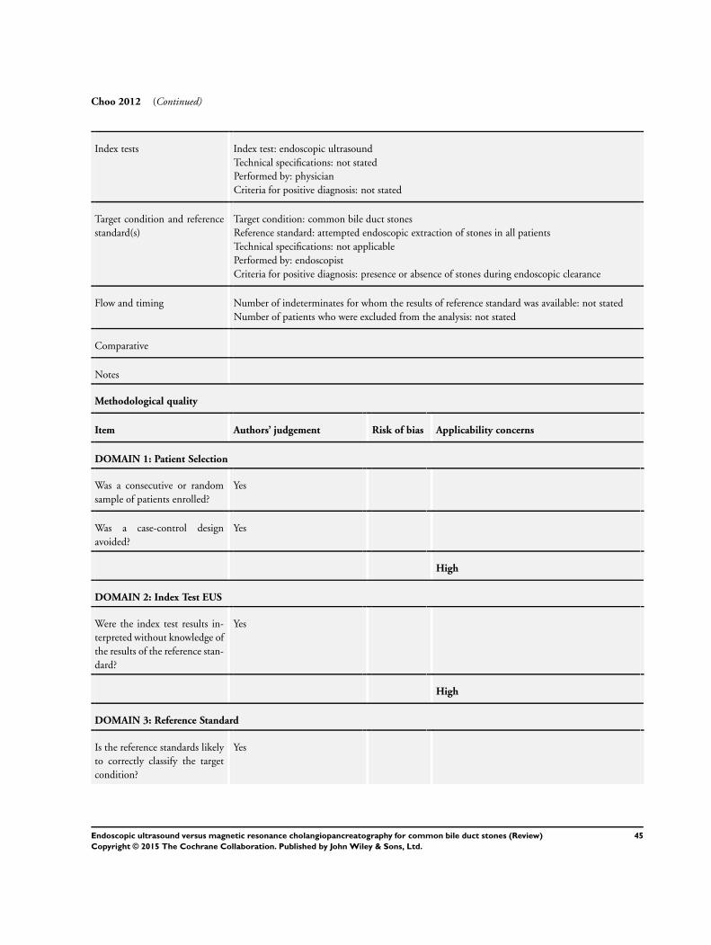
Choo 2012 (Continued)
Index tests Index test: endoscopic ultrasoundTechnical specifications: not statedPerformed by: physicianCriteria for positive diagnosis: not stated
Target condition and referencestandard(s)
Target condition: common bile duct stonesReference standard: attempted endoscopic extraction of stones in all patientsTechnical specifications: not applicablePerformed by: endoscopistCriteria for positive diagnosis: presence or absence of stones during endoscopic clearance
Flow and timing Number of indeterminates for whom the results of reference standard was available: not statedNumber of patients who were excluded from the analysis: not stated
Comparative
Notes
Methodological quality
Item Authors’ judgement Risk of bias Applicability concerns
DOMAIN 1: Patient Selection
Was a consecutive or randomsample of patients enrolled?
Yes
Was a case-control designavoided?
Yes
High
DOMAIN 2: Index Test EUS
Were the index test results in-terpreted without knowledge ofthe results of the reference stan-dard?
Yes
High
DOMAIN 3: Reference Standard
Is the reference standards likelyto correctly classify the targetcondition?
Yes
45Endoscopic ultrasound versus magnetic resonance cholangiopancreatography for common bile duct stones (Review)
Copyright © 2015 The Cochrane Collaboration. Published by John Wiley & Sons, Ltd.

Choo 2012 (Continued)
Were the reference standard re-sultsinterpreted without knowledgeof the results of the index tests?
Yes
Low
DOMAIN 4: Flow and Timing
Was there an appropriate inter-val between index test and ref-erence standard?
Yes
Did all patients receive the samereference standard?
Yes
Were all patients included in theanalysis?
Unclear
De Ledinghen 1999
Study characteristics
Patient sampling Type of study: prospective studyConsecutive or random sample: unclear
Patient characteristics and set-ting
Sample size: 43Females: 25 (58.1%)Age: 61 yearsPresentation:Inclusion criteriaPatients with clinical or biochemical signs of choledocholithiasis according to the following criteria:1. Combination of epigastric or right upper quadrant pain with fever or jaundice2. One or two of the previous signs together with an elevation of serum alkaline phosphatase levelor an elevation of serum gamma glutamyl transpeptidase or transaminase level more than the upperlimit of normal3. Acute pancreatitis4. Unexplained cholestasis defined by an elevation of serum alkaline phosphatase level and anelevation of serum gamma glutamyl transpeptidase level to more than two times the upper limit ofnormalExclusion criteria1. Long-term daily alcohol intake exceeded 80 g2. Taking a hepatotoxic drug3. Serum hepatitis B or C antibodies were presentSetting: secondary care (Hepatogastroenterology Department, France)
46Endoscopic ultrasound versus magnetic resonance cholangiopancreatography for common bile duct stones (Review)
Copyright © 2015 The Cochrane Collaboration. Published by John Wiley & Sons, Ltd.

De Ledinghen 1999 (Continued)
Index tests Index test: magnetic resonance cholangiopancreatographyTechnical specifications: 1 T magnet; SiemensPerformed by: two experienced radiologists jointlyCriteria for positive diagnosis: a round, oval, or multifaceted area of signal void (hypointensity) waspresent within the lumen of the hyperintense bile ductIndex test: endoscopic ultrasoundTechnical specifications: GF EUM20; OlympusPerformed by: not statedCriteria for positive diagnosis: a hyperechoic structure within the common bile duct sometimesassociated with an acoustic shadow
Target condition and referencestandard(s)
Target condition: common bile duct stonesReference standard: attempted endoscopic or surgical extraction of stones in all patientsTechnical specifications: not applicablePerformed by: endoscopists and surgeonsCriteria for positive diagnosis: presence or absence of stones during endoscopic or surgical clearance
Flow and timing Number of indeterminates for whom the results of reference standard was available: not statedNumber of patients who were excluded from the analysis: 11 (25.6%)
Comparative
Notes
Methodological quality
Item Authors’ judgement Risk of bias Applicability concerns
DOMAIN 1: Patient Selection
Was a consecutive or randomsample of patients enrolled?
Unclear
Was a case-control designavoided?
Yes
High
DOMAIN 2: Index Test MRCP
Were the index test results in-terpreted without knowledge ofthe results of the reference stan-dard?
Yes
Low
DOMAIN 2: Index Test EUS
47Endoscopic ultrasound versus magnetic resonance cholangiopancreatography for common bile duct stones (Review)
Copyright © 2015 The Cochrane Collaboration. Published by John Wiley & Sons, Ltd.

De Ledinghen 1999 (Continued)
Were the index test results in-terpreted without knowledge ofthe results of the reference stan-dard?
Yes
Low
DOMAIN 3: Reference Standard
Is the reference standards likelyto correctly classify the targetcondition?
Yes
Were the reference standard re-sultsinterpreted without knowledgeof the results of the index tests?
Unclear
Low
DOMAIN 4: Flow and Timing
Was there an appropriate inter-val between index test and ref-erence standard?
Unclear
Did all patients receive the samereference standard?
Yes
Were all patients included in theanalysis?
No
Fazel 2002
Study characteristics
Patient sampling Type of study: unclear whether prospective or retrospective studyConsecutive or random sample: unclear
Patient characteristics and set-ting
Sample size: 40Females: not statedAge: not statedPresentation:Inclusion criteriaPatients with suspicion of biliary stone disease on the basis of symptoms and signs suggestiveof choledocholithiasis (biliary colic, abnormal liver function tests, or abnormal transabdominalultrasound)
48Endoscopic ultrasound versus magnetic resonance cholangiopancreatography for common bile duct stones (Review)
Copyright © 2015 The Cochrane Collaboration. Published by John Wiley & Sons, Ltd.

Fazel 2002 (Continued)
Setting: care setting not stated, USA
Index tests Index test: endoscopic ultrasoundTechnical specifications: not statedPerformed by: not statedCriteria for positive diagnosis: not stated
Target condition and referencestandard(s)
Target condition: common bile duct stonesReference standard: attempted endoscopic extraction of stones in all patientsTechnical specifications: not applicablePerformed by: endoscopists and surgeonsCriteria for positive diagnosis: presence or absence of stones during endoscopic or surgical clearance
Flow and timing Number of indeterminates for whom the results of reference standard was available: not statedNumber of patients who were excluded from the analysis: not stated
Comparative
Notes
Methodological quality
Item Authors’ judgement Risk of bias Applicability concerns
DOMAIN 1: Patient Selection
Was a consecutive or randomsample of patients enrolled?
Unclear
Was a case-control designavoided?
Yes
High
DOMAIN 2: Index Test EUS
Were the index test results in-terpreted without knowledge ofthe results of the reference stan-dard?
Unclear
High
DOMAIN 3: Reference Standard
Is the reference standards likelyto correctly classify the targetcondition?
Yes
49Endoscopic ultrasound versus magnetic resonance cholangiopancreatography for common bile duct stones (Review)
Copyright © 2015 The Cochrane Collaboration. Published by John Wiley & Sons, Ltd.

Fazel 2002 (Continued)
Were the reference standard re-sultsinterpreted without knowledgeof the results of the index tests?
Unclear
Low
DOMAIN 4: Flow and Timing
Was there an appropriate inter-val between index test and ref-erence standard?
Unclear
Did all patients receive the samereference standard?
Yes
Were all patients included in theanalysis?
Unclear
Fernandez-Esparrach 2007
Study characteristics
Patient sampling Type of study: prospective studyConsecutive or random sample: consecutive patients
Patient characteristics and set-ting
Sample size: 159Females: 74 (46.5%)Age: 68 yearsPresentation:Inclusion criteria1. Unexplained common bile duct dilation in standard US, independently of clinical symptoms2. Non-dilated common bile duct and a high probability of having choledocholithiasis (cholangitis,jaundice, non-severe pancreatitis, alkaline phosphatase < twice the upper normal limit or increasedgamma glutamyl transferase, alanine aminotransferase or aspartate aminotransferaseExclusion criteria1. Acute severe biliary pancreatitisSetting: secondary care (Gastroenterology and Surgery Departments, Spain)
Index tests Index test: magnetic resonance cholangiopancreatographyTechnical specifications: 1.5 T magnet; SiemensPerformed by: not statedCriteria for positive diagnosis: a round, oval, or multifaceted area of signal void (hypointensity) waspresent inside the lumen of the hyperintense bile ductIndex test: endoscopic ultrasoundTechnical specifications: GF UM20 or GF UM160; Olympus
50Endoscopic ultrasound versus magnetic resonance cholangiopancreatography for common bile duct stones (Review)
Copyright © 2015 The Cochrane Collaboration. Published by John Wiley & Sons, Ltd.

Fernandez-Esparrach 2007 (Continued)
Performed by: not statedCriteria for positive diagnosis: visualisation of one or more hyperechoic images inside the commonbile duct with or without acoustic shadow
Target condition and referencestandard(s)
Target condition: common bile duct stonesReference standard: endoscopic or surgical extraction of stones in patients with positive EUS andclinical follow-up of minimum 6 months in patients with negative EUSTechnical specifications: not applicablePerformed by: endoscopists, surgeons, and cliniciansCriteria for positive diagnosis: endoscopic or surgical extraction of stones in patients with positiveEUS and clinical follow-up of minimum 6 months in patients with negative EUS
Flow and timing Number of indeterminates for whom the results of reference standard was available: not statedNumber of patients who were excluded from the analysis: 24 (15.1%)
Comparative
Notes
Methodological quality
Item Authors’ judgement Risk of bias Applicability concerns
DOMAIN 1: Patient Selection
Was a consecutive or randomsample of patients enrolled?
Yes
Was a case-control designavoided?
Yes
Low
DOMAIN 2: Index Test MRCP
Were the index test results in-terpreted without knowledge ofthe results of the reference stan-dard?
Unclear
Low
DOMAIN 2: Index Test EUS
Were the index test results in-terpreted without knowledge ofthe results of the reference stan-dard?
Unclear
Low
51Endoscopic ultrasound versus magnetic resonance cholangiopancreatography for common bile duct stones (Review)
Copyright © 2015 The Cochrane Collaboration. Published by John Wiley & Sons, Ltd.
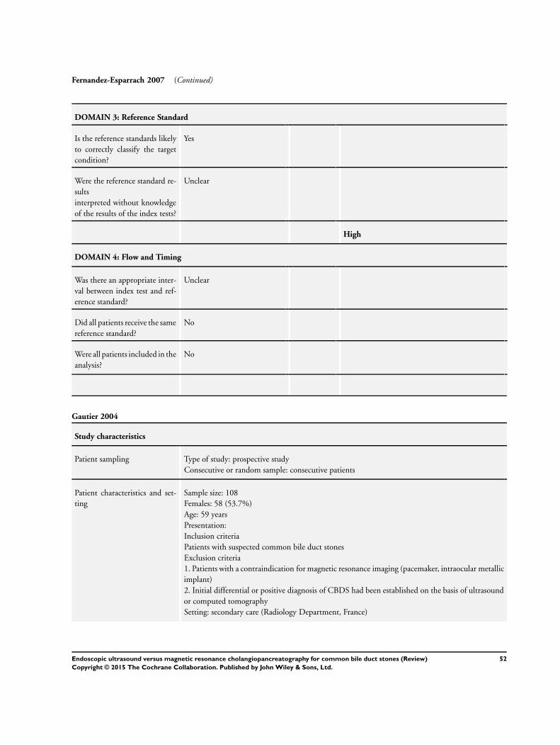
Fernandez-Esparrach 2007 (Continued)
DOMAIN 3: Reference Standard
Is the reference standards likelyto correctly classify the targetcondition?
Yes
Were the reference standard re-sultsinterpreted without knowledgeof the results of the index tests?
Unclear
High
DOMAIN 4: Flow and Timing
Was there an appropriate inter-val between index test and ref-erence standard?
Unclear
Did all patients receive the samereference standard?
No
Were all patients included in theanalysis?
No
Gautier 2004
Study characteristics
Patient sampling Type of study: prospective studyConsecutive or random sample: consecutive patients
Patient characteristics and set-ting
Sample size: 108Females: 58 (53.7%)Age: 59 yearsPresentation:Inclusion criteriaPatients with suspected common bile duct stonesExclusion criteria1. Patients with a contraindication for magnetic resonance imaging (pacemaker, intraocular metallicimplant)2. Initial differential or positive diagnosis of CBDS had been established on the basis of ultrasoundor computed tomographySetting: secondary care (Radiology Department, France)
52Endoscopic ultrasound versus magnetic resonance cholangiopancreatography for common bile duct stones (Review)
Copyright © 2015 The Cochrane Collaboration. Published by John Wiley & Sons, Ltd.

Gautier 2004 (Continued)
Index tests Index test: magnetic resonance cholangiopancreatographyTechnical specifications: 1.5 T magnet; SiemensPerformed by: two radiologists interpreted the scan independently and in the case of discrepancy, athird radiologist made the final decisionCriteria for positive diagnosis: low intensity intraductal signal surrounded by a high intensity liquidsignal
Target condition and referencestandard(s)
Target condition: common bile duct stonesReference standard: endoscopic or surgical extraction of stones or clinical follow-up of minimum 6monthsTechnical specifications: not applicablePerformed by: endoscopists, surgeons, and cliniciansCriteria for positive diagnosis: endoscopic, surgical extraction of stones, clinical follow-up of mini-mum 6 months
Flow and timing Number of indeterminates for whom the results of reference standard was available: not statedNumber of patients who were excluded from the analysis: 9 (8.3%)
Comparative
Notes
Methodological quality
Item Authors’ judgement Risk of bias Applicability concerns
DOMAIN 1: Patient Selection
Was a consecutive or randomsample of patients enrolled?
Yes
Was a case-control designavoided?
Yes
Low
DOMAIN 2: Index Test MRCP
Were the index test results in-terpreted without knowledge ofthe results of the reference stan-dard?
Unclear
Low
DOMAIN 3: Reference Standard
53Endoscopic ultrasound versus magnetic resonance cholangiopancreatography for common bile duct stones (Review)
Copyright © 2015 The Cochrane Collaboration. Published by John Wiley & Sons, Ltd.

Gautier 2004 (Continued)
Is the reference standards likelyto correctly classify the targetcondition?
Yes
Were the reference standard re-sultsinterpreted without knowledgeof the results of the index tests?
Unclear
High
DOMAIN 4: Flow and Timing
Was there an appropriate inter-val between index test and ref-erence standard?
Yes
Did all patients receive the samereference standard?
No
Were all patients included in theanalysis?
No
Guarise 2005
Study characteristics
Patient sampling Type of study: unclear whether prospective or retrospective studyConsecutive or random sample: consecutive patients
Patient characteristics and set-ting
Sample size: 170Females: not statedAge: not statedPresentation:Inclusion criteriaPatients who underwent magnetic resonance cholangiopancreatography and endoscopic retrogradecholangiopancreatography for clinically suspected biliary diseaseSetting: secondary care (Radiology Department, Italy)
Index tests Index test: magnetic resonance cholangiopancreatographyTechnical specifications: 1.5 T magnet; SiemensPerformed by: two radiologists with experience in gastrointestinal diseaseCriteria for positive diagnosis: a rounded and oval signal defect within the bile duct in at least twoprojections and located in the dependent portion of the duct
54Endoscopic ultrasound versus magnetic resonance cholangiopancreatography for common bile duct stones (Review)
Copyright © 2015 The Cochrane Collaboration. Published by John Wiley & Sons, Ltd.

Guarise 2005 (Continued)
Target condition and referencestandard(s)
Target condition: common bile duct stonesReference standard: endoscopic extraction of stones in all patientsTechnical specifications: not applicablePerformed by: endoscopists (the images were interpreted as consensus with radiologist)Criteria for positive diagnosis: endoscopic extraction of stones in all patients
Flow and timing Number of indeterminates for whom the results of reference standard was available: not statedNumber of patients who were excluded from the analysis: 23 (13.5%)
Comparative
Notes
Methodological quality
Item Authors’ judgement Risk of bias Applicability concerns
DOMAIN 1: Patient Selection
Was a consecutive or randomsample of patients enrolled?
Yes
Was a case-control designavoided?
Yes
Low
DOMAIN 2: Index Test MRCP
Were the index test results in-terpreted without knowledge ofthe results of the reference stan-dard?
Unclear
Low
DOMAIN 3: Reference Standard
Is the reference standards likelyto correctly classify the targetcondition?
Yes
Were the reference standard re-sultsinterpreted without knowledgeof the results of the index tests?
Yes
Low
55Endoscopic ultrasound versus magnetic resonance cholangiopancreatography for common bile duct stones (Review)
Copyright © 2015 The Cochrane Collaboration. Published by John Wiley & Sons, Ltd.
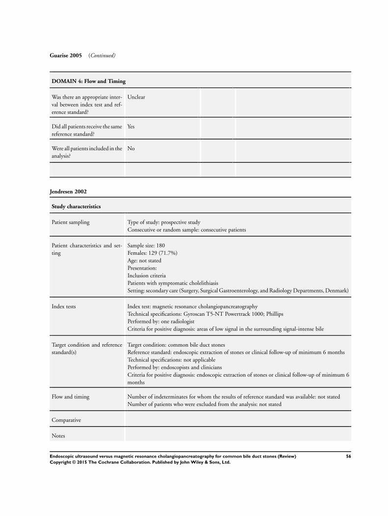
Guarise 2005 (Continued)
DOMAIN 4: Flow and Timing
Was there an appropriate inter-val between index test and ref-erence standard?
Unclear
Did all patients receive the samereference standard?
Yes
Were all patients included in theanalysis?
No
Jendresen 2002
Study characteristics
Patient sampling Type of study: prospective studyConsecutive or random sample: consecutive patients
Patient characteristics and set-ting
Sample size: 180Females: 129 (71.7%)Age: not statedPresentation:Inclusion criteriaPatients with symptomatic cholelithiasisSetting: secondary care (Surgery, Surgical Gastroenterology, and Radiology Departments, Denmark)
Index tests Index test: magnetic resonance cholangiopancreatographyTechnical specifications: Gyroscan T5-NT Powertrack 1000; PhillipsPerformed by: one radiologistCriteria for positive diagnosis: areas of low signal in the surrounding signal-intense bile
Target condition and referencestandard(s)
Target condition: common bile duct stonesReference standard: endoscopic extraction of stones or clinical follow-up of minimum 6 monthsTechnical specifications: not applicablePerformed by: endoscopists and cliniciansCriteria for positive diagnosis: endoscopic extraction of stones or clinical follow-up of minimum 6months
Flow and timing Number of indeterminates for whom the results of reference standard was available: not statedNumber of patients who were excluded from the analysis: not stated
Comparative
Notes
56Endoscopic ultrasound versus magnetic resonance cholangiopancreatography for common bile duct stones (Review)
Copyright © 2015 The Cochrane Collaboration. Published by John Wiley & Sons, Ltd.

Jendresen 2002 (Continued)
Methodological quality
Item Authors’ judgement Risk of bias Applicability concerns
DOMAIN 1: Patient Selection
Was a consecutive or randomsample of patients enrolled?
Yes
Was a case-control designavoided?
Yes
Low
DOMAIN 2: Index Test MRCP
Were the index test results in-terpreted without knowledge ofthe results of the reference stan-dard?
Yes
Low
DOMAIN 3: Reference Standard
Is the reference standards likelyto correctly classify the targetcondition?
Yes
Were the reference standard re-sultsinterpreted without knowledgeof the results of the index tests?
No
High
DOMAIN 4: Flow and Timing
Was there an appropriate inter-val between index test and ref-erence standard?
Yes
Did all patients receive the samereference standard?
No
Were all patients included in theanalysis?
Yes
57Endoscopic ultrasound versus magnetic resonance cholangiopancreatography for common bile duct stones (Review)
Copyright © 2015 The Cochrane Collaboration. Published by John Wiley & Sons, Ltd.

Kohut 2002
Study characteristics
Patient sampling Type of study: prospective studyConsecutive or random sample: unclear
Patient characteristics and set-ting
Sample size: 134Females: 109 (81.3%)Age: 57 yearsPresentation:Inclusion criteriaPatients suspected of having common bile duct stones based on the following features1. Biliary colic with elevated levels of biochemical values (bilirubin, transaminases, alkaline phos-phatase, g-glutamyl transpeptidase), and enlarged bile ducts (> 7 mm in patients with gallbladder insitu or > 9 mm in post-cholecystectomy patients) or suspicion of bile duct stones on conventionalultrasound, done currently or in the previous 6 monthsExclusion criteria1. Patients with suspicion of biliary or pancreatic malignancy on CT scan2. Current acute biliary pancreatitis or cholangitis, or bothSetting: secondary care (Gastroenterology Departments, Poland)
Index tests Index test: endoscopic ultrasound
Technical specifications: FG 32 UA; Pentax
Performed by: not statedCriteria for positive diagnosis: single or multiple hyperechoic structures within the biliary tree withacoustic shadowing were found
Target condition and referencestandard(s)
Target condition: common bile duct stonesReference standard: attempted endoscopic or surgical extraction of stones in all patientsTechnical specifications: not applicablePerformed by: endoscopists and surgeonsCriteria for positive diagnosis: presence or absence of stones during endoscopic or surgical clearance
Flow and timing Number of indeterminates for whom the results of reference standard was available: not statedNumber of patients who were excluded from the analysis: not stated
Comparative
Notes
Methodological quality
Item Authors’ judgement Risk of bias Applicability concerns
DOMAIN 1: Patient Selection
Was a consecutive or randomsample of patients enrolled?
Unclear
58Endoscopic ultrasound versus magnetic resonance cholangiopancreatography for common bile duct stones (Review)
Copyright © 2015 The Cochrane Collaboration. Published by John Wiley & Sons, Ltd.

Kohut 2002 (Continued)
Was a case-control designavoided?
Yes
High
DOMAIN 2: Index Test EUS
Were the index test results in-terpreted without knowledge ofthe results of the reference stan-dard?
Unclear
Low
DOMAIN 3: Reference Standard
Is the reference standards likelyto correctly classify the targetcondition?
Yes
Were the reference standard re-sultsinterpreted without knowledgeof the results of the index tests?
Unclear
Low
DOMAIN 4: Flow and Timing
Was there an appropriate inter-val between index test and ref-erence standard?
Yes
Did all patients receive the samereference standard?
Yes
Were all patients included in theanalysis?
Unclear
Liu 2001
Study characteristics
Patient sampling Type of study: prospective studyConsecutive or random sample: consecutive patients
59Endoscopic ultrasound versus magnetic resonance cholangiopancreatography for common bile duct stones (Review)
Copyright © 2015 The Cochrane Collaboration. Published by John Wiley & Sons, Ltd.

Liu 2001 (Continued)
Patient characteristics and set-ting
Sample size: 100Females: 49 (49.0%)Age: 61 yearsPresentation:Inclusion criteria1. Patients presenting with acute pancreatitisExclusion criteria1. Known diagnosis of recurrent pancreatitis related to chronic alcoholism or hyperlipidaemia2. Post-endoscopic retrograde cholangiopancreatography pancreatitis when performed for reasonsother than suspected biliary stonesSetting: secondary care (Surgery Department, Hong Kong, China)
Index tests Index test: endoscopic ultrasoundTechnical specifications: GF-UM20 or JF-UM20; Olympus; 7.5 MHz probePerformed by: endoscopistCriteria for positive diagnosis: a persistent echogenic focus with or without posterior acousticshadowing was considered a biliary stone, microlithiasis, or sludge
Target condition and referencestandard(s)
Target condition: common bile duct stonesReference standard: endoscopic extraction of stones in patients with positive EUS and clinicalfollow-up of minimum 12 months in patients with negative EUSTechnical specifications: not applicablePerformed by: endoscopists, surgeons, and cliniciansCriteria for positive diagnosis: endoscopic extraction of stones in patients with positive EUS andclinical follow-up of minimum 12 months in patients with negative EUS
Flow and timing Number of indeterminates for whom the results of reference standard was available: not statedNumber of patients who were excluded from the analysis: not stated
Comparative
Notes
Methodological quality
Item Authors’ judgement Risk of bias Applicability concerns
DOMAIN 1: Patient Selection
Was a consecutive or randomsample of patients enrolled?
Yes
Was a case-control designavoided?
Yes
Low
DOMAIN 2: Index Test EUS
60Endoscopic ultrasound versus magnetic resonance cholangiopancreatography for common bile duct stones (Review)
Copyright © 2015 The Cochrane Collaboration. Published by John Wiley & Sons, Ltd.

Liu 2001 (Continued)
Were the index test results in-terpreted without knowledge ofthe results of the reference stan-dard?
Unclear
Low
DOMAIN 3: Reference Standard
Is the reference standards likelyto correctly classify the targetcondition?
Yes
Were the reference standard re-sultsinterpreted without knowledgeof the results of the index tests?
Unclear
High
DOMAIN 4: Flow and Timing
Was there an appropriate inter-val between index test and ref-erence standard?
Yes
Did all patients receive the samereference standard?
No
Were all patients included in theanalysis?
Yes
Miletic 2006
Study characteristics
Patient sampling Type of study: prospective studyConsecutive or random sample: consecutive patients
Patient characteristics and set-ting
Sample size: 337Females: 174 (51.6%)Age: 65 yearsPresentation:Inclusion criteriaPatients with suspected CBD stones undergoing magnetic resonance cholangiopancreatographySetting: secondary care (Croatia)
61Endoscopic ultrasound versus magnetic resonance cholangiopancreatography for common bile duct stones (Review)
Copyright © 2015 The Cochrane Collaboration. Published by John Wiley & Sons, Ltd.

Miletic 2006 (Continued)
Index tests Index test: magnetic resonance cholangiopancreatographyTechnical specifications: 0.5 T magnet; ShimadzuPerformed by: two radiologists interpreted the scan independently and arrived at a consensusCriteria for positive diagnosis: not stated
Target condition and referencestandard(s)
Target condition: common bile duct stonesReference standard: endoscopic or surgical extraction of stones in patients and clinical follow-up ofminimum 12 months in patients with negative endoscopic retrograde cholangiopancreatographyTechnical specifications: not applicablePerformed by: endoscopists, surgeons, and cliniciansCriteria for positive diagnosis: endoscopic or surgical extraction of stones in patients and clinicalfollow-up of minimum 12 months in patients with negative endoscopic retrograde cholangiopan-creatography
Flow and timing Number of indeterminates for whom the results of reference standard was available: not statedNumber of patients who were excluded from the analysis: 29 (8.6%)
Comparative
Notes
Methodological quality
Item Authors’ judgement Risk of bias Applicability concerns
DOMAIN 1: Patient Selection
Was a consecutive or randomsample of patients enrolled?
Yes
Was a case-control designavoided?
Yes
Low
DOMAIN 2: Index Test MRCP
Were the index test results in-terpreted without knowledge ofthe results of the reference stan-dard?
Unclear
High
DOMAIN 3: Reference Standard
Is the reference standards likelyto correctly classify the targetcondition?
Yes
62Endoscopic ultrasound versus magnetic resonance cholangiopancreatography for common bile duct stones (Review)
Copyright © 2015 The Cochrane Collaboration. Published by John Wiley & Sons, Ltd.

Miletic 2006 (Continued)
Were the reference standard re-sultsinterpreted without knowledgeof the results of the index tests?
Unclear
High
DOMAIN 4: Flow and Timing
Was there an appropriate inter-val between index test and ref-erence standard?
Yes
Did all patients receive the samereference standard?
No
Were all patients included in theanalysis?
No
Montariol 1998
Study characteristics
Patient sampling Type of study: prospective studyConsecutive or random sample: consecutive patients
Patient characteristics and set-ting
Sample size: 240Females: 171 (71.3%)Age: 57 yearsPresentation:Inclusion criteria1. Patients with symptomatic cholelithiasis, scheduled for elective cholecystectomy or emergencyoperations within 48 hours for acute cholecystitisExclusion criteria1. Cholelithiasis was asymptomatic2. Preoperative risk of CBD stones was less than 5%3. Patients had symptomatic choledocholithiasis defined as combination of clinical symptoms (pan-creatic pain and jaundice), biochemical abnormalities (serum aminotransferase, alkaline phosphataseor γ -glutamyl transpeptidase levels more than twice normal values, serum bilirubin levels >50 µmol/L, and serum amylase and lipase levels more than fourfold and threefold, respectively), and mor-phologic features (presence of hyperechoic image in the CBD on ultrasonographySetting: secondary care (Surgery Departments, France)
Index tests Index test: endoscopic ultrasoundTechnical specifications: EUM3 and EUM20; Olympus; 7.5 MHz probePerformed by: experienced and selected operators
63Endoscopic ultrasound versus magnetic resonance cholangiopancreatography for common bile duct stones (Review)
Copyright © 2015 The Cochrane Collaboration. Published by John Wiley & Sons, Ltd.

Montariol 1998 (Continued)
Criteria for positive diagnosis: stones were described as hyperechoic images in the different partsof the CBD, identified because of their acoustic shadow and usually mobile spontaneously or withchanging positions
Target condition and referencestandard(s)
Target condition: common bile duct stonesReference standard: surgical extraction of stones in patients with positive EUS and clinical follow-up of minimum 12 months in patients with negative EUSTechnical specifications: not applicablePerformed by: surgeons and cliniciansCriteria for positive diagnosis: surgical extraction of stones in patients with positive EUS and clinicalfollow-up of minimum 12 months in patients with negative EUS
Flow and timing Number of indeterminates for whom the results of reference standard was available: not statedNumber of patients who were excluded from the analysis: 25 (10.4%)
Comparative
Notes
Methodological quality
Item Authors’ judgement Risk of bias Applicability concerns
DOMAIN 1: Patient Selection
Was a consecutive or randomsample of patients enrolled?
Yes
Was a case-control designavoided?
Yes
Low
DOMAIN 2: Index Test EUS
Were the index test results in-terpreted without knowledge ofthe results of the reference stan-dard?
Unclear
Low
DOMAIN 3: Reference Standard
Is the reference standards likelyto correctly classify the targetcondition?
Yes
64Endoscopic ultrasound versus magnetic resonance cholangiopancreatography for common bile duct stones (Review)
Copyright © 2015 The Cochrane Collaboration. Published by John Wiley & Sons, Ltd.

Montariol 1998 (Continued)
Were the reference standard re-sultsinterpreted without knowledgeof the results of the index tests?
No
High
DOMAIN 4: Flow and Timing
Was there an appropriate inter-val between index test and ref-erence standard?
Yes
Did all patients receive the samereference standard?
No
Were all patients included in theanalysis?
No
Ney 2005
Study characteristics
Patient sampling Type of study: prospective studyConsecutive or random sample: consecutive patients
Patient characteristics and set-ting
Sample size: 68Females: 49 (72.1%)Age: 57 yearsPresentation:Inclusion criteria1. Dilated CBD (> 7 mm on conventional ultrasound) and/or hepatic biochemical parameterabnormalities (AST > 2 times normal; elevated alkaline phosphatase)Exclusion criteria1. Jaundiced or had clinical signs of cholangitis2. Acute pancreatitis3. Unequivocal evidence of CBD stones on US or CT scans or magnetic resonance cholangiopan-creatographySetting: secondary care (Surgery Department, Brazil)
Index tests Index test: endoscopic ultrasoundTechnical specifications: GIF-UM20; Olympus; 7.5 or 12 MHz probePerformed by: not statedCriteria for positive diagnosis: stones were defined as mobile hyperechoic spots with an acousticshadow
65Endoscopic ultrasound versus magnetic resonance cholangiopancreatography for common bile duct stones (Review)
Copyright © 2015 The Cochrane Collaboration. Published by John Wiley & Sons, Ltd.

Ney 2005 (Continued)
Target condition and referencestandard(s)
Target condition: common bile duct stonesReference standard: endoscopic or surgical extraction of stones in patients with positive EUS andclinical follow-up minimum 11 months in patients with negative EUSTechnical specifications: not applicablePerformed by: endoscopists, surgeons, and cliniciansCriteria for positive diagnosis: endoscopic or surgical extraction of stones in patients with positiveEUS and clinical follow-up minimum 11 months in patients with negative EUS
Flow and timing Number of indeterminates for whom the results of reference standard was available: not statedNumber of patients who were excluded from the analysis: not stated
Comparative
Notes
Methodological quality
Item Authors’ judgement Risk of bias Applicability concerns
DOMAIN 1: Patient Selection
Was a consecutive or randomsample of patients enrolled?
Yes
Was a case-control designavoided?
Yes
Low
DOMAIN 2: Index Test EUS
Were the index test results in-terpreted without knowledge ofthe results of the reference stan-dard?
Unclear
Low
DOMAIN 3: Reference Standard
Is the reference standards likelyto correctly classify the targetcondition?
Yes
Were the reference standard re-sultsinterpreted without knowledgeof the results of the index tests?
Unclear
66Endoscopic ultrasound versus magnetic resonance cholangiopancreatography for common bile duct stones (Review)
Copyright © 2015 The Cochrane Collaboration. Published by John Wiley & Sons, Ltd.

Ney 2005 (Continued)
High
DOMAIN 4: Flow and Timing
Was there an appropriate inter-val between index test and ref-erence standard?
Yes
Did all patients receive the samereference standard?
No
Were all patients included in theanalysis?
Yes
Norton 1997
Study characteristics
Patient sampling Type of study: unclear whether prospective or retrospective studyConsecutive or random sample: unclear
Patient characteristics and set-ting
Sample size: 50Females: 34 (68.0%)Age: 63 yearsPresentation:Inclusion criteriaPatients with proven symptomatic gallstone disease and suspected bile duct stones because of thepresence of at least one of the following features1. Dilated (greater than 7 mm) bile duct on abdominal ultrasonography2. Clinical jaundice3. Gallstone pancreatitis4. Deranged liver functionSetting: secondary care (Surgery Department, United Kingdom)
Index tests Index test: endoscopic ultrasoundTechnical specifications: GF-UM 20; OlympusPerformed by: not statedCriteria for positive diagnosis: stones were recognized by their hyperechoic image and the acousticshadow commonly produced
Target condition and referencestandard(s)
Target condition: common bile duct stonesReference standard: endoscopic or surgical extraction of stones in patients with positive EUS andclinical follow-up minimum 6 months in patients with negative EUSTechnical specifications: not applicablePerformed by: endoscopists, surgeons, and cliniciansCriteria for positive diagnosis: endoscopic or surgical extraction of stones in patients with positive
67Endoscopic ultrasound versus magnetic resonance cholangiopancreatography for common bile duct stones (Review)
Copyright © 2015 The Cochrane Collaboration. Published by John Wiley & Sons, Ltd.
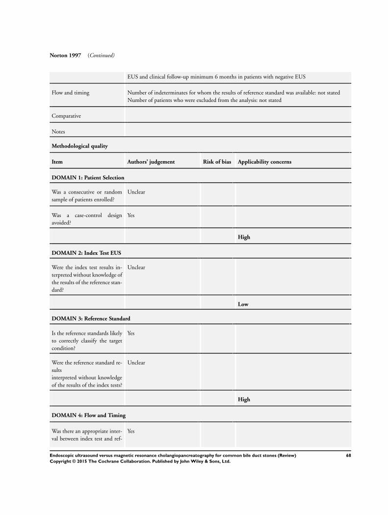
Norton 1997 (Continued)
EUS and clinical follow-up minimum 6 months in patients with negative EUS
Flow and timing Number of indeterminates for whom the results of reference standard was available: not statedNumber of patients who were excluded from the analysis: not stated
Comparative
Notes
Methodological quality
Item Authors’ judgement Risk of bias Applicability concerns
DOMAIN 1: Patient Selection
Was a consecutive or randomsample of patients enrolled?
Unclear
Was a case-control designavoided?
Yes
High
DOMAIN 2: Index Test EUS
Were the index test results in-terpreted without knowledge ofthe results of the reference stan-dard?
Unclear
Low
DOMAIN 3: Reference Standard
Is the reference standards likelyto correctly classify the targetcondition?
Yes
Were the reference standard re-sultsinterpreted without knowledgeof the results of the index tests?
Unclear
High
DOMAIN 4: Flow and Timing
Was there an appropriate inter-val between index test and ref-
Yes
68Endoscopic ultrasound versus magnetic resonance cholangiopancreatography for common bile duct stones (Review)
Copyright © 2015 The Cochrane Collaboration. Published by John Wiley & Sons, Ltd.

Norton 1997 (Continued)
erence standard?
Did all patients receive the samereference standard?
No
Were all patients included in theanalysis?
Unclear
Prat 1996
Study characteristics
Patient sampling Type of study: prospectiveConsecutive or random sample: consecutive patients
Patient characteristics and set-ting
Sample size: 121Females: 69 (57.0%)Age: 70 yearsPresentation:Inclusion criteria1. Strong suspicion of choledocholithiasis as determined by a combination of clinical symptoms(history of biliary colic, pancreatic pain, fever, jaundice), biochemical abnormalities (raised serumaminotransferases, alkaline phosphatase, or gamma-glutamyl transpeptidase more than twice thenormal value, serum bilirubin above 50 (micromol/L), and morphological features (common bileduct dilated to more than 8 mm in patients with the gallbladder in situ and 10 mm in patients withprevious cholecystectomy, or the presence of a hyperechoic image in the common bile duct).2. Endoscopic treatment would be chosen for the treatment of the stonesExclusion criteria1. Patients younger than 50 who had not had cholecystectomy2. Patients who declined to take partSetting: secondary care (Gastroenterology Department, France)
Index tests Index test: endoscopic ultrasoundTechnical specifications: GIF-EUM20; Olympus; 7.5 and 12 MHz probePerformed by: one of two experts in EUSCriteria for positive diagnosis: not stated
Target condition and referencestandard(s)
Target condition: common bile duct stonesReference standard: attempted endoscopic extraction of stones in all patientsTechnical specifications: not applicablePerformed by: endoscopists and surgeonsCriteria for positive diagnosis: presence or absence of stones during endoscopic clearance
Flow and timing Number of indeterminates for whom the results of reference standard was available: 1 (0.8%)Number of patients who were excluded from the analysis: 1 (0.8%)
69Endoscopic ultrasound versus magnetic resonance cholangiopancreatography for common bile duct stones (Review)
Copyright © 2015 The Cochrane Collaboration. Published by John Wiley & Sons, Ltd.

Prat 1996 (Continued)
Comparative
Notes
Methodological quality
Item Authors’ judgement Risk of bias Applicability concerns
DOMAIN 1: Patient Selection
Was a consecutive or randomsample of patients enrolled?
Yes
Was a case-control designavoided?
Yes
High
DOMAIN 2: Index Test EUS
Were the index test results in-terpreted without knowledge ofthe results of the reference stan-dard?
Unclear
High
DOMAIN 3: Reference Standard
Is the reference standards likelyto correctly classify the targetcondition?
Yes
Were the reference standard re-sultsinterpreted without knowledgeof the results of the index tests?
Yes
Low
DOMAIN 4: Flow and Timing
Was there an appropriate inter-val between index test and ref-erence standard?
Yes
Did all patients receive the samereference standard?
Yes
70Endoscopic ultrasound versus magnetic resonance cholangiopancreatography for common bile duct stones (Review)
Copyright © 2015 The Cochrane Collaboration. Published by John Wiley & Sons, Ltd.

Prat 1996 (Continued)
Were all patients included in theanalysis?
No
EUS: endoscopic ultrasound.MRCP: magnetic resonance cholangiopancreatography.
Characteristics of excluded studies [ordered by study ID]
Study Reason for exclusion
Adamek 1998 Not enough data for 2 x 2 table
Agapov 2006 Review of literature
Ahn 1998 Inappropriate reference standard
Ainsworth 2003 Not enough data for 2 x 2 table
Al-Jiffry 2010 Inappropriate reference standard
Alcaraz 2000 Inappropriate reference standard
Alhayaf 2008 Inappropriate reference standard
Aljebreen 2008 Inappropriate reference standard
Amouyal 1994 Inappropriate reference standard
Anderloni 2012 Not enough data for 2 x 2 table
Anderloni 2012a Not enough data for 2 x 2 table
Ang 2007 Inappropriate reference standard
Ang 2007a Inappropriate reference standard
Aube 2005 Inappropriate reference standard
Aubertin 1996 Inappropriate reference standard
Aubertin 1996a Inappropriate reference standard
71Endoscopic ultrasound versus magnetic resonance cholangiopancreatography for common bile duct stones (Review)
Copyright © 2015 The Cochrane Collaboration. Published by John Wiley & Sons, Ltd.

(Continued)
Basile 2000 Inappropriate reference standard
Becker 1997 Inappropriate reference standard
Berdah 2001 Inappropriate reference standard
Bhatt 2005 Not enough data for 2 x 2 table
Bilgin 2012 Inappropriate reference standard
Boboev 2012 Inappropriate reference standard
Bodula 2011 Inappropriate reference standard
Bokobza 1988 Review article
Boraschi 1999 Inappropriate reference standard
Brisbois 2001 Inappropriate reference standard
Calle 2006 Inappropriate reference standard
Calvo 2002 Inappropriate reference standard
Canto 1995 Not enough data for 2 x 2 table
Catalano 2000 Inappropriate reference standard
Cervi 2000 Inappropriate reference standard
Chak 1999 Inappropriate index test
Chan 1996 Not enough data for 2 x 2 table
Chan 2010 Inappropriate reference standard
Chandra 2010 Not enough data for 2 x 2 table
Chavez-Valencia 2009 Inappropriate reference standard
Chen 2003 Inappropriate reference standard
Chen 2012 Editorial
Chowdhury 1999 Inappropriate reference standard
Coakley 2002 Review article
72Endoscopic ultrasound versus magnetic resonance cholangiopancreatography for common bile duct stones (Review)
Copyright © 2015 The Cochrane Collaboration. Published by John Wiley & Sons, Ltd.

(Continued)
Contractor 2004 Inappropriate reference standard
Dalton 2005 Inappropriate reference standard
Danaci 2002 Inappropriate reference standard
Dancygier 1995 Not enough data for 2 x 2 table
De Waele 2007 Inappropriate reference standard
del Pozo 2011 Not enough data for 2 x 2 table
Demartines 2000 Inappropriate reference standard
Denis 1993 Inappropriate reference standard
Derodra 1986 Letter to editor
Di Angelo 2010 Not enough data for 2 x 2 table
Di Angelo 2011 Not enough data for 2 x 2 table
Dittrick 2005 Inappropriate reference standard
Duchmann 1999 Not enough data for 2 x 2 table
Dwerryhouse 1998 Inappropriate reference standard
Eshghi 2008 Inappropriate reference standard
Familiari 2004 Inappropriate reference standard
Fernandez 2001 Inappropriate reference standard
Filippone 2003 Inappropriate reference standard
Galvao 2007 Inappropriate reference standard
Griffin 2003 Inappropriate reference standard
Gul 2010 Inappropriate reference standard
Gupta 2008 Inappropriate reference standard
Hasan 2010 Inappropriate reference standard
73Endoscopic ultrasound versus magnetic resonance cholangiopancreatography for common bile duct stones (Review)
Copyright © 2015 The Cochrane Collaboration. Published by John Wiley & Sons, Ltd.

(Continued)
Hayashi 2002 Not enough data for 2 x 2 table
Ho 1999 Inappropriate reference standard
Hochwald 1998 Inappropriate reference standard
Holzknecht 1998 Inappropriate reference standard
Hrabar 2009 Inappropriate reference standard
Hussein 2002 Inappropriate reference standard
Isomoto 1998 Inappropriate reference standard
Ito 2001 Inappropriate reference standard
Janssen 2008 Not enough data for 2 x 2 table
Karakan 2009 Inappropriate reference standard
Kats 2003 Inappropriate reference standard
Kausar 2005 Not enough data for 2 x 2 table
Ke 2004 Inappropriate reference standard
Kejriwal 2004 Inappropriate reference standard
Kim 2002 Inappropriate reference standard
Kim 2005 Inappropriate reference standard
Kohut 2003 Not enough data for 2 x 2 table
Kondo 2005 Inappropriate reference standard
Lachter 2000 Inappropriate reference standard
Laghi 1998 Inappropriate reference standard
Laokpessi 2001 Inappropriate reference standard
Le Rhun 1999 Inappropriate reference standard
Lee 1996 Inappropriate reference standard
74Endoscopic ultrasound versus magnetic resonance cholangiopancreatography for common bile duct stones (Review)
Copyright © 2015 The Cochrane Collaboration. Published by John Wiley & Sons, Ltd.
(Continued)
Lee 2010 Inappropriate reference standard
Leytens 2001 Inappropriate reference standard
Liessi 1996 Inappropriate reference standard
Lim 2003 Inappropriate reference standard
Liu 1999 Inappropriate reference standard
Liu 2005 Inappropriate reference standard
Lomanto 1997 Inappropriate reference standard
Lomas 1999 Inappropriate reference standard
Lundorf 2000 Inappropriate reference standard
Magnuson 1997 Inappropriate reference standard
Magnuson 1999 Inappropriate reference standard
Makary 2005 Inappropriate reference standard
Maurea 2009 Not enough data for 2 x 2 table
Meduri 1998 Inappropriate reference standard
Mendler 1998 Inappropriate reference standard
Meroni 2004 Inappropriate reference standard
Miao 2008 Not enough data for 2 x 2 table
Mirbagheri 2005 Inappropriate reference standard
Mofidi 2008 Inappropriate reference standard
Moon 2005 Inappropriate reference standard
Morera 2006 Inappropriate reference standard
Morris-Stiff 2009 Inappropriate reference standard
Munir 2004 Inappropriate reference standard
75Endoscopic ultrasound versus magnetic resonance cholangiopancreatography for common bile duct stones (Review)
Copyright © 2015 The Cochrane Collaboration. Published by John Wiley & Sons, Ltd.

(Continued)
Musella 1998 Inappropriate reference standard
Nandalur 2008 Inappropriate reference standard
Nau 2011 Inappropriate reference standard
Nebiker 2009 Inappropriate reference standard
Neri 2000 Inappropriate reference standard
Norero 2008 Inappropriate reference standard
Okaniwa 2002 Inappropriate reference standard
Palazzo 1995 Inappropriate reference standard
Palazzo 1998 Overview article
Palmucci 2010 Inappropriate reference standard
Pamos 1998 Inappropriate reference standard
Pamos 2003 Inappropriate reference standard
Pavone 1996 Inappropriate reference standard
Pavone 1996a Inappropriate reference standard
Pavone 1996b Inappropriate reference standard
Pavone 1997 Inappropriate reference standard
Pavone 1997a Not enough data for 2 x 2 table
Polkowski 2001 Not enough data for 2 x 2 table
Pomakov 2007 Inappropriate reference standard
Pozo 2010 Not enough data for 2 x 2 table
Pulpeiro 2000 Inappropriate reference standard
Puri 2012 Not enough data for 2 x 2 table
Rahman 2010 Inappropriate reference standard
Regan 1996 Inappropriate reference standard
76Endoscopic ultrasound versus magnetic resonance cholangiopancreatography for common bile duct stones (Review)
Copyright © 2015 The Cochrane Collaboration. Published by John Wiley & Sons, Ltd.

(Continued)
Regan 1996a Not enough data for 2 x 2 table
Regan 1998 Inappropriate reference standard
Reinhold 1998 Inappropriate reference standard
Roig 1995 Not enough data for 2 x 2 table
Roig 1995a Not enough data for 2 x 2 table
Rudowicz-Pietruszewska 2002 Inappropriate reference standard
Sabbagh 2000 Not enough data for 2 x 2 table
Sajewicz 2006 Inappropriate reference standard
Sakai 2007 Not enough data for 2 x 2 table
Salmeron 1994 Inappropriate reference standard
Saruc 2001 Inappropriate reference standard
Scaffidi 2009 Inappropriate reference standard
Scheiman 2001 Inappropriate reference standard
Schmidt 2012 Inappropriate reference standard
Seifert 2004 Inadequate index test
Shafiq 2003 Not enough data for 2 x 2 table
Shamiyeh 2005 Inappropriate reference standard
Shanmugam 2005 Inappropriate reference standard
Shim 1995 Inappropriate reference standard
Simeone 1997 Inappropriate reference standard
Skorka 1982 Inappropriate reference standard
Soto 1996 Inappropriate reference standard
Soto 2000 Inappropriate reference standard
77Endoscopic ultrasound versus magnetic resonance cholangiopancreatography for common bile duct stones (Review)
Copyright © 2015 The Cochrane Collaboration. Published by John Wiley & Sons, Ltd.

(Continued)
Soto 2000a Inappropriate reference standard
Sotoudehmanesh 2007 Not enough data for 2 x 2 table
Sperlongano 2005 Not enough data for 2 x 2 table
Srinivasa 2010 Inappropriate reference standard
Stevens 1996 Inappropriate reference standard
Stiris 2000 Inappropriate reference standard
Sugiyama 1997 Inappropriate reference standard
Sugiyama 1998 Inappropriate reference standard
Sverrisson 2012 Not enough data for 2 x 2 table
Taylor 2002 Inappropriate reference standard
Tennoe 1999 Inappropriate reference standard
Topal 2003 Inappropriate reference standard
Tripathi 2002 Inappropriate reference standard
Uehara 1998 Inappropriate reference standard
Urban 2002 Inappropriate reference standard
Vaishali 2004 Inappropriate reference standard
Valji 1996 Inappropriate reference standard
Varghese 1999 Inappropriate reference standard
Varghese 2000 Inappropriate reference standard
Vazquez-Sequeiros 2005 Inappropriate reference standard
Vazquez-Sequeiros 2011 Inappropriate reference standard
Verma 2006 Systematic review
Watanabe 2003 Inappropriate reference standard
78Endoscopic ultrasound versus magnetic resonance cholangiopancreatography for common bile duct stones (Review)
Copyright © 2015 The Cochrane Collaboration. Published by John Wiley & Sons, Ltd.

(Continued)
Wehrmann 2009 Inappropriate reference standard
Wierzbicka-Paczos 1999 Not enough data for 2 x 2 table
Wong 2012 Inappropriate reference standard
Zaheer 2011 Not enough data for 2 x 2 table
Zaydan 2009 Inappropriate reference standard
Zhang 2012 Inappropriate reference standard
Zhi 2002 Not enough data for 2 x 2 table
Zidi 1997 Not enough data for 2 x 2 table
Zidi 1999 Inappropriate reference standard
79Endoscopic ultrasound versus magnetic resonance cholangiopancreatography for common bile duct stones (Review)
Copyright © 2015 The Cochrane Collaboration. Published by John Wiley & Sons, Ltd.

D A T A
Presented below are all the data for all of the tests entered into the review.
Tests. Data tables by test
TestNo. ofstudies
No. ofparticipants
1 Endoscopic ultrasound 13 15372 Magnetic resonance
cholangiopancreatography7 996
Test 1. Endoscopic ultrasound.
Review: Endoscopic ultrasound versus magnetic resonance cholangiopancreatography for common bile duct stones
Test: 1 Endoscopic ultrasound
Study TP FP FN TN Sensitivity Specificity Sensitivity Specificity
Ang 2012 33 0 0 79 1.00 [ 0.89, 1.00 ] 1.00 [ 0.95, 1.00 ]
Buscarini 2003 237 2 4 216 0.98 [ 0.96, 1.00 ] 0.99 [ 0.97, 1.00 ]
Canto 1998 16 1 3 44 0.84 [ 0.60, 0.97 ] 0.98 [ 0.88, 1.00 ]
Choo 2012 3 0 1 5 0.75 [ 0.19, 0.99 ] 1.00 [ 0.48, 1.00 ]
De Ledinghen 1999 10 1 0 21 1.00 [ 0.69, 1.00 ] 0.95 [ 0.77, 1.00 ]
Fazel 2002 15 1 2 22 0.88 [ 0.64, 0.99 ] 0.96 [ 0.78, 1.00 ]
Fernandez-Esparrach 2007 59 11 2 63 0.97 [ 0.89, 1.00 ] 0.85 [ 0.75, 0.92 ]
Kohut 2002 85 2 6 41 0.93 [ 0.86, 0.98 ] 0.95 [ 0.84, 0.99 ]
Liu 2001 33 1 1 65 0.97 [ 0.85, 1.00 ] 0.98 [ 0.92, 1.00 ]
Montariol 1998 35 12 6 162 0.85 [ 0.71, 0.94 ] 0.93 [ 0.88, 0.96 ]
Ney 2005 32 0 1 35 0.97 [ 0.84, 1.00 ] 1.00 [ 0.90, 1.00 ]
Norton 1997 21 1 3 25 0.88 [ 0.68, 0.97 ] 0.96 [ 0.80, 1.00 ]
Prat 1996 73 1 5 40 0.94 [ 0.86, 0.98 ] 0.98 [ 0.87, 1.00 ]
0 0.2 0.4 0.6 0.8 1 0 0.2 0.4 0.6 0.8 1
80Endoscopic ultrasound versus magnetic resonance cholangiopancreatography for common bile duct stones (Review)
Copyright © 2015 The Cochrane Collaboration. Published by John Wiley & Sons, Ltd.

Test 2. Magnetic resonance cholangiopancreatography.
Review: Endoscopic ultrasound versus magnetic resonance cholangiopancreatography for common bile duct stones
Test: 2 Magnetic resonance cholangiopancreatography
Study TP FP FN TN Sensitivity Specificity Sensitivity Specificity
Boraschi 2002 37 4 2 52 0.95 [ 0.83, 0.99 ] 0.93 [ 0.83, 0.98 ]
De Ledinghen 1999 10 6 0 16 1.00 [ 0.69, 1.00 ] 0.73 [ 0.50, 0.89 ]
Fernandez-Esparrach 2007 54 2 7 72 0.89 [ 0.78, 0.95 ] 0.97 [ 0.91, 1.00 ]
Gautier 2004 22 1 1 75 0.96 [ 0.78, 1.00 ] 0.99 [ 0.93, 1.00 ]
Guarise 2005 78 7 9 53 0.90 [ 0.81, 0.95 ] 0.88 [ 0.77, 0.95 ]
Jendresen 2002 20 1 6 153 0.77 [ 0.56, 0.91 ] 0.99 [ 0.96, 1.00 ]
Miletic 2006 112 7 3 186 0.97 [ 0.93, 0.99 ] 0.96 [ 0.93, 0.99 ]
0 0.2 0.4 0.6 0.8 1 0 0.2 0.4 0.6 0.8 1
A D D I T I O N A L T A B L E S
Table 1. Application of the QUADAS-2 tool for assessing methodological quality of included studies
Domain 1: Patientsampling
Signallingquestion
Signallingquestion
Signallingquestion
Risk of bias Concerns for ap-plicability
Patient sampling Was a consecutive orrandom sample ofpatients enrolled?
Was a case-controldesign avoided?
Did the study avoidinappropriate exclu-sions?
Could the selectionof patients have in-troduced bias?
Were there concernsthat the includedpatients and settingdid not match thereview question?
Yes: all consecutivepatients or randomsample of patientswith suspected com-mon bile duct stoneswere enrolledNo: selected pa-tients were enrolledUnclear: this wasnot clear form thereport
Yes: case-control de-sign was avoided.No: case-control de-sign was not avoidedUnclear: this wasnot clear from thereport.
Yes: thestudy avoided inap-propriate exclusions(i.e., difficult to di-agnose patients)No: the study ex-cluded patients in-appropriatelyUnclear: this wasnot clear from thereport
Low risk: ’yes’ for allsignalling questionsHigh risk: ’no’ or’unclear’ for at leastone signalling ques-tion
Low concern: the se-lected patients rep-resent the patientsin whom the testswill be used in clin-ical practice (pleasesee diagnostic path-way (Figure 1)High concern: therewas high concernthat patient selec-tion was performed
81Endoscopic ultrasound versus magnetic resonance cholangiopancreatography for common bile duct stones (Review)
Copyright © 2015 The Cochrane Collaboration. Published by John Wiley & Sons, Ltd.

Table 1. Application of the QUADAS-2 tool for assessing methodological quality of included studies (Continued)
in a such a waythat the includedpatients did not rep-resent the patientsin whom the testswill be used in clin-ical practice
Domain 2: Indextest
Index test(s) Werethe index test resultsinterpreted withoutknowledge of the re-sults of the referencestandard?
If a threshold wasused, was it pre-specified?
Could the conductor interpretation ofthe index test haveintroduced bias?
Were there concernsthat the index test,its conduct, or inter-preta-tion differ from thereview question?
Yes: in-dex test results wereinterpreted withoutknowledge of the re-sults of the referencestandardNo: index test re-sults were inter-preted with knowl-edge of the results ofthe reference stan-dardUnclear: this wasnot clear from thereport
Not applicable Low risk: ’yes’ for allsignalling questionsHigh risk: ’no’ or’unclear’ for at leastone of the two sig-nalling questions
High concern: therewas high concernthat the conductor interpretation ofthe index test differsfrom the way it islikely to be used inclinical practiceLow concern: therewaslow concern that theconduct or interpre-tation of the indextest differs from theway it is likely to beused in clinical prac-tice
Domain 3: Refer-ence standard
Target con-dition and referencestandard(s)
Was the referencestandard likely tocorrectly classify thetarget condition?
Were the referencestandard results in-terpretedwithout knowledgeof the results of theindex tests?
Could the referencestandard, its con-duct, or its inter-pretation have in-troduced bias?
Were there concernsthat the target con-dition as defined bythe reference stan-dard does not matchthe review question?
Yes: all patients un-derwent the accept-able reference stan-
Yes: reference stan-dard results wereinterpreted without
Low risk: ’yes’ for allsignalling questionsHigh risk: ’no’ or
Low concern:patients underwentendoscopic or sur-
82Endoscopic ultrasound versus magnetic resonance cholangiopancreatography for common bile duct stones (Review)
Copyright © 2015 The Cochrane Collaboration. Published by John Wiley & Sons, Ltd.
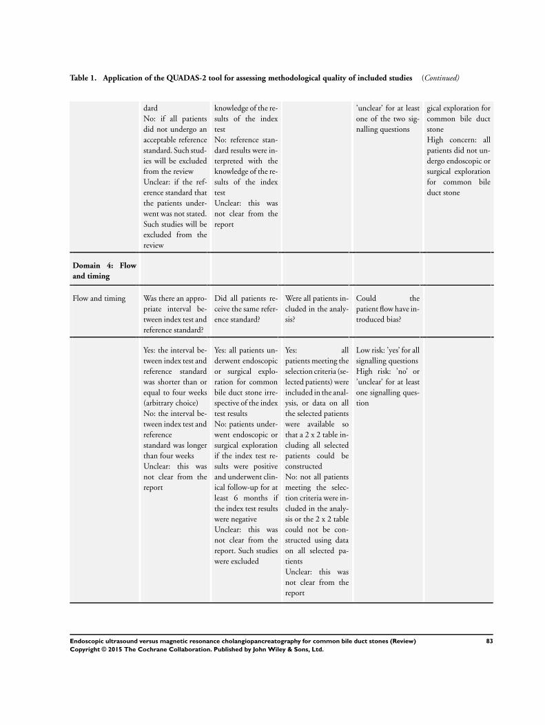
Table 1. Application of the QUADAS-2 tool for assessing methodological quality of included studies (Continued)
dardNo: if all patientsdid not undergo anacceptable referencestandard. Such stud-ies will be excludedfrom the reviewUnclear: if the ref-erence standard thatthe patients under-went was not stated.Such studies will beexcluded from thereview
knowledge of the re-sults of the indextestNo: reference stan-dard results were in-terpreted with theknowledge of the re-sults of the indextestUnclear: this wasnot clear from thereport
’unclear’ for at leastone of the two sig-nalling questions
gical exploration forcommon bile ductstoneHigh concern: allpatients did not un-dergo endoscopic orsurgical explorationfor common bileduct stone
Domain 4: Flowand timing
Flow and timing Was there an appro-priate interval be-tween index test andreference standard?
Did all patients re-ceive the same refer-ence standard?
Were all patients in-cluded in the analy-sis?
Could thepatient flow have in-troduced bias?
Yes: the interval be-tween index test andreference standardwas shorter than orequal to four weeks(arbitrary choice)No: the interval be-tween index test andreferencestandard was longerthan four weeksUnclear: this wasnot clear from thereport
Yes: all patients un-derwent endoscopicor surgical explo-ration for commonbile duct stone irre-spective of the indextest resultsNo: patients under-went endoscopic orsurgical explorationif the index test re-sults were positiveand underwent clin-ical follow-up for atleast 6 months ifthe index test resultswere negativeUnclear: this wasnot clear from thereport. Such studieswere excluded
Yes: allpatients meeting theselection criteria (se-lected patients) wereincluded in the anal-ysis, or data on allthe selected patientswere available sothat a 2 x 2 table in-cluding all selectedpatients could beconstructedNo: not all patientsmeeting the selec-tion criteria were in-cluded in the analy-sis or the 2 x 2 tablecould not be con-structed using dataon all selected pa-tientsUnclear: this wasnot clear from thereport
Low risk: ’yes’ for allsignalling questionsHigh risk: ’no’ or’unclear’ for at leastone signalling ques-tion
83Endoscopic ultrasound versus magnetic resonance cholangiopancreatography for common bile duct stones (Review)
Copyright © 2015 The Cochrane Collaboration. Published by John Wiley & Sons, Ltd.

A P P E N D I C E S
Appendix 1. Search strategies
Database Period of Search Search Strategy
MEDLINE (PubMed) 1946 until September 2012 (((bile duct[tiab] or biliary[tiab] OR CBD[tiab]) AND(stone[tiab] OR stones[tiab] OR calculus[tiab] OR cal-culi[tiab])) OR choledocholithiasis[tiab] OR cholelithi-asis[tiab] OR “Choledocholithiasis”[Mesh] OR “Com-mon Bile Duct Calculi ”[MESH] OR “Cholelithia-sis ”[MESH]) AND (CT[tiab] OR tomodensitome-try[tiab] OR MRI[tiab] OR NMRI[tiab] OR zeug-matogra*[tiab] OR ((computed[tiab] OR comput-erised[tiab] OR computerized[tiab] OR magneti*[tiab]OR MR[tiab] OR NMR[tiab] OR proton[tiab]) AND(tomogra*[tiab] OR scan[tiab] OR scans[tiab] OR imag-ing[tiab] OR cholangiogra*[tiab])) OR “Tomography,X-Ray Computed”[Mesh] OR “Magnetic ResonanceImaging”[Mesh] OR echogra*[tiab] OR ultrason*[tiab]OR ultrasound[tiab] OR EUS[tiab] OR “Ultra-sonography”[Mesh] OR “Endosonography”[Mesh] ORcholangiogra*[tiab] OR cholangio?pancreatogra*[tiab]OR cholangiosco*[tiab] OR choledochosco*[tiab] ORERCP[tiab] OR MRCP[tiab] OR “Cholangiogra-phy”[Mesh] OR “Cholangiopancreatography, MagneticResonance”[Mesh] OR liver function test[tiab] OR liverfunction tests[tiab] OR “Liver Function Tests”[Mesh])
EMBASE (OvidSP) 1947 until September 2012 1. (((bile duct or biliary or CBD) adj5 (stone or stones orcalculus or calculi)) or choledocholithiasis or cholelithia-sis).tw.2. exp common bile duct stone/ or exp bile duct stone/or exp cholelithiasis/3. 1 or 24. (CT or tomodensitometry or MRI or NMRI or zeug-matogra* or ((computed or computerised or computer-ized or magneti* or MR or NMR or proton) adj5 (tomo-gra* or scan or scans or imaging or cholangiogra*))).tw.5. exp computer assisted tomography/6. exp nuclear magnetic resonance imaging/7. (echogra* or ultrason* or ultrasound or EUS).tw.8. exp ultrasound/9. (cholangiogra* or cholangio?pancreatogra* or cholan-giosco* or choledochosco* or ERCP or MRCP).tw.10. exp cholangiography/11. (liver function test or liver function tests).tw.12. exp liver function test/13. 4 or 5 or 6 or 7 or 8 or 9 or 10 or 11 or 1214. 3 and 13
84Endoscopic ultrasound versus magnetic resonance cholangiopancreatography for common bile duct stones (Review)
Copyright © 2015 The Cochrane Collaboration. Published by John Wiley & Sons, Ltd.
(Continued)
Science Citation Index Expanded (ISI Webof Knowledge)
1898 until September 2012 #1 TS=(((bile duct or biliary OR CBD) AND (stone ORstones OR calculus OR calculi)) OR choledocholithiasisOR cholelithiasis)#2 TS=(CT OR tomodensitometry OR MRI OR NMRIOR zeugmatogra* OR ((computed OR computerised ORcomputerized OR magneti* OR MR OR NMR OR pro-ton) AND (tomogra* OR scan OR scans OR imagingOR cholangiogra*)))#3 TS=(echogra* OR ultrason* OR ultrasound OR EUS)#4 TS=(cholangiogra* OR cholangio?pancreatogra* ORcholangiosco* OR choledochosco* OR ERCP ORMRCP)#5 TS=(liver function test OR liver function tests)#6 #5 OR #4 OR #3 OR #2#7 #1 AND #6
BIOSIS (ISI Web of Knowledge) 1969 until September 2012 #1 TS=(((bile duct or biliary OR CBD) AND (stone ORstones OR calculus OR calculi)) OR choledocholithiasisOR cholelithiasis)#2 TS=(CT OR tomodensitometry OR MRI OR NMRIOR zeugmatogra* OR ((computed OR computerised ORcomputerized OR magneti* OR MR OR NMR OR pro-ton) AND (tomogra* OR scan OR scans OR imagingOR cholangiogra*)))#3 TS=(echogra* OR ultrason* OR ultrasound OR EUS)#4 TS=(cholangiogra* OR cholangio?pancreatogra* ORcholangiosco* OR choledochosco* OR ERCP ORMRCP)#5 TS=(liver function test OR liver function tests)#6 #5 OR #4 OR #3 OR #2#7 #1 AND #6
Clinicaltrials.gov September 2012 (bile duct) OR CBD OR choledocholithiasis ORcholelithiasis
Database of Abstracts of Reviews of Effects(DARE) andHealth Technology Assessment (HTA) inThe Cochrane Library (Wiley)
September 2012 #1 (((bile duct or biliary or CBD) NEAR/5 (stone ORstones OR calculus OR calculi)) OR choledocholithiasisOR cholelithiasis):ti,ab,kw#2 MeSH descriptor Choledocholithiasis explode all trees#3 (#1 OR #2)#4 (CT OR tomodensitometry OR MRI OR NMRIOR zeugmatogra* OR ((computed OR computerised ORcomputerized OR magneti* OR MR OR NMR OR pro-ton) NEAR/5 (tomogra* OR scan OR scans OR imagingOR cholangiogra*))):ti,ab,kw#5 MeSH descriptor Tomography, X-Ray Computed ex-plode all trees#6 MeSH descriptor Magnetic Resonance Imaging ex-plode all trees
85Endoscopic ultrasound versus magnetic resonance cholangiopancreatography for common bile duct stones (Review)
Copyright © 2015 The Cochrane Collaboration. Published by John Wiley & Sons, Ltd.

(Continued)
#7 (echogra* OR ultrason* OR ultrasound OR EUS):ti,ab,kw#8 MeSH descriptor Ultrasonography explode all trees#9 MeSH descriptor Endosonography explode all trees#10 (cholangiogra* OR cholangio?pancreatogra* ORcholangiosco* OR choledochosco* OR ERCP ORMRCP):ti,ab,kw#11 MeSH descriptor Cholangiography explode all trees#12 MeSH descriptor Cholangiopancreatography, Mag-netic Resonance explode all trees#13 (liver function test OR liver function tests):ti,ab,kw#14 MeSH descriptor Liver Function Tests explode alltrees#15 (#4 OR #5 OR #6 OR #7 OR #8 OR #9 OR #10OR #11 OR #12 OR #13 OR #14)#16 (#3 AND #15)
Medion (www.mediondatabase.nl/) September 2012 We will conduct four separate searches of the abstractusing the terms:bile ductCBDcholedocholithiasischolelithiasis
ARIF (www.birmingham.ac.uk/research/activity/mds/projects/HaPS/PHEB/ARIF/databases/index.aspx)
September 2012 (bile duct) OR CBD OR choledocholithiasis ORcholelithiasis
C O N T R I B U T I O N S O F A U T H O R S
KSG designed the search strategies. VG wrote the first draft review. VG, GP and DH independently evaluated references for inclusionin this review. VG and KSG independently extracted data from included studies and assessed the methodological quality of includedstudies. YT and KSG performed the analysis, critically commented on the interpretation of the results, and revised sections of thereview. DS and BRD critically commented on the review.
D E C L A R A T I O N S O F I N T E R E S T
Vanja Giljaca: none.
Kurinchi S Gurusamy: none.
David Higgie: none.
Goran Poropat: none.
Davor Stimac is a co-author in one of included studies (Miletic 2006). Davor Stimac is not affiliated with Shimadzu, Siemens, orOlympus.
Brian R Davidson: none.
Yemisi Takwoingi: none.
86Endoscopic ultrasound versus magnetic resonance cholangiopancreatography for common bile duct stones (Review)
Copyright © 2015 The Cochrane Collaboration. Published by John Wiley & Sons, Ltd.

S O U R C E S O F S U P P O R T
Internal sources
• University College London, UK.
External sources
• University of Rijeka - Medical Faculty, Croatia.TransMedRi project, EU FP7 REGPOT-2010-5 programme (grant agreement No. 256686)
• National Institute of Health Research, UK.
D I F F E R E N C E S B E T W E E N P R O T O C O L A N D R E V I E W
1. We used the statistical package Stata instead of SAS to fit the bivariate models.
2. We performed one main analysis. In this analysis indeterminate test results were excluded. The planned sensitivity analyses wereconsidered inappropriate because of sparse data.
3. Author order changed: Vanja Giljaca, Kurinchi Selvan Gurusamy, Yemisi Takwoingi, David Higgie, Goran Poropat, DavorStimac, Brian R Davidson.
N O T E S
This review is based on a common protocol which needed to be split in to three reviews (Giljaca 2013).
87Endoscopic ultrasound versus magnetic resonance cholangiopancreatography for common bile duct stones (Review)
Copyright © 2015 The Cochrane Collaboration. Published by John Wiley & Sons, Ltd.