THE EXCITATION-CONTRACTION COUPLING OF THE SKELETAL …
14
THE EXCITATION-CONTRACTION COUPLING OF THE SKELETAL MUSCLE AND THE ' GLYCEROL EFFECT' Toshio YAMAGUCHI, Tatsuaki MATSUSHIMA, Masahiro FUJINO ANDTorao NAGAI* Department of Physiology, Sapporo Medical College, Sapporo In the recent studies concerning the excitation-contraction coupling (E-C coupling) of muscles, the close relationship between the coupling process and both the plasma membrane and the intracellular structures joined to this mem- brane has been assumed (HUXLEY, 195913); MASHIMA, 195919)). Further, it is well known that glycerol is a constitutional member of lipid part in lipoprotein molecule, which is a constituent substance not only of the plasma membrane but also of the intracellular membrane systems (DANIELLI, 19585)). On the other hand, in the study of muscle contraction, glycerol treated muscles have been used widely (SENT-GYORGYI, 195124)) and are prepared by treating fresh muscles with glycerol solution of high concentration for a long time, so that the muscle membranes are considered to be completely destroyed, though the contractile elements are not suffered greatly (KOREY, 195015)). Considering these points, it could be expected that a short treatment of muscle with glycerol of a suitable concentration caused a moderate alteration of the plasma membrane of muscle cells and the joining structures to the membrane so that the alteration would give rise to some change in the E-C coupling process in question. It is from this view point that the present study was undertaken to clarify the mechanism of the coupling process. In the present study performed on the effects of glycerol of moderate concentrations contained in Ringer solution on the electrical and mechanical responses of frog's muscles, an interesting phenomenon, which could clarify the mechanism of the E-C coupling process, was found, and the following description will be made mainly on this phenomenon, which will be called 'glycerol effect'. MATERIALS AND METHODS Materials and preparations Experiments were performed on muscles of hind legs, ventral abdomen, and heart ventricle of frog (Rana nigromaculata or R. japonica). For the experimental purposes, muscle fibre bundles, single muscle fibres or whole Received for publication October 1, 1961 * 山 口俊 夫 ,松 島達明,藤 野和宏,永 井寅男 129
Transcript of THE EXCITATION-CONTRACTION COUPLING OF THE SKELETAL …

THE EXCITATION-CONTRACTION COUPLING OF THE
SKELETAL MUSCLE AND THE ' GLYCEROL EFFECT'
Toshio YAMAGUCHI, Tatsuaki MATSUSHIMA,
Masahiro FUJINO AND Torao NAGAI*
Department of Physiology, Sapporo Medical College, Sapporo
In the recent studies concerning the excitation-contraction coupling (E-C
coupling) of muscles, the close relationship between the coupling process and
both the plasma membrane and the intracellular structures joined to this mem-
brane has been assumed (HUXLEY, 195913); MASHIMA, 195919)).
Further, it is well known that glycerol is a constitutional member of lipid
part in lipoprotein molecule, which is a constituent substance not only of the
plasma membrane but also of the intracellular membrane systems (DANIELLI,19585)). On the other hand, in the study of muscle contraction, glycerol treated
muscles have been used widely (SENT-GYORGYI, 195124)) and are prepared by
treating fresh muscles with glycerol solution of high concentration for a long
time, so that the muscle membranes are considered to be completely destroyed,
though the contractile elements are not suffered greatly (KOREY, 195015)).
Considering these points, it could be expected that a short treatment of muscle
with glycerol of a suitable concentration caused a moderate alteration of the
plasma membrane of muscle cells and the joining structures to the membraneso that the alteration would give rise to some change in the E-C coupling
process in question. It is from this view point that the present study was
undertaken to clarify the mechanism of the coupling process. In the present
study performed on the effects of glycerol of moderate concentrations contained
in Ringer solution on the electrical and mechanical responses of frog's muscles,
an interesting phenomenon, which could clarify the mechanism of the E-C
coupling process, was found, and the following description will be made mainly
on this phenomenon, which will be called 'glycerol effect'.
MATERIALS AND METHODS
Materials and preparations Experiments were performed on muscles of hind legs,
ventral abdomen, and heart ventricle of frog (Rana nigromaculata or R. japonica).For the experimental purposes, muscle fibre bundles, single muscle fibres or whole
Received for publication October 1, 1961* 山 口俊夫,松 島達 明,藤 野和宏,永 井寅男
129

130 T. YAMAGUCHI, T. MATSUSHITA, M. FUJINO AND T. NAGAI
muscles were prepared; the bundles and single fibres were obtained from M. semiten-
dinosus, M. iliofibularis, or from M. rectus formis anticus, and as a whole muscle the
sartorius was used. Preparation of rectus abdominis muscle was a strip of 5mm in
width dissected from shoulder girdle to pelvic along median line. Heart ventricle
strip was prepared in the same way as described by KOTOWSKI, et al. (1959).16)Recording of transmembrane potentials The muscle bundle preparation was suspended
at both tendons on movable hooked glass rods in a trough. Intracellular glass capillary
electrodes filled with 3M KCl (electrial resistance, 10-30MĦ) were inserted into single
muscle fibres of the bundle under a microscope by means of a micromanipulator. The
potential differences were fed into the balanced d. c. amplifier set in a dual oscilloscopethrough a cathode follower preamplifier.
The transmembrane potentials traced on the Broun tube was photographed. An
Ag-AgCl plate immersed in the Ringer pool, serving as the indifferent electrode, was
grounded by way of a calibration circuit.
Recording of mechanical responses The twitch tensions of a single muscle fibre were
recorded by means of a mechano-electronic transducer (RCA 5734). The single musclefibre was mounted in the above trough in the following procedures; first a tendon of
the fibre was hooked to one of the above glass rods and the other tendon was on a
hooked glass capillary lever of 4cm in length, which was attached to the movable
anode of the transducer. The fibre was, then, stretched just tautly and horizontallyin the pool. The voltage change induced by the tension development of the fibre
was fed into the oscilloscope and photographed. The resonance frequency of the trans-
ducer with its glass lever was 250 cycle, and its damping was small. This did not
cause any appreciable distortion of the recording of the twitch responses.
In order to record the tensions developed in the rectus abdominis and heart yen-
tricle strips, the preparations were mounted vertically in the cylindrical trough and
their lower ends were fixed on a hook at the lower part of the trough and their upper
ends were connected with silk thread to the glass capillary lever of 1 cm in length,
which was attached to the movable anode of the transducer. The output voltage was
fed into a balanced d. c. amplifier and recorded by an ink writing oscillograph.
The contraction height of sartorius muscle was recorded on a smoked drum of a
kymograph by means of an isotonic lever.
Stimulation system The muscles were stimulated with square pulses generated
from an electronic stimulator through Ag-AgCl wire electrodes, unless stated otherwise.
When the transmembrane potentials were recorded, the pulses were isolated from
ground by an isolating circuit, in order to minimize the stimulus artifact.Exchanging the solutions The trough employed for recording potentials and tensions
of single muscle fibres had a capacity of 6.5ml. For exchanging solutions in this
trough, sucking and pouring were carried out simultaneously by the use of two syringes
of 20ml which were operated as one unit (FURUKAWA, 1957).9) On exchanging thesolutions, they flowed along the fibre through the narrow spaces made by septa in the
trough, so that a mechanical distortion by the flow on the fibres was negligible. By
introducing dye, the residue after exchanging the solution was estimated at 1/25 of thetotal content in the trough, which was negligible for the purpose of the experiments.
In the experiments of the whole muscle and both strips of rectus abdominis and
heart ventricle, cylindrical trough was used. In this case, after the solution in the
trough was drawn out by opening the outlet at the bottom, another solution was poured
into from the upper.
Measurement of muscle weight Before the measurement was done every three mi-
nutes by means of a torsion balance, the sartorius muscles, which were just taken out
from the bathing solution, were blotted at both sides, turning several times on a

E-C COUPLING AND 'GLYCEROL EFFECT' 131
piece of filter paper. After the muscle reached to equilibrium state or showing con-stant weight, the bathing solution was exchanged for 420mM glycerol Ringer solution
(see below) or for the other hypertonic Ringer solution made by adding excess sodiumchloride.
Solutions The normal Ringer solution had the following composition NaCl, 122mM;
KH2PO4, 2mM; CaCl2, 1.3mM; and NaHCO3, 1.4mM; and it was buffered by adding
M/8 NaHCO3. 420mM glycerol Ringer solution was made by adding suitable amounts ofboth 3M glycerol solution and deionized water to 10 times hypertonic Ringer solution;
that is, a part of water of normal Ringer solution was replaced by glycerol and the
final concentration of glycerol was 420mM. The other Ringer solutions, which contained
substances to be tested, were also made in the similar way.
All experiments were performed at room temperature (17-25•Ž).
RESULTS
I. Resting and action potentials of single muscle fibres immersed in 420mM
glycerol Ringer solution This experiment was performed on eight muscle fibrebundle preparations. At first, resting and action potentials were recorded from 1-3
fibres of each bundle in normal Ringer solution. Magnitudes of resting poten-
tials, which were determined before the stimuli were given, amounted to 82-
97mV and the average value for them 90mV (1•}0.1). The action potentials
had the spike height of 123-148mV and the average value was 133mV (1•}0.16).
Then, after exchanging for the glycerol Ringer solution, recording of the poten-
tials was carried out with consideration of time elapsed. FIG. 1 represents the
FIG. 1. Magnitudes of resting and action potentials (up to
peak) obtained from single muscle fibres of frog in 420mMglycerol Ringer solution. Solid circles, resting potentials: hol-
low circles, action potentials. RP and AP indicate average
values of resting and action potentials in normal solution, res-
pectively. Abscissa, time after exchanging the solution in
min: ordinate, potential values in mV. For further explanation,
see text.

132 T. YAMAGUCHI, T. MATSUSHITA, M. FUJIN() AND T. NAGAI
Record A Record B
FIG. 2. Electrical and mechanical responses of single muscle fibres of frog
at different times after exchanging the bathing solution for420 mM glycerol
Ringer solution.
Record A. action potentials recorded with intracellular capillary electrode.
1, control response in normal Ringer, dislodging the electrode caused by move-
ment of fibres results in displacement of tracing to 0mV at right hand-side
2-5, responses in 420mM glycerol solution at 3, 24, 45 and 60min after exchang-
ing the solution, respectively.
Record B, mechanical response from one single muscle fibre. 1, control
response in normal Ringer 2-5, responses in 420mM glycerol at 1, 24, 44 and
60min after exchanging the solution, respectively.

E-C COUPLING AND 'GLYCEROL EFFECT' 133
values thus obtained. The result shows that the magnitudes of resting poten-
tials recorded for about sixty minutes coincide with those of the control. The
same conclusion is deduced from the result of the spike heights of action poten-
tials although they are dispersed somewhat widely.
Sixteen muscle fibres tested showed a great difference in their maximum
rates of rise of action potentials recorded in normal Ringer solution and the
rates amounted to 238-800V/sec, ten of them 200-450V/sec. During the time
of about sixty minutes, most fibres immersed in the glycerol Ringer solution
showed the value of 200-450V/sec. It can be said, therefore, that glycerol Ringer
solution has no effect on the maximum rate of rise.
The maximum rates of fall of action potentials in normal Ringer solution,
amounting to 48-182V/sec, were obtained from 17 fibres and the values from
13 fibres of them ranged from 48 to 113 V/sec. During about sixty minutes,
the values in the glycerol Ringer solution were in the latter range. But, as a
whole, for initial twenty minutes, the values were decreased by a little. This
may suggest that glycerol Ringer solution seems to have a possibility of in-
fluencing on the maximum rate of fall.
FIG. 2-A shows a typical example of electrical phenomenon of single muscle
fibres immersed in normal Ringer solution and at various times immersed in
420 mM glycerol Ringer solution.
II. Mechanical responses and weight of muscles in some hypertonic Ringer solutions
a) Twitch tensions of single muscle fibre immersed in 420mM glycerol Ringer
solution After recording the twitch tension in normal Ringer solution, a short-
ly lasting fibrillation occurred when the normal Ringer solution was exchanged
for the 420mM glycerol one. For the first some minutes under the latter con-
dition the mechanical twitch responses to electrical stimuli diminished greatly
and the twitch tension showed only 0-5 percent of that in normal Ringer
solution; but at this time, excitability remained unaltered, as shown in FIG.
2-A. These facts indicate, therefore, that in the glycerol Ringer solution,
muscle fibre does not contract, although the cell membrane maintains normal
excitability (FIG. 1, 2-A).It has been already reported that such phenomenon due to the hypertonic
conditions occurs (HODGKIN and HOROWICZ, 195711); HOWARTH, 195812)). But
the peculiar evidence found in the present experiment is that the contraction,
which has been once diminished or abolished, is restored again; twitch tension
begins to increase in several minutes after exchanging the solution, and after
50-60 minutes reaches the maximum, which amounts to 60-70 percent of that
in normal Ringer solution. The rising time of the twitch in the glycerol Rin-
ger solution was 1.5-2.5 times longer than that in the normal Ringer solution.
FIG. 2-B shows a typical example of the results mentioned here.
b) The mechanical responses and weight of whole muscles immersed in glyce-rol Ringer solutions The experiment on isotonic contraction of the whole mus-

134 T. YAMAGUCHI, T. MATSUSHITA, M. FUJINO AND T. NAGAI
cle in the glycerol Ringer solution yielded the almost same result as the above.
On exchanging the Ringer solutions for the glycerol one, small contracture
occurred. For the first several minutes, the twitch heights responding to the
successive single stimuli diminished gradually with the time lapse and then
they were restored again (FIG. 3).
FIG. 3. Diminution and restora-
tion of successive twitch heights of
sartorius muscle in 420mM glycerol
Ringer solution. The muscle was
stimulated every 5 min. Arrow mark
indicates the time exchanging the
bathing solution for glycerol Ringer
solution.
The diminution of twitch responses, which appeared just after the ex-
changing the bathing solution, was abrupt in the case of a single muscle fibre,
but gradual in the case of whole muscle. This may be due to the time re-
quiring for glycerol to diffuse into the extracellular spaces in whole muscle:
the effects of glycerol on the fibres situated at the surface may occur earlier
than that on the inner ones of the muscle, so that in the case of whole mus-
cle the contraction recorded may express the total of responses of all the fibres
(FRANK, 1960)8).The above phenomenon of restoration in the twitch responses was able to
be observed in a considerable wide range of concentrations of glycerol contain-
ed in the Ringer solutions, and the most prominent phenomenon was induced
in 420mM glycerol Ringer solution; the restoration in 840mM became indistinct
due to the decrease of its grade, while that in 120mM due to the less dimi-
nution of the initial twitch responses.
The beginning and the duration of the increase of the twitch responses
did not depend on the stimulus intervals 1-9 min, but only on the duration of
immersion of the muscle in glycerol Ringer solutions.
The restoration of contractility of the muscle immersed in glycerol Ringer
solution was able to be observed also in the case of a short complete tetanus.
In 420mM glycerol Ringer solution, the weight of sartorius muscle dropped
rapidly and, in a few minutes, it reached the minimum value, which was 74-
82 percent of that in normal Ringer solution. After reaching the minimum,
the weight was kept constant for about an hour and returned to the initial,
when the muscle was replaced in normal Ringer solution (FIG. 4). Namely, the
restoration of contractility already appeared, while the osmotic effect was

E-C COUPLING AND 'GLYCEROL EFFECT' 135
FIG. 4. Decrease in weight of
sartorius muscle in 420mM glycerol
Ringer solution, and restoration
after returning to normal solution.
FIG. 5. Mechanical responses of rectus abdominis strip (A)and heart ventricle strip (B) to electrical stimulation in glycerolRinger solutions.
kept constant from several minutes to scores after immersion in the hypertonic
glycerol Ringer solution.
c) The mechanical responses of rectus abdominis muscle and heart ventricle
muscle immersed in the glycerol Ringer solution The result of the mechanical
responses in 420mM glycerol Ringer solution to electrical stimuli of the rectus

136 T. YAMAGUCHI, T. MATSUSHITA, M. FUJINO AND T. NAGAI
abdominis strip, of which muscle is reported to belong to slow fibre system
(KUFFLER & VAUGHAN WILLIAMS, 1953'7)), was just the same as that of sartoriusmuscle or others from hind leg, which are constituted from fast fibres pre-
dominantly.17) FIG. 5-A shows the result obtained from rectus abdominis strip.
Differing from the skeletal muscle, the heart ventricle strip did not show
the restored mechanical responses to electrical stimuli in hypertonic glycerol
Ringer solutions and the tensions developed were diminished only with the
time elapsed. The higher the concentration of glycerol in the solution, the
more rapidly the twitch responses were diminished, and were abolished in five
minutes in 2M glycerol Ringer solution (FIG. 5-B).
d) The mechanical responses of whole muscle in hypertonic Ringer solutions
made by other substances It was found that the twitch heights of sartorius
muscles diminished only with time elapsed in hypertonic Ringer solutions made
by the following concentrations of the substances, 150-255 mM NaCl, 50-400 mM
mannitol, 200-400mM glucose, 50-300mM monoacetin (CH2OH•ECHOHCH2OCOCH3)
and 200-300mM sucrose, respectively (FIG. 6). Such phenomenon obtained in
muscles in hypertonic Ringer solutions has been already investigated by several
FIG. 6. Diminutions of twitch heights of sartorius muscles in some hyper-
tonic Ringer solutions. A, 150mM NaCl: B, 100mM mannitol: C, 200mM
glucose: D, 50mM monoacetin: E, 200mM sucrose. Arrow marks indicate the
time exchanging the solutions; the muscles were stimulated every one min if
B and D, and every two in A, C and E. Rotation speeds of smoked drums were
changed at "a".

F-C COUPLING AND 'GLYCEROL EFFECT' 137
authors as afore mentioned (see p.134).
The twitch height of sartorius, which had previously been diminished by
immersing in 255mM NaCl-Ringer solution, was restored after exchanging the
bathing solution for the Ringer solution made more hypertonic by the addition
of 420mM glycerol. Under the former condition, the weight of muscle kept 80
percent of that in normal, while under the latter, it decreased further by 5
percent.
Unlike these substances, only urea showed a similar effect to the case of
glycerol Ringer solution, but the effect was not so remarkable. As soon as
the bathing solution was exchanged for 400mM urea solution, which had about
the same tonicity as the glycerol Ringer solution, the sartorius showed a con-
tracture and then relaxed very slowly. The twitch responses superimposed on
this shortening were abolished after about four minutes. In about fifteen
minutes, however, there appeared just a discriminative mechanical responses,
which increased by and by and thirty minutes after exchanging the solution,
reached the maximum which was sustained.
But the height was much smaller than those in the glycerol Ringer solution
(FIG. 7). With higher concentrations of urea, the greater became the contractureand also the later the time of reappearance of mechanical responses became;
at last restoration could not be seen in the Ringer solutions containing more
than 800mM urea.
FIG. 7. Abolition and restora-
tion of successive twitch responses
of sartorius muscle in 400mM urea
Ringer solution. The muscle was
stimulated by 0.1cps square pulses
through Ag-AgCl wire electrodes.
e) Effect of glycerol on the mechanical responses of whole muscle to A. C.
electric field stimulation under excess K+ condition The effect of glycerol on themechanical responses to the A. C. electric field stimulation under the condition
of partially depolarized sartorius muscle by excess K+ (CSAPO & SUZUKI, 1958;
STEN-KNUDSEN, 196022)), was investigated. The excess K+-Ringer solution was
made by replacing a part of NaCl with KCl, and the final concentration of
potassium was 13mM. The hypertonic glycerol solution used in this experiment
was made so as to contain glycerol of 420mM in the excess K+ Ringer solution.
The muscle was stimulated at appropriate intervals by A. C. electric field
(50cps; 8-10V/cm; duration of 94msec) along both transverse and longitudinal
138 T. YAMAGUCHI, T. MATSUSHITA, M. FUJINO AND T. NAGAI
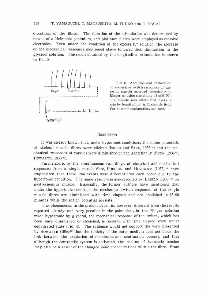
directions of the fibres. The duration of the stimulation was determined by
means of a Helmholz pendulum, and platinum plates were employed as massive
electrodes. Even under the condition of the excess K+ solution, the increase
of the mechanical responses mentioned above followed their diminution in the
glycerol solution. The result obtained by the longitudinal stimulation is shown
in FIG. 8.
FIG. 8. Abolition and restoration
of successive twitch responses of sar-
torius muscle recorded isotonically in
Ringer solution containing 13mM K.
The muscle was stimulated every 5
min by longitudinal A. C. electric field.
For further explanation, see text.
DISCUSSION
It was already known that, under hypertonic conditions, the action potentials
of skeletal muscle fibres were elicited (Ismico and SATO, 19571) and the me-chanical responses of muscles were diminished or abolished finally (FENN, 1936'3);
HOWARTH, 195812)).
Furthermore, by the simultaneous recordings of electrical and mechanical
responses from a single muscle fibre, HODGKIN and HOROWICZ (1957)11) have
emphasized that these two events were differentiated each other due to the
hypertonic condition. The same result was also reported by LASZLO (1958)18) on
gastrocnemius muscle. Especially, the former authors have mentioned that
under the hypertonic condition the mechanical twitch responses of the single
muscle fibres are diminished with time elapsed and are abolished in 15-90
minutes while the action potential persists.
The phenomenon in the present paper is, however, different from the results
reported already and very peculiar in the point that, in the Ringer solution
made hypertonic by glycerol, the mechanical response of the twitch, which has
been once diminished or abolished, is restored with time elapsed even under
dehydrated state (FIG. 4). The evidence would not support the view presented
by HOWARTH (1958)12) that the tonicity of the outer medium does not block the
link between the excitation of membrane and contraction process, and that
although the contractile system is activated, the decline of isometric tension
may also be a result of the changed ionic concentrations within the fibre. From

E-C COUPLING AND GLYCEROL EFFECT 139
the results of the present study, however, it would be indicated that the hyper-
tonicity of the outer medium suppresses or blocks the link between excitation
and contraction, and on the other hand the link is restored by actions of gly-
cerol. MASHIMA (1959)19) has also stated that the hypertonicity may block theE-C coupling based on the fact that caffeine produces contracture on the muscle
even in hypertonic Ringer solutions.
The above restoration of the twitch response of the skeletal muscles ob-
served in the glycerol Ringer solution could not be seen in other hypertonic
Ringer solutions made by substances except urea. This suggests that the
above phenomenon is considerably specific for glycerol and it may be suitable
to call the phenomenon 'glycerol effect'.
BOZLER pointed out (personal communication, 1960) that it could not be
considered that substances, which had the molecular weight as that of glycerol,
could not penetrate into the muscle cells within the time when the mechanical
responses were restored. This may be supposed by the result shown in FIG. 4
on muscle weight: any increase of the muscle weight does not occur in glycerol
Ringer solution over the wide range of the time course. Therefore, two possible
causes of the 'glycerol effect' are considered: the one is the effect of the
glycerol on the membrane itself, and another is that, though glycerol penetrates
into the cell, the glycerol is localized in a portion within the cell, which is in
the vicinity of the membrane and the amount of the penetrated glycerol is so
small that it causes no change in weight. In other words, the 'glycerol effect'
would occur in connection with the membrane or a portion in its vicinity.
It can be considered generally that the mechanism in the 'glycerol effect'
is either chemical or physicochemical. In order to clarify which of the two is
concerned, further experiment was performed by the use of urea, of which
molecular weight is smaller than glycerol. The result, that showed a similar
phenomenon as 'glycerol effect' might suggest that the mechanism of the'glycerol effect' is more physicochemical rather than chemical
.
But, even if so, one problem still remains unsolved. According to the pre-
sent result on urea, the time of beginning of the restoration of the twitch
response was retarded much more and the heights of them were much smaller
than the case of glycerol. Considering that a relatively large amount of urea
penetrates into cells in a short time (BOZLER, 19593) BARANY, et al., 19602)),urea must come to the membrane or a portion of the cell in the vicinity of the
membrane much faster, and thus restoration of the twitch response must occur
in the earlier stage. A possible explanation for this inconsistency is that the
effect of urea may be weaker than that of glycerol. But, we cannot exclude
the possibility that the urea might have directly an inhibitory influence on
actomyosin system (BARANY, et al., 19602)).
KUFFLER and VAUGHAN WILLIAMS (1953)' discussed the differences of the
physiological properties between fast and slow fibres of the skeletal muscle, and

140 T. YAMAGUCHI, T. MATSUSHITA, M. FUJINO AND T. NAGAI
according to PAUSCHINGER (1960),21) one of the differences may be due to the
difference of the content of calcium ion. From the result that no difference
could be seen between the two kinds of muscle fibres in connection with'glycerol effect' it is suggested that the 'glycerol effect' may be independent
of the Ca content of muscles.
It is considered that the contractions of both skeletal and heart muscles
are initiated by depolarization of membrane potentials (FRANK, 19607); NIEDER-
GERKE, 195920)). On the other hand, SUZUKI23) has suggested that there is some
differences between the two muscles with respect to the mechanism in the E-C
coupling; namely, from the experiments performed by employing caffeine, which
induces contracture without depolarization, it was shown that in the case of
skeletal muscles contracture could be elicited under the Ca free condition
(FRANK, 19607); AXELSSON and THESLEFF, 19581)), while Suzum23) demonstratedthe necessity of Ca++ to induce the contracture of the heart ventricle muscles.
The present result that the 'glycerol effect' did not occur on heart ventricle
muscle is the additional interesting evidence to the above facts that there
would be different mechanism between the E-C couplings of the two muscles.
The 'glycerol effect' was produced in the case of the A. C. stimulation on
sartorius muscle under excess K+ condition. Based on the results obtained by
A. C field stimulation of muscle under the same K+ condition it has been dis-
cussed in detail by CSAPO and SUZUKI (1958)4), STEN-KNUDSEN (1960)22) or by
others, how muscle contraction is operated. The present result would suggest
that the affected part by electric currents, whether they are longitudinal or
transverse, is the membrane or its vicinity and not the contractile system
within the cell.
SUMMARY
In order to clarify the mechanism of E-C coupling process of the muscle,
electrical and mechanical responses to the electrical stimulation and the relation-
ship between their changes and the muscle weight were investigated in Ringer
solution made hypertonic by the addition of glycerol or of the other several
substances.
1. For about sixty minutes, for which the observation was made, features of
resting and action potentials from single muscle fibres in 420mM glycerol
Ringer solution were almost the same as those in normal Ringer solution.
2. The mechanical twitch responses of sartorius muscle immersed in glycerol
Ringer solution were at first diminished and then restored with time elapsed.
The most prominent restoration occurred in 420mM glycerol Ringer solution,
and the maximum restored twitch tension of a single fibre, which attained to
50-60 minutes after exchanging the bathing solution for glycerol one, was 60-70
percent of that in normal Ringer solution.

E-C COUPLING AND 'GLYCEROL EFFECT' 141
3. In 420mM glycerol Ringer solution, the weight of sartorius muscle dropped
to 74-82 percent of that in normal Ringer solution within a few minutes, and
then the dropped value kept constant for about an hour.
4. The similar mechanical phenomenon was observed in rectus abdominis muscle
but not in heart ventricle. It was also found in partially depolarized sartorius
muscle stimulated by A. C. electric field.
5. The restoration of the mechanical twitch response did not occur in sartorius
muscles under the condition of Ringer solutions made hypertonic by the addi-
tion of excess NaCl, mannitol, glucose, monoacetin and sucrose respectively,
and the observable change was only a gradual diminution of mechanical twitch
response. In the case of urea Ringer solution, however, the restoration ap-
peared, though the grade was considerably less than that in glycerol.
6. From these results, the mechanism of the peculiar effect of glycerol was
discussed in connection with the excitation-contraction coupling process of
muscle.
REFERENCES
1) AXELSSON, J. AND THESLEFF, S. Activation of the contractile mechanism in striatedmuscle. Acta Physiol. Scand. 44: 55-66, 1958.
2) BARANY, M., BARANY, K. UND TRAUTWEIN, W. Hemmung der Aktin-L-MyosinInteraktion in lebenden und extrahierten Muskeln durch Urea. Biochim. Biophys.
Acta 45: 317-335, 1960.
3) BOZLER, E. Osmotic effects and diffusion of nonelectrolytes in muscle. Am. J. Phy-siol. 197: 505-510, 1959.
4) CSAPO, A. AND SUZUKI, T. The effectiveness of the longitudinal field, coupledwith depolarization in activating frog twitch muscles. J. gen. Physiol. 41: 1083-1098,
1958.
5) DANIELLI, J. F. Surface Phenomena in Chemistry and Biology DANIELLI, J. F., PANK-HURST, K. G. A. AND RIDDIFORD, A. C. (editors), p.246. Pergamon Press, London,1958.
6) FENN, W. O. The role of tissue spaces in the osmotic equilibrium of frog musclesin hypotonic and hypertonic solutions. J. cell. comp. Physiol. 9: 93-103, 1936.
7) FRANK, G. B. Effects of changes in extracellular calcium concentration on the
potassium induced contracture of frogs skeletal muscle. J. Physiol. 151: 518-538,1960.
8) FRANK, G. B. Maximum activation of the contractile mechanism in frog's skeletalmuscle by potassium depolarization. J. Physiol. 154: 345-353, 1960.
9) FURUKAWA, T. [Guide to the Microelectrode Technique[ (Japanese). p.107, KinpodoPublishing Co., Ltd., Tokyo. 1957.
10) HAUROWITZ, F. Chemistry and Biology of Proteins. Academic Press, New York. 1950.11) HODGKIN, A. L. AND HOROWICZ, P.The differential action of hypertonic solutions
on the twitch and action potential of a muscle fibre. J. Physiol. 136: 17 p.1957.
12) HOWARTH, J. V. The behaviour of frog muscle in hypertonic solutions . J. Physiol.144: 167-175, 1958.
13) HUXLEY, A. F. Local activation of muscle . Ann. N. Y. Acad. Sci. 81, Pt. 2: 446-452,1959.

142 T. YAMAGUCHI, T. MATSUSHITA, M. FUJINO AND T. NAGAI
14) ISHIKO, N. AND SATO, M. The effect of calcium ions on electrical properties ofstriated muscle fibres. Jap. J. Physiol. 7: 51-63, 1957.
15) KOREY, S. Some factors influencing the contractility of a nonconducting fiber prepar-ation. Biochim. Biophys. Acta 4: 58-67, 1950.
16) KOTOWSKI, H., ANTONI, H. UND FLECKENSTEIN, A. Elektrophysiologische Studienzur Aufhebung der Kaliumlahmung des Froschmyocards durch ATP. Pfliigers Arch.
ges. Physiol. 270: 85-102, 1959.
17) KUFFLER, S. W. AND VAUGHAN WILLIAMS, E. M. Properties of the " slow " skeletalmuscle fibres of the frog. J. Physiol. 121: 318-340, 1953.
18) LASZLO, N. Die Muskeltatigkeit p. 295. Ernst, E. (author) Verlag d. UngarischenAkademie d. Wissenschaf ten, Budapest. 1958.
19) MASHIMA, H. On the excitation-contraction coupling in the skeletal muscle. (Japa-nese). Medicine of Japan in 1959 5: 333-340, 1959.
20) NIEDERGERKE, R. Calcium and the activation of contraction. Experientia 15: 128-130, 1959.
21) PAUSCHINGER, P. Uber die Beeinflussung von Kontrakturen langsamer (tonischer)and schneller (phasischer) Skeletmuskeln durch Calcium. Pfliigers Arch. ges. Physiol.272: 43-44, 1960.
22) STEN-KNUDSEN, O. Is muscle contraction initiated by internal current flow? J.Physiol. 151: 363-384, 1960.
23) SUZUKI, K. to be published.24) SENT-GYORGYI, A. Chemistry of muscular contraction. 2nd ed. Academic Press, New
York. 1951.