Smooth Muscle Excitation - Contraction
33
Smooth Muscle Excitation - Contraction Mike Clark, M.D.
-
Upload
quemby-stanley -
Category
Documents
-
view
72 -
download
2
description
Smooth Muscle Excitation - Contraction. Mike Clark, M.D. Smooth Muscle. Found in walls of most hollow organs (except heart) Usually in two layers (longitudinal and circular). Longitudinal layer of smooth muscle (shows smooth muscle fibers in cross section). Small intestine. Mucosa. - PowerPoint PPT Presentation
Transcript of Smooth Muscle Excitation - Contraction
Smooth MuscleExcitation - Contraction
Mike Clark, M.D.
Smooth Muscle
• Found in walls of most hollow organs(except heart)
• Usually in two layers (longitudinal and circular)
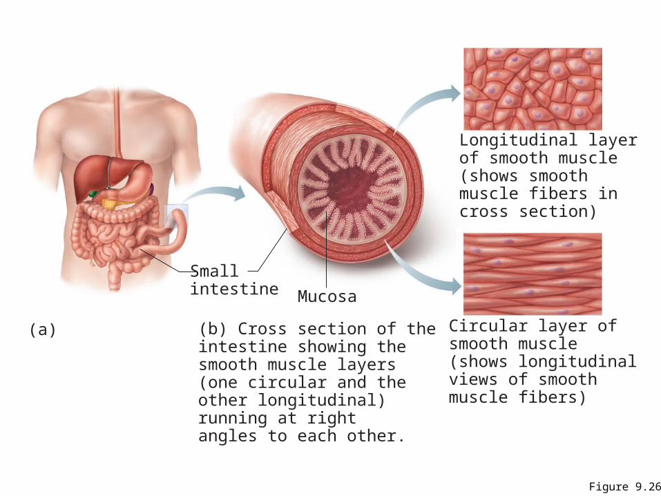
Figure 9.26
Smallintestine
(a) (b) Cross section of theintestine showing thesmooth muscle layers(one circular and theother longitudinal)running at rightangles to each other.
Mucosa
Longitudinal layerof smooth muscle (shows smooth muscle fibers in cross section)
Circular layer ofsmooth muscle (shows longitudinalviews of smooth muscle fibers)
Peristalsis
• Alternating contractions and relaxations of smooth muscle layers that mix and squeeze substances through the lumen of hollow organs– Longitudinal layer contracts; organ dilates and
shortens – Circular layer contracts; organ constricts and
elongates
Microscopic Structure
• Spindle-shaped fibers: thin and short compared with skeletal muscle fibers
• Connective tissue: endomysium only• SR: less developed than in skeletal muscle • Pouchlike infoldings (caveolae) of sarcolemma
sequester Ca2+
• No sarcomeres, myofibrils, or T tubules
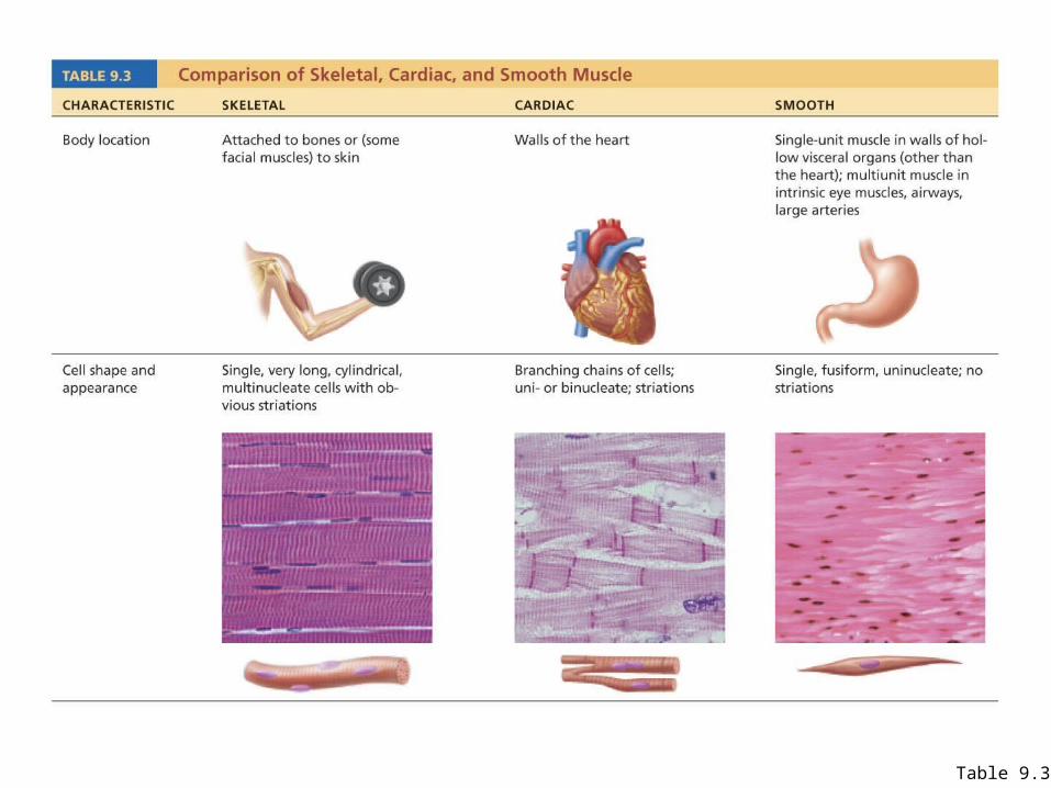
Table 9.3
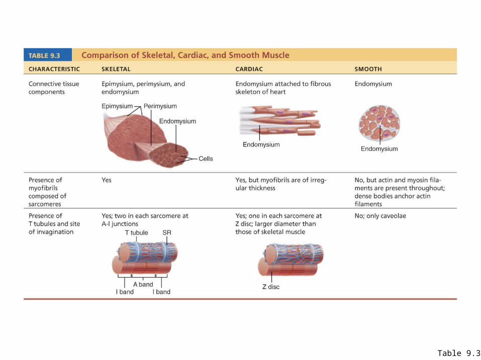
Table 9.3
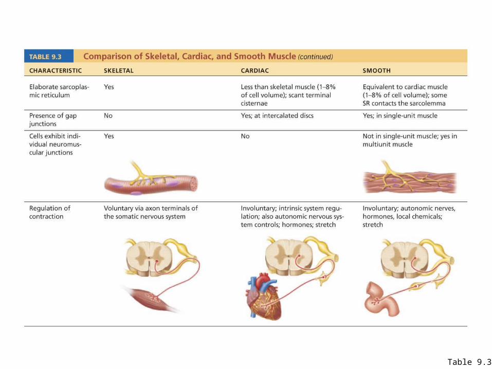
Table 9.3
Innervation of Smooth Muscle
• Autonomic nerve fibers innervate smooth muscle at diffuse junctions
• Varicosities (bulbous swellings) of nerve fibers store and release neurotransmitters
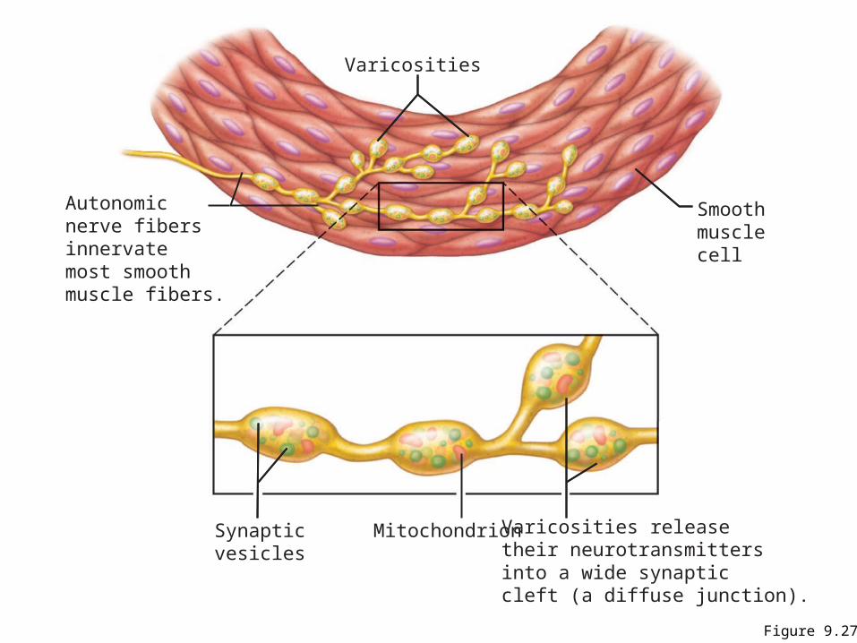
Figure 9.27
Smoothmusclecell
Varicosities releasetheir neurotransmittersinto a wide synaptic cleft (a diffuse junction).
Synapticvesicles
Mitochondrion
Autonomicnerve fibersinnervatemost smoothmuscle fibers.
Varicosities
Myofilaments in Smooth Muscle
• Ratio of thick to thin filaments (1:13) is much lower than in skeletal muscle (1:2)
• Thick filaments have heads along their entire length
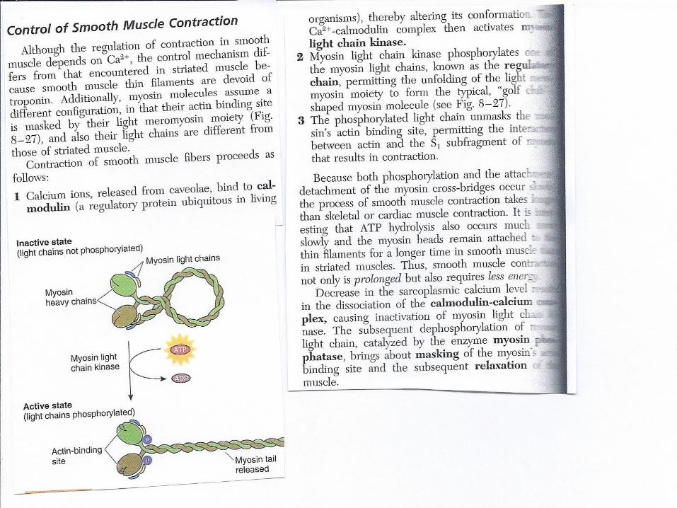
• No troponin complex; protein calmodulin binds Ca2+
Myofilaments in Smooth Muscle
• Myofilaments are spirally arranged, causing smooth muscle to contract in a corkscrew manner
• Dense bodies: proteins that anchor noncontractile intermediate filaments to sarcolemma at regular intervals – the dense bodies also attach to the Actin filaments – thus acting as a type of Z-line
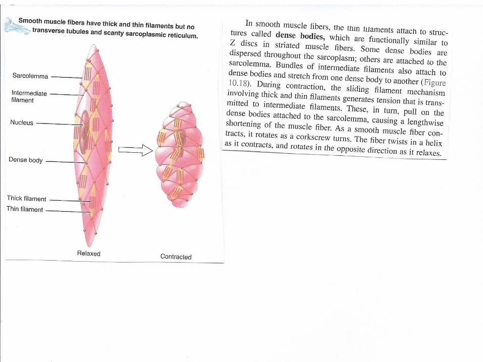
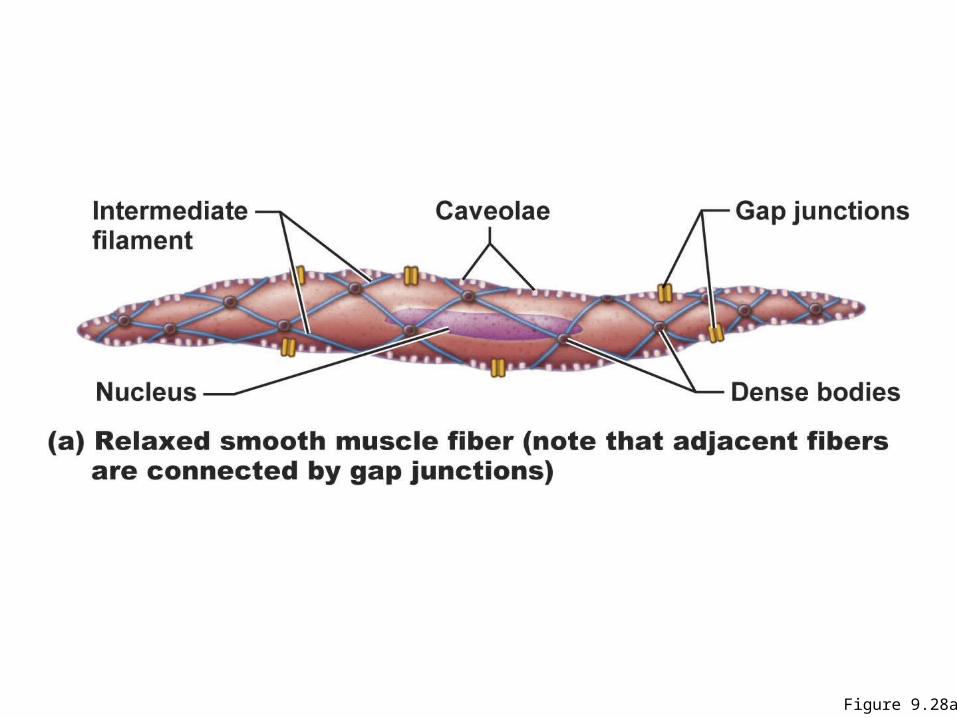
Figure 9.28a
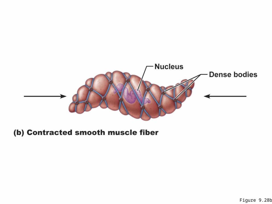
Figure 9.28b
Contraction of Smooth Muscle
• Slow, synchronized contractions • Cells are electrically coupled by gap junctions• Some cells are self-excitatory (depolarize
without external stimuli); act as pacemakers for sheets of muscle
• Rate and intensity of contraction may be modified by neural and chemical stimuli
Contraction of Smooth Muscle
• Sliding filament mechanism• Final trigger is intracellular Ca2+
• Ca2+ is obtained from the SR and extracellular space
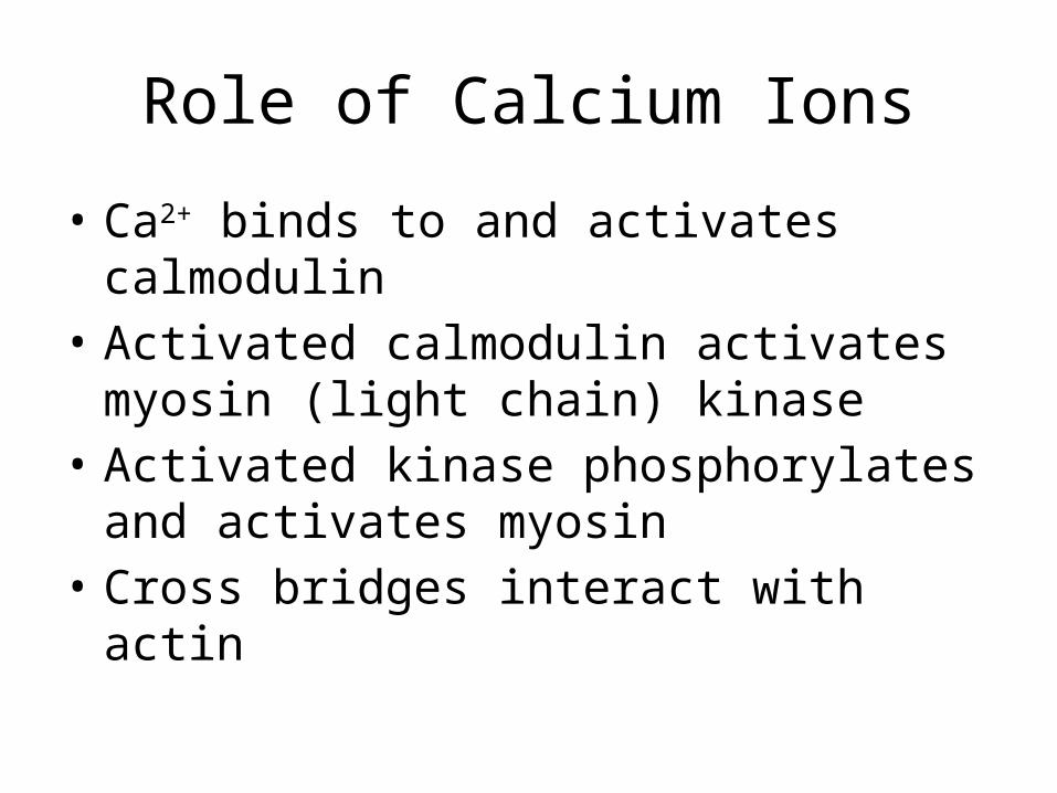
Role of Calcium Ions
• Ca2+ binds to and activates calmodulin • Activated calmodulin activates myosin (light
chain) kinase• Activated kinase phosphorylates and activates
myosin • Cross bridges interact with actin
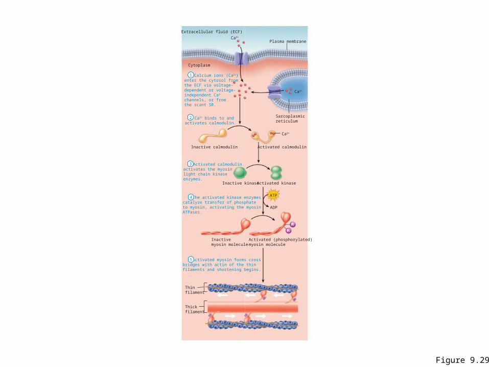
Figure 9.29
Calcium ions (Ca2+)enter the cytosol fromthe ECF via voltage-dependent or voltage-independent Ca2+
channels, or fromthe scant SR.
ATP
Pi
Pi
Extracellular fluid (ECF)
ADP
Ca2+
Ca2+
Ca2+
Plasma membrane
Sarcoplasmicreticulum
Inactive calmodulin
Inactive kinase
Inactivemyosin molecule
Activated (phosphorylated)myosin molecule
Activated kinase
Activated calmodulin
Cytoplasm
Ca2+ binds to andactivates calmodulin.
Activated calmodulinactivates the myosinlight chain kinaseenzymes.
The activated kinase enzymescatalyze transfer of phosphateto myosin, activating the myosinATPases.
Activated myosin forms crossbridges with actin of the thinfilaments and shortening begins.
Thinfilament
Thickfilament
1
2
3
4
5
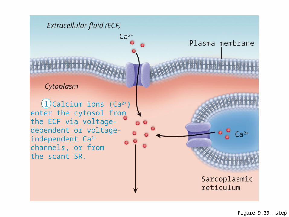
Figure 9.29, step 1
Calcium ions (Ca2+)enter the cytosol fromthe ECF via voltage-dependent or voltage-independent Ca2+
channels, or fromthe scant SR.
Extracellular fluid (ECF)
Ca2+
Ca2+
Plasma membrane
Sarcoplasmicreticulum
Cytoplasm
1
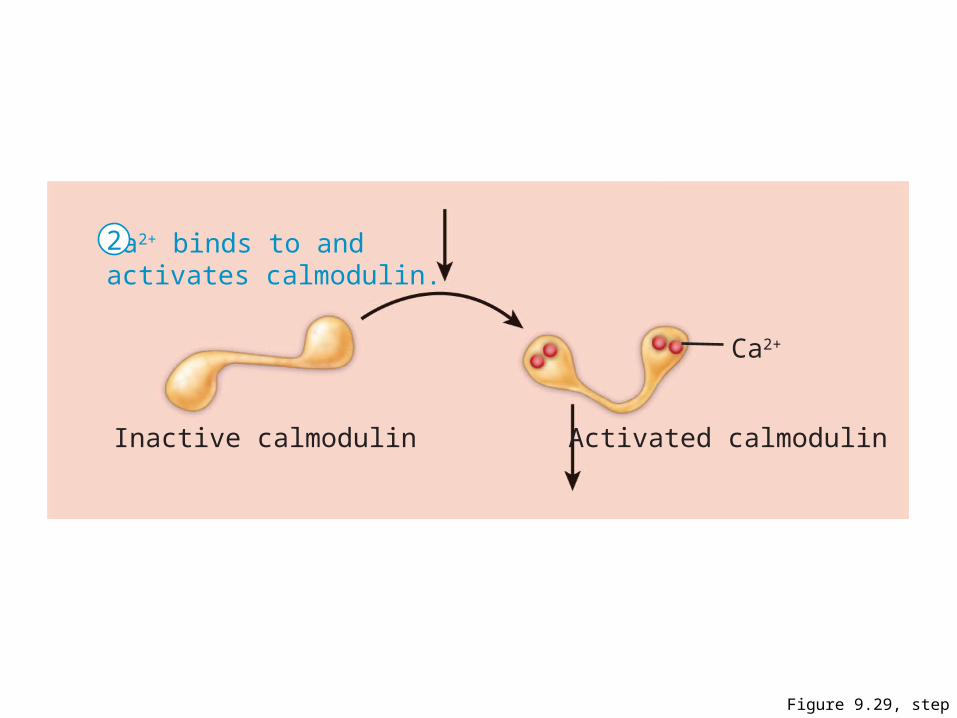
Figure 9.29, step 2
Ca2+
Inactive calmodulin Activated calmodulin
Ca2+ binds to andactivates calmodulin.
2
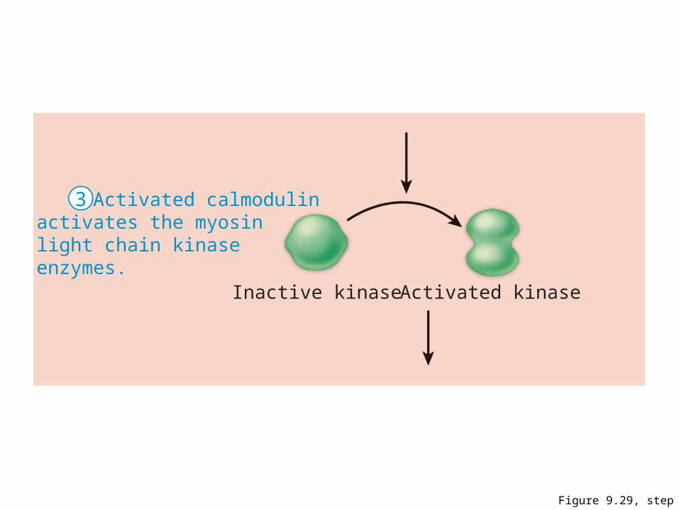
Figure 9.29, step 3
Inactive kinase Activated kinase
Activated calmodulinactivates the myosinlight chain kinaseenzymes.
3
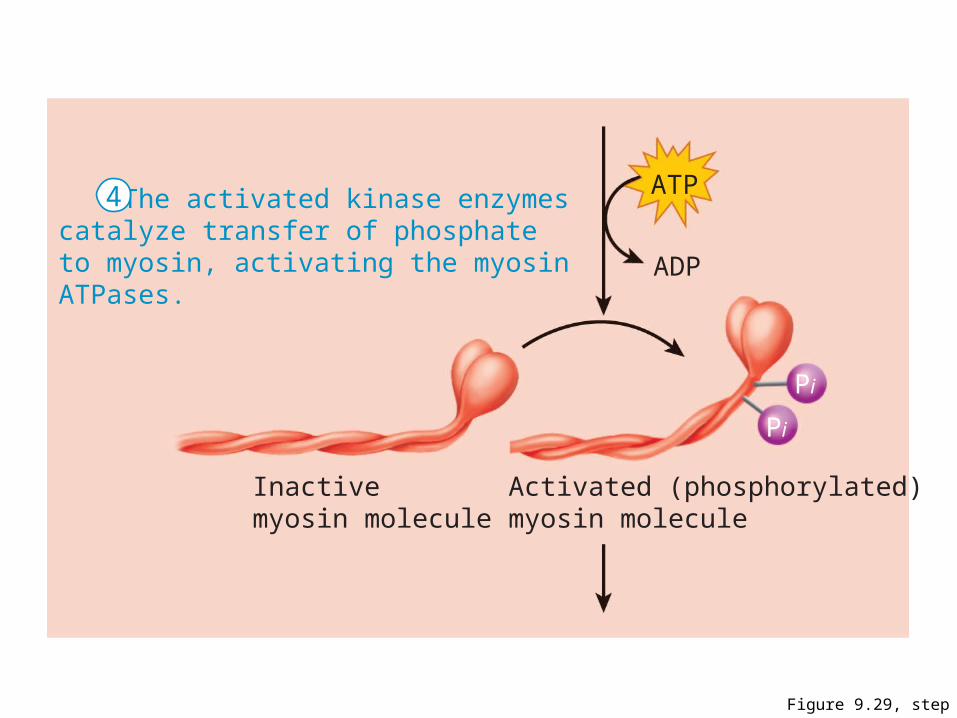
Figure 9.29, step 4
ATP
Pi
Pi
ADP
Inactivemyosin molecule
Activated (phosphorylated)myosin molecule
The activated kinase enzymescatalyze transfer of phosphateto myosin, activating the myosinATPases.
4
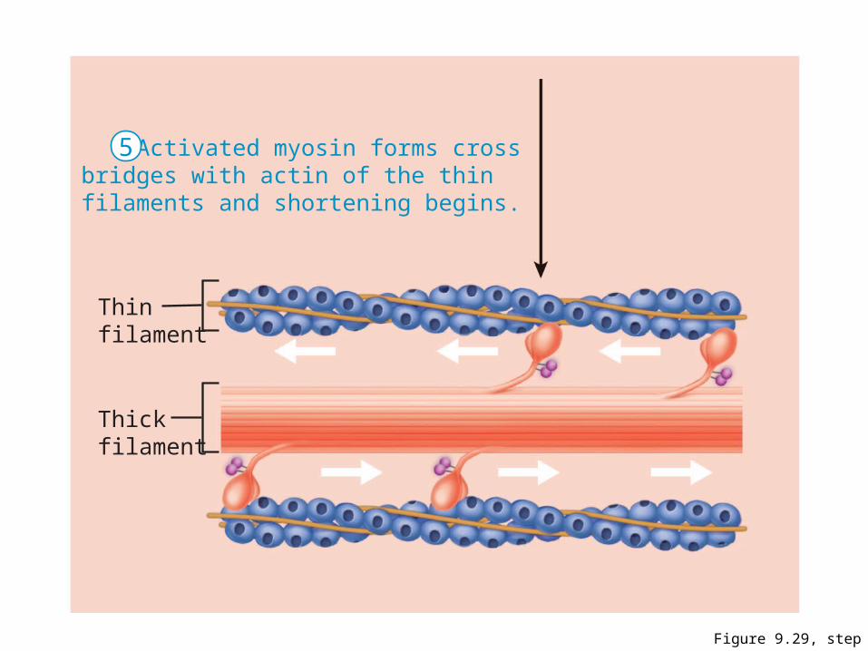
Figure 9.29, step 5
Activated myosin forms crossbridges with actin of the thinfilaments and shortening begins.
Thinfilament
Thickfilament
5
Contraction of Smooth Muscle
• Very energy efficient (slow ATPases)• Myofilaments may maintain a latch state for
prolonged contractionsRelaxation requires:• Ca2+ detachment from calmodulin• Active transport of Ca2+ into SR and ECF• Dephosphorylation of myosin to reduce
myosin ATPase activity
Regulation of Contraction
Neural regulation:• Neurotransmitter binding [Ca2+] in
sarcoplasm; either graded (local) potential or action potential
• Response depends on neurotransmitter released and type of receptor molecules
Regulation of Contraction
Hormones and local chemicals:– May bind to G protein–linked receptors– May either enhance or inhibit Ca2+ entry
Special Features of Smooth Muscle Contraction
Stress-relaxation response: – Responds to stretch only briefly, then adapts to
new length– Retains ability to contract on demand– Enables organs such as the stomach and bladder
to temporarily store contentsLength and tension changes:
– Can contract when between half and twice its resting length
Special Features of Smooth Muscle Contraction
Hyperplasia:– Smooth muscle cells can divide and increase their
numbers– Example:
• estrogen effects on uterus at puberty and during pregnancy
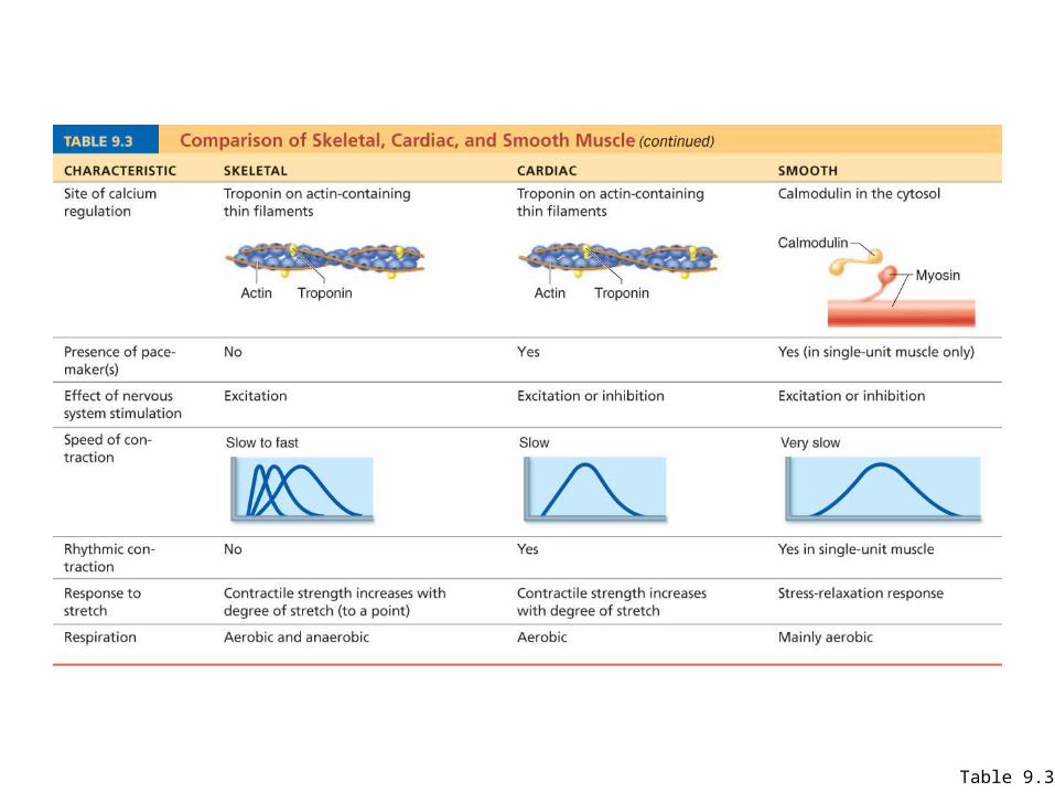
Table 9.3
Types of Smooth Muscle
Single-unit (visceral) smooth muscle: – Sheets contract rhythmically as a unit (gap
junctions)– Often exhibit spontaneous action potentials– Arranged in opposing sheets and exhibit stress-
relaxation response
Types of Smooth Muscle: Multiunit
Multiunit smooth muscle:– Located in large airways, large arteries, arrector
pili muscles, and iris of eye– Gap junctions are rare– Arranged in motor units– Graded contractions occur in response to neural
stimuli