The Degradation Mechanism of an Epoxy- Phenolic … · Coatings, Birmingham, UK) were then cast...
18
1 The Degradation Mechanism of an Epoxy- Phenolic Can Coating S. Morsch * , 1 S. Lyon, 1 S. R. Gibbon 2 1 Corrosion and Protection Centre, School of Materials, The University of Manchester, The Mill, Sackville St, Manchester, M13 9PL, UK 2 AkzoNobel Supply Chain, Research & Development, Stoneygate Lane, Felling, Gateshead, Tyne & Wear, NE10 0JY, UK * To whom correspondence should be addressed. [email protected] tel: +44 161 306 2914
Transcript of The Degradation Mechanism of an Epoxy- Phenolic … · Coatings, Birmingham, UK) were then cast...

1
The Degradation Mechanism of an Epoxy-
Phenolic Can Coating
S. Morsch*,1S. Lyon,1 S. R. Gibbon2
1 Corrosion and Protection Centre,
School of Materials,
The University of Manchester,
The Mill,
Sackville St,
Manchester, M13 9PL, UK
2 AkzoNobel Supply Chain, Research & Development,
Stoneygate Lane,
Felling,
Gateshead,
Tyne & Wear, NE10 0JY, UK
* To whom correspondence should be addressed. [email protected] tel: +44 161 306 2914

2
ABSTRACT
Paint remains a widely employed approach to corrosion control due its relatively low
cost and proven efficacy. Nonetheless, the processes governing long-term deterioration
of intact organic coatings (in the absence of defects) are not fully understood. In this
contribution, we investigate the degradation mechanism of a corrosion resistant epoxy-
phenolic can coating. In-situ time-resolved ATR FTIR is applied to monitor both the
chemical integrity of the coating and water uptake as a function of immersion time in
water or electrolyte. Ion transport is assessed across free standing films, and
morphological changes accompanying immersion are examined using ex-situ advanced
scanning probe microscopy techniques. Coatings are found to deform as a result of
water sorption during immersion in electrolyte or water, yielding regions of
heterogeneous hydrophilicity, yet no change in functional group chemistry is found to
occur.
HIGHLIGHTS
Immersion in water or electrolyte does not affect the chemical structure of an
epoxy-phenolic can-coating.
Ionic transport occurs across free-standing films independently of water-induced
damage.
Non-Fickian water sorption is associated with deformation of the coating.
AFM-IR is used to show that hydrophilic pores form during immersion.
3
1. INTRODUCTION
Paint remains the most widely employed approach to corrosion control due its relatively low
cost and proven efficacy. Nevertheless, the long-term deterioration of coating barrier
properties (associated with the onset of underfilm corrosion) is not fully understood, and
continues to be a matter of significant scientific and industrial interest. For organic coatings,
it is widely accepted that failures in corrosion protection are most commonly instigated by the
presence of a defect traversing the film (as a consequence of e.g., mechanical damage
during the coating lifetime, inconsistent application, or trapped solvent during the cure).
Consequently, research efforts have focussed on corrosion initiation in the presence of
macroscopic defects, and coatings are routinely formulated to mitigate corrosive phenomena
at and around defect sites (by e.g., the inclusion of leachable inhibitor additives).[1–7] Little
attention has been paid to the long-term failure of intact organic coatings, a process which
becomes increasingly significant as formulation and application techniques continue to
improve. In the case of intact organic coatings, it can be seen that any long term failure in
corrosion protection must involve the spontaneous degradation of polymeric barrier
properties, yet the mechanism of this process remains elusive.
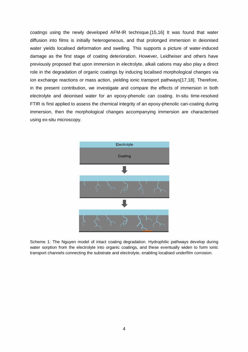
At present, the prevailing model describing the long term degradation of intact
organic coatings entails the spontaneous formation of conducting pathways through the
coating, followed by localised corrosion initiation (i.e., the base of the ionic pathway behaves
in the same manner as a macroscopic defect), Scheme 1. Nguyen proposed that in the
presence of neutral electrolyte, these ionic transport pathways are introduced by the action
of water during immersion; via hydrolysis reactions, leaching of unreacted material or else
the gradual evolution of hydrophilic channels traversing the film.[8] In the case of epoxy
resins, (widely used as the binders in corrosion resistant paints) little evidence exists for
hydrolytic breakdown under ambient conditions, and in particular for food-contact epoxy-
phenolic coatings (such as those studied here) negligible leaching effects have been well
documented.[9–12] Thus, hydrophilic (and ionic) transport pathways are considered to
evolve spontaneously in the absence of chain scission or leaching phenomena, and result in
localised failures. However, whilst this mechanism of degradation was proposed in the
Nguyen model almost two decades ago, few studies have addressed the formation of
discrete hydrophilic or ionic transport channels through intact coatings. Taylor et al have
previously reported evidence for ionic transport pathways through epoxy coatings, detected
using fluorescence microscopy (after aging in electrolyte and exposure to an ion-specific
fluorescent probes, or directly using cationic chromophores).[13,14] In addition, we recently
reported the first direct observations of heterogeneous water sorption into epoxy-phenolic
4
coatings using the newly developed AFM-IR technique.[15,16] It was found that water
diffusion into films is initially heterogeneous, and that prolonged immersion in deionised
water yields localised deformation and swelling. This supports a picture of water-induced
damage as the first stage of coating deterioration. However, Leidheiser and others have
previously proposed that upon immersion in electrolyte, alkali cations may also play a direct
role in the degradation of organic coatings by inducing localised morphological changes via
ion exchange reactions or mass action, yielding ionic transport pathways[17,18]. Therefore,
in the present contribution, we investigate and compare the effects of immersion in both
electrolyte and deionised water for an epoxy-phenolic can coating. In-situ time-resolved
FTIR is first applied to assess the chemical integrity of an epoxy-phenolic can-coating during
immersion, then the morphological changes accompanying immersion are characterised
using ex-situ microscopy.
Scheme 1: The Nguyen model of intact coating degradation. Hydrophilic pathways develop during
water sorption from the electrolyte into organic coatings, and these eventually widen to form ionic
transport channels connecting the substrate and electrolyte, enabling localised underfilm corrosion.

5
2. EXPERIMENTAL
2.1 Sample Preparation
Electrolytic chrome-coated steel pieces (4 cm2) were degreased by rinsing in ethanol (Fisher
Scientific, >99%). Thin films of epoxy-phenolic Vitalure 345 Lacquer (AkzoNobel Packaging
Coatings, Birmingham, UK) were then cast onto the steel pieces at 2000 rpm for 30 s
(Photoresist spin coater, Headway Research Inc., 1-10,000 rpm). Specimens were cured by
placing in an oven maintained at 200 °C for 15 min, resulting in a dry film thickness of 4.3 ±
0.3 µm. Free-standing films were acquired by delamination from PTFE (Polyflon). PTFE
films were first attached to steel substrates, then coated and cured under identical
conditions.
2.2 Film Characterisation
For infrared analysis the epoxy-phenolic lacquer was manually bar coated (using an 8 µm
spiral bar, Elcometer) onto a germanium internal reflection element and cured for 15 min at
200 °C. In order to assess water uptake, 5 % (w/w) NaCl solution in D2O (>99 %, Aldrich)
was prepared and injected into an ATR fluid cell, forming a reservoir against the coating,
Scheme 1. Using this set up, infrared spectra can be gathered continuously without
adjustment of the sample or realignment of the laser. Spectra are the result of 32
accumulations collected in ATR mode (with a 45° angle of incidence) using an FTIR
spectrometer (Spectrum 2000, Perkin Elmer), operating at 4 cm-1 resolution across the 700 –
4000 cm-1 range. FTIR spectra were gathered every 60 min over a ten day period.
Scheme 2. The ATR fluid cell set-up with a germanium internal reflection element (IRE) used for in-situ immersion FTIR measurements.
Atomic force microscope images were obtained using a Multimode 8 (Bruker, Santa
Barbara) operating in Peakforce Nanomechanical mode using a Pt-Ir coated SCMPIT probe
(Bruker, nominal spring constant 2 N/m, nominal resonance frequency of 80 kHz).

6
Nanoscale infrared analysis was performed on a NanoIR2 system (Anasys
Instruments) operating with top-down illumination. During AFM-IR, the sample is subjected
to rapid pulses (10 ns duration at a repetition rate of 1 KHz) from a tuneable infrared source
(optical parametric oscillator). Absorbance of infrared radiation induces abrupt thermal
expansion of the sample, and this is detected by deflection of an AFM probe in contact with
the surface. The recorded AFM-IR signal is either the maximum of peak-to-peak deflection
during the cantilever ring-down corresponding to IR pulses, or the amplitude of induced
oscillation after fast Fourier transform, Scheme 3. It has been shown that plotting these
signals as a function of IR wavelength yields spectra closely matched to those obtained by
macroscopic transmission-mode FTIR.[19] Furthermore, since the IR pulse (10 ns duration),
thermal expansion, and damping down of the induced oscillation occur on a faster timescale
than the feedback electronics of the AFM, simultaneous contact-mode topographical
measurement and IR absorbance mapping can be performed at a given wavelength.[20–23]
For the present study, AFM-IR images were collected in contact mode at a scan rate of 0.1
Hz using a gold-coated silicon nitride probe (0.07 – 0.4 N/m spring constant, 13 ± 4 kHz
resonant frequency, Anasys Instruments). For mapping, the amplitude of the induced
vibration was recorded at a given wavelength using 16 co-averages for 1024 points per 300
scan lines. The specimen and AFM head were contained within a sample chamber equipped
with a portable temperature and humidity logger (Lascar Electronics). In order to raise
humidity, saturated NaCl solution in D2O was placed in a recrystallizing dish within the
sample chamber and allowed to equilibrate for 24 h.
Scheme 3. The AFM-IR experiment with top-down illumination. The infrared source is pulsed,
inducing rapid thermal expansion of the sample, detected by deflection of the AFM probe cantilever.
The recorded AFM-IR signal is either given as the maximum peak-to-peak deflection during the
cantilever ring-down following 10 ns infrared pulses, or the amplitude at a given frequency following a
fast Fourier transform of the deflection signal.

7
3. RESULTS
3.1 In-situ FTIR Analysis
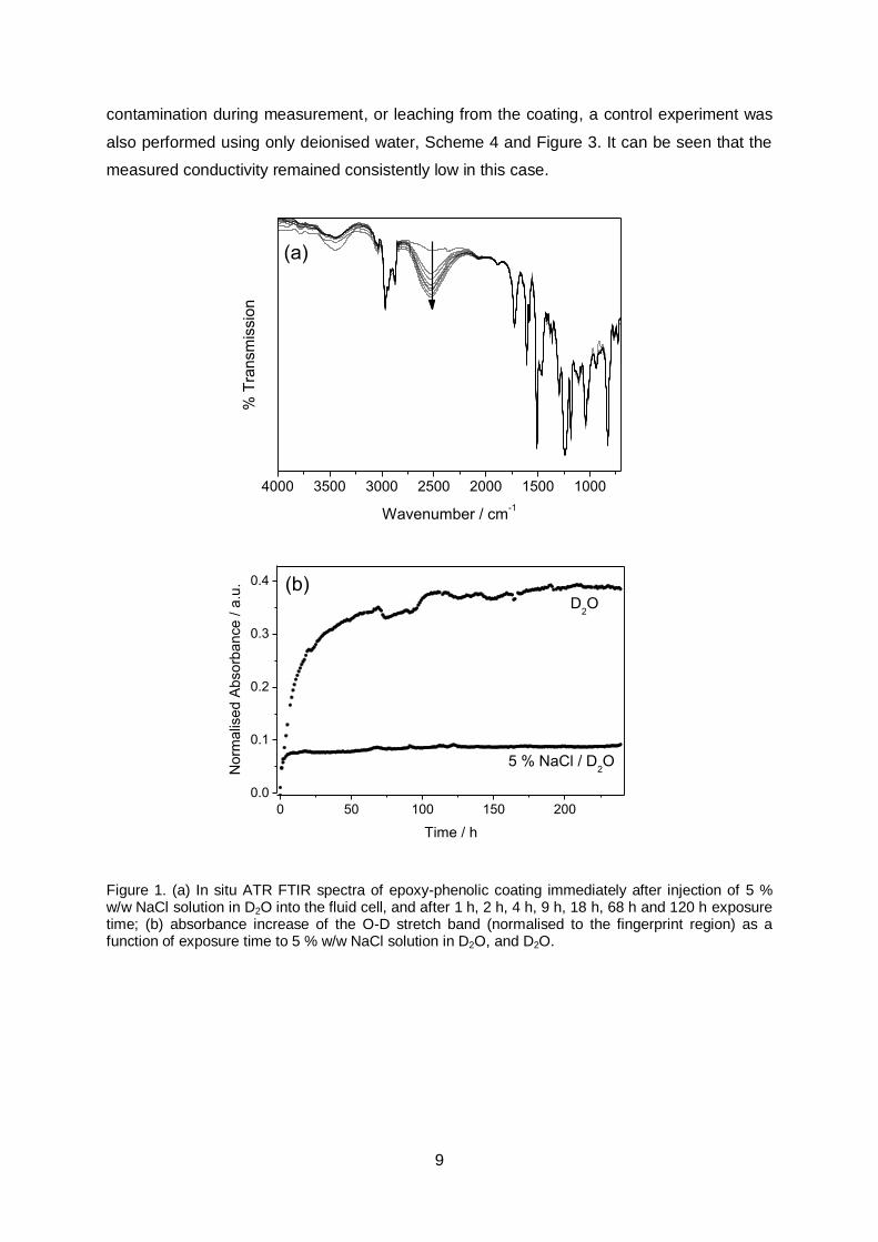
The chemical integrity of epoxy phenolic coatings was initially evaluated using in-situ ATR
FTIR spectroscopy in the presence of electrolyte. Spectra were collected at 60 min intervals
over a period of 10 days, commencing directly after injection of a 5 % w/w solution of NaCl in
D2O into the ATR fluid cell, Scheme 2. It is notable that infrared absorbance in the fingerprint
region associated with polymeric functionality (700 – 2000 cm-1) remained unaffected
throughout exposure to the electrolyte environment, confirming that the coatings do not
delaminate during exposure (since coating peak intensity would be expected to diminish
following delamination, due to an effectively lowered concentration within the infrared
evanescent wave sampling depth).[24] In addition, during exposure the appearance (and
then incremental increase) of a broad absorbance centred around 2550 cm-1 was observed,
Figure 1a. This band is attributed to the O-D stretch of deuterium oxide absorbed by the
epoxy-phenolic coating from the electrolyte solution.[25] Again, the position and intensity of
peaks in the spectral fingerprint region remained completely unchanged in the presence of
this absorbed water, confirming that no disruption of macromolecular structure occurs due to
bulk hydrolysis reactions, Figure 1a.
Because the infrared absorbance of the O-D stretch vibration does not overlap with
any native infrared peaks associated with the epoxy-phenolic polymer, moisture uptake into
the coating is directly proportional to the absorbance increase in this region, and was
quantified by integration of the 2550 cm-1 band at each time point. For comparison, a second
experiment was also performed using only deuterium oxide as the exposure medium, and
the measured area of the O-D stretch bands were normalised relative to the area of the
entire fingerprint region in both cases, Figure 1b. A degree of noise is generated in water
sorption profiles by fluctuating CO2 bands (centred on 2280 cm-1), which overlap with the
broad O-D stretch band. Nonetheless, it is clear that water sorption occurs in both cases,
and from generated profiles it can be seen that significantly enhanced water sorption occurs
in the presence of D2O. This is in keeping with previous reports, where the kinetics and
equilibrium content for water uptake into epoxy resins has been found to depend on the
water activity of the immersion medium.[26]
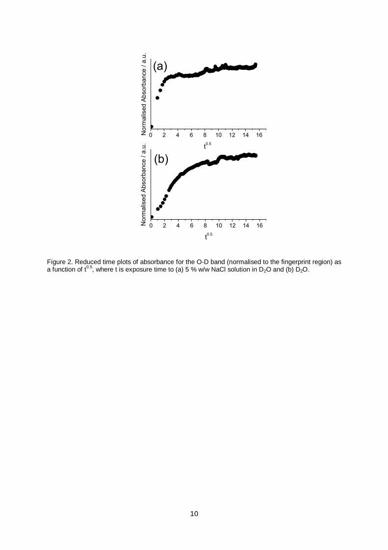
It is well-known that water sorption kinetics correlate to the transport mechanism
through organic networks. In general, Fickian diffusion is associated with fast polymer
relaxation or rapid sorption into the available free volume (i.e., diffusion limited by mobility of

8
the penetrant solvent), whereas non-Fickian kinetics is associated with polymeric relaxation
and penetration into the bulk (i.e., transport is limited by the mobility of polymer segments)
[27–30] In the present case, for moisture sorption from electrolyte, the reduced time plot
indicates Fickian diffusion at early exposure times (<4 h, a linear correlation is evident),
Figure 2a. At longer exposure times, it can be seen that the kinetics deviate from ideal
Fickian behaviour; i.e., an equilibrium water content is not established, and instead water
uptake continues at a slower rate, characteristic of non-Fickian diffusion. Such two-stage
water sorption kinetics are typical for epoxy resins, where water is proposed to rapidly
penetrate the available free volume via Fickian diffusion, whilst relaxation of water-
plasticised polymer chains drives further, slower sorption by exposing further hydrogen
bonding sites and isolated pockets of free volume. Moreover, two-stage kinetics are
consistent with previous water sorption results for these epoxy phenolic coatings, where it
has been shown that rapid water sorption under humid conditions initially occurs via diffusion
into the available free volume (60 min exposure time)[31], whereas deformation occurs only
after longer term (3 days) immersion in water (swelling and deformation are associated with
non-Fickian sorption, since this process involves disruption of the network)[15].
For water sorption directly from D2O, no Fickian diffusion stage was detected (no
linear correlation is observed in the reduced time plot), Figure 2b. We propose this is
because the initial rapid penetration into the available free volume occurs prior to the first
measured time point (60 min). Instead, the kinetic profile is characteristic of an auto-
accelerated process (S-shaped curve). This can be attributed to water-plasticised polymer
relaxation (i.e., the presence of absorbed water accelerates the non-Fickian sorption
process). Indeed, we have previously reported water plasticisation of these epoxy-phenolic
can coatings, where the measured Tg was depressed by 4 ºC (from 89 ºC to 85 ºC) after 7
days immersion in deionised water and drying.[15]
Since the penetration depth of the evanescent wave for a germanium internal
reflection element at 2550 cm-1 can be calculated to be <500 nm[32], a further notable result
for FTIR experiments is the rapid penetration of water through the entirety of the (nominally 8
µm thick) coatings (<60 min), demonstrating that water can freely diffuse through the intact
coating structure. To assess whether this could be accompanied by ion transport when the
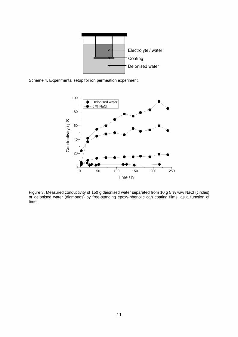
immersion medium is electrolyte, permeation experiments were undertaken. 4.9 cm2 free
standing films were used to separate 10 g 5 % w/w NaCl electrolyte and 150 g deionised
water in a sealed vessel, Scheme 4. Conductivity of the water was then monitored over a ten
day period, Figure 3. Despite significant sample-to-sample variation, it can be seen that the
conductivity of the surrounding solution consistently rises, indicating that ion transport is
possible across intact free-standing films. To ensure that this was not the result of
9
contamination during measurement, or leaching from the coating, a control experiment was
also performed using only deionised water, Scheme 4 and Figure 3. It can be seen that the
measured conductivity remained consistently low in this case.
4000 3500 3000 2500 2000 1500 1000
% T
ransm
issio
n
Wavenumber / cm-1
(a)
0.0
0.1
0.2
0.3
0.4
0 50 100 150 200
No
rma
lise
d A
bso
rba
nce
/ a
.u. (b)
D2O
5 % NaCl / D2O
Time / h
Figure 1. (a) In situ ATR FTIR spectra of epoxy-phenolic coating immediately after injection of 5 % w/w NaCl solution in D2O into the fluid cell, and after 1 h, 2 h, 4 h, 9 h, 18 h, 68 h and 120 h exposure time; (b) absorbance increase of the O-D stretch band (normalised to the fingerprint region) as a function of exposure time to 5 % w/w NaCl solution in D2O, and D2O.
10
0 2 4 6 8 10 12 14 16
0 2 4 6 8 10 12 14 16
No
rma
lise
d A
bso
rba
nce
/ a
.u.
t0.5
(a)
No
rma
lise
d A
bso
rba
nce
/ a
.u.
t0.5
(b)
Figure 2. Reduced time plots of absorbance for the O-D band (normalised to the fingerprint region) as a function of t
0.5, where t is exposure time to (a) 5 % w/w NaCl solution in D2O and (b) D2O.
11
Scheme 4. Experimental setup for ion permeation experiment.
0
20
40
60
80
100
0 50 100 150 200 250
5 % NaCl
Co
nductivity /
S
Deionised water
Time / h
Figure 3. Measured conductivity of 150 g deionised water separated from 10 g 5 % w/w NaCl (circles) or deionised water (diamonds) by free-standing epoxy-phenolic can coating films, as a function of time.
12
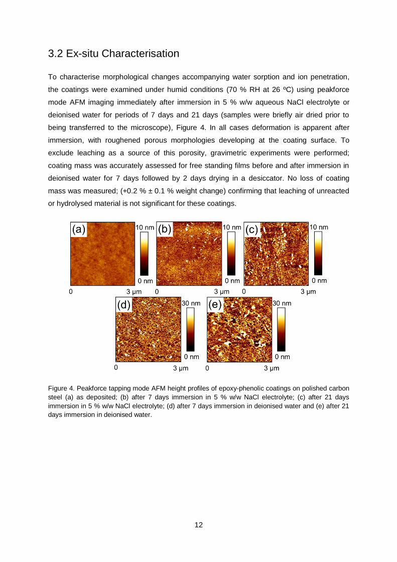
3.2 Ex-situ Characterisation
To characterise morphological changes accompanying water sorption and ion penetration,
the coatings were examined under humid conditions (70 % RH at 26 ºC) using peakforce
mode AFM imaging immediately after immersion in 5 % w/w aqueous NaCl electrolyte or
deionised water for periods of 7 days and 21 days (samples were briefly air dried prior to
being transferred to the microscope), Figure 4. In all cases deformation is apparent after
immersion, with roughened porous morphologies developing at the coating surface. To
exclude leaching as a source of this porosity, gravimetric experiments were performed;
coating mass was accurately assessed for free standing films before and after immersion in
deionised water for 7 days followed by 2 days drying in a desiccator. No loss of coating
mass was measured; (+0.2 % ± 0.1 % weight change) confirming that leaching of unreacted
or hydrolysed material is not significant for these coatings.
Figure 4. Peakforce tapping mode AFM height profiles of epoxy-phenolic coatings on polished carbon
steel (a) as deposited; (b) after 7 days immersion in 5 % w/w NaCl electrolyte; (c) after 21 days
immersion in 5 % w/w NaCl electrolyte; (d) after 7 days immersion in deionised water and (e) after 21
days immersion in deionised water.

13
Finally, AFM-IR analysis was applied to determine the relative hydrophilicity across
the deformed coatings. The specimen displaying the greatest degree of morphological
disruption was selected, i.e., that pre-soaked in deionised water for 21 days. Localised water
sorption was assessed by monitoring the O-D stretch absorbance at 2550 cm-1 before and
after exposure to D2O vapour, Figure 5. AFM-IR spectra were obtained by stepping the
wavelength of incident infrared illumination through the 2400 cm-1 – 3600 cm-1 range and
recording the amplitude of thermally induced cantilever oscillations, Scheme 3 and Figure 5.
For infrared mapping, the pulsed incident beam was held at 2550 cm-1, and the induced
amplitude of cantilever oscillation was recorded during a contact mode scan. Under ambient
conditions (prior to the introduction of D2O vapour into the sample chamber) a degree of
background absorbance was detected at 2550 cm-1, however infrared amplitude mapping
demonstrated that the intensity of this absorbance does not vary across the sample, Figure
5a-c. In the presence of D2O vapour (50 % RH at 25 ºC) the intensity of detected infrared
absorbance (i.e., the amplitude of induced oscillations) at 2550 cm-1 increased, due to the
presence of D2O adsorbed from the humid atmosphere. The location of this enhanced
absorbance thus gives an indication of the surface hydrophilicity. Infrared amplitude mapping
demonstrated that a higher concentration of deuterium oxide was adsorbed onto pore walls,
indicating these regions are more wettable (hydrophilic) than the surrounding resin, Figure
5d-f. Finally, following analysis under humid conditions, silica desiccant was placed in the
sample chamber and the specimen was allowed to dry for 48 h. Subsequently, spectra
revealed that the infrared absorbance at 2550 cm-1 (corresponding to the O-D stretch)
returned to its original intensity, and greatly reduced contrast was observed in infrared
amplitude images, Figure 5g-i. This demonstrates that the detected deuterium oxide species
are desorbed from the sample surface, without affecting the coating structure (note that
height profiles remain unchanged throughout, demonstrating that no further deformation
occurs during exposure to humid conditions).

14
Figure 5. AFM-IR images and spectra of an epoxy-phenolic coating on polished carbon steel after 21
day immersion in deionised water and drying. AFM-IR height profiles, the corresponding infrared
amplitude maps (at 2550 cm-1
), and spectra obtained under (a) ambient conditions (30 % RH); (b)
raised humidity after exposure to D2O vapour (50 % RH) and (c) after drying for 48 h (20 % RH).
Infrared amplitude maps have been flattened line-by-line about zero to show only local fluctuations in
thermally induced amplitude.

15
4. DISCUSSION
The Nguyen model of intact coating degradation entails the establishment of hydrophilic
pathways through an organic coating as a precursor to the ionic transport channels
necessary for corrosion onset.[8] It was proposed therein that organic coatings contain
intrinsic ‘hydrophilic’ regions, predicted to be of low molecular weight (i.e., partially reacted or
hydrolysed), or else regions of low cross-link density within the polymer network, through
which water and ion transport freely occur. Corrosion is then thought to initiate when stress
relaxation of the polymer joins these hydrophilic regions to create a percolating pathway
through the coating. However, for the epoxy-phenolic coatings studied here, the presence of
partially reacted low molecular weight material may be discounted, since the can coatings
are shown to be fully cured (confirmed by the absence of any epoxy absorbance at 916 cm-1
in FTIR spectra, Figure 1, and no endothermic peak associated with excess cure observed
in DSC thermograms, reported previously[31]). Furthermore, the absence of hydrolysis
reactions was here confirmed using in-situ FTIR analysis during immersion, Figure 1, and
leaching effects were ruled out using gravimetric analysis. It is, on the other hand, well
established that nanoscale fluctuations in cross-linking density do occur within thermoset
epoxy resins.[33–35] Furthermore, we have previously shown that short term water uptake
into model epoxy-phenolic resins is indeed heterogeneous at the nanoscale, and
corresponds to slight variations in the degree of cross-linking density, in keeping with the
Nguyen model.[16] Since water has been shown to diffuse through the intact coatings
rapidly, it is feasible that the distribution of free-volume voids provided by these fluctuations
in cross-link density provide favourable diffusive pathways through the coating. Furthermore,
we have found that water sorption during immersion does indeed induce polymeric
relaxation as a consequence of water plasticisation, in keeping with numerous previously
reported studies.[36–38] However, it is important to note that this process is in itself
associated with deformation of the coating, as water binds to hydrogen-bonding sites
exposed along the relaxing chain and disrupts the long range network structure. In the
present case, this manifests in the development of hydrophilic pores and swollen regions
capable of retaining water. This is in keeping with a study by MacQueen and Granata, where
free volume cavities measured using positron annihilation lifetime spectroscopy (PALS) were
found to increase in size during water uptake into unpigmented epoxy coatings. Thus, an
important distinction with the Nguyen hypothesis is that long-term water sorption (and the
associated plasticisation of the polymer network) appears to drive the creation of hydrophilic
regions, rather than simply joining pre-existing heterogeneities within the network.

16
It can readily be envisioned that these macroscopic hydrophilic regions may form a
percolating network during long-term immersion, in keeping with the Nguyen model of
coating degradation. However, at no point during ion permeation experiments was a sudden
increase in conductivity measured, as would be expected if a channel capable of supporting
rapid ionic transport were to spontaneously form. Furthermore, it is important to note that
prior to any deformation due to water sorption (detectable only after several days
immersion[15]), ion transport was already found to occur across the intact coatings,
indicating that ion penetration is not wholly dependent on water-induced degradation.
Similarly, sodium ion diffusion has previously been reported to proceed slowly through intact
polyester coatings, but preferentially though defects.[39][40] It is feasible that ion transport is
locally accelerated following water induced morphological changes in the polymer coating,
and this may correspond to corrosion onset. However for corrosion initiation, further
parameters to consider include bonding at the coating-substrate interface and substrate
potentials driving ion transport (rather than a concentration gradient). Finally, an important
result in the present study is the lack of evidence for direct ion-induced degradation. Whilst
ionic species were shown to penetrate the resin, the level of sample deformation in fact
appears to be lower for coatings exposed to electrolyte, in keeping with a mechanism of
water-induced deformation (since significantly less water sorption occurs into films exposed
to electrolyte vs. deionised water), Figures 1 and 4.
5. CONCLUSIONS
The chemical and morphological changes taking place within an epoxy-phenolic can-coating
were investigated during immersion in aqueous electrolyte or water. In situ FTIR analysis
confirmed the absence of hydrolysis reactions during prolonged immersion, accompanied by
a non-Fickian water sorption process associated with network disruption, plasticisation and
swelling. Ex-situ microscopy techniques revealed that this corresponds to the development
of a hydrophilic porous structure. Comparable results were obtained for samples immersed
in electrolyte, indicating that the ionic species do not significantly affect the mechanism of
coating degradation.
6. ACKNOWLEDGEMENTS
The Authors are grateful to AkzoNobel for financial support and materials.

17
7. REFERENCES
[1] M. Doherty, J.M. Sykes, Corros. Sci. 46 (2004) 1265.
[2] W. Fürbeth, M. Stratmann, Corros. Sci. 43 (2001) 207.
[3] W. Fürbeth, M. Stratmann, Prog. Org. Coatings 39 (2000) 23.
[4] B. Reddy, M.J. Doherty, J.M. Sykes, Electrochim. Acta 49 (2004) 2965.
[5] B. Reddy, J.M. Sykes, Prog. Org. Coatings 52 (2005) 280.
[6] A.P. Nazarov, D. Thierry, Prot. Met. Phys. Chem. Surfaces 45 (2009) 735.
[7] A. Nazarov, M.-G. Olivier, D. Thierry, Prog. Org. Coatings 74 (2012) 356.
[8] T. Nguyen, J.B. Hubbard, J.M. Pommersheim, J. Coatings Technol. 68 (1996) 45.
[9] Z.W. Wicks, F.N. Jones, S.P. Pappas, D.A. Wicks, Organic Coatings Science and
Technology, 3rd ed., Wiley-Interscience, Hoboken, NJ, 2007.
[10] T. Yoshida, M. Horie, Y. Hoshino, H. Nakazawa, Food Addit. Contam. 18 (2001) 69.
[11] C.-M. Chang, C.-C. Chou, M.-R. Lee, Anal. Chim. Acta 539 (2005) 41.
[12] J.-H. Kang, F. Kondo, Res. Vet. Sci. 73 (2002) 177.
[13] P. Moongkhamklang, S.. Taylor, Prog. Org. Coatings 46 (2003) 259.
[14] S.R. Taylor, F. Contu, R. Santhanam, P. Suwanna, Prog. Org. Coatings 73 (2012)
169.
[15] S. Morsch, S. Lyon, P. Greensmith, S.D. Smith, S.R. Gibbon, Faraday Discuss. 180
(2015) 1.
[16] S. Morsch, S. Lyon, S.D. Smith, S.R. Gibbon, Prog. Org. Coatings 86 (2015) 173.
[17] H. Leidheiser, R.D. Granata, R. Turoscy, Corros. NACE 43 (1987) 296.
[18] W.S. Tait, K.A. Handrich, Corrosion 50 (1994) 373.
[19] B. Lahiri, G. Holland, A. Centrone, Small 9 (2013) 439.
[20] A. Dazzi, F. Glotin, R. Carminati, J. Appl. Phys. 107 (2010) 124519.
[21] K. Kjoller, J.R. Felts, D. Cook, C.B. Prater, W.P. King, Nanotechnology 21 (2010)
185705.
[22] B. Van Eerdenbrugh, M. Lo, K. Kjoller, C. Marcott, L.S. Taylor, J. Pharm. Sci. 101 (2012) 2066.

18
[23] C. Mayet, A. Dazzi, R. Prazeres, J.-M. Ortega, D. Jaillard, Analyst 135 (2010) 2540.
[24] T. Nguyen, E. Byrd, D. Bentz, C. Lin, Prog. Org. Coatings 27 (1996) 181.
[25] D. Lin-Vien, N.B. Colthup, W.G. Fateley, J.G. Grasselli, The Handbook of Infrared and Raman Characteristic Frequencies of Organic Molecules, Academic Press Ltd, London, 1991.
[26] S. Cotugno, G. Mensitieri, P. Musto, L. Sanguigno, Macromolecules 38 (2005) 801.
[27] Y.C. Lin, X. Chen, Polymer 46 (2005) 11994.
[28] S. Roy, W.X. Xu, S.J. Park, K.M. Liechti, J. Appl. Mech. 67 (2000) 391.
[29] M.R. Vanlandingham, R.F. Eduljee, J.W. Gillespie, J. Appl. Polym. Sci. 71 (1999) 787.
[30] A. Mubashar, I.A. Ashcroft, G.W. Critchlow, A.D. Crocombe, J. Adhes. 85 (2009) 711.
[31] S. Morsch, S. Lyon, P. Greensmith, S.D. Smith, S.R. Gibbon, Prog. Org. Coatings 78 (2015) 293.
[32] T. Nguyen, E. Byrd, C. Lin, J. Adhes. Sci. Technol. 5 (1991) 697.
[33] C. Sahagun, S. Morgan, ACS Appl. Mater. Interfaces 4 (2012) 564.
[34] X. Gu, T. Nguyen, M. Oudina, D. Martin, B. Kidah, J. Jasmin, A. Rezig, L. Sung, E. Byrd, J.W. Martin, D.L. Ho, Y.C. Jean, JCT Res. 2 (2005) 547.
[35] C. Sahagun, K.M. Knauer, S.E. Morgan, J. Appl. Polym. Sci. 126 (2012) 1394.
[36] C. Wu, W. Xu, Polymer 48 (2007) 5440.
[37] T.C. Wong, L.J. Broutman, Polym. Eng. Sci. 25 (1985) 521.
[38] L.-R. Bao, A.F. Yee, C.Y.-C. Lee, Polymer 42 (2001) 7327.
[39] F. Deflorian, S. Rossi, J. Adhes. Sci. Technol. 17 (2003) 291.
[40] F. Deflorian, S. Rossi, Electrochim. Acta 51 (2006) 1736.