The Cell Membrane - Biology for Life€¦ · The Cell Membrane Question of the Day: ... Many...
30
Transcript of The Cell Membrane - Biology for Life€¦ · The Cell Membrane Question of the Day: ... Many...


The Cell Membrane
Question of the Day:
How Is the Structure of a
Membrane Related to Its
Function?
Big Idea
Membranes are “Fluid Mosaics” in which
proteins move within layers of lipids
– The phospholipid bilayer Is the fluid
portion of the membrane
– A mosaic of proteins is embedded in the
membrane
– Membranes are dynamic, ever-changing
structures

The plasma membrane is a bilayer of phospholipids that form a fluid matrix in which
various proteins (blue) are embedded. Many proteins have carbohydrates attached to
them, forming glycoproteins. Three of the five major types of membrane proteins are
illustrated here: recognition, receptor, and transport proteins.

The Phospholipid Bilayer
Phospholipids are the basis of membrane
structure

Head is a phosphate bonded to
a glycerol

Tails are fatty acids (chains of
hydrogen and carbon atoms, termed
“hydrocarbon chain”)

Hydrocarbon tail can be
saturated or unsaturated• Saturated
– Have as many
hydrogen present as
can bond to the
carbons (so they lack
double bonds
between the carbons)
– Causes a straight
chain (which
decreases fluidity of
the membrane)

Hydrocarbon tail can be
saturated or unsaturated• Unsaturated
– Have fewer hydrogen present then can bond to the carbons (so there are double bonds between the carbons)
– Causes kinked chains (which increases fluidity in the membrane because kinks at the carbon-to-carbon double bond hinder close packing of phospholipids.)


The Phospholipid Bilayer
• The cell exterior and interior face watery
environments

• Hydrophilic (“water
loving”) head
portions are
exposed to water.
• Hydrophobic (“water
fearing”) tail portions
of are oriented
inside the bilayer.

Phospholipid bilayer is flexible, allowing for
cellular shape changes
Membrane lipids (and some proteins) can
drift laterally within the membrane.
– Individual phospholipid molecules are not
bonded to one another
– Proteins drift more slowly than lipids
• Some membrane proteins are tethered to the
cytoskeleton and cannot move far.
Why called “FLUID”
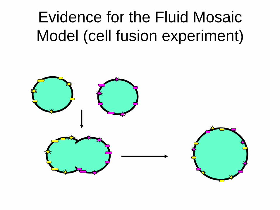
Evidence for the Fluid Mosaic
Model (cell fusion experiment)

Membranes must be fluid to
work properly!!!!!


• In eukaryotes, a fat
molecule called
cholesterol modulates
the membrane fluidity
by making the
membrane:
– Less fluid at warmer
temperatures
– More fluid at lower
temperatures

Membrane proteins form a “Mosaic”
• Integral Proteins (span the
membrane)
• Peripheral Proteins (not
embedded, attached to
membrane surface)


Membrane Proteins Form a Mosaic
• Categories of membrane proteins
– Receptor Proteins
– Recognition Proteins
– Enzymatic Proteins
– Adhesion Proteins
– Transport Proteins

Functions of membrane proteins
• Receptor proteins
bind to specific
molecules found
outside the cell
(like hormones)
and trigger
changes in cell
action


Membrane Proteins Form a Mosaic
• Recognition Proteins
– Serve as identification tags on the surface of
a cell
– Often times these are GLYCOPROTEINS-
proteins with an attached small sugar
molecule (called an oligosaccharide)
Membrane Proteins Form a Mosaic
• Enzymes
– Promote chemical reactions that synthesize
or break apart biological molecules

Membrane Proteins Form a Mosaic
• Adhesion Proteins
– Anchor the cell membrane to inner
cytoskeleton, to proteins outside the cell,
and to other cells

• Transport proteins allow substances to move into or out of the cell through the membrane
– Channel proteins serve as pores through which substances can move
– Carrier proteins bind specific substances and change shape to force the material across the membrane (some need energy input to work)


Video clip (fluid mosaic)