The Appendicular Skeleton Slide 5.32a Copyright © 2003 Pearson Education, Inc. publishing as...
19
The Appendicular Skeleton The Appendicular Skeleton Slide 5.32a right © 2003 Pearson Education, Inc. publishing as Benjamin Cummings
-
Upload
luke-walker -
Category
Documents
-
view
214 -
download
0
Transcript of The Appendicular Skeleton Slide 5.32a Copyright © 2003 Pearson Education, Inc. publishing as...

The Appendicular SkeletonThe Appendicular Skeleton
Slide 5.32aCopyright © 2003 Pearson Education, Inc. publishing as Benjamin Cummings

The Appendicular SkeletonThe Appendicular Skeleton
Slide 5.32aCopyright © 2003 Pearson Education, Inc. publishing as Benjamin Cummings
Limbs (appendages) – arms & legs
Pectoral girdle (shoulders)
Pelvic girdle (hips)

Bones of the Shoulder GirdleBones of the Shoulder Girdle
Slide 5.34aCopyright © 2003 Pearson Education, Inc. publishing as Benjamin Cummings
Figure 5.20a, b

The Pectoral (Shoulder) GirdleThe Pectoral (Shoulder) Girdle Composed of 2 bones: clavicle & scapula
Clavicle (collarbone)
attaches to sternum & to the scapula
keeps shoulder from dislocating and the arm from flopping forward onto chest
Scapula (shoulder blade)
Only attaches to the axial skeleton at the clavicle, so allows lots of movement
Drawbacks: clavicle breaks; scapula dislocates

Slide 5.34bCopyright © 2003 Pearson Education, Inc. publishing as Benjamin Cummings
Figure 5.20c, d

The ScapulaThe Scapula Scapula
Has 3 important landmarks or areas: acromion process, coracoid process & glenoid cavity
Acromion process – where clavicle connects
Coracoid process – forms top of shoulders & gives arm muscles a place to attach
Glenoid cavity – shallow socket where humerus fits (ball & joint)

Bones of the Upper LimbBones of the Upper Limb
Slide 5.35aCopyright © 2003 Pearson Education, Inc. publishing as Benjamin Cummings
Humerus = upper arm
Round at the top for shoulder joint
2 depressions at lower end to allow ulna to move freely
Figure 5.21a, b

Bones of the Upper LimbBones of the Upper Limb
Slide 5.35bCopyright © 2003 Pearson Education, Inc. publishing as Benjamin Cummings
• The forearm has two bones: ulna & radius
• Radius is on thumb side
• Ulan & radius touch at top & bottom (radioulnar joints) & are joined in the middle by interosseous membrane
• Radius allows arm to rotate Figure 5.21c

Bones of the Upper LimbBones of the Upper Limb
Slide 5.36Copyright © 2003 Pearson Education, Inc. publishing as Benjamin Cummings
The hand:
Carpals – wrist, 8 bones – 2 rows of 4
Metacarpals – palm; 5 total, #1 = thumb
Phalanges – fingers; 14 total (3 per finger except the thumb)
Figure 5.22

Slide 5.37Copyright © 2003 Pearson Education, Inc. publishing as Benjamin Cummings
The PelvisThe Pelvis
Slide 5.38aCopyright © 2003 Pearson Education, Inc. publishing as Benjamin Cummings
Figure 5.23a

Slide 5.37Copyright © 2003 Pearson Education, Inc. publishing as Benjamin Cummings
The PelvisThe Pelvis
Slide 5.38bCopyright © 2003 Pearson Education, Inc. publishing as Benjamin Cummings
Figure 5.23b

Bones of the Pelvic GirdleBones of the Pelvic Girdle
Slide 5.37Copyright © 2003 Pearson Education, Inc. publishing as Benjamin Cummings
Composed of 2 hip bones (coxal bones) Does not include the sacrum & coccyx Composed of three pair of fused bones
Ilium – large upper part, most of the hip bone Ischium – lower back part, what you sit on Pubic bone – lower front part
All 3 are heavy bones with deep sockets & lots of ligament
Attaches to the axial skeleton & entire weight of body rests on the pelvis

Bones of the Pelvic GirdleBones of the Pelvic Girdle
Slide 5.37Copyright © 2003 Pearson Education, Inc. publishing as Benjamin Cummings
2 main functions Bear weight is the main function Second function is to protects several organ
Reproductive organs Urinary bladder Part of the large intestine
Several differences between male & female pelvic girdles so can easily tell gender on a skeleton
One big difference is the size of the interior cavity since women give birth

Gender Differences of the PelvisGender Differences of the Pelvis
Slide 5.39
Copyright © 2003 Pearson Education, Inc. publishing as Benjamin Cummings
Figure 5.23c

Bones of the Lower LimbsBones of the Lower Limbs
Slide 5.40a
Carry our entire weight when erect, so large & thick compared to arms
Femur - thigh bone
Only bone in the thigh
Lower leg
Tibia & fibula Figure 5.35a, b
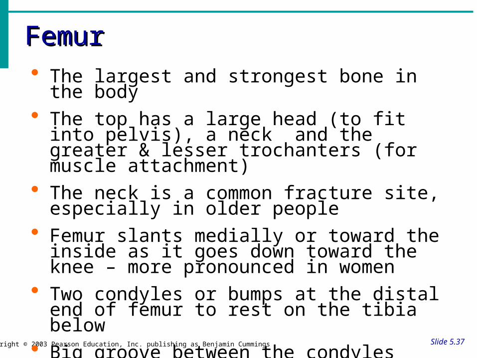
FemurFemur
Slide 5.37Copyright © 2003 Pearson Education, Inc. publishing as Benjamin Cummings
The largest and strongest bone in the body The top has a large head (to fit into pelvis), a
neck and the greater & lesser trochanters (for muscle attachment)
The neck is a common fracture site, especially in older people
Femur slants medially or toward the inside as it goes down toward the knee – more pronounced in women
Two condyles or bumps at the distal end of femur to rest on the tibia below
Big groove between the condyles where the patella moves back and forth with movement

Bones of the Lower LegBones of the Lower Leg
Slide 5.40b
Copyright © 2003 Pearson Education, Inc. publishing as Benjamin Cummings
The leg has two bones: tibia & fibula
Very similar to ulna and radius
Tibia (shinbone) is bigger & medial – upper end is where patella attaches; lower end forms ankle
Figure 5.35c

Bones of the FootBones of the Foot
Slide 5.41
Copyright © 2003 Pearson Education, Inc. publishing as Benjamin Cummings
Three parts like the hand
Tarsus – ankle
Metatarsals – sole
Phalanges – toes
2 jobs: support weight & act as a lever to help us move forward Figure 5.25

Arches of the FootArches of the Foot
Slide 5.42
Copyright © 2003 Pearson Education, Inc. publishing as Benjamin Cummings
Bones of the foot are arranged to form three strong arches
Two longitudinal
One transverse
Allows flexibility and springiness
Figure 5.26