
Sub1550
5
International Journal of Science and Research (IJSR) ISSN (Online): 2319-7064 Index Copernicus Value (2013): 6.14 | Impact Factor (2013): 4.438 Volume 4 Issue 1, January 2015 www.ijsr.net Licensed Under Creative Commons Attribution CC BY An Intelligent System for Lung Cancer Diagnosis Using Fusion of Support Vector Machines and Back Propagation Neural Network Gurpreet Kaur 1 , Harpreet Singh 2 1 M-Tech Research Scholar, Swami Vivekananad Institute of Engineering and Technology, Banur, Punjab India. 2 Assistant Professor, Swami Vivekanand Institute of Engineering and Technology, Banur Punjab India Abstract: Cancer is the most important cause of death for both men and women. The early detection of cancer can be helpful in curing the disease completely. So the requirement of techniques to detect the occurrence of cancer nodule in early stage is increasing. A disease that is commonly misdiagnosed is lung cancer. Neural Networks (NNs) play a vital role in the medical field in solving various health problems like acute diseases and even other mild diseases. Earlier diagnosis of Lung Cancer saves enormous lives, failing which may lead to other severe problems causing sudden fatal end. Its cure rate and prognosis depends mainly on the early detection and diagnosis of the disease. This thesis provides a Neural Network and SVM model for early detection of lung cancer. The model consists of an input layer, a hidden layer and an output layer. The network is trained with one hidden layer and one output layer by giving twelve inputs. One of the most common forms of medical malpractices globally is an error in diagnosis. By using the fusion of SVM and BPNN we achieved the accuracy of 98%.The performance simulation is taken place in MATLAB 7.10 environment. The MATLAB has inbuilt Neural Network toolbox and SVM has been implemented using two steps training and testing phases. Keywords: SVM, Neural Network, Lung Cancer, Survival Rate 1. Introduction Lung cancer is the leading cause of cancer deaths in both women and men. It is estimated that 1.2 million people are diagnosed with this disease every year (12.3% of the total number of cancer diagnosed), and about 1.1 million people are dying of this disease yearly (17.8% of the total cancer death) [1]. The survival rate is higher if the cancer is detected at early stages. The early detection of lung cancer is not an easy task. About 80% patients are diagnosed correctly at the middle or advanced stage of cancer [2]. Computer-aided diagnosis sys-tem is very helpful for radiologist in detection and diagnosing abnormalities earlier and faster [3]. The computer aided diagnosis is a second opinion for radiologists before suggesting a biopsy test finally compare this with individual SVM and BPNN to show the improvement in proposed work. [4]. In recent research literature, it is observed that principles of neural networks have been widely used for the detection of lung cancer in medical images [5].For classification of lung cancer, few methods based on neural network have been reported in the literature. Abdulla et al. [6] proposed a computer aided diagnosis based on artificial neural networks for classification of lung cancer. The features used for the mode of the area of the new pre classification are area, perimeter and shape. The maximum classification accuracy obtained is 90%.Camarlinghi et al. [7] proposed a computer-aided detection algorithm for automatic lung nodule identification. The sensitivity obtained is 80% with 3 FP/scan. Al-Kadi et al. [8] proposed classification method based on fractal texture features. The classification accuracy obtained is 83.3%. van Ginneken et al.[9] compared and combined six computer aided detection algorithms for pulmonary nodules. The combination of six algorithms is able to detect 80% of all nodules at the expense of only two false positive detections per scan and 65% of all nodules with only 0.5 false positives. Cascio et al. [10] proposed computer-aided detection (CAD) system for the selection of lung nodules in computer tomography (CT) images. The detection rate of the system is 88.5% with 6.6 FPs/CT on 15 CT scans. A reduction to 2.47 FPs/CT is achieved at 80% efficiency. But in proposed work, we will show the efficiency of the proposed work by hybridization of SVM and BPNN. Then finally compare this with individual SVM and BPNN to show the improvement in proposed work. 2. Proposed Methodology Figure 1: Lung Cancer Detection Above Figure 1 shows the main working of the project of the lung cancer detection using the neural network. The project contains two section , the first section is called the training section and the another section is called the testing section .In the training section , the system is trained on the basis of affected diacom images taken from the medical institutes [11]. The first process of the training is the feature extraction. To extract the features, the system requires the training folder Paper ID: SUB1550 87
-
Upload
international-journal-of-science-and-research-ijsr -
Category
Documents
-
view
44 -
download
0
Transcript of Sub1550
International Journal of Science and Research (IJSR) ISSN (Online): 2319-7064
Index Copernicus Value (2013): 6.14 | Impact Factor (2013): 4.438
Volume 4 Issue 1, January 2015 www.ijsr.net
Licensed Under Creative Commons Attribution CC BY
An Intelligent System for Lung Cancer Diagnosis Using Fusion of Support Vector Machines and Back
Propagation Neural Network
Gurpreet Kaur1, Harpreet Singh2
1M-Tech Research Scholar, Swami Vivekananad Institute of Engineering and Technology, Banur, Punjab India.
2Assistant Professor, Swami Vivekanand Institute of Engineering and Technology, Banur Punjab India
Abstract: Cancer is the most important cause of death for both men and women. The early detection of cancer can be helpful in curing the disease completely. So the requirement of techniques to detect the occurrence of cancer nodule in early stage is increasing. A disease that is commonly misdiagnosed is lung cancer. Neural Networks (NNs) play a vital role in the medical field in solving various health problems like acute diseases and even other mild diseases. Earlier diagnosis of Lung Cancer saves enormous lives, failing which may lead to other severe problems causing sudden fatal end. Its cure rate and prognosis depends mainly on the early detection and diagnosis of the disease. This thesis provides a Neural Network and SVM model for early detection of lung cancer. The model consists of an input layer, a hidden layer and an output layer. The network is trained with one hidden layer and one output layer by giving twelve inputs. One of the most common forms of medical malpractices globally is an error in diagnosis. By using the fusion of SVM and BPNN we achieved the accuracy of 98%.The performance simulation is taken place in MATLAB 7.10 environment. The MATLAB has inbuilt Neural Network toolbox and SVM has been implemented using two steps training and testing phases. Keywords: SVM, Neural Network, Lung Cancer, Survival Rate 1. Introduction Lung cancer is the leading cause of cancer deaths in both women and men. It is estimated that 1.2 million people are diagnosed with this disease every year (12.3% of the total number of cancer diagnosed), and about 1.1 million people are dying of this disease yearly (17.8% of the total cancer death) [1]. The survival rate is higher if the cancer is detected at early stages. The early detection of lung cancer is not an easy task. About 80% patients are diagnosed correctly at the middle or advanced stage of cancer [2]. Computer-aided diagnosis sys-tem is very helpful for radiologist in detection and diagnosing abnormalities earlier and faster [3]. The computer aided diagnosis is a second opinion for radiologists before suggesting a biopsy test finally compare this with individual SVM and BPNN to show the improvement in proposed work. [4]. In recent research literature, it is observed that principles of neural networks have been widely used for the detection of lung cancer in medical images [5].For classification of lung cancer, few methods based on neural network have been reported in the literature. Abdulla et al. [6] proposed a computer aided diagnosis based on artificial neural networks for classification of lung cancer. The features used for the mode of the area of the new pre classification are area, perimeter and shape. The maximum classification accuracy obtained is 90%.Camarlinghi et al. [7] proposed a computer-aided detection algorithm for automatic lung nodule identification. The sensitivity obtained is 80% with 3 FP/scan. Al-Kadi et al. [8] proposed classification method based on fractal texture features. The classification accuracy obtained is 83.3%. van Ginneken et al.[9] compared and combined six computer aided detection algorithms for pulmonary nodules. The combination of six algorithms is able to detect 80% of all nodules at the expense of only two false positive detections per scan and 65% of all nodules with
only 0.5 false positives. Cascio et al. [10] proposed computer-aided detection (CAD) system for the selection of lung nodules in computer tomography (CT) images. The detection rate of the system is 88.5% with 6.6 FPs/CT on 15 CT scans. A reduction to 2.47 FPs/CT is achieved at 80% efficiency. But in proposed work, we will show the efficiency of the proposed work by hybridization of SVM and BPNN. Then finally compare this with individual SVM and BPNN to show the improvement in proposed work. 2. Proposed Methodology
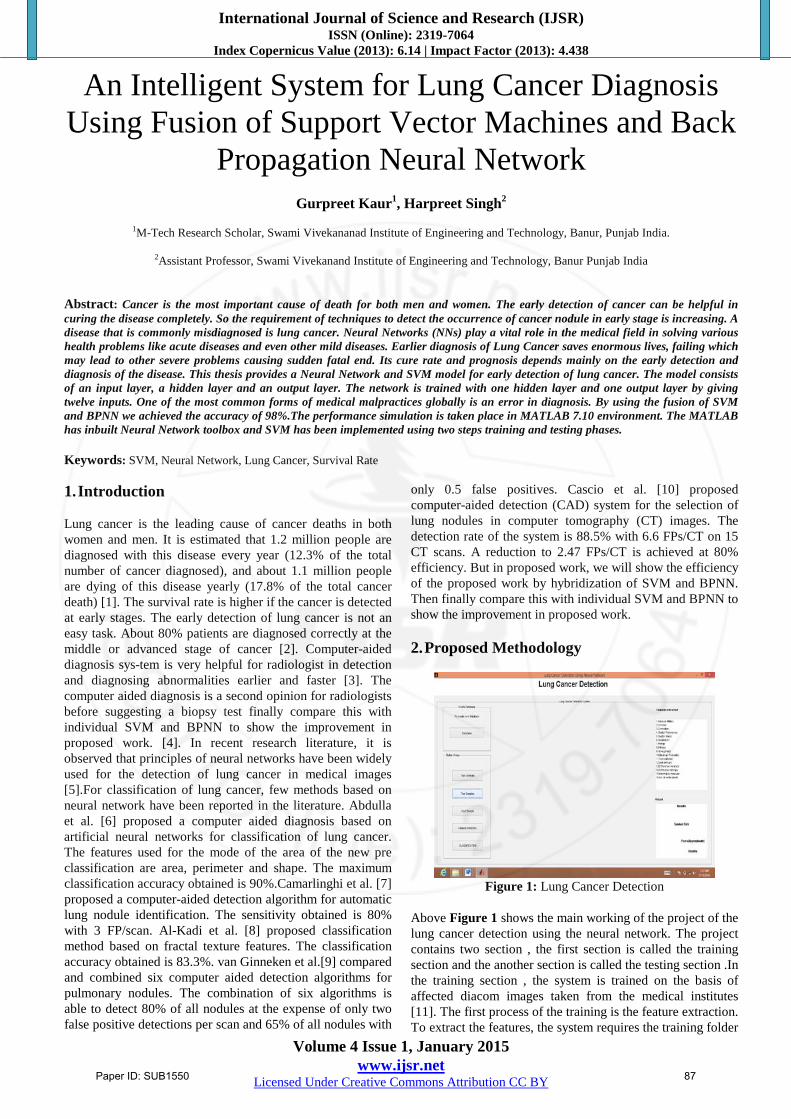
Figure 1: Lung Cancer Detection
Above Figure 1 shows the main working of the project of the lung cancer detection using the neural network. The project contains two section , the first section is called the training section and the another section is called the testing section .In the training section , the system is trained on the basis of affected diacom images taken from the medical institutes [11]. The first process of the training is the feature extraction. To extract the features, the system requires the training folder
Paper ID: SUB1550 87

International Journal of Science and Research (IJSR) ISSN (Online): 2319-7064
Index Copernicus Value (2013): 6.14 | Impact Factor (2013): 4.438
Volume 4 Issue 1, January 2015 www.ijsr.net
Licensed Under Creative Commons Attribution CC BY
which can be uploaded using a button provided at the GUI. In the same manner the user will have to specify that through which folder he has to confirm the images.
Figure 2: Training Section
Above Figure 2 shows the training section of project. The project contain two section, the first section is called training section. Training is done by using Neural network as well as SVM method. Both of the classifiers are used for traning and the section of testing. Traning section is uploaded the tested diacom images that has been tested by both of the classifier. To extract the features, the system requires the training folder which can be uploaded using a button provided at the GUI.
Figure 3: Training System uploaded
Above Figure 3 shows that the system is trained on the basis of diacom images taken from the medical institute.
Figure 4: Input Image
Above Figure 4 shows the input image that has been taken for the training of the image.
Figure 5: Testing section
Second section is shown by Figure 5 that is called testing section, that include affected or not affected..testing section defines the original output of the diacom images.
Figure 6: Testing is done
Above Figure 6 shows that testing is done on the image that has been inputted to the system.
Figure 7: Select image for feature extraction
Above Figure 7 shows the window for the extraction of features of the inputted image.
Paper ID: SUB1550 88

International Journal of Science and Research (IJSR) ISSN (Online): 2319-7064
Index Copernicus Value (2013): 6.14 | Impact Factor (2013): 4.438
Volume 4 Issue 1, January 2015 www.ijsr.net
Licensed Under Creative Commons Attribution CC BY
Figure 8: Feature Extraction Process
First process of the training is feature extraction feature extraction describes the different types of symptom’s that’s bring in a logical way feature extraction is done by Principle Component Analysis.
Figure 9: Feature extractions done
Above Figure 9 shows that after the feature extraction classification is done of the lung cancer detection.
Figure 10: Neural network toolbox
Above Figure 10 shows that for classification neural network has been used, It has 5 iterations that has been used. In proposed neural network 2 input layers, 3 hidden layers and 1 output layer has been used.
Figure 11: Classification done
Above Figure 11 Shows that classification is done after applying SVM [12] and neural network only or in hybridisation. 3. Pseudo Code of Implementation do for (Neural Network) tmp = sigmoid(I) for I = 1 to element_second_layer if tmp is greater than θi yi =tmp flag = check_with_expected() if flag is equal to false adjust_weight() return false else yi = 0 I weighted sum of y tmp = sigmoid(I) if tmp > θi flag = check_with_expected() if flag is equal to false adjust_weight() return false else return confirm else return negative do for (SVM) initialize radial basis kernel function for training • Operator1: substitute one existing input feature with one unselected variable. • Operator2: add one input feature • Step 1: do for each Operatork, k=1,2 • Step 2: do for each variable i=1, … ,93 (i≠ the variable selected as input features) 1. Trial new SVM: SVM*=Operatork (SVM) 2. ROC evaluation; 3. If k=2, i=93, and SVM is not replaced, stop construction. • parameter C and σ are selected for testing • matching : if yes = stop • if no= go ahead
Paper ID: SUB1550 89
International Journal of Science and Research (IJSR) ISSN (Online): 2319-7064
Index Copernicus Value (2013): 6.14 | Impact Factor (2013): 4.438
Volume 4 Issue 1, January 2015 www.ijsr.net
Licensed Under Creative Commons Attribution CC BY
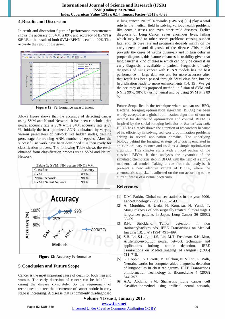
4. Results and Discussion In result and discussion figure of performance measurement shows the accuracy of SVM is 89% and accuracy of BPNN is 98%.But the result of both SVM+BPNN is eual to 99%.That accurate the result of the given.
Figure 12: Performance measurement
Above figure shows that the accuracy of detecting cancer using SVM and Neural Network. It has been concluded that neural accuracy rate is 98% while SVM accuracy rate is 89 %. Initially the best optimized ANN is obtained by varying various parameters of network like hidden nodes, training percentage for training ANN, number of epochs. After the successful network have been developed it is then ready for classification process. The following Table shows the result obtained from classification process using SVM and Neural Network.
Table 1: SVM, NN versus NN&SVM Classifier Accuracy SVM 89 % Neural network 98 % SVM +Neural Network 99%
Figure 13: Accuracy Performance
5. Conclusion and Future Scope Cancer is the most important cause of death for both men and women. The early detection of cancer can be helpful in curing the disease completely. So the requirement of techniques to detect the occurrence of cancer nodule in early stage is increasing. A disease that is commonly misdiagnosed
is lung cancer. Neural Networks (BPNNs) [13] play a vital role in the medical field in solving various health problems like acute diseases and even other mild diseases. Earlier diagnosis of Lung Cancer saves enormous lives, failing which may lead to other severe problems causing sudden fatal end. Its cure rate and prognosis depends mainly on the early detection and diagnosis of the disease .This model prevents the cases of wrong diagnosis and in turn delay in proper diagnosis, this feature enhances its usability given that lung cancer is kind of disease which can only be cured if an early diagnosis is available to patient. Prognosis of early diagnosis of Lung cancer with BPNN models has the best performance in large data sets and for more accuracy after that result has been passed through SVM classifier, but the hybridization leads to more enhancements [14, 15]. We get the accuracy of this proposed method i.e fusion of SVM and NN is 99%, 98% by using neural and by using SVM it is 89 % Future Scope lies in the technique where we can use BFO, Bacterial foraging optimization algorithm (BFOA) has been widely accepted as a global optimization algorithm of current interest for distributed optimization and control. BFOA is inspired by the social foraging behavior of Escherichia coli. BFOA has already drawn the attention of researchers because of its efficiency in solving real-world optimization problems arising in several application domains. The underlying biology behind the foraging strategy of E.coli is emulated in an extraordinary manner and used as a simple optimization algorithm. This chapter starts with a lucid outline of the classical BFOA. It then analyses the dynamics of the simulated chemotaxis step in BFOA with the help of a simple mathematical model. Taking a cue from the analysis, it presents a new adaptive variant of BFOA, where the chemotactic step size is adjusted on the run according to the current fitness of a virtual bacterium. References [1] D.M. Parkin, Global cancer statistics in the year 2000,
LancetOncology 2 (2001) 533–543. [2] A. Motohiro, H. Ueda, H. Komatsu, N. Yanai, T.
Mori,Prognosis of non-surgically treated, clinical stage I lungcancer patients in Japan, Lung Cancer 36 (2002) 65–69.
[3] R.N. Strickland, Tumor detection in non stationarybackgrounds, IEEE Transactions on Medical Imaging 13(June) (1994) 491–499.
[4] S.B. Lo, S.L. Lou, J.S. Lin, M.T. Freedman, S.K. Mun, Artificialconvolution neural network techniques and applications forlung nodule detection, IEEE Transactions on MedicalImaging 14 (August) (1995) 711–718.
[5] G. Coppini, S. Diciotti, M. Falchini, N. Villari, G. Valli, Neuralnetworks for computer aided diagnosis: detection of lungnodules in chest radiograms, IEEE Transactions onInformation Technology in Biomedicine 4 (2003) 344–357.
[6] A.A. Abdulla, S.M. Shaharum, Lung cancer cell classificationmethod using artificial neural network,
Paper ID: SUB1550 90

International Journal of Science and Research (IJSR) ISSN (Online): 2319-7064
Index Copernicus Value (2013): 6.14 | Impact Factor (2013): 4.438
Volume 4 Issue 1, January 2015 www.ijsr.net
Licensed Under Creative Commons Attribution CC BY
InformationEngineering Letters 2 (March) (2012) 50–58.
[7] N. Camarlinghi, et al., Combination of computer-aideddetection algorithms for automatic lung noduleidentification, International Journal of Computer AssistedRadiology and Surgery 7 (2012) 455–464.
[8] O.S. Al-Kadi, D. Watson, Texture analysis of aggressive andnon aggressive lung tumor CE CT images, IEEE Transactionson Biomedical Engineering 55 (2008) 1822–1830.
[9] B. van Ginneken, et al., Comparing and combiningalgorithms for computer-aided detection of pulmonarynodules in computed tomography scans: the ANODE09study, Medical Image Analysis 14 (2010) 707–722.
[10] R. Bellotti, D. Cascio, et al., A CAD system for noduledetection in low-dose lung CTs based on region growing anda new active contour model, International Journal ofMedical Physics Research and Practice 34 (2007) 4901–4911
[11] R. M. Haralick, K. Shanmugam and I. Dinstein, "Texture parameters for image classification", IEEE Transaction. Systems, Man, Cybernatics, vol. SMC-3, no. 6, pp. 610-621, 1973.
[12] Frenster, J.H (1990)., "Neural Networks for PatternRecognition in Medical Diagnosis", Annual International Conference in the IEEE Engineering in Medicine and Biology Society, vol. 12, No. 3, issued 1990, pp. 1423-1424
[13] Peter M. Ravdin, et al(1992), "A practical application of neural network analysis for predicting outcome of individual breast cancer patients", Breast Cancer Research & Treatment, vol. 22, pp. 285-293 (1992)
[14] Hernandez, C.A. et al (1993)., "How to Choose the Training Data for Neural Network Medical Diagnosis Systems", ISA, pp. 283-290 (1993).
[15] N.B. Karayiannis, A.N. Venetsanopoulos, “Efficient Learning Algorithms for Neural Networks,” IEEE , vol. 23, 1993, pp. 1372 -1383.
Author Profile
Gurpreet Kaur is Student of M.Tech in the department of Computer Science and engineering at Swami vivekanand institute of engineering, Banur under Punjab technical university (SVIET) , She has done B.Tech from Yadvindra College of engineering ,
Talwandi Sabo Under Punjabi University, Patiala in 2012.
Harpreet Singh is Assistant Professor in the department of Computer Science Engineering at Swami Vivekanand institute of engineering, (SVIET) Banur, under Punjab Technical University, Jalandhar. He has done M.Tech from UCoE (Punjabi University, Patiala)
in 2013 and B.Tech from Lovely Professional University Jalandhar in 2011.
Paper ID: SUB1550 91


