Studies on Fetal response to Prozac Treatment
20
The Egyptian Journal of Hospital Medicine (April 2011) Vol., 43: 145 – 161 145 Studies on Fetal response to Prozac Treatment Nehal A, Abou Naja* , Fatma A. Eid * and Khadija Abdul jalil Fadladdeen** *Zoology Department,Faculty of Science (Girls), Al – Azhar University. **Zoology Department,Faculty of Science,King Abd El-Aziz University,KSA. Abstract Aim of the work: :A variety of adverse effects are reported post-treatment with Prozac(fluoxetine)especially during pregnancy.The percentage of these changes often reflects increased rates with rising doses. This study aimed to study the possible histopathological and histochemical changes in skin of fetuses maternally treated with Prozac with 3 different doses(0.72&1.44&2.88 mg/kg b.wt.). Material and methods: Mature male and virgin female albino rats of pure strain (Albino rattus norvegicus) ranging from 220-280 gm were used. Males were used only for mating. Pregnant rats were categorized into the following groups: Group (1): control group. Group (2): 10 pregnant rats treated daily with 0.72 mg/kg. b.wt. Prozac (T1) (treatment started one month before pregnancy and continued till day19 of gestation) Group (3): 10 pregnant rats treated daily with 1.44 mg/kg. b.wt. (T2). Group (4):10 pregnant rats treated daily with 2.88 mg/kg. b.wt. Prozac (T3). Pregnant mothers from all groups were sacrificed on day 19 of gestation and small pieces of fetal skin were taken for the histological and histochemical studies. Results: Many histological and histochemical changes were observed in fetal skin of all the treated groups compared with control ones. The severity of these changes increased with increasing the doses. Conclusion: Maternally Prozac treatment caused deleterious changes in the fetal skin, therefore the use of this drug during pregnancy should be under strict precautions and further studies are recommended due to the potential risks to the developing fetuses. Key words: Prozac (fluoxetine),pregnant rats, pregnant women,fetus,skin. Introduction Pregnancy should be carefully evaluated because it is a period during which women go through many physical, hormonal and psychic changes which, in turn influence their mental health. It has been recognized that gestation can be complicated by emotional problems such as depression, thus, heavily impacting both mother and fetus (Costei et al., 2002; Crews and Frederic, 2007). The use of medication during pregnancy requires special attention due to the potential risks to the developing fetus. Pregnant women often need psychiatric treatment in face of emotional disorders caused by stress, anxiety and depression (Richelson, 2001). Antidepressants are capable of crossing the placental barrier, and their use has been evaluated with respect to their biosecurity. Recent researches report the use of tricyclic antidepressants and serotonin reuptake inhibitors, especially fluoxetine, in pregnant women (Chubak et al., 2007, 2009, and 2011). Some authors have proposed new studies to assess the risk-benefit ratio of the use of antidepressants during gestation. Likewise, serotonin and noradrenaline are involved in the physiopathology of affective disorders, Imipramine inhibits
Transcript of Studies on Fetal response to Prozac Treatment

The Egyptian Journal of Hospital Medicine (April 2011) Vol., 43: 145 – 161
145
Studies on Fetal response to Prozac Treatment
Nehal A, Abou Naja* , Fatma A. Eid * and Khadija Abdul jalil Fadladdeen**
*Zoology Department,Faculty of Science (Girls), Al – Azhar University.
**Zoology Department,Faculty of Science,King Abd El-Aziz University,KSA.
Abstract
Aim of the work: :A variety of adverse effects are reported post-treatment with
Prozac(fluoxetine)especially during pregnancy.The percentage of these changes often
reflects increased rates with rising doses. This study aimed to study the possible
histopathological and histochemical changes in skin of fetuses maternally treated with
Prozac with 3 different doses(0.72&1.44&2.88 mg/kg b.wt.).
Material and methods: Mature male and virgin female albino rats of pure strain (Albino rattus norvegicus) ranging from 220-280 gm were used. Males were used only for mating. Pregnant
rats were categorized into the following groups: Group (1): control group. Group (2): 10
pregnant rats treated daily with 0.72 mg/kg. b.wt. Prozac (T1) (treatment started one month
before pregnancy and continued till day19 of gestation)
Group (3): 10 pregnant rats treated daily with 1.44 mg/kg. b.wt. (T2). Group (4):10 pregnant
rats treated daily with 2.88 mg/kg. b.wt. Prozac (T3). Pregnant mothers from all groups were sacrificed on day 19 of gestation and small pieces of fetal skin were taken for the histological
and histochemical studies.
Results: Many histological and histochemical changes were observed in fetal skin of all the
treated groups compared with control ones. The severity of these changes increased with
increasing the doses.
Conclusion: Maternally Prozac treatment caused deleterious changes in the fetal skin, therefore
the use of this drug during pregnancy should be under strict precautions and further studies are
recommended due to the potential risks to the developing fetuses. Key words: Prozac (fluoxetine),pregnant rats, pregnant women,fetus,skin.
Introduction
Pregnancy should be carefully evaluated because it is a period during
which women go through many physical,
hormonal and psychic changes which, in
turn influence their mental health. It has been recognized that gestation can be
complicated by emotional problems such as
depression, thus, heavily impacting both mother and fetus (Costei et al., 2002;
Crews and Frederic, 2007). The use of medication during
pregnancy requires special attention due to the potential risks to the developing fetus.
Pregnant women often need psychiatric
treatment in face of emotional disorders
caused by stress, anxiety and depression
(Richelson, 2001).
Antidepressants are capable of crossing the placental barrier, and their use
has been evaluated with respect to their
biosecurity. Recent researches report the use of tricyclic antidepressants and
serotonin reuptake inhibitors, especially
fluoxetine, in pregnant women (Chubak et
al., 2007, 2009, and 2011). Some authors have proposed new
studies to assess the risk-benefit ratio of the
use of antidepressants during gestation. Likewise, serotonin and noradrenaline are
involved in the physiopathology of
affective disorders, Imipramine inhibits

Studies on….
146
noradrenaline and serotonin reuptake in the
central nervous system, while fluoxetine selectively inhibits serotonin reuptake
(Alwan et al., 2007).
During organogenesis, medication can be
considered as a teratogenic factor, thereby causing congenital malformations and
serious damages that may lead to abortion.
Although, if used during the second and third trimesters, medication is no longer
able to produce significant malformations,
it can affect the fetus's functional development and growth (Chambers et al.,
2006).
Fluoxetine was selected for being the
antidepressant of choice for pregnant women and most researches have focused
on fluoxetine because of its high selectivity
and negligible affinity for several receptor subtypes.
The administration of antidepressant drug
to rats induced variable histopathological changes on different organs such as the
brain, lung, heart and muscle. Such as areas
of necrosis, intestinal lymphocytic
infiltration, and congestion were detected (Hassan, 1990).
Fluoxetine causes an acute increase in
serotonin levels, thus leading to a transient reduction in uterine blood flow. This, in
turn, decreases the oxygen and nutrient
supply to the fetus, reduces its growth and
leads it to premature birth. In addition, fluoxetine mainly affects the fetus neural
development (Kallen, 2004; Chubak et al.,
2007, 2009, 2011). This study was designed to evaluate the
possible histopathological and
histochemical changes in the skin of fetuses maternally exposed to fluoxetine with three
different doses (0.72&1.44&2.88 mg/kg
b.wt.).
In2011,Fadladdeen noticed numerous histopathological and histological
changes in some fetal organs
maternally treated with different doses of Prozac. In this respect, the
histopathological and histological
changes in the fetuses due to Prozac treatment are rare ,so, This study was
designed to evaluate the possible
histopathological and histochemical
changes in the skin of fetuses maternally exposed to fluoxetine with three different
doses (0.72&1.44&2.88 mg/kg b.wt.).
Material and Methods
Mature male and virgin female albino rats
of pure strain (Albino rattus norvegicus)
ranging from 220-280 gm body weight were used and kept under normal conditions
of temperature, light and relative humidity.
Estrous cycle was determined according to Taylor (1986). Pregnant rats were
randomly assigned to control and treated
groups. The gestation period in the pregnant
rats was 21 day. Prozac doses were determined after
conversion from human doses according to
Paget and Barnes (1964). Pregnant female rats were categorized into
the following groups: Group (1):10
pregnant rats were kept under normal conditions (control group). Group (2): 10
pregnant rats were treated daily with 0.72
mg/kg. b.wt. Prozac (T1) which was
dissolved in distilled water (treatment started one month before pregnancy and
continued till day 19 of
gestation).Pregnancy was determined according to Eda et al.(2009).
Group (3): 10 pregnant rats were treated
daily with 1.44 mg/kg. b.wt. Prozac as group 2(T2).
Group (4):10 pregnant rats were treated
daily with 2.88 mg/kg. b.wt. Prozac (T3).
Pregnant mothers from all groups were sacrificed on day 11 of gestation and small
pieces of skin were taken for the
histological and histochemical studies. These pieces were fixed in 10% neutral
buffered formol solution and Carnoy's fluid
for the histological and histochemical
studies. Paraffin sections were prepared 5 µm
thicknesses and stained with Harris
haematoxylin and eosin (Bancroft and
Gamble, 2002). Polysaccharides were
detected by PAS (periodic acid Schiff)
method (Hotchkiss, 1948).Total proteins were detected by mercuric bromophenol
blue method (Mazia et al., 1953). DNA
content were detected by Feulgen method
(Pears, 1977). Collagen fibers were stained by Mallory's trichorome stain (Pears,
1977).
Image analysis: The thickness of skin layers were measured
(µm) by Bel micro Image Analyzer,
software for microscopy ver. 2.3.

Nehal Abou Naja… et al
147
In addition, the optical transparency (pexil)
of the total protein, PAS+ve materials and DNA content were recorded and all data
were statistically analyzed by using T- test
microsoft Excel 2007.
Results
Skin (Integumentary system) The skin is the heaviest single organ in the body and it represents about 16% of the
total body weight.
-The skin is mainly formed of the following layers:
1-Epidermis:
-It is the epithelial layer of the skin formed of stratified squamous keratinized
epithelium.
-It is of ectodermal origin.
2-Dermis: -It is the connective tissue layer of the skin
and can be divided into-2 main layers:
A-Papillary layer. B-Reticular layer.
-It is of mesodermal origin.
-The subcutaneous connective tissue layer (hypodermis) is not considered as skin
layer.
Studies on….
148
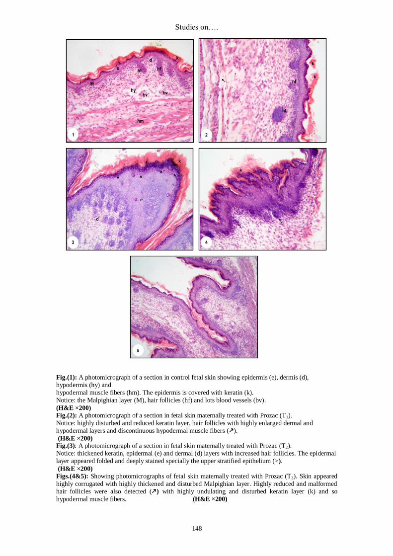
Fig.(1): A photomicrograph of a section in control fetal skin showing epidermis (e), dermis (d),
hypodermis (hy) and
hypodermal muscle fibers (hm). The epidermis is covered with keratin (k).
Notice: the Malpighian layer (M), hair follicles (hf) and lots blood vessels (bv).
(H&E ×200)
Fig.(2): A photomicrograph of a section in fetal skin maternally treated with Prozac (T1).
Notice: highly disturbed and reduced keratin layer, hair follicles with highly enlarged dermal and
hypodermal layers and discontinuous hypodermal muscle fibers ().
(H&E ×200)
Fig.(3): A photomicrograph of a section in fetal skin maternally treated with Prozac (T2).
Notice: thickened keratin, epidermal (e) and dermal (d) layers with increased hair follicles. The epidermal
layer appeared folded and deeply stained specially the upper stratified epithelium (>).
(H&E ×200)
Figs.(4&5): Showing photomicrographs of fetal skin maternally treated with Prozac (T3). Skin appeared
highly corrugated with highly thickened and disturbed Malpighian layer. Highly reduced and malformed
hair follicles were also detected () with highly undulating and disturbed keratin layer (k) and so
hypodermal muscle fibers. (H&E ×200)
Nehal Abou Naja… et al
149
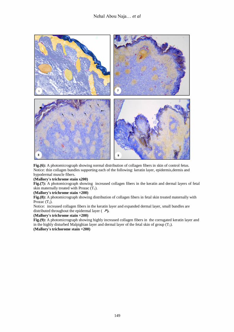
Fig.(6): A photomicrograph showing normal distribution of collagen fibers in skin of control fetus.
Notice: thin collagen bundles supporting each of the following: keratin layer, epidermis,dermis and
hypodermal muscle fibers.
(Mallory's trichrome stain x200) Fig.(7): A photomicrograph showing increased collagen fibers in the keratin and dermal layers of fetal
skin maternally treated with Prozac (T1).
(Mallory's trichrome stain ×200)
Fig.(8): A photomicrograph showing distribution of collagen fibers in fetal skin treated maternally with
Prozac (T2).
Notice: increased collagen fibers in the keratin layer and expanded dermal layer, small bundles are
distributed throughout the epidermal layer ( ).
(Mallory's trichrome stain ×200)
Fig.(9): A photomicrograph showing highly increased collagen fibers in the corrugated keratin layer and
in the highly disturbed Malpighian layer and dermal layer of the fetal skin of group (T3).
(Mallory's trichorome stain ×200)

Studies on….
150
Fig.(10): A photomicrograph showing normal distribution of PAS +ve materials in the control fetal skin.
Notice: Notice dense stain ability in the walls of blood vessels, muscle fibers, hair follicles and Malpighian
layer.
(PAS ×200) Fig.(11): A photomicrograph showing increased PAS +ve materials in few hair follicles and
hypodermal muscle fibers, but decreased stain affinity was detected in the different layers of the fetal
skin of group(T1).
(PAS ×200) Fig.(12): A photomicrograph showing increased PAS+ve materials in few hair follicles of fetal skin of
group(T2),while decreased stain ability could be observed in the different layers of the skin.
(PAS ×200) Figs. (13&14): A photomicrograph showing distribution of PAS+ ve materials in fetal skin of group (T3).
(Fig. (13): Showing deeply stained aggregations of PAS+ ve materials above the corrugated epidermal
layer ( ), with increased stain ability in the hypodermal muscle fibers.
Fig. (14): Showing depleted layers of the fetal skin in another sample.
(PAS ×200)

Nehal Abou Naja… et al
151
Fig.(15): A photomicrograph showing normal distribution of total proteins in the epidermal, dermal and
hypodermal layers.
Notice: increased stain ability in the epidermal layer, hair follicles and walls of blood vessels and
hypodermal muscle fibers.
(Mercuric bromophenol blue ×200)
Fig.(16): A photomicrograph showing increased total proteins in the folded keratin , epidermis and
dermis layers in fetal skin of group (T1).
(Mercuric bromophenol blue ×200)
Fig.(17): A photomicrograph showing increased stain ability of total protein in the thickened keratin and
Malpighian layers of fetal skin of group(T2).
(Mercuric bromophenol blue ×200)
Fig.(18): A photomicrograph showing fetal skin of group(T3) with increased stain affinity of total
proteins in the keratin layer, highly thickened epidermal layer and hair follicles.
(Mercuric bromophenol blue ×200)


Studies on….
152
Fig.(19): A photomicrograph showing normal distribution of DNA content in the control fetal skin.
Notice: increased stain ability in the nuclei of the epidermal layer, hair follicles and numerous nuclei in the
dermal and hypodermal layers.
(Feulgen reaction ×200)
Fig.(20): A photomicrograph showing decreased DNA content in the stratified squamousal epithelial cells
of the epidermis and also decreased stain ability in the dermal and hypodermal layers in the fetal skin treated maternally with Prozac(T1).
(Feulgen reaction ×200)
Fig.(21): A photomicrograph showing decreased DNA stain affinity in the thickened epidermal layer of
fetal skin of group(T2), but few aggregations were detected in this layer. While thickened dermal layer
showed reduced stain ability of DNA, hair follicles appeared deeply stained.
(Feulgen reaction ×200)
Fig.(22): A photomicrograph of a section in fetal skin of group(T3), showing decreased stain ability of
DNA in the epidermal layer, in spite of enlarged dermal layer, the nuclei of this layer appeared less
stained.
(Feulgen reaction×200)

Nehal Abou Naja… et al
153
The epidermis is firmly attached to the dermis and may form one layer while the hypodermis is
loosely attached from the overlying dermis. Normal histological pattern of skin of the control fetus is shown in fig. (1). Different layers of the skin could be observed. These layers include:
keratin, epidermis and dermis.
Numerous hair follicles were detected in the dermal layer. Fetuses of group T1 showed many
deleterious changes in the skin. These changes include reduced keratin layer (38.77± 9.59) compared with the control group (47.72±10.97) and hair follicles with enlarged epidermis
(120.29± 13.13) compared with the control (50.39±7.84),dermis (1072.22±423.53) compared
with the control (255.83±74.52) and hypodermal layers with discontinuous hypodermal muscle fibers (fig.2).
Fetuses maternally treated with Prozac T2 showed highly thickened keratin layer (81.54± 17.48),
epidermis (412.38±253.96) and dermis (790.42±63.59790.42) with increased number of hair follicles. The epidermal layer was folded and deeply stained (fig.3).
Highly corrugated and thickened Malpighian layer was detected in skin of fetuses of group T3
with highly reduced hair follicles. Undulating, distorted and highly reduced keratin layer
(32.79± 7.32), highly thickened epidermis (95.86±14.65), with non significant reduction in the dermal layer (248.07±94.44) and reduced hypodermal muscle fibers were demonstrated (figs.4,
5 & table 4 and hist.1, 2, 3).
Fig. (6) Showing thin collagen bundles supporting the different skin layers of the control fetus. Increased collagen fibers were realized in skin of fetuses of groups T1, T2, and T3
(figs.7, 8, 9). Normal distribution of polysaccharides in skin of a control fetus was observed in fig. (10). Dense stain affinity was noticed in walls of the blood vessels, muscle fibers, hair follicles and
the Malpighian layer.
Highly decreased PAS +ve materials were demonstrated in the fetuses' skin of group T1. These
results were confirmed by the mean optical transparency which reached 29.71±13.06 compared with the control 69.48±19.56 ,but increased stain affinity was realized in few hair
follicles and hypodermal muscle fibers (fig. 11).
Also increased PAS +ve materials was noticed in few hair follicles in the fetal skin of group T2 , while , highly significant decreased stain affinity was observed in the different layers of the
skin (fig. 12)(MOT reached 26.38±13.28).
Numerous deeply stained aggregations of PAS +ve materials were detected in the epidermal
layer of skin of fetuses of group T3 with increased stain affinity in the hypodermal muscle fibers (MOT reached 72.18±31.87). Some areas were depleted (MOT values reached 16.74±8.53)
(figs.13,14&table 5 and hist.4).
Normal distribution of total proteins was realized in skin of a control fetus (fig.15). Dense stain affinity was observed in the keratin layer, epidermis, hair follicles, walls of the blood vessels
and the hypodermal muscle fibers.
Figs. (16,17,18) showing increased stain affinity of total proteins in skin of fetuses of groups T1, T2, and T3. MOT were (89.28±17.35, 88.75±22.7, 90.44± 13.38) in epidermis of T1, T2, T3
respectively compared with the control (72.28±7.36), they were (36.11±14.79, 43.87±14.59,
36.44±22.47) in the dermal layers of T1, T2, T3 respectively compared with the
control(30.28±14.26)(table 6&hist.5), Normal DNA content was demonstrated in the control fetal skin (fig.19).
Some deeply stained nuclei of the epidermis, dermis, hypodermal layer and hair follicle were
detected. Decreased stain affinity of DNA was recorded in skin of fetuses of all the treated groups T1, T2
and T3, (figs.20, 21, and 22&table 7&hist.6). The MOT values were (63.22±23.16,
40.04±15.02), and 70.18±18.37 in T1, T2 and T3 respectively compared with the control value 93.73± 17.57)( fig.19).
Studies on….
154
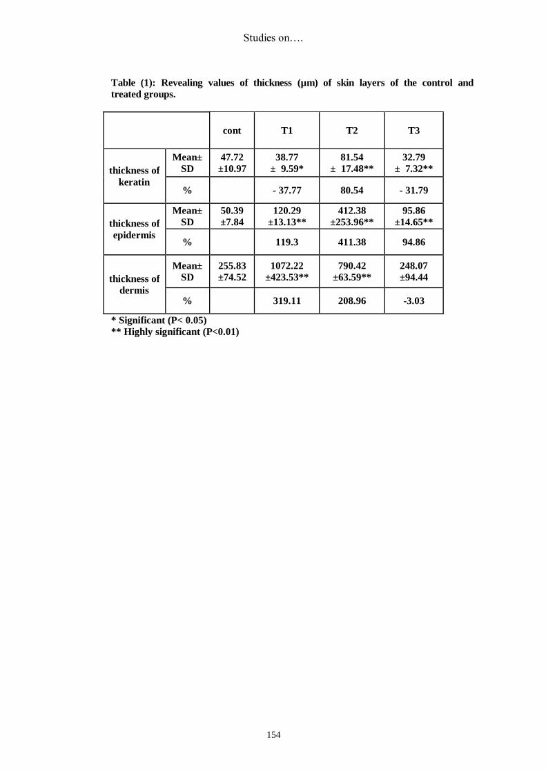
Table (1): Revealing values of thickness (µm) of skin layers of the control and
treated groups.
cont T1 T2 T3
thickness of
keratin
Mean±
SD
47.72
±10.97
38.77
± 9.59*
81.54
± 17.48**
32.79
± 7.32**
% - 37.77 80.54 - 31.79
thickness of
epidermis
Mean±
SD
50.39
±7.84
120.29
±13.13**
412.38
±253.96**
95.86
±14.65**
% 119.3 411.38 94.86
thickness of
dermis
Mean±
SD
255.83
±74.52
1072.22
±423.53**
790.42
±63.59**
248.07
±94.44
% 319.11 208.96 -3.03
* Significant (P< 0.05)
** Highly significant (P<0.01)

Nehal Abou Naja… et al
155
0
50
100
150
200
250
300
350
400
450
cont T1 T2 T3
Mea
n
Thickness of epidermis
Histogram
(1):
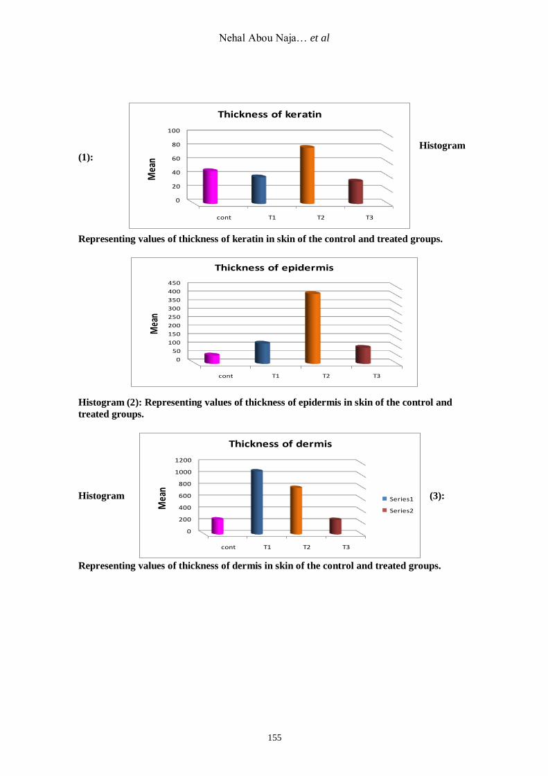
Representing values of thickness of keratin in skin of the control and treated groups.
Histogram (2): Representing values of thickness of epidermis in skin of the control and
treated groups.
Histogram (3):
Representing values of thickness of dermis in skin of the control and treated groups.
0
20
40
60
80
100
cont T1 T2 T3
Mea
n
Thickness of keratin
0
200
400
600
800
1000
1200
cont T1 T2 T3
Mea
n
Thickness of dermis
Series1
Series2
Studies on….
156
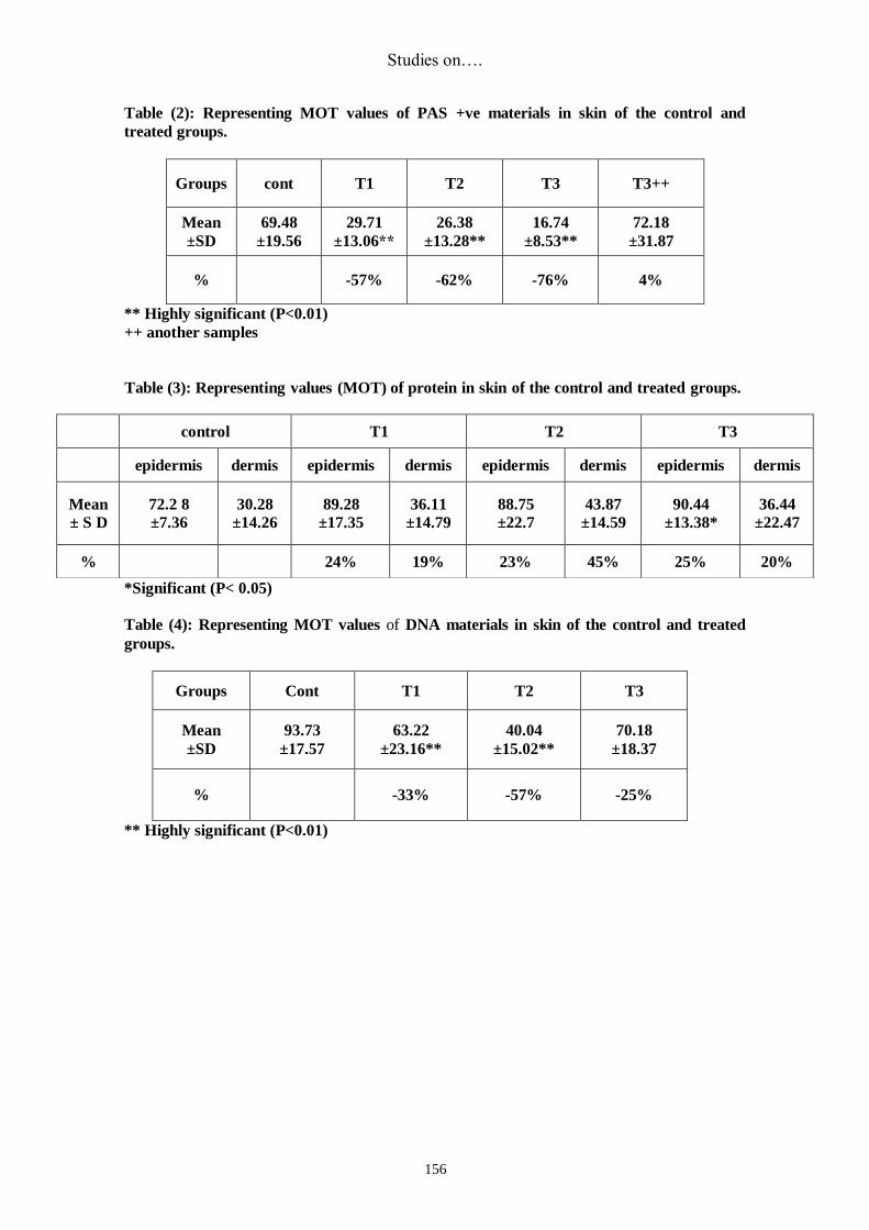
Table (2): Representing MOT values of PAS +ve materials in skin of the control and
treated groups.
Groups cont T1 T2 T3 T3++
Mean
±SD
69.48
±19.56
29.71
±13.06**
26.38
±13.28**
16.74
±8.53**
72.18
±31.87
% -57% -62% -76% 4%
** Highly significant (P<0.01)
++ another samples
Table (3): Representing values (MOT) of protein in skin of the control and treated groups.
*Significant (P< 0.05)
Table (4): Representing MOT values of DNA materials in skin of the control and treated
groups.
Groups Cont T1 T2 T3
Mean
±SD
93.73
±17.57
63.22
±23.16**
40.04
±15.02**
70.18
±18.37
% -33% -57% -25%
** Highly significant (P<0.01)
control T1 T2 T3
epidermis dermis epidermis dermis epidermis dermis epidermis dermis
Mean
± S D
72.2 8
±7.36
30.28
±14.26
89.28
±17.35
36.11
±14.79
88.75
±22.7
43.87
±14.59
90.44
±13.38*
36.44
±22.47
% 24% 19% 23% 45% 25% 20%
Nehal Abou Naja… et al
157
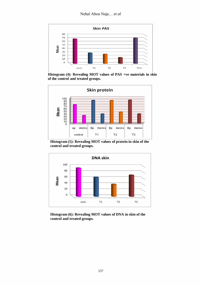
Histogram (4): Revealing MOT values of PAS +ve materials in skin
of the control and treated groups.
Histogram (5): Revealing MOT values of protein in skin of the
control and treated groups.
0
20
40
60
80
100
cont T1 T2 T3
Mea
n
DNA skin
Histogram (6): Revealing MOT values of DNA in skin of the
control and treated groups.


Studies on….
158
Discussion
Antidepressants may affect growth of
embryos and fetuses during pregnancy and
lactation (Ballone, 2005). According to Crews and Frederic
(2007) that by 2020 depression will become
the second leading cause of world wide disability, behind only heart disease, and
that depression is already the single leading
cause of disability for people in midlife and for women of all ages.
Fluoxetine is one of the most
important SSRIs and numerous researches
had focused on fluoxetine because of its high selectively and negligible affinity for
several receptors subtypes. (Cabrera-Vera
et al., 1997; Chubak et al., 2007, 2009,
2011). Fetal skin was chosen for the present study
because the skin is considered as a mirror for the internal body organs and its healthy
look gives a good idea about the state of
such organs.
In the present study, fetuses maternally treated with fluoxetine (Prozac)
showed numerous histopathological and
histochemical changes in the fetal skin. Deleterious changes in the fetal skin
of group T1 were observed. Reduced keratin
layer and hair follicles with highly enlarged
dermal and hypodermal layers were noticed. Discontinuous hypodermal muscle
fibers were realized. Fetuses of group T2
showed increased proliferation, highly thickened keratin layer, epidermis and
dermis with increased number of hair
follicles. Folded and deeply stained epidermal layer was also realized. Fetuses
of group T3 showed highly reduced hair
follicles, corrugated and distorted keratin
layer and hypodermal muscle fibers.
Stanford and Patton (1993) reported that the skin hematoma was
detected in offspring of pregnant gravid Sprague-Dawley rats treated with fluoxetine
beginning on day 7 of gestation and ending
the day of birth and they suggested caution in the prolonged use of this medication
during pregnancy and in patients with
predisposing conditions that may increase
the chances of bleeding. This bleeding was also observed by Al-Nasser (2008), she
treated pregnant rats with 0.7, 1.3 and 2.6
mg/kg b.w. and noted hematoma under the
skin of fetuses; also, mild skin reaction (rash) was reported by Borg and Brodin
(1992) in a small percentage of fluoxetine
treated patients. Increased collagen fibers
were observed in skin of fetuses of all the treated groups of this experiment, this result
was also observed by Al-Nasser (2008) in
some organs of pregnant rats and their fetuses treated with Prozac. Concerning
polysaccharides content, increased PAS +ve
materials was noted in few hair follicles and hypodermal muscle fibers in skin of fetuses
of group T1, but decreased stain affinity
was realized in the remnant layers of the
skin(MOT values reached 29.71 compared with the control group 69.48). Also,
decreased PAS +ve materials was detected
in the different layers of the fetal skin of group T2.Cells of hair follicles showed
increased stain affinity of PAS +ve
materials(MOT reached 26.38). Fetuses of group T3 showed
numerous aggregations of PAS +ve in the
epidermal layer with increased stain affinity
in the hypodermal muscle fibers (MOT was72.18), but remnant layers were
depleted (MOT was16.74).
Increased stain affinity of PAS +ve materials was realized by Eid and Al-
Nasser (2008) in lung tissue of pregnant
rats treated with fluoxetine (0.143, 0.286
and 0.572 mg / kg b.w.). They also noticed diffused polysaccharides inside the blood
vessels of lungs of all the treated groups.
In accordance with the present results, Hutchins and Rogers (1970)
noticed increased polysaccharides in brain
of mice treated with antidepressant drugs, this may be dependent on adrenocortical
activity (Mills, 1986).
In the present study increased stain
affinity of total proteins was detected in skin fetuses of all the treated groups (MOT
values reached 89.28,88.75,90.44 in T1, T2,
T3 respectively compared with the control group 72.28) Increased total proteins was
also noted by Kim et al (2004) and Eid
and Al-Nasser (2008) in lungs of treated pregnant rats with fluoxetine.
Kim et al. (2004) stated that
fluoxetine has high affinity to bind with
proteins. Decreased stain affinity of DNA
was recorded in skin of fetuses of all the
treated groups, in spite of the presence of
Nehal Abou Naja… et al
159
deeply stained pyknotic nuclei (MOT
values reached 63.22, 40.04, 70.18 in T1, T2, T3 respectively compared with the
control group 93.73).In 2011,Fadladdeen
noticed numerous histological and
histopathological changes in many fetal organs treated maternally with
Prozac.These changes include: internal
hemorrhage in the gastrointestinal tract ,destructed muscle fibers and altered
PAS+ve materials,total protein and DNA
content. In 1979, Fawthrop et al., tried to discuss fragmentation or dissolution of
DNA material in the degenerated cell and
they stated that two distinct morphological
patterns of cells death have been recognized, either by necrosis or apoptosis.
Apoptosis occurs in both physiological and
pathological conditions. It arises due to an elevation of cytosolic free calcium
concentration resulting in activation of the
nuclear enodonuclease.Activated endonucleases produce oligonucleosome-
length DNA fragments. This DNA cleavage
can directly lead to cell death. They added
that cytoskeleton disruption, activation of degenerative enzymes such as proteases and
phospholipase A2 and stimulation of other
enzymes such as ADP-ribose polymerase play an important role in cell killing. Also,
Ritter (1987) suggested that necrosis or
cellular degeneration may be either due to
progressive action of intracellular enzymes of the injured cells or to a metabolic
disturbance and inhibition of synthesis
needed for DNA and hence protein synthesis .
In 2008, Su et al., found a relation
between decreased omega-3 polyunsaturated fatty acid and depression.
Thus pregnant females must take enough
amounts of foods rich with omega-3 during
pregnancy for safety of mothers and their newborns.
Conclusion
Results of the present study showed that
maternal use of Prozac has been associated
with dystrophic changes in the fetuses and increased risk of fetal complications. These
findings should be taken into considerate
before using of Prozac during pregnancy
and future researches on the nervous system and placenta can lead to better
understanding of the effects of Prozac use
during pregnancy to improve public health
outcomes
References
Al-Nasser, F. (2008): Effect of Prozac
(fluoxetine) on some tissues of the pregnant rats
and their foetuses. M.Sc.College of Science ,King Faisal, university. Dammam ,K.S.A.
Alwan, S.; Reefhuis, J.; Rasmussen. S.;
Oinev, R. and Friedman, J. (2007): Use of
selective serotonin reuptake inhibitors in
pregnancy and the risk of birth defects. N. Engl.
J. Med., 356(26): 2684-2692.
Ballone, G. (2005): J. Gravideze Medicamentos
- In .PsiqWeb. Disponivel em:
http://www.psiqweb.med.br.2005.
Bancroft, J. and Gamble, M. (2002): Theory
and Practice of Histology
Techniques.5thed.Churchil Livingstone, London. pp: 150-152.
Borg, S. and Brodin, K. (1992): Antidepressant Drugs. Dukes, M.N. 12th ed.
Amsterdam.pp:30-78.
Cabrera-Vera, TM.; Garcia, F.; Pinto, W.
and Battaglia, G. (1997): Effect of prenatal
fluoxetine (Prozac) exposure on brain serotonin
neurons in prepubescent and adult male rat
offspring. J. Pharmacol. Exp. Ther., 280(1):
138-145.
Chambers, C.; Hernandez-Diaz, S.; Marter,
L.; Werler, M.; Louik, C.; Jones, K. and
Mitchell ,A. (2006): Selective serotonin-
reuptake inhibitors and risk of persistent
pulmonary hypertension of the newborn. N. E.
J. M., 354(6): 579-587.
Chubak, J.; Buist, D.; Boudreau, D.; Rossing,
M.; Lumley, T. and Weiss, N. (2007): Breast
cancer recurrence risk in relation to
antidepressant use after diagnosis. Breast
Cancer Res. Treatment, 112(1):123-132.
Chubak ,J.; Bowles, .J.; Terry ,M.
B.;Trentham-D ietz,A.and Buist ,D.S.(2009): Antidepressant medications and change in
mammographic density in postmenopausal
women.Cancer Epidemiol.Biomarkers Prey.,
18(2):676-679.
Chubak, J.; Boudreau, D.M.; Rulyak, S.J.
and Mandelson, M.T.(2011):Colorectal cancer
risk in relation to antidepressant medication
use.Int.J.Cancer,128(1):227-229.
Costei, AM.; Kozer, E.; Ho, T.; lto, S. and
Koren, G. (2002): Perinatal outcome following
third trimester exposure to paroxetine. Aroh. Pediatr. Adolesc. Med., 156: 1129-1132.
Crews, F. and Frederic, C. (2007): Talking
Back to Prozac. New York Review of Books.
54(19):215-220.
Eda, K.; Buyuknacar, H.; Gocmen, C.;
Evruke, I. and Onder, S. (2009): Differential
effect of neocuproine, a copper (I) chelator, on

Studies on….
160
contractile activity in isolated ovariectomized
non-pregnant rat, pregnant rat and pregnant
human uterus. European Journal of
Pharmacology, 605: 158-163.
Eid, F. and Al-Nasser, F. (2008): Histological
and histochemical changes in lung of pregnant rats treated with an antidepressant drug
(Prozac). J. Biol. Pham. Sci., 6: 99-114.
Fadladdeen,K.A.(2011):Studies on fetal
response to Prozac
treatment.Ph.D.Thesis.Zoology
Departement,Faculty of Science,Al-Azhar
University.
Fawthrop, J. F. ; Boobis, A. and Davies.E.
(1979): Mechanism of death. Lancet. 24(1):
412-413.
Hassan, E. M. K. (1990): Toxicological and
histopathological effects of some antidepressant drugs. Ph.D. Thesis, Al-Azahar University, pp:
192-194.
Hotchkiss, R. D. (1948): A microchemical
reaction resulting in the staining of
polysaccharide structure in fixed tissue
preparation. Arch. Biochem., 16: 131-136.
Hutchins, D. A. and Rogers, K. J. (1970): Physiological and drug-induced changes in the
glycogen content of mouse brain. Br. J.
Pharmacol., 39: 9-25.
Kallen, B. (2004): Neonate characteristics after maternal use of antidepressants in late
pregnancy. Arch. Pediatr. Adolesc. Med., 158:
312-316.
Kim, J.; Wayne Riggs, D. and Rurak, D.
(2004): Stereo selective pharmacokinetics of
fluoxetine and norfluoxetine in pregnant sheep.
Am. Soc. Pharmacol. Exper. Therap., 32: 212–
221.
Mazia, D.; Brewer, P. and Alfert, M. (1953): The cytochemical staining and measurement of
protein with mercuric bromophenol blue. Biol.
Bull., 104: 57-67.
Mills, J. N. (1986): Human circadian rhythms.
Physiol. Rev., 46: 128-171.
Paget, G. E. and Barnes, J. M. (1964): Interspecies dosage conversion scheme in
evaluation of results and quantitative application
in different species. In: Evaluation of Drug
Activities: Pharmacometric. Vol. 1, Laurence,
D. R. and Bacharach, A. L. (Eds.); Academic
Press, London and USA. 160-162.
Pearse, A. G. (1977): Histochemistry,
Theoretical, and Applied. 3th ed., vol. 1.
Churchill Livingstone, London.
Richelson, E. (2001): Pharmacology of
Antidepressants. Myo .Clin. Proc., 6: 511-527. Ritter, E. (1987): Altered biosynthesis. In Hand
Book of Teratology. Vol. 2, Plenum Press, New
York, 23-26.
Stanford, M. and Patton, J. (1993): In utero
exposure to fluoxetine HCI increases hematoma
frequency at birth. Pharm. Biochem. Behav., 45:
959-962.
Taylor, P. (1986): Handling the reproductive
cycle and mating. In: Practical Teratology.
Academic Press. Inc. London. Copyright. C. P.:
3-9.
Su, K.; Huang, K.; Huang, C. and Pariante,
C. (2008): Omega-3 fatty acids for major
depressive disorder during pregnancy. J. Clin.
Psychiatry, 18: 1-8.
Nehal Abou Naja… et al
161
على الاستجابه الجنينية للمعالجة بالبروزاكدراسات
** سضج فعو اىض* فبغ عض, * به أث اىجب جضح-جبعخ اىيل عجض اىعؼؼ-ميخ اىعي**0جبعخ الأػغ -ميخ اىعي–قس عي اىذا *
عي أجخ اىجغطا اىذاو ىقض عقبع عبص ىلإمزئبة( اىفيمسز)اسزضفذ ظ اىغسبىخ رأثغ اىجغػاك
اسزشضذ اىظمع لإرب Albino rattus norvegicusاسزشض ف ظ اىضعاسخ إبس طمع اىجغطا جس
: عيخ اىزؼاج فقػ
:ر رقس اىجغطا ىيجعبد اىزبىخ
.اىجعخ اىعبثطخ رع إبس اىجغطا اىغغ عبيخ ثبىجغػاك . أ
:ىزجغجخ رقس إىاىجعبد ا . ة
اىجعخالأى رع اىجغطا اىذاو اىعبيخ ثعقبع اىجغػاك ثجغعخ رنبفئ صف -1
(.مج ػ اىجس/ج 2,,0)اىجغعخ اىعلاجخ ىلإسب
اىجعخ اىثبخ رع اىجغطا اىذاو اىعبيخ ثعقبع اىجغػاك ثجغعخ رنبفئ اىجغعخ -2
(.مج ػ اىجس/ج 1,44)اىعلاجخ ىلإسب
اىجعخ اىثبىثخ رع اىجغطا اىذاو اىعبيخ ثعقبع اىجغػاك ثجغعخ رنبفئ ظعف -3
(.مج ػ اىجس/ج 2,22)اىجغعخ اىعلاجخ ىلإسب
.مسزجخ اىجغطا اىذاو اىعبيخ ثبىفياشزيذ ظ اىضعاسخ عي اىزغغد اىسجخ اىنبسجخ ف جيض أ
.ىدع أ اىعبيخ ىيجغطا اىذاو ثجغعبد شزيفخ اىجغػاك قض أصد إى ظع رغغاد عضضح ثجيض الأجخ
ىدع أعب اسزؼاه شضض ف غجقخ اىنغار قص عضص دصلاد اىشعغ ع رعش شضض ف غجقخ الأصخ
.مج ػ اىجس/ج 2,,0ثجغعخ قضاعب اىعبيخ T1رذذ الأصخ طىل ف أجخ اىجغطا ىيجعخ
مج ػ اىجس فيقض ىدع رغيع شضض ف /ج 1,44اىعبيخ ثجغعخ قضاعب T2ثبىسجخ ىيجعخ اىثبخ
أب ثبىسجخ , غجقخ اىنغار اىجشغح الأصخ ع ػبصح يذظخ ف دصلاد اىشعغ رعغج غجقخ اىجشغح
مج ػ اىجس فيقض ىدع اسزؼاه شضض ف عضص /ج 2,22عبيخ ثجغعخ قضاعب اى T3ىيجعخ
.دصلاد اىشعغ ع رعغج اظطغاة غجقخ اىنغار غجقخ يجج اىز ظغد سنخ جضا
ىقض ىدع رؼاض الأىبف اىنلاجخ ف جيض الأجخ ىجع اىجعبد اىعبيخ ع رغغاد عضضح ف ذز
. DNAىاص عضضح اىزسنغ اىجغربد اىنيخ اىذط اى ا
بسبج ىنسبجخ اىشزيفبخ عي اىزغمبت اىن SSRI اىلادع ضعح جص أثذبس ف جبه رأثغ اىجغػاك أ
ىن عظ الأثذبس اىسبثقخ ارجذ ىضعاسخ اىزغبغاد اىظبغبخ فب الأجبخ اىزغبغاد فب . بد اىذاولأجخ الأ
نيببب اىببضاسي أ رصببغفبد لببغبع اىاىببض اىقببضعح عيبب الاسببزعبة اىببظمب أ ػ غببه عببضص الأجببخ فبب
ب ىيجببدث لإجبغا اىؼبض ب الأثذببس ىزظبخ عب فئ ظا اىجبه عزجغ جبلا ثنغا سصبج. اىثضبد اىشزيفخ
اىزغغاد اىسجخ اىفسىجخ اىنبسبجخ اىجغبخ اىضققبخ فب مبو أسبجخ الأ اىذببو اىعبيبخ ثببىجغػاك
ذزببج الأبغ مبظىل إىب رزجبع . مظىل أجزب سبلخ اىجبػ اىعصج ع عثػ ظ اىزغغاد ثبىزغغاد فب اىشبخ
ىعو اىسؤاه اىظ طغح فس بب ببطا ىب ادزبجبذ الأببد اىذابو . اه اىاىض ثعض فزغاد عضضح اىلاصحأد
سلاه فزغح اىذو ىعو جؼ الإجبثخ عي ظا اىسؤاه الإب ثقضع الله سبغ ىزعبغ أدض عبصاد الامزئبة
عببغ الأغعبخ اىشبغثبد اىزب رذزب عيب اىذبط شغ اىجعض ع اىزعغض ىيعبغغ اىفسبخ اىشبضضح بع ر
الأ اىزغثزفب اىذصه عي مبد مبفخ ظ اىشس اىقب ثبىزبع اىغبظخ لأ طىل غفع سبز
اىسغر فب اىجسب بب بؤص إىب رذسب اىبؼاج رببه الأيظبخ اىزب رذزب عيب اىسبغر ثبو اىفطبغ
اىببباص اىعبببصح ىنمسبببضح ببب 0ىبببػ اىشببر اىنببب اىجببػ عظببب اىفبمبببخ اىشعببغادالأجبببو ا
اىعببغف أ اىسببغر جببص ثنثببغح فبب الأيظببخ اىغببخ ثبببىجغر ثببو اىجببط الأىجببب ىذبب اىجقببغ اىببضل
اىسل اىأملاد اىجذغبخ اىغ عزجغ اىفه اىسصا فه اىصصب صضع جض ىيزغثزفب مظىل ذز
اىجبصح ثنثبغح فب 3جت الإزب مظىل ثزعببغ صب الأجبب . اىنسغاد ػذ اىؼز عي اىزغثزفب
ػبذ اىسبل الأسببك مببظىل جبت رببه اىذعبببد اىشبب اىفبلبىب اىجقىبببد اىذجبة اىنبيبخ سبلببخ
الأسزه مو ظ اىعاو ىب اىقضعح عي رؼاض جص اىسغر ف اىبز اىشعغلادزائ عي دط اىفزبل
اىض ثبىزبى رجت دضس الامزئبة ثئط الله