sph302 articulation 01.ppt - Macquarie...
56
Speech Physiology Articulation and Resonance Lecture 1 Lecture 1 Dr Robert H. Mannell Department of Linguistics Macquarie University 1
Transcript of sph302 articulation 01.ppt - Macquarie...

Speech Physiology
Articulation and ResonanceLecture 1Lecture 1
Dr Robert H. MannellDepartment of Linguistics
Macquarie University1

Introduction (1)Introduction (1)
• In this series of lectures we will examine the anatomy and functional physiology of y p y gythe structures that together can be manipulated to create coordinatedmanipulated to create coordinated patterns of vocal tract shape.
f• These patterns of changing vocal tract shape are know as the “articulatory” p ypatterns of speech production.
2

Introduction (2)Introduction (2)
Wh f t th “ l t t”• When we refer to the “vocal tract” we are usually referring to the space (or cavity) above the glottis (known as the “supra-glottal” cavity)
• This cavity consists of the oral and nasal cavity.ca ty
• When we talk about changing patterns of vocal tract shape we are always referringvocal tract shape we are always referring to the shape of the cavity.
3

Introduction (3)Introduction (3)
Ch i th h d/ iti f• Changing the shape and/or position of certain organs changes the shape and size of the supra-glottal cavity.
• Some of the structures around the vocal tract cavity are (for the most part) passive walls of the tract. These include the hard a s o t e t act ese c ude t e a dpalate of the roof of the mouth, most of the surrounding walls of the nasal cavity, andsurrounding walls of the nasal cavity, and the back wall of the pharynx.
4

Introduction (4)Introduction (4)
Oth t t i th l tt l l• Other structures in the supra-glottal vocal tract can be manipulated by various muscles to change their position and shape. This results in changes in the size and shape of the supra-glottal cavity.
• Such structures are referred to as Suc st uctu es a e e e ed to as“articulators” (or “active articulators”) as their position and shape can be “articulated”their position and shape can be articulated (or finely controlled).
5

Introduction (5)Introduction (5)The major active articulators of the vocal tract are:vocal tract are:-
1. Tongue
2. Lips
3. Velum (soft palate)
4. Mandible (jaw)
Hardcastle (1976) 6

Introduction (6)Introduction (6)
Here we can see anHere we can see an anterior (front) view of the oral cavity showing the lips, tongue, teeth and hard and soft palatehard and soft palate (and a few additional features)features).
Seikel et al (1997) 7

Vocal Tract Tissues (1)Vocal Tract Tissues (1)
There are four major types and all can be found in the vocal tract. They are:-y
E ith li l ti• Epithelial tissue• Connective TissueCo ect e ssue• Muscle Tissue• Nervous Tissue
8

Vocal Tract Tissues (2)Vocal Tract Tissues (2)
Epithelial tissues: cover the surface of the body, cover and line internal organs, include th l d Th i f ti i l dthe glands. Their functions include protection, secretion, absorption and excretion In the vocal tract they include:a. skin on the face surrounding the lipsexcretion. In the vocal tract they include:-
g pb. mucous membrane covering the lips, the
inner surfaces of the oral and nasalinner surfaces of the oral and nasal cavities and the surface of the tongue.
c salivary glandsc. salivary glands9

Vocal Tract Tissues (3)Vocal Tract Tissues (3)
C ti ti t bi d dConnective tissues: support, bind and protect body structures. Bones: In the vocal tract they include the bones of the skull, jaw, hard palate and , j , pnasal cavity which underlie the shape of these structures. Bones also provide points t ese st uctu es o es a so p o de po tsof origin for many of the muscles involved in articulation. The hyoid bone in the neck isarticulation. The hyoid bone in the neck is also the point of origin of a number of muscles involved in articulation.muscles involved in articulation.
10

Vocal Tract Tissues (4)Vocal Tract Tissues (4)
C ti tiConnective tissues:Cartilage is a lighter, more flexible structural g g ,tissue than bone. As we already know, it constitutes the main structures of the larynx. yIt also constitutes the structural component of the epiglottis (which is not used as an o t e ep g ott s ( c s ot used as aarticulator in speech – except perhaps in pre-linguistic sounds of neonates). Therepre linguistic sounds of neonates). There are also some cartilaginous structural components in the nasal cavity.components in the nasal cavity.
11
Vocal tract Tissues (5)Vocal tract Tissues (5)
C ti tiConnective tissues:There are a number of different types of ypfibrous tissue that is quite flexible but can also be an important structural component of p pmany organs. In the vocal tract, the most important of these is a central fibrous po ta t o t ese s a ce t a b ous“septum” running along the centre of the tongue and providing the point of origin oftongue and providing the point of origin of certain tongue muscles.
12

Vocal Tract Tissues (6)Vocal Tract Tissues (6)
C ti tiConnective tissues:• Ligaments connect bone to bone, bone to g ,
cartilage and cartilage to cartilage. There are many of these but perhaps the most y p pimportant in the articulatory system are the ligaments (one each side of the jaw) that ga e ts (o e eac s de o t e ja ) t atconnect the condylar process of the jaw to the Zygomatic arch of the skull.the Zygomatic arch of the skull.
• Tendons connect muscles to bone, cartilage and some fibrous structurescartilage and some fibrous structures.
13

Vocal tract Tissues (7)Vocal tract Tissues (7)
Connective tissues:Loose connective tissue lies below the skinLoose connective tissue lies below the skin and mucous membrane in most parts of the vocal tract For example it makes up part ofvocal tract. For example, it makes up part of the mass beneath the mucous membrane of
fthe soft palate, the nasal cavity and the region below the front of the tongue. g g
14
Vocal Tract Tissues (8)Vocal Tract Tissues (8)
Teeth are unique in the body. They are not considered to be bones as they differ ysignificantly from bones in certain ways (they are harder than bones and their(they are harder than bones and their structure and protein content is different). In the vocal tract the upper teeth providethe vocal tract the upper teeth provide passive articulatory targets for some speech sounds. They also contribute to the overall shape of the oral cavity.p y
15

Vocal tract Tissues (9)Vocal tract Tissues (9)
Muscles: Muscles are often described in terms of:-
a) Muscle Point of origin: The attachment point at the more massive lesspoint at the more massive, less moveable end of the muscle. In the supra-glottal vocal tract this is usually a bone, but some muscles in the tongue goriginate from a central fibrous septum.
16

Vocal tract Tissues (10)Vocal tract Tissues (10)
b) Muscle point of insertion: the other end of the muscle (it might be a lighter, more ( g g ,moveable bone than the point of origin, or it might insert into another muscle) Forit might insert into another muscle). For example, some tongue muscles insert into other musclesinto other muscles.
17

Vocal Tract Tissues (11)Vocal Tract Tissues (11)
• For a number of articulatory organs its convenient to talk about intrinsic musclesand extrinsic muscles.Intrinsic muscles are found wholly within• Intrinsic muscles are found wholly within the structure and may change its shape.
• Extrinsic muscles insert into the structure but have their point of origin elsewherebut have their point of origin elsewhere. They can change both shape and position.
18
Vocal Tract Tissues (12)Vocal Tract Tissues (12)
Th i l t k f i l• There is a large network of cranial nerves that attach to the various structures of the head including the vocal tract articulators. These include both motor neurons and sensory neurons. They will be dealt with in more detail later in the semester.
• A motor unit is a group of muscle fibres that are controlled by a single motorthat are controlled by a single motor neuron fibre.
19

The skull (1)The skull (1)
I th f ll i t i i• In the following two images we examine the front and back of the skull (plus the jaw).
• Several bones of the skull cover brain lobes of the same name.
• A process is a prominent projection on a• A process is a prominent projection on a bone. A foramen is an opening thro gh the bone• A foramen is an opening through the bone for blood vessels, nerves or ligaments.
20

The skull (2)
21

The skull (3)
22

The nasal cavity (1)The nasal cavity (1)
I th f ll i i th b• In the following image we can see the bone and cartilage structures of the nasal cavity.
• The superior (upper) surface is a sinus filled part of the skull. p
• The inferior (lower) surface consists of two bones of the hard palate and the tissue ofbones of the hard palate and the tissue of the soft palate.These str ct res are co ered b loose• These structures are covered by loose connective tissue and mucous membrane.
23
The nasal cavity (2)The nasal cavity (2)
• The structures of the nasal cavity, with the exception of the soft palate (velum) are p p ( )fixed in shape and dimensions and thus fixed acoustic (resonance) characteristicsfixed acoustic (resonance) characteristics.
• Their resonance characteristics are only ff faffected by degree of velum opening and
by tissue swelling during a head cold, hay y g g yfever or similar affliction.
24

The nasal cavity (3)
25
The hard palate (1)The hard palate (1)• In the next slide we can see the bony• In the next slide we can see the bony
structure of the hard palate.Lik h f th l t t t t• Like much of the vocal tract, structures are mirrored on the left and right side.
• The bones are covered by loose connective tissue which is in turn covered by mucous ymembrane.
• The suture down the middle if it fails to join• The suture down the middle, if it fails to join during foetal development, results in a cleft palatepalate.
26

The hard palate (2)
27
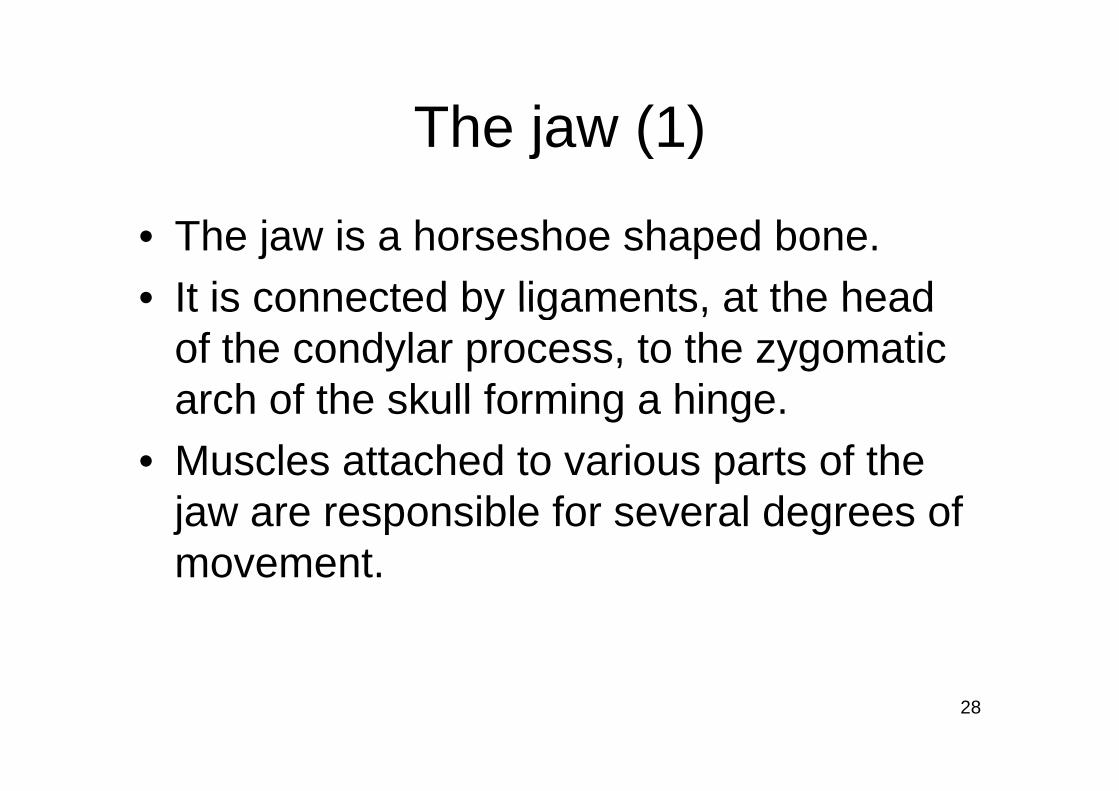
The jaw (1)The jaw (1)
• The jaw is a horseshoe shaped bone.• It is connected by ligaments at the headIt is connected by ligaments, at the head
of the condylar process, to the zygomatic arch of the skull forming a hingearch of the skull forming a hinge.
• Muscles attached to various parts of the jaw are responsible for several degrees of movement.movement.
28

The jaw (2)
29
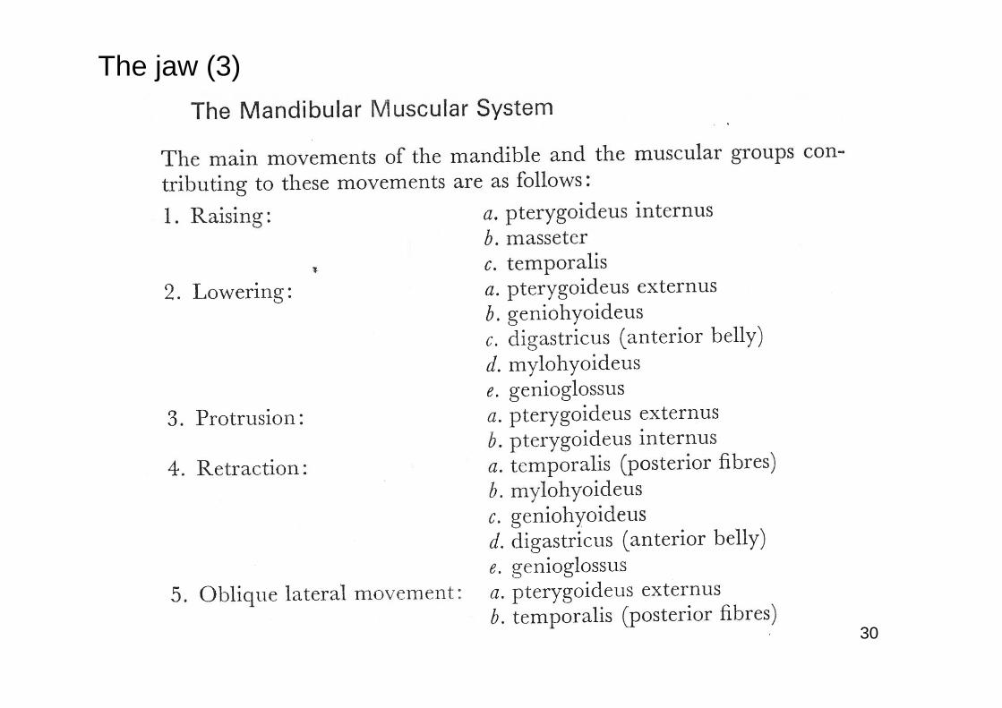
The jaw (3)
30

The jaw (4)The jaw (4)
• In speech the relevant dimensions of movement are jaw raising (for many j g ( ylingual (tongue articulated) consonants and for high vowels) and jaw lowering (forand for high vowels) and jaw lowering (for low vowels).
f• The muscles of jaw lowering are assisted by the action of gravity, which has quite a y g y qsignificant effect on the relatively massive body of the jaw.body of the jaw.
31

The jaw (5)
32

The jaw (6)The jaw (6)
Th j i th t i ( i h th• The jaw is the most massive (weighs the most) of the four major articulators and so it is also the slowest moving articulator. In other words, it has the highest degree of inertia.
• The jaw is also the least important of the e ja s a so t e east po ta t o t efour major articulators. This can be seen by the fact that its possible to speak fairlythe fact that its possible to speak fairly clearly (although not completely clearly) with teeth clenched (ie. with the jaw up).teeth clenched (ie. with the jaw up).
33

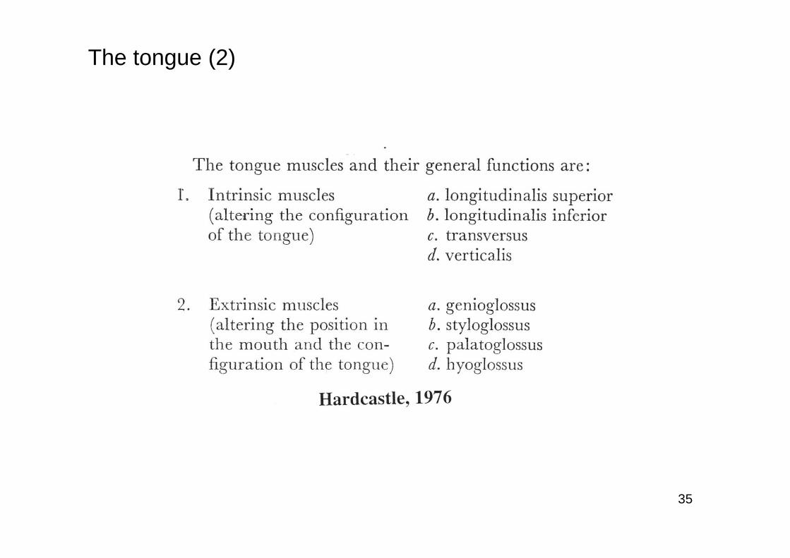
The tongue (1)The tongue (1)
I th t lid th t th• In the next slide we can see that the tongue consists of intrinsic muscles that are wholly within the tongue and extrinsic muscles that originate outside the tongue and insert into the body of the tongue.
• Note that in the next slide the more Latin-ote t at t e e t s de t e o e atlike spellings of these muscles are used. The alternative non Latinate spellings areThe alternative non Latinate spellings are also acceptable.
34
The tongue (2)
35
The tongue (3)
36

The tongue (4)The tongue (4)
• In the next image we see greatly simplified vector representations of the direction of paction of the extrinsic muscles (plus the intrinsic longitudinal superior muscle)intrinsic longitudinal superior muscle).
• The arrows indicate the direction that the tongue body will move with the contraction of a particular muscle (all else being p ( gequal). Extrinsic muscles pull the tongue towards their point of origin.towards their point of origin.
37

The tongue (5)The tongue (5)
It h ld l b t d th t i th f• It should also be noted that in the case of the longitudinal superior muscle the points of origin and insertion isn’t obvious.
• The muscle extends from the back of the tongue to the tip of the tongue.
• The more massive anchor point is regarded• The more massive anchor point is regarded as the point of origin. As the back of the body of the tongue is more massive thanbody of the tongue is more massive than the tip of the tongue contraction of this muscle will mostly move the tip backwardsmuscle will mostly move the tip backwards.
38

The tongue (6)
39
The tongue (7)The tongue (7)
I th t i th t li d• In the next image we see another stylised diagram of the relative locations of the muscles (with exaggerated separation of the muscles). We can see all of the intrinsic muscles and two of the extrinsic muscles.
• The transversus muscle originates in the e t a s e sus usc e o g ates t ecentral fibrous septum.
• The verticalis muscle can be said to• The verticalis muscle can be said to originate in the less movable base of the tonguetongue.
40

The tongue (8)
41

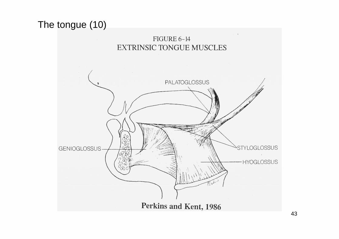
The tongue (9)The tongue (9)
• The next diagram has removed the intrinsic muscles of the tongue and only g yshows the course of the extrinsic muscles. These muscles fan out as they reach theirThese muscles fan out as they reach their point of insertion in the tongue. This is shown more accurately here than in theshown more accurately here than in the vector diagram whilst the vector diagram shows more clearly the effect of muscle contraction.
42
The tongue (10)
43

The tongue (11)The tongue (11)
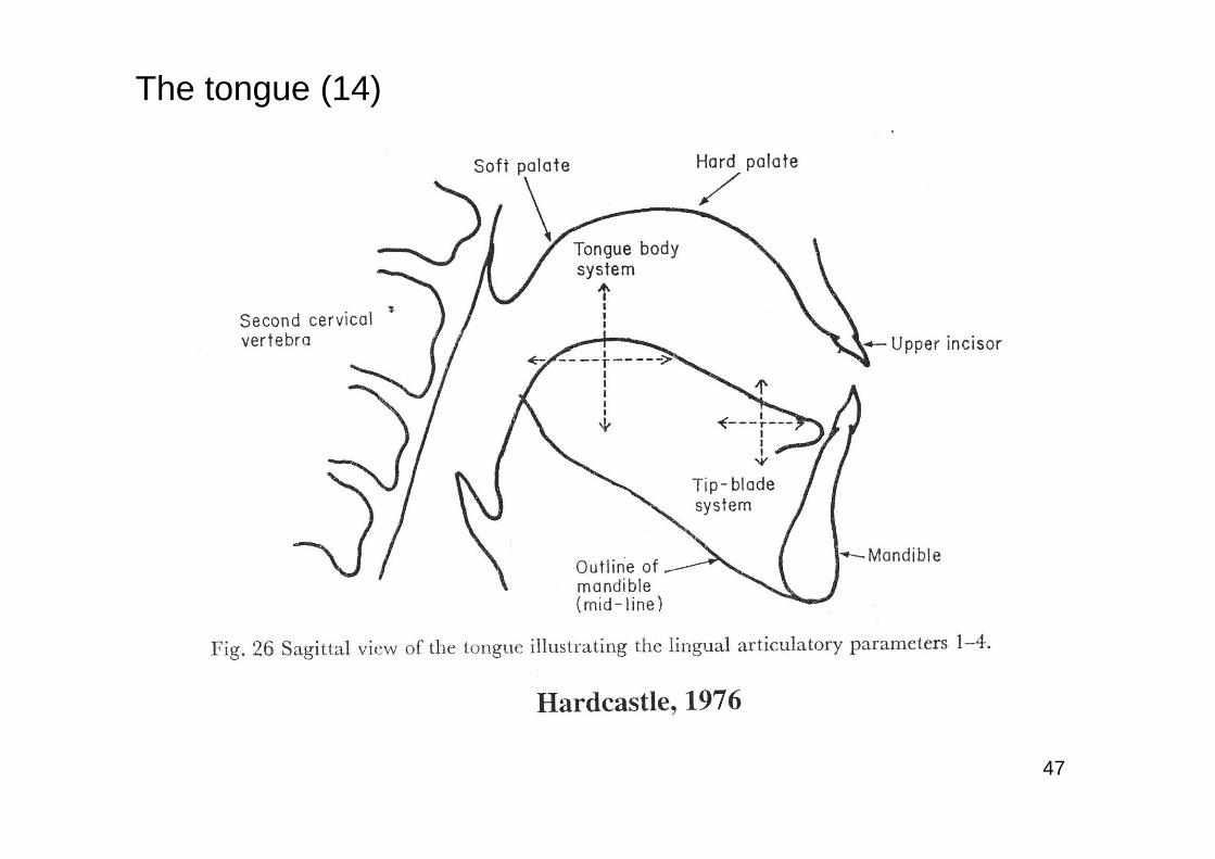
• The next 5 slides describe and illustrate seven articulatory dimensions or yparameters that result in phonetically important tongue articulationsimportant tongue articulations.
• For further details on this model you are freferred to:-
W.J Hardcastle, 1976, Physiology of Speech Production, Academic Press, London, pp 100-106
44

The tongue (12)
45

The tongue (13)
46
The tongue (14)
47

The tongue (15)
48

The tongue (16)
49

The tongue (17)The tongue (17)
• As we can see (particularly in slide #14), the body and the tip of the tongue can to a y p gfair extent be moved independently of each other Its possible for example toeach other. Its possible, for example, to move the tip back whilst moving the body forward The tongue tip being lighter (lessforward. The tongue tip, being lighter (less inertia), can move and change direction more rapidly then the tongue body.
50
The pharynx (1)The pharynx (1)Th h b di id d i t th h• The pharynx can be divided into the oro-pharnx and the naso-pharynx. The naso-pharynx is the region at and above the point where the velumregion at and above the point where the velum closes. The oro-pharynx is below that point.
• The oro pharynx can be considered and• The oro-pharynx can be considered and articulatory organ, but in most cases its a passive articulatory organ The active articulator is thearticulatory organ. The active articulator is the root of the tongue. eg. for low vowels the place of greatest constriction is the oro-pharynx and the g p yconstriction is created by the back of the tongue bulging back towards the rear of the pharynx.
51

The pharynx (2)The pharynx (2)
• In some languages (eg. Arabic) the oro-pharynx is an active articulator for p ypharyngeal and pharyngealised consonants.
• The next two images show the location of• The next two images show the location of the main pharyngeal constrictor muscles, the stylopharyngeus, and the superior, middle and inferior pharyngeal constrictor p y gmuscles.
52

The pharynx (3)
53

The pharynx (4)
54

Further ReadingFurther Reading• Clark, J., Yallop, C. & Fletcher, J. (2007), An introduction
to phonetics and phonology, 3rd. edition, Blackwell, Oxford (pages 84 90 and Section 7 17)Oxford (pages 84-90 and Section 7.17)
• J.A. Seikel, D.W. King and D.G. Drumright, (1997), Anatomy and Physiology for Speech Language andAnatomy and Physiology for Speech, Language, and Hearing, Singular, San Diego (or a more recent edition of the same text)
55

ReferencesReferences
The following books were used as sources for materials used in this lecture:-
• W.J Hardcastle, 1976, Physiology of Speech Production, Academic P L dPress, London
• J.A. Seikel, D.W. King and D.G. Drumright, 1997, Anatomy and Physiology for Speech, Language, and Hearing, Singular, San y gy p , g g , g, g ,Diego
• D. Shier, J. Butler and R. Lewis, 2004, Hole’s Human Anatomy and Physiology 10th edn McGraw Hill BostonPhysiology, 10th edn., McGraw Hill, Boston.
56