Pulmonary Fibrosis in Sarcoidosis - · PDF filePulmonary Fibrosis in Sarcoidosis Robert P....
63
Pulmonary Fibrosis in Sarcoidosis Robert P. Baughman Interstitial Lung Disease and Sarcoidosis Clinic University of Cincinnati
Transcript of Pulmonary Fibrosis in Sarcoidosis - · PDF filePulmonary Fibrosis in Sarcoidosis Robert P....

Pulmonary Fibrosis in
Sarcoidosis
Robert P. Baughman
Interstitial Lung Disease and
Sarcoidosis Clinic
University of Cincinnati

Pharmaceutical Support for Dr.
Baughman
• Centocor: Research Grants, Consultant
• Celgene: Research Grants
• Actelion: Research Grants
• Cephalon: Research Grants
• Intermune: Research Grants
• Genetech: Research Grants
• Gilead: Research Grants
• Glaxo Smith Kline: Consultant
Off Label Use of Therapy
• Prednisone and ACTHAR are the only
drugs approved for use in pulmonary
sarcoidosis
• All other drugs discussed here are off label
use for treatment of sarcoidosis
Introduction
• Pulmonary fibrosis occurs a significant proportion of pulmonary sarcoidosis patients
• It is associated with morbidity and some mortality
• However not all patients with fibrosis are impaired by their disease
• Treatment options are unclear
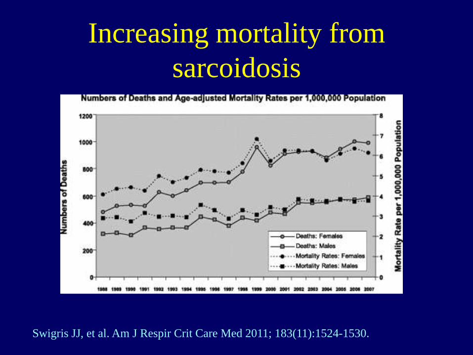
Increasing mortality from
sarcoidosis
Swigris JJ, et al. Am J Respir Crit Care Med 2011; 183(11):1524-1530.
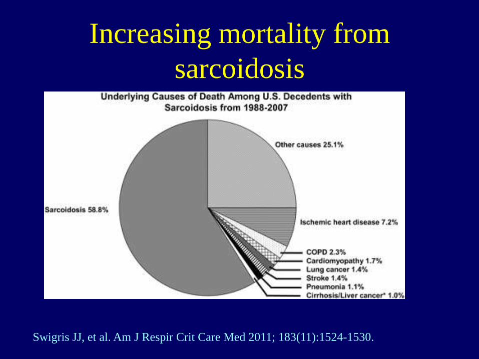
Increasing mortality from
sarcoidosis
Swigris JJ, et al. Am J Respir Crit Care Med 2011; 183(11):1524-1530.
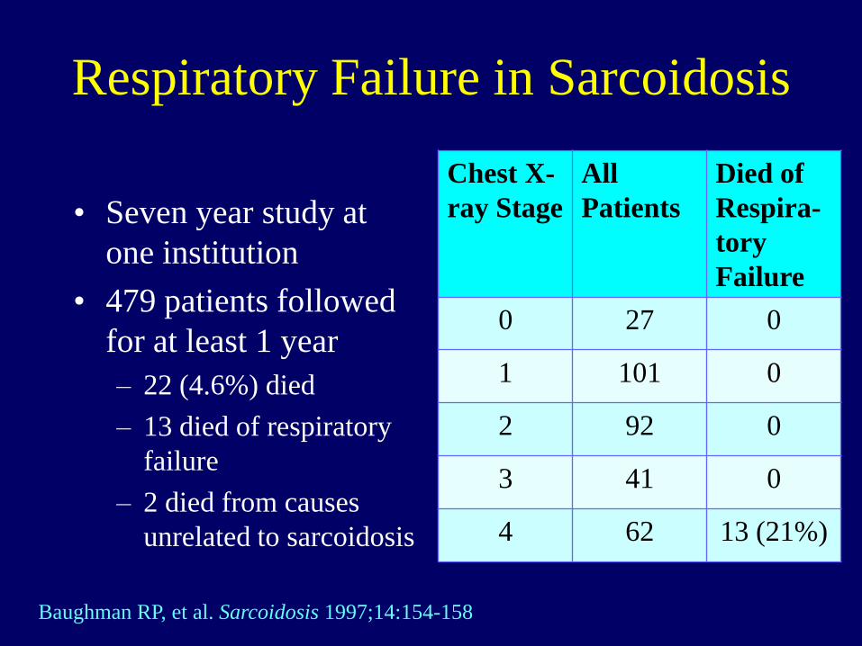
Respiratory Failure in Sarcoidosis
• Seven year study at
one institution
• 479 patients followed
for at least 1 year
– 22 (4.6%) died
– 13 died of respiratory
failure
– 2 died from causes
unrelated to sarcoidosis
Chest X-
ray Stage
All
Patients
Died of
Respira-
tory
Failure
0 27 0
1 101 0
2 92 0
3 41 0
4 62 13 (21%)
Baughman RP, et al. Sarcoidosis 1997;14:154-158
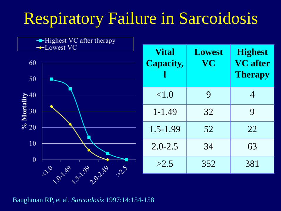
Respiratory Failure in Sarcoidosis
Vital
Capacity,
l
Lowest
VC
Highest
VC after
Therapy
<1.0 9 4
1-1.49 32 9
1.5-1.99 52 22
2.0-2.5 34 63
>2.5 352 381
Baughman RP, et al. Sarcoidosis 1997;14:154-158

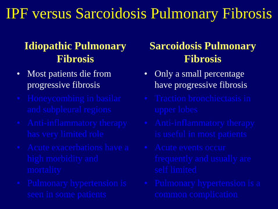
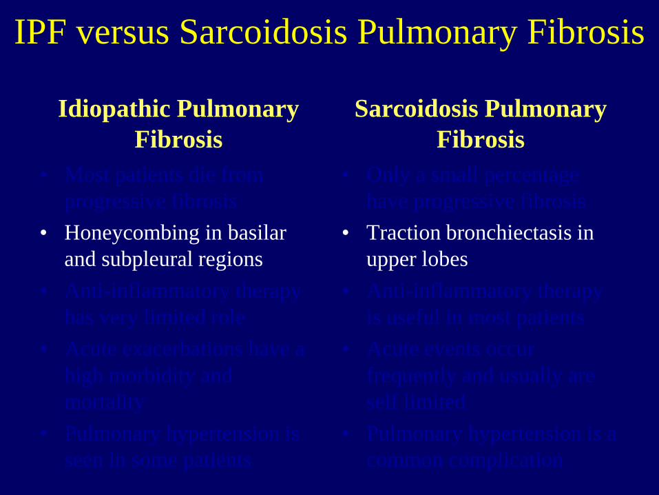
IPF versus Sarcoidosis Pulmonary Fibrosis
Idiopathic Pulmonary
Fibrosis
• Most patients die from
progressive fibrosis
• Honeycombing in basilar
and subpleural regions
• Anti-inflammatory therapy
has very limited role
• Acute exacerbations have a
high morbidity and
mortality
• Pulmonary hypertension is
seen in some patients
Sarcoidosis Pulmonary
Fibrosis
• Only a small percentage
have progressive fibrosis
• Traction bronchiectasis in
upper lobes
• Anti-inflammatory therapy
is useful in most patients
• Acute events occur
frequently and usually are
self limited
• Pulmonary hypertension is a
common complication
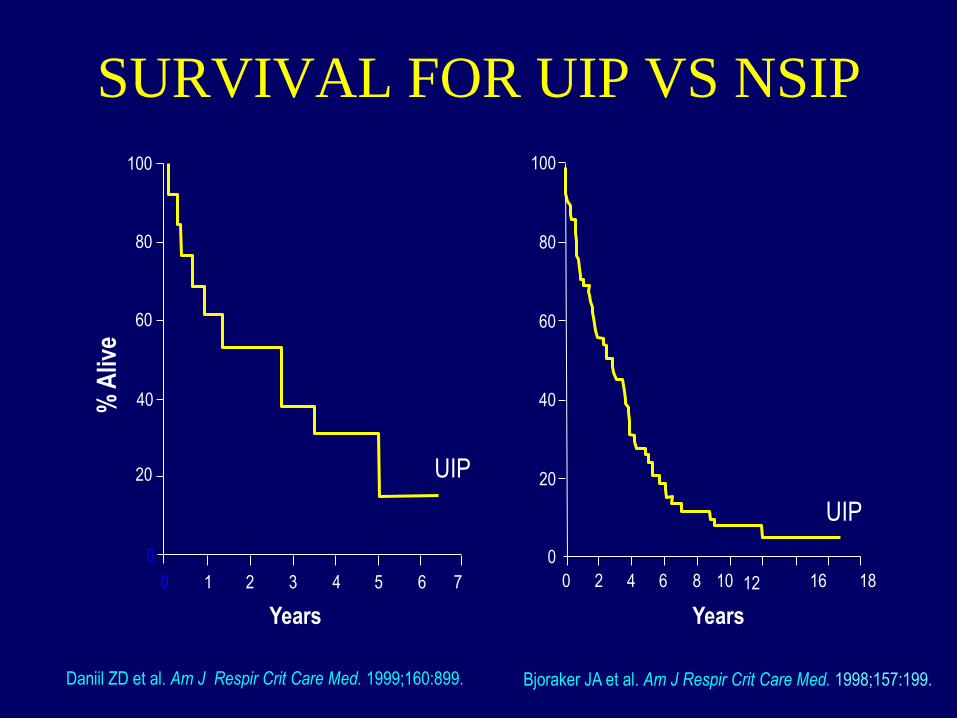
Daniil ZD et al. Am J Respir Crit Care Med. 1999;160:899. Bjoraker JA et al. Am J Respir Crit Care Med. 1998;157:199.
Years
SURVIVAL FOR UIP VS NSIP
Years
7 6 5 4 3 2 1 0
0
20
40
60
80
100
UIP
0 2 4 6 8 10 12 16 18
0
20
40
60
80
100
UIP
% A
live
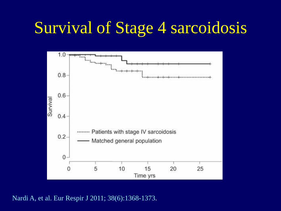
Survival of Stage 4 sarcoidosis
Nardi A, et al. Eur Respir J 2011; 38(6):1368-1373.

CPI
CPI >40 CPI > 40
MPAD/AAD > 1
or
Extent of fibrosis on HRCT >20%
Yes
High risk/poor prognosis
No
Low risk/good prognosis
CPI=91.0-(0.65*percent predicted DLCO)-(0.53*percent predicted
FVC)+(0.34*percent predicted FEV-1)
Walsh SL, et al. Lancet Respir Med 2014; 2(2):123-30.
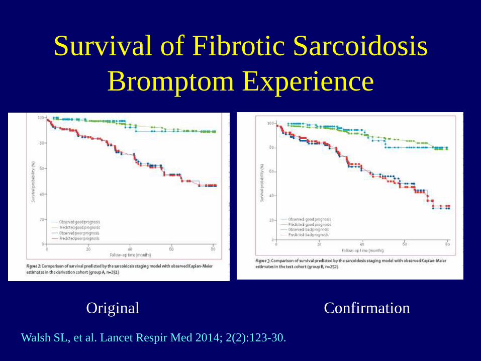
Survival of Fibrotic Sarcoidosis
Bromptom Experience
Walsh SL, et al. Lancet Respir Med 2014; 2(2):123-30.
Original Confirmation
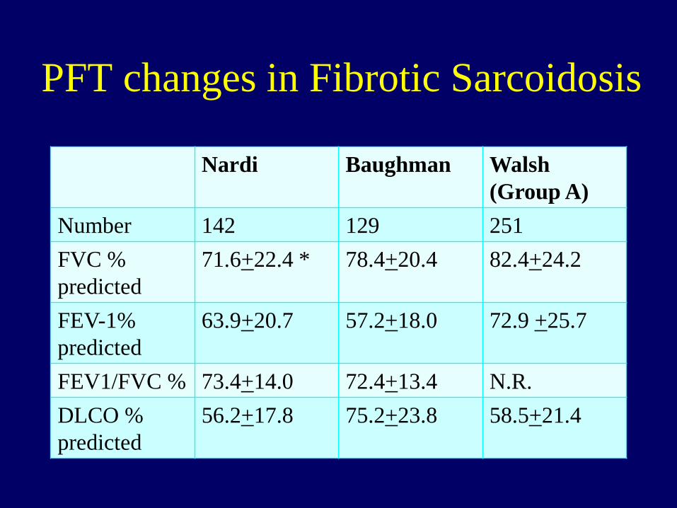
PFT changes in Fibrotic Sarcoidosis
Nardi Baughman Walsh
(Group A)
Number 142 129 251
FVC %
predicted
71.6+22.4 * 78.4+20.4 82.4+24.2
FEV-1%
predicted
63.9+20.7 57.2+18.0 72.9 +25.7
FEV1/FVC % 73.4+14.0 72.4+13.4 N.R.
DLCO %
predicted
56.2+17.8 75.2+23.8 58.5+21.4
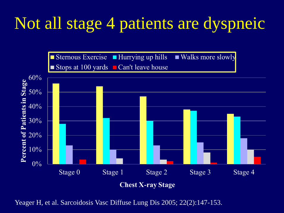
Not all stage 4 patients are dyspneic
Yeager H, et al. Sarcoidosis Vasc Diffuse Lung Dis 2005; 22(2):147-153.
IPF versus Sarcoidosis Pulmonary Fibrosis
Idiopathic Pulmonary
Fibrosis
• Most patients die from
progressive fibrosis
• Honeycombing in basilar
and subpleural regions
• Anti-inflammatory therapy
has very limited role
• Acute exacerbations have a
high morbidity and
mortality
• Pulmonary hypertension is
seen in some patients
Sarcoidosis Pulmonary
Fibrosis
• Only a small percentage
have progressive fibrosis
• Traction bronchiectasis in
upper lobes
• Anti-inflammatory therapy
is useful in most patients
• Acute events occur
frequently and usually are
self limited
• Pulmonary hypertension is a
common complication

HRCT Pattern of
Usual Interstitial Pneumonia
Raghu G, et al. Am J Respir Crit Care Med 183: 788-824, 2011.

Progressive fibrosis in sarcoidosis
patient on prednisone
2008 2014

Asymptomatic Patient with Sarcoidosis
Associated Pulmonary Fibrosis
2009

Asymptomatic Patient with Sarcoidosis
Associated Pulmonary Fibrosis
2012

Fibrosis patient with no symptoms
2009 2012
FVC 2.47 2.42
FVC %
predicted
99% 100%
FEV-1 1.80 1.63
FEV1/FVC 73% 68%
DLCO 9.39 10.46
DLCO %
predicted
52% 59%

HRCT in sarcoidosis: Major Features
• Three main CT patterns
– Bronchial distortion,
– Honeycombing
– Linear opacities.
• Other patterns
– Endobronchial granulomatous lesions
– Aspergilloma colonization
– Bronchiectasis
– Air trapping
Naccache JM, et al J Comput Assist Tomogr 2008;32:905-912.
Hennebicque AS,et al Eur Radiol 2005;15:23-30.
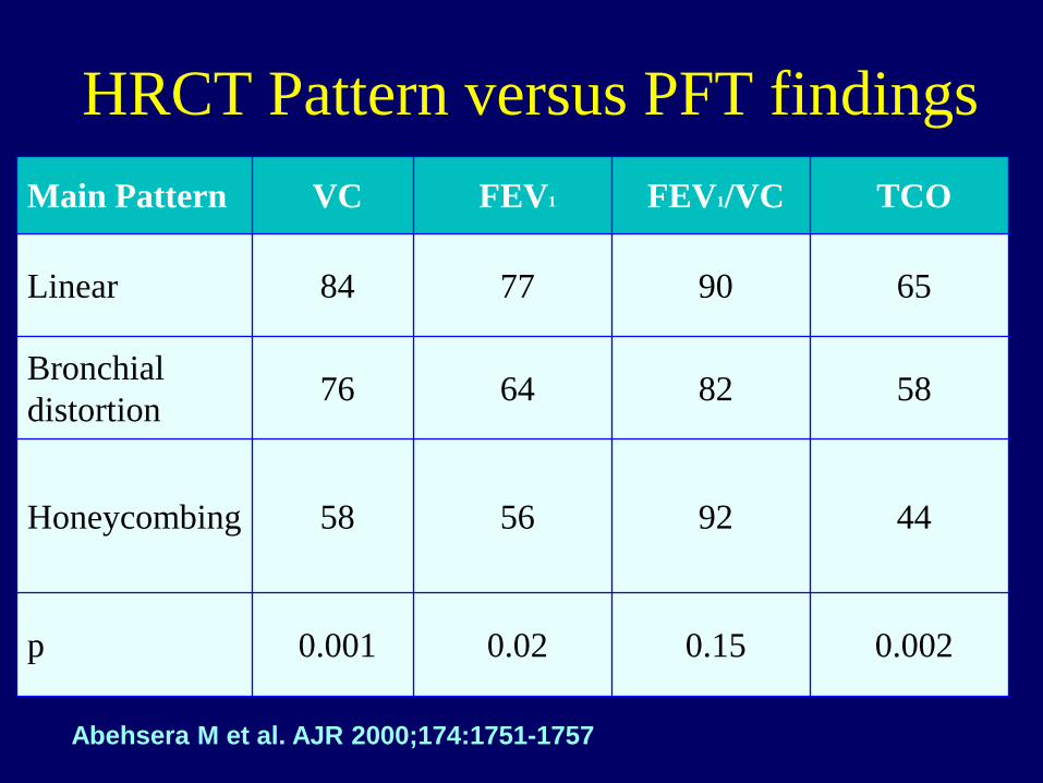
HRCT Pattern versus PFT findings
Main Pattern VC FEV1 FEV1/VC TCO
Linear 84 77 90 65
Bronchial
distortion 76 64 82 58
Honeycombing 58 56 92 44
p 0.001 0.02 0.15 0.002
Abehsera M et al. AJR 2000;174:1751-1757
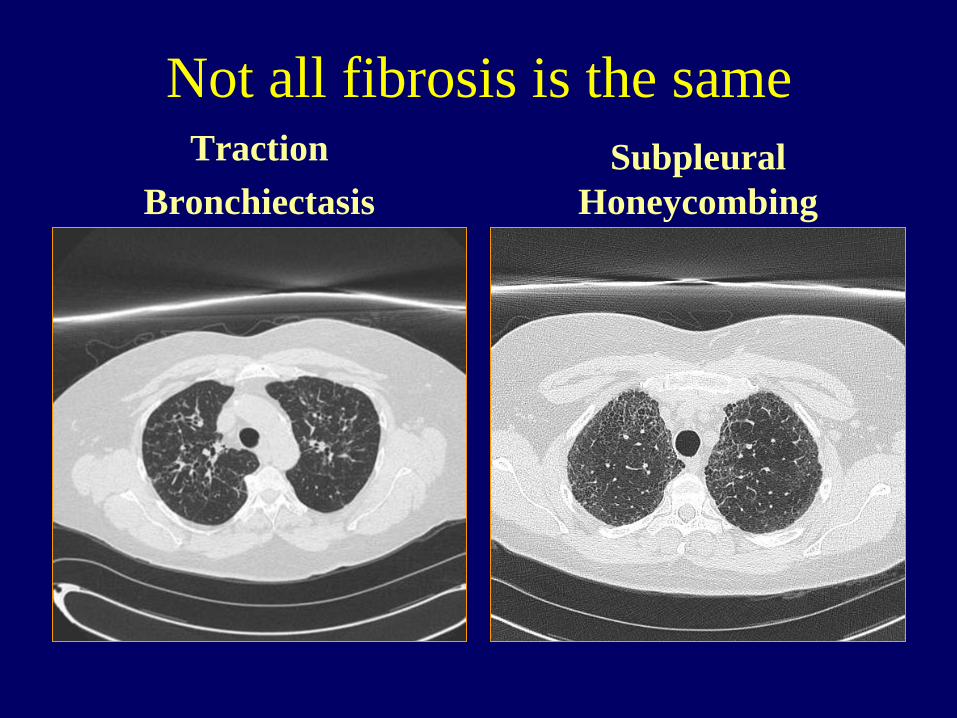
Not all fibrosis is the same Traction
Bronchiectasis
Subpleural
Honeycombing
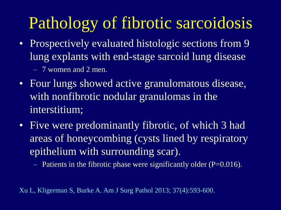
Pathology of fibrotic sarcoidosis • Prospectively evaluated histologic sections from 9
lung explants with end-stage sarcoid lung disease – 7 women and 2 men.
• Four lungs showed active granulomatous disease,
with nonfibrotic nodular granulomas in the
interstitium;
• Five were predominantly fibrotic, of which 3 had
areas of honeycombing (cysts lined by respiratory
epithelium with surrounding scar). – Patients in the fibrotic phase were significantly older (P=0.016).
Xu L, Kligerman S, Burke A. Am J Surg Pathol 2013; 37(4):593-600.
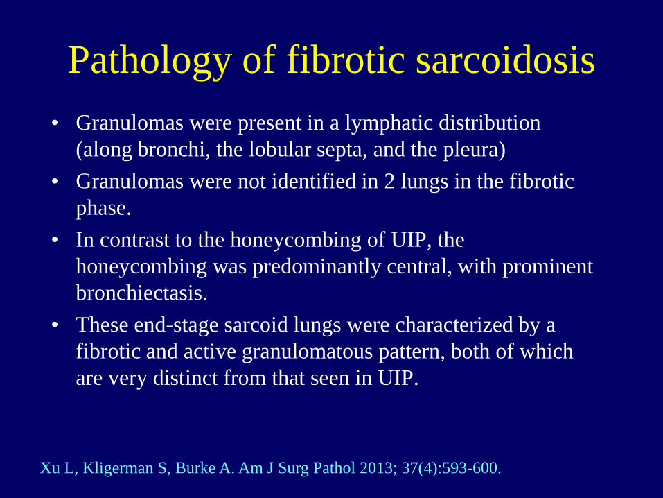
Pathology of fibrotic sarcoidosis
• Granulomas were present in a lymphatic distribution
(along bronchi, the lobular septa, and the pleura)
• Granulomas were not identified in 2 lungs in the fibrotic
phase.
• In contrast to the honeycombing of UIP, the
honeycombing was predominantly central, with prominent
bronchiectasis.
• These end-stage sarcoid lungs were characterized by a
fibrotic and active granulomatous pattern, both of which
are very distinct from that seen in UIP.
Xu L, Kligerman S, Burke A. Am J Surg Pathol 2013; 37(4):593-600.
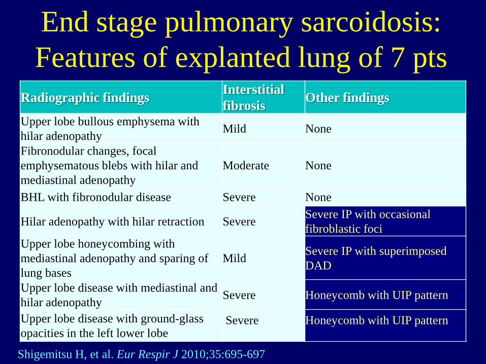
End stage pulmonary sarcoidosis:
Features of explanted lung of 7 pts
Radiographic findings Interstitial
fibrosis Other findings
Upper lobe bullous emphysema with
hilar adenopathy Mild None
Fibronodular changes, focal
emphysematous blebs with hilar and
mediastinal adenopathy Moderate None
BHL with fibronodular disease Severe None
Hilar adenopathy with hilar retraction Severe Severe IP with occasional
fibroblastic foci
Upper lobe honeycombing with
mediastinal adenopathy and sparing of
lung bases Mild
Severe IP with superimposed
DAD
Upper lobe disease with mediastinal and
hilar adenopathy Severe Honeycomb with UIP pattern
Upper lobe disease with ground-glass
opacities in the left lower lobe Severe
Honeycomb with UIP pattern
Shigemitsu H, et al. Eur Respir J 2010;35:695-697
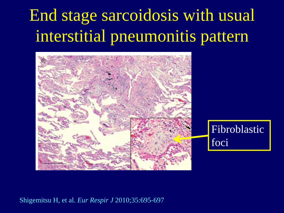
End stage sarcoidosis with usual
interstitial pneumonitis pattern
Fibroblastic
foci
Shigemitsu H, et al. Eur Respir J 2010;35:695-697
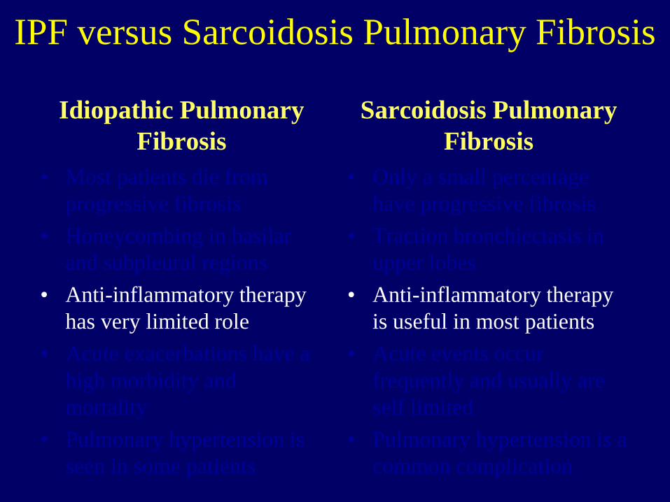
IPF versus Sarcoidosis Pulmonary Fibrosis
Idiopathic Pulmonary
Fibrosis
• Most patients die from
progressive fibrosis
• Honeycombing in basilar
and subpleural regions
• Anti-inflammatory therapy
has very limited role
• Acute exacerbations have a
high morbidity and
mortality
• Pulmonary hypertension is
seen in some patients
Sarcoidosis Pulmonary
Fibrosis
• Only a small percentage
have progressive fibrosis
• Traction bronchiectasis in
upper lobes
• Anti-inflammatory therapy
is useful in most patients
• Acute events occur
frequently and usually are
self limited
• Pulmonary hypertension is a
common complication
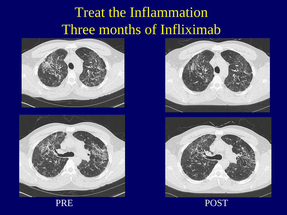
Treat the Inflammation
Three months of Infliximab
PRE POST

Survival of Stage 4 sarcoidosis
Nardi A, et al. Eur Respir J 2011; 38(6):1368-1373.
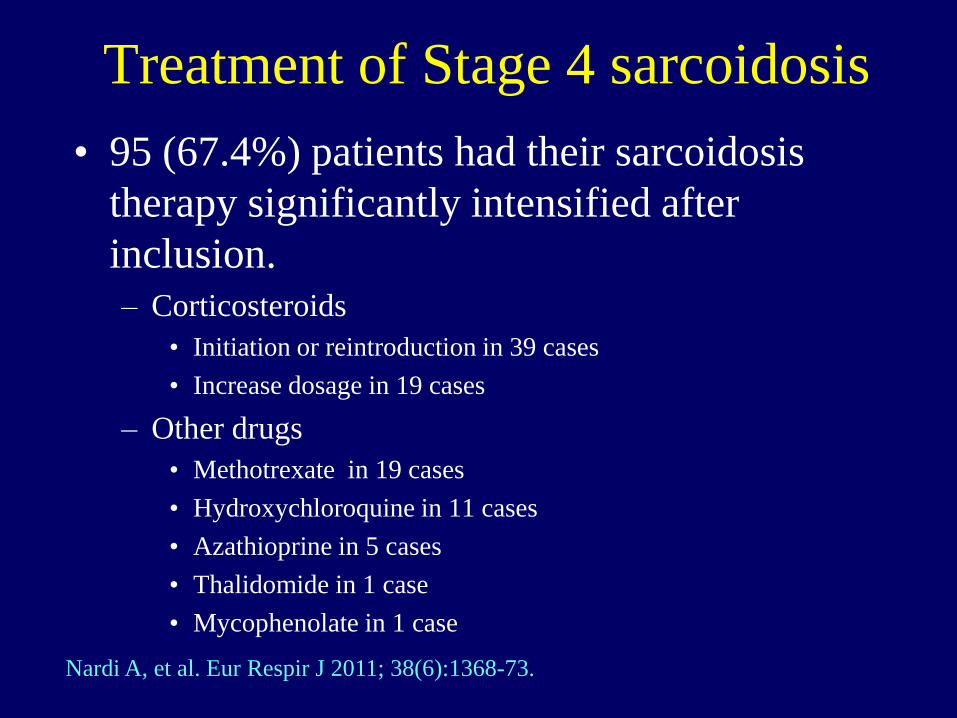
Treatment of Stage 4 sarcoidosis
• 95 (67.4%) patients had their sarcoidosis
therapy significantly intensified after
inclusion. – Corticosteroids
• Initiation or reintroduction in 39 cases
• Increase dosage in 19 cases
– Other drugs
• Methotrexate in 19 cases
• Hydroxychloroquine in 11 cases
• Azathioprine in 5 cases
• Thalidomide in 1 case
• Mycophenolate in 1 case
Nardi A, et al. Eur Respir J 2011; 38(6):1368-73.
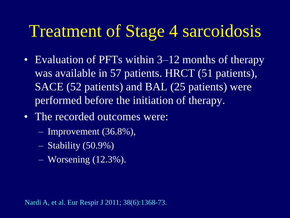
Treatment of Stage 4 sarcoidosis
• Evaluation of PFTs within 3–12 months of therapy
was available in 57 patients. HRCT (51 patients),
SACE (52 patients) and BAL (25 patients) were
performed before the initiation of therapy.
• The recorded outcomes were:
– Improvement (36.8%),
– Stability (50.9%)
– Worsening (12.3%).
Nardi A, et al. Eur Respir J 2011; 38(6):1368-73.
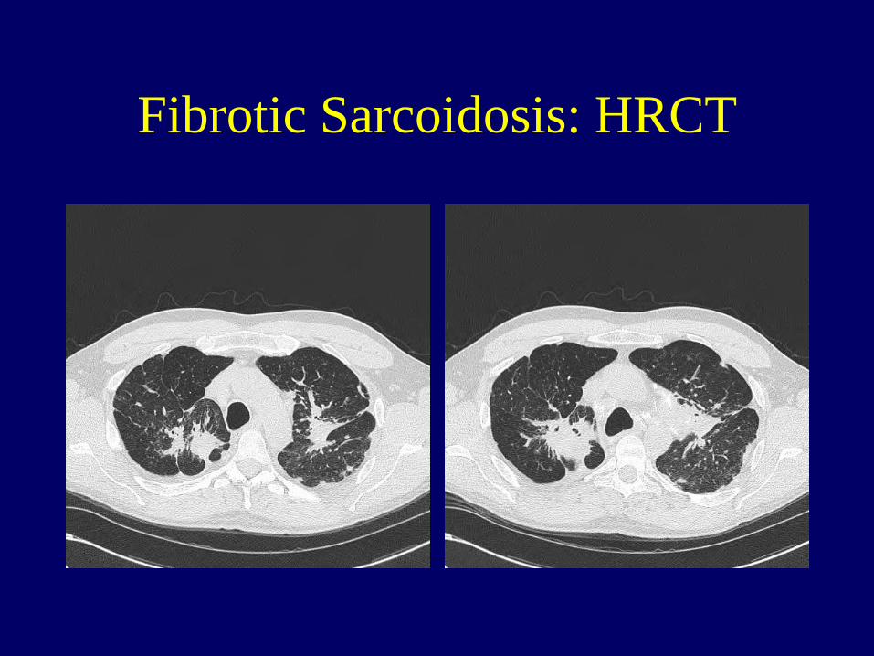
Fibrotic Sarcoidosis: HRCT
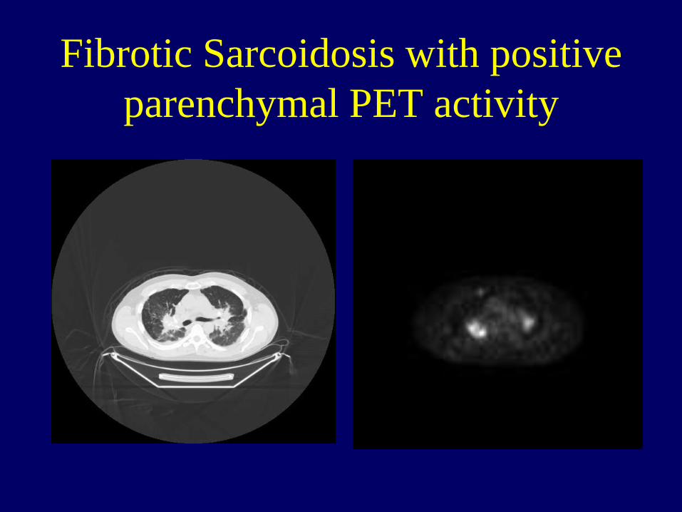
Fibrotic Sarcoidosis with positive
parenchymal PET activity
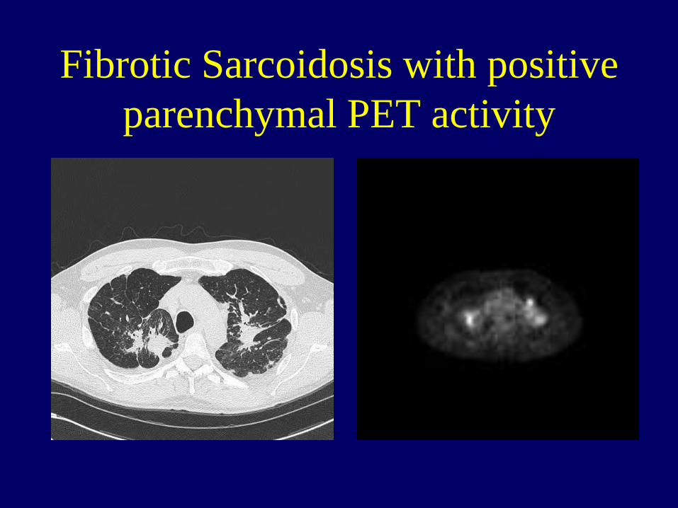
Fibrotic Sarcoidosis with positive
parenchymal PET activity

PET scan predicting response to
therapy in sarcoidosis: FVC change
Keijsers RG, et al. Sarcoidosis Vasc Diffuse Lung Dis 2011; 28(2):123-129
N=11 N=16 N=16
P<0.01
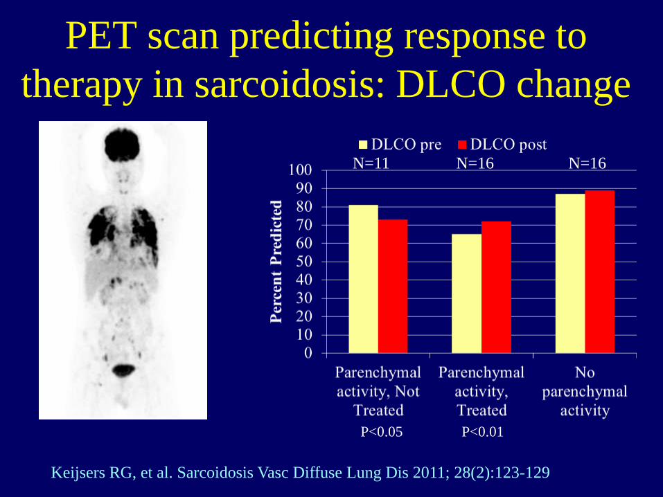
PET scan predicting response to
therapy in sarcoidosis: DLCO change
Keijsers RG, et al. Sarcoidosis Vasc Diffuse Lung Dis 2011; 28(2):123-129
N=11 N=16 N=16
P<0.05 P<0.01
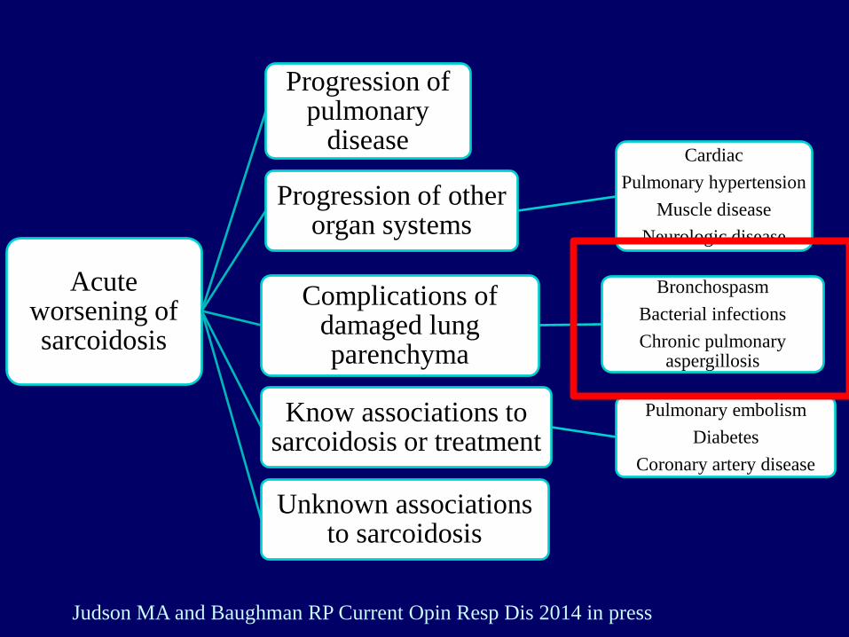
Acute worsening of sarcoidosis
Progression of pulmonary
disease
Progression of other organ systems
Cardiac
Pulmonary hypertension
Muscle disease
Neurologic disease
Complications of damaged lung parenchyma
Bronchospasm
Bacterial infections
Chronic pulmonary aspergillosis
Know associations to sarcoidosis or treatment
Pulmonary embolism
Diabetes
Coronary artery disease
Unknown associations to sarcoidosis
Judson MA and Baughman RP Current Opin Resp Dis 2014 in press
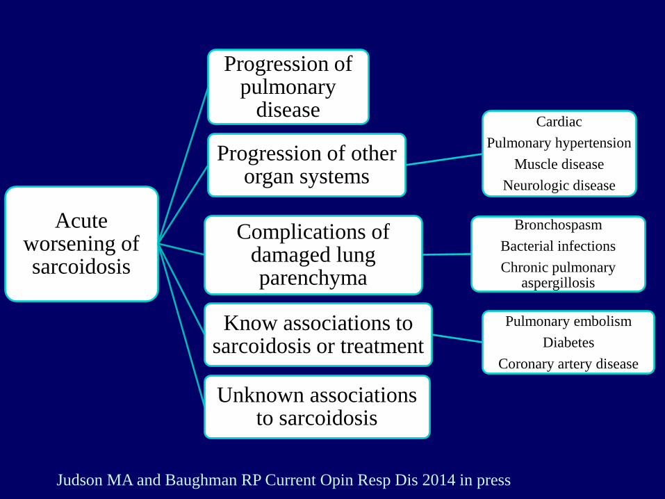

FVC % predicted versus Chest X-ray stage
Significant difference between groups Chi square=72.9, P<0.0001
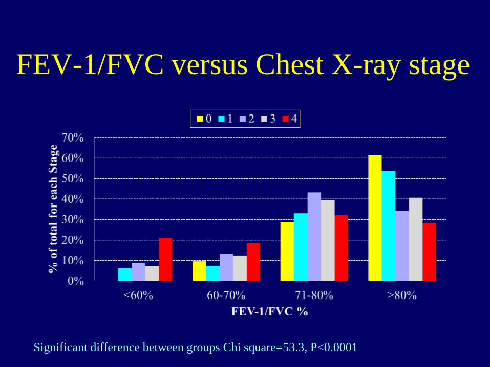
FEV-1/FVC versus Chest X-ray stage
Significant difference between groups Chi square=53.3, P<0.0001
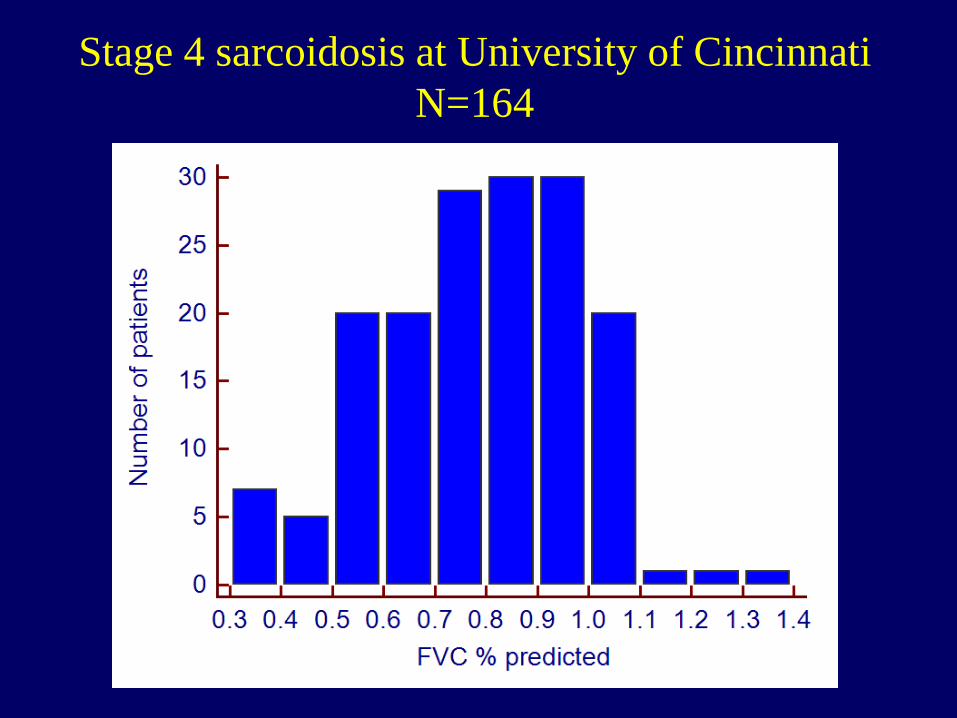
Stage 4 sarcoidosis at University of Cincinnati
N=164
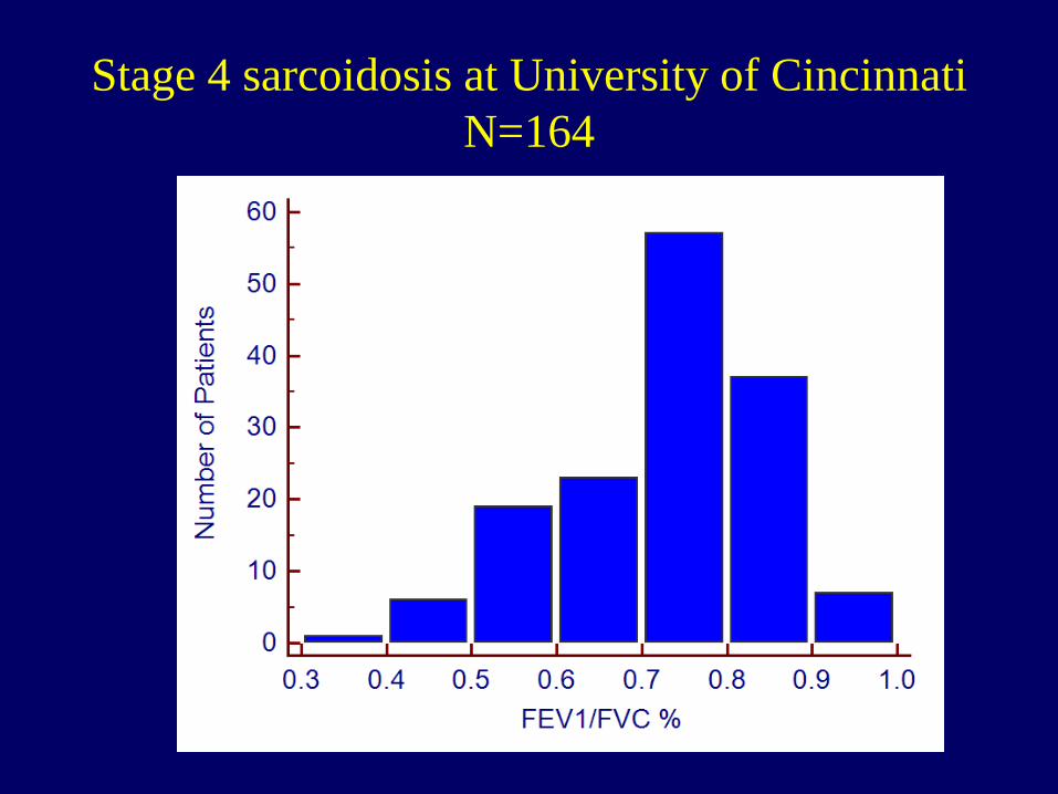
Stage 4 sarcoidosis at University of Cincinnati
N=164

Medial segment
Lateral segment occluded Lateral segment patent
RML view from its orifice RML view post dilation
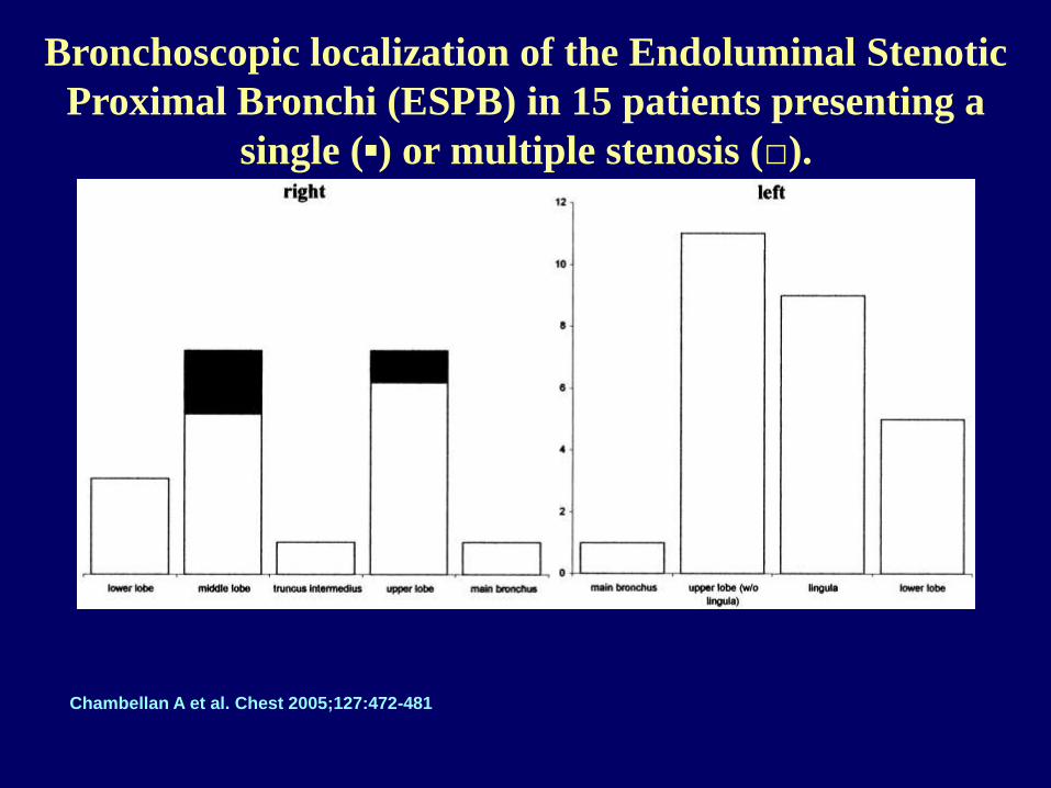
Bronchoscopic localization of the Endoluminal Stenotic
Proximal Bronchi (ESPB) in 15 patients presenting a
single (▪) or multiple stenosis (□).
Chambellan A et al. Chest 2005;127:472-481

Relationship between FEV1/FVC and the number
of Endoluminal Stenotic Proximal Bronchi
(ESPB) at baseline.
Chambellan A et al. Chest 2005;127:472-481
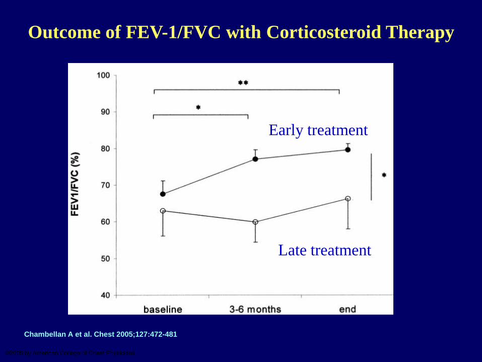
Outcome of FEV-1/FVC with Corticosteroid Therapy
Chambellan A et al. Chest 2005;127:472-481
©2005 by American College of Chest Physicians
Early treatment
Late treatment

IPF versus Sarcoidosis Pulmonary Fibrosis
Idiopathic Pulmonary
Fibrosis
• Most patients die from
progressive fibrosis
• Honeycombing in basilar
and subpleural regions
• Anti-inflammatory therapy
has very limited role
• Acute exacerbations have a
high morbidity and
mortality
• Pulmonary hypertension is
seen in some patients
Sarcoidosis Pulmonary
Fibrosis
• Only a small percentage
have progressive fibrosis
• Traction bronchiectasis in
upper lobes
• Anti-inflammatory therapy
is useful in most patients
• Acute events occur
frequently and usually are
self limited
• Pulmonary hypertension is a
common complication
Acute worsening of sarcoidosis
Progression of pulmonary
disease
Progression of other organ systems
Cardiac
Pulmonary hypertension
Muscle disease
Neurologic disease
Complications of damaged lung parenchyma
Bronchospasm
Bacterial infections
Chronic pulmonary aspergillosis
Know associations to sarcoidosis or treatment
Pulmonary embolism
Diabetes
Coronary artery disease
Unknown associations to sarcoidosis
Judson MA and Baughman RP Current Opin Resp Dis 2014 in press

Acute exacerbations in IPF are associated
with significant short term mortality
Abe S, et al. Intern Med 2012; 51:1487-91
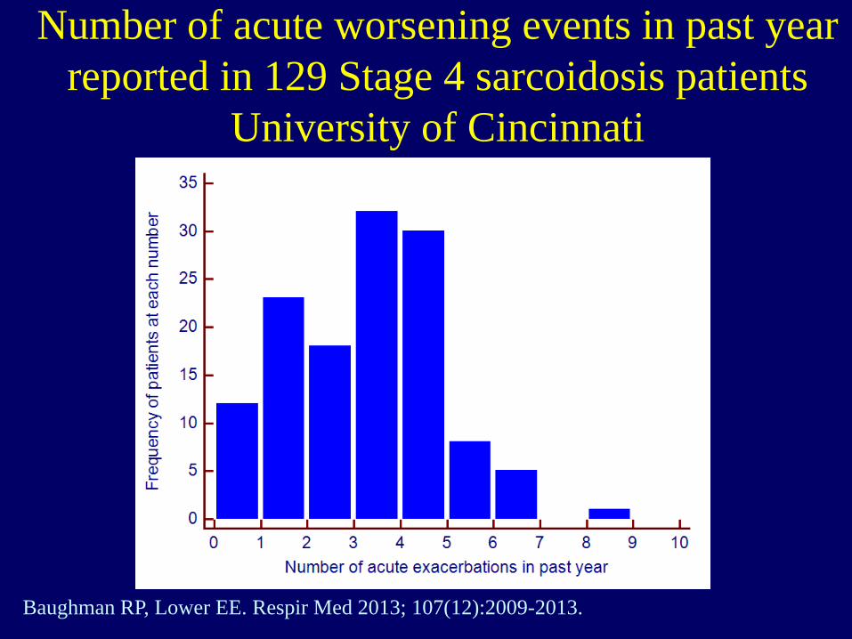
Number of acute worsening events in past year
reported in 129 Stage 4 sarcoidosis patients
University of Cincinnati
Baughman RP, Lower EE. Respir Med 2013; 107(12):2009-2013.

Number of events were higher in Bronchiectasis patients
(n=63) versus those without Bronchiectasis (n=66)
Baughman RP, Lower EE. Respir Med 2013; 107(12):2009-2013.
P<0.0001

IPF versus Sarcoidosis Pulmonary Fibrosis
Idiopathic Pulmonary
Fibrosis
• Most patients die from
progressive fibrosis
• Honeycombing in basilar
and subpleural regions
• Anti-inflammatory therapy
has very limited role
• Acute exacerbations have a
high morbidity and
mortality
• Pulmonary hypertension is
seen in some patients
Sarcoidosis Pulmonary
Fibrosis
• Only a small percentage
have progressive fibrosis
• Traction bronchiectasis in
upper lobes
• Anti-inflammatory therapy
is useful in most patients
• Acute events occur
frequently and usually are
self limited
• Pulmonary hypertension is a
common complication

Pulmonary Hypertension in
Sarcoidosis
0
10
20
30
40
50
60
70
80
Per
cen
t w
ith
Pu
lmon
ary
Hyp
erte
nsi
on
Kyoto Detroit Milan New York Cincinnati Transplant
All patients Only Dyspneic Patients
Blue bars indicate those centers who confirmed pulmonary hypertension by right heart cath

Pulmonary Hypertension associated
with Stage 4 disease
Baughman RP, et al. Chest 2010;138:1078-1085.
Sulica R, et al. Chest 2005;128:1483-1489.
Barnett CF, et al. Chest 2009;135:1455-1461.
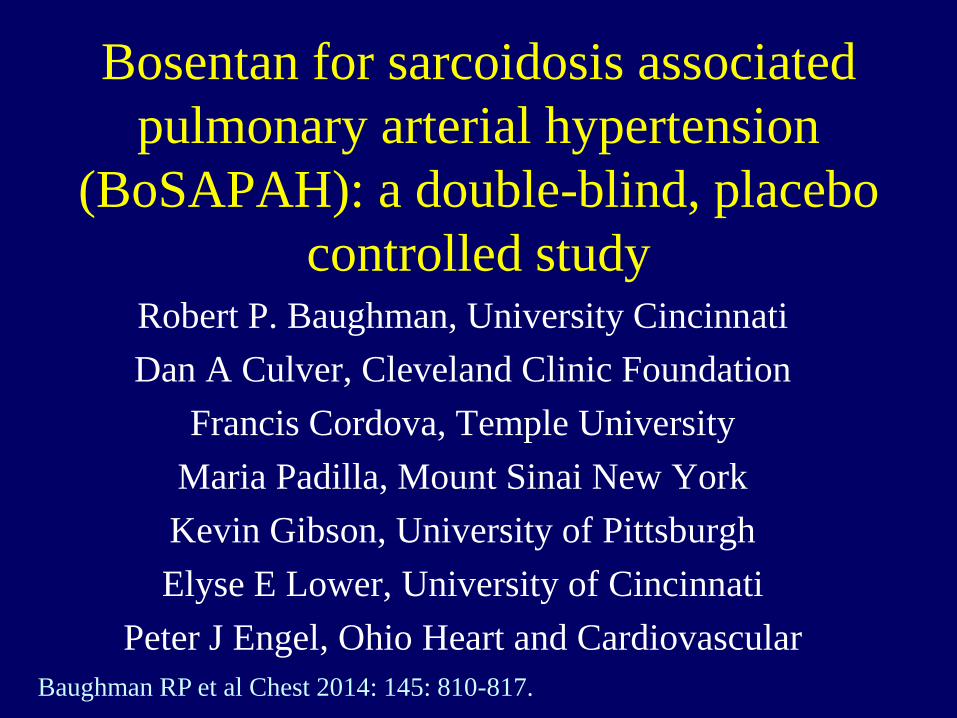
Bosentan for sarcoidosis associated
pulmonary arterial hypertension
(BoSAPAH): a double-blind, placebo
controlled study Robert P. Baughman, University Cincinnati
Dan A Culver, Cleveland Clinic Foundation
Francis Cordova, Temple University
Maria Padilla, Mount Sinai New York
Kevin Gibson, University of Pittsburgh
Elyse E Lower, University of Cincinnati
Peter J Engel, Ohio Heart and Cardiovascular
Baughman RP et al Chest 2014: 145: 810-817.
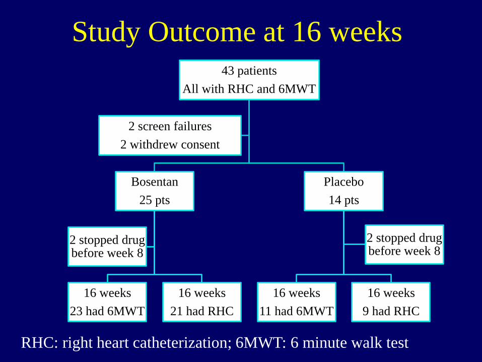
Study Outcome at 16 weeks 43 patients
All with RHC and 6MWT
Bosentan
25 pts
16 weeks
23 had 6MWT
16 weeks
21 had RHC
2 stopped drug before week 8
Placebo
14 pts
16 weeks
11 had 6MWT
16 weeks
9 had RHC
2 stopped drug before week 8
2 screen failures
2 withdrew consent
RHC: right heart catheterization; 6MWT: 6 minute walk test

PA Mean pressure before and
after 16 weeks of therapy

Conclusion
• Pulmonary fibrosis is a significant problem
in pulmonary sarcoidosis
• Not all patients with pulmonary fibrosis are
dyspneic
• For the dyspneic patient, there is significant
mortality
• Treatment may helpful in the dyspneic
patient
Acknowledgements
• Dr. Elyse Lower
– Co-director of ILD/Sarcoidosis Clinic
• Dr. Peter Engel
– Co-Director of PH Clinic
• Research coordinators
– Felicia Thompson
– Joyce Zeigler
• Our patients