Protective effect of 14-3-3 antibodies on stressed ... · stressed neuroretinal cells via the...
13
RESEARCH ARTICLE Open Access Protective effect of 14-3-3 antibodies on stressed neuroretinal cells via the mitochondrial apoptosis pathway Katharina Bell, Corina Wilding, Sebastian Funke, Norbert Pfeiffer and Franz H. Grus * Abstract Background: Previous studies demonstrate changes of autoantibody concentrations against retinal and optic nerve head antigens in the serum of glaucoma patients in comparison to healthy persons. These antibodies belong to the natural autoimmunity. Previous studies showed up regulated, but also significantly down-regulated autoantibody levels. These antibodies have the ability to influence protein profiles of neuroretinal cells and possibly hold neuroprotective potential, as we have been able to demonstrate before. Aim of this study was to analyse the serum and antibody effect of glaucoma patients on neuroretinal cells in more detail and also determine the impact of antibodies found down- regulated in glaucoma patients on the pathogenesis of the neurodegenerative disease glaucoma. Methods: Neuroretinal cells (RGC-5) were incubated with serum either from glaucoma patients or healthy controls for 24 h. Mass spectrometric analysis was performed after cell lysis. Furthermore the neuroretinal cells were preincubated with different and concentrations of 14-3-3 antibodies (0.005, 0.1, 0.5, 1, 5 and 10 μg/ml) and then stressed with H2O2, staurosporine or glutamate. Viability tests were performed with crystal violet and ROS tests with DCFH-DA. Antibody location in the cell after antibody incubation was performed with immunoccytochemical methods. Additionally mass spectrometric analysis was performed with the cells after antibody incubation. Results: Protein expression analysis with Maldi-Orbitrap MS showed changes in the expression level of regulatory proteins in cells incubated with glaucoma serum, e.g. an up-regulation of 14-3-3 and a down-regulation of Calmodulin. After preincubation of the cells with anti-14-3-3 antibody and stressing the cells, we detected an increase in viability of up to 22 % and a decrease in reactive oxygen species (ROS) of up to 31 %. Proteomic 1 analysis involvement of the mitochondrial apoptosis pathway in this protective effect and immunohistochemical analysis showed an antibody uptake in the cells. Conclusion: We found significant effects of serum antibodies on proteins of neuroretinal cells especially of the mitochondrial apoptosis pathway. Furthermore we detected a protective potential of antibodies down-regulated in glaucoma patients. The changed autoantibodies belong to the natural autoimmunity. We conclude that changes in the natural autoimmunity of patients with glaucoma can negatively impact regulatory functions. Keywords: Autoantibodies, Glaucoma, Neurodegeneration, Natural autoimmunity, Neuroprotection * Correspondence: [email protected] Experimental Ophthalmology, Department of Ophthalmology, University Medical center of the Johannes Gutenberg University, Langenbeckstraße 1, 55131 Mainz, Germany © 2015 Bell et al. This is an Open Access article distributed under the terms of the Creative Commons Attribution License (http://creativecommons.org/licenses/by/4.0), which permits unrestricted use, distribution, and reproduction in any medium, provided the original work is properly credited. The Creative Commons Public Domain Dedication waiver (http:// creativecommons.org/publicdomain/zero/1.0/) applies to the data made available in this article, unless otherwise stated. Bell et al. BMC Ophthalmology (2015) 15:64 DOI 10.1186/s12886-015-0044-9
Transcript of Protective effect of 14-3-3 antibodies on stressed ... · stressed neuroretinal cells via the...

Bell et al. BMC Ophthalmology (2015) 15:64 DOI 10.1186/s12886-015-0044-9
RESEARCH ARTICLE Open Access
Protective effect of 14-3-3 antibodies onstressed neuroretinal cells via the mitochondrialapoptosis pathway
Katharina Bell, Corina Wilding, Sebastian Funke, Norbert Pfeiffer and Franz H. Grus*Abstract
Background: Previous studies demonstrate changes of autoantibody concentrations against retinal and optic nervehead antigens in the serum of glaucoma patients in comparison to healthy persons. These antibodies belong to thenatural autoimmunity. Previous studies showed up regulated, but also significantly down-regulated autoantibody levels.These antibodies have the ability to influence protein profiles of neuroretinal cells and possibly hold neuroprotectivepotential, as we have been able to demonstrate before. Aim of this study was to analyse the serum and antibody effectof glaucoma patients on neuroretinal cells in more detail and also determine the impact of antibodies found down-regulated in glaucoma patients on the pathogenesis of the neurodegenerative disease glaucoma.
Methods: Neuroretinal cells (RGC-5) were incubated with serum either from glaucoma patients or healthy controls for24 h. Mass spectrometric analysis was performed after cell lysis. Furthermore the neuroretinal cells were preincubatedwith different and concentrations of 14-3-3 antibodies (0.005, 0.1, 0.5, 1, 5 and 10 μg/ml) and then stressed with H2O2,staurosporine or glutamate. Viability tests were performed with crystal violet and ROS tests with DCFH-DA. Antibodylocation in the cell after antibody incubation was performed with immunoccytochemical methods. Additionally massspectrometric analysis was performed with the cells after antibody incubation.
Results: Protein expression analysis with Maldi-Orbitrap MS showed changes in the expression level of regulatoryproteins in cells incubated with glaucoma serum, e.g. an up-regulation of 14-3-3 and a down-regulation of Calmodulin.After preincubation of the cells with anti-14-3-3 antibody and stressing the cells, we detected an increase in viability ofup to 22 % and a decrease in reactive oxygen species (ROS) of up to 31 %. Proteomic 1 analysis involvement of themitochondrial apoptosis pathway in this protective effect and immunohistochemical analysis showed an antibodyuptake in the cells.
Conclusion: We found significant effects of serum antibodies on proteins of neuroretinal cells especially of themitochondrial apoptosis pathway. Furthermore we detected a protective potential of antibodies down-regulated inglaucoma patients. The changed autoantibodies belong to the natural autoimmunity. We conclude that changes inthe natural autoimmunity of patients with glaucoma can negatively impact regulatory functions.
Keywords: Autoantibodies, Glaucoma, Neurodegeneration, Natural autoimmunity, Neuroprotection
* Correspondence: [email protected] Ophthalmology, Department of Ophthalmology, UniversityMedical center of the Johannes Gutenberg University, Langenbeckstraße 1,55131 Mainz, Germany
© 2015 Bell et al. This is an Open Access article distributed under the terms of the Creative Commons Attribution License(http://creativecommons.org/licenses/by/4.0), which permits unrestricted use, distribution, and reproduction in any medium,provided the original work is properly credited. The Creative Commons Public Domain Dedication waiver (http://creativecommons.org/publicdomain/zero/1.0/) applies to the data made available in this article, unless otherwise stated.

Bell et al. BMC Ophthalmology (2015) 15:64 Page 2 of 13
BackgroundThe pathogenesis of neurodegenerative diseases is oftenpoorly understood. Neurodegenerative diseases are char-acterised by progressive nervous system dysfunction andan accompanying atrophy of the affected central or per-ipheral nervous system [1]. As in other neurodegenerativediseases, such as amyotrophic lateral sclerosis, Alzheimer’sor Parkinson disease, glaucoma leads to the apoptotic lossof one specific neuron population, the retinal ganglioncells (rgc) [2]. An atrophy of central structures such as thelateral geniculate nucleus [3] can also be found. With anestimated prevalence of at least 60 million cases world-wide [4], glaucoma can be counted to the list of the mostcommon neurodegenerative diseases [5].This heterogeneous group of eye diseases, with a still
unknown pathogenesis, demonstrates with a progressiveloss of retinal ganglion cells (rgc), optic nerve degener-ation and visual fields loss, finally leading to blindness[6]. 2.65 % of the world’s population above the age of 40suffers from glaucoma [7]. The major risk factor for de-veloping glaucoma found in approximately 70 % of thepatients is an increased intraocular pressure (IOP) [8, 9].Other pathogenesis factors leading to apoptosis of rgc
[10, 11] such as elevated levels of reactive oxygen species(ROS) [12, 13] or elevated glutamate levels are discussed[14, 15]. Furthermore, there is strong evidence that animmunologic component is involved in glaucoma patho-genesis. Altered autoantibody levels in the serum ofglaucoma patients e.g. against heat shock protein (hsp)60 [16], alpha crystallin and hsp27, gamma enolase [17]and glycosaminoglycans as well as against human retinalantigens, such as against cellular retinaldehyde-bindingprotein and retinal-S-antigen [18, 19] have been demon-strated. Interestingly, the studies were not only able todetect higher concentrations of different autoantibodiesin glaucoma patients, but also lower concentrations ofmany autoantibodies in comparison to healthy people[20]. Many of the serum immunoglobulins in healthypeople belong to the so called natural autoimmunity [21,22]. These autoantibodies do not cause diseases and incontrast are considered as regulatory factors [23]. Ingeneral it is known that up-regulated autoantibodies canbe auto-aggressive and lead to pathogenic conditions,such as the antibody against postsynaptic nicotinicacetylcholine receptor in patients suffering from myas-thenia gravis [24]. The role of the down-regulated auto-antibodies found e.g. in glaucoma patients, but also inpatients suffering from other neurodegenerative diseases,such as Alzheimer’s disease [25], so far is not known.We assume that the down-regulation of some of theantibodies can lead to changes in the regulatory functionof these antibodies and therefore could be involved inthe pathogenesis of the neurodegenerative diseaseglaucoma.
The aim of this study was to investigate the inducedeffect of glaucomatous serum and an antibody founddown-regulated in glaucoma patients on viability, react-ive oxygen levels (ROS) as well as the proteomics ofneuroretinal cells. In previous studies we were able todemonstrate that the antibodies of glaucoma patients ingeneral have a large influence (59 %) on the protein pro-files of neuroretinal cells [26]. Therefore we analysed thechanges of proteins and their pathways in more detail.Additionally we enlighten whether down-regulated anti-bodies could have an impact on the disease glaucoma.In summary we demonstrate that serum of glaucoma
patients can change intracellular concentrations of im-portant regulatory proteins such as the protein kinaseinhibitor 14-3-3, which has a major regulatory functionon the MAPK/ERK pathway, or Calmodulin. Further-more we were able to show that the antibody against14-3-3, which is down-regulated in glaucoma patients(p < 0.01) (see Additional file 1: Figure S1), has protect-ive effects on stressed neuroretinal cells, documentedby an increased viability and reduced ROS-level of thecells.
MethodsReagentsThe used reagents were purchased from Sigma Aldrich,St. Louis, MO, US if not stated otherwise. L-alanyl-L-glutamine was purchased from Biochrom AG (Berlin,Germany). The rabbit anti-14-3-3 abs for cell incubationand the secondary antibody goat-anti-rabbit IgG-H&L(FITC) were purchased from Abcam (Cambridge, UK),H2O2 and paraformaldehyde from Carl Roth GmbH(Karlsruhe, Germany), Staurosporine from Calbiochem(San Diego, CA), 70 % ethanol, formic acid, trifluoroace-tic acid (TFA) and acetonitrile (ACN) from Merck(Darmstadt, Germany), 14-3-3 antibodys for immunohis-tochemical staining from Lifespan (Seattle, WA), theBCA Pierce Protein Assay kit from Fisher scientific(Waltham, MA), Trypsin from Promega (Mannheim,Germany), wheat germ agglutinin conjugate with tetra-methylrhodamin (WGA) from Invitrogen (Karlsruhe,Germany), vectashield mounting medium with 4’,6-dia-midino-2-phenylindole (DAPI) from Vector Laboratories(California, USA) and HPLC H2O from Applichem(Darmstadt, Germany).
Serum samplesThe serum was collected from patients suffering fromPOAG according to the classification of the guidelines ofthe European glaucoma society as well as from healthyvolunteers after given written informed consent. Further-more, patients suffering from autoimmune diseases orother neurodegenerative diseases were excluded from thestudy. The studies were performed in accordance with the

Bell et al. BMC Ophthalmology (2015) 15:64 Page 3 of 13
Declaration of Helsinki on medical research involving hu-man subjects. The samples were age matched. Ethicsapproval: No: 837.219.07 (5754); Ethics committee of theLandesärztekammer Rhineland-Palatinate.
Cell cultureRGC-5 cells, a neuroretinal cell line of mouse origin,provided by Dr. Neeraj Agarwal, were used as model forretinal cells of neuronal origin [27]. They were grown in75 culture flasks in Dulbecco’s modified eagle mediumsupplemented with 10 % fetal calf serum (FCS), 100 U/ml penicillin, 100 U/ml streptomycin and 4 % L-alanyl-L-glutamine. The cells were cultivated in a humidifiedincubator at 37 °C with 95 % air and 5 % CO2 and werepassaged when they reached a confluence of 80 %.
Cell treatment with different serum types250 000 RGC-5 cells were transferred to culture disheswith an 82.7 mm inner diameter. After 24 h the cellswere incubated with medium containing 5 % FCS andeither 5 % serum from healthy people or 5 % serumfrom POAG patients (n in each experimental group: 8).Each individual serum was used for an individual run.The cells were incubated with the different serum typesfor 24 h. After 24 h the medium was discarded and thecells were washed twice with phosphate buffered saline(PBS). Cell lysis was performed and the detached cellswere transferred to an Eppendorf tube with lysis buffer[Urea 9.5 M, Chaps 2 %, DTT 1 % + proteinase inhibitormix (P 1860)]. Furthermore an ultrasonic pulse echo in-strument [Labsonic®M (Sartorius)] was used to performadditional cell lysis. Protein concentration of the sampleswas determined with the method of Lowry [28]. Afterthe protein concentration was determined, a sample-pool for each experimental group was created with atotal amount of 80 μg protein. The pooled samples wereseparated with a 1D SDS gel. Each lane was divided into15 pieces and digested with trypsin in order to measurethe peptide profile with Maldi- Orbitrap MSMS.
Protein profiling with Maldi- Orbitrap MSMSThe protein profiles were analysed with Maldi- LTQOrbitrap XL using Maldi-steel targets. The samples weredried in a concentrator and acidified with 0.1 % TFA. C-18 ZipTips (Millipore, Billerica, MA) were used to purifythe samples and the peptides were eluted directly onto aMaldi Target with 40 % ACN and 60 % ACN. Measure-ments were performed according to the manufacturers’protocol.
Protein profiling with capillary LC-ESI-MSMSThe protein profiles were analysed with capillary LC-ESI-MSMS using a C-18 pre-column (30 mm x 0.5 mm)and a C18 analytical column (150 mm x 0.5 mm, both
Thermo Scientific,). A Rheos Allegro HPLC Pump(Thermo Scientific) was the solvent delivery system. Thepump flow rate was 200 μl/min, and reduced to a col-umn flow of 10 μl/min (M-472 graduated microsplitvalve (Upchurch, Scientific, USA). With two runningbuffers (A (98 % H2O, 1.94 % ACN, 0.06 % methanol,0.05 % TFA) + B (95 % ACN, 3 % methanol, 2 % H2O,0.05 % TFA) a linear gradient of 80 min was performed(0–47 min: 0–100 % B, 47–49 min: 100 % B, 49–58:100 %–0 % B, 58–80 min: 0 % B). Mass spectra were ob-tained using an LTQ OrbitrapXL.
Cell treatment with 14-3-3 antibodies and different stressfactorsRGC-5 cells were seeded in 24 well plates. Dependingon the stress factor and therefore the overall incubationtime, the cells were either seeded with 45000 cells perwell (for the experiments with H2O2 and staurosporine)or 40000 cells per well (experiments with the stress fac-tor glutamate). The cells were preincubated with differ-ent concentrations of chicken polyclonal anti 14-3-3sigma antibodies (0.005, 0.1, 0.5, 1, 5 and 10 μg/ml) for3 h. As an additional control group the cells were incu-bated with a non-retina specific antibody against myo-globin. To induce apoptosis the cells were stressed withstaurosporine (1.5 μM for 5 h) or glutamate (20 mM for24 h). Furthermore oxidative stress was induced by incu-bating the cells with 50 μM H2O2 for 1 h (n in each ex-perimental group = 4). We used this amount of H2O2
because test showed that we were able to detect a rise inROS without a loss of viability of the cells using thisconcentration. Subsequently cell viability tests and ROSmeasurements were performed. Figure 1 shows an over-view of the experimental setup.
Cell viability testCell viability was assessed with crystal violet staining. Thecells were fixed with 3 % paraformaldehyde (15 min) andrinsed with PBS. Subsequently the cells were stained with0.1 % crystal violet solution for 20 min. Excess stain wasremoved by washing the plates with distilled water threetimes. The bound stain of the viable cells was resolved in70 % ethanol for 3 h. The supernatants were read with theMultiscan ascent plate reader (Thermo scientific) at570 nm. The absorption was expressed as a percentage ofthe control cells, which were only treated with the stressfactor. An unpaired student t-test was used to comparethe data obtained and was realized with Statistica (Statsoft,Tulsa, Oklahoma, USA). A p value < 0.05 was significantand a p value < 0.01 was declared highly significant.
ROS-testTo quantify ROS we used 2’,7’-dichlorodihydrofluores-cein-diacetate (DCFH-DA). Intracellular esterases’ and

Fig. 1 Experimental setup for RGC-5 cells incubated with 14-3-3 ab. The cells were seeded in 24 well plates and were let to rest for 24 h. Thenantibody preincubation of the cells with different antibody concentrations was performed for 3 h. The control cells not incubated with the antibodywere incubated with normal medium for the same amount of time. After 3 h the medium containing the antibodies or the control medium wasreplaced with medium containing one of the stress factors (glutamate, staurosporine or H2O2). Depending on the stress factor, the incubation timevaried. After stressing the cells, viability tests with crystal violet staining was performed with all cells, and ROS level measurements were performed inthe cells stressed with H2O2
Bell et al. BMC Ophthalmology (2015) 15:64 Page 4 of 13
ROS convert the non-fluorescence stain 2’, 7’ dichlorodi-hydrofluorescein (DCFH) to the fluorescent staindichlorofluorescein (DCF). Cells were loaded with10 mM DCFH-DA in the incubation chamber for15 min. Then the culture medium was replaced, to re-move the unbound DCFH-DA. To generate ROS, 50 μMH2O2 was added. The fluorescence was measured byusing the microplate reader fluoroscan ascent (Thermoscientific) with excitation/emission wavelengths of 485/538 nm. The absorption was expressed as a percentageof the control cells, which were only treated with 50 μMH2O2. The ROS- level was normalised by measuring theviability of the cells in the same well. An unpaired stu-dent t-test was used to compare the data obtained andwas realized with Statistica.
Immunocytochemical stainingRGC-5 cells were grown in μ-slide IV (Ibidi GmbH,Munich, Germany) and subsequently washed with PBS.Then the cells were fixed with 3 % paraformaldehyde for15 min and incubated with 0.25 % Triton-X-100 for12 min. After 3 wash steps with PBS, the cells weretreated with 1 % bovine serum albumin for 20 min.Afterwards, the cells were incubated with 2 μg/ml rabbitpolyclonal anti 14-3-3 sigma antibodies overnight, then
gently washed 3 times with PBS and incubated withGoat polyclonal secondary antibody to rabbit IgG-H&Lconjugated with FITC for 1.5 h. After 3 washing stepswith PBS the cells were visualized with a fluorescencemicroscope (Leica Microsystem, Heidelberg, Germany).To investigate the antibody uptake in living cells, thecells were preincubated with 10 μg/ml rabbit polyclonalanti 14-3-3 sigma abs for 3 h and then washed with PBSto remove unbound antibodies. Controls were preincu-bated with medium not containing the polyclonal anti14-3-3 sigma abs. The cells then were treated as de-scribed above and visualized with a Leica fluorescencemicroscope and using Lucia G/F software.
Cell lysate preparationFor proteomic analysis the cells incubated with anti-14-3-3 antibody were grown in 60 x 15 mm cell culture dishesand incubated with 0.5 μg/ml chicken polyclonal anti 14-3-3 sigma antibodies. Cells were detached using cell dis-sociation solution (CDS) and lysed by freezing at −80 °Cafter adding 0.1 % Dodecyl-D-β- Maltosid with proteinaseinhibitor. Additionally, the cells were treated with a sonic-ation bath for 1 min. After centrifugation, the supernatantwas used for determining the protein concentration byBCA Pierce Protein Assay kit.

Bell et al. BMC Ophthalmology (2015) 15:64 Page 5 of 13
SDS PAGE separation and in-gel digestionProtein separation was performed with a denaturing gelelectrophoresis. Each lane was cut into 17 pieces, incu-bated with acetonitrile (ACN) and ammonium bicarbon-ate (AB) and dried in a concentrator. The pieces weredigested with trypsin (0.7 μg Trypsin in 80 % HPLCH2O, 10 % ACN, 10 % AB) over night. The supernatantwas collected and the remaining proteins were dissolvedwith an extraction buffer (38 % HPLC H2O, 2 % formicacid, 60 % ACN) for 30 min. The supernatants thenwere pooled, dried in a concentrator and acidified with0.1 % trifluoroacetic acid. C-18 ZipTips were used topurify the samples. The samples were measured with ca-pillary LC-ESI-MSMS as described above.
Data processingThe obtained mass spectra measured with Maldi- Orbi-trap MSMS were used for an identification and quantifi-cation of the proteins. Using Mascot search engine, thespectra were transferred to SwissProt database. Theidentification of the proteins was performed using Musmusculus as taxonomy and trypsin as digesting enzyme.This information is necessary for database to calculatethe theoretical mass. Furthermore one missed cleavagewas allowed. As MALDI was used, the charge state wasset to 1+. The error window of the mass was set at100 ppm and 0.8 Da. The normalisation and quantifica-tion of the peptides was performed with PSP (formerP2M), our in-house proteomics pipeline software andtransferred to Statistica software for quantification, asdescribed before [29].The obtained mass spectra measured with LC-ESI-
MSMS were used for identification and quantificationwith Maxquant (Max Planck Institute of Biochemistry,Martinsried, Germany). The tolerance in mass precisionfor MS/MS was 20 ppm and 0.5 Da. The protein andpeptide false discovery rate were set to 0.01 and theminimum peptide length was 6 amino acids. The evalu-ation was implemented with Ingenuity Pathway Analysis(IPA) Software. Only proteins with a 2-fold changed ex-pression upon 14-3-3 sigma antibody treatment were in-cluded in the analysis. The statistical significance of eachpathways was calculated by IPA using a Fisher Exact testp < 0.05.
ResultsChanges in protein profiles of cells incubated with POAGserumWe were able to detect complex protein profiles ofRGC-5 cells incubated either with primary open angleglaucoma (POAG) or healthy serum. The measurementsof the pooled samples (one of each group, consisting ofa mixture of eight samples) with Maldi-Orbitrap MSMSshowed 182 identified proteins of which 39 were
significantly differently regulated (< −2 fold down-regulated or > 2 fold up-regulated) in cells incubatedwith POAG serum in comparison to healthy serum. Sig-nificant changes could be found throughout the cell, e.g.cytosolic proteins, as well as mitochondrial or nucleolusproteins. The most significantly up-regulated proteinwas identified as 14-3-3, a regulatory protein ineukaryotic cells. 14-3-3 protein was up-regulated 18 foldin cells incubated with POAG serum (Fig. 2, 14-3-3F_MOUSE). In comparison, Calmodulin (Fig. 2, CALM),a binding partner of 14-3-3 [30], was shown to be down-regulated nearly 6 fold (−5.6). Proteins, known from otherglaucoma studies, such as zink-finger protein (CNPB),were detected to also be differently regulated in this study(Fig. 2).
Protective effect of 14-3-3 antibodiesThe effect of 14-3-3 antibodies (ab) on the cells was de-termined with crystal violet and DCFH-DA.An increased cell viability of 22 % (p < 0.01) was mea-
sured after preincubation of the cells with 10 μg/ml 14-3-3 ab and additional stress with H2O2 (Fig. 3), as well as asignificantly decreased ROS-production of 31 % (p < 0.01)(Fig. 3). We could indicate a significantly increased viabil-ity of 12 % (p < 0.01) when incubating the cells with0.5 μg/ml 14-3-3 ab and of 7 % (p < 0.05) when incubatingthe cells with 1 μg/ml 14-3-3 ab and stressing with staur-osporine (Fig. 4a).The cells treated with glutamate showed a decreased
viability of 51 %. We detected that cells incubated with0.5, 1 and 5 μg/ml 14-3-3 antibodies showed an in-creased viability of up to 12 % (p < 0.01) in comparisonto the control cells stressed with glutamate (Fig. 4b). Incontrast, no positive or negative effect of the anti-myoglobin antibody, which served as a control, on theviability of the cells could be detected (see Additionalfile 2: Figure S2). Furthermore we could not detect anyeffect of the 14-3-3 antibody on non-stressed RGC-5cells (see Additional file 3: Figure S3).
Expression of 14-3-3 and 14-3-3 antibody bindingTo determine, whether RGC-5 cells express 14-3-3 andwere the antibody binds in living cells we performed anindirect immunofluorescence staining. In permeabilisedRGC-5 cells we could show binding of the 14-3-3 anti-body in the cytoplasm (see Fig. 5). We further investi-gated the uptake of 14-3-3 antibodies in living RGC-5cells. We were able to show the uptake of 14-3-3 anti-bodies in vesicles after preincubation of living RGC-5for 3 h (Fig. 6).
Proteomic analysisTo further investigate the effect of 14-3-3 abs on theprotein expression of RGC-5, proteomic analyses were

Fig. 3 Protective effects of 14-3-3 antibody on RGC-5 stressed withH2O2. RGC-5 cells were preincubated with different concentration of14-3-3 antibodies for 3 h and additionally stressed with 50 μM H2O2
for 1 h. This amount of H2O2 was used, as we did not want to pro-duce a loss of viability through H2O2 our aim was to just increaseROS in the cells. The graph shows the ROS levels in % in the cells(blue line) as well as the viability (red line) of the cells after pre-incubation with different 14-3-3 antibody concentrations and stresswith 50 μM H2O2 for 1 h. N in each experimental group is 4. Signifi-cantly decreased ROS levels (−31 %) were measured in the cells in-cubated with 10 μg/ml 14-3-3 antibodies. ROS-production wasmeasured using DCFH-DA and expressed as percent of the controlcells, which were only treated with H2O2. Significantly increased via-bility (+22 %) was measured for the cells incubated with 10 μg/ml14-3-3 antibodies. Viability was measured using crystal violet andexpressed as percent of the control cells additionally incubated withH2O2 (* = p < 0.05; **p < 0.01)
Fig. 2 Changes of proteins in RGC-5 cells incubated with POAG serum versus healthy serum. Proteomic measurements of the pooled samples ofthe cells incubated with POAG serum or healthy serum were performed. Each pooled sample contained protein from each of the 8 samples ofeither the POAG or the healthy group. After identification of the proteins a quantification of the proteins of the cells incubated with POAG serumin comparison to healthy serum was performed. The graph shows the changes in %. The most up-regulated protein in the cells incubated withPOAG serum was 14-3-3 eta. But also proteins such as Calmodulin (CALM) or zink-finger protein (CNBP) were significantly differently regulated.Proteins up- or down-regulated more than 2 fold (100 %) were considered to be statistically significant
Bell et al. BMC Ophthalmology (2015) 15:64 Page 6 of 13
performed. Using a pooled sample, we could identify1204 proteins of which 225 were significantly differentlyregulated in cells incubated with 14-3-3 abs (>2 fold in-creased or < 2 fold decreased) (see Additional file 4:Table S1). The pathway analysis (performed with IPA)showed that many of the differently regulated proteinsbelong to apoptosis signalling pathways of the cells. Wecould indicate several changed proteins, such as BAX,BIRC6, PRFA2, S100A4, VDAC 1/2/3 and ERK1, whichare involved in the regulation of the mitochondrial apop-tosis pathways. BAX, PRFA1, VDAC 1/2/3 and S100A4were significant down-regulated and BIRC6 and ERK1were significant up-regulated in cells treated with 14-3-3abs in comparison to untreated cells (Fig. 7a).
Discussion14-3-3 proteins, a group of proteins with many bindingpartners, play an important role in a wide range of cellu-lar processes and are well known to be altered in manyneurodegenerative diseases [31]. In general 14-3-3, aprotein kinase inhibitor, has adjudicated anti- apoptoticfeatures. Studies have been able to show the regulatorfunction of 14-3-3 on the MAPK/ERK pathway, protect-ing cells from apoptosis [32]. The MAPK/ERK pathwaycan be activated by several extracellular signals andstressors, such as oxidative stress [33], as well as intra-cellular signals and pathways [34]. In an inactive state14-3-3 is bound to Raf-1. Briefly, after binding activatedRas and dephosphorylation of S259, 14-3-3 is releasedfrom Raf-1 [35]. This evokes activation of the MAPK/ERK pathway with resulting changes of the gene tran-scription and protein expression, resulting in differentcell reactions such as proliferation, but also apoptosis
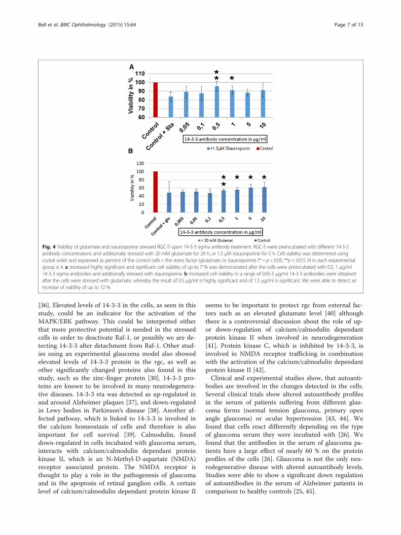
Fig. 4 Viability of glutamate and staurosporine stressed RGC-5 upon 14-3-3 sigma antibody treatment. RGC-5 were preincubated with different 14-3-3antibody concentrations and additionally stressed with 20 mM glutamate for 24 h, or 1.5 μM stauorsporine for 5 h. Cell viability was determined usingcrystal violet and expressed as percent of the control cells + the stress factor (glutamate or staurosporine) (* = p< 0.05; **p< 0.01). N in each experimentalgroup is 4. a: Increased highly significant and significant cell viability of up to 7 % was demonstrated after the cells were preincubated with 0.5, 1 μg/ml14-3-3 sigma antibodies and additionally stressed with staurosporine. b: Increased cell viability in a range of 0.05-5 μg/ml 14-3-3 antibodies were obtainedafter the cells were stressed with glutamate, whereby the result of 0.5 μg/ml is highly significant and of 1.5 μg/ml is significant. We were able to detect anincrease of viability of up to 12 %
Bell et al. BMC Ophthalmology (2015) 15:64 Page 7 of 13
[36]. Elevated levels of 14-3-3 in the cells, as seen in thisstudy, could be an indicator for the activation of theMAPK/ERK pathway. This could be interpreted eitherthat more protective potential is needed in the stressedcells in order to deactivate Raf-1, or possibly we are de-tecting 14-3-3 after detachment from Raf-1. Other stud-ies using an experimental glaucoma model also showedelevated levels of 14-3-3 protein in the rgc, as well asother significantly changed proteins also found in thisstudy, such as the zinc-finger protein [30]. 14-3-3 pro-teins are known to be involved in many neurodegenera-tive diseases. 14-3-3 eta was detected as up-regulated inand around Alzheimer plaques [37], and down-regulatedin Lewy bodies in Parkinson’s disease [38]. Another af-fected pathway, which is linked to 14-3-3 is involved inthe calcium homeostasis of cells and therefore is alsoimportant for cell survival [39]. Calmodulin, founddown-regulated in cells incubated with glaucoma serum,interacts with calcium/calmodulin dependant proteinkinase II, which is an N-Methyl-D-aspartate (NMDA)receptor associated protein. The NMDA receptor isthought to play a role in the pathogenesis of glaucomaand in the apoptosis of retinal ganglion cells. A certainlevel of calcium/calmodulin dependant protein kinase II
seems to be important to protect rgc from external fac-tors such as an elevated glutamate level [40] althoughthere is a controversial discussion about the role of up-or down-regulation of calcium/calmodulin dependantprotein kinase II when involved in neurodegeneration[41]. Protein kinase C, which is inhibited by 14-3-3, isinvolved in NMDA receptor trafficking in combinationwith the activation of the calcium/calmodulin dependantprotein kinase II [42].Clinical and experimental studies show, that autoanti-
bodies are involved in the changes detected in the cells.Several clinical trials show altered autoantibody profilesin the serum of patients suffering from different glau-coma forms (normal tension glaucoma, primary openangle glaucoma) or ocular hypertension [43, 44]. Wefound that cells react differently depending on the typeof glaucoma serum they were incubated with [26]. Wefound that the antibodies in the serum of glaucoma pa-tients have a large effect of nearly 60 % on the proteinprofiles of the cells [26]. Glaucoma is not the only neu-rodegenerative disease with altered autoantibody levels.Studies were able to show a significant down regulationof autoantibodies in the serum of Alzheimer patients incomparison to healthy controls [25, 45].

Fig. 5 Expression of 14-3-3 sigma in RGC-5 revealed by indirect immunfluorescence. RGC-5 cells were fixed, permeabilised, blocked and incubatedwith rabbit polyclonal anti 14-3-3 antibodies. Subsequently the cells were incubated with Goat polyclonal secondary antibody to rabbit IgG-H&Lconjugated with FITC. The cells were visualized with a fluorescence microscope. The pictures show that 14-3-3 was expressed in all cells and itseems to be distributed in the cytoplasm
Bell et al. BMC Ophthalmology (2015) 15:64 Page 8 of 13
The effect of one of the down-regulated antibodieswas tested within this study and we found that low con-centrations of 14-3-3 abs had protective effects on neu-roretinal cells.Our proteomic measurements show that these effects
are mediated via the mitochondrial apoptosis pathway.The proteins BAX, BIRC6. PRFA2, S100A4, VDAC 1/2/3and ERK1 where regulated in a significantly differentway in cells incubated with the abs and are involved inthe regulation of the intrinsic apoptosis pathway (Fig. 7b).Pro-apoptotic BAX, a member of the Bcl-2 family, wasdown-regulated in cells incubated with 14-3-3 abs. Itplays an important role in the intrinsic apoptotic path-way by binding mitochondrial VDAC which leads to therelease of cytochrome c and finally to the initiation ofapoptosis [46]. BAX, as well as its transcription factorp53, are associated with neurodegenerative diseases [47].Deficiency of BAX in DBA/2 J mice protects rgc fromcell death [48]. It also is increased in a mouse model ofglaucoma [49]. Tumour suppressor p53 is a key regula-tor of apoptosis and also plays a role in glaucoma [50].14-3-3 interacts with p53 and positively regulates itstranscriptional activity. It also stabilizes p53 and is in-volved in the nuclear export and modulation of tumoursuppressing activity [51]. Other proteins also were regu-lated in an anti-apoptotic manner such as S1004 andPRAF2 which were down-regulated in this study. Theyboth can lead to apoptosis [52, 53]. The anti-apoptotic
protein BIRC6 belongs to the inhibitor of apoptosis(IAP) family and was up-regulated in cells incubatedwith 14-3-3 antibodies. BIRC6 is up-regulated in tu-mours and inhibits active caspase 3 through bindingwith its BIR domain [54].Additionally an increased expression of ERK was
found in 14-3-3 ab incubated cells. Activated ERK1 isable to phosphorylate many cytoplasmic as well as nu-clear targets, which leads to cell proliferation and differ-entiation [55] and participates in mitochondrialapoptosis [56, 57]. Studies with experimental rat glau-coma models show that the activation of ERK leads toincreased survival of rgc after ocular hypertension sur-gery [58]. Staurosporine leads to changes in the phos-phorylation as well as activation status of ERK [59]. Theincreased ERK expression in the cells incubated with 14-3-3 antibodies could be the link to the protective effectof the 14-3-3 antibody on staurosporine stressed cells.Furthermore, increased ERK1 expression could result inphosphorylation of BAD [60, 61] and provide survival ofstressed RGC-5. This could be the mechanism for theincreased viability and reduced ROS levels of the cellsstressed with H2O2. Cellular H2O2 stress induces ele-vated ROS expression, which increases the expression ofJNK-mitogen activated protein kinase (MAPK). JNK it-self leads to caspase activation, which contributes toapoptosis of cells [62]. JNK activation also leads to phos-phorylation of 14-3-3 and this consequently promotes
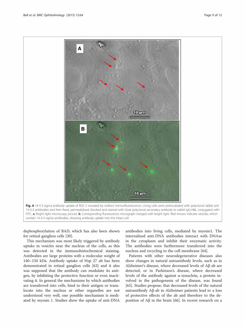
Fig. 6 14-3-3 sigma antibody uptake of RGC-5 revealed by indirect immunfluorescence. Living cells were preincubated with polyclonal rabbit anti14-3-3 antibodies and then fixed, permeabilised, blocked and stained with Goat polyclonal secondary antibody to rabbit IgG-H&L conjugated withFITC. a: Bright light microscopy picture. b: Corresponding fluorescence micrograph merged with bright light. Red Arrows indicate vesicles, whichcontain 14-3-3 sigma antibodies, showing antibody uptake into the intact cell
Bell et al. BMC Ophthalmology (2015) 15:64 Page 9 of 13
dephosphorylation of BAD, which has also been shownfor retinal ganglion cells [30].This mechanism was most likely triggered by antibody
uptake in vesicles near the nucleus of the cells, as thiswas detected in the immunohistochemical staining.Antibodies are large proteins with a molecular weight of140–150 kDa. Antibody uptake of Hsp 27 ab has beendemonstrated in retinal ganglion cells [63] and it alsowas supposed that the antibody can modulate its anti-gen, by inhibiting the protective function or even inacti-vating it. In general the mechanisms by which antibodiesare transferred into cells, bind to their antigen or trans-locate into the nucleus or other organelles are notunderstood very well, one possible mechanism is medi-ated by myosin 1. Studies show the uptake of anti-DNA
antibodies into living cells, mediated by myosin1. Theinternalized anti-DNA antibodies interact with DNAsein the cytoplasm and inhibit their enzymatic activity.The antibodies were furthermore transferred into thenucleus and recycling to the cell membrane [64].Patients with other neurodegenerative diseases also
show changes in natural autoantibody levels, such as inAlzheimer’s disease, where decreased levels of Aβ-ab aredetected, or in Parkinson’s disease, where decreasedlevels of the antibody against α-synuclein, a protein in-volved in the pathogenesis of the disease, was found[65]. Studies propose, that decreased levels of the naturalautoantibody Aβ-ab in Alzheimer patients lead to a lossof protective effects of the ab and therefore to the de-position of Aβ in the brain [66]. In recent research on a

Fig. 7 (See legend on next page.)
Bell et al. BMC Ophthalmology (2015) 15:64 Page 10 of 13
(See figure on previous page.)Fig. 7 Changed mitochondrial apoptosis pathway in RGC-5 conditioned of treatment with 14-3-3 sigma antibodies. a: This graph shows severalof the significantly differently regulated proteins in the cells incubated with 14-3-3 antibodies in comparison to control cells. The proteins listedhere in some way all are involved in the mitochondrial apoptosis pathway. The changes of the proteins are shown in percent. b: This graphschematically visualizes the significantly changed proteins of the mitochondrial apoptosis pathway. Proteins highlighted in green were found tobe significantly down-regulated) and proteins highlighted in red significantly up-regulated. The protein 14-3-3 interacts with p53, which is alsoshown in the graph. It is conceivable that the modulation of 14-3-3 through 14-3-3 sigma antibodies leads to a changed interaction of p53 andthereby to changed expression of p53 target genes such as PRAF2, S100A4 and BAX, which are significantly changed in the cells incubated with14-3-3 abs. BAX plays a role in releasing cytochrome c from the mitochondrion. Cytochrome C interacts with caspase3, which triggers apoptosisof the cells. It also is conceivable that the interaction of 14-3-3 and STAT3 is altered, which comes to the up-regulation of BIRC6. Another proteinwhich plays also a role in mitochondrial apoptosis is the anti-apoptotic ERK1, which is up-regulated in 14-3-3 sigma treated RGC-5 as well thedown-regulation of VDAC 1/2/3
Bell et al. BMC Ophthalmology (2015) 15:64 Page 11 of 13
new Alzheimer’s therapy, promising studies have beenperformed with intravenous Immunoglobulin’s (Ig) show-ing a reduction of plasma concentration of Aβ1–42 [67].These results underline our hypothesis, that changes
found in the natural autoimmunity of patients sufferingfrom neurodegenerative diseases such as Alzheimer’s orglaucoma have an impact on the pathogenesis of the dis-ease, showing that also the down-regulated autoanti-bodies have an impact on the regulatory functions andtherefore their reduction is most possibly causing vul-nerability to damage. We are aware of the fact thatRGC-5 cells are not a pure retinal ganglion cell line.Nevertheless, a recently published article summarisedthe characteristics of RGC-5 and their usage as a cellmodel line. The authors stated that the majority of thepublished articles characterising RGC-5 cells showedBrn3, as well as Thy 1 staining, whereas only two paperswere not able to detect these markers. Furthermore nes-tin expression was detected in RGC-5 cells, leading tothe conclusion that these cells are of neuronal origin.The authors conclude that RGC-5 cells can act as retinalcell line of neuronal origin in order to follow up initialhypothesis [27].
ConclusionIn conclusion we were able to show changes in the pro-tein profiles of the cells incubated with POAG serum,especially of proteins involved in regulatory cell mecha-nisms. In a further step we were able to demonstratethat 14-3-3 abs, which are down-regulated in glaucomapatients have protective effects on RGC-5 cells and aretransported into the cells. We were able to detect severalproteins changed in an anti-apoptotic manner in thosecells incubated with 14-3-3 abs. Altogether these resultsunderline our hypothesis that the changes in the naturalautoimmunity found in the serum of patients with theneurodegenerative disease glaucoma play a role in thedisease pathogenesis. These results can be seen in ac-cordance with results derived from studies performedfor Alzheimer’s disease. We believe that by losing regula-tory effects of down-regulated autoantibodies, retinalganglion cells could become more vulnerable to other
stress factors and apoptosis of the cells could be pro-voked e.g. by an elevated IOP.
Additional files
Additional file 1: Figure S1. This graph shows the autoantibody levelsof the 14-3-3 antibody in the serum of glaucoma patients in comparison tohealthy subjects (n = 45). Microarray meassurements of the sera wereperformed. Glas slides coated with nitrocellulose were spotted with differentantigens, also including the 14-3-3 antigen. Then the slides were incubatedwith the sera of either healthy people or patients suffering from primaryopen angle glaucoma. The antibodies in the serum bind to their antigen.The bound antibodies are then detected using a secondary antibody to IgG(bound with Cy3). Using a Affymetrix Array scanner, the spots are scanned.The intensity of the bound antibody is analyzed using the intensity of thelabelling of the secondary antibody and a comparison between healty andglaucomatous serum is performed. A discrimance analysis was performed inorder to detect significant differences. Shown ist the mean of the Intensity(U) with the standard error. We were able to detect a significantly lowerlevel of 14-3-3 antibody in the serum of POAG patients (p < 0.01).
Additional file 2: Figure S2. RGC-5 were preincubated with differentanti-myoglobin antibody concentrations and additionally stressed with20 mM glutamate for 24 h, or 1.5 μM stauorsporine for 5 h. Cell viability wasdetermined using crystal violet and expressed as percent of the controlcells + the stress factor (glutamate or staurosporine) (* = p < 0.05; **p < 0.01).This graph shows the results of the cells stressed with glutamate. Noprotective effect of the antibody can be seen.
Additional file 3: Figure S3. Non-stressed RGC-5 cells were incubatedwith the different 14-3-3 ab concentrations (0.05, 0.1, 0.5, 1, 5 and 10 μg/ml). Viability was measured with crystal violet. We were not able to detectany significant changes in the viability of the cells. The graph shows thedifferent cell groups on the X- axis and the viability in % on the Y- axis.
Additional file 4: Table S1 We were able to identify 1204 proteins inthe cells. 225 of the proteins were significantly differently regulated inthe cells incubated with 14-3-3 antibody in comparison to control cells(>2 fold increased or <2 fold decreased). The significantly changedproteins are listed in Additional file 4: Table S1.
AbbreviationsRgc: Retinal ganglion cell; BAX: bcl-2 associated- x-protein; BIRC6: BaculoviralIAP repeat containing 6; S100A4: S100 calcium binding protein A4;PRAF2: PRA1 family protein 2; VDAC 1/2/3: Voltage dependent anionchannel; ERK1: Extracellular regulated protein kinase; ROS: Reactive oxygenspecies; hsp: Heat shock protein; PBS: Phosphate buffered saline, DCFH-DA,2’,7’-dichlorodihydrofluorescein-diacetate; DCFH: 2’, 7’dichlorodihydrofluorescein; DCF: 2’,7’-dichlorfluorescein; CDS: Cell dissociationsolution; CAN: Acetonitril; AB: Ammoniumbicarbonate; IPA: Ingenuitypathway analysis; IAP: Inhibitor of apoptosis protein; RSK: 90 kDa ribosomalS6 kinase.
Competing interestsThe authors declare that they have no competing interests.

Bell et al. BMC Ophthalmology (2015) 15:64 Page 12 of 13
Authors’ contributionsKB designed the study, carried out the incubation of the cells with serum ofPOAG patients, performed statistical analysis, interpreted the data and drafted themanuscript. CW performed and designed the incubation of the cells withthe 14-3-3 antibody, performed the statistical analysis. SF carried out themass spectrometric measurements, and made substantial contributions tointerpretation of the data. NP conceived of the study, contributed toacquisition of the data. FG made substantial contributions to conception ofthe study and revised the manuscript. All authors read and approved thefinal version manuscript.
Authors’ informationNP is Head of Department of the Clinics for Ophthalmology of the Medicalcenter of the Johannes Gutenberg University Mainz.FG is Head of Department of the Experimental Ophthalmology within theClinics of Ophthalmology of the Medical center of the Johannes GutenbergUniversity Mainz.
AcknowledgementForschungsschwerpunkt Translationale Neurowissenschaften (FTN) of theJohannes Gutenberg-University, Mainz, Germany and Stiftung Rheinland-Pfalzfür Innovation“funded by the German State Rhineland-Palatinate.
Received: 8 September 2014 Accepted: 20 May 2015
References1. Przedborski S, Vila M, Jackson-Lewis V. Neurodegeneration: what is it and
where are we? J Clin Invest. 2003;111(1):3–10.2. Quigley HA. Neuronal death in glaucoma. Prog Retin Eye Res. 1999;18(1):39–57.3. Weber AJ, Chen H, Hubbard WC, Kaufman PL. Experimental glaucoma and
cell size, density, and number in the primate lateral geniculate nucleus.Invest Ophthalmol Vis Sci. 2000;41(6):1370–9.
4. Quigley HA. Glaucoma. Lancet. 2011;377(9774):1367–77.5. Gupta N, Yucel YH. Glaucoma as a neurodegenerative disease. Curr Opin
Ophthalmol. 2007;18(2):110–4.6. Quigley HA. Medical progress - open-angle glaucoma. N Engl J Med.
1993;328(15):1097–106.7. Quigley HA, Broman AT. The number of people with glaucoma worldwide
in 2010 and 2020. Br J Ophthalmol. 2006;90(3):262–7.8. Coleman AL, Kodjebacheva G. Risk factors for glaucoma needing more
attention. Open Ophthalmol J. 2009;3:38–42.9. Mizoguchi T, Ozaki M, Wakiyama H, Ogino N. Additive intraocular pressure-
lowering effect of dorzolamide 1 %/timolol 0.5 % fixed combination onprostaglandin monotherapy in patients with normal tension glaucoma. ClinOphthalmol. 2011;5:1515–20.
10. Nickells RW. From ocular hypertension to ganglion cell death: a theoreticalsequence of events leading to glaucoma. Can J Ophthalmol.2007;42(2):278–87.
11. Garcia-Valenzuela E, Shareef S, Walsh J, Sharma SC. Programmed cell deathof retinal ganglion cells during experimental glaucoma. Exp Eye Res.1995;61(1):33–44.
12. Ferreira SM, Lerner SF, Brunzini R, Evelson PA, Llesuy SF. Oxidative stressmarkers in aqueous humor of glaucoma patients. Am J Ophthalmol.2004;137(1):62–9.
13. Izzotti A, Sacca SC, Cartiglia C, De Flora S. Oxidative deoxyribonucleic aciddamage in the eyes of glaucoma patients. Am J Med. 2003;114(8):638–46.
14. Dreyer EB, Grosskreutz CL. Excitatory mechanisms in retinal ganglion celldeath in primary open angle glaucoma (POAG). Clin Neurosci.1997;4(5):270–3.
15. Dreyer EB, Zurakowski D, Schumer RA, Podos SM, Lipton SA. Elevatedglutamate levels in the vitreous body of humans and monkeys withglaucoma. Arch Ophthalmol. 1996;114(3):299–305.
16. Wax MB, Tezel G, Saito I, Gupta RS, Harley JB, Li Z, et al. Anti-Ro/SS-Apositivity and heat shock protein antibodies in patients with normal-pressure glaucoma. Am J Ophthalmol. 1998;125(2):145–57.
17. Ikeda Y, Maruyama I, Nakazawa M, Ohguro H. Clinical significance of serumantibody against neuron-specific enolase in glaucoma patients. Jpn JOphthalmol. 2002;46(1):13–7.
18. Reichelt J, Joachim SC, Pfeiffer N, Grus FH. Analysis of autoantibodiesagainst human retinal antigens in sera of patients with glaucoma andocular hypertension. Curr Eye Res. 2008;33(3):253–61.
19. Tezel G, Edward DP, Wax MB. Serum autoantibodies to optic nerve headglycosaminoglycans in patients with glaucoma. Arch Ophthalmol.1999;117(7):917–24.
20. Grus FH, Joachim SC, Hoffmann EM, Pfeiffer N. Complex autoantibodyrepertoires in patients with glaucoma. Mol Vis. 2004;10:132–7.
21. Lacroix-Desmazes S, Kaveri SV, Mouthon L, Ayouba A, Malanchere E,Coutinho A, et al. Self-reactive antibodies (natural autoantibodies) in healthyindividuals. J Immunol Methods. 1998;216(1–2):117–37.
22. Avrameas S. Natural autoantibodies: from ‘horror autotoxicus’ to ‘gnothiseauton’. Immunol Today. 1991;12(5):154–9.
23. Poletaev A, Osipenko L. General network of natural autoantibodies asimmunological homunculus (Immunculus). Autoimmun Rev. 2003;2(5):264–71.
24. Meriggioli MN. Myasthenia gravis with anti-acetylcholine receptorantibodies. Front Neurol Neurosci. 2009;26:94–108.
25. Brettschneider S, Morgenthaler NG, Teipel SJ, Fischer-Schulz C, Burger K, DodelR, et al. Decreased serum amyloid beta (1–42) autoantibody levels inAlzheimer’s disease, determined by a newly developed immuno-precipitationassay with radiolabeled amyloid beta (1–42) peptide. Biol Psychiatry.2005;57(7):813–6.
26. Bell K, Funke S, Pfeiffer N, Grus FH. Serum and antibodies of glaucomapatients lead to changes in the proteome, especially cell regulatoryproteins, in retinal cells. PLoS One. 2012;7(10):e46910.
27. Sippl C, Tamm ER. What is the nature of the RGC-5 cell line? Adv Exp MedBiol. 2014;801:145–54.
28. Lowry OH, Rosebrough NJ, Farr AL, Randall RJ. Protein measurement withthe Folin phenol reagent. J Biol Chem. 1951;193(1):265–75.
29. Funke S, Azimi D, Wolters D, Grus FH, Pfeiffer N. Longitudinal analysis oftaurine induced effects on the tear proteome of contact lens wearers anddry eye patients using a RP-RP-Capillary-HPLC-MALDI TOF/TOF MSapproach. J Proteomics. 2012;75(11):3177–90.
30. Yang X, Luo C, Cai J, Pierce WM, Tezel G. Phosphorylation-dependentinteraction with 14-3-3 in the regulation of bad trafficking in retinalganglion cells. Invest Ophthalmol Vis Sci. 2008;49(6):2483–94.
31. Steinacker P, Aitken A, Otto M. 14-3-3 proteins in neurodegeneration. SeminCell Dev Biol. 2011;22(7):696–704.
32. Xing H, Zhang S, Weinheimer C, Kovacs A, Muslin AJ. 14-3-3 proteins blockapoptosis and differentially regulate MAPK cascades. EMBO J.2000;19(3):349–58.
33. Chen L, Liu L, Yin J, Luo Y, Huang S. Hydrogen peroxide-induced neuronalapoptosis is associated with inhibition of protein phosphatase 2A and 5,leading to activation of MAPK pathway. Int J Biochem Cell Biol.2009;41(6):1284–95.
34. Kim EK, Choi EJ. Pathological roles of MAPK signaling pathways in humandiseases. Biochim Biophys Acta. 1802;4:396–405.
35. Dhillon AS, Yip YY, Grindlay GJ, Pakay JL, Dangers M, Hillmann M, et al. TheC-terminus of Raf-1 acts as a 14-3-3-dependent activation switch. Cell Signal.2009;21(11):1645–51.
36. Kyriakis JM, Avruch J. Mammalian mitogen-activated protein kinase signaltransduction pathways activated by stress and inflammation. Physiol Rev.2001;81(2):807–69.
37. Gozal YM, Duong DM, Gearing M, Cheng D, Hanfelt JJ, Funderburk C, et al.Proteomics analysis reveals novel components in the detergent-insolublesubproteome in Alzheimer’s disease. J Proteome Res. 2009;8(11):5069–79.
38. Shimada T, Fournier AE, Yamagata K. Neuroprotective function of 14-3-3proteins in neurodegeneration. Biomed Res Int. 2013;2013:564534.
39. Yamniuk AP, Vogel HJ. Calmodulin’s flexibility allows for promiscuity in itsinteractions with target proteins and peptides. Mol Biotechnol.2004;27(1):33–57.
40. Fan W, Li X, Cooper NG. CaMKIIalphaB mediates a survival response inretinal ganglion cells subjected to a glutamate stimulus. Invest OphthalmolVis Sci. 2007;48(8):3854–63.
41. Ashpole NM, Hudmon A. Excitotoxic neuroprotection and vulnerability withCaMKII inhibition. Mol Cell Neurosci. 2011;46(4):720–30.
42. Yan JZ, Xu Z, Ren SQ, Hu B, Yao W, Wang SH, et al. Protein kinase Cpromotes N-methyl-D-aspartate (NMDA) receptor trafficking by indirectlytriggering calcium/calmodulin-dependent protein kinase II (CaMKII)autophosphorylation. J Biol Chem.2011;286(28):25187–200.

Bell et al. BMC Ophthalmology (2015) 15:64 Page 13 of 13
43. Joachim SC, Pfeiffer N, Grus FH. Autoantibodies in patients with glaucoma: acomparison of IgG serum antibodies against retinal, optic nerve, and opticnerve head antigens. Graefes Arch Clin Exp Ophthalmol. 2005;243(8):817–23.
44. Joachim SC, Reichelt J, Berneiser S, Pfeiffer N, Grus FH. Sera of glaucomapatients show autoantibodies against myelin basic protein and complexautoantibody profiles against human optic nerve antigens. Graefes ArchClin Exp Ophthalmol. 2008;246(4):573–80.
45. Britschgi M, Olin CE, Johns HT, Takeda-Uchimura Y, LeMieux MC, Rufibach K,et al. Neuroprotective natural antibodies to assemblies of amyloidogenicpeptides decrease with normal aging and advancing Alzheimer’s disease.Proc Natl Acad Sci U S A. 2009;106(29):12145–50.
46. Kumarswamy R, Chandna S. Putative partners in Bax mediated cytochrome-crelease: ANT, CypD, VDAC or none of them? Mitochondrion. 2009;9(1):1–8.
47. Nickells RW. Apoptosis of retinal ganglion cells in glaucoma: an update ofthe molecular pathways involved in cell death. Surv Ophthalmol. 1999;43Suppl 1:S151–61.
48. Libby RT, Li Y, Savinova OV, Barter J, Smith RS, Nickells RW, et al.Susceptibility to neurodegeneration in a glaucoma is modified by Bax genedosage. PLoS Genet. 2005;1(1):17–26.
49. Ji J, Chang P, Pennesi ME, Yang Z, Zhang J, Li D, et al. Effects of elevatedintraocular pressure on mouse retinal ganglion cells. Vision Res.2005;45(2):169–79.
50. Yuan S, Fu Y, Wang X, Shi H, Huang Y, Song X, et al. Voltage-dependentanion channel 1 is involved in endostatin-induced endothelial cellapoptosis. FASEB J. 2008;22(8):2809–20.
51. Yang HY, Wen YY, Chen CH, Lozano G, Lee MH. 14-3-3 sigma positivelyregulates p53 and suppresses tumor growth. Mol Cell Biol.2003;23(20):7096–107.
52. Grigorian M, Andresen S, Tulchinsky E, Kriajevska M, Carlberg C, Kruse C, et al.Tumor suppressor p53 protein is a new target for the metastasis-associatedMts1/S100A4 protein: functional consequences of their interaction. J BiolChem. 2001;276(25):22699–708.
53. Wolter KG, Hsu YT, Smith CL, Nechushtan A, Xi XG, Youle RJ. Movement ofBax from the cytosol to mitochondria during apoptosis. J Cell Biol.1997;139(5):1281–92.
54. Bartke T, Pohl C, Pyrowolakis G, Jentsch S. Dual role of BRUCE as anantiapoptotic IAP and a chimeric E2/E3 ubiquitin ligase. Mol Cell.2004;14(6):801–11.
55. Davis RJ. The mitogen-activated protein kinase signal transduction pathway.J Biol Chem. 1993;268(20):14553–6.
56. Fisher TL, Blenis J. Evidence for two catalytically active kinase domains inpp90rsk. Mol Cell Biol. 1996;16(3):1212–9.
57. Dalby KN, Morrice N, Caudwell FB, Avruch J, Cohen P. Identification ofregulatory phosphorylation sites in mitogen-activated protein kinase(MAPK)-activated protein kinase-1a/p90rsk that are inducible by MAPK. J BiolChem. 1998;273(3):1496–505.
58. Abu-Hamad S, Sivan S, Shoshan-Barmatz V. The expression level of thevoltage-dependent anion channel controls life and death of the cell. ProcNatl Acad Sci U S A. 2006;103(15):5787–92.
59. Antonsson A, Persson JL. Induction of apoptosis by staurosporine involvesthe inhibition of expression of the major cell cycle proteins at the G (2)/mcheckpoint accompanied by alterations in Erk and Akt kinase activities.Anticancer Res. 2009;29(8):2893–8.
60. Shimamura A, Ballif BA, Richards SA, Blenis J. Rsk1 mediates a MEK-MAPkinase cell survival signal. Curr Biol. 2000;10(3):127–35.
61. Zha J, Harada H, Yang E, Jockel J, Korsmeyer SJ. Serine phosphorylation ofdeath agonist BAD in response to survival factor results in binding to 14-3-3not BCL-X (L). Cell. 1996;87(4):619–28.
62. Chun HS, Gibson GE, DeGiorgio LA, Zhang H, Kidd VJ, Son JH.Dopaminergic cell death induced by MPP (+), oxidant and specificneurotoxicants shares the common molecular mechanism. J Neurochem.2001;76(4):1010–21.
63. Tezel G, Wax MB. The mechanisms of hsp27 antibody-mediated apoptosisin retinal neuronal cells. J Neurosci. 2000;20(10):3552–62.
64. Yanase K, Smith RM, Puccetti A, Jarett L, Madaio MP. Receptor-mediatedcellular entry of nuclear localizing anti-DNA antibodies via myosin 1. J ClinInvest. 1997;100(1):25–31.
65. Besong-Agbo D, Wolf E, Jessen F, Oechsner M, Hametner E, Poewe W, et al.Naturally occurring alpha-synuclein autoantibody levels are lower in patientswith Parkinson disease. Neurology. 2013;80(2):169–75.
66. Magga J, Puli L, Pihlaja R, Kanninen K, Neulamaa S, Malm T, et al. Humanintravenous immunoglobulin provides protection against Abeta toxicity bymultiple mechanisms in a mouse model of Alzheimer’s disease. JNeuroinflammation. 2010;7:90.
67. Dodel R, Rominger A, Bartenstein P, Barkhof F, Blennow K, Forster S, et al.Intravenous immunoglobulin for treatment of mild-to-moderate Alzheimer’sdisease: a phase 2, randomised, double-blind, placebo-controlled, dose-finding trial. Lancet Neurol. 2013;12(3):233–43.
Submit your next manuscript to BioMed Centraland take full advantage of:
• Convenient online submission
• Thorough peer review
• No space constraints or color figure charges
• Immediate publication on acceptance
• Inclusion in PubMed, CAS, Scopus and Google Scholar
• Research which is freely available for redistribution
Submit your manuscript at www.biomedcentral.com/submit