Pneumocystis carinii Presented by: Samantha Todd & Sandra Thorbus.
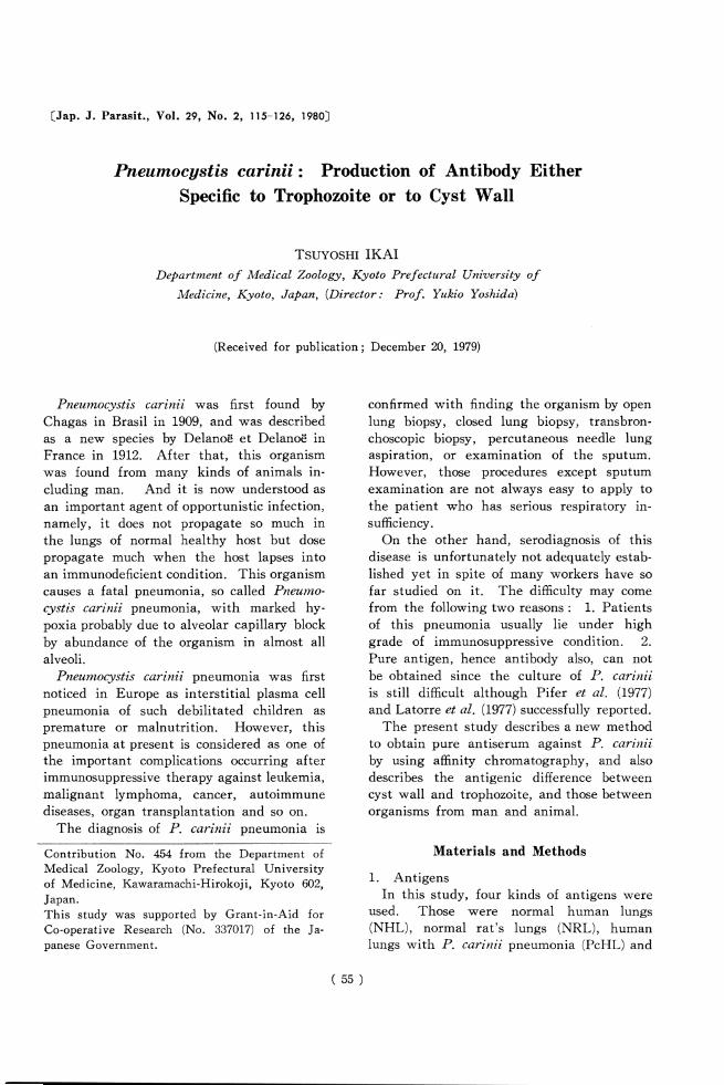
CJap. J. Parasit., Vol. 29, No. 2, 115-126, 1980]
Pneumocystis carinii: Production of Antibody Either
Specific to Trophozoite or to Cyst Wall
Tsuyoshi IKAI
Department of Medical Zoology, Kyoto Prefectural University of
Medicine, Kyoto, Japan, (Director: Prof. Yukio Yoshidd)
(Received for publication; December 20, 1979)
Pneumocystis carinii was first found by
Chagas in Brasil in 1909, and was described
as a new species by Delanoe et Delanoe in
France in 1912. After that, this organism
was found from many kinds of animals in
cluding man. And it is now understood as
an important agent of opportunistic infection,
namely, it does not propagate so much in
the lungs of normal healthy host but dose
propagate much when the host lapses into
an immunodeficient condition. This organism
causes a fatal pneumonia, so called Pneumo
cystis carinii pneumonia, with marked hy-
poxia probably due to alveolar capillary block
by abundance of the organism in almost all
alveoli.
Pneumocystis carinii pneumonia was first
noticed in Europe as interstitial plasma cell
pneumonia of such debilitated children as
premature or malnutrition. However, this
pneumonia at present is considered as one of
the important complications occurring after
immunosuppressive therapy against leukemia,
malignant lymphoma, cancer, autoimmune
diseases, organ transplantation and so on.
The diagnosis of P. carinii pneumonia is
Contribution No. 454 from the Department of
Medical Zoology, Kyoto Prefectural University
of Medicine, Kawaramachi-Hirokoji, Kyoto 602,
Japan.
This study was supported by Grant-in-Aid for
Co-operative Research (No. 337017) of the Ja
panese Government.
confirmed with finding the organism by open
lung biopsy, closed lung biopsy, transbron-
choscopic biopsy, percutaneous needle lung
aspiration, or examination of the sputum.
However, those procedures except sputum
examination are not always easy to apply to
the patient who has serious respiratory in
sufficiency.
On the other hand, serodiagnosis of this
disease is unfortunately not adequately estab
lished yet in spite of many workers have so
far studied on it. The difficulty may come
from the following two reasons : 1. Patients
of this pneumonia usually lie under high
grade of immunosuppressive condition. 2.
Pure antigen, hence antibody also, can not
be obtained since the culture of P. carinii
is still difficult although Pifer et al. (1977)
and Latorre et al. (1977) successfully reported.
The present study describes a new method
to obtain pure antiserum against P. carinii
by using affinity chromatography, and also
describes the antigenic difference between
cyst wall and trophozoite, and those between
organisms from man and animal.
Materials and Methods
1. Antigens
In this study, four kinds of antigens were
used. Those were normal human lungs
(NHL), normal rat's lungs (NRL), human
lungs with P. carinii pneumonia (PcHL) and
(55)
116
rat's lungs with P. carinii dominant infec
tion (PcRL).
NHL were obtained from legal autopsies.
Only lungs with neither any pathological
changes nor any pathogens were used for
the experiment. PcHL were obtained in
pathological autopsies from the patients who
died by apparent P. carinii pneumonia in
the course of immunosuppressive therapy
against their underlying diseases. The lungs
with other pathogens like bacteria and fungi,
were also excluded from the material.
The rats used in the present study were all
Wistar-strain young adults weighing around
200 g. NRL were obtained from healthy rats
as soon as they arrived us from an animal
dealer, and were checked the absence of P.
carinii and other pathogens.
PcRL were obtained from P. carinii pro-
vocated rats by administration of cortisone
acetate 25 mg twice a week and 0.05 % solu
tion of tetracycline hydrochloride in drinking
water daily for seven to ten weeks. Those
lungs were stored in a deep freezer at — 70 C
until use.
Cysts of P. carinii were prepared from
human and rat's lungs by cyst purification
method (Ikai et al., 1979).
2. Extraction of antigens
Each of the four kinds of lung materials
stored at —70 C, was thawed under room
temperature at use. About 30 g to 50 g of
the lungs were cut into pieces of about 1 cm3
in size, and were lyophilized. Dried lungs
were powdered in a bowel, then about 100
ml of 0.1% NaCl were added. This turbid
fluid was frozen then smashed at room tem
perature until it thawed. This freeze and
smash procedure was repeated ten times.
Although the trophic stages of the organism
were almost completely broken by this proce
dure, the cystic stages were usually remained
without change. The fluid was then cen-
trifuzed at 10,000 rpm for 30 minutes at 4 C.
The supernate was dialyzed against distilled
water overnight at 4 C, and was lyophilized.
The crude antigens (NHL-extract, PcHL-ext.,
NRL-ext. and PcRL-ext.) thus obtained were
stored in a desiccator.
3. Immunization of rabbits
Nine male, white rabbits weighing 2.5±
0.3 kg were divided into three groups (L 2
and 3) of three each. Group 1 was immu
nized with the emulsion of 3 mg NHL-ext.
powder in 0.5 ml distilled water and 0.5 ml
Freund's complete adjuvant. The emulsion
was subcutaneously given, dividing in 0.1-
0.2 ml, to the back of the rabbits, once a
week, for 10 weeks. Group 2 was immu
nized with the emulsion of 3 mg PcHL-ext.
powder and Freund's complete adjuvant as
the same manner as group 1. Group 3 was
immunized with the emulsion of 106 cysts in
0.5 ml distilled water and 0.5 ml Freund's
complete adjuvant by the same way as group
1 and 2.
In all groups, blood samples were collected
from the auricular vein one week after the
tenth injection. In order to collect antisera
as much as possible, further booster injec
tions with the same manner and blood col
lections were repeated 4 to 5 times with 1
week interval in each procedure. Those sera
were all stored at —20 C until use.
4. Combination of NHL-ext. with AH-Se
pharose 4B
AH-Sepharose 4B (Pharmacia Fine Chemi
cals) was activated by the method of Cam-
biaso et al. (1975). Fifteen grams of AH-
Sepharose 4 B were swollen in 200 to 300 ml
of distilled water for 10 to 15 minutes with
gentle stir. After removing the excess water
and washing with 2 to 3 liters of 0.5 M Na-
HCO3 through glassfilter, AH-Sepharose 4B
was put in a beaker. Then, 140 ml of 0.5 M
NaHCOs and 20 ml of 25% glutaraldehyde
(final concentration 2.5%) were added to it.
This gel was stirred gently for 10 to 15
minutes at room temperature until gel be
came yellowish. After that, glutaraldehyde
was removed through glassfilter, and the
activated AH-Sepharose 4B was washed with
2 to 3 liters of 0.5 M NaHCO3.
The activated AH-Sepharose 4B was again
put in a beaker, and about 100 ml of 0.5 M
NaHCO3 containing 2g of NHL-ext. was
added to it. This mixture was stirred gently
at 4 C overnight. After removing the excess
( 56 )
117
NHL-ext. through glassfilter, NHL-ext.-com
bined AH-Sepharose 4B was washed with 2
to 3 liters of saline. The product thus ob
tained was stored in 300 to 500 ml of saline
containing with 0.02% NaN3, at 4 C until
use.
When the quantity of NHL-ext. is pro
portionally scant to that of activated AH-
Sepharose 4B, it is necessary to block the
uncoupling activated AH-Sepharose 4B with
0.2 M monoethanolamine in 0.1 M sodium
borate buffer (pH 8.5) for 16 hours at 4 C.
However, such blocking procedure is unnec
essary when the quantity of NHL-ext. is
excess, for example 2 g or more as in the
present medium.
5. Purification of antibodies
In order to purify anti-Pc antibody, crude
anti-PcHL-ext. antibody and crude anti-Pc
cyst antibody were treated through affinity
chromatography which contained NHL-ext.-
combined AH-Sepharose 4B. Firstly, f-gk)-
bulin was separated from each antiserum of
rabbits by (NH^SC^ precipitation method.
Each of final precipitate of ^-globulin (mainly
IgG) was resolved in 30 to 50 ml of saline
and was dialyzed against saline at 4 C over
night. Since these f-globulins contain both
anti-Pc antibody and anti-NHL-ext. antibody,
the latter antibody should be removed as the
next step.
Each of those f-globulin solution was treat
ed by repeated passing through NHL-ext. -
combined AH-Sepharose 4B gel in a special
column which had perista mini-pump SJ-1211
type (Atto Corp.) recycle system at 4 C over
night. Then, uncombined ^-globulin was col
lected from the chromatographic glassfilter
tube. NHL-ext.-combined AH-Sepharose 4B
coupled with anti-NHL-ext. antibody was
washed with 1 to 2 liters of saline on glass-
filter, and was put back to the chromato
graphic column again. After that, anti-
NHL-ext. antibody was removed from NHL-
ext.-combined AH-Sepharose 4B by adding
5 to 10 ml of 8M urea. NHL-ext.-combined
AH-Sepharose 4B was finally washed "with
1 to 2 liters of saline. The gel can be used
several times by this regeneration method.
The procedure mentioned above was repeated
six times or more on one sample to obtain
pure antibody. Finally those sera were con
centrated to about 50mg/ml of protein con
tent in saline with minicon B15 system
(Amicon Corp.). They were stored at 4 C
after adding 0.02% NaN3.
Anti-Pc cyst antibody was purified by the
same manner as mentioned above since the
cyst suspension still might have small amount
of host elements even though cysts were
obtained by purification method.
Hereafter, the author abbreviated the puri
fied anti-PcHL-ext. antibody as T antibody
since it is considered to be anti-trophozoite
antibody, and the purified anti-Pc cyst anti
body as C antibody.
6. Techniques for demonstration of anti
bodies
Double diffusion test (DD test) in agar gel
of 0.9% agar in veronal buffer saline (pH
8.6) was performed. Central well of 3 mm
diameter was filled with 5 fil of antibody,
and peripheral wells, 3 mm diameter and 5
mm apart from the center one, were filled
with 5 [A of antigen solutions, each of which
contained 0.5 mg of NHL-ext., PcHL-ext.,
NRL-ext. and PcRL-ext.. The incubation
of the plate was carried out firstly at room
temperature for 24 hours then at 4 C for 2
days both in a moist chamber.
Immunoelectrophoresis (IEP) was perform
ed on 1 % agarose in veronal buffer saline
(pH 8.6). Wells were filled with 0.05 ml of
antigen solution containing 5 mg of NHL-
ext., PcHL-ext., NRL-ext. and PcRL-ext..
After electrophoresis was done by 2 mA/cm
for 90 minutes in the same buffer system,
T antibody was poured into each trough.
Precipitin patterns were allowed to develop
in a moist chamber at room temperature for
24 hours then at 4 C for 2 days.
Indirect fluorescent antibody technique
(IFA) was performed between purified anti
bodies (T and C) and antigens on tissue
sections or purified cysts on smear. For IFA
test on tissue section, Hamashima and Kyo-
goku's method (1965) was mainly used. The
details are as follows : P. carinii infected
( 57 )
118
lungs of rats provocated the organism by
Frenkel's method (1966) for ten weeks, were
cut into 2 to 4 mm3 in size, and were im
mediately fixed in cold 95% ethyl alcohol
at —70 C for one hour then kept at 4 C
overnight. Dehydration with absolute ethyl
alcohol at 4C for 3 days changing alcohol
every day and dealcoholization with xylene
three times at 4 C for 20 minutes each were
performed. The lungs were put into paraffin
three times at 57 C for 5 minutes each then
embedded. The embedded lungs were stored
at — 20 C until use. The tissue sections of
about 5 ft thick were stretched on warm
water, transfered onto a glass slide, and
dryed at 37 C for 30 minutes then in a de
siccator for 15 minutes under room tempera
ture. Deparaffinization with xylene three
times 3 minutes for each, and dexylenization
with absolute ethyl alcohol two times, 90%,
70% and 45% ethyl alcohol one minute for
each were performed. The glass slide was
then moved into cold phosphate buffered
saline (pH 7.2) and washed by changing PBS
four times. One drop of antiserum diluted
to 3 to 5 mg/ml of protein content, was put
on the section, and it was incubated at 37 C
for one hour. A glass slide was washed with
cold PBS several times for at least one hour
at 4 C. Then one drop of fluorescein iso-
thiocyanate conjugated anti-rabbit IgG (Fuji-
zoki Parmaceutical Co.) which contained 11
mg/ml of IgG and 1.3 of fluorescein per pro
tein ratio, was put on the glass slide and
incubated at room temperature for one hour.
The glass slide was washed with cold PBS,
and embedded in 10% glycerin in PBS.
On the other hand, P. carinii cysts ob
tained by cyst purification method (Ikai et al.,
1979) were spread on some glass slides, and
dried quickly. Those slides were fixed in
acetone at 37 C for 10 minutes, then dried
at 37 C for 30 minutes. IFA test was car
ried out by the same way as described above.
Those materials were examined with fluores
cent microscope Model BHF (Olympus Co.).
7. Absorption of purified antisera
Ten mg of NHL-ext. and PcHL-ext. was
added to each of 0.1 ml of purified antiserum
at room temperature for 3 hours followed by
at 4C overnight. Then sera were centri-
fuged at 2,000 rpm for 15 minutes and super-
nates were used in DD test and IEP test.
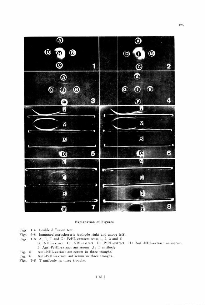
Results
1. Double diffusion test of four kinds of
antigens with anti-NHL-ext. antiserum
or anti-PcHL-ext. antiserum
Prior to use of purified antiserum, anti-
NHL-ext. antiserum or anti-PcHL-ext. anti
serum were attempted to react with extracts
of NHL (B), PcHL (A, E, F, G), NRL (C)
and PcRL (D). Fig. 1 is a pattern resulted
between anti-NHL-ext. antiserum (H) and
four kinds of extracts, showing multiplicity
of precipitin bands in each antigen-antibody
system including faint cross-reactivity be
tween human and rat's lung antigenicity.
The same finding was obtained when anti-
PcHL-ext. antiserum (I) was used. Specific
Pc antigen and antibody reaction seems to
be hidden under the reaction between anti
gen and antibody responsible for the lung
component (Fig. 2).
2. Double diffusion test of four kinds of
antigens with purified T antibody
As mentioned before, T antibody was ob
tained by affinity chromatography by which
the activity of anti-NHL-ext. antibody was
completely absorbed out.
As shown in Fig. 3, single band was pro
duced not only between T antibody (J) and
PcHL-ext. (A), but also between T antibody
and PcRL-ext. (D). And no band was seen
between T antibody and NHL-ext. (B) and
between T antibody and NRL-ext. (C).
Thus, this is evidently resultant of Pc anti
gen-antibody reaction.
3. Double diffusion test of three more hu
man lung extracts of P. carinii pneumo
nia with purified T antibody
Case 1 corresponds to PcHL-ext. (A) men
tioned above. Three other autopsied human
lungs (case 2-4, E, F, G) containing P. carinii
were used for the experiment. As shown
in Fig. 4, a clear single band between each
of antigen and antibody was seen with
( 58 )
119
apparent fusion one another.
From the fact that C antibody did not
show a precipitin band with any of NHL-
ext., PcHL-ext., NRL-ext. and PcRL-ext.,
the antigen and antibody responsible for it
must be derived from the trophozoite (T
antigen and T antibody).
4. Immunoelectrophoresis of four kinds of
antigens with crude and purified antibody
IEP patterns between anti-NHL-ext. anti-
serum (in trough) and NHL-ext. (B), PcHL-
ext. (A), NRL-ext. (C) and PcRL-ext. (D)
(Fig. 5) and those between anti-PcHL-ext.
antiserum (in trough) and NHL-ext. (B),
PcHL-ext. (A), NRL-ext. (C) and PcRL-ext.
(D) (Fig. 6) are considered mostly to be re
actions responsible for host tissue antigen
and antibody. However, when the purified
T antibody (in trough) was used (Fig. 7), a
single band was clearly seen between T anti
body and PcHL-ext. (A), and between T
antibody and PcRL-ext. (D). Neither preci
pitin band was found between T antibody
and NHL-ext. (B), nor between T antibody
and NRL-ext. (C). The antigen taking part
in this precipitin band showed slight mobility
to anodic direction in pH 8.6 veronal buffered
saline.
5. Immunoelectrophoresis of three more hu
man lung extracts of P. carinii pneu
monia with purified T antibody
IEP patterns between T antibody and each
of PcHL-extracts from three other clinical
cases (E, F, G) showed clear precipitin bands
(Fig. 8). These results were well corres
ponded with those obtained in DD test (Fig.
3 and 4). Furthermore, these DD and IEP
reactions were inhibited by absorption with
adding PcHL-ext. to T antibody, but not
inhibited with NHL-ext.. C antibody did
not show any reaction with four kinds of
antigens.
Thus, the purified antibody obtained by
affinity chromatography in this experiment
is considered to be P. carinii origin and that
mostly from trophozoite.
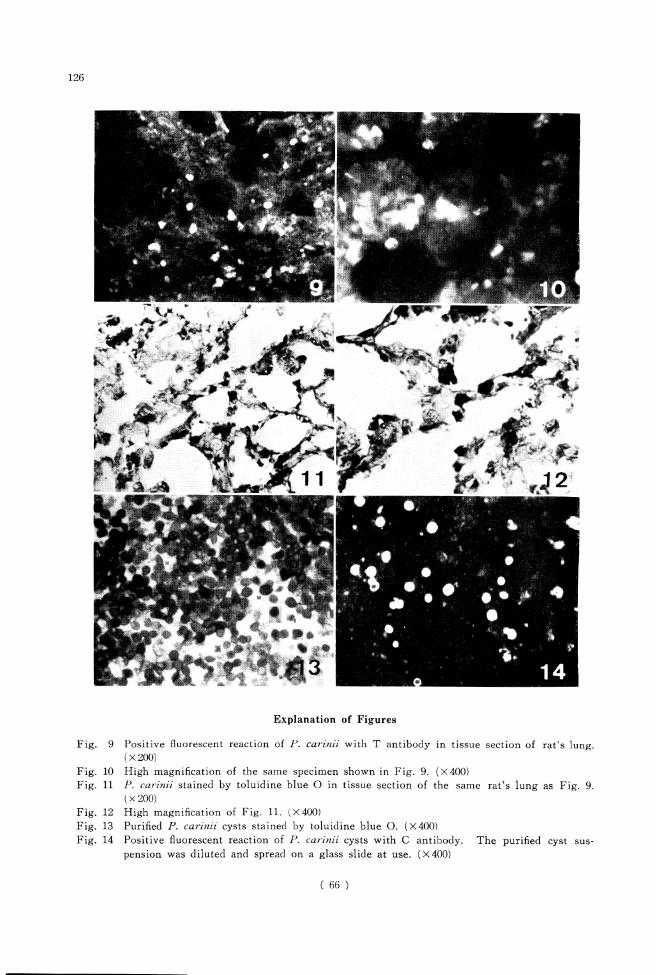
6. Indirect fluorescent antibody test
As the next step, IFA test was carried
out. The reactions of T antibody on a tissue
section of P. carinii infected rat's lung were
shown in Fig. 9 (magnification 200 X) and
Fig. 10 (400X). Tissue sections stained with
toluidine blue O were also shown in Fig. 11
(200 X) and Fig. 12 (400 X) for comparison.
The fluorescein activity was noted not only
on trophozoites but also on cysts, which
seemed to come from intracystic contents
rather than the cyst wall.
The final step is the comparison of anti-
genicity between T antigen and C antigen
using IFA test. As described before (Ikai
et al., 1979), the harvest by cyst purification
method do not contain the trophozoite at
all, and intracystic bodies also could not be
found by Giemsa stain probably due to de
generation and lysis during this disruptive
procedure. However, the cyst wall was usu
ally remained intact as shown in Fig. 13.
The fact that C antibody showed marked
positive fluorescein reactions with purified
cysts derived not only from human lungs
but also from rat's lungs, (Fig. 14) although
T antibody did not react with the same
material, suggests that the antigenicity of
trophozoite derived antigen is somewhat dif
ferent from that of cyst wall derived one.
Discussion
In the present study, two different methods
were used for preparation of P. carinii anti
gen. One was freezing and thawing method
of P. carinii infected human lungs, and the
other was cyst purification method by passing
of millipore filter system (Ikai et al., 1979).
It became evident that the P. carinii antigen
obtained by the former method was tropho
zoite derived one (T antigen), and that by
the later method cyst wall derived antigen
(C antigen). By the immunization of rabbits
with these two types of antigens, T antibody
(anti-trophozoite antibody) and C antibody
(anti-cyst wall antibody) were obtained. The
present study proposed a new technique for
purifying these antibodies by absorption of
the antibodies responsible for the host ele
ment with NHL-ext.-combined AH-Sepharose
4B.
( 59 )
120
All of DD test, IEP test and IFA test ap
parently demonstrated antigen-antibody reac
tion responsible for P. carinii. The fact that
T antibody and C antibody did not show
any cross-reaction, suggests that antigenicity
of cyst wall is entirely different from that
of trophozoite. And it is auther's specula
tion that soluble antigen is only derived from
components of the trophozoite, not from the
cyst wall.
The extractions of P. carinii antigen from
autopsied human lungs were attempted by
many investigators. Dvoracek et al. (1953),
Barta et al. (1955) and Barta (1966, 1969)
extracted the antigens with 96% ethyl al
cohol. Vivell (1954, 1955), Jirovec (1954),
Kucera (1967) and Meuwissen and Leeuwen-
berg (1972) made the antigens by homogena-
tion or by freezing and thawing method.
It is presumed, from the present study that
their soluble antigens are trophozoite derived
antigen. They tried complement fixation test
or intradermal reaction with those antigens.
On the other hand, trials to collect the
cysts as purely as possible have also been
performed by several workers by different
techniques as follows : Norman and Kagan
(1972) by sucrose density gradient, Meuwis
sen et al. (1973, 1977) pronase digestion, Lim
et al. (1973) trypsin digestion, Ikai et al. (1979)
millipore filter, and Walzer et al. (1979) col-
lagenase and hyaluronidase followed by ficoll-
hypaque density gradient respectively. They
carried out IFA test and titration of anti
body with those cysts as antigen. Those
antigens are mainly cyst wall antigen.
Histopathological examination of antigen
icity of P. carinii was first described by
Brzosko and Nowoslawski (1965). They ex
amined fluorescent reactions on lung section
of P. carinii pneumonia with FITC-conju-
gated antihuman IgG and IgM. PAS stain
ing was also done on the same material.
They concluded from the results of those
two staining methods that glyco- or muco-
proteins of P. carinii might be the active
antigenic components of the organism.
Attempts to obtain the anti-P. carinii anti-
serum in rabbits have already been performed
by Minielly et al. (1970), Lim et al. (1971,
1973), Kim et al. (1972), Kagan and Norman
(1976), Pifer et al. (1977, 1978), and Walzer
et al. (1979). Minielly et al. (1970) immu
nized rabbits with an antigen extracted from
PcHL in saline, then purified the antibody
by absorption with NHL. Their antigen and
antibody are thought to be T antigen and T
antibody. They examined the reaction be
tween their antibody and P. carinii in tissue
sections of infected human lungs with IFA
test, and found positive results. However,
they did neither mention about the result
of DD test and IEP test nor about origin of
the antigenicity.
Kim et al. (1972) immunized rabbits with
cysts collected from rat's lungs, and purified
the obtained antibody by absorption with
normal rat's lungs. Their antibody showed
positive fluorescence against cysts and tro-
phozoites collected from rat's lungs, whereas
the cysts from human lungs were nagative.
So, they stated that antigenicity of P. carinii
in rat's lungs was different from that in
human lungs. Frenkel (1976) separated Pneu
mocystis in man from that in rat, and gave
a new specific name, Pneumocystis jiroveci,
to it. However, the present author failed
to demonstrate any immunological difference
between Pneumocystis derived from man and
rat.
Lim et al. (1971) produced antiserum in
rats which were treated with cortisone ace
tate for 8 weeks, then kept for another 8
weeks without cortisone to allow the anti
body titer to rise. They found positive fluo
rescent reactions between P. carinii in rat's
lungs and their antiserum. Because they did
not find various morphological pneumocystic
forms rather than the cystic form by IFA
test, they stated that antigenic differences
might exist among various developmental
stages. Lim et al. (1973) also immunized
rabbits with relatively cleaned cysts collected
from rat's lungs and human lungs by sucrose
density gradient method, and absorbed the
obtained antiserum with mouse powder and
rat's serum or human serum. They exam
ined antigen-antibody reactions by IFA, DFA
( 60)
121
and DD tests. DD test showed a single
band between antiserum and sonically dis
rupted soluble antigen of P. carinii. IFA
test was also positive between P. carinii and
the antiserum. Cross reactions of P. carinii
in rat's and human lungs were found, but
higher titer was noted in the homologous
system. From those data, they suggested
that antigenicity of P. carinii from different
hosts might not be entirely identical. It is
suspected that their antibody cotained T
antibody and C antibody. Their data partly
corresponded with the present study except
no immunological difference between Pneu-
mocystis derived from man and rat was found
in the later.
Kagan and Norman (1976) immunized rab
bits and monkeys with the antigens which
were extracted from PcHL, NHL, PcRL and
NRL by Viveil's method (Vivell, 1955). The
anti-PcHL antiserum and anti-PcRL anti
serum were examined with each of the anti
gens mentioned above after absorbing with
NHL and NRL. Since they did not find any
specific band to P. carinii in that system,
they presumed that Pneumocystis cysts per
se were poor antigens and that component
present in infected lungs were closely host
related. Their results are quite different
from many other investigators' including the
present author. The present study used a
different method in preparing antigens and
absorbing antisera from them, and demon
strated two types of antigens (T antigen and
C antigen), hence two types of antigen-anti
body reactions. The antigenicity of P. carinii
proved not to be poor as the results.
Recently, Pifer et at. (1977) and Latorre
et at. (1977) reported on successful cultiva
tion of P. carinii in vitro. Pifer et at. (1977)
immunized rabbits with the cultivated P.
carinii. They measured antibody titer by
IFA test, and found positive fluorescence in
as much as 1 : 1,024 to 1 : 2,048 dilutions of
the antiserum. They utilized that antiserum
in diagnosis of P. carinii pneumonia in man.
However, they did not mention about the
difference of the antigenicity between cyst
wall and trophozoite.
Walzer et al. (1979) reported a separation
method of P. carinii from the lung tissue by
digestion with collagenase and hyaluronidase
followed by ficoll-hypaque density gradient.
They immunized rabbits with the cysts or
trophozoites thus collected from the rat's
lungs, and examined antigen-antibody reac
tion by IFA test. Although they found
positive fluorescence between P. carinii and
their antiserum, they did not examined the
cross reaction between P. carinii from rat's
lungs and from human lungs. They also
did not describe the difference of antigenicity
between cyst wall and trophozoite.
Nowadays, cultivation of P. carinii is still
difficult, and so, it is very important to es
tablish another method to obtain pure anti
gen and pure antibody of P. carinii.
A new absorption technique with NHL-
ext.-combined AH-Sepharose 4B proposed
here is very useful to absorb and remove
the anti-NHL-ext. antibody without contami
nation of NHL-ext..
Summary
In the present status of difficulty to obtain
pure antigen of P. carinii the purification of
antibody was performed by absorption with
affinity chromatography.
Two kinds of crude antigens were used.
One was trophozoite derived soluble antigen
which was extracted by freezing and thawing
the P. carinii infected human lungs. The
other was purified cyst per se collected from
human lungs by cyst purification method.
Two kinds of antisera obtained by immu
nization of rabbits with the crude antigens,
were thoroughly treated by affinity chromato
graphy to remove antibodies responsible for
the host elements. Thus, the purified T
antibody (trophozoite derived) and C anti
body (cyst wall derived) were produced.
Double diffusion test and immunoelectro-
phoresis confirmed the purity of T antibody
by showing a clear single precipitin band
between this antibody and P. carinii infected
human and rat's lung extracts, and also by
not showing any band between this antibody
( 61 )
122
and normal human and normal rat's lung
extracts. C antibody did not show any pre-
cipitin band with P. carinii infected human
and rat's lung extracts.
In indirect fluorescent antibody test, T
antibody showed positive reaction to P.
carinii on tissue section of infected rat's
lung, probably with trophozoites and intra-
cystic elements concerned, whereas negative
on cyst smear preparation. However, the
smeared cysts showed marked positive fluo-
rescein when C antibody was used.
From the facts mentioned above, it can
be said that the antigenicity of trophozoite
is different from that of cyst wall. In addi
tion, the antigenicity either of T antigen
or C antigen, was not different between P.
car'mii of human origin and rat's origin.
Acknowledgements
The author wishes to express his sincere ap
preciation to Professor Yukio Yoshida for his
interest, guidance and encouragement through this
study and for his critical reading of the manu
script, and also sincere thanks are due to Dr.
Yutaka Katayama and Dr. Tohru Sugimoto for
their kind advice on techniques.
References
1) Barta, K., Dvoracek, C. und Kadlec, A. (1955) :
Komplementbindungs-Reaction bei Pneumo-
zysten-Pneumonien. Schweiz. Z. Path. Bakt.,
18, 22-32.
2) Barta, K. (1966) : Zur Frage der antigen-
wirksamen Komponenten der Lunge bei der
Pneumocysten-Pneumonie. Path. Microbiol.,
29, 63-74.
3) Barta, K. (1969) : Complement fixation test
for pneumocystosis. Ann. Int. Med.. 70, 235.
4) Brzosko, W. J. and Nowoslawski, A. (1965) :
Identification of Pneumocystis carinii antigens
in tissues. Bull. Acad. Polon. Sci. Ser. Sci.
BioL, 13, 49-54.
5) Cambiaso, C. L., Gofnnet, A., Vaerman, J. P.
and Haremans, J. F. (1975) : Glutaraldehyde-
activated aminohexyl-derivative of Sepharose
4B as a new versatile immunoabsorbent. Im-
munochemistry, 12, 273-278.
6) Chagas, C. (1909) : Nova tripanozomiaze hu-
mana. Mem. Inst. Oswaldo Cruz, 1, 159-218.
7) Delanoe, P. et Delanoe, E. (1912) : Sur les
rapports des Kystes de Carini du poumon des
rats avec le Trypa?iozo?na lexvisi. C. R. Acad.
Sci., 155, 658-660.
8) Dvoracek, C, Barta, K. and Kadlec, A. (1953) :
Vazba komplementu u pneumocystovych zanetu
plic. Lek. Listy, 8, 537-539.
9) Frenkel, J. K., Good, J. T. and Shultz, J. A.
(1966) : Latent Pneumocystis infection of
rats, relapse and chemotherapy. Lab. Invest.,
15, 1559-1577.
10) Frenkel, J. K. (1976) : Pneumocystis jiroveci
n. sp. from man : Morphology, physiology,
and immunology in relation to pathology.
National Cancer Institute Monograph, 43, 13-
27.
11) Hamashima Y. and Kyogoku, M. (1965) : Im-
munohistology, Igaku Shoin, 82-84. (in Ja
panese)
12) Ikai. T., Yoshida, Y., Yamada, M., Ogino,
K. and Takeuchi, S. (1979) : Studies on Pneu
mocystis carinii and Pneumocystis carinii
pneumonia VI. Cyst purification method.
Jap. J. Parasit., 28, 71-80 (Japanese with
English summary).
13) Jirovec, O. (1954) : Uber die durch Pneumo
cystis carinii verursachte interstitielle Pneu-
monie der Sauglinge. Mschr. Kinderheilk.,
102, 476-485,
14) Kagan, I. G. and Norman, L. G. (1976): Sero-
logy of pneumocystosis. National Cancer
Institute Monograph, 43, 121-125.
15) Kim, H. K., Hughes, W. T. and Feldman, S.
(1972) : Studies of morphology and immuno-
fluorescence of Pneumocystis carinii. Proc.
Soc. Exp. Biol. Med., 141, 304-309.
16) Kucera, K. (1967) : La pneumocystose en tant
qu'anthropozoonose. Ann. Parasit. Hum.
Comp., 42, 465-482.
17) Latorre, C. R., Sulzer, A. J. and Norman.
L. G. (1977) : Serial propagation of Pneumo
cystis carinii in cell line cultures, Appl.
Environ. Microbiol., 33, 1204-1206.
18) Lim, S. K., Jones, R. H. and Eveland, W. C.
(1971) : Fluorescent antibody studies on ex
perimental pneumocystosis. Proc. Soc. Exp.
Biol. Med., 136, 675-679.
19) Lim, S. K., Eveland, W. C. and Porter, R. J.
(1973) : Development and evaluation of a
direct fluorescent antibody method for the
diagnosis of Pneumocystis carinii infections
in experimental animals. Appl. Microbiol.,
26, 666-671.
20) Meuwissen, J. H. E. Th. and Leeuwenberg,
( 62 )
123
A. D. E. M. (1972) : A microcomplement fixa
tion test applied to infection with Pneumo-
cystis carinii. Trop. Geogr. Med., 24, 282-
291.
21) Meuwissen, J. H. E. Th., Leeuwenberg, A. D.
E. M., Heeren, J. and Stumpel, A. (1973) :
New method for study of infections with
Pneu?7iocystis carinii. J. Infect. Dis., 127,
209-210.
22) Meuwissen, J. H. E. Th., Tauber, L, Leeu
wenberg, A. D. E. M., Beckers, P. J. A. and
Sieben, M. (1977) : Parasitologic and sero-
logic observations of infection with Pneu-
mocystis in humans. J. Infect. Dis., 136,
43-49.
23) Minielly, J. A., McDuffie, F. C. and Holley,
K. E. (1970) : Immunofluorescent identifica
tion of Pneumocystis carinii. Arch. Path.,
90, 561-566.
24) Norman, L, and Kagan, I. G. (1972) : A pre
liminary report of an indirect fluorescent anti
body test for detecting antibodies to cysts of
Pneumocystis carinii in human sera. A. J. C.
P., 58, 170-176.
25) Pifer, L. L., Hughes, W. T. and Murphy,
M. J. Jr. (1977) : Propagation of Pneumocystis
carinii in vitro. Pediat. Res., 11, 305-316.
26) Pifer, L. L., Hughes, W. T., Stagno, S. and
Woods, D. (1978) : Pneumocystis carinii in
fection : Evidence for high prevalence in
normal and immunosuppressed children. Pe
diat., 61, 35-41.
27) Vivell, O. (1954) : Uber eine neue serodia-
gnostische Methode bei der interstitiellen
plasmazellularen Pneumonie junger Sauglinge
und Friihgeburten. Dtsch. Med. Wschr., 79,
358-360.
28) Vivell, O. (1955) : Ein neues stabiles Anti
gen fur die Serodiagnose der interstitiellen
plasmazellularen Pneumonie junger Sauglinge
und Friihgeburten. Dtsch. Med. Wschr., 80,
1357.
29) Walzer, P. D., Rutledge, M. E., Yoneda, K.
and Stahr, B. J. (1979) : Pneumocystis carinii :
New separation method from lung tissue.
Exp. Parasit., 47, 356-368.
( 63 )
124
Pneumocystis carinii: #l trophozoite ziJi cyst wall
Pneumocystis carinii
affinity chromatography ^rfflV^T P. carinii \
(PcHL-ext.),
P. carinii *S
(NHL-ext.)
VIZ. 10,000rpm
, P. carinii <D trophozoite
—^, P. carinii <Di/
1979) T? P. canmV )ffi^S#W«3: D M#>fc>
^ffift^ttfflffiJS^tt^tl^iX 3mgfo, ^^ ht*
Freund's complete adjuvant t hi>fo]&
f^^L^jLjiL»<0 5*>tfL PcHL-
P. wnmi v^^. bik?fHftfc5E^5
affinity chromatography T*iDSlR
PcHL-ext.
2, 5, 6
NHL-ext. ik?f ^[^# Figs. 1,
NHL-ext. t PcHL-ext.
(PcRL-ext. *5 J: U NRL-ext.) t <D fflV %
Figs. 3, 7 \C7jki~X ? \C PcHL-ext. t PcRL-ext.
Figs. 4, 8 fr£
NHL-ext. ^ NRL-
ext.
extract
Affinity chromatography JCJi Sepharose 4B §:ffi
V^fCo NHL-ext. t Sepharose 4B ^ glutaraldehyde
feT?jfrfr£ii:, £*l/fc perista mini-pump $^fflV^TS:
jfiLttSrjftDSL^aLTSlSS^jLifiLitt^tt NHL-
ext. S#S®R^*L^
p.
PcHL-ext. Jtft: (gJt
Figs. 9, 10 T^"tJ; 5
P.
, C #l#: Tit Fig. 14
P. can'mi
P. cfln
cyst wall
trophozoite
(64)
125
Figs.
Figs.
Figs.
Fig.
Fig.
Figs.
1-4
5-8
1-8
5
6
7-8
Explanation of Figures
Double diffusion test.
Immunoelectrophoresis (cathode right and anode left).
A, E, F and G : PcHL-extracts (case 1, 2, 3 and 4)
B : NHL-extract C : NRL-extract D : PcRL-extract
I : Anti-PcHL-extract antiserum J : T antibody
Anti-NHL-extract antiserum in three troughs.
Anti-PcHL-extract antiserum in three troughs.
T antibody in three troughs.
H : Anti-NHL-extract antiserum
( 65 )
126
Explanation of Figures
Fig. 9 Positive fluorescent reaction of P. car'inii with T antibody in tissue section of rat's lung.
(X200)
Fig. 10 High magnification of the same specimen shown in Fig. 9. (X400)
Fig. 11 P. carinii stained by toluidine blue O in tissue section of the same rat's lung as Fig. 9.
(x 200)
Fig. 12 High magnification of Fig. 11. (X400)
Fig. 13 Purified P. carinii cysts stained by toluidine blue O. (X400)
Fig. 14 Positive fluorescent reaction of P. carinii cysts with C antibody. The purified cyst sus
pension was diluted and spread on a glass slide at use. (X400)
( 66 )