PELVIC OSTEOTOMY FOR THE TREATMENT OF THE YOUNG ADULT WITH HIP PAIN Emmanuel Illical, Adult...
19
PELVIC OSTEOTOMY FOR THE TREATMENT OF THE YOUNG ADULT WITH HIP PAIN Emmanuel Illical, Adult Reconstruction Fellow
-
Upload
primrose-walton -
Category
Documents
-
view
221 -
download
3
Transcript of PELVIC OSTEOTOMY FOR THE TREATMENT OF THE YOUNG ADULT WITH HIP PAIN Emmanuel Illical, Adult...

PELVIC OSTEOTOMYFOR THE TREATMENT OF
THE YOUNG ADULT WITH HIP PAIN
Emmanuel Illical, Adult Reconstruction Fellow
OUTLINE• Pelvic osteotomy introduction• Background: hip dysplasia• Bernese periacetabular osteotomy (PAO)
– Technique & Surgical adjuncts
– Advantages & Complications
– Clinical Results
• Background: acetabular retrotorsion• Reverse PAO
– Principles
– Clinical Results
• Other osteotomies• Salvage Osteotomies• Summary

• Treatment of choice for acetabular structural disorders for young pts– classical developmental hip dysplasia
– retrotorsional acetabular abnormalities
• Indications– symptomatic patient (pain / progressive limp)
– radiographically negative for advanced OA
– adequate and relatively painless passive ROM
• Rationale– restore normal hip anatomy and biomechanics
• increase joint congruity
• optimize center of rotation
– relieve symptoms
– prevent (possibly delay?) degenerative changes
• Challenges: diagnosis & predictability of outcome
PELVIC OSTEOTOMY: INTRODUCTION

• Acetabular abnormalities– shallow
– anteverted (retrortosion in up to 25%)
– lateralized
– femoral head coverage deficient anteriorly, laterally, superiorly
• Femoral abnormalities– proximal migration– femoral head small and deformed– femoral neck short and narrrow w/ varying but but anteversion– valgus neck shaft angle– femoral canal narrow
• Secondary degenerative joint disease over time contact area btwn femoral head and acetabulum
– excessive lateralization of body weight lever arm body weight lever arm
– relatively high forces transmitted through surface area
BACKGROUND: HIP DYSPLASIA

BACKGROUND: HIP DYSPLASIA

BACKGROUND: HIP DYSPLASIA

BERNESE PAO: TECHNIQUE

• Anterior arthrotomy: before or after PAO– labral pathology: debridement / repair
– femoral head neck junction: osteochondroplasty
• Proximal femoral osteotomy– severe coxa valga / vara varus / valgus producing intertrochanteric osteotomy
BERNESE PAO: SURGICAL ADJUNCTS

• Only one incision that spares ABductors• Reproducible extra-articular osteotomies• Allows large corrections in all directions• Posterior column remains intact
– minimal internal fixation required
– early mobilization w/o external immobilization
• Preservation of acetabular fragment vascularity– intra-articular examinzation w/o further risk of devascularization
• True pelvis shape is unchanged– child bearing & vaginal delivery not affected
BERNESE PAO: ADVANTAGES

• Most important factor affecting incidence = surgeon experience
• Most common complication = nerve dysfunction– lateral femoral cutaneous nerve (35%)
– femoral nerve
– sciatic / peroneal nerve
• Vascular – related ilioinguinal approach: femoral / iliac artery thrombosis
• Inadvertent extension of osteotomy to undesirable location– intra-articular extension of infra / supra – acetabular osteotomies
– sciatic notch extension of iliac osteotomies
• Femoroacetabular impingement
• Osteonecrosis of acetabular fragment
• Nonunion
• Other: HO, loss of correction, femoral head subluxation
BERNESE PAO: COMPLICATIONS

BERNESE PAO: CLINICAL RESULTS

BERNESE PAO: CLINICAL RESULTS

• Posteriorly oriented acetabular opening (sagittal plane)• Etiology
– isolated entity
– associated w/ classic hip dysplasia
– injury to tri-radiate cartilage in growing child
– associated with LCP, bladder extrophy, neuromuscular d/o
• Typical presentation is groin pain reproduced with “impingement signs”
• Recurrent impingement has been implicated in development of 2* arthrosis
BACKGROUND: ACETABULAR RETROTORSION

BACKGROUND: ACETABULAR RETROTORSION

• Treatment of choice when acetabular retrotorsion exists– +ve crossover sign AND +ve posterior wall sign (poor posterior coverage)
– addresses lack of posterior wall coverage by increasing anteversion
• Technique– same approach and osteotomies as PAO
– re-orientation achieved by combined flexion + IR of acetabular fragment
– goals: eliminate xray signs+ sufficient impingement free ROM (flexion + IR)
– posterior over-coverage is a concern
– arthrotomy / SHD to address femoral head neck offset & labral pathology
• Contra-indications– excessive posterior wall coverage / AI < 0* impingement
– significant combined CAM / pincer deformity requires surgical hip dislocation
– advanced cartilage degeneration area would end up in weight bearing zone
REVERSE PAO: PRINCIPLES

• Siebenrock et al. JBJS Am 2003. Impingement due to acetabular retroversion. Treatment with PAO.– 29 reverse PAOs to reorient retroverted acetabulum (+ve cross over / pw sign)– concominant femoral head neck osteochondroplasty in 24 hips– avg 30 month f/u– significant increase in flexion / IR / ADduction– significant d’Aubigne hip score improvement: 14.0 16.9– 28 “good / excellent” results– no pt had radiographic signs of OA– 3 revisions
• Buchler et al. JBJS Br 2011. Symptomatic acetabular retroversion: mean 10 year fu after treatment with PAO.– mean f/u 10.6 year – overall d’Aubinge score improved: 14.0 16.3– all patients had symptomatic relief at final f/u– ROM and functional scores improved in all cases– vast majority of pts continued to demonstrate no signs of radiographic OA
REVERSE PAO: CLINICAL RESULTS

OTHER OSTEOTOMIES
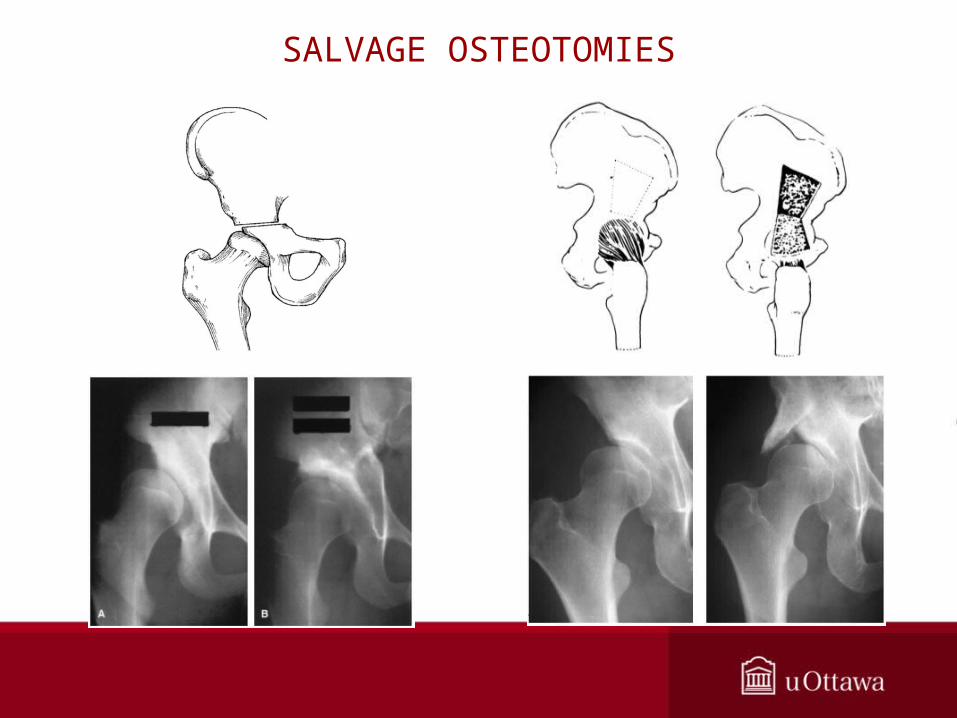
SALVAGE OSTEOTOMIES

• Full pre-operative work-up– assess degree of dysplasia and acetabular version
– assess femoral head neck offset
– assess labral pathology
• Select patients appropriately– symptomatic
– be aware of age
– no more than mild to moderate articular degenerative changes (Tonnis grade < 2)
– reasonable joint congruity (obtain functional xrays if necessary)
• Surgeon experience key to minimizing complications
SUMMARY