Severe Gummy Smile with Class II Malocclusion Treated with ... · LeFort I Osteotomy Combined with...
6
Severe Gummy Smile with Class II Malocclusion Treated with LeFort I Osteotomy Combined with Horseshoe Osteotomy and Intraoral Vertical Ramus Osteotomy Tsuyoshi Shimo a , Akiyoshi Nishiyama a* , Tokiari Jinno b , and Akira Sasaki a a Department of Oral and Maxillofacial Surgery, Okayama University Graduate School of Medicine, Dentistry and Pharmaceutical Sciences, Okayama 700-8558, Japan, and b Jinno Orthodontic Office, Okayama 700-0815, Japan In this article, we report the successful surgical treatment of a patient, 34 years of age, who had a severe gummy smile and a class II malocclusion. The patient had an 11- mm gingival exposure during full smile and a convex profile. A LeFort I osteotomy combined with a horseshoe osteotomy was used for the superior repositioning of the maxilla; then, an intraoral vertical ramus osteotomy (IVRO) and genioplasty were performed for mandibular advancement. The maxilla was acceptably impacted 8 mm at the first incisor and 5 mm at the first molar. Both the occlusion and facial appearance were signifi- cantly improved by this surgical - orthodontic treatment. Our results suggest that the combination of a horseshoe osteotomy with a LeFort I osteotomy is a useful technique for reliable superior reposition- ing of the maxilla. Key words: LeFort I osteotomy, horseshoe osteotomy, gummy smile gummy smile is an aesthetic problem for some patients and has been treated by orthodontics alone or orthodontic surgery to reposition the maxilla [1, 2]. However, since a severe gummy smile is characterized by overgrowth due to anterior vertical maxillary excess [3], conventional orthodontic treat- ment alone is not an option. Orthodontic surgery can provide significant skeletal improvement [4]; in addi- tion, orthodontic surgery, such as that provided by a LeFort I osteotomy, provides a more aesthetically pleasing result and affords patients with severe gummy smiles a more acceptable outcome than orthodontic treatment alone. Various studies have shown that superior reposi - tioning of the maxilla is indicated in patients with a vertical maxillary excess, in cases of a long face and open bite [5, 6]. However, superior repositioning of the maxilla by a single LeFort I osteotomy is some- times difficult for high impactions because of the bar- rier of the bone around the descending palatine artery. Although the bone around the palatine artery can be trimmed equally for superior repositioning of the maxilla during LeFort I osteotomy, there is always the potential risk of damaging or cutting the artery, which may lead to severe hemorrhage and avascular necrosis of the maxilla [7]. Therefore, Bell and McBride introduced the horseshoe palatal osteotomy in combination with the LeFort I osteotomy [8]. The greatest advantage of this technique is that it allows higher superior repositioning of the maxilla, espe- cially in the posterior portion, because there is no need for bone trimming around the palatine vessel. Successful LeFort I osteotomy combined with horse- shoe osteotomy has been recommended for high- A Acta Med. Okayama, 2013 Vol. 67, No. 1, pp. 55ン60 CopyrightⒸ 2013 by Okayama University Medical School. Case Report http: // escholarship.lib.okayama-u.ac.jp / amo/ Received July 5, 2012 ; accepted September 18, 2012. * Corresponding author. Phone: + 81ン86ン235ン6702; Fax: + 81ン86ン235ン6704 E- mail:[email protected]- u.ac.jp (A. Nishiyama)
Transcript of Severe Gummy Smile with Class II Malocclusion Treated with ... · LeFort I Osteotomy Combined with...
Severe Gummy Smile with Class II Malocclusion Treated with LeFort I Osteotomy Combined with Horseshoe Osteotomy and
Intraoral Vertical Ramus Osteotomy
Tsuyoshi Shimoa, Akiyoshi Nishiyamaa*, Tokiari Jinnob, and Akira Sasakia
aDepartment of Oral and Maxillofacial Surgery, Okayama University Graduate School of Medicine, Dentistry and Pharmaceutical Sciences, Okayama 700-8558, Japan, and bJinno Orthodontic Office, Okayama 700-0815, Japan
In this article, we report the successful surgical treatment of a patient, 34 years of age, who had a severe gummy smile and a class II malocclusion. The patient had an 11-mm gingival exposure during full smile and a convex profile. A LeFort I osteotomy combined with a horseshoe osteotomy was used for the superior repositioning of the maxilla; then, an intraoral vertical ramus osteotomy (IVRO) and genioplasty were performed for mandibular advancement. The maxilla was acceptably impacted 8mm at the first incisor and 5mm at the first molar. Both the occlusion and facial appearance were signifi-cantly improved by this surgical-orthodontic treatment. Our results suggest that the combination of a horseshoe osteotomy with a LeFort I osteotomy is a useful technique for reliable superior reposition-ing of the maxilla.
Key words: LeFort I osteotomy, horseshoe osteotomy, gummy smile
gummy smile is an aesthetic problem for some patients and has been treated by orthodontics
alone or orthodontic surgery to reposition the maxilla [1, 2]. However, since a severe gummy smile is characterized by overgrowth due to anterior vertical maxillary excess [3], conventional orthodontic treat-ment alone is not an option. Orthodontic surgery can provide significant skeletal improvement [4]; in addi-tion, orthodontic surgery, such as that provided by a LeFort I osteotomy, provides a more aesthetically pleasing result and affords patients with severe gummy smiles a more acceptable outcome than orthodontic treatment alone. Various studies have shown that superior reposi-tioning of the maxilla is indicated in patients with a
vertical maxillary excess, in cases of a long face and open bite [5, 6]. However, superior repositioning of the maxilla by a single LeFort I osteotomy is some-times difficult for high impactions because of the bar-rier of the bone around the descending palatine artery. Although the bone around the palatine artery can be trimmed equally for superior repositioning of the maxilla during LeFort I osteotomy, there is always the potential risk of damaging or cutting the artery, which may lead to severe hemorrhage and avascular necrosis of the maxilla [7]. Therefore, Bell and McBride introduced the horseshoe palatal osteotomy in combination with the LeFort I osteotomy [8]. The greatest advantage of this technique is that it allows higher superior repositioning of the maxilla, espe-cially in the posterior portion, because there is no need for bone trimming around the palatine vessel. Successful LeFort I osteotomy combined with horse-shoe osteotomy has been recommended for high-
A
Acta Med. Okayama, 2013Vol. 67, No. 1, pp. 55ン60CopyrightⒸ 2013 by Okayama University Medical School.
Case Report http ://escholarship.lib.okayama-u.ac.jp/amo/
Received July 5, 2012 ; accepted September 18, 2012.*Corresponding author. Phone : +81ン86ン235ン6702; Fax : +81ン86ン235ン6704E-mail : [email protected] (A. Nishiyama)

impaction cases [9, 10]. However, there are few reports about cases of severe gummy smile. In this report, we present the case of severe gummy smile with class II malocclusion treated with LeFort I osteotomy combined with horseshoe osteotomy, intraoral ramus osteotomy (IVRO), and genioplasty.
Case Report
A 34-year-old woman with class II malocclusion came to our department complaining of a gummy smile. Her past medical history was insignificant, including a lack of family history of malformation. In the extraoral examination, a severe dolichofacial condition with retrognathia was observed. The upper gingiva was exposed about 11mm from the cervical line to the
upper lip while smiling (Fig. 1). Intraoral examina-tion revealed a class II malocclusion with an excessive positive overbite (3mm) and overjet (6mm). Repre-sentative preoperative cephalometric values were as follows: SNA, 82.7° (+1.1 SD); SNB, 87° (-0.3 SD); ANB, 8.5° (+2.2 SD); A to N perpendicular to FH -0.8° (+1.5 SD); pogonion to N perpendicular to FH, -24.5mm (-1.0 SD); mx1 Crown to FN, 70.5mm; mx6 Crown to FH, 59mm; Mandibular Plane, 42.4 deg (+3.1 SD); Pos Face Height (S-Go), 86.8mm (-0.5 SD); Ant. Face Height (N-Me), 147.6mm (+2.3 SD); S-Go/N-Me, 58.8オ (-0.5 SD); Ricketʼs VERT index, -8.8 (Fig. 1 and Table 1). The patient was diagnosed as having an angle class II malocclusion with a skeletal class II jaw base rela-tionship and a severe gummy smile. The treatment
56 Acta Med. Okayama Vol. 67, No. 1Shimo et al.
Fig. 1 Pretreatment extraoral and intraoral views.
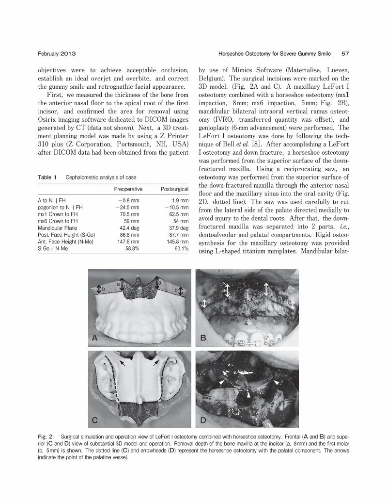
objectives were to achieve acceptable occlusion, establish an ideal overjet and overbite, and correct the gummy smile and retrognathic facial appearance. First, we measured the thickness of the bone from the anterior nasal floor to the apical root of the first incisor, and confirmed the area for removal using Osirix imaging software dedicated to DICOM images generated by CT (data not shown). Next, a 3D treat-ment planning model was made by using a Z Printer 310 plus (Z Corporation, Portsmouth, NH, USA) after DICOM data had been obtained from the patient
by use of Mimics Software (Materialise, Lueven, Belgium). The surgical incisions were marked on the 3D model. (Fig. 2A and C). A maxillary LeFort I osteotomy combined with a horseshoe osteotomy (mx1 impaction, 8mm; mx6 impaction, 5mm; Fig. 2B), mandibular bilateral intraoral vertical ramus osteot-omy (IVRO, transferred quantity was offset), and genioplasty (6-mm advancement) were performed. The LeFort I osteotomy was done by following the tech-nique of Bell et al. [8]. After accomplishing a LeFort I osteotomy and down fracture, a horseshoe osteotomy was performed from the superior surface of the down-fractured maxilla. Using a reciprocating saw, an osteotomy was performed from the superior surface of the down-fractured maxilla through the anterior nasal floor and the maxillary sinus into the oral cavity (Fig. 2D, dotted line). The saw was used carefully to cut from the lateral side of the palate directed medially to avoid injury to the dental roots. After that, the down-fractured maxilla was separated into 2 parts, i.e., dentoalveolar and palatal compartments. Rigid osteo-synthesis for the maxillary osteotomy was provided using L-shaped titanium miniplates. Mandibular bilat-
57Horseshoe Osteotomy for Severe Gummy SmileFebruary 2013
Table 1 Cephalometric analysis of case
Preoperative Postsurgical
A to N -| FH -0.8 mm 1.9 mmpogonion to N -| FH -24.5 mm -10.5 mmmx1 Crown to FH 70.5 mm 62.5 mmmx6 Crown to FH 59 mm 54 mmMandibular Plane 42.4 deg 37.9 degPost. Face Height (S-Go) 86.8 mm 87.7 mmAnt. Face Height (N-Me) 147.6 mm 145.8 mmS-Go / N-Me 58.8% 60.1%
A B
C D
ab
ab a a
bb
Fig. 2 Surgical simulation and operation view of LeFort I osteotomy combined with horseshoe osteotomy. Frontal (A and B) and supe-rior (C and D) view of substantial 3D model and operation. Removal depth of the bone maxilla at the incisor (a, 8mm) and the first molar (b, 5mm) is shown. The dotted line (C) and arrowheads (D) represent the horseshoe osteotomy with the palatal component. The arrows indicate the point of the palatine vessel.
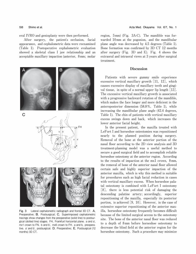
eral IVRO and genioplasty were then performed. After surgery, the patientʼs occlusion, facial appearance, and cephalometric data were reexamined (Table 1). Postoperative cephalometric evaluation showed a skeletal class I jaw relationship and an acceptable maxillary impaction (anterior, 8mm; molar
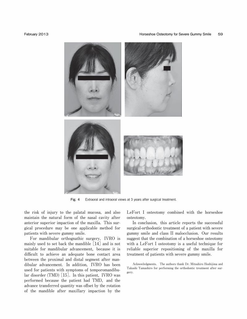
region, 5mm) (Fig. 3A-C). The mandible was for-warded 10mm at the pogonion, and the mandibular plane angle was decreased by 4.5 degrees (Table 1). Bone formation was confirmed by 3D CT 12 months after surgery (Fig. 3D and E). Fig. 4 shows the extraoral and intraoral views at 3 years after surgical treatment.
Discussion
Patients with severe gummy smile experience excessive vertical maxillary growth [11, 12], which causes excessive display of maxillary teeth and gingi-val tissue, in spite of a normal upper lip length [13]. The excessive vertical maxillary growth is associated with a progressive backward rotation of the mandible, which makes the face longer and more deficient in the anteroposterior dimension (58.8オ, Table 1), while increasing the mandibular plane angle (42.4 degrees, Table 1). The chin of patients with vertical maxillary excess swings down and back, which increases the lower anterior facial height. In the present patient, the maxilla treated with LeFort I and horseshoe osteotomies was repositioned nearly to the planned position during surgery. Removal of the bone at the anterior portion of the nasal floor according to the 2D view analysis and 3D treatment-planning model was a useful method to secure a good surgical field and to accomplish reliable horseshoe osteotomy at the anterior region. According to the results of impaction at the mx1 crown, 8mm, the removal of bone of the anterior nasal floor allowed certain safe and highly superior impaction of the anterior maxilla, which is why this method is suitable for procedures such as high facial reduction in cases with vertical maxillary excess. When horseshoe pala-tal osteotomy is combined with LeFort I osteotomy [8], there is less potential risk of damaging the descending palatine artery; accordingly, superior repositioning of the maxilla, especially its posterior portion, is achieved [9, 10]. However, in the case of extensive superior repositioning of the anterior max-illa, horseshoe osteotomy frequently becomes difficult because of the limited surgical access to the osteotomy site. The bone of the anterior nasal floor was reduced to a depth of 8mm before horseshoe osteotomy to decrease the blind field at the anterior region for the horseshoe osteotomy. Such a procedure may minimize
58 Acta Med. Okayama Vol. 67, No. 1Shimo et al.
A B
C
D E
FH
a a’
b b’
Fig. 3 Lateral cephalometric radiograph and frontal 3D CT. A, Preoperative; B, Postsurgical; C, Superimposed cephalometric tracings show changes from the preoperative (solid line) to postsur-gical (dotted line) stages. FH, Frankfurt horizontal plane. a and aʼ, mx1 crown to FN. b and bʼ, mx6 crown to FH. a and b, preopera-tive. aʼ and bʼ, postsurgical. D, Preoperative; E, Postsurgical (12 months) 3D CT.
the risk of injury to the palatal mucosa, and also maintain the natural form of the nasal cavity after anterior superior impaction of the maxilla. This sur-gical procedure may be one applicable method for patients with severe gummy smile. For mandibular orthognathic surgery, IVRO is mainly used to set back the mandible [14] and is not suitable for mandibular advancement, because it is difficult to achieve an adequate bone contact area between the proximal and distal segment after man-dibular advancement. In addition, IVRO has been used for patients with symptoms of temporomandibu-lar disorder (TMD) [15]. In this patient, IVRO was performed because the patient had TMD, and the advance transferred quantity was offset by the rotation of the mandible after maxillary impaction by the
LeFort I osteotomy combined with the horseshoe osteotomy. In conclusion, this article reports the successful surgical-orthodontic treatment of a patient with severe gummy smile and class II malocclusion. Our results suggest that the combination of a horseshoe osteotomy with a LeFort I osteotomy is a useful technique for reliable superior repositioning of the maxilla for treatment of patients with severe gummy smile.
Acknowledgments. The authors thank Dr. Mitsuhiro Hoshijima and Takashi Yamashiro for performing the orthodontic treatment after sur-gery.
59Horseshoe Osteotomy for Severe Gummy SmileFebruary 2013
Fig. 4 Extraoral and intraoral views at 3 years after surgical treatment.
References
1. Kaku M, Kojima S, Sumi H, Koseki H, Abedini S, Motokawa M, Fujita T, Ohtani J, Kawata T and Tanne K: Gummy smile and facial profile correction using miniscrew anchorage. Angle Orthod (2012) 82: 170-177.
2. Redlich M, Mazor Z and Brezniak N: Severe high Angle Class II Division 1 malocclusion with vertical maxillary excess and gummy smile: a case report. Am J Orthod Dentofacial Orthop (1999) 116: 317-320.
3. Van der Geld P, Oosterveld P, Schols J and Kuijpers-Jagtman AM: Smile line assessment comparing quantitative measurement and visual estimation. Am J Orthod Dentofacial Orthop (2011) 139: 174-180.
4. Capelozza Filho L, Cardoso Mde A, Reis SA and Mazzottini R: Surgical-orthodontic correction of long-face syndrome. J Clin Orthod (2006) 40: 323-332; quiz 308.
5. Bailey LJ, Phillips C, Proffit WR and Turvey TA: Stability follow-ing superior repositioning of the maxilla by Le Fort I osteotomy: five-year follow-up. Int J Adult Orthodon Orthognath Surg (1994) 9: 163-173.
6. Greebe RB and Tuinzing DB: Superior repositioning of the maxilla by a Le Fort I osteotomy: a review of 26 patients. Oral Surg Oral Med Oral Pathol (1987) 63: 158-161.
7. Lanigan DT, Hey JH and West RA: Aseptic necrosis following maxillary osteotomies: report of 36 cases. J Oral Maxillofac Surg
(1990) 48: 142-156. 8. Bell WH and McBride KL: Correction of the long face syndrome by
Le Fort I osteotomy. A report on some new technical modifications and treatment results. Oral Surg Oral Med Oral Pathol (1977) 44: 493-520.
9. Harada K, Sumida E, Enomoto S and Omura K: Post-operative stability of the maxilla treated with Le Fort I and horseshoe osteot-omies in bimaxillary surgery. Eur J Orthod (2002) 24: 471-476.
10. Yoshioka I, Khanal A, Kodama M, Furuta N and Tominaga K: Postoperative skeletal stability and accuracy of a new combined Le Fort I and horseshoe osteotomy for superior repositioning of the maxilla. Int J Oral Maxillofac Surg (2009) 38: 1250-1255.
11. Ellis E 3rd: The nature of vertical maxillary deformities: implica-tions for surgical intervention. J Oral Maxillofac Surg (1985) 43: 756-762.
12. Monaco A, Streni O, Marci MC, Marzo G, Gatto R and Giannoni M: Gummy smile: clinical parameters useful for diagnosis and thera-peutical approach. J Clin Pediatr Dent (2004) 29: 19-25.
13. Fish LC, Wolford LM and Epker BN: Surgical-orthodontic correc-tion of vertical maxillary excess. Am J Orthod (1978) 73: 241-257.
14. Tornes K, Gilhuus-Moe O, McCallum CA, Jr. and Wisth PJ: Positioning and mobility of the mandibular condyle after surgical correction of the asymmetric, prognathic mandible. Int J Adult Orthodon Orthognath Surg (1990) 5: 29-34.
15. Bell WH, Yamaguchi Y and Poor MR: Treatment of temporoman-dibular joint dysfunction by intraoral vertical ramus osteotomy. Int J Adult Orthodon Orthognath Surg (1990) 5: 9-27.
60 Acta Med. Okayama Vol. 67, No. 1Shimo et al.