Outline 25.1 DNA, Chromosomes, and Genes 25.2Composition of Nucleic Acids 25.3The Structure of...
57
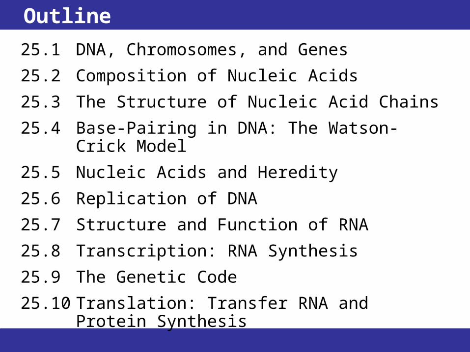
Outline 25.1 DNA, Chromosomes, and Genes 25.2 Composition of Nucleic Acids 25.3 The Structure of Nucleic Acid Chains 25.4 Base-Pairing in DNA: The Watson- Crick Model 25.5 Nucleic Acids and Heredity 25.6 Replication of DNA 25.7 Structure and Function of RNA 25.8 Transcription: RNA Synthesis 25.9 The Genetic Code 25.10 Translation: Transfer RNA and Protein Synthesis
-
Upload
florence-caldwell -
Category
Documents
-
view
219 -
download
3
Transcript of Outline 25.1 DNA, Chromosomes, and Genes 25.2Composition of Nucleic Acids 25.3The Structure of...
Outline
25.1 DNA, Chromosomes, and Genes
25.2 Composition of Nucleic Acids
25.3 The Structure of Nucleic Acid Chains
25.4 Base-Pairing in DNA: The Watson-Crick Model
25.5 Nucleic Acids and Heredity
25.6 Replication of DNA
25.7 Structure and Function of RNA
25.8 Transcription: RNA Synthesis
25.9 The Genetic Code
25.10 Translation: Transfer RNA and Protein Synthesis

Goals1. What is the composition of the nucleic acids, DNA and
RNA? Be able to describe and identify the components of nucleosides, nucleotides, DNA, and RNA.
2. What is the structure of DNA? Be able to describe the double helix and base pairing in DNA.
3. How is DNA reproduced? Be able to explain the process of DNA replication.
4. What are the functions of RNA?
Be able to list the types of RNA, their locations in the cell, and their functions.
5. How do organisms synthesize messenger RNA? Be able to explain the process of transcription.
• How does RNA participate in protein synthesis? Be able to explain the genetic code, and describe the initiation, elongation, and termination steps of translation.
25.1 DNA, Chromosomes, and Genes
• When a cell is not dividing, its nucleus is occupied by chromatin, DNA (deoxyribonucleic acid), twisted around organizing proteins known as histones.
• During cell division, chromatin organizes itself into chromosomes.
• Each chromosome contains a different DNA molecule; the DNA is duplicated so each new cell receives a complete copy.

25.1 DNA, Chromosomes, and Genes
• Each DNA molecule is composed of genes—individual segments of the DNA molecule containing the instructions that direct the synthesis of a polypeptide.
• Some genes code for functional RNA molecules.
• Organisms differ widely in their numbers of chromosomes. A horse has 64 chromosomes (32 pairs), a cat has 38 chromosomes (19 pairs), a mosquito has 6 chromosomes (3 pairs), and a corn plant has 20 chromosomes (10 pairs). A human has 46 chromosomes (23 pairs).
25.2 Composition of Nucleic Acids
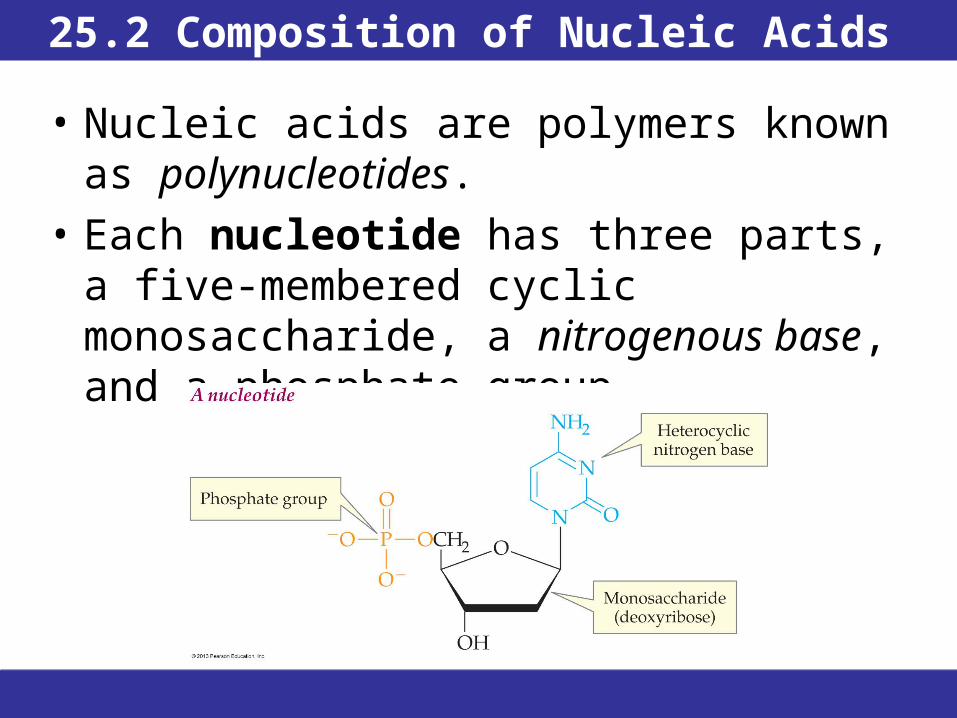
• Nucleic acids are polymers known as polynucleotides.
• Each nucleotide has three parts, a five-membered cyclic monosaccharide, a nitrogenous base, and a phosphate group.

25.2 Composition of Nucleic Acids
• Nucleic acids are polymers known as polynucleotides.
• There are two types of nucleic acids, DNA and RNA (ribonucleic acid).
• Each nucleotide has three parts: a five-membered cyclic monosaccharide, a nitrogenous base, and a phosphate group.

25.2 Composition of Nucleic Acids
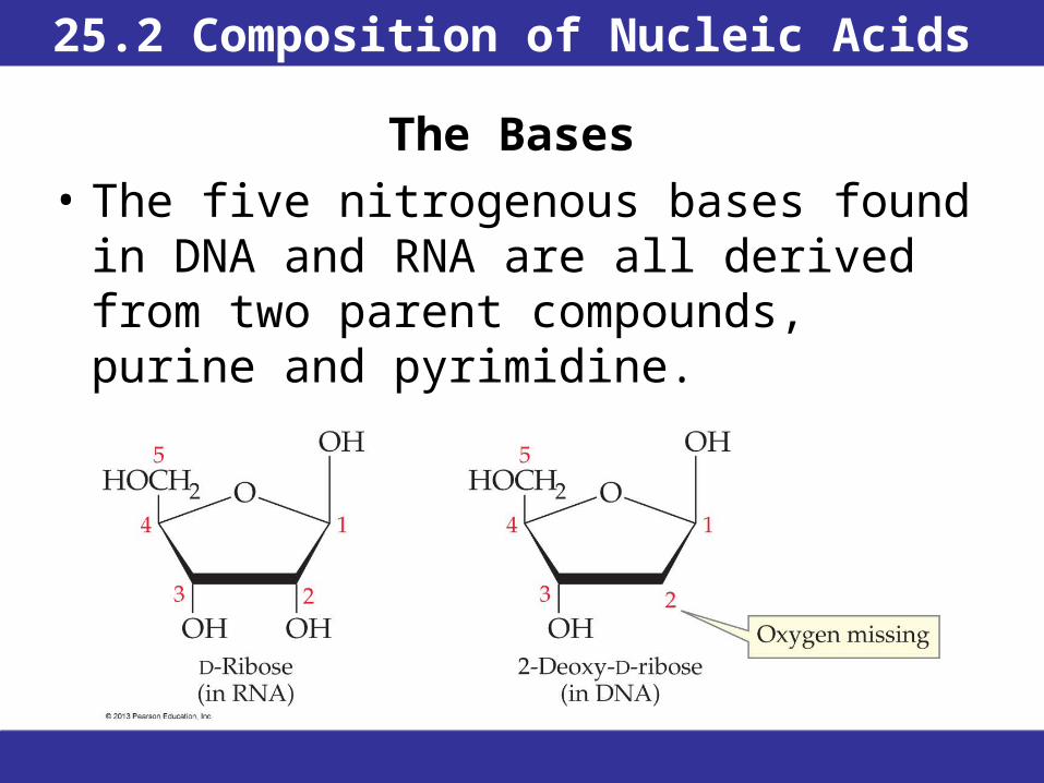
The Sugars
• In RNA, the sugar is D-ribose, as indicated by the name ribonucleic acid.
• In DNA, the sugar is 2-deoxyribose, giving deoxyribonucleic acid.
25.2 Composition of Nucleic Acids
The Bases
• The five nitrogenous bases found in DNA and RNA are all derived from two parent compounds, purine and pyrimidine.
25.2 Composition of Nucleic Acids
The Bases
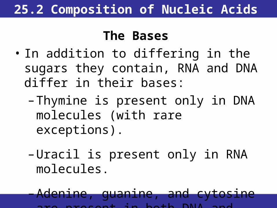
• In addition to differing in the sugars they contain, RNA and DNA differ in their bases:
– Thymine is present only in DNA molecules (with rare exceptions).
– Uracil is present only in RNA molecules.
– Adenine, guanine, and cytosine are present in both DNA and RNA.
25.2 Composition of Nucleic Acids
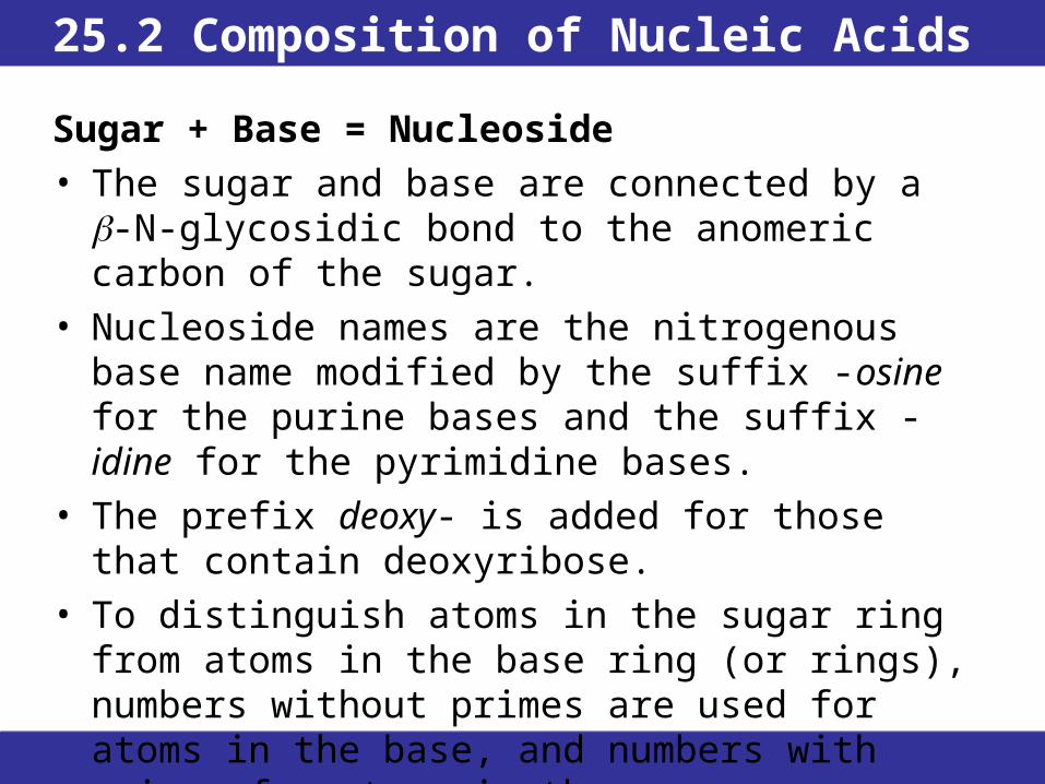
Sugar + Base = Nucleoside
• The sugar and base are connected by a -N-glycosidic bond to the anomeric carbon of the sugar.
• Nucleoside names are the nitrogenous base name modified by the suffix -osine for the purine bases and the suffix -idine for the pyrimidine bases.
• The prefix deoxy- is added for those that contain deoxyribose.
• To distinguish atoms in the sugar ring from atoms in the base ring (or rings), numbers without primes are used for atoms in the base, and numbers with primes for atoms in the sugar.
25.2 Composition of Nucleic Acids
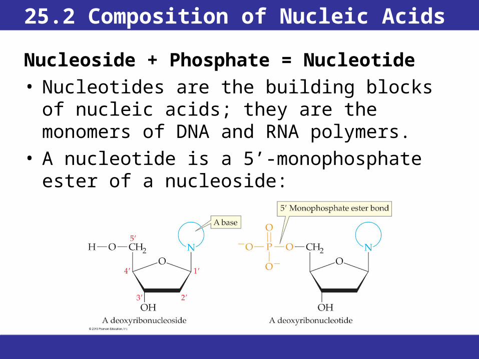
Nucleoside + Phosphate = Nucleotide• Nucleotides are the building blocks of nucleic
acids; they are the monomers of DNA and RNA polymers.
• A nucleotide is a 5’-monophosphate ester of a nucleoside:

25.2 Composition of Nucleic Acids
Nucleoside + Phosphate = Nucleotide
• Nucleotides are named by adding 5’-monophosphate at the end of the nucleoside name.
• Nucleotides containing ribose are ribonucleotides.
• Those that contain 2-deoxy-D-ribose are deoxyribonucleotides, designated by leading their abbreviations with a lower case “d”.
• Phosphate groups can be added to any of the nucleotides to form diphosphate or triphosphate esters. These esters are named with the nucleoside name plus diphosphate or triphosphate.

25.2 Composition of Nucleic Acids
25.2 Composition of Nucleic Acids
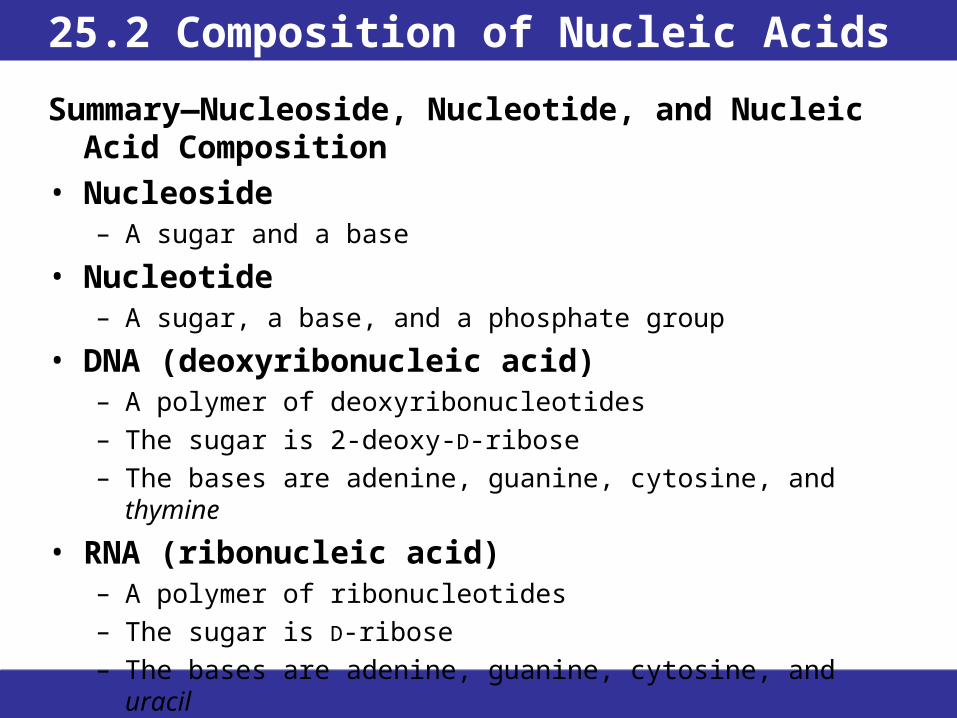
Summary—Nucleoside, Nucleotide, and Nucleic Acid Composition
• Nucleoside– A sugar and a base
• Nucleotide– A sugar, a base, and a phosphate group
• DNA (deoxyribonucleic acid)– A polymer of deoxyribonucleotides– The sugar is 2-deoxy-D-ribose– The bases are adenine, guanine, cytosine, and thymine
• RNA (ribonucleic acid)– A polymer of ribonucleotides– The sugar is D-ribose– The bases are adenine, guanine, cytosine, and uracil
25.3 The Structure of Nucleic Acid Chains
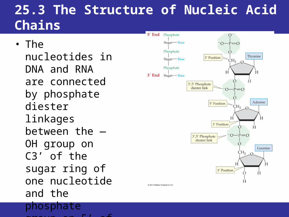
• The nucleotides in DNA and RNA are connected by phosphate diester linkages between the —OH group on C3’ of the sugar ring of one nucleotide and the phosphate group on 5’ of the next nucleotide.
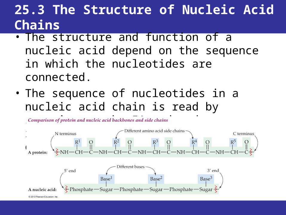
25.3 The Structure of Nucleic Acid Chains
• The structure and function of a nucleic acid depend on the sequence in which the nucleotides are connected.
• The sequence of nucleotides in a nucleic acid chain is read by starting at the 5’ end and identifying the bases in the order of occurrence.

25.4 Base-Pairing in DNA: The Watson-Crick Model
• Analysis of the nitrogenous bases in DNA samples from many different species revealed that the amounts of adenine and thymine were always equal, and the amounts of cytosine and guanine were always equal (A=T and G=C).
• The proportions of each (A/T:G/C) vary from one species to another.
• This is Chargoff’s Rule, and it suggests that the bases occur in discrete pairs.
25.4 Base-Pairing in DNA: The Watson-Crick Model
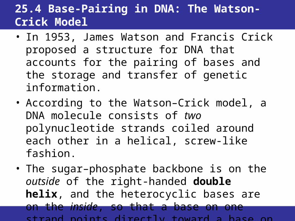
• In 1953, James Watson and Francis Crick proposed a structure for DNA that accounts for the pairing of bases and the storage and transfer of genetic information.
• According to the Watson–Crick model, a DNA molecule consists of two polynucleotide strands coiled around each other in a helical, screw-like fashion.
• The sugar–phosphate backbone is on the outside of the right-handed double helix, and the heterocyclic bases are on the inside, so that a base on one strand points directly toward a base on the second strand.

25.4 Base-Pairing in DNA: The Watson-Crick Model

25.4 Base-Pairing in DNA: The Watson-Crick Model
• The two strands of the DNA double helix are said to be antiparallel.
• The stacking of hydrophobic bases in the interior and the alignment of the hydrophilic groups on the exterior provide stability to the structure.
• Each pair of bases in the center of the double helix is connected by hydrogen bonding. Adenine and thymine (A-T) form two hydrogen bonds, and cytosine and guanine (C-G) form three hydrogen bonds.

25.4 Base-Pairing in DNA: The Watson-Crick Model
• The pairing of the bases is complementary. • Wherever a thymine occurs in one strand, an adenine
falls opposite it in the other strand. • Wherever a cytosine occurs in one strand, a guanine
falls opposite it on the other strand. • A and T and C and G occur in equal amounts.
25.5 Nucleic Acids and Heredity
• Duplication, transfer, and expression of genetic information occur as the result of three fundamental processes: replication, transcription, and translation.– Replication is the process by which a replica, or
identical copy, of DNA is made when a cell divides, so that each daughter cell has the same DNA.
– Transcription is the process by which DNA is read and copied. The products of transcription are ribonucleic acids, which carry the instructions stored by DNA to the sites of protein synthesis.
– Translation is the process by which the messages carried by RNA are used to build proteins.

25.6 Replication of DNA
• DNA replication begins in the nucleus with partial unwinding of the double helix; this process involves enzymes known as helicases.
• The unwinding occurs simultaneously in many specific origins of replication.
• The DNA strands separate, exposing the bases and forming a “bubble” in which the replication process can begin.
• At either end are branch points known as replication forks.
25.6 Replication of DNA
• Multisubunit enzymes called DNA polymerases facilitate transcription of the single-stranded DNA.
• Nucleoside triphosphates carrying each of the four bases are available, and move into place by forming hydrogen bonds with the bases exposed on the DNA template strand.
• DNA polymerase catalyzes covalent bond formation between the 5’ phosphate group of the arriving nucleoside triphosphate and the 3’ —OH at the end of the growing polynucleotide strand.

25.6 Replication of DNA
• The template strand can only be read in the 3’ to 5’ direction, and the new DNA strand can grow only in the 5’ to 3’ direction.
• In each new double helix, one strand is the template and the other is the newly synthesized strand. This is semiconservative replication.
25.6 Replication of DNA
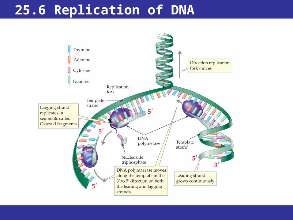
• Only the leading strand, is able to grow continuously; the DNA polymerase traveling in the 3’ to 5’ direction, is moving in the same direction as the replication fork.
• On the other strand, the DNA polymerase is moving in the opposite direction as the replication fork.
• The lagging strand is replicated in short segments called Okazaki fragments. These short DNA segments are joined together by DNA ligase.
• A human cell contains 3 billion base pairs. Yet a random error occurs only about once in each 10 billion to 100 billion bases. The complete copying process in human cells takes several hours.
25.6 Replication of DNA
25.7 Structure and Function of RNA
• The three types of RNA make it possible for the information carried by DNA to be put to use in the synthesis of proteins.
• Ribosomal RNAs Outside the nucleus are ribosomes—small granular organelles where protein synthesis takes place. Each ribosome is a complex consisting of about 60% ribosomal RNA (rRNA) and 40% protein, with a total molecular mass of approximately 5,000,000 amu.
25.7 Structure and Function of RNA
• Transfer RNAs (tRNA) are smaller RNAs that deliver amino acids one by one to protein chains growing at ribosomes. Each tRNA carries only one amino acid.
• Messenger RNAs (mRNA) carry information transcribed from DNA. They are formed in the nucleus and transported to ribosomes, where proteins are synthesized. They are polynucleotides that carry the same code for proteins as does the DNA.

25.7 Structure and Function of RNA
It’s a Ribozyme!• The reaction that removes unneeded sections from newly
synthesized mRNA is accomplished by the mRNA itself acting as the reaction catalyst.
• The slicing out of unneeded bases (introns) and splicing together of the rest of the mRNA is termed spliceosome activity.
• Since then more than 500 ribozymes in different organisms have been identified.
• During protein synthesis in a ribosome, 23S RNA catalyzes the formation of the peptide bond between amino acids in the growing protein molecule. The associated proteins, all 31 of them, appear to provide the necessary structure to maintain the correct three-dimensional relationship among the molecules involved in protein synthesis.
• Basic research into the nature and functions of ribozymes continues. However, applied research is directed toward their use in medical treatments.

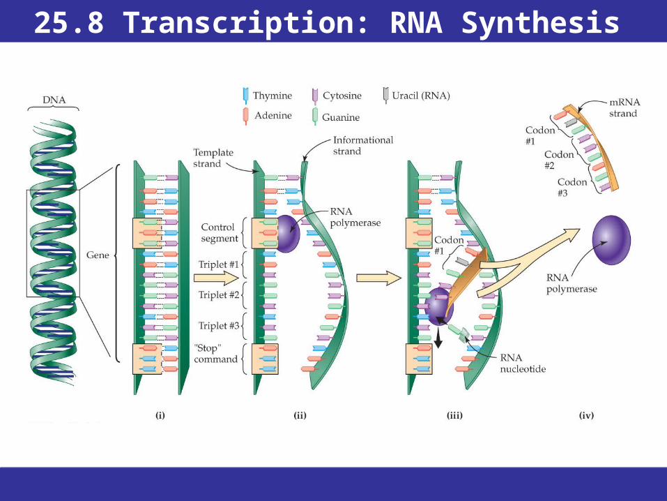
25.8 Transcription: RNA Synthesis
• Ribonucleic acids are synthesized in the cell nucleus.
• Before leaving the nucleus, all types of RNA molecules are modified in ways that enable them to perform different functions.
• In transcription, a small section of the DNA double helix unwinds, the bases on the strands are exposed, and complementary nucleotides are attached.

25.8 Transcription: RNA Synthesis
• rRNA, tRNA, and mRNA are synthesized in the same manner.
• The DNA strand that is transcribed is the template strand; its complement in the original helix is the informational strand.
• The mRNA molecule is an exact RNA-duplicate of the DNA informational strand, with the exception that a U replaces each T in the DNA strand.

25.8 Transcription: RNA Synthesis
• Transcription begins when RNA polymerase recognizes a control segment in DNA that precedes the nucleotides to be transcribed.
• RNA polymerase moves down the DNA segment, adding complementary nucleotides to the growing RNA strand.
• Transcription ends when the RNA polymerase reaches a termination sequence that signals the end of the sequence to be copied.
25.8 Transcription: RNA Synthesis

25.8 Transcription: RNA Synthesis
• At the end of transcription, the mRNA molecule contains a matching base for every base that was on the informational DNA strand.
• Only about 10% of the base pairs in DNA code for genes.
• The code for a gene is contained in one or more small sections of DNA called an exon.
• The code for a given gene may be interrupted by a non-coding sequence of bases called an intron.
25.8 Transcription: RNA Synthesis
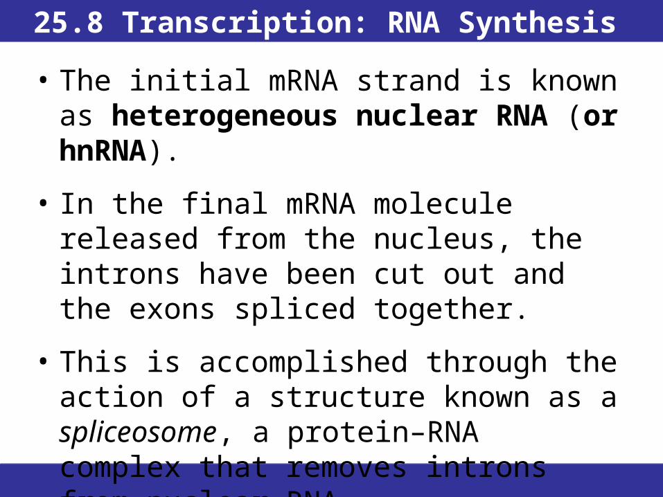
• The initial mRNA strand is known as heterogeneous nuclear RNA (or hnRNA).
• In the final mRNA molecule released from the nucleus, the introns have been cut out and the exons spliced together.
• This is accomplished through the action of a structure known as a spliceosome, a protein–RNA complex that removes introns from nuclear RNA.
25.9 The Genetic Code
• The sequence in an mRNA is a coded sentence that spells out the order in which amino acid residues should be joined to form a protein.
• Codon A sequence of three ribonucleotides that codes for a specific amino acid or stops translation.
• Genetic code The sequence of nucleotides, coded in triplets (codons) in mRNA, that determines the sequence of amino acids in protein synthesis.

25.9 The Genetic Code
• Of the 64 possible three-base combinations in RNA, 61 code for specific amino acids and 3 code for chain termination (the stop codons).
• The “meaning” of each codon is the genetic code, and is universal to all but a few living organisms.
• Most amino acids are specified by more than one codon.
• Codons are always written in the 5’ to 3’ direction.

25.9 The Genetic Code

25.9 The Genetic Code
DNA informational strand:
5’ ATG CCA GTA GGC CAC TTG TCA 3’
DNA template strand:
3’ TAC GGT CAT CCG GTG AAC AGT 5’
mRNA:
5’ AUG CCA GUA GGC CAC UUG UCA 3’
Protein:
Met Pro Val Gly His Leu Ser

25.9 The Genetic CodeViruses and AIDS
• Viruses are infectious agents that can replicate only inside living cells.
• Virus particles consist of only some nucleic acid and a protein coating.
• Once a virus enters a living cell, it takes over the host cell and forces it to produce virus copies.
• The replication of DNA viruses is straightforward: the cell replicates the viral DNA, the viral DNA is transcribed to RNA and many copies of the capsid proteins are made.
• After an RNA virus infects a cell either the cell must transcribe and produce proteins directly from the viral RNA template, or else it must first produce DNA from the viral RNA by reverse transcription. Reverse transcriptase is provided by the virus itself.
• The human immunodeficiency virus (HIV-1) responsible for most cases of AIDS (Acquired Immune Deficiency Syndrome) is a retrovirus. Development of drugs for the treatment of AIDS is especially challenging because HIV has the highest mutation rate of any known virus.
• The best success thus far has been with false nucleosides: once incorporated into the viral DNA, they then slow down production of new viral RNA.
• Some AIDS vaccines have shown promise, but no effective vaccine has yet been developed.

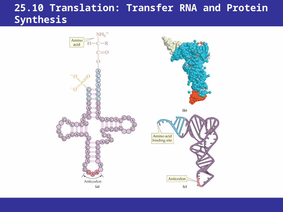
25.10 Translation: Transfer RNA and Protein Synthesis
• Protein synthesis occurs at ribosomes, which are located outside the nucleus in the cytoplasm of cells.
• mRNA binds to the ribosome.
• Amino acids are delivered one-by-one by transfer RNA (tRNA) molecules.
• Amino acids are joined into a specific protein by the ribosomal “machinery.”

25.10 Translation: Transfer RNA and Protein Synthesis
• Every cell contains more than 20 different tRNAs, each designed to carry a specific amino acid.
• A tRNA molecule is a single polynucleotide chain held together by regions of base pairing in a partially helical structure like a cloverleaf.
• In three dimensions, a tRNA molecule is L-shaped.
25.10 Translation: Transfer RNA and Protein Synthesis
25.10 Translation: Transfer RNA and Protein Synthesis
• At one end of the L-shaped tRNA molecule, an amino acid is bonded to its specific tRNA by an ester linkage between the —COOH of the amino acid and an —OH group on the last ribose at the 3’ end of the tRNA chain.
• Individual synthetase enzymes are responsible for connecting each amino acid with its partner tRNA in an energy-requiring reaction.
• This reaction is referred to as charging the tRNA. Once charged, the tRNA is ready to be used in the synthesis of new protein.

25.10 Translation: Transfer RNA and Protein Synthesis
• At the other end of the tRNA “L” is an anticodon.
• The anticodon of each tRNA is complementary to the mRNA codon designating the amino acid that the tRNA carries.
• This is how the genetic message of the codons, is translated into the sequence of amino acids in a protein.

25.10 Translation: Transfer RNA and Protein Synthesis

25.10 Translation: Transfer RNA and Protein Synthesis
Translation Initiation• Each ribosome is made up of two subunits called the
small subunit and the large subunit which contain protein enzymes and ribosomal RNA (rRNA).
• Protein synthesis begins with the binding of an mRNA to the small subunit of a ribosome, joined by the first tRNA. The first codon on the 5’ end of mRNA, an AUG, acts as a “start” signal for the translation machinery and codes for a methionine-carrying tRNA.
• Initiation is completed when the large ribosomal subunit joins the small one and the methionine-bearing tRNA occupies one of the two binding sites on the united ribosome.
• Met may be removed by post-translational modification.

25.10 Translation: Transfer RNA and Protein Synthesis
Translation Elongation
• A second binding site where the next codon on mRNA is exposed and the tRNA carrying the next amino acid is attached.
• A ribozyme in the large subunit catalyzes the new peptide bond and breaks the bond linking amino acid 1 to its tRNA.
• These energy-requiring steps are fueled by the hydrolysis of GTP to GDP.
• The first tRNA leaves the ribosome, and the ribosome shifts three positions (one codon) along the mRNA chain. A second binding site is opened up to accept the tRNA carrying the next amino acid.
• A single mRNA can be “read” simultaneously by many ribosomes. The growing polypeptides increase in length as the ribosomes move down the mRNA strand.

25.10 Translation: Transfer RNA and Protein Synthesis
Translation Termination
• When synthesis of the protein is completed, a “stop” codon signals the end of translation.
• An enzyme called a releasing factor then catalyzes cleavage of the polypeptide chain from the last tRNA, the tRNA and mRNA molecules are released from the ribosome, and the two ribosome subunits separate.
• This step also requires energy from GTP.
• Adding one amino acid to the growing polypeptide chain requires four molecules of GTP, excluding the energy needed to charge the tRNA.

25.10 Translation: Transfer RNA and Protein Synthesis
Influenza—Variations on a Theme• Flu is caused by the influenza virus, of which there are three major
types—A, B, and C.• Getting a flu shot can prevent illness from types A and B influenza.
You have to be re-immunized yearly because the influenza virus mutates.
• Flu can cause either an epidemic or a pandemic. Both have occurred.
• Many subtypes of influenza A viruses are also found in animals. Birds are susceptible to all known subtypes of the influenza A virus and serve as a reservoir.
• Because pigs are susceptible to infection by both avian and human viruses, they can serve as a “mixing vessel” for the scrambling of genetic material from human and avian viruses.
• In 1918, a strain of influenza that became known as Spanish flu killed an estimated 20–50 million people worldwide.
Chapter Summary
1. What is the composition of the nucleic acids, DNA and RNA?
• Nucleic acids are polymers of nucleotides. Each nucleotide contains a sugar, a base, and a phosphate group.
• The sugar is D-ribose in ribonucleic acids (RNAs) and 2-deoxy-D-ribose in deoxyribonucleic acids (DNAs). The C5-OH of the sugar is bonded to the phosphate group, and the anomeric carbon of the sugar is connected by an N-glycosidic bond to one of five heterocyclic nitrogen bases.
• A nucleoside contains a sugar and a base, but not the phosphate group.
• In DNA and RNA, the nucleotides are connected by phosphate diester linkages between the 3’—OH group of one nucleotide and the 5’ phosphate group of the next nucleotide. DNA and RNA both contain adenine, guanine, and cytosine; thymine occurs in DNA and uracil occurs in RNA.
Chapter Summary, Continued
2. What is the structure of DNA? • The DNA in each chromosome consists of two
polynucleotide strands twisted together in a double helix.
• The sugar–phosphate backbones are on the outside, and the bases are in the center of the helix. The bases on the two strands are complementary—opposite every thymine is an adenine, opposite every guanine is a cytosine.
• The base pairs are connected by hydrogen bonds (two between T and A; three between G and C). Because of the base pairing, the DNA strands are antiparallel: One DNA strand runs in the 5’ to 3’ direction and its complementary partner runs in the 3’ to 5’ direction.
Chapter Summary, Continued
3. How is DNA reproduced? • Replication requires DNA polymerase enzymes and
deoxyribonucleoside triphosphates. • The DNA helix partially unwinds and the enzymes
move along the separated DNA strands, synthesizing a new strand with bases complementary to those on the unwound DNA strand being copied.
• The enzymes move only in the 3’ to 5’ direction along the template strand (and thus new DNA strands only grow in the 5’ to 3’ direction), so that one strand is copied continuously and the other strand is copied in segments as the replication fork moves along.
• In each resulting double helix, one strand is the original template strand and the other is the new copy.
Chapter Summary, Continued
4. What are the functions of RNA?
• Messenger RNA (mRNA) carries the genetic information out of the nucleus to the ribosomes in the cytosol, where protein synthesis occurs.
• Transfer RNAs (tRNAs) circulate in the cytosol, where they bond to amino acids that they then deliver to ribosomes for protein synthesis.
• Ribosomal RNAs (rRNAs) are incorporated into ribosomes.

Chapter Summary, Continued
5. How do organisms synthesize messenger RNA? • In transcription, one DNA strand serves as the
template and the other, the informational strand, is not copied.
• Nucleotides carrying bases complementary to the template bases between a control segment and a termination sequence are connected one by one to form mRNA.
• The primary transcript mRNA (or hnRNA) is identical to the matching segment of the informational strand, but with uracil replacing thymine.
• Introns, which are base sequences that do not code for amino acids in the protein, are cut out before the final transcript mRNA leaves the nucleus.
Chapter Summary, Continued
6. How does RNA participate in protein synthesis? • The genetic information is read as a sequence of codons—triplets
of bases that give the sequence of amino acids in a protein. • Of the 64 possible codons, 61 specify amino acids and 3 are stop
codons. Each tRNA has at one end an anticodon consisting of three bases complementary to those of the mRNA codon that specifies the amino acid it carries.
• Initiation of translation is the coming together of the large and small subunits of the ribosome, an mRNA, and the first amino acid–bearing tRNA connected at the first of the two binding sites in the ribosome.
• Elongation proceeds as the next tRNA arrives at the second binding site, its amino acid is bonded to the first one, the first tRNA leaves, and the ribosome moves along so that once again there is a vacant second site. These steps repeat until the stop codon is reached. The termination step consists of separation of the two ribosome subunits, the mRNA, and the protein.