Kinetics of Left Ventricular Strains and Torsion During...
32
1 Kinetics of Left Ventricular Strains and Torsion During Incremental Exercise in Healthy Subjects: The Key Role of Torsional Mechanics for Systolic-Diastolic Coupling Running title: LV Torsion During Incremental Exercise Grégory Doucende 1 , MS, Iris Schuster 2 , MD, PhD, Thomas Rupp 1 , PhD, Aliona Startun 2 , MD, Michel Dauzat 2 , MD, PhD, Philippe Obert 1 , PhD, Stéphane Nottin 1 , PhD 1 EA 4278, Physiology and Physiopathology of Cardio-vascular Adaptations to Exercise, Faculty of Sciences, 33 Rue Louis Pasteur, 84000 Avignon, France. 2 EA 2992, Dysfunction of Vascular Interfaces Research Laboratory, Faculty of Medicine, Montpellier I University and Nîmes University Hospital Center, Place Prof. Debre, 30029 Nîmes, France Correspondence to: Stéphane Nottin EA 4278, Laboratoire de physiologie et physiopathologie des adaptations cardiovasculaires à l’exercice 33, Rue Louis Pasteur 84000 Avignon France Tel. +334 901 629 31 Fax: +334 901 444 09 E-mail: [email protected] Journal Subject Codes: [26], [31], [125], [150] nce. . f niversity and Nîmes University Hospital Center, Place Prof. D function of Vascular Interfaces Research Laboratory, Faculty niversity and Nîmes University Hospital Center, Place Prof. D by guest on June 9, 2018 http://circimaging.ahajournals.org/ Downloaded from
Transcript of Kinetics of Left Ventricular Strains and Torsion During...
1
Kinetics of Left Ventricular Strains and Torsion During Incremental
Exercise in Healthy Subjects: The Key Role of Torsional Mechanics for
Systolic-Diastolic Coupling
Running title: LV Torsion During Incremental Exercise
Grégory Doucende1, MS, Iris Schuster2, MD, PhD, Thomas Rupp1, PhD, Aliona Startun2,
MD, Michel Dauzat2, MD, PhD, Philippe Obert1, PhD, Stéphane Nottin1, PhD
1 EA 4278, Physiology and Physiopathology of Cardio-vascular Adaptations to Exercise,
Faculty of Sciences, 33 Rue Louis Pasteur, 84000 Avignon, France.
2 EA 2992, Dysfunction of Vascular Interfaces Research Laboratory, Faculty of Medicine,
Montpellier I University and Nîmes University Hospital Center, Place Prof. Debre, 30029
Nîmes, France
Correspondence to: Stéphane Nottin EA 4278, Laboratoire de physiologie et physiopathologie des adaptations cardiovasculaires à l’exercice 33, Rue Louis Pasteur 84000 Avignon France Tel. +334 901 629 31 Fax: +334 901 444 09 E-mail: [email protected]
Journal Subject Codes: [26], [31], [125], [150]
nce. .
f
niversity and Nîmes University Hospital Center, Place Prof. D
function of Vascular Interfaces Research Laboratory, Faculty
niversity and Nîmes University Hospital Center, Place Prof. D
by guest on June 9, 2018http://circim
aging.ahajournals.org/D
ownloaded from
2
ABSTRACT
Background – Dynamics of systolic and diastolic strains and torsional mechanics of
the left ventricle (LV) and their relation to diastolic filling have never been evaluated at
various exercise intensities.
Methods and Results – Speckle tracking echocardiography (STE) was performed in
20 healthy sedentary subjects at rest and during a progressive submaximal exercise test at
20%, 30% and 40% of maximal aerobic power. LV twist increased progressively with
exercise intensity (10.5±3.2 to 15.8±4.5 deg, P<0.001), whereas longitudinal strain remained
unchanged after the first workload, underlining the key role of torsional “reserve” in systolic-
diastolic-coupling during exercise. The increase in diastolic untwisting (-88.7±34.2 to -
182.9±53.5 deg.s-1, P<0.01) was correlated to enhanced systolic twist (R=0.61, P<0.001), and
its magnitude of increase was significantly higher compared to diastolic longitudinal and
circumferential strain rates (119±64 versus 65±44 and 57±24 %, respectively), emphasizing
its contribution to diastolic filling. The timing of peak untwisting and the chronology of
diastolic mechanical events were unchanged during effort. Untwisting was driven mainly by
apical rotation and determined mitral opening and isovolumic relaxation time (R=0.47 and
0.61 respectively, P<0.001), whereas basal rotation, longitudinal and circumferential diastolic
strain rates were major determinants of increased early diastolic filling (R=0.64, 0.79 and
0.81 respectively, P<0.001).
Conclusions – The use of STE gives new insights into physiologic adaptive LV mechanics
during incremental exercise in healthy subjects, underlining the key role of torsional
mechanics. It might be useful to better understand the mechanisms of diastolic dysfunction
and exercise intolerance in various pathologic conditions.
Key Words: speckle tracking echocardiograhy – exercise – left ventricular torsion - left
ventricular untwisting
wisi titiiiiiingngngngngngng ( ((((((-8-8-8-8-8-8-88.8.88888 7±7±7±7±7±7±7±34343434343434
P
i d
t m
o diastolic filling The timing of peak untwisting and the chronol
-1, P<0.01) was correlated to enhanced systolic twist (R=0.61, P
increase was significantly higher compared to diastolic longitud
train rates (119±64 versus 65±44 and 57±24 %,d respectively), em
o diastolic filling The timing of peak untwisting and the chronol
by guest on June 9, 2018http://circim
aging.ahajournals.org/D
ownloaded from
3
INTRODUCTION
During diastole, left ventricle (LV) filling results from a complex interplay between
numerous factors such as myocardial relaxation, LV compliance, untwisting and loading
conditions. During systole, contraction of the cardiomyocytes induces not only normal, but
also shear strains, including LV torsion (i.e. basal clockwise rotation and apical
counterclockwise rotation) due to the helical orientation of myofibers 1-3. LV diastolic
untwisting is a consequence of both an active relaxation of the myocardium and a release of
the energy stored in compressed titin 4 and elastic components of the intersitium 5.
Importantly, untwisting occurs predominantly during isovolumic relaxation (IVR) and
promotes LV “suction” by increasing LV intraventricular (i.e from apex to base) pressure
gradients 6, 7. Interestingly, LV untwisting is decreased and/or delayed in states associated
with diastolic dysfunction such as tachycardia-induced heart failure 8, dilated cardiomyopathy
9, aortic stenosis 10, or following strenuous exercise 11. The evaluation of LV normal and shear
strains and their timing during diastole is therefore of significant interest to assess diastolic
dysfunction.
Exercise stress echocardiography is a powerful tool to provide additional diagnostic
and prognostic information in a variety of diseases 12 and allows early detection of subtle
myocardial dysfunction. During exercise, the increase in stroke volume is limited by diastolic
filling, 13, 14 as the increase in heart rate shortens the duration of diastole. Additionally, left
ventricular filling must be accomplished at relatively low filling pressures to avoid pulmonary
vascular congestion. Early studies underlined the key role of untwisting in LV filling during
exercise 15-17, but the time course of diastolic mechanical events, including both LV strains
and torsional mechanics, has not been fully described. Moreover, no data are available
fffffff t t t t t t thehehehehehehe i i i i i i intntntntntntntererererererersisisisisisisitititititititiumumumumumumum 5
c relaxaxaxaxaxaxaxatatatatatatatioioioioiooonnnnnnn (I(I(I(I(I(I(IVRVRVVVVVg p y g (
erestin y, LV untwisting is decreased and/or delayed in states as
s d
g p y g (
ction” by increasing LV intraventricular (i.e from apex to base)
erestingly, LV untwisting is decreased and/or delayed in states as
sfunction such as tachycardia-induced heart failure 8, dilated card
by guest on June 9, 2018http://circim
aging.ahajournals.org/D
ownloaded from
4
regarding kinetics with increasing exercise intensity and timing of diastolic myocardial events
during an incremental exercise. Recently, speckle tracking echocardiography (STE) provides
the ability to quantify LV strains 18 and torsion 19 at rest and during exercise 15, 16.
In the present study, we aimed to describe dynamics and timing of LV myocardial
mechanical events during an incremental exercise test. We used STE at rest and during an
exercise conducted on a dedicated ergometer. We hypothesized that (1) diastolic untwisting
would increase progressively with exercise intensity paralleling enhanced systolic twisting (2)
amplitude and timing of LV diastolic mechanics would adapt to preserve or enhance LV
filling when diastole shortens during effort.
METHODS
Study population
We evaluated 20 young, healthy and sedentary adult males (mean age: 25 ± 9 yr old).
None of them reported regular training habits nor had any clinical or anamnestic evidence of
cardiovascular disease or arterial hypertension. Subjects were excluded if resting
echocardiography demonstrated ejection fraction < 50%, significant valvular disease,
abnormal right ventricle function, or systolic arterial pulmonary pressure > 35 mmHg. This
study received approval from the local ethics committee and written informed consent was
obtained from all subjects.
Experimental protocol
nn
by guest on June 9, 2018http://circim
aging.ahajournals.org/D
ownloaded from
5
Body height and mass were assessed. The maximal aerobic power was initially
estimated via the Wasserman equation for the subject’s age and body mass, and corrected for
the semi-supine position (20 % was removed from normal values).
The subjects were installed on a dedicated semi-supine cycling ergometer (E-Bike
ergometer, GE Healthcare, Horten, Norway). After a 15 to 20 minutes resting period, each
subject underwent an exercise test including 3 stages of 6 minutes duration at 20%, 30% and
40% of their maximal aerobic power (respectively labeled W1, W2, and W3), and then stages
of 1 minute from 50% to exhaustion by 10% increments. The pedaling rate was kept constant
at 70 rpm for all subjects.
Two-dimensional (2-D) and Doppler Echocardiographic data were recorded at the end
of the resting period and during the last 4 minutes of W1, W2, and W3 stages. During the last
30 seconds of the test we measured stroke volume. Gas exchanges were measured
continuously by means of a cardiopulmonary exercise system (Ergocard, Medisoft S.A,
Sorinnes, Belgium). Systemic arterial blood pressure was measured during each stage of
exercise in the left arm using manual sphygmomanometry and auscultation. Mean arterial
pressure was calculated as 1/3 × systolic pressure + 2/3 × diastolic pressure.
Echocardiographic data acquisition
Images were obtained using a commercially available system (Vivid 7, GE Healthcare,
Horten, Norway) with a 3.5 Mhz sector scanning electronic transducer. We recorded cine
loops in parasternal short axis (basal, papillary muscle and apical levels) and in apical 4-
chambers views. Two-dimensional grayscale harmonic images were obtained at a rate of 65 to
90 frames per second, and color tissue velocity images were acquired at a rate of 120 to 140
frames per second. Images were acquired in cine loops triggered to the QRS complex and
dadadadadadadatatatatataata w w wwwwwererererererere e e e eee rererererererecococococococordrdrdrdrdrdrd
nd WWWWWWW3333333 stststststststagagagagagagageseseseseseses DuDDDDDDg g
e test we measured stroke volume. Gas exchan s were measure
m o
m . stemic arterial blood essure was measured duri each s
g g
e test we measured stroke volume. Gas exchanges were measure
means of a cardiopulmonary exercise system (Ergocard, Mediso
m). Systemic arterial blood pressure was measured during each s
by guest on June 9, 2018http://circim
aging.ahajournals.org/D
ownloaded from
6
saved digitally for subsequent off-line analysis with dedicated software (EchoPac 6.0, GE
Healthcare, Horten, Norway).
2-D and Tissue Doppler echocardiography
M-Mode measurements were obtained off-line from the parasternal short-axis view
recorded at the papillary muscle level. Pulsed Doppler LV inflow (E and/or A waves)
recordings were performed in the apical 4-chamber view. Aortic blood flow velocity was
recorded in the ascending aorta with a 2.0 Mhz transducer (Pedof) placed at the suprasternal
notch to assessed stroke volume and cardiac output, as previously used in our laboratory 20, 21.
Systemic vascular resistance was estimated at each workload as mean arterial pressure
divided by cardiac output. We measured the time delay in milliseconds from the onset of the
ECG QRS interval to the onset of aortic blood flow (aortic opening, AO), the peak of aortic
blood flow (Peak-S), the end of aortic blood flow (aortic closure, AC), the onset of early
filling blood flow (mitral opening, MO), the peak of early filling blood flow (Peak-E) and the
end of early filling blood flow (End-E). Isovolumic relaxation time (IVRT) was calculated as
MO - AC.
Tissue Doppler evaluation was performed off-line from color cine loops recorded in
the apical 4-chamber view. We assessed wall motion velocities at the mitral annulus level on
the septal and lateral walls. The ratio transmitral peak-E on peak early myocardial velocity of
the lateral wall was used as an index of LV filling pressure 22.
Speckle tracking echocardiography
lly y y y y y y usususususususededededededed i ii i i in n n n n n n ououououououour r rr rrr lalalalalalalabbbbbbb
meaaaaaaannnnnnn arararararararteteteteteteteriririririririalalalalalalal ppppppprerrrp
ac output. We measured the time delay in milliseconds from the
al to the onset of aortic blood flow aortic opening, AO the p a
-S the end of aortic blood flow ortic closure, A , the onset
p
ac output. We measured the time delay in milliseconds from the
al to the onset of aortic blood flow (aortic opening, AO), the pea
-S), the end of aortic blood flow (aortic closure, AC), the onset
by guest on June 9, 2018http://circim
aging.ahajournals.org/D
ownloaded from
7
Analysis of strain and torsion was conducted as previously described 11, 23. After
manually tracing the endocardial border on the end-systolic frame of the 2D sequence, the
software automatically tracked myocardial motion. Whenever the software signaled poor
tracking efficiency, the observer readjusted the endocardial trace line and/or the region of
interest width until a better tracking score could be obtained. Results were averaged on three
to five cardiac cycles. LV longitudinal strain and strain rate (SR) were assessed using an
apical 4-chamber view. Circumferential strain, SR and LV rotation and rotational rate were
assessed from short-axis views at basal and apical levels. Care was taken to ensure that the
basal short-axis plane contained the mitral valve, and that the apical plane was acquired with
the probe in a caudal position to improve LV apical rotation measurement 24.
2D-Strain data were processed with a specific toolbox (Scilab 4.1, Consortium Scilab,
INRIA-ENPC, Paris, France) developed in our laboratory. For temporal analysis, this
software adjusted all strain variables for inter-subjects differences in heart rate and transducer
frame rate acquisition. The time sequence was normalized to the percentage of systolic and
diastolic duration (i.e. AC represented 100% of systole and end of cardiac cycle represented
100% of diastole) using interpolations. After normalization, the software averaged each data
from 3 to 5 cardiac cycles and performed the detection of peak strains events and their timing
(expressed in percentage of systolic duration). Net LV torsion was calculated as the
instantaneous difference between LV apical - LV basal rotations. We calculated the following
indexes of diastolic function during isovolumic relaxation (IVR): 1) untwisting angle (UT°,°)
= twist at AC - twist at end-IVR; 2) percentage of untwisting during IVRT (%UTIVRT, %) =
(UT° / twist at AC)*100; and 3) mean rate of untwisting during the IVRT (mean UTIVRT, °.s-1)
= -UT° / IVRT. To assess the dynamics of global LV torsion and its relation to radial
displacement (reflecting volumetric changes of the LV) throughout the cardiac cycle, we
constructed twist-radial displacement loops 11, 25. Radial displacement data from 6 segments in
aaaasususususususurerererereerememememememementntntntntntnt 24..
cilabbbbbbb 4444444 1111111 CoCoCoCoCoCoConsnsnsnsnsnsnsorooop p (
a ,
d n
ition. The time s uence was normalized to the rcenta of y
p p
aris, France) developed in our laboratory. For temporal analysis,r
d all strain variables for inter-subjects differences in heart rate an
ition. The time sequence was normalized to the percentage of sy
by guest on June 9, 2018http://circim
aging.ahajournals.org/D
ownloaded from
8
basal and apical short axis planes were averaged to obtained the mean value of radial
displacement.
Statistical analysis
All values in the text and tables are expressed as mean ± SD and are shown as mean ±
SE in figures. The statistical analysis was performed using specific software (Statview 5.0,
SAS Insitute Inc. Cary, USA). For each cardiac variable, an analysis of variance with repeated
measures was performed with post hoc test using Bonferroni correction. Linear regressions
were performed to determine the relations between IVRT, peak-E and LV diastolic
mechanical events. A multiple stepwise regression analysis was done to determine the
mechanical events responsible for the increase in early filling during effort. Statistical
significance for all analysis was assumed if P < 0.05. Intraobserver reproducibility of speckle
tracking evaluation was previously assessed in 12 subjects and was inferior at 8% for both
strains and rotations.
RESULTS
Height and body mass of the subjects were 177 ± 5 cm and 72 ± 8 kg, respectively.
Resting echocardiographic and blood pressure data are presented in table 1. At the end of
exercise, the maximal values were as follows: aerobic power: 221 ± 33 W, oxygen uptake: 34
± 5 mL.min-1.kg-1, heart rate: 179 ± 12 bpm, systolic and diastolic pressures: 197 ± 8 and 99 ±
7 mmHg, SV: 115.9 ± 17.3 mL and cardiac output: 20.6 ± 2.7 L.min-1. The W1, W2, and W3
stages were performed at 19 ± 1, 28 ± 1 and 38 ± 2% of maximal aerobic power, respectively.
Heart rate during each stage was respectively 100 ± 12, 110 ± 12 and 121 ± 12 bpm.
EEEEEEE a a a a a aandndndndndndnd L L LLL L LV V V V V V V didididididdiasasasasasasastototototototolllllll
doneeeeeee tototototototo dddddddetetetetetetetererererererermimimimmmm np p g y
ts re onsible for the increase in earl fillin duri effort. Statis
a t
o %
g y
ts responsible for the increase in early filling during effort. Statis
all analysis was assumed if P < 0.05. Intraobserver reproducibilit
on was previously assessed in 12 subjects and was inferior at 8%
by guest on June 9, 2018http://circim
aging.ahajournals.org/D
ownloaded from
9
Kinetics of LV hemodynamic parameters
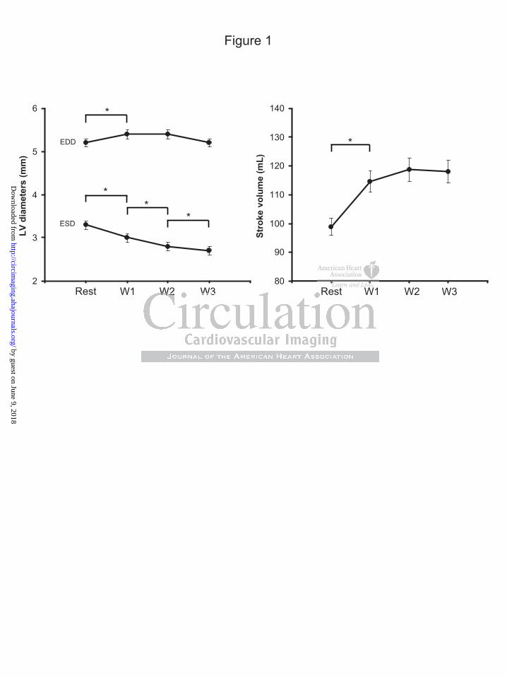
Kinetics of LV internal diameters and SV are shown in figure 1. LV end-diastolic
diameter increased from rest to W1, and then slightly decreased from W1 to W3. LV end-
systolic diameter progressively decreased from rest to W3. Stroke volume increased by 14 ± 8
% from rest to W1 and then remained constant until W3. Peak-E increased from rest to W2
(0.88 ± 0.19 versus 1.23 ± 0.17 m.s-1, P<0.0001) then remained constant (1.23 ± 0.17 versus
1.28 ± 0.17 m.s-1, NS), whereas mean peak early diastolic myocardial velocity increased only
between rest and W1 (12.4 ± 1.7 versus 14.6 ± 1.3 cm.s-1, P<0.0001) then plateaued until W3
(14.6 ± 1.3 versus 14.7 ± 1.9 cm.s-1, NS). LV filling pressure increased from rest to W1 (6.5 ±
1.1 versus 7.5 ± 1.4, P<0.0001), and then remained constant (7.5 ± 1.4 versus 8.1 ± 1.5, NS).
Kinetics of LV systolic strains
Kinetics of LV longitudinal and circumferential (averaged from basal and apical
levels) peak systolic strains and LV rotations are shown in figure 2. LV longitudinal strain
increased only from rest to W1, whereas LV circumferential strain and rotations progressively
increased from rest to W3. As a result, LV torsion increased progressively from rest to W3
(10.5 ± 3.2 to 15.8 ± 4.5°, P<0.0001). Times to peak strains and torsion did not change during
exercise excepted for LV apical rotations which appeared earlier at W2 and W3 (table 2).
Peak LV systolic SR and rotational and twisting rates progressively increased from
rest to W3. Respective times to peak were not affected by exercise intensity except for LV
peak basal rotational rate which was significantly delayed during exercise. LV peak apical
00000000001010101010101) ) ) ) ) ) ) thththththththenenenenenenen p p p p ppplalalalaaaateteteteteteteaauauauauaa
reasssededededededed fffffffrororororororommmmmmm rererererererestssss) g p
1 1
y
) g p
1.4, P<0.0001), and then remained constant (7.5 ± 1.4 versus 8.1
ystolic strains
by guest on June 9, 2018http://circim
aging.ahajournals.org/D
ownloaded from
10
rotational rate showed no significant change from rest to exercise, and occurred earlier than
all other peak SR (table 3) independently of exercise intensity.
Kinetics of LV diastolic strains
During diastole, peak LV SR, rotational and untwisting rates increased progressively
from rest to W3 (table 3). A strong correlation was observed between peak LV torsion and
peak LV untwisting rate (R=0.61, P<0.001). The magnitude of increase (expressed in
percentage compared to resting conditions) was significantly higher for LV untwisting rate
(119 ± 64 %) than for changes of peak longitudinal and circumferential diastolic SR (65 ± 44
and 57 ± 24 %, respectively) (figure 3). Whatever the exercise intensity, peak LV apical
rotational and untwisting rates occurred closely to MO, whereas peak LV basal rotational and
strain rates were concomitant with transmitral peak-E (figure 4). Peak untwisting rate and
peak LV diastolic apical rotational rate were correlated with IVRT (R=0.63, P<0.001 and
R=0.48, P<0.001, respectively), while peak LV diastolic basal rotational rate and longitudinal
and circumferential SR were related to peak-E (R=0.64, 0.79 and 0.81 respectively, P<0.001).
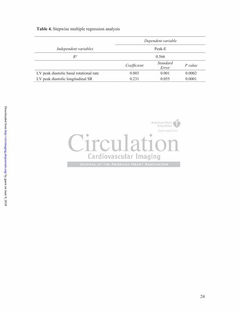
The stepwise regression analysis shown that these mechanical events mainly explained the
increase in peak-E with exercise intensity (table 4).
Mean UTIVRT progressively increased from rest to W3 (table 5). %UTIVRT decreased
from rest to W1 (45.3 ± 22.5 versus 28.0 ± 15.4%, P=0.0003) then remained unchanged
during exercise (from 28.0 ± 15.4 to 30.6 ± 16.6%, NS). Peak untwisting rate occurred during
the IVR period at rest and was delayed after MO during exercise when the IVRT shortened
(figure 4). UTIVRT (deg) was stable from rest to W3 (between 3.5 ± 1.6 and 4.1 ± 2.1°, NS).
Twist-radial displacement loops (figure 5) indicated that exercise intensity did not
affect the relationship between radial displacement and torsion during the cardiac cycle.
eeeeeerererererererentntntntntntntiaiaiaiaiaiaial l ll l l dididididididiasasasasasasastotototototoolililililililic cc c cc c
ntensssssssititititititityyyyyyy pppppppeaeaeaeaeaeaeakkkkkkk LVLLLLLLp y) ( g ) y p
t o
concomitant with transmitral peak-E ure . Peak untwisting
c 0
y g y
twisting rates occurred closely to MO, whereas peak LV basal ro
concomitant with transmitral peak-E (figure 4). Peak untwisting
c apical rotational rate were correlated with IVRT (R=0.63, P<0
by guest on June 9, 2018http://circim
aging.ahajournals.org/D
ownloaded from

11
Systole was characterized by roughly linear relationship between torsion and radial
displacement, and diastole by rapid untwisting before radial displacement.
DISCUSSION
This study based on STE reports comprehensive kinetics of LV systolic and diastolic
strains and torsion and their relationship to diastolic filling at different submaximal exercise
intensities in healthy sedentary subjects. The major findings of the study were (1) LV systolic
twist increased progressively with exercise intensity, whereas systolic longitudinal strain
remained unchanged after the first workload, underlining the key role of torsional “reserve” in
systolic-diastolic-coupling during exercise (2) Increased diastolic untwist, paralleling
enhanced systolic twist, was driven mainly by early apical rotation and determined early
mitral valve opening, whereas basal rotation, longitudinal and circumferential diastolic strain
rates were major determinants of increased early diastolic filling.
The key role of LV torsional reserve in systolic-diastolic coupling during exercise
Very few data are available regarding the kinetics of LV strains during an incremental
exercise test 26. Using STE, we observed that whereas longitudinal strains remained
unchanged after the first workload of 20% of maximal aerobic power (figure 1), LV
circumferential strains, rotations and torsion increased regularly with exercise intensity. The
underlying mechanisms responsible for this heterogeneous response of LV strains are not well
understood. LV torsion result from a complex arrangement of myocardial fibers within the
LV wall 27, 28. Contraction of the opposite helically oriented fibers creates LV torsion which
follows subepicardial layers because of their longer lever 1, and the progressive increase in
yyyyyyy r r r r r r rololololololole e e e ee e ofofofofofofof t t tt t ttorororororororsisisisisisiionononononononaaaaaaa
c unnnnnntwtwtwtwtwtwtwisisisisisisisttttttt ppppppparararararararalalalalalaa lelp g g ( ) p
c twist, was driven mainly b earl ical rotation and determine
n a
g g
c twist, was driven mainly by early apical rotation and determine
ning, whereas basal rotation, longitudinal and circumferential dia
determinants of increased early diastolic filling.
by guest on June 9, 2018http://circim
aging.ahajournals.org/D
ownloaded from
12
LV torsion during effort reflected a higher contribution of subepicardial versus
subendocardial layers 29. An explanation could be that subendocardial contractility could be
blunted during effort due to its higher sensibility to local ischemia 30. Another explanation
could be based on the regular decrease in LV end-diastolic diameters observed with exercise
intensity (figure 1). Indeed, a decrease in LV internal diameters improves the mechanical
advantage of subepicardial layers 29, 31 that in turn could progressively enhance LV torsion
during effort.
During the incremental exercise, the increase in LV untwisting rate was progressive
and correlated to enhanced systolic torsion. The magnitude of increase was significantly
higher for untwisting rate than for other mechanical components (table 3 and figure 3),
confirming its key role when diastolic time shortens 15-17. During diastole, whatever the
exercise intensity, LV untwisting occurs earlier compared to LV normal strains, inducing an
LV intraventricular pressure gradient that drives LV filling 6. The extent of untwisting
increase was highly significant even between W2 and W3, suggesting that there is still an
« untwisting reserve » when exercise intensities are higher.
This important torsional reserve probably plays a key role in systolic-diastolic
coupling during exercise: a decreased end-diastolic volume due to a shortened filling period
could induce enhanced systolic LV twist, which in turn results in increased diastolic
untwisting in order to enable rapid filling and thus support stroke volume. Previous study
demonstrated the close functional relationship in normal patients 6, 17 between systolic
twisting and early diastolic untwisting, generating ventricular recoil and negative
intraventricular pressure gradient or suction. However, this is the first study reporting kinetics
of torsional mechanics with increasing exercise intensities. Our results suggest that the
contribution of LV torsional mechanics might become more important to LV filling as
diastole shortens with increasing heart rates. Thus, the storage of energy during LV twist
(t(t(t(t(t(t(tababababababablelelelelelele 3 333333 a a a aaaandndndndndndnd ff ff fffigigigigigigiguuuuuuu
g diaaaaaaastststststststolololololololeeeeeee wwwwwwwhahahahahahahatetetetttt vy g
y, LV untwisting occurs earlier compared to LV normal strains, i
l i
hl si ificant even between W2 and W3, su esti that there i
y g
y, LV untwisting occurs earlier compared to LV normal strains, i
lar pressure gradient that drives LV filling 6. The extent of untwi
hly significant even between W2 and W3, suggesting that there i
by guest on June 9, 2018http://circim
aging.ahajournals.org/D
ownloaded from
13
which is released during early diastole seems to be a fundamental mechanism to support
diastolic filling with increasing workloads.
Unchanged timing of mechanical diastolic events
The timing of diastolic mechanics are of major interest to better understand diastolic
function, but studies in normal subjects according to exercise intensity are lacking 16, 17. Our
results indicated that all time periods of the cardiac cycle were reduced proportionally with
increasing heart rate (figure 4) and that the chronology of different diastolic mechanical
events was respected when time was expressed in percentage of systolic duration. The time to
peak untwisting rate during effort was not different from resting values and was not affected
by exercise intensity (table 3 and figure 4).Diastolic untwisting always occurred close to MO,
preceding peak diastolic longitudinal and circumferential SR which occurred close to
transmitral peak-E (figure 4), as evidenced by the similar profile of the torsion-radial
displacement loops during diastole at rest and during exercise (figure 5). The %UTIVRT
decreased only from rest to W1, but was not further affected by exercise intensity. Very few
studies reported %UTIVRT during effort. Notomi et al. 17 obtained similar results at a HR of
112 ± 10 bpm, whereas Esch et al. 16 did not found any difference between rest and effort in
healthy young subjects. UTIVRT was approximately 4° at rest and whatever the exercise
intensity (table 5), implying that a constant angle of untwist enabled a sufficient drop in LV
pressure to MO. During effort, at end-systole, the atrio-ventricular pressure gradient is higher
compared to resting values 32, suggest that, for a given elastic recoil (i.e. for a given
untwisting angle), the drop in LV pressure is higher during effort.
The specific roles of apical and basal rotations
sysysysysysysyststststststtolololololololicicicicicicic d d ddd ddurururururururatatatatatatatioioioioioioionnnnnnn
valuuuuuuueseseseseseses aaaaaaandndndndndndnd wwwwwwwasasasasaasa ng g
s c
i s
E a
g g
sity (table 3 and figure 4).Diastolic untwisting always occurred c
iastolic longitudinal and circumferential SR which occurred clos
E (figure 4), as evidenced by the similar profile of the torsion-ra
by guest on June 9, 2018http://circim
aging.ahajournals.org/D
ownloaded from
14
Previous studies documented that the LV apex rotates earlier than the LV base at rest
16, 17, 33, but information on timing of LV rotational events during exercise is missing. Our
results highlighted that this chronology was respected during effort (figure 4). Moreover, the
difference between time to peak apical and basal rotation was majored when exercise intensity
increased. Peak apical rotation occurred early in systole and time to peak shortened when
exercise intensity increased (table 2), whereas peak basal rotation occurred after AC and time
to peak remained constant during effort.
Consequently, during diastole, peak rotational rate occurred earlier at the apex, close
to MO, whereas peak basal rotation occurred later, close to peak-E. At rest and during
incremental exercise, diastolic untwisting rate closely followed apical rotation and their peaks
occurred closely to MO. Strong correlations were found between peak untwisting, diastolic
apical rotational rate and IVRT, underlying their role for early MO when heart rate increases.
Peak basal rotational rate appeared close to transmitral peak-E (table 3 and 5). Strong
correlations were found between peak-E and peak basal rotational rate, and stepwise
regression analysis (table 4) underlined that 57% of peak-E was determined by two main
factors: peak basal rotational rate (which was the strongest predictor) and longitudinal SR.
Previous studies observed also an apex-to-base dispersion in regional timing of LV
strain events 34-36. The post systolic strains of basal regions, due in part to a higher electrical –
mechanical delay 37, has already been reported as an active mechanism enhancing the apex-to-
base gradient of relaxation that facilitates rapid expansion of LV cavity near the apex, causing
a rapid decrease of LV pressure during IVRT 35. Our results demonstrated that this
mechanism is also observed during effort.
Clinical implications
-E-E-E-E-E-EE. . . .. . AtAtAtAtAtAtAt r r r r r r resesesesesesest t tt tt t ananananananand dd d dd d dddddudud
apicaaaaaaalllllll rororororororotatatatatatatatititititititiononononononon aaaaang y p
to MO. Strong correlations were found between peak untwisti g
rate and IVRT, under ing their role for ear MO when heart ra
onal rate a eared close to transmitral eak-E able 3 and 5 St
g y p
to MO. Strong correlations were found between peak untwisting
rate and IVRT, underlying their role for early MO when heart ra
onal rate appeared close to transmitral peak-E (table 3 and 5). St
by guest on June 9, 2018http://circim
aging.ahajournals.org/D
ownloaded from
15
This is the first study reporting dynamics of LV torsion and strains during an
incremental submaximal exercise test in healthy sedentary subjects. It gives new insights into
physiologic adaptive mechanisms of myocardial mechanics to preserve stoke volume when
diastolic time shortens during effort, underlining the key role of torsional “reserve” in
systolic-diastolic coupling with increasing workloads.
This potential of STE to assess the kinetics of LV diastolic mechanics and their
interaction with LV filling during an incremental exercise is of major interest to investigate
LV diastolic dysfunction in various pathologic conditions 9, 17. Previous studies documented
altered torsional mechanics in aortic stenosis, hypertrophic cardiomyopathy and dilated
cardiomyopathy 11, 19, 23, but subtle dysfunction might become more apparent with exercise.
Few studies investigated LV torsion in patients at rest and at one single exercise load with a
maximal heart rate 100 bpm. In heart failure patients with preserved ejection fraction 38 LV
twist failed to rise normally with exercise and diastolic untwisting rate was significantly lower
compared to controls. In a study of 7 patients with hypertrophic cardiomyopathy 17, systolic
torsion was higher in patients compared to controls at rest, but failed to increase normally
with exercise, whereas untwisting was delayed and non-enhanced.
The possibility to follow dynamics of these variables at different submaximal exercise
intensities in patients should allow better unterstanding of the underlying mechanisms of
exercise intolerance. Besides altered relaxation and/or increased ventricular stiffness, altered
untwisting should be considered as one possible mechanism contributing to exertional
dyspnea.
.
Study limitations
ioioooooomymymymymymymyopopopopopopopatatatatatatathyhyhyhyhyhyhy a a a a aaandndndndndndnd d d d d dd
moree aaaaaaapppppppppppppparararararararenenenenenenenttttttt wiwwwwww ty g pp
stigated LV torsion in patients at rest and at one single exercise l
t r
e normally with exercise and diastolic untwisting rate was sign f
y g pp
stigated LV torsion in patients at rest and at one single exercise l
te 100 bpm. In heart failure patients with preserved ejection fr
e normally with exercise and diastolic untwisting rate was signif
by guest on June 9, 2018http://circim
aging.ahajournals.org/D
ownloaded from

16
The use of STE limits the frame rate so that strain mechanics can only be explored at
submaximal exercise intensities. However STE, which has greater feasibility and
reproducibility than tissue Doppler imaging, allowed timing of different mechanical events
with sufficient accuracy for the explored exercise intensities and with a mean heart rates of
120 bpm at W3. Furthermore, stroke volume levelled off at W2 and remained unchanged
between W2 and W3, so we can assume that major adaptive mechanisms during exercise took
place during the 3 explored workloads. Previous studies indicated that exercise at 120-130
bpm was sufficient to detect diastolic dysfunction occurring during effort 12, 17.
The exact location of the basal and apical planes may be different from patient to
patient. However, previous studies have validated the accuracy of STE versus tagged MRI 19
and care was taken to be at similar planes both at rest and during exercise.
oooooof f f f f f f STSTSTSTSTSTSTE E E EEEE veveveveveveversrsrsrsrsrsrsususususususus tatatatatatataggggggg
g exeeeeeeercrcrcrcrcrcrcisisisisisisiseeeeeeep gp g
by guest on June 9, 2018http://circim
aging.ahajournals.org/D
ownloaded from

17
ACKNOWLEDGMENTS
We thank Medical Sports Center of Nîmes city for support.
DISCLOSURES
None.
by guest on June 9, 2018http://circim
aging.ahajournals.org/D
ownloaded from
18
REFERENCES
1. Ingels NB, Jr., Hansen DE, Daughters GT, 2nd, Stinson EB, Alderman EL, Miller DC. Relation between longitudinal, circumferential, and oblique shortening and torsional deformation in the left ventricle of the transplanted human heart. Circ Res.1989;64:915-927.
2. Morgenstern S. The Robert L. Levy collection of William Harvey and the cardiac classics. Acad Bookman. 1974;27:3-14.
3. Streeter DD, Jr., Spotnitz HM, Patel DP, Ross J, Jr., Sonnenblick EH. Fiber orientation in the canine left ventricle during diastole and systole. Circ Res.1969;24:339-347.
4. Helmes M, Lim CC, Liao R, Bharti A, Cui L, Sawyer DB. Titin determines the Frank-Starling relation in early diastole. J Gen Physiol. 2003;121:97-110.
5. Ashikaga H, Criscione JC, Omens JH, Covell JW, Ingels NB, Jr. Transmural left ventricular mechanics underlying torsional recoil during relaxation. Am J Physiol Heart Circ Physiol. 2004;286:H640-647.
6. Notomi Y, Popovic ZB, Yamada H, Wallick DW, Martin MG, Oryszak SJ, Shiota T, Greenberg NL, Thomas JD. Ventricular untwisting: a temporal link between left ventricular relaxation and suction. Am J Physiol Heart Circ Physiol. 2008;294:H505-513.
7. Rademakers FE, Buchalter MB, Rogers WJ, Zerhouni EA, Weisfeldt ML, Weiss JL, Shapiro EP. Dissociation between left ventricular untwisting and filling. Accentuation by catecholamines. Circulation. 1992;85:1572-1581.
8. Tibayan FA, Lai DT, Timek TA, Dagum P, Liang D, Daughters GT, Ingels NB, Miller DC. Alterations in left ventricular torsion in tachycardia-induced dilated cardiomyopathy. J Thorac Cardiovasc Surg. 2002;124:43-49.
9. Wang J, Buergler JM, Veerasamy K, Ashton YP, Nagueh SF. Delayed untwisting: the mechanistic link between dynamic obstruction and exercise tolerance in patients with hypertrophic obstructive cardiomyopathy. J Am Coll Cardiol. 2009;54:1326-1334.
10. Stuber M, Scheidegger MB, Fischer SE, Nagel E, Steinemann F, Hess OM, Boesiger P. Alterations in the local myocardial motion pattern in patients suffering from pressure overload due to aortic stenosis. Circulation. 1999;100:361-368.
11. Nottin S, Doucende G, Schuster I, Tanguy S, Dauzat M, Obert P. Alteration in Left Ventricular Strains and Torsional Mechanics After Ultralong Duration Exercise in Athletes. Circulation-Cardiovascular Imaging. 2009;2:323-330.
12. Phan TT, Abozguia K, Nallur Shivu G, Mahadevan G, Ahmed I, Williams L, Dwivedi G, Patel K, Steendijk P, Ashrafian H, Henning A, Frenneaux M. Heart failure with preserved ejection fraction is characterized by dynamic impairment of active relaxation and contraction of the left ventricle on exercise and associated with myocardial energy deficiency. J Am Coll Cardiol. 2009;54:402-409.
13. Gledhill N, Cox D, Jamnik R. Endurance athletes' stroke volume does not plateau: major advantage is diastolic function. Med Sci Sports Exerc. 1994;26:1116-1121.
14. Phan TT, Shivu GN, Abozguia K, Gnanadevan M, Ahmed I, Frenneaux M. Left ventricular torsion and strain patterns in heart failure with normal ejection fraction are similar to age-related changes. Eur J Echocardiogr. 2009;10:793-800.
15. Burns AT, La Gerche A, MacIsaac AI, Prior DL. Augmentation of left ventricular torsion with exercise is attenuated with age. J Am Soc Echocardiogr. 2008;21:315-320.
mmmmmmmpopopopopopoporararararararal l llll l lililililililinknknknknknknk b b b bbbbetetetetetetetweweweweweweweirc PhPhPhPhPhPhPhysysysysysysysioioioioioioiol.l.lllll 2 222222000000000000008888888
ers FE, Buchalter MB, Rogers WJ, Zerhouni EA, Weisfeldt LEP. Dissociation between left ventricular untwisting and filling. Ao
FA, Lai DT, Timek TA, Dagum P, Liang D, Dau ters GT, IngeroBuergler JM Veerasamy K Ashton YP Nagueh SF Delayed un
ers FE, Buchalter MB, Rogers WJ, Zerhouni EA, Weisfeldt MLEP. Dissociation between left ventricular untwisting and filling. Aolamines. Circulation. 1992;85:1572-1581.
FA, Lai DT, Timek TA, Dagum P, Liang D, Daughters GT, Ingerations in left ventricular torsion in tachycardia-induced dilated opathy. J Thorac Cardiovasc Surg. 2002;124:43-49. Buergler JM Veerasamy K Ashton YP Nagueh SF Delayed un
by guest on June 9, 2018http://circim
aging.ahajournals.org/D
ownloaded from
19
16. Esch BT, Scott JM, Warburton DE, Thompson R, Taylor D, Baron JC, Paterson I, Haykowsky MJ. Left ventricular torsion and untwisting during exercise in heart transplant recipients. J Physiol. 2009;587:2375-2386.
17. Notomi Y, Martin-Miklovic MG, Oryszak SJ, Shiota T, Deserranno D, Popovic ZB, Garcia MJ, Greenberg NL, Thomas JD. Enhanced ventricular untwisting during exercise: a mechanistic manifestation of elastic recoil described by Doppler tissue imaging. Circulation. 2006;113:2524-2533.
18. Amundsen BH, Helle-Valle T, Edvardsen T, Torp H, Crosby J, Lyseggen E, Stoylen A, Ihlen H, Lima JA, Smiseth OA, Slordahl SA. Noninvasive myocardial strain measurement by speckle tracking echocardiography: validation against sonomicrometry and tagged magnetic resonance imaging. J Am Coll Cardiol.2006;47:789-793.
19. Helle-Valle T, Crosby J, Edvardsen T, Lyseggen E, Amundsen BH, Smith HJ, Rosen BD, Lima JA, Torp H, Ihlen H, Smiseth OA. New noninvasive method for assessment of left ventricular rotation: speckle tracking echocardiography. Circulation.2005;112:3149-3156.
20. Vinet A, Nottin S, Lecoq AM, Guenon P, Obert P. Reproducibility of cardiac output measurements by Doppler echocardiography in prepubertal children and adults. Int J Sports Med. 2001;22:437-441.
21. Schuster I, Thoni GJ, Ederhy S, Walther G, Nottin S, Vinet A, Boccara F, Khireddine M, Girard PM, Mauboussin JM, Rouanet I, Dauzat M, Cohen A, Messner-Pellenc P, Obert P. Subclinical cardiac abnormalities in human immunodeficiency virus-infected men receiving antiretroviral therapy. Am J Cardiol. 2008;101:1213-1217.
22. Nagueh SF, Kopelen HA, Quinones MA. Assessment of left ventricular filling pressures by Doppler in the presence of atrial fibrillation. Circulation. 1996;94:2138-2145.
23. Notomi Y, Lysyansky P, Setser RM, Shiota T, Popovic ZB, Martin-Miklovic MG, Weaver JA, Oryszak SJ, Greenberg NL, White RD, Thomas JD. Measurement of ventricular torsion by two-dimensional ultrasound speckle tracking imaging. J Am Coll Cardiol. 2005;45:2034-2041.
24. van Dalen BM, Vletter WB, Soliman OI, ten Cate FJ, Geleijnse ML. Importance of transducer position in the assessment of apical rotation by speckle tracking echocardiography. J Am Soc Echocardiogr. 2008;21:895-898.
25. Beyar R, Yin FC, Hausknecht M, Weisfeldt ML, Kass DA. Dependence of left ventricular twist-radial shortening relations on cardiac cycle phase. Am J Physiol.1989;257:H1119-1126.
26. Quintana M, Saha SK, Rohani M, Del Furia F, Bjernby J, Lind B, Brodin LA. Assessment of the longitudinal and circumferential left ventricular function at rest and during exercise in healthy elderly individuals by tissue-Doppler echocardiography: relationship with heart rate. Clin Sci (Lond). 2004;106:451-457.
27. Greenbaum RA, Ho SY, Gibson DG, Becker AE, Anderson RH. Left ventricular fibre architecture in man. Br Heart J. 1981;45:248-263.
28. Torrent-Guasp F, Buckberg GD, Clemente C, Cox JL, Coghlan HC, Gharib M. The structure and function of the helical heart and its buttress wrapping. I. The normal macroscopic structure of the heart. Semin Thorac Cardiovasc Surg. 2001;13:301-319.
29. Taber LA, Yang M, Podszus WW. Mechanics of ventricular torsion. J Biomech.1996;29:745-752.
30. Henein MY, Gibson DG. Long axis function in disease. Heart. 1999;81:229-231.
net A,A,AAAAA B BBBBBBocococococococcacacacacacacararararararara F FF FF FFohennnnnnn AAAAAAA MMMMMMMesesesesesesessnsnsnsnsnsnsneereee
S v.f9
Y, L ns P, Setser RM, Shiota T, P ovic ZB, Martin-MikloA Oryszak SJ Greenberg NL White RD Thomas JD Measure
Subclinical cardiac abnormalities in human immunodeficiency vving antiretroviral therapy. Am J Cardiol. 2008;101:1213-1217.F, Kopelen HA, Quinones MA. Assessment of left ventricular fby Doppler in the presence of atrial fibrillation. Circulation. 199
Y, Lysyansky P, Setser RM, Shiota T, Popovic ZB, Martin-MikloA Oryszak SJ Greenberg NL White RD Thomas JD Measure
by guest on June 9, 2018http://circim
aging.ahajournals.org/D
ownloaded from
20
31. Gibbons Kroeker CA, Tyberg JV, Beyar R. Effects of load manipulations, heart rate, and contractility on left ventricular apical rotation. An experimental study in anesthetized dogs. Circulation. 1995;92:130-141.
32. Cheng CP, Igarashi Y, Little WC. Mechanism of augmented rate of left ventricular filling during exercise. Circ Res. 1992;70:9-19.
33. van Dalen BM, Soliman OI, Vletter WB, ten Cate FJ, Geleijnse ML. Age-related changes in the biomechanics of left ventricular twist measured by speckle tracking echocardiography. Am J Physiol Heart Circ Physiol. 2008;295:H1705-1711.
34. Goetz WA, Lansac E, Lim HS, Weber PA, Duran CM. Left ventricular endocardial longitudinal and transverse changes during isovolumic contraction and relaxation: a challenge. Am J Physiol Heart Circ Physiol. 2005;289:H196-201.
35. Sengupta PP, Khandheria BK, Korinek J, Wang J, Jahangir A, Seward JB, Belohlavek M. Apex-to-base dispersion in regional timing of left ventricular shortening and lengthening. J Am Coll Cardiol. 2006;47:163-172.
36. Zwanenburg JJ, Gotte MJ, Kuijer JP, Heethaar RM, van Rossum AC, Marcus JT. Timing of cardiac contraction in humans mapped by high-temporal-resolution MRI tagging: early onset and late peak of shortening in lateral wall. Am J Physiol Heart Circ Physiol. 2004;286:H1872-1880.
37. Prinzen FW, Augustijn CH, Allessie MA, Arts T, Delhaas T, Reneman RS. The time sequence of electrical and mechanical activation during spontaneous beating and ectopic stimulation. Eur Heart J. 1992;13:535-543.
38. Tan YT, Wenzelburger F, Lee E, Heatlie G, Leyva F, Patel K, Frenneaux M, Sanderson JE. The pathophysiology of heart failure with normal ejection fraction: exercise echocardiography reveals complex abnormalities of both systolic and diastolic ventricular function involving torsion, untwist, and longitudinal motion. J Am Coll Cardiol. 2009;54:36-46.
aaaas s s s s s s T,T,T,T,T,T,T, R R R R R R Renenenenenenenemememememememananananananan R R R R RR Rsponnnnnnntatatatatatataneneneneneneneououououououous ssssss bebebebebebebeatatatatatatat
Wenzelburger F, Lee E, Heatlie G, Leyva F, Patel K, Frennea xn JE. The th hysiol of heart failure with normal ejection e cventricular function involving torsion, untwist, and lon tudinal d
Wenzelburger F, Lee E, Heatlie G, Leyva F, Patel K, Frenneauxn JE. The pathophysiology of heart failure with normal ejection echocardiography reveals complex abnormalities of both systolicventricular function involving torsion, untwist, and longitudinal diol. 2009;54:36-46.
by guest on June 9, 2018http://circim
aging.ahajournals.org/D
ownloaded from

21
Table 1. Resting echocardiographic and blood pressure data
Heart Rate, bpm 66 ± 9 Stroke volume, mL 99 ± 14 Cardiac output, L.min-1 6.4 ± 1.4Blood Pressure, mmHg Systolic 127 ± 9 Diastolic 86 ± 7 LV-EDV, mL 132 ± 17 LV-ESV, mL 43 ± 8 IVST, cm 0.9 ± 0.1 PWT, cm 0.9 ± 0.1 LV-EDV: Left ventricular end-diastolic volume ; LV-ESV: Left ventricular end-systolic volume ; IVST: interventricular septum thickness ; PWT: posterior wall thickness ioioioioiooior r rrrrr wawawawawawawalllllllllllll t tttttthihihihihihihickckckckckckcknenenenenenenesssssss
by guest on June 9, 2018http://circim
aging.ahajournals.org/D
ownloaded from
22
Table 2. Time to peak of left ventricular strains, rotations and torsion
Rest Workload 1 Workload 2 Workload 3 Longitudinal strain, % 103 ± 8 103 ± 5 102 ± 6 100 ± 6 Circumferential strain, % 103 ± 8 104 ± 6 104 ± 6 104 ± 7 Basal rotation, % 108 ± 20 109 ± 13 109 ± 16 111 ± 17 Apical rotation, % 91 ± 7 89 ± 10 78 ± 18* 72 ± 18 Torsion, % 92 ± 4 93 ± 7 92 ± 5 92 ± 6 %: percentage of systolic duration *: Significant difference from previous workload
by guest on June 9, 2018http://circim
aging.ahajournals.org/D
ownloaded from
23
Table 3. Peak and time to peak strain rates, rotational rates and untwisting rate during systole and diastole
Rest Workload 1 Workload 2 Workload 3 Systole Longitudinal strain rate, s-1 Peak -0.96 ± 0.14 -1.36 ± 0.22* -1.51 ± 0.23* -1.75 ± 0.23*
TTP 44 ± 11 43 ± 7 44 ± 7 39 ± 8 Circumferential strain rate, s-1 Peak -1.18 ± 0.15 -1.49 ± 0.23* -1.86 ± 0.30* -1.98 ± 0.25*
TTP 46 ± 5 47 ± 7 46 ± 6 47 ± 9 Basal rotational rate, deg.s-1 Peak -72.0 ± 16.5 -92.2 ± 32.6* -107.5 ± 35.5* -117.7 ± 29.4
TTP 60 ± 9 65 ± 11* 66 ± 10 68 ± 11 Apical rotational rate, deg.s-1 Peak 79.1 ± 16.8 105.8 ± 21.3* 117.5 ± 23.6 133.1 ± 23.7*
TTP 24 ± 4 23 ± 5 23 ± 5 23 ± 6 Twisting rate, deg.s-1 Peak 68.3 ± 15.6 97.1 ± 31.9* 114.9 ± 28.8* 124.1 ± 35.3
TTP 56 ± 11 52 ± 18 55 ± 17 53 ± 18 Diastole Longitudinal strain rate, s-1 Peak 1.24 ± 0.34 1.77 ± 0.36* 1.89 ± 0.36 1.95 ± 0.38
TTP 142 ± 6 143 ± 7 143 ± 8 143 ± 10 Circumferential strain rate, s-1 Peak 1.00 ± 0.14 1.42 ± 0.21* 1.79 ± 0.26* 1.87 ± 0.27
TTP 145 ± 6 145 ± 8 146 ± 11 146 ± 12 Basal rotational rate, deg.s-1 Peak 49.9 ± 18.3 79.1 ± 21.0* 90.8 ± 29.5* 109.2 ± 27.9*
TTP 135 ± 16 141 ± 18 144 ± 19 148 ± 21 Apical rotational rate, deg.s-1 Peak -78.5 ± 31.3 -115.1 ± 38.4* -139.5 ± 53.2* -167.3 ± 61.8*
TTP 114 ± 10 119 ± 9 119 ± 6 114 ± 6 Untwisting rate, deg.s-1 Peak -88.7 ± 34.2 -119.3 ± 42.8* -157.1 ± 53.0* -182.9 ± 53.5*
TTP 119 ± 9 121 ± 7 122 ± 8 122 ± 7 *: Significant difference from previous workload
55 177
6* 1.1.1.1.1.1.1.89898989898989 ±±±±±±± 0.0.0.0.0.0.0.36333333
n
e, deg.s Peak -78 5 ± 31 3 -115 1 ± 38 4* -139 5 ± 53 2
TTP 142 ± 6 143 ± 7 143 ± 8 n rate, s-1 Peak 1.00 ± 0.14 1.42 ± 0.21* 1.79 ± 0.26
TTP 145 ± 6 145 ± 8 146 ± 11 deg.s-1 Peak 49.9 ± 18.3 79.1 ± 21.0* 90.8 ± 29.5
TTP 135 ± 16 141 ± 18 144 ± 19 e, deg.s-1 Peak -78 5 ± 31 3 -115 1 ± 38 4* -139 5 ± 53 2
by guest on June 9, 2018http://circim
aging.ahajournals.org/D
ownloaded from
24
Table 4. Stepwise multiple regression analysis
Dependent variable
Independent variables Peak-E
R² 0.566
Coefficient Standard Error P value
LV peak diastolic basal rotational rate 0.003 0.001 0.0002 LV peak diastolic longitudinal SR 0.231 0.055 0.0001
by guest on June 9, 2018http://circim
aging.ahajournals.org/D
ownloaded from

25
Table 5. Left ventricular diastolic parameters
Rest Workload 1 Workload 2 Workload 3 Mean UTIVRT, deg.s-1 64.1± 28.8 76.9± 35.7 103.6± 47.7* 110.7± 47.6 UTIVRT, deg 3.9± 1.5 3.5± 1.6 4.1± 2.1 3.8± 1.9 %UTIVRT, % 45.3± 22.5 28.0± 15.4* 30.6± 16.6 29.8± 20.4 IVRT ms 66± 18 46± 13* 41± 12 35± 10 % systolic duration 20± 6 17± 5* 16± 5 15± 5 Time to transmitral peak-E ms 480± 33 394± 35* 364± 33* 342± 35* % systolic duration 149± 9 149± 10 149± 10 151± 10 Mean UTIVRT: mean untwisting rate during IVRT, UTIVRT: untwisting angle during IVRT, %UTIVRT: percentage of untwisting during IVRT. IVRT: isovolumic relaxation time, ms: time in milliseconds, % systolic duration: time in percentage of systolic duration. *: Significant difference from previous workload
by guest on June 9, 2018http://circim
aging.ahajournals.org/D
ownloaded from

26
FIGURE LEGENDS
Figure 1 – Kinetics of left ventricular end-diastolic (EDD), end-systolic diameters (ESD) and
stroke volume. W1, workload 1; W2, workload 2; W3, workload 3. Significant difference: *.
Figure 2 – Kinetics of left ventricular systolic strains. W1, workload 1; W2, workload 2; W3,
workload 3. Significant difference: *.
Figure 3 – Variation from resting condition to workload 1 ( ), workload 2 ( ) and
workload 3 ( ) of myocardial SR and twist/untwist rate during systole and diastole. SRL,
longitudinal strain rate; SRC, circumferential strain rate; TR/UTR, twist/untwist rate.
Significant difference: *.
Figure 4 – Myocardial strain and twist/untwist rates averaged on 20 healthy subjects. Values
are expressed in percentage of systolic and diastolic duration and scale to their respective
duration. Black continuous line: twist/untwisting rate, dotted line: apical rotational rate,
discontinuous dotted line: basal rotational rate, blue line: longitudinal SR, red line:
circumferential SR. AO, aortic opening; Peak S, peak ejection velocity; AC, aortic valve
closure; MO, mitral opening; Peak E, peak early filling velocity; End E, end of early filling.
Figure 5 – Twist-radial displacement loops. ED, end-diastole; ES, end-systole; , aortic
valve closure; , mitral opening; , peak untwisting rate. W1, workload 1; W2, workload 2;
W3, workload 3.
sysysysysysysystststststtstololololololole e eeeee ananananananand d ddddd dididididididiasasasasasasastt
R twiwiwiwiwiwiwiststststststst/u/u/u/u/u/u/untntntntntntntwiwiwiwiwiwiwistsstsss r
e
ardial strain and twist/untwist rates avera d on 20 health su e
ence: *.
ardial strain and twist/untwist rates averaged on 20 healthy subje
by guest on June 9, 2018http://circim
aging.ahajournals.org/D
ownloaded from
80
90
100
110
120
130
140
Stro
ke v
olum
e (m
L)
2
3
4
5
6
LV d
iam
eter
s (m
m)
Rest W1 W2 W3 Rest W1 W2 W3
*
*
**
*
EDD
ESD
Figure 1
W2 W3 Rest W1W2W2 W3W3 RestRest W1W1
by guest on June 9, 2018http://circim
aging.ahajournals.org/D
ownloaded from
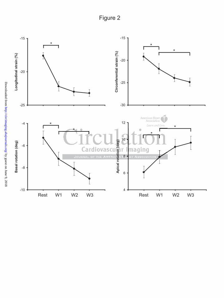
Figure 2
4
6
8
10
12A
pica
l rot
atio
n (d
eg)
**
Rest W1 W2 W3
-25
-20
-15
Long
itudi
nal s
trai
n (%
)
*
-30
-25
-20
-15
Circ
onfe
rent
ial s
trai
n (%
)
**
-10
-8
-6
-4
Bas
al ro
tatio
n (d
eg)
**
Rest W1 W2 W3
o
8
10
otat
ion
(deg
)
**
by guest on June 9, 2018http://circim
aging.ahajournals.org/D
ownloaded from

Figure 3
0
40
80
120
160
Diff
eren
ce fr
om re
stin
g co
nditi
on (%
)
0
40
80
120
160
Diff
eren
ce fr
om re
stin
g co
nditi
on (%
)
SRL SRC TR/UTR
Systole
Diastole
**
** *
* *
**
* *
by guest on June 9, 2018http://circim
aging.ahajournals.org/D
ownloaded from
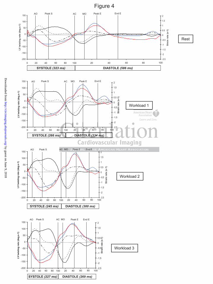
Figure 4
0 20 40 60 80 100-2.5
-2
-1.5
-1
-0.5
0
0.5
1
1.5
2
Stra
in ra
te (s
-1)
-200
-150
-100
-50
0
50
100
150
0 20 40 60 80 100
LV tw
istin
g ra
te (d
eg.s
-1)
-200
-150
-100
-50
0
50
100
150
0 20 40 60 80 100
LV tw
istin
g ra
te (d
eg.s
-1)
0 20 40 60 80 100-2,5
-2
-1,5
-1
-0,5
0
0,5
1
1,5
2
Stra
in ra
te (s
-1)
-200
-150
-100
-50
0
50
100
150
0 20 40 60 80 100
LV tw
istin
g ra
te (d
eg.s
-1)
0 20 40 60 80 100-2,5
-2
-1,5
-1
-0,5
0
0,5
1
1,5
2
Stra
in ra
te (s
-1)
-200
-150
-100
-50
0
50
100
150
0 20 40 60 80 100
LV tw
istin
g ra
te (d
eg.s
-1)
0 20 40 60 80 100-2,5
-2
-1,5
-1
-0,5
0
0,5
1
1,5
2
Stra
in ra
te (s
-1)
SYSTOLE (323 ms)
AC MO Peak E End EAO Peak S
DIASTOLE (586 ms)0 20 40 60 80 100 20 40 60 80 100
Rest
SYSTOLE (266 ms) DIASTOLE (334 ms)
AC MO Peak E End EAO Peak S
0 20 40 60 80 100 20 40 60 80 100
Workload 1
SYSTOLE (245 ms) DIASTOLE (300 ms)
AC MO Peak E End EAO Peak S
0 20 40 60 80 100 20 40 60 80 100
Workload 2
SYSTOLE (227 ms) DIASTOLE (269 ms)
AC MO Peak E End EAO Peak S
0 20 40 60 80 100 20 40 60 80 100
Workload 3
2
2
s)
A
-2,5
-2
2
s) DIASTOLE (334 ms)
100 20 40 60 80 100
AC MO Peak E End E
by guest on June 9, 2018http://circim
aging.ahajournals.org/D
ownloaded from

Figure 5
-2
0
2
4
6
8
10
12
14
16
18
-1 0 1 2 3 4 5 6 7 8Radial displacement (mm)
LV to
rsio
n (d
eg)
ES
ED
Rest
W1
W2
W3
by guest on June 9, 2018http://circim
aging.ahajournals.org/D
ownloaded from
Stéphane NottinGrégory Doucende, Iris Schuster, Thomas Rupp, Aliona Startun, Michel Dauzat, Philippe Obert and
Subjects: The Key Role of Torsional Mechanics for Systolic-Diastolic CouplingKinetics of Left Ventricular Strains and Torsion During Incremental Exercise in Healthy
Print ISSN: 1941-9651. Online ISSN: 1942-0080 Copyright © 2010 American Heart Association, Inc. All rights reserved.
TX 75231is published by the American Heart Association, 7272 Greenville Avenue, Dallas,Circulation: Cardiovascular Imaging
published online June 25, 2010;Circ Cardiovasc Imaging.
http://circimaging.ahajournals.org/content/early/2010/06/25/CIRCIMAGING.110.943522World Wide Web at:
The online version of this article, along with updated information and services, is located on the
http://circimaging.ahajournals.org//subscriptions/
is online at: Circulation: Cardiovascular Imaging Information about subscribing to Subscriptions:
http://www.lww.com/reprints Information about reprints can be found online at: Reprints:
document. Permissions and Rights Question and Answer this process is available in the
located, click Request Permissions in the middle column of the Web page under Services. Further information aboutnot the Editorial Office. Once the online version of the published article for which permission is being requested is
can be obtained via RightsLink, a service of the Copyright Clearance Center,Circulation: Cardiovascular Imaging Requests for permissions to reproduce figures, tables, or portions of articles originally published inPermissions:
by guest on June 9, 2018http://circim
aging.ahajournals.org/D
ownloaded from


![[XLS] · Web view2174 25 2175 25 2176 25 2177 25 2178 25 2179 25 2180 25 2181 25 2182 25 2183 25 2184 25 2185 25 2186 25 2187 25 2188 25 2189 25 2190 25 2191 25 2192 25 2193 25 2194](https://static.fdocuments.net/doc/165x107/5a9f94857f8b9a76178cfd21/xls-view2174-25-2175-25-2176-25-2177-25-2178-25-2179-25-2180-25-2181-25-2182-25.jpg)