Heterogeneity of Intramural Function in Hypertrophic...
35
Heterogeneity of Intramural Function in Hypertrophic Cardiomyopathy: Mechanistic Insights from MRI Late Gadolinium Enhancement and High-resolution DENSE Strain Maps Aletras et al: Intramural Function in HCM using LGE and DENSE MRI Anthony H. Aletras PhD †‡ , Gauri S. Tilak MD, PhD, MPH † Li-Yueh Hsu DSc † Andrew E. Arai MD † †Cardiovascular and Pulmonary Branch, National Heart, Lung and Blood Institute, National Institutes of Health, Department of Health and Human Services, Bethesda, MD, U.S.A. ‡Department of Computer Science and Biomedical Informatics University of Central Greece, Lamia, Greece Correspondence to: Andrew E. Arai, MD Cardiovascular and Pulmonary Branch National Heart, Lung, and Blood Institute National Institutes of Health Bldg 10, B1D-461, MSC 1061 10 Center Drive Bethesda, MD 20892-1061 USA Phone: 301-496-3658 Fax: 301-402-2389 Email: [email protected] Journal Subject Codes: [30] CT and MRI Lu Lu Lu Lu Lu Lu Lung ng ng ng ng ng ng a a a a a a and nd nd nd nd nd nd B B B B B B Blo lo lo lo lo lo lood od od od od od od l Institutes of Health, Departm nt of Health and Human Serv e artment of Com ter Science and Biomedical Informati s University of Central Greece l Institutes of Health, Departme nt of Health and Human Serv Bethesda, MD, U.S.A. epartment of Computer Science and Biomedical Informatics University of Central Greece by guest on May 19, 2018 http://circimaging.ahajournals.org/ Downloaded from
Transcript of Heterogeneity of Intramural Function in Hypertrophic...

Heterogeneity of Intramural Function in Hypertrophic Cardiomyopathy:
Mechanistic Insights from MRI Late Gadolinium Enhancement
and High-resolution DENSE Strain Maps
Aletras et al: Intramural Function in HCM using LGE and DENSE MRI
Anthony H. Aletras PhD†‡, Gauri S. Tilak MD, PhD, MPH†
Li-Yueh Hsu DSc† Andrew E. Arai MD†
†Cardiovascular and Pulmonary Branch, National Heart, Lung and Blood Institute,
National Institutes of Health, Department of Health and Human Services,
Bethesda, MD, U.S.A.
‡Department of Computer Science and Biomedical Informatics
University of Central Greece,
Lamia, Greece
Correspondence to: Andrew E. Arai, MD Cardiovascular and Pulmonary Branch National Heart, Lung, and Blood Institute National Institutes of Health Bldg 10, B1D-461, MSC 1061 10 Center Drive Bethesda, MD 20892-1061 USA Phone: 301-496-3658 Fax: 301-402-2389 Email: [email protected]
Journal Subject Codes: [30] CT and MRI
LuLuLuLuLuLuLungngngngngngng a a a aaa andndndndndndnd B B B B BBBlololololololoododododododod
l Institutes of Health, Departm nt of Health and Human Serv
e artment of Com ter Science and Biomedical Informati s
University of Central Greece
l Institutes of Health, Department of Health and Human Serv
Bethesda, MD, U.S.A.
epartment of Computer Science and Biomedical Informatics
University of Central Greece
by guest on May 19, 2018
http://circimaging.ahajournals.org/
Dow
nloaded from

Abstract
Background In HCM, myocardial abnormalities are commonly heterogeneous. Two patterns
of LGE have been reported: a bright “confluent” and an intermediate intensity abnormality
termed “diffuse,” each representing different degrees of myocardial scarring. We used MRI to
study the relation between intramural cardiac function and the extent of fibrosis in HCM. The
aim of this study was to determine whether excess collagen or myocardial scarring, as
determined by late gadolinium enhancement (LGE) MRI, are the primary mechanisms leading to
heterogeneous regional contractile function in patients with hypertrophic cardiomyopathy
(HCM).
Methods and Results Intramural left ventricular (LV) strain, transmural LV function, and
regions of myocardial fibrosis/scarring were imaged in 22 patients with HCM using
displacement encoding with stimulated echoes (DENSE), cine MRI and LGE. DENSE systolic
strain maps were qualitatively and quantitatively compared with LGE images. Intramural
systolic strain by DENSE was significantly depressed within areas of confluent and diffuse LGE
but also in the core of the most hypertrophic non-enhanced segment (all p<0.001 vs. non-
hypertrophied segments). DENSE demonstrated an unexpected inner rim of largely preserved
contractile function and a non-contracting outer wall within hypertrophic segments in 91% of
patients.
Conclusions LGE predicted some but not all of the heterogeneity of intramural contractile
abnormalities. This indicates that myocardial scarring or excess interstitial collagen deposition
does not fully explain the observed contractile heterogeneity in HCM. Thus, myofibril disarray
or other non-fibrotic processes affect systolic function in a large number of patients with HCM.
Key Words: hypertrophic cardiomyopathy; magnetic resonance imaging; DENSE; displacement
encoding with stimulated echoes; myocardial function; strain; late gadolinium enhancement
tststststststs w w w w ww wititititititith h hh hh h HCHCHCHCHCHCHCM M M M M M M ususususususus
MRI ananananananandd d dd d d LGLGLGLGLGLGLGE.E.E.E.E.E.E D D D D D DD
a n
E a
o 1
e
alitatively and quantitatively compared with LGE images. In
ENSE was significantly depressed within areas of confluent a
of the most hypertrophic non-enhanced segment (all p<0.001
ents). DENSE demonstrated an unexpected inner rim of larg
by guest on May 19, 2018
http://circimaging.ahajournals.org/
Dow
nloaded from

Hypertrophic cardiomyopathy (HCM) is a genetic disease characterized by left
ventricular (LV) hypertrophy1, 2 due to hyperplasia and hypertrophy of myocytes and hyperplasia
of several other cell types. Myofiber disarray and interstitial fibrosis occur commonly 3, 4.
Hypertrophy and fibrosis are associated with LV systolic and diastolic dysfunction, heart failure,
arrhythmias, morbidity, and mortality 5-8. Myocardial ischemia, infarction and fibrosis occur in
HCM despite widely-patent epicardial coronary artery vessels 9, 10 6. MRI late gadolinium
enhancement (LGE) has revealed two types of abnormalities which may be related to infarction
or fibrosis. Confluent LGE appears in most respects comparable to the type of bright gadolinium
enhancement seen in the myocardial infarctions of patients with ischemic heart disease 11.
Diffuse LGE likely represents less severe myocardial fibrosis or milder degrees of excess
collagen deposition. It has also been postulated that the interstitial fibrosis may be a secondary
and reversible phenotype 5, 12.
Extrapolations from transmural measurements of myocardial strain have led some
investigators to postulate that patients with HCM may have preserved endocardial strain 13, 14
while others suggest there should be reduced endocardial strain 15, 16. A study by Tseng et al. 4
shows that myocardial fiber disarray is related to reduced myocardial strain. However, this study
does not address the presence or absence of myocardial fibrosis within the hypokinetic regions.
A more recent echocardiographic study by Popovic et al. 17 reports that non-fibrotic myocardial
segments exhibit increased strain compared to segments with fibrosis. This finding suggests that
perhaps some function is preserved in non-fibrotic hypertrophic segments. However, this could
also reflect partial volume effects since average midwall strain measurements were studied in a
12 segment model.
to the type of f brbbbbbb
ischeheeeeeemimimimimimimic c c c c c heheheheheheheararararararartttt t tt dd
o
b
o
epresents less severe myocardial fibrosis or milder degrees o
It has also been postulated that the interstitial fibrosis may b
otype 5, 12.
by guest on May 19, 2018
http://circimaging.ahajournals.org/
Dow
nloaded from

High resolution imaging of intramural strain might provide insight into the relative
contribution of myocardial fiber disarray and myocardial fibrosis as distinct mechanisms causing
regional LV dysfunction. DENSE 18, 19 (Displacement Encoding with Stimulated Echoes) MRI
can quantify short axis regional systolic function at high spatial resolution. DENSE allows direct
measurement of intramural myocardial strain expressed in terms of circumferential shortening
(CS) and radial thickening (RT) - a significant advance over transmural measures of regional
function such as absolute systolic change in wall thickness (in mm) or percent wall thickening.
This study aimed to determine whether excess collagen or myocardial scarring, as
determined by gadolinium enhanced MRI, are the primary mechanisms leading to heterogeneous
regional contractile function in patients with HCM. We hypothesized that if intramural
contractile function in HCM is heterogeneous due to excess collagen deposition or myocardial
scarring, then hypokinesis will be localized primarily in areas with LGE. However, if extensive
hypokinesis is observed outside the areas of LGE, then excess collagen or myocardial scarring is
unlikely to be the only mechanism leading to heterogeneous regional contractile function in
patients with HCM. Finally, it is possible that severely hypertrophic myocardium has diffusely
abnormal function regardless of regional fibrosis. This possibility is sometimes referred to as
“muscle bound”. We also hypothesized that high resolution intramyocardial DENSE strain maps
would be able to image these functional abnormalities. Such imaging should be able to resolve
these three possibilities.
An unexpected finding of this work was the presence of largely preserved inner wall
strain in hypertrophic segments with overall low transmural strain. While an endocardial ring of
preserved contractility in HCM has been postulated to exist, 17 no direct visualization has been
anisms leadingngngngnggg t
sized d ddddd thththththththatatatatatatt i i i iiif f f f f ff inininininininttttrtrtt a
i
v
v a
in HCM is heterogeneous due to excess collagen deposition
inesis will be localized primarily in areas with LGE. Howev
ved outside the areas of LGE, then excess collagen or myoca
by guest on May 19, 2018
http://circimaging.ahajournals.org/
Dow
nloaded from

provided to date. Therefore, additional analysis of this pattern of intramural strain was performed
since it was not predicted by geometric models of myocardial hypertrophy.
Methods
Study Population
Twenty-two patients with HCM were studied under research protocol 09-H-N237
approved by the National Heart Lung and Blood Institute’s review board. The diagnosis of HCM
was established by echocardiography (>15 mm LV wall thickness in the absence of another
cause for the increased cardiac mass). Patients with a history of alcohol septal ablation or
cardiac surgery were excluded. Only patients who underwent both DENSE and LGE imaging
were included. LV outflow tract gradients at rest were obtained by cardiac catheterization.
MRI Studies
Subjects were scanned in a 1.5 T MRI scanner using a cardiac phased array coil. LV
systolic function and mass were imaged with cine MRI along contiguous short axis slices
(typically with 1.4x1.4 mm/pixel and 8 mm slice thickness). Intramural circumferential
shortening (CS) and radial thickening (RT) measurements were made with DENSE with a
volumetric preparation. Displacement was mapped for every pixel of the end-systolic image
allowing systolic strain (CS and RT) maps to be computed. End-systole was determined from the
cine MRI. LGE imaging was performed with an inversion recovery method 20 after a dose of 0.2
mmol/kg gadopentetate dimeglumine (typical resolution of 1.4x1.4 mm/pixel, slice thickness of
8 mm) with the inversion time optimized to null normal myocardium.
alcohol septal l ll abaaaaaa
oth DDDDDDDENENENENENENENSESESESESESESE a aaaaaandndndndndndnd
t
e scanned in a 1.5 T MRI scanner using a cardiac phased arr
utflow tract gradients at rest were obtained by cardiac cathet
e scanned in a 1.5 T MRI scanner using a cardiac phased arr
by guest on May 19, 2018
http://circimaging.ahajournals.org/
Dow
nloaded from

Data analysis
LV ejection fraction and total myocardial mass were measured using computer assisted
planimetry. The signal intensity was quantified in confluent and diffuse lesions21, from normal
myocardium and the LV blood pool. In short, confluent lesions were defined as qualitatively
having bright signal similar to that found in non-viable myocardium in patients with CAD.
Diffuse lesions were defined as qualitatively having signal brighter than that of nulled
myocardium and signal less bright than that of confluent lesions. The “most hypertrophic non-
enhancing segment” was defined as the thickest segment (as seen on end-diastolic cine images)
without LGE. To mitigate potential through-plane motion misregistration issues between
DENSE and LGE, the adjacent slices to the one containing the “most hypertrophic non-
enhancing segment” were examined for absence of LGE. Remote myocardium was defined as
myocardium diametrically opposite to the hypertrophic segments. Following endocardial and
epicardial segmentation, DENSE displacement maps were automatically processed to produce
circumferential shortening (CS) and radial thickening (RT) strain maps 22. CS and RT maps were
scaled using a standardized color scale that displayed quantitative regional function on a pixel-
by-pixel basis. Region of Interest (ROI) analysis was performed to measure CS and RT within
confluent, diffuse and most hypertrophic non-enhanced areas. Only one slice per patient was
analyzed in order to preserve the independence of the samples. Apical slices were excluded from
data analysis to avoid partial volume effects 23.
Statistical Analysis
Continuous measurements are expressed as mean ± standard deviation (SD) for
descriptive purposes. Standard error of the mean (SEM) was used in figures where the primary
gistration issueeeeeees ssssss
moststttttt h h h h h hhypypypypypypypererererererertrtrtrtrtrtrtropopopopopopophh
w
r d
t s
were examined for absence of LGE. Remote myocardium w
rically opposite to the hypertrophic segments. Following end
tion, DENSE displacement maps were automatically processa
by guest on May 19, 2018
http://circimaging.ahajournals.org/
Dow
nloaded from

purpose was to depict differences between two average values. Comparisons used a paired t-test
with Bonferroni correction for multiple comparisons (p-values are presented unadjusted and the
appropriate level of significance is used within each group of comparisons).
Results
Demographics
Six patients (Table 1) had severe symptoms related to LV outflow obstruction while others had
mild or no symptoms. Four were asymptomatic, seven presented with shortness of breath (SOB),
three with chest pain (CP) and eight with a combination of SOB and CP. The average pulmonary
capillary wedge pressure (PCWP) was 12±5 mmHg but 6 patients had significantly elevated
PCWP. The maximum wall thickness averaged 25±5 mm. Median value of LV mass was 185
grams (range: 76 to 567) and the ejection fraction averaged 66±6%. Median value of LGE was
5.8% of the left ventricle (range:1.8 to 41.5). Nineteen of the patients had asymmetric septal
hypertrophy (ASH), two with severe concentric hypertrophy (CON) and one with hypertrophy
localized to the mid-ventricle (MID). Of the 22 individuals evaluated, only 4 had identified
mutations after an unbiased screening of the Beta-Myosin head (exons 1-23) (Table 1). Patient
numbers 1 and 20 are related and have the R403Q mutation. Patient number 5 has the K847E
mutation. Patient number 15 has a Val95Ala mutation in Alpha-tropomyosin. On the basis of the
unbiased screening, it can be concluded that the other patients are very unlikely to have
mutations in the Beta- Myosin head.
and CP. The aaaaaaavvevvvvv
ts haaaaaaad d d dddd sisisisisisisigngngngngngngnififififififificicicicicicicanaaaaaa
u m
567) and the ejection fraction averaged 66±6%. Median va u
r m
um wall thickness averaged 25±5 mm. Median value of LV m
567) and the ejection fraction averaged 66±6%. Median valu
ricle (range:1.8 to 41.5). Nineteen of the patients had asymm
by guest on May 19, 2018
http://circimaging.ahajournals.org/
Dow
nloaded from

DENSE Detects Patterns of Intramural Contractile Function associated with LGE
DENSE strain maps and LGE images can directly show that LGE is not a prerequisite for
the presence of intramural functional abnormalities. DENSE functional abnormalities that are
either more extensive than LGE imaging predicts or that are present in the absence of LGE can
directly disprove the primary hypotheses. Figures 1-4 summarize the relationship between LGE
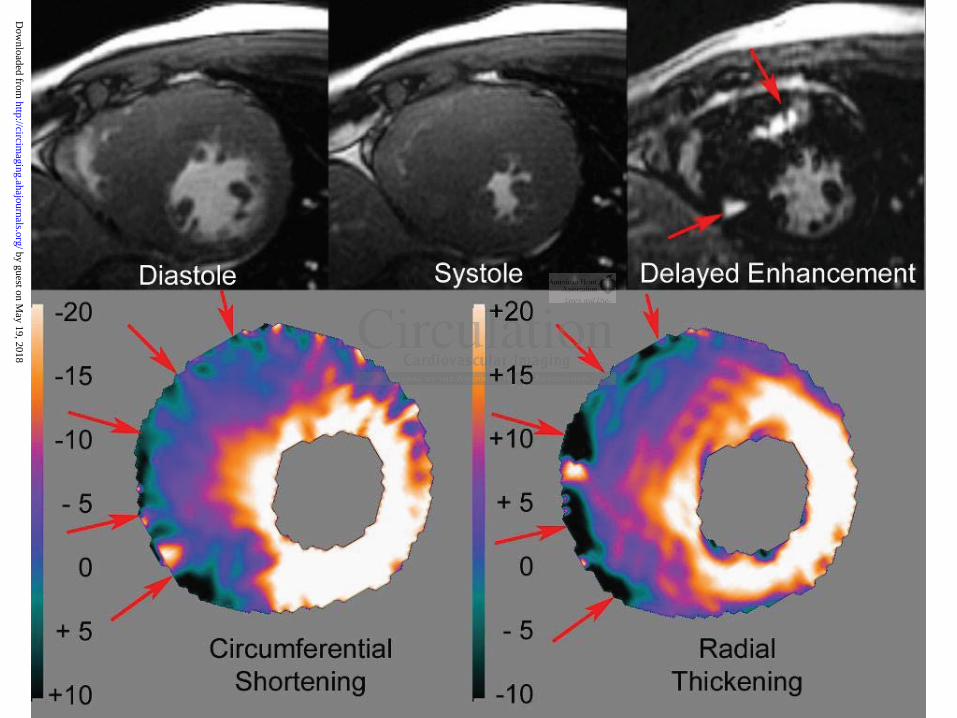
and intramural cardiac function as depicted by DENSE CS and RT color strain maps. The color
scale represents intramural percent circumferential shortening and percent radial thickening
respectively. Normal CS and RT are generally mapped white to bright orange. Dark orange and
purple hues represent mild and severe strain abnormalities. The transition from blue to green
represents zero systolic strain. Green to black represents dyskinetic strain (i.e. systolic
circumferential stretching or systolic radial thinning). Figures 1-4 also show diastolic and
systolic frames from cine MRI (first and second images on the top row respectively) as well as
LGE (third image on the top row).
An example that supports the concept that myocardium with confluent LGE does not
contract normally in HCM is shown in Figure 1. This patient has a confluent abnormality of LGE
in the inferior septum which corresponds to a severe focal abnormality in systolic strain as
represented by the blue and purple patches corresponding to less than 10% circumferential
shortening and radial thickening. Note that the signal intensity on the LGE is similar to what is
typically observed in patients with myocardial infarction due to coronary artery disease (i.e. the
myocardial enhanced zone and the left ventricular blood pool present with similar signal
intensity) except that the abnormality is not located in the subendocardium.
bright orange.e...... D
transnssssssitititititittioioioioioioion nn n nnn frfrfrfrfrfrfromomomomomomom b
o y
c s
v
olic strain. Green to black represents dyskinetic strain (i.e. sy
ching or systolic radial thinning). Figures 1-4 also show dias
cine MRI (first and second images on the top row respectivd
by guest on May 19, 2018
http://circimaging.ahajournals.org/
Dow
nloaded from

Diffusely enhancing myocardium also does not contract in HCM (Figure 2). In this
example, a diffuse pattern of LGE, which corresponds to a severe intramural strain abnormality
on color-coded DENSE, can be seen in the anterior wall and anteroseptum. The myocardial
signal intensity in the late gadolinium enhanced zone is much lower than in cases with confluent
LGE. The DENSE CS and RT maps show normal strain (orange to white) in the septum, inferior
and lateral walls as well as in the inner 1/3 of the anterior and anteroseptal segments. Thus, there
is a substantial amount of myocardium in all segments with normal strain and a zone of very low
strain in the outer half of the segment with diffuse gadolinium enhancement. Cine MRI showed
normal systolic endocardial excursion and ejection fraction in this patient but could not assess
intramural function.
Intramural functional abnormalities can be more extensive than predicted by the extent of
LGE. Figure 3 shows a patient with more severe septal hypertrophy but a similar sized
abnormality of confluent LGE to the one seen in Figure 1. Despite severely depressed strain in
the two thirds of the septum near the right ventricle, DENSE maps of intramural strain again
showed a nearly circumferential inner ring of largely normal contraction. The amount of
myocardium with normal strain (orange-white) is comparable in all segments around this short
axis slice despite large functional abnormalities in the remainder of the septum. Systolic strain in
this patient is more heterogeneous than LGE would predict. Cine MRI shows global systolic
function (corresponding to an EF of 64%) that almost obliterates the mid left ventricular cavity.
Intramural functional abnormalities in HCM also exist in the absence of significant LGE.
Figure 4 shows an example where the DENSE maps of intramural strain show two large distinct
contractile abnormalities (purple to blue/black patches) separated by a zone of nearly normally
contracting myocardium in the mid-septum. In this patient, there is only a small region of
is patient but cccccccouoooooo
unctional abnormalities can be more extensive than predict d
w a
r
unctional abnormalities can be more extensive than predicted
ws a patient with more severe septal hypertrophy but a simila
uent LGE to the one seen in Figure 1. Despite severely depr
by guest on May 19, 2018
http://circimaging.ahajournals.org/
Dow
nloaded from

confluent LGE within the inferoseptum. Cine MRI showed hyperdynamic global systolic
function in this patient.
The examples shown in Figures 2-4 indicate that reduced contractile function is not
necessarily associated with LGE. In most of the patients (17 out of 22) the LGE was not
localized to the inner third of the heart consistent with viable myocardium in the regions where
strain appeared normal in most of these patients (Table 1).
Heterogeneous Intramural Function Irrespective of Pattern or Presence of Enhancement
Intramural function by DENSE was not only depressed within areas of both confluent
and diffuse LGE but also at the core of the most hypertrophic non-enhanced segment (Figure 5).
Specifically, CS was significantly reduced within the confluent late gadolinium enhanced regions
compared to a normal remote non-enhanced non-hypertrophic region (6.1±1.6 vs. 20.4±1.8% CS
respectively, p<0.001). Diffuse late gadolinium enhanced regions exhibited disproportionally
reduced intramural CS compared to a normal remote non-enhanced non-hypertrophic region
(7.5±0.9 vs. 18.4.8±2.1% CS respectively, p<0.001). Interestingly, the most hypertrophic non-
enhanced segment also had significantly reduced intramural strain when compared to a normal
remote non-enhanced non-hypertrophic region (9.3±1.5 vs. 19.1±2.2% CS respectively,
p<0.001). Radial thickening DENSE data showed similar results (confluent enhancement 7.6±1.3
vs. normal 29.5±1.3%, p<0.001; diffuse enhancement 9.1±0.9 vs. normal 23.4±1.9%, p<0.001;
most hypertrophic non-enhanced 9.5±1.2 vs. normal 28.3±1.4%, p<0.001).
On late gadolinium enhanced images, the signal intensity was significantly different for
confluent and diffusely enhancing versus normal myocardium (Figure 6, both p<0.001). The
signal intensity in the most hypertrophic non-enhancing myocardium was not significantly
within areas of f fff bbobbbbb
n-ennnnnnnhahahahahahahancncncncncncncededededededed s s s s s s segegegegegee m
s e
a s
r
s significantly reduced within the confluent late gadolinium e
al remote non-enhanced non-hypertrophic region (6.1±1.6 vs
1). Diffuse late gadolinium enhanced regions exhibited dispr
by guest on May 19, 2018
http://circimaging.ahajournals.org/
Dow
nloaded from

different than normal myocardium, an important control measurement that indicates the
qualitative characterization of amount of gadolinium in these regions was not simply due to the
way the images were displayed.
Preserved Absolute Wall Thickening and Inner wall Contractile Function
DENSE intramural contractile function demonstrated a complete circumferential inner
rim of largely normal contracting myocardium in 77% of the patients (17 out of 22). This group
of patients had on average a normal ejection fraction (66.8%±4.3). Predominantly transmural
hypokinesis in the hypertrophic zone was observed in 2 out of 22 patients. These two patients
had reduced ejection fractions (<55%). The remaining 3 out of 22 patients presented with a
mixture of both patterns of intramural contractile function within the hypertrophic zones
(ejection fraction 67.7%±5.8). Thus, 91% of patients had a complete or significant amount of
myocardium with normal strain in the inner layers of the left ventricle.
Absolute systolic wall thickening by CINE MRI was overall preserved in these HCM
patients (Figure 7A). In particular, absolute systolic wall thickening averaged more than 6 mm
even with an end-diastolic wall thickness of 20 mm. Even the most hypertrophic segments (end-
diastolic thickness greater than 25 mm) exhibited normal absolute systolic wall thickening
defined as >3 mm 24. On average, more than 5 mm of absolute systolic wall thickening was
observed by CINE MRI for both hypertrophic and non-hypertrophic segments independent of the
extent of LGE. These findings of overall preserved absolute systolic wall thickening are
concordant with the observed preserved ejection fractions (Table 2). Conversely, a normal
ejection fraction or significant regional wall thickening did not preclude large patches of severely
depressed intramural systolic function.
2 patients. Theeeeeeesesesssss
2 paaaaaaatitititititit enenenenenenentststststssts p p p p p pprererererereresesesssss n
e
. a
o
erns of intramural contractile function within the hypertroph
.7%±5.8). Thus, 91% of patients had a complete or significar
ormal strain in the inner layers of the left ventricle.
by guest on May 19, 2018
http://circimaging.ahajournals.org/
Dow
nloaded from

On average, percent systolic wall thickening (Figure 7B) was inversely related to end-
diastolic wall thickness suggesting that transmural contractile function in the most hypertrophic
myocardial segments is very low.
In non-hypertrophic segments (Figure 8, EDWth < 12 mm), percent wall thickening was
inversely related to increasing extent of segment LGE. In hypertrophic segments (Figure 8,
EDWth >=12 mm), percent wall thickening was low irrespective of extent of LGE.
Discussion
The DENSE systolic strain maps show heterogeneous myocardial function in HCM
which requires a heterogeneous mechanism rather than a diffuse or global process. LGE predicts
some but not all of the heterogeneity of intramural contractile abnormalities. Since LGE is a
highly sensitive method 11, 25, myocardial scar and excess collagen deposition cannot explain the
spectrum of heterogeneous intramural function encountered in HCM. Myofiber disarray is one
mechanism that could impair function without showing abnormalities on late enhancement
images4. The frequently observed pattern of an inner ring of normal intramural strain provides a
mechanism that can explain preserved global left ventricular function despite severe strain
abnormalities in the remainder of that segment. That observation could not be predicted by
“uniform models” or assumptions about cardiac contraction. There are several factors that lead to
heterogeneity of intramural function in patients with HCM and most of these mechanisms cannot
be visualized with transmural or low resolution methods. Thus, the ability to spatially register
high resolution strain maps with LGE images allows interrogation of the relationships between
function and myocardial scar/collagen at an unprecedented level.
yocararararararardididididididialalalalalalal f f fff f funununununununctctctctctctctioioioioioioio
e s
he heterogeneity of intramural contractile abnormalities. S n
h n
i t l f ti t d i HCM M fib df
erogeneous mechanism rather than a diffuse or global proces
he heterogeneity of intramural contractile abnormalities. Sin
hod 11, 25, myocardial scar and excess collagen deposition can
i t l f ti t d i HCM M fib dff
by guest on May 19, 2018
http://circimaging.ahajournals.org/
Dow
nloaded from

Histopathological studies have identified more than one kind of intramural heterogeneity
in HCM that could relate to contractile abnormalities. Sarcomeric disarray is highly
heterogeneous 26, 27 and not even confined within hypertrophic segments 26. Sarcomeric
hypertrophy occurs in the endocardium and the mid-wall 28, 29. Diffuse interstitial fibrosis and
myocardial scarring are common in HCM 28, 30, 31. Moreover, a high fraction of young patients
with HCM but no coronary artery disease have patchy signs of acute-subacute ischemia post-
mortem 31.
At a clinical level, HCM is characterized by many types of heterogeneous abnormalities.
Resting myocardial blood flow is heterogeneous despite metabolic demands similar to that of
normal myocardium 32 and is related to LGE 10. Multi-focal patterns of LGE have also been
identified in asymptomatic and mildly symptomatic HCM patients 23. These patterns, which may
represent infarction, fibrosis, or excess collagen deposition 33, 34, were later correlated with
progressive dilation of the ventricle and risk of sudden death 21.
Characterization of intramural left ventricular strain based on simple geometrical models
does not apply in HCM since the assumption of a homogeneously contracting medium is not
valid. Based on such models, there is a largely linear endocardial to epicardial gradient of
circumferential strain and HCM 35. Pioneering myocardial tagging studies may have masked the
intramural strain heterogeneity due to the coarse spatial resolution (tag spacing of 7mm) 35. Other
MRI tagging studies in patients with HCM have previously reported abnormal contractile
function in the left ventricle. Both radial displacement 36 and circumferential shortening are
reduced in the septum 37. Similar results were also obtained for total systolic strain recently with
methods that allow full cardiac cycle interrogations 38 whereas diastolic strain was reduced in all
segments.
ic demands simimimmmmm
erns ooooooofffffff LLLLLLLGEGEGEGEGEGEGE h hh h h hhavaaaaaafffffff
o t
e
o
omatic and mildly symptomatic HCM patients 23. These patt
fibrosis, or excess collagen deposition 33, 34, were later corre
of the ventricle and risk of sudden death 21.
by guest on May 19, 2018
http://circimaging.ahajournals.org/
Dow
nloaded from

Late gadolinium enhancement is not as closely linked to the full range of contractile
abnormalities detected in HCM whereas in coronary artery disease regional function varies
inversely with the transmural extent of gadolinium delayed enhancement 20. In patients with
HCM, the extent of delayed enhancement is inversely related to systolic segmental percent wall
thickening 23. While the current study also found an inverse relationship between percent wall
thickening and the extent of LGE, this only applied to the less severely hypertrophic segments
(Figure 8). In the thickest regions of the heart, transmural systolic contraction was independent
of the extent of LGE (Figure 8). While not addressed by this dataset, myocardial edema or
inflammation may also explain the discrepancy between regional contractile function and LGE
39, 40.
The DENSE MRI acquisition has many advantages in producing cardiac strain
measurements. DENSE has been used to measure post-infarct recovery of function 41.
Myocardial tagging 42, harmonic phase image processing 43 and DENSE have yield comparable
strain measurements 43-46 and have been recently shown to rely on the same physics for mapping
motion 47. As implemented in this study, DENSE provided approximately 250 measurements of
intramural contractile function per short axis slice -- an order of magnitude more measurements
than conventional tagged MRI. Therefore, fitting the strain measurements to a predetermined
model of the heart 48 was not necessary -- a significant advantage for the irregularly shaped heart
in patients with HCM. Since a volumetric preparation is used to sensitize the image to motion,
the method intrinsically avoids through-plane motion artifacts.
Regional wall motion assessment by cine MRI and echocardiography can potentially be
misleading depending on whether one chooses to report percent wall thickening (i.e. transmural
strain) or absolute systolic change in wall thickness (in mm). Percent wall thickening is
l contractile fuuuuuuunncnnnnn
N
4 e
MRI acquisition has many advantages in producing cardiac
NSE has been used to measure post-infarct recovery of functi
42, harmonic phase image processing 43 and DENSE have yie
by guest on May 19, 2018
http://circimaging.ahajournals.org/
Dow
nloaded from

inversely proportional to end diastolic wall thickness yet the average amount of wall thickening
in mm remains normal or hypercontractile in the same hearts. However, the greatest hazard of
interpreting percent wall thickening data is the extrapolation to infer the mechanisms leading to
abnormalities in regional contractile function in HCM. High-resolution images of intramural
systolic function can properly address this issue. The fact that LGE and intramural strain show
abnormalities with different spatial distributions suggests that the information may be
complementary. Larger studies will need to be performed to determine the prognostic
significance of these findings.
Limitations
The current data do not address right ventricular involvement specifically since it was not
possible to delineate the RV-LV border within the septum. Multi-slice analysis with appropriate
statistical methods to correct for data dependence could have provided insight into intra-subject
heterogeneity. However, single slice analysis was favored both for simplicity sake and for
preserving the statistical significance lever, which degrades by testing multiple hypotheses (e.g.
through the use of the Bonferroni correction). LGE images were acquired during diastole while
DENSE images during systole. Despite all efforts to properly place the relevant regions of
interest, some partial volume averaging may have influenced the results. Further
histopathological studies are needed to shed light on how abnormalities by DENSE may indicate
diffuse, interstitial fibrosis in visually-negative LGE regions.
o s
e
t f d t d d ld h id d i i ht
o not address right ventricular involvement specifically s
e the RV-LV border within the septum. Multi-slice analysis
t f d t d d ld h id d i i ht
by guest on May 19, 2018
http://circimaging.ahajournals.org/
Dow
nloaded from
Conclusions
In conclusion, just as high resolution LGE images allowed intramural assessments of
myocardial infarction and fibrosis, high-resolution DENSE strain maps provide the most detailed
view of intramural function obtained to date in patients with HCM. These results show that LGE
as a measure of myocardial fibrosis does not fully explain the observed contractile heterogeneity
in these patients. Direct visualization of an inner rim of preserved strain in the endocardium and
mid-wall along with otherwise hypokinetic regions is a contractile pattern unique to HCM that
has not been previously visualized with other methods.
by guest on May 19, 2018
http://circimaging.ahajournals.org/
Dow
nloaded from

Sources of Funding
This study was supported by the Intramural Research Program of the National Heart, Lung and
Blood Institute at the National Institutes of Health.
Disclosures
None.
by guest on May 19, 2018
http://circimaging.ahajournals.org/
Dow
nloaded from

References
1. Hansen MW, Merchant N. Mri of hypertrophic cardiomyopathy: Part 2, differential diagnosis, risk stratification, and posttreatment mri appearances. AJR Am J Roentgenol. 2007; 189:1344-1352.
2. Hansen MW, Merchant N. Mri of hypertrophic cardiomyopathy: Part i, mri appearances. AJR Am J Roentgenol. 2007; 189:1335-1343.
3. Shirani J, Pick R, Roberts WC, Maron BJ. Morphology and significance of the left ventricular collagen network in young patients with hypertrophic cardiomyopathy and sudden cardiac death. J Am Coll Cardiol. 2000; 35:36-44.
4. Tseng WY, Dou J, Reese TG, Wedeen VJ. Imaging myocardial fiber disarray and intramural strain hypokinesis in hypertrophic cardiomyopathy with mri. J Magn Reson Imaging. 2006; 23:1-8.
5. Weber KT. Targeting pathological remodeling: Concepts of cardioprotection and reparation. Circulation. 2000; 102:1342-1345.
6. Kitamura M, Shimizu M, Ino H, Okeie K, Yamaguchi M, Funjno N, Mabuchi H, Nakanishi I. Collagen remodeling and cardiac dysfunction in patients with hypertrophic cardiomyopathy: The significance of type iii and vi collagens. Clin Cardiol. 2001; 24:325-329.
7. Spirito P, Bellone P, Harris KM, Bernabo P, Bruzzi P, Maron BJ. Magnitude of left ventricular hypertrophy and risk of sudden death in hypertrophic cardiomyopathy. N Engl J Med. 2000; 342:1778-1785.
8. Dumont CA, Monserrat L, Soler R, Rodriguez E, Fernandez X, Peteiro J, Bouzas B, Pinon P, Castro-Beiras A. [clinical significance of late gadolinium enhancement on cardiovascular magnetic resonance in patients with hypertrophic cardiomyopathy]. Rev Esp Cardiol. 2007; 60:15-23.
9. Soler R, Rodriguez E, Monserrat L, Mendez C, Martinez C. Magnetic resonance imaging of delayed enhancement in hypertrophic cardiomyopathy: Relationship with left ventricular perfusion and contractile function. J Comput Assist Tomogr. 2006; 30:412-420.
10. Knaapen P, van Dockum WG, Gotte MJ, Broeze KA, Kuijer JP, Zwanenburg JJ, Marcus JT, Kok WE, van Rossum AC, Lammertsma AA, Visser FC. Regional heterogeneity of resting perfusion in hypertrophic cardiomyopathy is related to delayed contrast enhancement but not to systolic function: A pet and mri study. J Nucl Cardiol. 2006; 13:660-667.
11. Kim RJ, Wu E, Rafael A, Chen EL, Parker MA, Simonetti O, Klocke FJ, Bonow RO, Judd RM. The use of contrast-enhanced magnetic resonance imaging to identify reversible myocardial dysfunction. N Engl J Med. 2000; 343:1445-1453.
12. Lim DS, Lutucuta S, Bachireddy P, Youker K, Evans A, Entman M, Roberts R, Marian AJ. Angiotensin ii blockade reverses myocardial fibrosis in a transgenic mouse model of human hypertrophic cardiomyopathy. Circulation. 2001; 103:789-791.
13. Carasso S, Rakowski H. Myocardial fibrosis and regional function in hypertrophic cardiomyopathy: May the force be with you. J Am Soc Echocardiogr. 2008; 21:1306-1308.
, F FFFFFFunununununununjnjnjnjnjnjnjnoo o o o oo N,N,N,N,N,N,N, M M M MMMMabababababababn in n papapapapapapatitititititit enenenenenenentststststststs w w w w wwwiti
thy: The significance of type iii and vi collagens. Clin Cardi
llone P, Harris KM, Bernabo P, Bruzzi P, Maron BJ. Magnity m
,tro-Beiras A [clinical significance of late gadolinium enhan
thy: The significance of type iii and vi collagens. Clin Cardi
llone P, Harris KM, Bernabo P, Bruzzi P, Maron BJ. Magnitypertrophy and risk of sudden death in hypertrophic cardiomk 342:1778-1785. Monserrat L, Soler R, Rodriguez E, Fernandez X, Peteiro J,
tro-Beiras A [clinical significance of late gadolinium enhan
by guest on May 19, 2018
http://circimaging.ahajournals.org/
Dow
nloaded from

14. Carasso S, Yang H, Woo A, Vannan MA, Jamorski M, Wigle ED, Rakowski H. Systolic myocardial mechanics in hypertrophic cardiomyopathy: Novel concepts and implications for clinical status. J Am Soc Echocardiogr. 2008; 21:675-683.
15. Nagueh SF, McFalls J, Meyer D, Hill R, Zoghbi WA, Tam JW, Quinones MA, Roberts R, Marian AJ. Tissue doppler imaging predicts the development of hypertrophic cardiomyopathy in subjects with subclinical disease. Circulation. 2003; 108:395-398.
16. Ashrafian H, Redwood C, Blair E, Watkins H. Hypertrophic cardiomyopathy:A paradigm for myocardial energy depletion. Trends Genet. 2003; 19:263-268.
17. Popovic ZB, Kwon DH, Mishra M, Buakhamsri A, Greenberg NL, Thamilarasan M, Flamm SD, Thomas JD, Lever HM, Desai MY. Association between regional ventricular function and myocardial fibrosis in hypertrophic cardiomyopathy assessed by speckle tracking echocardiography and delayed hyperenhancement magnetic resonance imaging. J Am Soc Echocardiogr. 2008; 21:1299-1305.
18. Aletras AH, Ding S, Balaban RS, Wen H. Dense: Displacement encoding with stimulated echoes in cardiac functional mri. J Magn Reson. 1999; 137:247-252.
19. Aletras AH, Wen H. Mixed echo train acquisition displacement encoding with stimulated echoes: An optimized dense method for in vivo functional imaging of the human heart. Magn Reson Med. 2001; 46:523-534.
20. Kim RJ, Fieno DS, Parrish TB, Harris K, Chen EL, Simonetti O, Bundy J, Finn JP, Klocke FJ, Judd RM. Relationship of mri delayed contrast enhancement to irreversible injury, infarct age, and contractile function. Circulation. 1999; 100:1992-2002.
21. Moon JC, McKenna WJ, McCrohon JA, Elliott PM, Smith GC, Pennell DJ. Toward clinical risk assessment in hypertrophic cardiomyopathy with gadolinium cardiovascular magnetic resonance. J Am Coll Cardiol. 2003; 41:1561-1567.
22. Aletras AH, Arai AE. Meta-dense complex acquisition for reduced intravoxel dephasing. J Magn Reson. 2004; 169:246-249.
23. Choudhury L, Mahrholdt H, Wagner A, Choi KM, Elliott MD, Klocke FJ, Bonow RO, Judd RM, Kim RJ. Myocardial scarring in asymptomatic or mildly symptomatic patients with hypertrophic cardiomyopathy. J Am Coll Cardiol. 2002; 40:2156-2164.
24. Kwong RY, Schussheim AE, Rekhraj S, Aletras AH, Geller N, Davis J, Christian TF, Balaban RS, Arai AE. Detecting acute coronary syndrome in the emergency department with cardiac magnetic resonance imaging. Circulation. 2003; 107:531-537.
25. Ricciardi MJ, Wu E, Davidson CJ, Choi KM, Klocke FJ, Bonow RO, Judd RM, Kim RJ. Visualization of discrete microinfarction after percutaneous coronary intervention associated with mild creatine kinase-mb elevation. Circulation. 2001; 103:2780-2783.
26. Maron BJ, Roberts WC. Quantitative analysis of cardiac muscle cell disorganization in the ventricular septum of patients with hypertrophic cardiomyopathy. Circulation. 1979; 59:689-706.
27. Kuribayashi T, Roberts WC. Myocardial disarray at junction of ventricular septum and left and right ventricular free walls in hypertrophic cardiomyopathy. Am J Cardiol. 1992; 70:1333-1340.
28. Unverferth DV, Baker PB, Pearce LI, Lautman J, Roberts WC. Regional myocyte hypertrophy and increased interstitial myocardial fibrosis in hypertrophic cardiomyopathy. Am J Cardiol. 1987; 59:932-936.
al imaging of ththththththhe
nettitiiiiii O O OOOOO, , ,, , ,, BuBuBuBuBuBuBundndndndndndndyy yyyyy Judd RM. Relationship of mri delayed contrast enhancement t
-cKenna WJ, McCrohon JA, Elliott PM, Smith GC, Pennell Da moA vn 2004; 169:246-249
udd RM. Relationship of mri delayed contrast enhancement taat age, and contractile function. Circulation. 1999; 100:1992-cKenna WJ, McCrohon JA, Elliott PM, Smith GC, Pennell Dassessment in hypertrophic cardiomyopathy with gadoliniumonance. J Am Coll Cardiol. 2003; 41:1561-1567. Arai AE. Meta-dense complex acquisition for reduced intravn 2004; 169:246-249
by guest on May 19, 2018
http://circimaging.ahajournals.org/
Dow
nloaded from

29. Hoshino T, Fujiwara H, Kawai C, Hamashima Y. Myocardial fiber diameter and regional distribution in the ventricular wall of normal adult hearts, hypertensive hearts and hearts with hypertrophic cardiomyopathy. Circulation. 1983; 67:1109-1116.
30. Factor SM, Butany J, Sole MJ, Wigle ED, Williams WC, Rojkind M. Pathologic fibrosis and matrix connective tissue in the subaortic myocardium of patients with hypertrophic cardiomyopathy. J Am Coll Cardiol. 1991; 17:1343-1351.
31. Basso C, Thiene G, Corrado D, Buja G, Melacini P, Nava A. Hypertrophic cardiomyopathy and sudden death in the young: Pathologic evidence of myocardial ischemia. Hum Pathol. 2000; 31:988-998.
32. Nienaber CA, Gambhir SS, Mody FV, Ratib O, Huang SC, Phelps ME, Schelbert HR. Regional myocardial blood flow and glucose utilization in symptomatic patients with hypertrophic cardiomyopathy. Circulation. 1993; 87:1580-1590.
33. Tanaka M, Fujiwara H, Onodera T, Wu DJ, Hamashima Y, Kawai C. Quantitative analysis of myocardial fibrosis in normals, hypertensive hearts, and hypertrophic cardiomyopathy. Br Heart J. 1986; 55:575-581.
34. Lombardi R, Betocchi S, Losi MA, Tocchetti CG, Aversa M, Miranda M, D'Alessandro G, Cacace A, Ciampi Q, Chiariello M. Myocardial collagen turnover in hypertrophic cardiomyopathy. Circulation. 2003; 108:1455-1460.
35. Kramer CM, Reichek N, Ferrari VA, Theobald T, Dawson J, Axel L. Regional heterogeneity of function in hypertrophic cardiomyopathy. Circulation. 1994; 90:186-194.
36. Maier SE, Fischer SE, McKinnon GC, Hess OM, Krayenbuehl HP, Boesiger P. Evaluation of left ventricular segmental wall motion in hypertrophic cardiomyopathy with myocardial tagging. Circulation. 1992; 86:1919-1928.
37. Young AA, Kramer CM, Ferrari VA, Axel L, Reichek N. Three-dimensional left ventricular deformation in hypertrophic cardiomyopathy. Circulation. 1994; 90:854-867.
38. Ennis DB, Epstein FH, Kellman P, Fananapazir L, McVeigh ER, Arai AE. Assessment of regional systolic and diastolic dysfunction in familial hypertrophic cardiomyopathy using mr tagging. Magn Reson Med. 2003; 50:638-642.
39. Knaapen P, van Dockum WG, Bondarenko O, Kok WE, Gotte MJ, Boellaard R, Beek AM, Visser CA, van Rossum AC, Lammertsma AA, Visser FC. Delayed contrast enhancement and perfusable tissue index in hypertrophic cardiomyopathy: Comparison between cardiac mri and pet. J Nucl Med. 2005; 46:923-929.
40. Abdel-Aty H, Cocker M, Strohm O, Filipchuk N, Friedrich MG. Abnormalities in t2-weighted cardiovascular magnetic resonance images of hypertrophic cardiomyopathy: Regional distribution and relation to late gadolinium enhancement and severity of hypertrophy. J Magn Reson Imaging. 2008; 28:242-245.
41. Aletras AH, Tilak GS, Natanzon A, Hsu LY, Gonzalez FM, Hoyt RF, Jr., Arai AE. Retrospective determination of the area at risk for reperfused acute myocardial infarction with t2-weighted cardiac magnetic resonance imaging: Histopathological and displacement encoding with stimulated echoes (dense) functional validations. Circulation. 2006; 113:1865-1870.
42. Axel L, Dougherty L. Mr imaging of motion with spatial modulation of magnetization. Radiology. 1989; 171:841-845.
en turnover innnnnnn h h
n J, AxAxAxAxAxAxAxelelelelelell L L L LLLL. .. ... ReReRRRRR gy of function in hypertrophi di th Ci ulation. 1
scher SE, McKinnon GC, Hess OM, Krayenbuehl HP, Bof left ventricular segmental wall motion in hypertrophic carddK oeformation in hypertrophic cardiomyopathy Circulation 19
y of function in hypertrophic cardiomyopathy. Circulation. 1
scher SE, McKinnon GC, Hess OM, Krayenbuehl HP, Boesf left ventricular segmental wall motion in hypertrophic carddial tagging. Circulation. 1992; 86:1919-1928. Kramer CM, Ferrari VA, Axel L, Reichek N. Three-dimensioeformation in hypertrophic cardiomyopathy Circulation 19
by guest on May 19, 2018
http://circimaging.ahajournals.org/
Dow
nloaded from

43. Osman NF, Kerwin WS, McVeigh ER, Prince JL. Cardiac motion tracking using cine harmonic phase (harp) magnetic resonance imaging. Magn Reson Med. 1999; 42:1048-1060.
44. Gilson WD, Yang Z, French BA, Epstein FH. Complementary displacement-encoded mri for contrast-enhanced infarct detection and quantification of myocardial function in mice. Magn Reson Med. 2004; 51:744-752.
45. Kim D, Gilson WD, Kramer CM, Epstein FH. Myocardial tissue tracking with two-dimensional cine displacement-encoded mr imaging: Development and initial evaluation. Radiology. 2004; 230:862-871.
46. Gilson WD, Yang Z, French BA, Epstein FH. Measurement of myocardial mechanics in mice before and after infarction using multislice displacement-encoded mri with 3d motion encoding. Am J Physiol Heart Circ Physiol. 2005; 288:H1491-1497.
47. Kuijer JP, Hofman MB, Zwanenburg JJ, Marcus JT, van Rossum AC, Heethaar RM. Dense and harp: Two views on the same technique of phase-based strain imaging. J Magn Reson Imaging. 2006; 24:1432-1438.
48. Guttman MAZ, E. A.;McVeigh, E. Analysis of cardiac function from mr images. IEEE Comput. Graph. Appl. 1997; 17:30-38.
by guest on May 19, 2018
http://circimaging.ahajournals.org/
Dow
nloaded from

Table 1. Demographics, clinical and hemodynamic findings HCM LVOT
Patient Age Gender NYHA Symptoms Pattern SAM Gradient PCWP Mutation EP inducible Arrhythmias, other findings
1 35 F 3 CP, SOB ASH No 14 19 BMHC, R403Q No SVT, AF
2 48 M 1 None CON Yes 4 10 NA NA 1st deg AVB, SVT 3 51 F 2 SOB ASH No 6 16 NA No VT 4 50 M 3 CP, SOB ASH No 0 12 NA NA SVT, VT
5 49 F 2 CP ASH No 22 10 BMHC, K847E NA PAT
6 66 F 2 CP, SOB ASH Yes 12 4 NA NA VT, Provocable LVOT obstruction 7 32 F 2 SOB ASH No 6 8 NA NA 8 41 M 1 SOB ASH No 0 9 NA No VT 9 49 F 3 CP, SOB ASH No 5 14 NA NA AF, incomplete LBBB, VT
10 23 M 1 None ASH No 8 8 NA NA Brief run NSVT during EP testing, Negative holter 11 27 F 1 SOB ASH Yes 12 9 NA No Negative holter 12 26 M 2 CP, SOB ASH No 0 7 NA No Negative holter 13 32 M 1 None MID No 20 8 NA NA Provocable LVOT obstruction 14 15 M 1 None ASH No 0 11 NA No Provocable RVOT obstruction
15 33 F 3 SOB ASH No 0 26 a trop, Val95Ala NA
16 35 M 2 CP, SOB ASH Yes 10 NA NA VT AT, VT 17 24 F 2 CP ASH Yes 104 10 NA NA Provocable LVOT obstruction 18 44 M 3 CP, SOB ASH No 0 10 NA No VT 19 34 F 2 SOB ASH Yes 6 20 NA NA
20 49 F 3 CP, SOB ASH No 2 19 BMHC, R403Q NA
21 52 M 2 SOB CON No 0 10 NA No TIA, Paroxysmal AF, NSVT 22 19 M 1 CP ASH No NA NA NA NA Near syncope while driving, Negative holter
NYHA = New York Heart Association classification; M = male; F = female; CP = Chest Pain; SOB = Shortness of Breath; HCM = Hypertrophic Cardiomyopathy; SAM = Systolic Anterior Motion of the mitral valve; LVOT = Left Ventricular Outflow Tract; RVOT = right ventricular outflow tract; EP = Electrophysiology; NA = not available; AF = atrial fibrillation; AVB = atrioventricular block; PAT = paroxysmal atrial tachycardia; VT = ventricular tachycardia; NSVT = non-sustained VT; TIA = transient ischemic attack
NNNNNNNo o o o o ooNANANANANANANA
No 8 8 NA NA Yes 12 9 NA No No 0 7 NA No No 20 8 NA NA No 0 11 NA No
No 8 8 NA NA Yes 12 9 NA No No 0 7 NA No No 20 8 NA NA No 0 11 NA No
by guest on May 19, 2018
http://circimaging.ahajournals.org/
Dow
nloaded from

Table 2. CMR findings Max WTh LGE LGE Strain LVEF LV
Patient (mm) (% of LV) patterns patterns (%) Mass (g) 1 17 13.0 Confluent Both 65 107 2 28 5.1 Confluent Endo 63 567 3 25 8.1 Diffuse Endo 67 179 4 23 9.9 Diffuse Trans 55 240 5 24 3.8 Diffuse Endo 73 111 6 22 4.3 Diffuse Endo 67 129 7 25 10.5 Both Both 64 107 8 21 12.3 Both Endo 61 192 9 20 5.8 Confluent Both 74 136
10 28 2.2 Diffuse Endo 68 190 11 28 5.8 Diffuse Endo 72 161 12 33 14.6 Confluent Endo 64 219 13 27 11.1 Diffuse Endo 59 207 14 28 2.2 Diffuse Endo 65 267 15 22 4.6 Confluent Endo 73 144 16 34 5.5 Both Endo 73 367 17 33 3.5 Diffuse Endo 66 263 18 25 17.9 Both Endo 68 135 19 25 3.5 Confluent Endo 65 213 20 17 12.5 Confluent Endo 62 76 21 24 41.5 Confluent Trans 53 224 22 21 1.8 Diffuse
Endo 70 167
LV = Left Ventricle; Max WTh = Maximum Wall Thickening; LGE = Late Gadolinium Enhancement; LVEF = Left Ventricular Ejection Fraction, Endo=endocardial, Trans=transmural.
ddo o oo 6161 h 74747474747474
28 2 2 Diffuse E d 682 use 33 14.6 Confluent Endo 64 27 11.1 D se E o 59 28 2.2 Diff Endo 65
28 2.2 Diffuse Endo 68 28 5.8 Diffuse Endo 72 33 14.6 Confluent Endo 64 27 11.1 Diffuse Endo 59 28 2.2 Diffuse Endo 65
by guest on May 19, 2018
http://circimaging.ahajournals.org/
Dow
nloaded from

Figure Legends
Figure 1.
Patient with confluent late gadolinium enhancement in the inferior septum which colocalizes
with a severe focal abnormality in radial DENSE (see Results for details).
Figure 2.
The diffuse pattern of late gadolinium enhancement seen in the anterior septum matches the
contractile abnormality seen with DENSE (see Results for details).
Figure 3.
Patient where the systolic strain deficit extends well beyond the two clearly defined confluent
lesions on the late gadolinium enhanced images (see Results for details).
Figure 4.
Patient with extensive systolic intramural strain abnormality despite minimal late gadolinium
gadolinium enhancement (see Results for details).
Figure 5.
Abnormal systolic strain is seen not only in areas of confluent and diffuse late gadolinium
enhancement but also in the most hypertrophic non-enhanced segment. Intramural function is
reduced irrespective of type or presence of late gadolinium enhancement pattern in hypertrophic
segments indicating that myocardial scarring and interstitial fibrosis cannot predict the entire
spectrum of functional abnormalities.
s n
a
stolic strain deficit extends well beyond the two clearly defin
adolinium enhanced images (see Results for details). d
by guest on May 19, 2018
http://circimaging.ahajournals.org/
Dow
nloaded from
Figure 6.
Signal intensities in late gadolinium enhancement images varied depending on presence and type
of late gadolinium enhancement. Confluent and diffuse patterns had significantly higher signal
intensity than normal myocardium. The signal intensity in the most hypertrophic non-enhancing
segment was not significantly different from that of normal myocardium.
Figure 7.
Left panel 7A: Irrespective of segmental end-diastolic thickness, the myocardium exhibits on
average more than 3 mm absolute systolic change in wall thickness. Note that excluding the most
hypertrophic segments, absolute systolic change in wall thickness is on average more than 6 mm.
In normal subjects, >3 mm represents normal absolute systolic change in wall thickness.
Right panel 7B: With increasing end-diastolic thickness the myocardium exhibits reduced
percent systolic wall thickening, which is a measure of transmural systolic strain. This seems
paradoxical given that these patients have preserved ejection fractions and fairly extensive left
ventricular hypertrophy.
(Sample size per category was <6:572, 6-12:1272, 12-15:327, 15-20:449, 20-25:306, >25:134)
Figure 8.
Gadolinium delayed enhancement does not fully explain the observed functional abnormalities.
For segments with end-diastolic wall thickness (EDWTh) < 12mm percent wall thickening was
inversely proportional to the extent of delayed enhancement. However, for segments with
EDWTh 12mm, transmural percent wall thickening was depressed irrespective of the extent of
gadolinium delayed enhancement.
ess. Note that eeeeeeexx
s is ooooooon n nn n n n avavavavavavaverererererereragagagagagagage ee m
> h
h t
n
>3 mm represents normal absolute systolic change in wall th
h increasing end-diastolic thickness the myocardium exhibit
thickening, which is a measure of transmural systolic strain
by guest on May 19, 2018
http://circimaging.ahajournals.org/
Dow
nloaded from

by guest on May 19, 2018
http://circimaging.ahajournals.org/
Dow
nloaded from

by guest on May 19, 2018
http://circimaging.ahajournals.org/
Dow
nloaded from
by guest on May 19, 2018
http://circimaging.ahajournals.org/
Dow
nloaded from

by guest on May 19, 2018
http://circimaging.ahajournals.org/
Dow
nloaded from

by guest on May 19, 2018
http://circimaging.ahajournals.org/
Dow
nloaded from

by guest on May 19, 2018
http://circimaging.ahajournals.org/
Dow
nloaded from

by guest on May 19, 2018
http://circimaging.ahajournals.org/
Dow
nloaded from

by guest on May 19, 2018
http://circimaging.ahajournals.org/
Dow
nloaded from

by guest on May 19, 2018
http://circimaging.ahajournals.org/
Dow
nloaded from

Anthony H. Aletras, Gauri S. Tilak, Li-Yueh Hsu and Andrew E. Araifrom MRI Late Gadolinium Enhancement and High-resolution DENSE Strain Maps
Heterogeneity of Intramural Function in Hypertrophic Cardiomyopathy: Mechanistic Insights
Print ISSN: 1941-9651. Online ISSN: 1942-0080 Copyright © 2011 American Heart Association, Inc. All rights reserved.
TX 75231is published by the American Heart Association, 7272 Greenville Avenue, Dallas,Circulation: Cardiovascular Imaging
published online May 16, 2011;Circ Cardiovasc Imaging.
http://circimaging.ahajournals.org/content/early/2011/05/16/CIRCIMAGING.110.958751World Wide Web at:
The online version of this article, along with updated information and services, is located on the
http://circimaging.ahajournals.org//subscriptions/
is online at: Circulation: Cardiovascular Imaging Information about subscribing to Subscriptions:
http://www.lww.com/reprints Information about reprints can be found online at: Reprints:
document. Permissions and Rights Question and Answer this process is available in the
located, click Request Permissions in the middle column of the Web page under Services. Further information aboutnot the Editorial Office. Once the online version of the published article for which permission is being requested is
can be obtained via RightsLink, a service of the Copyright Clearance Center,Circulation: Cardiovascular Imaging Requests for permissions to reproduce figures, tables, or portions of articles originally published inPermissions:
by guest on May 19, 2018
http://circimaging.ahajournals.org/
Dow
nloaded from