Iim+Pm+Dm+Necrotosing Myopathy
17
Polymyositis, Dermatomyositis, and Autoimmune Necrotizing Myopathy: Clinical Features Sabiha Khan, MD a , Lisa Christopher-Stine, MD, MPH b, * Idiopathic inflammatory myopathies (IIMs) are a heterogeneous group of autoimmune disorde rs predominantl y affec ting skeletal muscles, resul ting in muscl e inflammation and weakness. Along with the musculoskeletal manifestations, involvement of other organ systems is seen, including the skin, cardiac, gastrointestinal, and pulmonary sys tems. The 3 mos t common inflammatory myopat hies are pol ymyosi tis (PM), dermatomyosi tis (DM), and inclusion body myositis (I BM). Several much rarer syndromes ar e also described under the br oad spectru m of inflammat or y myopathies. This review details the clinical findings noted in PM, DM, and the emergin g entity of autoimmune necrotizing myopathy. The Bohan and Peter 1,2 criteria have been traditionally used to define and diagnose PM and DM. IBM was not part of the origin al classifica tion but has been include d over ti me. The Bohan and Peter cr ite ria combine cl inical , lab orator y, and pathol ogic features to define PM and DM. More recently, newer criteria have been proposed for the classification of myositis but are not yet widely accepted. EPIDEMIOLOGY There is a variation in the age at which PM and DM occur. Although DM has a bimodal incidence pattern, peaking during childhood and then again between 50 and 70 years Fundin g/Supp ort: Dr Christ opher- Stine’s work is supported by NIH grant K23-AR-053197. a Division of Rheumatology, Depart ment of Medicine, Johns Hopkins Uni ver sity School of Medicine, 5200 Eastern Avenue, Mason F. Lord Center Tower, Suite 4100, Baltimore, MD 21224, USA b Division of Rheumatol ogy , Department of Medicine, Johns Hopkins Uni ver sit y School of Medicine, 5200 Eastern Avenue, Mason F. Lord Center Tower, Suite 4500, Baltimore, MD 21224, USA * Corresponding author. E-mail address: [email protected] KEYWORDS Myositis Polymyositis Dermatomyositis Inflammatory myopathy Rheum Dis Clin N Am 37 (2011) 143–158 doi:10.1016/j.rdc.2011.01.001 rheumatic.theclinics.com 0889-857X/11/$ – see front matter Ó 2011 Elsevier Inc. All right s reserved.
Transcript of Iim+Pm+Dm+Necrotosing Myopathy

8/6/2019 Iim+Pm+Dm+Necrotosing Myopathy
http://slidepdf.com/reader/full/iimpmdmnecrotosing-myopathy 1/16

8/6/2019 Iim+Pm+Dm+Necrotosing Myopathy
http://slidepdf.com/reader/full/iimpmdmnecrotosing-myopathy 2/16
of age, 3 PM is rare in childhood and occurs mainly after the second decade of life. 4
Both conditions are more common in women. The rep orted incidence of both PMand DM is 4 to 10 cases per million population per year. 5
DEFINITION
The most commonly used criteria for the diagnosis and classification of PM and DMare those defined by Bohan and Peter: (1) symmetric proximal muscle weakness; (2)elevation of serum skeletal muscle enzyme levels, including creatine kinase andaldolase; (3) electromyographic (EMG) evidence of the classic pattern of muscularimpairment, with polyphasic, short, small motor unit potentials, fibrillation, positivesharp waves, increased insertional irritability, and repetitive high-frequencydischarges; (4) muscle biopsy specimens with typical histopathologic findings of degeneration, regeneration, necrosis, and interstitial mononuclear infiltrates; and (5)characteristic cutaneous manifestation of DM, including heliotrope rash or Gottronsign ( Box 1 ).1,2 As mentioned earlier, over time, IBM has been included in these criteriaas its own entity.
MUSCULAR MANIFESTATIONS
The classic clinical finding of both PM and DM is the progressive development of symmetric proximal muscle and truncal weakness that develops relatively slowly,over the course of weeks to months. Patients usually report progressive difficultywith everyday tasks requiring the use of proximal muscles, such as rising from a chair,climbing steps, lifting objects, or combing their hair. Fine motor movements thatrequire the use of distal muscles, such as buttoning shirts and writing, are affectedonly late in the course of the disease, and when found early in the illness, thes eaffected movements should prompt a search for another neuromuscular disorder. 6
Facial muscles remain unaffected; however, pharyngeal and respiratory musclescan become affected, resulting in complications that are discussed in detail later inthis review. 6 Often erroneously thought to be painless diseases, both PM and DMmay have associated myalgias and muscle tenderness early in the disease course,more often seen in DM.
The exception to this pattern of muscle involvement is amyopathic DM (ADM), inwhich patients have the classic dermatologic findings of DM without the accompa-nying muscular findings. 7
Box 1The Bohan and Peter classification criteria
1. Symmetric proximal muscle weakness
2. Elevation of skeletal muscle enzyme levels
3. Abnormal EMG results a
4. Muscle biopsy abnormalities b
5. Typical skin rash of DM c
a Polyphasic, short, small motor unit potentials; fibrillation; positive sharp waves; insertionalirritability; and bizarre, high-frequency, repetitive discharges.b Degeneration/regeneration, perifascicular atrophy, necrosis, phagocytosis, fiber size varia-tion, and mononuclear inflammatory infiltrate.c Gottron sign and heliotrope rash.
Khan & Christopher-Stine144

8/6/2019 Iim+Pm+Dm+Necrotosing Myopathy
http://slidepdf.com/reader/full/iimpmdmnecrotosing-myopathy 3/16
EXTRAMUSCULAR MANIFESTATIONS
The joints, skin, cardiac and pulmonary systems, and the gastrointestinal tract areaffected. Extramuscular organ involvement, such as intersti tial lung disease (ILD)and cardiac involvement, is associated with worse prognosis. 8
JOINT MANIFESTATIONS
Joint involvement can occur in those with PM and DM and is characterized by arthral-gias and arthritis. It is usually noted early in the course of disease, involving wrists,knees, and the small joints of the hands. Joint involvement is classically nonerosiveand frequently responsive to the treatment of the underlying inflammatory myopathy. 9
DERMATOLOGIC MANIFESTATIONS
Of the IIMs, 5 have cutaneous manifestations: adult DM, juvenile DM (JDM), DM asso-ciated with malign a ncy, DM with overlap syndrome, and ADM. 10
Bohan and Peter 2 proposed 5 diagnostic criteria for DM, 4 of which focus on clinical,histopathologic, EMG, and chemical evaluation of muscle inflammation. The presenceof cutaneous manifestations is the fifth criterion.
Cutaneous manifestations of DM can precede the onset of myositis by severalmonths, and for up to 2 years. However, it is uncommon for myositis to occur beforethe onset of cutaneous manifestations. Skin involvement can be the most activecomponent of DM and can be recalcitrant to therapy that is otherwise adequate fortreatment of muscle symptoms. 10
The hallmark cutaneous manifestations of DM can be divided into 5 categories:pathognomonic, highly characteristic, characteristic, more common in JDM, andrare in DM. 10
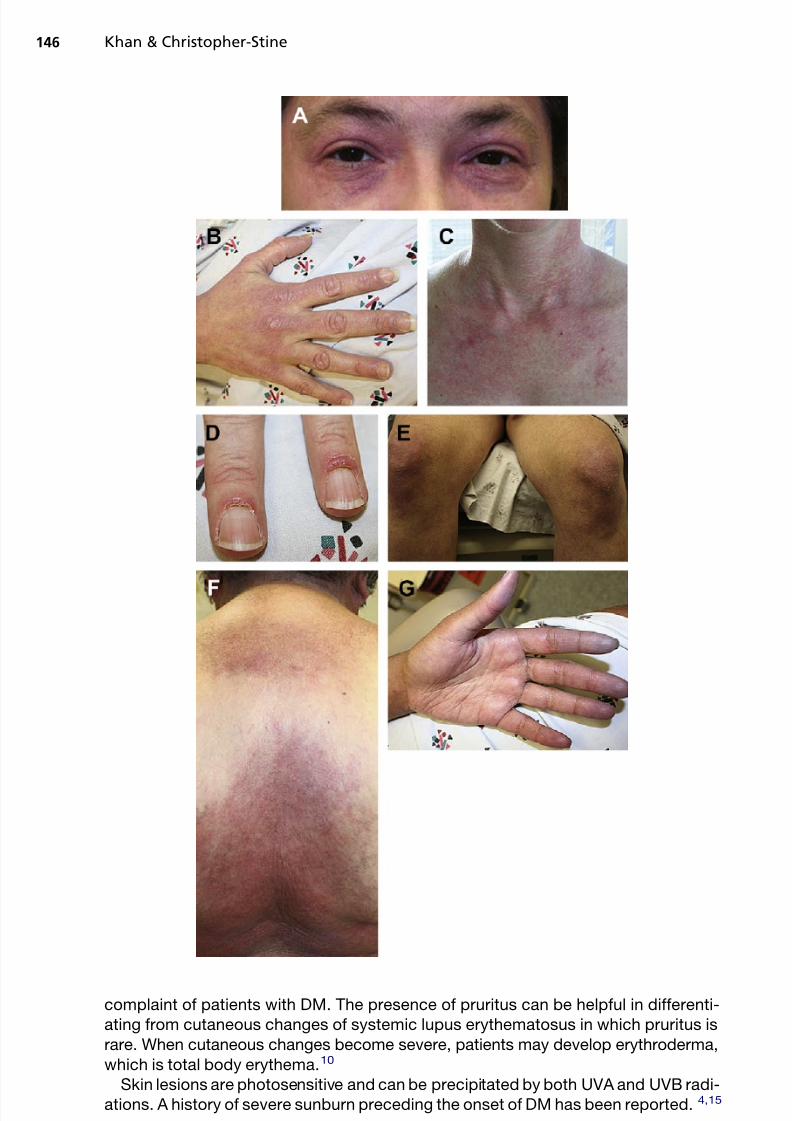
Gottron papules, observed in more than 80% of patients with DM, are consideredpathognomonic for DM. They are violaceous flat-topped papules and plaques locatedover the dorsal aspect of interphalangeal or metacarpophalangeal joints ( Fig. 1 E).Over time, these papules may evolve and develop atrophic, depressed, white centerswith prominent telangiectasias. 10,11
The heliotrope rash, considered highly characteristic of DM, is a periorbital viola-ceous erythema with or without associated edema of the eyelids and periorbital tissue
(see Fig. 1 A).11 The rash can become more confluent and involve the entire face. 10
Characteristic manifestations include Gottron sign, which is a symmetric macularviolaceous erythema with or without edema overlying the dorsal aspect of the inter-phalangeal or metaca rp ophalangeal joints, olecranon process, patella, and medialmalleoli (see Fig. 1 B).11 Other characteristic manifestations include macular viola-ceous erythema in symmetric distribution in classic areas; shawl sign on the napeof the neck, shoulders, and upper back ( se e Fig. 1 F); “V sign” on the V-shaped regionof the neck and upper chest (see Fig. 1 C)12 ; and linear extensor erythema involving theextensor aspects of the legs, thighs, arms, fingers, hands, and feet.
Mechanic’s hands, also considered characteristic, present with hyperkeratosis,scaling, and horizontal fissuring of the palms and fingers bilaterally. Mechanic’s handscan be a manifestation of the antisynthetase syndrome, which is discussed later. 13
This finding is frequently mistaken for contact dermatitis. 10
Other characteristic findings include nail fold telangiectasias, cuticular overgrowth,and prominent periungual erythema, which are frequently seen in patients with DM(see Fig. 1 D).11 Nail fold telangiectasias occur in 30% to 60% of patients early inthe course of the illness. 14 Pruritus is a common, but often underrecognized,
PM, DM, and Autoimmune Necrotizing Myopathy 145
8/6/2019 Iim+Pm+Dm+Necrotosing Myopathy
http://slidepdf.com/reader/full/iimpmdmnecrotosing-myopathy 4/16
complaint of patients with DM. The presence of pruritus can be helpful in differenti-ating from cutaneous changes of systemic lupus erythematosus in which pruritus israre. When cutaneous changes become severe, patients may develop erythroderma,which is total body erythema. 10
Skin lesions are photosensitive and can be precipitated by both UVA and UVB radi-ations. A history of severe sunburn preceding the onset of DM has been reported. 4,15
Khan & Christopher-Stine146

8/6/2019 Iim+Pm+Dm+Necrotosing Myopathy
http://slidepdf.com/reader/full/iimpmdmnecrotosing-myopathy 5/16
Cutaneous calcinosis, although more common in JDM, occurring in 30% to 70%of JDM cases, is also seen in adult DM, occurring in 10% of cases. 16,17 It is anoften-disabling complication in which subcutaneous calcium deposition occurs invarious patterns. Deep deposits occur with linear masses, superficial depo sits canerupt through the skin, and reticular subcutaneous deposits can also occur. 13 Cuta-neous calcinosis frequently occurs at sites of compression, such as elbows andbuttocks, and is associated with increased disease activity and duration. When occur-ring over the extensor surfaces, cutaneous calcinosis can be painful and can lead tochronic ulceration and infections. 9,16,18
Rare findings include nonscarring alopecia, erythroderma, vesiculobullous lesions,leukocytoclastic vasculitis, and livedo reticularis. Flagellate erythema has also beennoted. It is characterized by erythematous linear lesions on the trunk, back, and prox-imal extremities and is associated with active disease. 19
Poikiloderma, a speckled pattern of hypopigmented and hyperpigmented maculesinterspersed with telangiectasia and cutaneous atrophy on a background of erythema,is a manifestation of disease chronicity that is most commonly found over photo-exposed areas. 20 Severe atrophy of involved skin can lead to fragility, superficialerosions, and ulcerations. 21
A small subset of patients with DM develop hyperkeratotic follicular erythematouspapules that occur in a linear distribution over the dorsal aspect of the hands andfeet, frequently over the bony prominences. These papules are known as pityriasisrubra pilaris–like lesions or type Wong DM. The lesions clinically resemble pityriasisrubra pilaris, but they do not have the characteristic findings such as palmoplantardiscoloration or desquamating hyperkeratosis. Some of these patients may have
concomitant atopic dermatitis.22
Cutan eo us vasculitis may occur in severe acute forms of DM, w ith palpablepurpura, 23 urticaria-like lesions, livedo reticularis, and digital ulcerations. 24 The pres-ence of leukocytoclastic vasculitis is concerning for an underlying malignancy. 23 Ra y-naud phenomenon occurs in approximately 25% of patients with DM (see Fig. 1 G).25
ADM
ADM is characterized by the presence of the cutaneous manifestations of DM for 6months or more in individuals who have normal muscle enzyme levels withoutevidence of muscle weakness. 26 The term ADM was first coined in 1979 by Pearson, 27
and ADM was eventually recognized as a subset of DM in the 1990s.28
ADM occursat an incidence of 9.63 per million persons and represents about 10% to 20% of casesof DM. 29
Fig. 1. Examples of dermatologic manifestations of DM.( A) Heliotrope rash is a very commonsign of DM. It can become more confluent and involve the entire face. ( B) Gottron papules,another characteristic manifestation, are symmetric macular violaceous erythema with orwithout edema overlying the dorsal aspect of the interphalangeal or metacarpophalangeal joints, olecranon process, patella, and medial malleoli. 11 (C ) Other characteristic manifesta-
tions include the V sign that can occur on the neck and upper chest. ( D) Image showing cutic-ularerythema andhypertrophy. Other characteristic findings includenail fold telangiectasias,cuticular overgrowth, and prominent periungual erythema, which are frequently seen inpatients with DM. 11 (E ) Gottron sign, also called Gottron papules, is observed in more than80% of patients with DM. Gottron papules are violaceous flat-topped papules and plaqueslocated over the dorsal aspect of the interphalangeal or metacarpophalangeal joints. ( F )Shawl sign, another characteristic manifestation, occurs on the nape of the neck, shoulders,and upper back. ( G) Raynaud phenomenonoccurs in approximately 25%of patients with DM.
=
PM, DM, and Autoimmune Necrotizing Myopathy 147

8/6/2019 Iim+Pm+Dm+Necrotosing Myopathy
http://slidepdf.com/reader/full/iimpmdmnecrotosing-myopathy 6/16
Although previously thought less likely, some case studies have suggested that ADM may p rog ress to frank myopathy for up to 10 years after the onset of cutaneoussymptoms. 12,29 One study of 16 patients noted that 18 .75% of patients developedmuscle weakness within 5 years of diagnosis of ADM. 26
Patients with ADM are at risk for developing the sa me disease complications asthose with DM, such as lung disease and malignancy. 26 The rate of diagnosis of malig-nanc y oc curring close to the diagnosis of ADM has been reported between 15% and28%. 29,30 There have been some reports of the possibility of increased malignancy in ADM compared with DM 30–32 ; however, one study of 29 patients with ADM and DMdid not find a statistically significant difference between the 2 groups. 29
CARDIAC MANIFESTATIONS
Cardiac involvement in myositis was first described by Oppenheim 33 in 1899. Histor-
ically, it was believed that myositis spared the heart. However, based on autopsystudies, it has become evident that cardiac involv em ent, although usually asymptom-atic, is more common than previously be lieved. 34 Cardiac involvement is a majorcause of mortality in patients with myositis. 34 In a long-term follow-up study, cardio-vascular involvement was found to be the most common cause of death in patientswith myositis. 8
The myocardium has been reported to be affected in patients with DM withvarying frequencies. Up to 50% of patients with DM evaluated by noninvasivestudies have asymptomatic cardiac manifestations. 21,34 Noninvasive studies haveshown that up to 85% of patients have abnormal findings on electrocardiography,
77% on ambulatory monitoring, 42% on echocardiography, and 15% on radionu-clide ventriculography. 35
Cardiac manifestations included arrhythmia, conduction abnormalities, cardiacarrest, congestive heart failure (CHF), myocarditis, pericarditis, angina, and secondaryfibrosis. 7,8,34
The frequency of cardiac involvement in myositis has been reported between 6%and 75%. 25,36 Cardiac involvement as a cause of death in PM has been reportedbetween 10% and 20%, 37 although this number is uncertain because large studiesare unavailable.
Conduction abnormalities are the most common asymptomatic cardiac manifesta-
tion, which are observed in 32% to 72% of patients. 34,36,37 These abnormalitiesinclude ST-T changes, bundle branch block, atrioventricular blocks, PR prolongation,Q-wave ab norm alities, and arrhythmias. In some cases, patients required pacemakerplacement. 25,37
Arrhyt hmias and CHF secondary to myocarditis rarely occur in patients with acutedisease. 34 In chronic DM, heart failure is seen more frequently and is in fact themost common symptomatic manifestation. The frequency of CHF has been reportedto be 3% to 45% in patients with myositis. 37 In chronic DM, heart failure has beenattributed to the effects of long-standing hypertension secondary to steroid use. 6 Ithas also been theorized that the cause of CHF is secondary to myocarditis, leadingto left ventricular dysfunction and restrictive cardiomyopathy, or possibly becauseof fibrosis, resulting in chamber stiffness. 36 It is thought that myocardial involvementresulting in myocarditis occurs by the same mechanism that results in skeletal muscleinvolvement. Mononuclear inflammatory cells infiltrate into the endomysium and peri-vascular areas, resulting in degeneration of cardiac myocytes. Histopathologicchanges similar to those in the myocardium were also observed in the conductionsystem, which could explain the cause of heart block. 38
Khan & Christopher-Stine148

8/6/2019 Iim+Pm+Dm+Necrotosing Myopathy
http://slidepdf.com/reader/full/iimpmdmnecrotosing-myopathy 7/16
Angina secondary to Raynaud phenomenon, Prinzmetal angina, and small vesseldisease has also been reported in patients with DM. 39
Pericardial tamponade is very rare, with a frequency reported at about 10%. 25,35,36
However, beca use tamponade can be fatal, electrocardiography of every case isrecommended. 40 Clinically symptomatic cardiac in vo lvement is uncommon, andwhen present, it is associated with a poor prognosis. 36
There have also been reported associations bet ween cardiac involvement and anti–signal recognition particle (SRP) antibodies. 13,41 However, more recent evidencesuggests that anti-SRP antibodies may not contribute to cardiac involvement to thedegree that was once suspected. 42,43
PULMONARY MANIFESTATIONS
Pulmonary c omp lications are a major cause of morbidity and mortality in patients withPM and DM. 8,44 These complications occur primarily or secondarily due to muscleweakness. Three distinct pulmonary complications of PM and DM have beendescribed: hypoventilation, aspiration pneumonia, and ILD. 45
Respiratory failure due to hypoventilation has been historically thought to be a rarecomplication reported in less than 5% of patients with myositis. 46 However, a morerecent, larger, retrospective study of patients with DM and PM reported a higher prev-alence of 21.8%. 47 Hypoventilation occurs in patients with severe muscle weaknessand inflammation, involving respiratory muscles. As a result, restrictive lung functionimpairment is noted on pulmonary function tests (PFTs). Patients are noted to have
reduced lung volumes and maximal inspiratory and expiratory pressures, along withincreased residual volumes and normal forced expiratory volume in the first secondof expiration (FEV 1 ) to forced vital capacity (FVC) ratio. 48 Chest radiographs revealsmall lung volumes and basal atelectasis. 44
Aspiration pneumonia is a frequent complication of PM and DM, occurring inapproximately 17% of patients. 49 Patients suffering from aspiration pneumonia alsofrequently complain of dysphagia, which results from disease involvement of the stri-ated muscles of the pharynx and upper esophagus. 44 Aspiration pneumonia is morelikely to occur in patients with more extensive muscle and skin disease. 50
ILD is an inflammatory lung disorder of unknown cause, characterized by infiltrates
of monocytes, lymphocytes, and neutrophils, as well as interstitial fibrosis. ILD isa common complication in PM and DM. The inc idence of ILD has been reportedbetween 5% and 46% in cross-sectional studies. 44,46,49 No signif icant difference inthe prevalence of ILD exists between patients with PM and DM. 51 ILD associatedwith myosi tis m ay occur before, concomitantly, or after the onset of skin or musclesymptoms. 44,47 ILD as soci ated with ADM is a distinct subset in which the lung diseaseis rapidly progressive. 44,52
Myositis-associated ILDoccurs in 3 differentpatterns: (1)acuteonsetof symptoms, inwhich patients develop apparent progressive hypoxemiawithin a month of lung involve-ment; (2) chronic slowly progressive symptoms; and (3) asymptomatic progression, inwhich ILD is demonstrated only by abnormal results of chest imaging or PFTs. 51,53
Cough and dyspnea are the most frequently reported symptoms in ILD, althoughpatients can be asymptomatic. In one study, 27% of patients with ILD wereasymptomatic. 54 PFTs demonstrate a restrictive ventilatory impairment and canshow decreased total lung capacity, functional residual capacity, residual volume,FEV1 , and FVC, with a normal or elevated FEV 1 /FVC ratio and reduced diffusingcapacity of lung for carbon monoxide. 44
PM, DM, and Autoimmune Necrotizing Myopathy 149

8/6/2019 Iim+Pm+Dm+Necrotosing Myopathy
http://slidepdf.com/reader/full/iimpmdmnecrotosing-myopathy 8/16
Although chest radiography is a useful screening test, high-resolution computedtomography (HRCT) of the lungs is the standard technique for detecting ILD. HRCTis thought to be useful in distinguishing fibrotic disease from active inflammation,with th e former presenting as a reticular pattern and the latter as ground-glasspattern. 55
Bronchoalveolar lavage is not specific for the diagnosis of ILD, although it is useful indifferentiating pulmonary s ymp toms, such as evaluating for infection, drug-inducedreaction, and malignancy. 44,56 However, similar to idiopathic pulmonary fibrosis,neutrophil-predominant alveolitis and increased eosinophil counts may indicatemore progressive disease. 47 Lung biopsy is not routinely performed for diagnosisbecause of the increased morbidity associated with the procedure.
Most frequently seen HRCT changes are those of idiopathic nonspecific interstitialpneumonia (NSIP), with irregular linear opacities and with areas of consolidation andground-glass pattern. 44 However, other patterns such as usual interstitial pneumonia,bronchiolitis obliterans with organizing pneumonia, and diffuse alveolar damage (DAD)are also seen. 44,47
The presence of antiaminoacyl–transfer RNA (tRNA) synthetase antibodies, of whichantihistidyl-tRNA synthetase antibody (anti–Jo-1) is most frequently found, is thestrongest predictive marker for ILD. 44 The prevalence of ILD in patients with anti–Jo-1 antibodies is more than 70%. 13,47,54 There are also other markers for ILD,including PM-Scl autoantibodies. Krebs von den Lungen 6, a glycoprotein expressedon type 2 alveolar pneumocytes and bronchiolar epithelial cells, and serum surfactantprotein D have been suggested as useful markers for ILD in patients with myositis.However, these markers are not routinely used in clinical practice. 44,57 Serum cytoker-
atin (CK) 19 fragment, a component of bronchoepithelial cells, is also associated withILD in myositis. Patients with DAD have higher levels of CK-19 than patients withNSIP. 58
ILD is considered a major risk factor for premature death. It is unclear if the prog-nosis of ILD in patients with PM is different from those with DM. However, one studyshowed that patients with DM-assoc iated ILD were less responsive to steroid therapythan those with PM-associated ILD. 59
Spontaneous pneumomediastinum has also been reported as a rare complication of myositis-associated ILD, occurring with a prevalence of 8.3% in patients with PM andDM. Pneumomediastinum usually results from rupture of subpleural or paracardial
blebs in ILD. In a recent retrospective study, most cases of pneumomediastinumoccurred in patients with DM or ADM. Poor survival was associated with an absenceof muscle weakness and severe pulmonary involvement before the onset of pneumomediastinum. 60
Because of the concomitant use of immunosuppressive drugs in the treatment of inflammatory myopathies, infections are another complication to be considered,particularly opportunistic infections. Bronchoalveolar lavage is a useful tool in makingthe diagnosis. 56
GASTROINTESTINAL MANIFESTATIONS
Dysphagia to liquids and solids secondary to pharyngeal and esophageal abnormal-ities has been reported in 32% to 84% of patients with myositis. 61–63 Because of the loss of pharyngoesophageal muscle tone, patients develop nasal speech, hoarse-ness, nasal regurgitation, and aspiration pneumonia. On examination, tongue weak-ness, flaccid vocal cords, poor palatal motion, and pooling of secretions can beseen. Proximal esophageal skeletal muscle dysfunction can be seen on manometry
Khan & Christopher-Stine150

8/6/2019 Iim+Pm+Dm+Necrotosing Myopathy
http://slidepdf.com/reader/full/iimpmdmnecrotosing-myopathy 9/16
with evidence of low-amplitude or no pharyngeal contractions and decreased upperesophageal sphincter pressure. 61
MUSCLE BIOPSY
A muscle biopsy is required to make a definitive diagnosis of inflammatory myopathy.The biopsy not only confirms the diagnosis but also enables the clinician to rule outother conditions that resemble myositis. It is recommended that a moderately weakmuscle is selected for biopsy because it provides the best chance for obtaining infor-mation. Because the disease is usually symmetric, the muscle is often best located byEMG testing of the contralateral side (so as not to contaminate the biopsy site with theEMG needle) in combination with magnetic resonance imaging (MRI)-guided biopsy toprovide the highest degree of accuracy. An open m uscle biopsy is preferred overa needle biopsy because it provides a larger sample. 64
Characteristic findings of DM are perifascicular atrophy, muscle infarcts, and capil-lary necrosis with membrane attack complex deposition on vessel walls. Perifascicularatrophy sometimes involves grouped or scattered fibers inside fascicles. It is notuncommon to find punched-out areas of myofibrillar loss that appear as vacuoles atthe edge of the fascicle. Muscle fiber necrosis is also seen around the periphery of the fascicle or in a wedgelike distribution suggestive of microinfarcts. Regeneratingfibers are also seen. Biopsy results also reveal inflammatory infiltrates in the septa,around vessels, or inside fascicles, although in some instances, inflammation maynot be significant. When the inflammation is not significant, the diagnosis hinges onthe presence of perifascicular atrophy and reduced capillary density. 64
Characteristic findings of PM are partial invasion of nonnecrotic muscle fibers byCD8 1 cytotoxic T cells and activated macrophages. Also seen is the expression of major histocompatibility complex class I on muscle fiber surfaces, which is not alteredby prior immunosuppressive treatment. 64
LABORATORY TESTING
Laboratory testing reveals an elevation in muscle enzyme levels (CK and aldolase).Extreme elevations in CK levels to of more than 100-fold higher than the upper limitof the reference range are rare in myositis and require evaluation for alternative
diagnosis.7
ANTIBODIES
The role of antibodies in inflammatory myopathies is unclear. Whether they areinvolved in pathophysiology or are simply an epiphenomenon is not yet understood.
Autoantibodies to nuclear RNAs and certain cytoplasmic antigens involved inprotein synthesis are observed in up to 55% of patients with PM and DM. Anti–Jo-1antibodies, detected in 20% to 30% of patients with myositis, were the first describedautoantibodies in myositis. 13,65 Several strong associations between autoantibodiesand clinical phenotypes have been noted. For example, ILD has been observedfrequently in patients with anti–Jo-1 autoantibodies. 13
Antibodies in myositis are categorized into myositis-specific autoantibodies (MSAs)and myositis-associated autoantibodies (MAAs). MSAs are generally found only inpatients with inflammatory myositis or in patients with ILD with subclinical myositis,whereas MAAs are also encountered in other connective tissue diseases without signsof myositis ( Table 1 ).13
PM, DM, and Autoimmune Necrotizing Myopathy 151

8/6/2019 Iim+Pm+Dm+Necrotosing Myopathy
http://slidepdf.com/reader/full/iimpmdmnecrotosing-myopathy 10/16
Table 1Autoantibodies in IIM
Antibody Antigen Clinical Manifestation
Myositis-specific autoantibodies
Antisynthetase autoantibodiesAnti–Jo-1 Histidyl-tRNA synthetase PM, DM 1 ILDAnti–PL-7 Threonyl-tRNA synthetase PM, DM 1 ILD
Anti–PL-12 Alanyl-tRNA synthetase ILD>myositis a
Anti-EJ Glycyl-tRNA synthetase PM>DM 1 ILDAnti-OJ Isoleucyl-tRNA synthetase ILD 1 PM/DMAnti-KS Asparaginyl-tRNA synthetase ILD>myositis a
Anti-Zo Phenylalanyl-tRNA synthetase ILD 1 myositis a
Anti-Ha Tyrosyl-tRNA synthetase ILD 1 myositis a
Nonsynthetase autoantibodiesAnti-SRP SRP Severe, acute, resistant
necrotizing myopathyAnti–Mi-2 DNA helicase DM with rash > muscle
symptoms, treatmentresponsive
Anti-HMGCR (anti-200/100) HMGCR Necrotizing myopathy relatedto statin use in majority;most patients are statinexposed, but myopathy alsoreported in a minority ofstatin-naive patients
Anti-MDA5 (anti–CADM 140) MDA5 DM: CAM, DM with rapidlyprogressive lung disease,pneumomediastinum
Anti-155/140 Transcriptional intermediaryfactor 1 g
CAM
Anti-140 Nuclear matrix protein(NXP-2)
JDM
Anti-SAE SAE DM: CAM, DM with rapidlyprogressive lung disease,pneumomediastinum
Myositis-associated autoantibodies
PM-Scl Unidentified PM or DM/SSc overlapU1RNP U1 small RNP Mixed connective tissue
disease (overlap syndrome)Non-U1 snRNPs U2, U4/6, U5, U3 snRNPs PM or DM/SSc or SSc overlapKu DNA-binding proteins Myositis a /SSc/SLE overlapRo (SS-A), includes Ro60 and
Ro52RNA protein Myositis often with SS or SLE,
may be associated with ILD(especially Ro52)
56 kDa RNP particle Myositis, often with Jo-1KJ Unidentified translation factor PM, ILD, RPFer Elongation factor 1a MyositisMas tRNASer -related antigen Myositis, rhabdomyolysis,
chronic hepatitisMJ Unidentified nuclear pore JDMhPMS1 Protein related to DNA repair Myositis
Abbreviations: CAM, cancer-associated myositis; HMGCR, HMG-CoA reductase; MDA5, melanoma differ-entiation–associated gene 5; RNP, ribonucleoprotein; RP, Raynaud phenomenon; SLE, systemic lupuserythematosus; snRNP, small nuclear RNP; SAE, small ubiquitin-like modifier-activating enzyme; SS-A,Sjogren syndrome A; SSc, systemic sclerosis or scleroderma; >, more often than.
a May be either PM or DM.
152

8/6/2019 Iim+Pm+Dm+Necrotosing Myopathy
http://slidepdf.com/reader/full/iimpmdmnecrotosing-myopathy 11/16
STUDIES/IMAGING
EMG is useful in the diagnosis of myositis and reveals findings in 70% to 90% of patients. Frequent findings include increased spontaneous and insertional activitywith fibrillation potentials, positive sharp waves, complex repetitive discharges, early
recruitment, and small polyphasic motor unit potentials. Ongoing disease activity isreflected in the amount of spontaneous activity because later in the disease, inser-tional activity can become decreased because of fibrosis. However, these findingsare nonspecific because they can be detected in other muscular diseases. 66
Along with EMG and muscle biopsy, imaging techniques are important tools in earlydiagnosis. Imaging studies allow the clinician to determine the extent of disease andalso guide biopsy.
Initially, the most important histologic finding at the onset of disease is the presenceof inflammatory signs with muscle edema. After an extended period of diseaseactivity, usually in cases that are recalcitrant to therapy, the presence of fat or fibrous
tissue changes is detected within muscles. Muscle atrophy is seen after a prolongeddisease course but is nonspecific. Although rare, calcification within the muscles canalso be seen. 67
Muscle edema is the most important finding in myositis. 68 Ultrasonography showsincreased muscle echogenicity, and fasciae/septa can become obscured with alter-ation of normal architecture. 67 In a comparison study between patients with IIM andcontrols, the sensitivity of muscle ultrasonography in detecting myositis was notsignificantly different from that of EMG or serum CK level analysis. 69 However, ultra-sonography offers the possibility of real-time imaging, without exposure to radiation.Doppler imaging in PM a nd DM reveals hypervascularization and correlates with signsof inflammation on M RI.70 Ultrasonography is also useful in the detection of subcuta-neous calcifications. 71
Computed tomography (CT) has also been used in the assessment of myositis. CTshows small areas of hypodensity and is useful for detecting fatty infiltration seen inchronic d ise ase. Calcification within the muscles is also more easily detected by CTthan MRI. 67
MRI is more sensitive for the evaluation of muscle edema. The areas of inflammationappear hyperintense on T2-weighted images. Because fat can interfere with the inter-pretation of MRI images, T2-weighted images with fat suppression or short tau inver-sion recovery sequences are used to obtain clearer images. 71 Focal areas or diffuseincreased signal intensity is a sign of disease activity. 72 The lesions are usuallysymmetric and prominent in proximal muscles. MRI changes in PM and DM aresimilar; however, in DM, changes associated with edema are more common thanmuscle atrophy. 67
DIFFERENTIAL DIAGNOSIS
When considering the diagnosis of inflammatory myopathy in a patient presenting withmuscle weakness, other disease entities must be considered because the clinicalpresentation may resemble that of myositis.
In patients presenting with early muscle fatigue, elevated muscle enzyme levels, andnormal strength, the diagnosis of a metabolic myopathy should be considered. Inolder patients, the differential diagnosis also includes polymyalgia rheumatica, inwhich there is proximal muscle stiffness and elevated inflammatory marker levels.However, neither the elevation of muscle enzyme levels nor muscle pathology onbiopsy is seen. Polyarteritis limited to muscles has also been reported, in which the
PM, DM, and Autoimmune Necrotizing Myopathy 153

8/6/2019 Iim+Pm+Dm+Necrotosing Myopathy
http://slidepdf.com/reader/full/iimpmdmnecrotosing-myopathy 12/16
patient presents with elevated inflammatory marker levels, myalgias, and slightlyelevated muscle enzyme levels.
Another cause to consider is drug-related myopathy, as seen with statins, colchi-cines, corticosteroids, D-penicillamine, ipecac, cocaine, alcohol, and zidovudine.Hypothyroidism, leading to fatigue, weakness, arthritis, Raynaud phenomenon, andelevated muscle enzyme levels, should also be a consideration. The list of differentialdiagnoses also includes ne uromuscular disorders such as myasthenia gravis andEaton-Lambert syndrome. 73
AUTOIMMUNE NECROTIZING MYOPATHY AND THE STATIN CONNECTION
The presence of inflammatory infiltrates in muscle biopsy specimens is a well-recognized feature of autoimmune myopathies. However, muscle biopsy specimensfrom some patients with autoimmune myopathies contain few, if any, inflammatory
cell infiltrates. For example, patients with MSAs directed against components of theSRP have biopsy specimens containing notable for degenerating, necrotic, andregenerating muscle cells without extensive inflammatory cell infiltrates. Theauthors’ group recently tested their hypothesis that patients with otherwise undiag-nosed necrotizing myopathies might also have unique autoantibodies that could beused for diagnosis. 74 Among 38 patients with predominantly necrotizing myopathieswho underwent muscle biopsy, 12 patients were definitively diagnosed with specificconditions after extensive laboratory testing, who were largely patients with anti-SRP or antisynthetase myositis. The authors screened the sera of the remaining26 patients for the presence of novel autoantibodies and found that 16 of them
immunoprecipitated a pair of proteins with approximate molecular weights of 200and 100 kDa. The patients with anti–200/100 antibodies did not have other knownautoantibodies, including anti-SRP. Thus, anti–200/100 antibodies characterizea unique subset of myopathic patients, representing 62% of the patients with idio-pathic necrotizing myopathies.
In many respects, the clinical features of patients with the anti–200/100 antibodyimmunospecificity are similar to those with other forms of immune-mediated myop-athy; the latter typically experienced the subacute onset of proximal muscle weak-ness with elevated muscle enzyme levels, irritable myopathic findings on EMG,evidence of edema on MRI, and, in most cases, a clear response to immunosup-
pressive therapy. However, the authors noted several unique features in the patientswith anti–200/100 antibodies. First, several patients had very high CK levels (3000–8000 IU/L), with only minimal muscle weakness. Second, in more than 60% of thepatients, statin therapy preceded the development of muscle symptoms thatpersisted despite the cessation of the therapy. Importantly, the authors found thatthis association was strongest in older patients. Nearly 90% of patients aged 50years or older who showed positive results for anti-200/100 antibodies had beenexposed to statins. In this age-matched comparison, statin use was significantlyincreased in the patients with anti-200/100 antibodies when compared with patientswith DM ( P 5 .002), PM ( P 5 .011), and IBM ( P 5 .003).
Two recent reports described patients who developed necrotizing myopathy withstatin use that progressed despite discontinuation of this myotoxic agent. 75,76 In thelarger of the 2 series, Grable-Esposito and colleagues 76 described 25 patients whodeveloped an apparently immune-mediated, statin-associated necrotizing myopathythat shares many clinical features with the authors’ cohort of patients with anti-200/ 100 antibodies. For example, these patients had proximal muscle weakness, includingmen and women in almost equal numbers; had a mean CK level of 8203 IU/L; required
Khan & Christopher-Stine154
8/6/2019 Iim+Pm+Dm+Necrotosing Myopathy
http://slidepdf.com/reader/full/iimpmdmnecrotosing-myopathy 13/16
multiple immunosuppressive medications to achieve improved strength; and relapsedwhen immunosuppressive medications were tapered.
SUMMARY
PM and DM are IIMs, each with unique identifying clinical features that reflect differingpathogeneses. Thus, DM is not merely PM plus a rash. Autoimmune necrotizingmyopathy is increasingly recognized as a separate category and may have associatedprimary inflammation, but regeneration, degeneration, and necrosis are the predomi-nant histologic findings. Along with findings of inflammatory muscle involvementresulting in weakness, there are several extramuscular manifestations, including cuta-neous (in DM only), cardiac, pulmonary, and gastrointestinal manifestations. Althoughimaging techniques, EMG, and muscle biopsies allow accurate diagnosis, informationobtained from autoantibody testing allows correlation with specific phenotypes.
Although studies over the recent years have provided much information about thesedisease entities, they also reveal that much is yet to be learned.
REFERENCES
1. Bohan A, Peter JB. Polymyositis and dermatomyositis II. N Engl J Med 1975;292:403–7.
2. Bohan A, Peter JB. Polymyositis and dermatomyositis I. N Engl J Med 1975;292:344–7.
3. Weitoft T. Occurrence of polymyositis in the country of Ga ¨ vleborg, Sweden.
Scand J Rheumatol 1997;26:104–6.4. Rider LG, Okada S, Sherry DD, et al. Epidemiological features of environmentalexposures associated with illness onset in juvenile idiopathic inflammatory myop-athy. Arthritis Rheum 1995;38(Suppl):362.
5. Oddis CV, Conte CG, Steen VD, et al. Incidence of polymyositis-dermatomyositis:a 20-year study of hospital diagnosed cases in Allegheny County, PA 1963–1982.J Rheumatol 1990;17:1329–34.
6. Dalakas M. Polymyositis, dermatomyositis, and inclusion-body myositis. N Engl JMed 1991;325:1487–98.
7. Christopher-Stine L, Plotz P. Adult inflammatory myopathies. Best Pract Res Clin
Rheumatol 2004;18:331–44.8. Danko K, Ponvi A, Constantin T, et al. Long-term survival of patients with idio-
pathic inflammatory myopathies according to clinical features: a longitudinalstudy of 162 cases. Medicine (Baltimore) 2004;83(1):35–42.
9. Spiera R, Kagen L. Extramuscular manifestations in idiopathic inflammatorymyopathies. Curr Opin Rheumatol 1998;10:556–61.
10. Santmyire-Rosenberger B, Dugan EM. Skin involvement in dermatomyositis. CurrOpin Rheumatol 2003;15(6):714–22.
11. Sontheimer RD, Provost TT. Cutaneous manifestations of rheumatic diseases.2nd edition. Philadelphia: Lippincott Williams & Wilkins; 2004.
12. Sontheimer RD. Dermatomyositis: an overview of recent progress with emphasison dermatologic aspects. Dermatol Clin 2002;20:387–408.
13. Love LA, Leff RL, Fraser DD, et al. A new approach to the classification of idio-pathic inflammatory myopathy: myositis-specific autoantibodies define usefulhomogeneous patient groups. Medicine 1991;70:360–74.
14. Kovacs SO, Kovacs SC. Dermatomyositis. J Am Acad Dermatol 1998;39:899–920.
PM, DM, and Autoimmune Necrotizing Myopathy 155

8/6/2019 Iim+Pm+Dm+Necrotosing Myopathy
http://slidepdf.com/reader/full/iimpmdmnecrotosing-myopathy 14/16
15. Cheong WK, Hughes GR, Norris PG, et al. Cutaneous photosensitivity in derma-tomyositis. Br J Dermatol 1994;131:205–8.
16. Orlow SJ, Watsky KL, Bolognia JL. Skin and bones II. J Am Acad Dermatol 1991;25:447–62.
17. Dalakas MC, Hohlfeld R. Polymyositis and dermatomyositis. Lancet 2003;362:971–82.
18. Dasgeb B, Phillips TJ. Adult-onset dermatomyositis complicated by calcinosiscutis. Wounds 2004;16(12):364–70.
19. Nousari HC, Ha VT, Laman SD, et al. Centripetal flagellate erythema: a cuta-neous manifestation associated with dermatomyositis. J Rheumatol 1999;26:692–5.
20. Dugan EM, Huber AM, Miller FW, et al. Photoessay of cutaneous manifestations ofthe idiopathic inflammatory myopathies. Dermatol Online J 2009;15:2.
21. Miller OF, Newman ED. Dermatomyositis and polymyositis. In: Arndt KA,Leboit PC, Robinson JK, et al, editors. Cutaneous medicine and surgery. Phila-delphia: WB Saunders; 1996. p. 283–90.
22. Tribonniere X, Delaporte E, Alfandari S, et al. Dermatomyositis with follicularhyperkeratosis. Dermatology 1995;191:242–4.
23. Feldman D, Hochberg MC, Zizic TM, et al. Cutaneous vasculitis in adult polymyo-sitis/dermatomyositis. J Rheumatol 1983;10:85–9.
24. Yosipovitch G, Feinmesser M, David M. Adult dermatomyositis with livedoreticularis and multiple skin ulcers. J Eur Acad Dermatol Venereol 1998;11(1):48–50.
25. Hochberg MC, Feldman D, Stevens MB. Adult onset polymyositis/dermatomyosi-
tis: an analysis of clinical and laboratory features and survival in 76 patients witha review of the literature. Semin Arthritis Rheum 1986;15(3):168–78.26. Cao H, Parikh TN, Zheng J. Amyopathic dermatomyositis or dermatomyositis-like
skin disease: retrospective review of 16 cases with amyopathic dermatomyositis.Clin Rheumatol 2009;28(8):979–84.
27. Pearson C. Polymyositis and dermatomyositis. In: McCarthy DJ, editor. Arthritisand allied conditions: a textbook of rheumatology. 9th edition. Philadelphia:Lea & Febiger; 1979. p. 742.
28. Euwer R, Sontheimer R. Amyopathic dermatomyositis (dermatomyositis sinemyositis). Presentation of six new cases and review of the literature. J Am
Acad Dermatol 1991;24:959–66.29. Bendewald MJ, Wetter DA, Li X, et al. Incidence of dermatomyositis and clinically
amyopathic dermatomyositis: a population-based study in Olmsted County, Min-nesota. Arch Dermatol 2010;146(1):26–30.
30. Sigurgeirsson B, Lindelo ¨ f B, Edhag O, et al. Risk of cancer in patients with der-matomyositis or polymyositis. A population-based study. N Engl J Med 1992;326(6):363–7.
31. El-Azhary RA, Pakzad SY. Amyopathic dermatomyositis: retrospective review of37 cases. J Am Acad Dermatol 2002;46(4):560–5.
32. Finger DR, Dunn CL, Gilliland WR, et al. Amyopathic dermatomyositis associated
with malignancy. Int J Dermatol 1996;35(9):663–4.33. Oppenheim H. Zur dermatomyositis. Berl Klin Wochenschr 1899;36:805–7
[in German].34. Gottdiener JS, Sherber HS, Hawley RJ, et al. Cardiac manifestations in polymyo-
sitis. Am J Cardiol 1978;41(7):1141–9.35. Taylor AJ, Worthman DC, Burge JR, et al. The heart in polymyositis: a prospective
evolution of 26 patients. Clin Cardiol 1993;16:802–6.
Khan & Christopher-Stine156

8/6/2019 Iim+Pm+Dm+Necrotosing Myopathy
http://slidepdf.com/reader/full/iimpmdmnecrotosing-myopathy 15/16
36. Gonzalez-Lopez L, Gamez-Nava JI, Sanchez L, et al. Cardiac manifestations indermato-polymyositis. Clin Exp Rheumatol 1996;14(4):373–9.
37. Lundberg IE. The heart in dermatomyositis and polymyositis. Rheumatology2006;45:iv18–21.
38. Haupt HM, Hutchins GM. The heart and cardiac conduction system inpolymyositis-dermatomyositis: a clinicopathologic study of 16 autopsied patients.Am J Cardiol 1982;50(5):998–1006.
39. Riemekasten G, Opitz C, Audring H, et al. Beware of the heart: the multiplepicture of cardiac involvement in myositis. Rheumatology 1999;38:1153–7.
40. Chraibi S, Ibnadeljalil H, Habbal R, et al. Pericardial tamponade as the first mani-festation of dermatopolymyositis. Ann Med Interne 1998;131:205–8.
41. Targoff IN, Johnson AE, Miller FW. Antibody to signal recognition particle in poly-myositis. Arthritis Rheum 1990;33:1361–70.
42. Hengstman GJ, van Engelen BG, Vree Egberts WT, et al. Myositis-specific autoanti-bodies: overview and recent developments. Curr Opin Rheumatol 2001;13:476–82.
43. Kao AH, Lacomis D, Lucas M, et al. Anti-signal recognition particle autoantibodyin patients with and patients without idiopathic inflammatory myopathy. ArthritisRheum 2004;50(1):209–15.
44. Fathi M, Lundberg IE, Tornling G. Pulmonary complications of polymyositis anddermatomyositis. Semin Respir Crit Care Med 2007;28(4):451–8.
45. Hepper NG, Ferguson RH, Howard FM Jr. Three types of pulmonary involvementin polymyositis. Med Clin North Am 1964;48:1031–42.
46. Dickey BF, Myers AR. Pulmonary disease in polymyositis/dermatomyositis. SeminArthritis Rheum 1984;14:60–76.
47. Marie J, Hachulla E, Cherin P, et al. Interstitial lung disease in polymyositis anddermatomyositis. Arthritis Rheum 2002;47:614–22.48. Braun NM, Arora NS, Rochester DF. Respiratory muscle and pulmonary function
in polymyositis and other proximal myopathies. Thorax 1983;38:616–23.49. Marie J, Hachulla E, Cherin P, et al. Opportunistic infections in polymyositis and
dermatomyositis. Arthritis Rheum 2005;53:155–65.50. Medsger TA Jr, Robinson H, Masi AT. Factors affecting survivorship in polymyo-
sitis: a life-table study of 124 patients. Arthritis Rheum 1971;14:249–58.51. Hirakata M, Nagai S. Interstitial lung disease in polymyositis and dermatomyosi-
tis. Curr Opin Rheumatol 2000;12:501–8.
52. Suda T, Fujisawa T, Enomoto N, et al. Nonspecific interstitial pneumonia with poorprognosis associated with amyopathic dermatomyositis. Intern Med 2004;43:838–42.
53. Frazier AR, Miller RD. Interstitial pneumonitis in association with polymyositis anddermatomyositis. Chest 1974;65:403–7.
54. Fathi M, Dastmalchi M, Rasmussen E, et al. Interstitial lung disease, a commonmanifestation of newly diagnosed polymyositis and dermatomyositis. Ann RheumDis 2004;63:297–301.
55. Muller NL, Staples CA, Miller RR, et al. Disease activity in idiopathic pulmonaryfibrosis: CT and pathologic correlation. Radiology 1987;165:731–4.
56. Ascherman DP. Pulmonary complications of inflammatory myopathy. Curr Rheu-matol Rep 2002;4:409–14.
57. Kubo M, Ihn H, Yamane K, et al. Serum KL-6 in adult patients with polymyositisand dermatomyositis. Rheumatology (Oxford) 2003;39:632–6.
58. Fujita J, Dobashi N, Tokuda M, et al. Elevation of cytokeratin 19 fragment inpatients with interstitial pneumonia associated with polymyositis/dermatomyosi-tis. J Rheumatol 1999;26:2377–82.
PM, DM, and Autoimmune Necrotizing Myopathy 157

8/6/2019 Iim+Pm+Dm+Necrotosing Myopathy
http://slidepdf.com/reader/full/iimpmdmnecrotosing-myopathy 16/16
59. Fujisawa T, Suda T, Nakamura Y, et al. Differences in clinical features and prog-nosis of interstitial lung diseases between polymyositis and dermatomyositis.J Rheumatol 2005;32:58–64.
60. Le Goff B, Cherin P, Cantagrel A, et al. Pneumomediastinum in interstitial lungdisease associated with dermatomyositis and polymyositis. Arthritis Rheum2009;61:108–18.
61. Ebert EC. Review article: the gastrointestinal complications of myositis. AlimentPharmacol Ther 2010;31(3):359–65.
62. Jacob H, Berkowitz D, McDonald E, et al. The esophageal motility disorder of pol-ymyositis. A prospective study. Arch Intern Med 1983;143:2262–4.
63. de Merieux P, Verity MA, Clements PJ, et al. Esophageal abnormalities anddysphagia in polymyositis and dermatomyositis. Arthritis Rheum 1983;26:961–8.
64. Dalakas MC. Muscle biopsy findings in inflammatory myopathies. Rheum Dis ClinNorth Am 2002;28(4):779–98, vi.
65. Nishikai M, Homma M. Circulatory autoantibody against human myoglobin in pol-ymyositis. JAMA 1977;237:1842–4.
66. Briani C, Doria A, Sarzi-Puttini P, et al. Update on idiopathic inflammatory myop-athies. Autoimmunity 2006;39(3):161–70.
67. Garcia J. MRI in inflammatory myopathies. Skeletal Radiol 2000;29:425–38.68. Fleckenstein JL, Reimers CD. Inflammatory myopathies. Radiol Clin North Am
1996;34:427–39.69. Reimers CD, Fleckenstein JL, Witt TN, et al. Muscular ultrasound in idiopathic
inflammatory myopathies of adults. J Neurol Sci 1993;116:82–92.
70. Weber MA, Krix M, Jappe U, et al. Pathologic skeletal muscle perfusion inpatients with myositis: detection with quantitative contrast-enhanced US—initialresults. Radiology 2006;238:640–9.
71. Walker U. Imaging tools for the clinical assessment of idiopathic inflammatorymyositis. Curr Opin Rheumatol 2008;20:656–61.
72. Fraser DD, Frank JA, Dalakas M, et al. Magnetic resonance imaging in the idio-pathic inflammatory myopathies. J Rheumatol 1991;18:1693–700.
73. Plotz P. Not myositis: a series of chance encounters. JAMA 1992;268(15):2074–7.74. Christopher-Stine L, Hong G, Casciola-Rosen LA, et al. A novel autoantibody
recognizing 200 and 100 kDa proteins is associated with an immune-mediated
necrotizing myopathy. Arthritis Rheum 2010;62(9):2757–66.75. Needham M, Fabian V, Knezevic W, et al. Progressive myopathy with up-
regulation of MHC-I associated with statin therapy. Neuromuscul Disord 2007;17(2):194–200.
76. Grable-Esposito P, Katzberg HD, Greenberg SA, et al. Immune-mediated necro-tizing myopathy associated with statins. Muscle Nerve 2010;41(2):185–90.
Khan & Christopher-Stine158