GALSTONE DISEASE: THE BIG PICTURE jaundice ... Longstanding low grade biliary obstruction may lead...
31
UNR ECHO PROJECT CLARK A. HARRISON, MD GASTROENTEROLOGY CONSULTANTS RENO, NEVADA GALLSTONE DISEASE: THE BIG PICTURE
Transcript of GALSTONE DISEASE: THE BIG PICTURE jaundice ... Longstanding low grade biliary obstruction may lead...
UNR ECHO PROJECTCLARK A. HARRISON, MDGASTROENTEROLOGY CONSULTANTSRENO, NEVADA
GALLSTONE DISEASE: THE BIG PICTURE
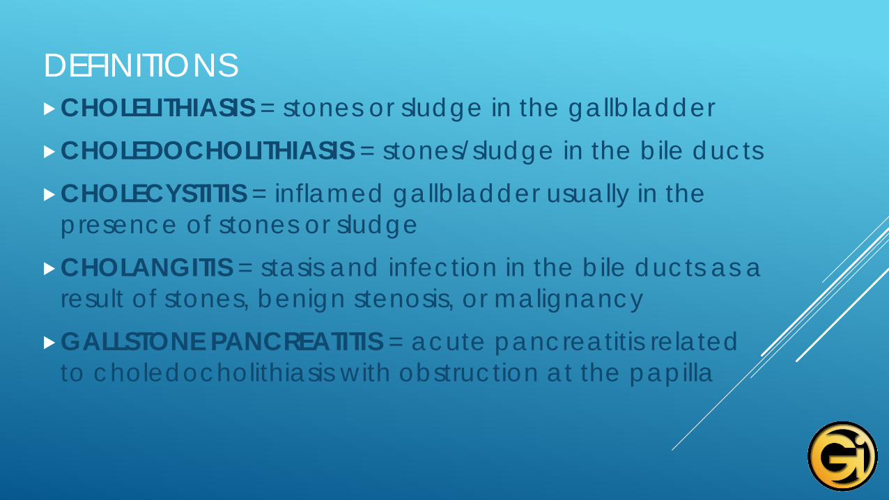
CHOLELITHIASIS = stones or sludge in the gallbladderCHOLEDOCHOLITHIASIS = stones/sludge in the bile ductsCHOLECYSTITIS = inflamed gallbladder usually in the
presence of stones or sludgeCHOLANGITIS = stasis and infection in the bile ducts as a
result of stones, benign stenosis, or malignancyGALLSTONE PANCREATITIS = acute pancreatitis related
to choledocholithiasis with obstruction at the papilla
DEFINITIONS

Gallbladder Cystic DuctRight and Left IntrahepticsCommon Hepatic DuctCommon Bile DuctAmpulla of VaterMajor Papilla
GALLBLADDER AND BILIARY ANATOMY
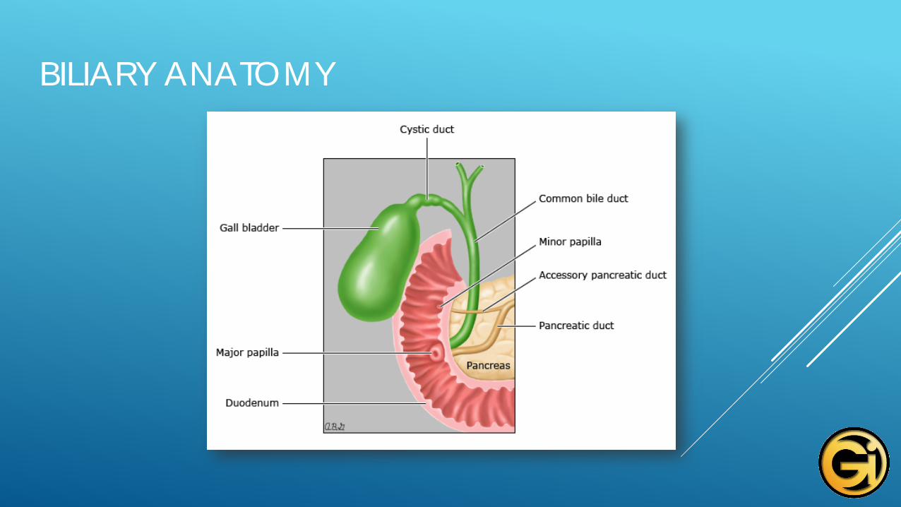
BILIARY ANATOMY

• A common and costly disease• US estimates are 6.3 million men and 14.2 million women
between ages of 20-74. • Prevalence among non-Hispanic white men and women is
8-16%.• Prevalence among Hispanic men and women is 9-27%.• Prevalence among African Americans is lower at 5-14%.• More common among Western Caucasians, Hispanics
and Native Americans• Less common among Eastern Europeans, African
Americans, and Asians
GALLSTONE EPIDEMIOLOGY
• Ethnicity• Female > Male• Pregnancy• Older age• Obesity• Rapid weight loss/bariatric surgery
GALLSTONE RISK FACTORS

• 15%-20% will develop symptoms• *Once symptoms develop, there is an increased risk of
complications.• Incidental or silent gallstones do not require treatment.• Special exceptions due to increased risk of gallbladder
cancer: Large gallstone > 3cm, porcelain gallbladder, gallbladder polyp/adenoma 10mm or bigger, and anomalous pancreatic duct drainage
GALLSTONES: NATURAL HISTORY

• Biliary colic which is a misnomer and not true colic• Episodic steady epigastric or RUQ pain often radiating to
the R scapular area• Peaks rapidly within 5-10 minutes and lasts 30 minutes to 6
hours or more• Frequently associated with N/V• Fatty meal is a common trigger, but symptoms may
occur day or night without a meal.
GALLSTONES: CLINICAL SYMPTOMS
• R chest pain• RLQ pain• Pain in general doesn’t cross the midline.• Bloating and distension• Postprandial fullness/early satiety• “Heartburn”
GALLSTONES: ATYPICAL SYMPTOMS

• Nonspecific• Normal abdomen• RUQ pain• Murphy’s sign refers to acute cholecystitis.
GALLSTONES: PHYSICAL EXAM

• Usually normal• If leukocytosis is present, consider acute cholecystitis.• If liver enzymes are elevated, consider choledocholithiasis.
GALLSTONES: LAB EXAM

• Ultrasound (US)- widely available, inexpensive, no radiation• Gallbladder stones, gravel, and sludge are all managed
similarly.• US has an 84% sensitivity and a 99% specificity.• A negative US can be repeated in a few weeks if symptoms
are consistent with biliary colic. • A CCK-HIDA can be obtained if US is negative and
gallbladder is still suspect.• CT insensitive- will miss most stones or sludge which
are isodense with bile
GALLSTONE DIAGNOSIS

• Asymptomatic gallstones require no treatment except for the exceptions mentioned earlier.
• Treatment for symptomatic stones is pain control with ketorolac or narcotic and surgical referral once stones or sludge become symptomatic.
• Once stones become symptomatic, patients are at increased risk of serious complications such as cholangitis or gallstone pancreatitis.
• Gallstone dissolution therapy rarely done nowadays but ursodiol has been used
GALLSTONE TREATMENT
• Syndrome of RUQ pain, fever, leukocytosis and gallbladder inflammation/wall edema on imaging are usually related to stones or sludge.
• Life threatening complications are gallbladder perforation and/or gangrene.
SOMETIMES THINGS GET COMPLICATED: ACUTE CHOLECYSTITIS

• Hospital admission for supportive care, antibiotics, and surgical therapy depending on the patient’s condition
• Suspected gangrene or perforation requires emergency cholecystectomy or percutaneous drainage (e.g. IR cholecystostomy).
• Mortality average 3% with a range of 1%-10%
ACUTE CHOLECYSTITIS TREATMENT
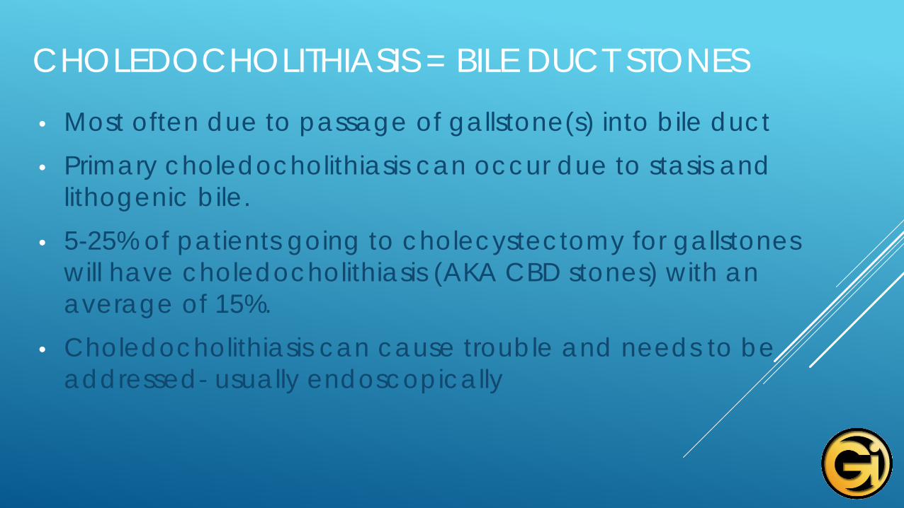
• Most often due to passage of gallstone(s) into bile duct• Primary choledocholithiasis can occur due to stasis and
lithogenic bile.• 5-25% of patients going to cholecystectomy for gallstones
will have choledocholithiasis (AKA CBD stones) with an average of 15%.
• Choledocholithiasis can cause trouble and needs to be addressed- usually endoscopically
CHOLEDOCHOLITHIASIS = BILE DUCT STONES
• May be asymptomatic but most have symptoms• Epigastric or substernal chest pain• Nausea and vomiting• Obstructive jaundice• Acute cholangitis: Charcot’s triad = RUQ pain, fever,
jaundice; sepsis may lead to hypotension and altered mental status (Reynold’s Pentad)
• Longstanding low grade biliary obstruction may lead to liver fibrosis and secondary biliary cirrhosis.
CHOLEDOCHOLITHIASIS: SYMPTOMS
• Labs: Elevated liver enzymes raise suspicion. ALT more sensitive than AST and can go as high as 1,000
• CBC and lipase should be obtained.• CT and US are insensitive. May show dilated duct > 6mm• MRCP and ERCP are sensitive, but both are expensive and
ERCP is invasive.
CHOLEDOCHOLITHIASIS: DIAGNOSIS

• Risk stratification• High risk patients- proceed with ERCP and stone removal• Intermediate risk patients don’t justify risk of ERCP
pancreatitis (5%). MRCP or EUS will diagnose bile duct stones.
• Low risk patients- laparoscopic cholecystectomy with IOC• Renown protocol is lap cholecystectomy with IOC for
intermediate risk patients. Avoids delay of ERCP or MRCP
CHOLEDOCHOLITHIASIS: TREATMENT
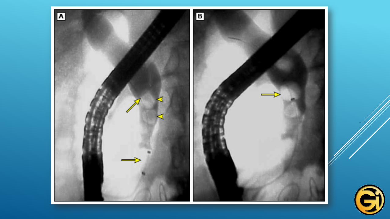
• Requires aggressive treatment; patients are often septic• Resuscitation with IV hydration• IV antibiotics: Zosyn or quinolone + metronidazole• Urgent drainage with either ERCP or if too ill for ERCP,
percutaneous transhepatic drainage by IR (PTC)• 70-80% will respond to support and IV antibiotics• Mortality is 11% in severe cases• Cholecystectomy can be done once patient
stabilizes if gallstones are present
CHOLEDOCHOLITHIASIS: COMPLICATIONSACUTE CHOLANGITIS

• Acute pancreatitis related to ampullary obstruction by stones or sludge, although the exact mechanism is unknown
• Syndrome of acute pancreatic inflammation characterized by abdominal pain with elevated liver and pancreatic enzymes
• Represents 35-405 of pancreatitis cases worldwide but 80-90% of cases of pancreatitis in my practice
GALLSTONE PANCREATITIS

• Presence of gallstones especially small stones which can escape the gallbladder
• Risks increase when stones become symptomatic.• Small stones, 5mm or less, are more likely to escape the
gallbladder and lodge or pass out of the ampulla into the duodenum.
GALLSTONE PANCREATITIS: RISKS
• Steady, unrelenting upper abdominal pain often with nausea and vomiting
• Elevated amylase or lipase; lipase more specific• Elevated liver enzymes; ALT > 150 has a 95% positive
predictive value• US demonstrating gallbladder stones or sludge
GALLSTONE PANCREATITIS: DIAGNOSIS

• Assessment of disease severity• 75% will have mild interstitial disease and a milder course.• 25% will have necrosis and a longer more difficult course.• Aggressive IV hydration and narcotic analgesia are the
keystones of treatment.• MRCP can be done to look for CBD stone if liver enzymes
are not improving. Best to avoid early ERCP if possible• ERCP or percutaneous drainage is mandatory if
there is concurrent cholangitis.
GALLSTONE PANCREATITIS: MANAGEMENT
• No need for CT scan if benign course with rapid improvement• CT ideally in 2-3 days for sicker patients to assess for pancreatic
necrosis; IV and oral contrast best but protect the kidneys• Avoid prophylactic antibiotics but treat if suspected infection• ERCP can be delayed if no cholangitis and if CBD stones are
suspected. Ideally should be done preoperatively• ERCP not indicated if liver enzymes are improving and
patient is improving; can worsen pancreatitis and can be difficult due to duodenal edema
GALLSTONE PANCREATITIS: MANAGEMENT

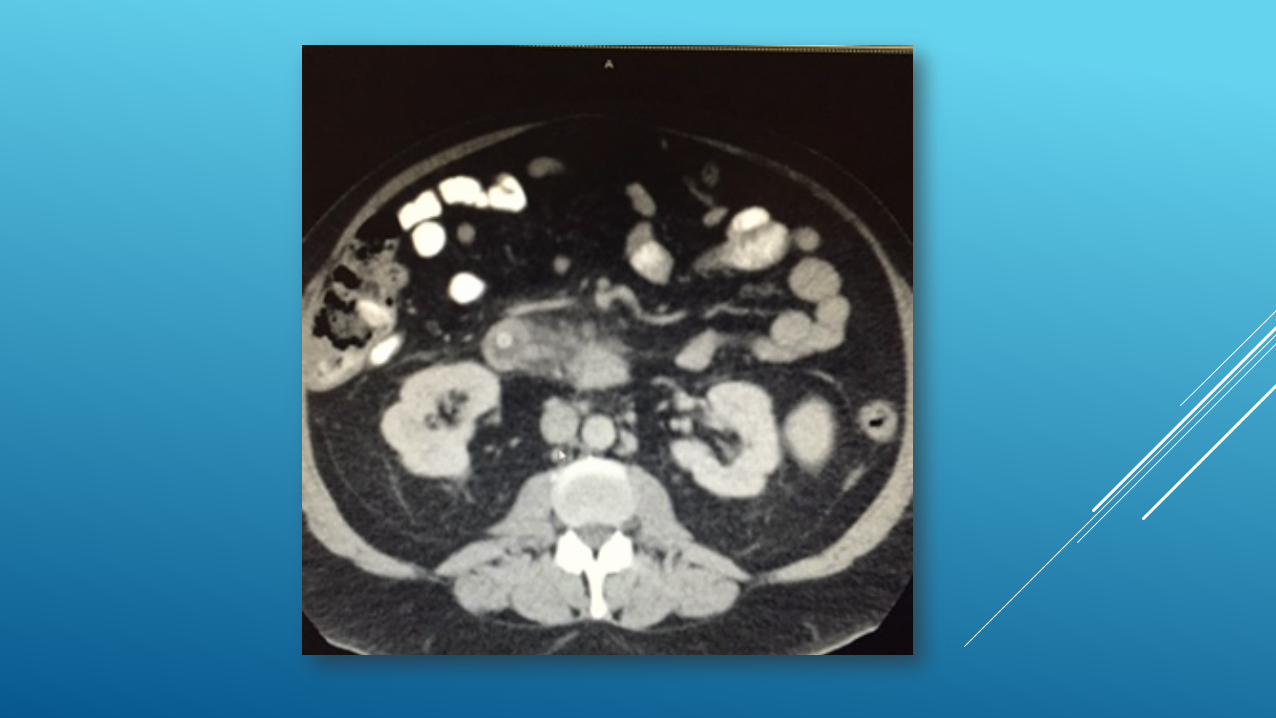
CT SCAN: GALLSTONE PANCREATITIS

• Cholecystectomy should be done ideally once pancreatitis subsides. Timing is controversial and some surgeons prefer to wait 6 weeks to allow edema to resolve.
• Delayed cholecystectomy is associated with a 25-30% risk of recurrent gallstone pancreatitis, cholecystitis, or cholangitis within the next 6-18 weeks.
GALLSTONE PANCREATITIS: MANAGEMENT
![Choledocholithiasis, Ascending Cholangitis, and Gallstone ... · bilirubinate to form biliary sludge, which can aggregate eventually into a gallbladder stone [10]. Black pigment stones](https://static.fdocuments.net/doc/165x107/5e04b56d64882534e3400732/choledocholithiasis-ascending-cholangitis-and-gallstone-bilirubinate-to-form.jpg)