Treatment of choledocholithiasis in patients with liver cirrhosis ...
6
ANNALS OF SURGERY Vol. 218, No. 1, 68-73 © 1993 J. B. Lippincott Company Treatment of Choledocholithiasis in Patients with Liver Cirrhosis Surgical Treatment or Endoscopic Sphincterotomy? Masanori Sugiyama, M.D., Yutaka Atomi, M.D., Akira Kuroda, M.D., and Tetsuichiro Muto, M.D. From the First Department of Surgery, Tokyo University School of Medicine, Tokyo, Japan Objective The clinical features of choledocholithiasis were analyzed in cirrhotic patients. The outcomes of surgical treatment and endoscopic sphincterotomy (EST) in this situation were compared and the risk factors predictive of an increased mortality rate were identified. Summary Background Data In cirrhotic patients, high risk for gallbladder stones in cholecystectomy has been established. Common bile duct stones can often exacerbate liver dysfunction and might be more difficult to treat. Methods Among 16 cirrhotic patients with choledocholithiasis, 9 underwent choledocholithotomy and T- tube placement (surgery group) and 7 underwent EST (EST group). Pretreatment clinical data were comparable between groups. Results Among 16 patients, 15 had biliary tract symptoms and 7 had cholangitis. The surgery group had excessive intraoperative hemorrhage (1576 mL) and a high morbidity rate (66.7%). The mortality rate was 44.4%: 0% in Child A or B classification patients and 80% in Child C patients. The common causes of death were liver failure, postoperative hemorrhage, and sepsis. The EST group had no complications related to procedures, but there was one death (14.3%) due to preexisting liver failure. Hepatic dysfunction, coagulopathy, and cholangitis were factors predictive of an increased mortality rate. Conclusions Choledocholithiasis in cirrhotic patients should be treated by EST after liver function and general condition are improved by medical management, except in emergency cases. Cholelithiasis often accompanies liver cirrhosis.",2 In and high morbidity and mortality rates.5'6 Common bile cirrhotic patients, gallstones are predominantly located duct stones are sometimes found in patients with liver in the gallbladder with few symptoms and complica- cirrhosis. These stones can often exacerbate liver dys- tions.3'4 However, cholecystectomy in cirrhotic patients function to the state of hepatic failure as a result of bili- has intraoperative and postoperative massive bleeding ary obstruction or cholangitis. In cirrhotic patients, the 68
Transcript of Treatment of choledocholithiasis in patients with liver cirrhosis ...

ANNALS OF SURGERYVol. 218, No. 1, 68-73© 1993 J. B. Lippincott Company
Treatment of Choledocholithiasis inPatients with Liver CirrhosisSurgical Treatment or EndoscopicSphincterotomy?Masanori Sugiyama, M.D., Yutaka Atomi, M.D., Akira Kuroda, M.D., and Tetsuichiro Muto, M.D.
From the First Department of Surgery, Tokyo University School of Medicine, Tokyo, Japan
ObjectiveThe clinical features of choledocholithiasis were analyzed in cirrhotic patients. The outcomes ofsurgical treatment and endoscopic sphincterotomy (EST) in this situation were compared andthe risk factors predictive of an increased mortality rate were identified.
Summary Background DataIn cirrhotic patients, high risk for gallbladder stones in cholecystectomy has been established.Common bile duct stones can often exacerbate liver dysfunction and might be more difficult totreat.
MethodsAmong 16 cirrhotic patients with choledocholithiasis, 9 underwent choledocholithotomy and T-tube placement (surgery group) and 7 underwent EST (EST group). Pretreatment clinical datawere comparable between groups.
ResultsAmong 16 patients, 15 had biliary tract symptoms and 7 had cholangitis. The surgery group hadexcessive intraoperative hemorrhage (1576 mL) and a high morbidity rate (66.7%). The mortalityrate was 44.4%: 0% in Child A or B classification patients and 80% in Child C patients. Thecommon causes of death were liver failure, postoperative hemorrhage, and sepsis. The ESTgroup had no complications related to procedures, but there was one death (14.3%) due topreexisting liver failure. Hepatic dysfunction, coagulopathy, and cholangitis were factorspredictive of an increased mortality rate.
ConclusionsCholedocholithiasis in cirrhotic patients should be treated by EST after liver function and generalcondition are improved by medical management, except in emergency cases.
Cholelithiasis often accompanies liver cirrhosis.",2 In and high morbidity and mortality rates.5'6 Common bilecirrhotic patients, gallstones are predominantly located duct stones are sometimes found in patients with liverin the gallbladder with few symptoms and complica- cirrhosis. These stones can often exacerbate liver dys-tions.3'4 However, cholecystectomy in cirrhotic patients function to the state of hepatic failure as a result of bili-has intraoperative and postoperative massive bleeding ary obstruction or cholangitis. In cirrhotic patients, the
68

Choledocholithiasis in Patients with Cirrhosis 69
treatment of common duct stones might be more diffi-cult than that of gallbladder stones.
In this study, the clinical features of choledocholithia-sis in patients with liver cirrhosis were compared withthose of cholecystolithiasis. Furthermore, we evaluatedthe outcomes of surgical treatment and endoscopicsphincterotomy (EST) for common duct stones in cir-rhotic patients and identified risk factors predictive ofanincreased mortality rate.
PATIENTS AND METHODSBetween 1972 and 1991, 16 patients with liver cirrho-
sis were treated for choledocholithiasis at the First De-partment of Surgery, Tokyo University School of Medi-cine, Tokyo, Japan. None of these patients had simulta-neous operations for diseases other than cholelithiasis.Of these 16 patients, 10 were men and 6 were women.The mean age was 63.3 years (range, 29 to 91 years).
Liver cirrhosis was diagnosed by inspection and liverbiopsy at laparotomy in the nine surgical cases. For theseven patients treated by EST, the diagnosis was basedon percutaneous liver biopsy in four, autopsy in one, orclinical signs, blood liver chemistry tests, and imagingstudies in two. Liver cirrhosis was linked to alcoholabuse in six patients, non-A non-B hepatitis in six (anti-hepatitis C virus was not tested; post-transfusion hepati-tis in three of the six), and B-type hepatitis in four. Pa-tients with primary or secondary biliary cirrhosis werenot included.The gallstones were located in the common bile duct
alone in ten patients (one of the ten had undergone cho-lecystectomy 22 years ago) and in both the gallbladderand common duct in six.Of these 16 patients, 9 had surgery (surgery group):
cholecystectomy, choledocholithotomy, and T-tubeplacement in 7 and choledocholithotomy and T-tubeplacement in 2. The remaining seven underwent EST(EST group). One ofthe seven had untreated gallbladderstones. Common bile duct stones were extracted by bas-ket forceps immediately after EST. Two ofthe seven hadendoscopic naso-biliary drainage (ENBD)7 after theseprocedures. We treated cirrhotic patients with choledo-cholithiasis surgically before 1985 and with EST after1986. Except in urgent cases, surgery or EST was per-formed after an attempt was made to improve the pa-tient's condition with intense medical management con-sisting ofintravenous hyperalimentation and the admin-istration of fresh frozen plasma, vitamin K, diuretics,
and antibiotics for malnutrition, coagulopathy, ascites,and cholangitis. This management was continued aftertreatment ofcommon bile duct stones.
In these patients, we studied signs and symptoms, pre-treatment complications, stone classification, bile cul-tures, and cholangiographic findings. Morbidity andmortality rates were compared between the surgerygroup and the EST group. Clinical data before or duringtreatment were analyzed to identify factors predictive ofsignificantly increased mortality. The data analyzedwere as follows: (1) blood liver chemistry tests,prothrombin times, platelet counts, cholangitis, andChild classification8 (all before treatment); (2) timing oftreatment; and (3) intraoperative hemorrhage.To provide comparative data, we reviewed 364 non-
cirrhotic patients treated for choledocholithiasis duringthe same years to determine intraoperative blood lossand morbidity and mortality rates. Of the 364 patients,240 were treated with surgery and 124 underwent EST.
Gallstones were classified as pure cholesterol, calciumbilirubinate, black, mixed, or combined stones accord-ing to the findings on cross section. Pure cholesterolstones had a radial pattern with a white to light yellowcolor. Calcium bilirubinate stones had multiple concen-tric layers with a brown color. Black stones were darkbrown or black, friable, and amorphous. These stonesmainly contained unconjugated bilirubin9 and are called"pigment stones" in the United States and Europe.Mixed stones had a pattern ofboth radial formation andmultiple concentric layers. Combined stones had multi-ple strata of different composition.
Statistical analyses were done using the chi square testand the Student's t test. Differences were considered tobe significant when the p value was less than 0.05.
RESULTSOf the 16 cirrhotic patients with choledocholithiasis,
15 had calcium bilirubinate stones and 1 had mixedstones. All six patients who had common duct bile sam-pled during surgery had bile cultures that were positivefor bacteria: Escherichia coli in three, Klebsiella in two,Citobacter in two, and Bacteroides in one.On endoscopic retrograde cholangiogram or intraoper-
ative cholangiogram, the mean diameters of the com-mon bile duct and the proximal part of the left mainhepatic duct were 15 mm (range, 9 to 25 mm) and 5 mm(range, 4 to 9 mm), respectively.On admission for the treatment ofgallstones, 12 (75%)
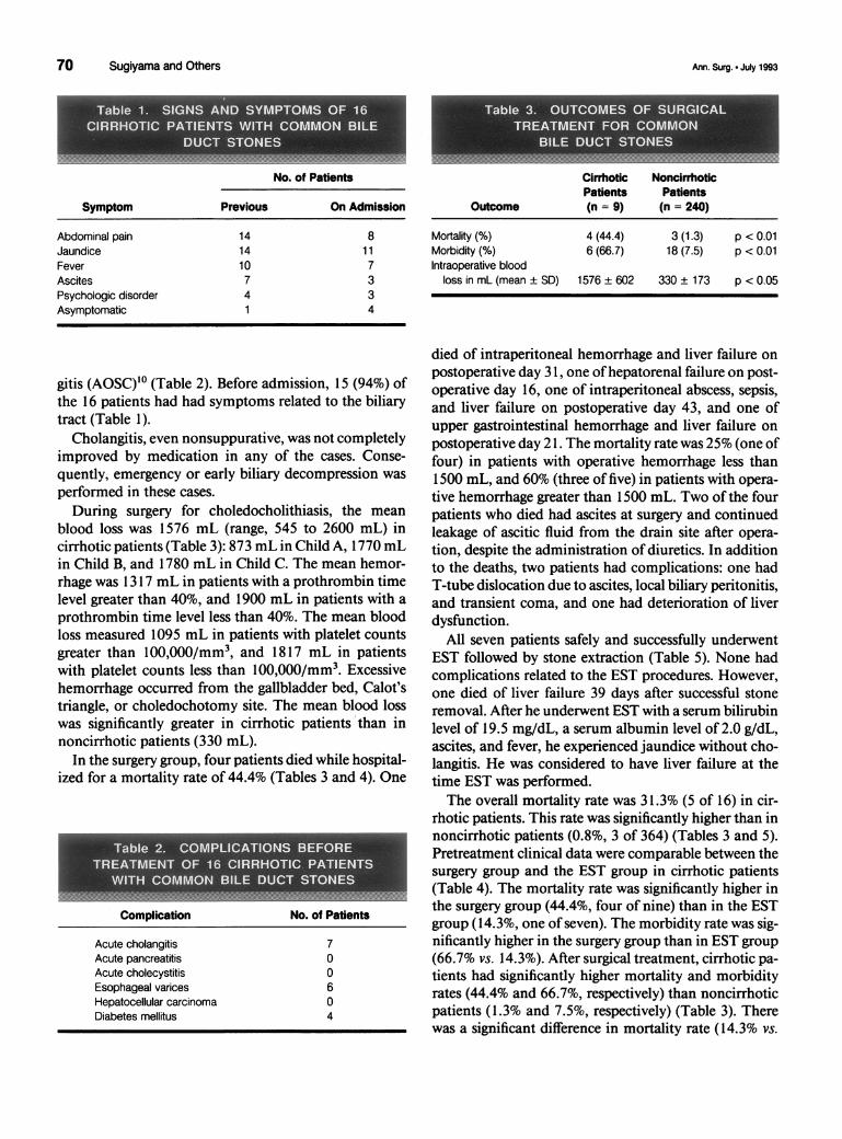
of these 16 patients had signs and symptoms ofcholedo-cholitiasis (abdominal pain, jaundice, and fever) (Table1). In addition, three had ascites and three had a psycho-logical disorder. Four had acute nonsuppurative cholan-gitis and three had acute obstructive suppurative cholan-
Address reprint requests to Masanori Sugiyama, M.D., First Depart-ment of Surgery, Tokyo University School of Medicine, 7-3-1,Hongo, Bunkyo-ku, Tokyo 113, Japan.
Accepted for publication October 9, 1992.
Vol. 218-No. 1
70 Sugiyama and Others
No. of Patients
Symptom Previous On Admission Outcome
CirrhoticPatients(n = 9)
NoncirrhoticPatients(n = 240)
Abdominal pain 14 8Jaundice 14 1 1Fever 10 7Ascites 7 3Psychologic disorder 4 3Asymptomatic 1 4
gitis (AOSC)10 (Table 2). Before admission, 15 (94%) ofthe 16 patients had had symptoms related to the biliarytract (Table 1).
Cholangitis, even nonsuppurative, was not completelyimproved by medication in any of the cases. Conse-quently, emergency or early biliary decompression was
performed in these cases.
During surgery for choledocholithiasis, the mean
blood loss was 1576 mL (range, 545 to 2600 mL) incirrhotic patients (Table 3): 873 mL in Child A, 1770 mLin Child B, and 1780 mL in Child C. The mean hemor-rhage was 1317 mL in patients with a prothrombin timelevel greater than 40%, and 1900 mL in patients with a
prothrombin time level less than 40%. The mean bloodloss measured 1095 mL in patients with platelet countsgreater than 100,000/mm3, and 1817 mL in patientswith platelet counts less than 100,000/mm3. Excessivehemorrhage occurred from the gallbladder bed, Calot'striangle, or choledochotomy site. The mean blood losswas significantly greater in cirrhotic patients than innoncirrhotic patients (330 mL).
In the surgery group, four patients died while hospital-ized for a mortality rate of 44.4% (Tables 3 and 4). One
..~ ~~~~~~~~~~~~~~~~.
Complication No. of Patients
Acute cholangitis 7Acute pancreatitis 0Acute cholecystitis 0Esophageal varices 6Hepatocellular carcinoma 0Diabetes mellitus 4
Mortality (%) 4 (44.4) 3 (1.3) p <0.01Morbidity (%) 6 (66.7) 18 (7.5) p < 0.01Intraoperative blood
loss in mL (mean ± SD) 1576 ± 602 330 ± 173 p < 0.05
died of intraperitoneal hemorrhage and liver failure onpostoperative day 3 1, one ofhepatorenal failure on post-operative day 16, one of intraperitoneal abscess, sepsis,and liver failure on postoperative day 43, and one ofupper gastrointestinal hemorrhage and liver failure onpostoperative day 21. The mortality rate was 25% (one offour) in patients with operative hemorrhage less than1500 mL, and 60% (three of five) in patients with opera-tive hemorrhage greater than 1500 mL. Two of the fourpatients who died had ascites at surgery and continuedleakage of ascitic fluid from the drain site after opera-tion, despite the administration of diuretics. In additionto the deaths, two patients had complications: one hadT-tube dislocation due to ascites, local biliary peritonitis,and transient coma, and one had deterioration of liverdysfunction.
All seven patients safely and successfully underwentEST followed by stone extraction (Table 5). None hadcomplications related to the EST procedures. However,one died of liver failure 39 days after successful stoneremoval. After he underwent EST with a serum bilirubinlevel of 19.5 mg/dL, a serum albumin level of 2.0 g/dL,ascites, and fever, he experienced jaundice without cho-langitis. He was considered to have liver failure at thetime EST was performed.The overall mortality rate was 31.3% (5 of 16) in cir-
rhotic patients. This rate was significantly higher than innoncirrhotic patients (0.8%, 3 of 364) (Tables 3 and 5).Pretreatment clinical data were comparable between thesurgery group and the EST group in cirrhotic patients(Table 4). The mortality rate was significantly higher inthe surgery group (44.4%, four of nine) than in the ESTgroup (14.3%, one of seven). The morbidity rate was sig-nificantly higher in the surgery group than in EST group(66.7% vs. 14.3%). After surgical treatment, cirrhotic pa-tients had significantly higher mortality and morbidityrates (44.4% and 66.7%, respectively) than noncirrhoticpatients (1.3% and 7.5%, respectively) (Table 3). Therewas a significant difference in mortality rate (14.3% vs.
111144AMtell I 4;-::.-;::-:;::-:;-;;;:-:- ... .. ;::.:.:..:::::::.::.:.:.: -: :..
GPIIIOIIL-iv"
Ann. Surg. * July 1993

Choledocholithiasis in Patients with Cirrhosis 71
EST (Table 4). These factors tended to increase mortal-ity rates in the surgery group also, but not significantly.
Factor
TotalTotal bilirubin level
(mg/dL)<22-5.5
Serum glutamicoxalacetictransaminaselevel (units)
<3536-99.100
Serum albumin level(g/dL)
.3.53.0-3.5<3.0
Prothrombin time (%)
.8040-80<40
Platelet count(X 104/mm3)
.2010-20.10
Child classificationABC
CholangitisAbsentNonsuppurative
cholangitisAOSC
Timing of treatmentElectiveEmergent
Surgery(Death/Total)*
4/9
0/30/24/4
0/21/33/4
1/31/32/3
0/21/33/4
0/10/24/6
0/20/24/5
0/5
2/22/2
1/63/3
EST(Death/Total)
1/7
0/20/11/4
0/20/31/2
0/20/11/4
0/30/21/2
0/10/31/3
0/10/11/5
0/4
1/20/1
0/51/2
tueu
* Cases of hospital death cases/total cases.
0%) between cirrhotic and noncirrhotic patients who un-
derwent EST, but there was no difference in morbidityrate ( 14.3% vs. 6.5%) (Table 5).For the 16 cirrhotic patients with choledocholithiasis,
the following factors, before or during treatment, were
associated with significantly increased mortality rates: a
total bilirubin level greater than 5 mg/dL, a serum glu-tamic oxalacetic transaminase level greater than 100units, a prothrombin time less than 40%, a platelet countless than 100,000/mm3, Child C classification, nonsup-
purative cholangitis or AOSC, and emergency surgery or
Both,__-A6 rr-&-i.k
DISCUSSIONatf/Tota The incidence of cholelithiasis in patients with liver
5/16 cirrhosis is reported to be twice that of the noncirrhotic
population.",2 Gallstones in cirrhotic patients are mostoften black stones' 2 ("pigment stones"). Black stones
0/5 are predominantly located in the gallbladder in both cir-0/3 rhotic' and noncirrhotic" patients. Compared with non-
5/8 cirrhotic patients, cholelithiasis in cirrhotic patients is
mostly asymptomatic and has a lower incidence ofcom-plications3'4 that include acute cholecystitis, obstructivejaundice, cholangitis, and acute pancreatitis. In our se-
0/4 ries of common bile duct stones, 94% of patients had
4/6 calcium bilirubinate stones. Calcium bilirubinate stonesare not commonly seen in the United States, but are themost common type of stones in noncirrhotic patients
1/5 with choledocholithiasis in Japan." These stones are as-1/4 sociated with bacterial infection ofthe biliary tract.'2 Al-3/7 though liver cirrhosis causes black stones to occur in the0/5 gallbladder, choledocholithiasis and cirrhosis might1/5 coexist in our series, which did not include cases of bili-4/6 ary cirrhosis, from a viewpoint of stone classification.
Choledocholithiasis has higher incidences of symptoms0/2 and complications than cholecystolithiasis in noncirrho-0/5 tic patients. Similarly, 94% of cirrhotic patients with5/9 choledocholithiasis had biliary tract symptoms such as
abdominal pain, jaundice, and fever. Acute cholangitis0/3 and AOSC accompanied 44% and 19% ofthese patients,5/10 respectively.
In noncirrhotic patients, even asymptomatic common0/9 bile duct stones are relative indications for treatment be-
cause of the high possibility of complications. Also, al-3/4 most all cirrhotic patients with choledocholithiasis, even2/3 if asymptomatic, should be treated because obstructive1/11 jaundice or cholangitis often causes liver failure.4/5 Many reports established high risk, including major
intraoperative hemorrhage and high morbidity and mor-tality rates, in biliary surgery for cirrhotic patients.36 In
Cirrhotic NoncirrhoticPatients Patients
Outcome (n = 7) (n = 124)
Success in stone extraction (%) 7 (100.0) 120 (96.8) NSMortality (%) 1 (14.3) 0 p < 0.01Morbidity (%) 1 (14.3) 8 (6.5) NS
v
Vol. 218-No. 1

72 Sugiyama and Others
this study, cirrhotic patients had greater blood loss insurgical treatment for choledocholithiasis than noncir-rhotic patients. The volume of hemorrhage was relatedto the Child classification of hepatic functional reserve.One of the reasons for excessive hemorrhage was the de-velopment of large collaterals in the gallbladder bed, Ca-lot's triangle, and hepatoduodenal ligament due to por-tal hypertension. Coagulopathy and thrombocytopeniaalso caused excessive blood loss, including major oozingfrom the incision and raw peritoneal surface. Coagulopa-thy and liver dysfunction should be corrected by preoper-ative and postoperative intense medical management in-cluding the administration of fresh frozen plasma.'3However, this management cannot necessarily precludemassive bleeding. Local hemostatic agents such as mi-crofibrillar collagen or vasoconstrictive agents such asvasopressin should be considered.'4 Cholecystolithot-omy,3 cholecystostomy,5 or subtotal cholecystectomy'5with the posterior wall left intact were advised for gall-bladder stones in cirrhotic patients to avoid massivebleeding from the gallbladder bed.The mortality rate was reported to be as high as 10% to
25% in surgical treatment of gallstones in cirrhotic pa-tients.3'5'6"3 Surgical treatment of choledocholithiasis incirrhotic patients had high morbidity and mortality ratesof 66.7% and 44.4%, respectively. The common causesofdeath were liver failure, postoperative hemorrhage (in-traperitoneal or upper gastrointestinal), and sepsis. Alldeaths resulted from liver failure. Surgical proceduresperformed on the liver and biliary tract, anesthesia, andexcessive intraoperative blood loss tended to exacerbateliver dysfunction because these cirrhotic patients did nothave hepatic functional reserve. Cirrhotic patients werealso susceptible to infection. Ascites leakage was a char-acteristic complication after biliary surgery in cirrhoticpatients. In one of three patients with ascites leakage,T-tube dislocation due to ascites resulted in biliary perito-nitis.
Factors that increased the surgical mortality rate werepreoperative liver dysfunction, preoperative cholangitis,emergency surgery, and massive intraoperative bloodloss. The increased mortality rate often reflected the de-gree of hepatic decompensation, which was indicated byabnormal blood liver test results (serum albumin, totalbilirubin, and serum glutamic pyruvic transaminase val-ues), prolonged prothrombin time, thrombocytopenia,and the presence of ascites. The surgical mortality ratewas 0% in Child A and B and 80% in Child C. Acutecholangitis often resulted in the deterioration of liverfunction and general condition in cirrhotic patients.Acute cholangitis, if resistant to Antibiotics, requiresearly biliary decompression in noncirrhotic patients."Emergency decompression is especially indicated forAOSC. Because surgical decompression in AOSC is as-
sociated with a high mortality rate, a nonsurgical ap-proach such as percutaneous transhepatic biliary drain-age (PTBD), EST, or ENBD is recommended.'" The ad-ministration of antibiotics could not improve evennonsuppurative cholangitis in cirrhotic patients. Surgi-cal decompression in these patients was associated with amortality rate of 100%. PTBD was difficult to perform incirrhotic patients because of liver atrophy and low-gradedilatation ofthe intrahepatic tree. Dilatation ofthe prox-imal part of the left hepatic duct, which was usuallypunctured in PTBD, was not sufficient to performPTBD, despite moderate to severe dilatation ofthe extra-hepatic duct. PTBD is contraindicated for patients withascites. However, EST was safely performed and im-proved cholangitis in cirrhotic patients. Even patientwith ascites underwent EST without complications. Be-cause emergency cases were often accompanied by se-vere cholangitis and excluded preparation to improvethe liver function and general condition, emergencycases had a high mortality rate. Excessive intraoperativebleeding6 caused the deterioration of liver function andled to liver failure.The EST group, which had liver dysfunction and coag-
ulation disturbance comparable to those of the surgerygroup, had no complications related to endoscopic pro-cedures; however, one death occurred (14.3%). Thecause of death was considered to be preexisiting liverfailure. These results were better than those from surgi-cal treatment. EST caused only minimal stress for thesehigh-risk patients and avoided the risk of major hemor-rhage during treatment. Although coagulopathy is pre-sented as a risk factor for bleeding after EST,'6 the practi-cal criteria are not well documented. In our series, evenpatients with severe coagulopathy and liver decompen-sation underwent EST safely. However, these abnormali-ties should be corrected before EST, if possible. In ex-tremely critical cases requiring emergency biliary de-compression, it might be safer to initially performENBD alone without sphincterotomy, although we didnot experience such situations. In these cases, commonduct stones should be extracted by EST after liver func-tion and general condition are improved. In general,EST is established as a first-choice treatment for com-mon duct stones in elderly, postcholecystectomy pa-tients at high risk. Patients with liver cirrhosis, particu-larly those with decompensated liver disease (Child C),should be treated with EST because EST is less invasiveand safer than surgical treatment. Because of a low inci-dence of cholecystitis after EST,'6 subsequent cholecys-tectomy is unnecessary in most cirrhotic patients at highsurgical risk, especially in patients without gallbladderstones.
In cirrhotic patients, common bile duct stones oftencause liver dysfunction resulting in hepatic failure and
Ann. Surg. * July 1993

Choledocholithiasis in Patients with Cirrhosis 73
making the general condition worse as a result ofextrahe-patic biliary obstruction or retrograde cholangitis. There-fore, almost all cases of choledocholithiasis with cirrho-sis, even if asymptomatic, are indications for treatment,despite the high risk. Child C cases have a very pooroutcome after surgery or EST. It is safer to treat gall-stones after improvement of Child C to Child A or B.Hepatic dysfunction, coagulation disturbance, and cho-langitis should be improved before the treatment of gall-stones, except in emergency cases such as AOSC. EST isrecommended as the first-choice treatment for choledo-cholithiasis in cirrhotic patients because of its effective-ness and safety.
References1. Nicholas P, Rinaudo PA, Conn HO. Increased incidence ofcholeli-
thiasis in Laennec's cirrhosis. Gastroenterology 1972; 63:112-121.2. Bouchier JA. Post-mortem study of the frequency of gallstones in
patients with cirrhosis of the liver. Gut 1969; 10:705-710.3. Castaing D, Houssin D, Lemoine J, Bismuth H. Surgical manage-
ment of gallstones in cirrhotic patients. Am J Surg 1983; 146:310-313.
4. Dunnington G, Sampliner R, Kogan F, et al. Natural history ofcholelithiasis in patients with alcoholic cirrhosis (cholelithiasis incirrhotic patients). Ann Surg 1987; 205:226-229.
5. Aranha GV, Sontag SJ, Greenlee HB. Cholecystectomy in cir-rhotic patients: a formidable operation. Am J Surg 1982; 143:55-60.
6. Bloch RS, Allaben RD, Walt AJ. Cholecystectomy in patients withcirrhosis: a surgical challenge. Arch Surg 1985; 120:669-672.
7. Cotton PB, Burney PGJ, Mason RR. Transnasal bile duct cathe-terisation after endoscopic sphincterotomy. Gut 1979; 20:285-287.
8. Child CG III. The liver and portal hypertension. Philadelphia: WBSaunders, 1964, p. 50.
9. Trotman BW, Soloway RD. Pigment vs cholesterol cholelithiasis;Clinical and epidemiological aspects. Am J Dig Dis 1975; 20:735-740.
10. Ellison EC, Carey LC. Cholangitis: nonsuppurative and suppura-tive. In Blumgart LH, ed. Surgery of the Liver and Biliary Tract.Edinbugh: Churchill Livingstone, 1988, pp 925-932.
11. Nagase M, Hikasa Y, Soloway RD, et al. Gallstones in WesternJapan: factors affecting the prevalence of intrahepatic gallstones.Gastroenterology 1980; 78:684-690.
12. Maki T. Pathogenesis of calcium bilirubinate gallstones: role of E.coli, beta-glucuronidase and coagulation by inorganic ions, poly-electrolytes and agitation. Ann Surg 1966; 164:90- 100.
13. Doberneck RC, Sterling WA, Allison DC. Morbidity and mortal-ity after operation in nonbleeding cirrhotic patients. Am J Surg1983; 146:306-309.
14. Schwartz SI. Biliary tract surgery and cirrhosis: a critical combina-tion. Surgery 1981; 90:577-583.
15. Neoptolemos JP, Carr-Locke DL. ERCP in cholangitis and pancre-atitis. In Jacobson IM, ed. ERCP: Diagnostic and Therapeutic Ap-plications. New York: Elsevier, 1989, pp 91-126.
16. OstroffJW, Shapiro HA. Complications ofendoscopic sphinctero-tomy. In Jacobson IM, ed. ERCP: Diagnostic and TherapeuticApplications. New York: Elsevier, 1989, pp 61-74.
Vol. 218-No. 1